Embed Size (px)
Citation preview

4. Vertebrate cardiovascular systems
w. BUR G G R E N
A. FAR R E II
H. l III Y W HIT E
Department of Biological Sciences, University of Nevada Las Vegas, Las Vegas, Nevada
Department of Biological Sciences, Simon Fraser University, Burnaby,British Columbia, Canada
Department of Zoology, University of Florida, Gainesville, Florida
CHAPTER CONTENTS
Diversity of Vertebrate Cardiovascular PatternsVertebrate origins and driving forces behind cardiovascular
evolutionCardiovascular patterns in vertebrates
General characteristics of the chordate circulationUrochordates and cephalochordatesThe Agnatha: hagfish and lampreysAquatic gnathostome fishes and the "venous heart"Air-breathing fishes: cardiovascular implications of multiple
respiratory sitesAmphibians: a dedicated gas-exchange circuitReptiles: masters of intracardiac shuntingMammals and birds: dedicated systemic and pulmonary circuits
Functional Properties of Vertebrate HeartsOverviewElectrical properties of cardiac cells
Cardiac pacemaker and the action potentialModulation of pacemaker rate and control of heart rateTransmission of the action potential
Excitation-contraction couplingMechanical properties of cardiac muscle
Effect of contraction frequency on contractilityEffect of temperature on contractilityEffect of l3-adrenergic stimulation on contractilityEffect of extracellular calcium on contractilityOther inotropic agentsArterial blood pressure and homeometric regulationThe Frank-Starling mechanism and control of SVCardiac filling and the role of the pericardium
Cardiac output and cardiac performanceCyclostomes, dipnoans, and phyletically ancient fishesElasmobranchsTeleost fishesAmphibiansChelonian and squamate reptilesCrocodilian reptiles
Coronary circulations, myocardial O2 consumption, andmyocardial O2 supply
Myocardial structure in relation to coronary circulationCoronary vascular arrangementsCoronary O2 supply and control of coronary blood flowMyocardial oxygen consumption
Peripheral Circulation and HemodynamicsArterial blood pressure and its regulation
Levels of arterial pressureBaroreflexes and neurogenic regulation of arterial pressureCentral neural control of arterial pressure
Blood volume and its regulationRenal sympathetic nerve responsesEndocrine and other factors affecting hemodynamics and
blood volumeParacrine, autocrine, and other factors influencing
hemodynamicsIntegrative responses to blood volume changesAutoregulationLong-term regulation of blood volume
Cardiovascular Performance under Special ConditionsAerobic exerciseBreath holding and diving
Cardiac output reductionShuntingO2 metering
Reduced metabolismDigestive stateResponses to gravityDevelopment of cardiovascular systems
Morphological developmentPhysiological development
Conclusions and Future DirectionsMechanistic unknownsAdaptive unknownsIntegrative unknownsDevelopmental unknowns
THE CARDIOVASCULAR SYSTEM OF VERTEBRATES is arguably one of the more critical of all organ systems.By virtue of its role in transporting nutrients, metabolicwaste products, respiratory gases, hormones, and heat,the cardiovascular system intervenes in a rate-limitingway between the acquisition of energy and its use inmetabolic processes. Although it is important at everypoint in an animal's life cycle, the pivotal role of thecardiovascular system in vertebrates is perhaps bestexemplified in the embryo, where it is the first organsystem to function. Only when this system is established can the materials needed for organogenesis betransported to the metabolically active areas of therapidly growing embryo. Serious cardiovascular defectsin embryonic stages usually result in diminished tissuedifferentiation and growth and often portend embry-
216 HANDBOOK OF PHYSIOLOGY-COMPARATIVE PHYSIOLOGY
CEPHALOCHORDATA
onic or early fetal death. Simply put, the cardiovascularsystem of vertebrates is a "choke-point" for all otherphysiological processes.
This chapter presents an overview of cardiovascularform and function in vertebrates. Where possible, wedescribe general characteristics of the vertebrate circulation. Many readers already familiar with circulatorysystems will recognize the gross oversimplification oftalking about the "vertebrate circulation" or the "vertebrate heart" since there is great inter- and intraclassvariation in vertebrate cardiovascular design. We beginthis chapter by considering the evolutionary diversityof vertebrate circulations.
DIVERSITY OF VERTEBRATECARDIOVASCULAR PATIERNS
Vertebrate Origins and Driving Forces behindCardiovascular Evolution
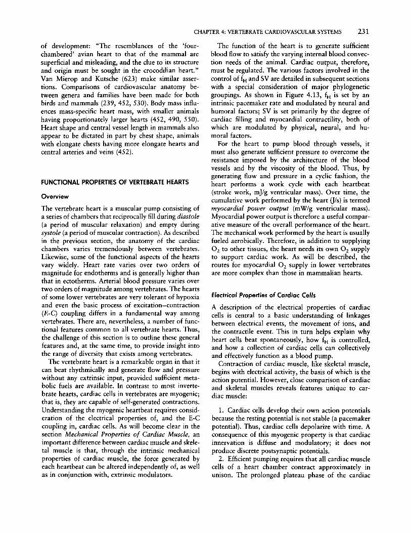
The evolutionary history of vertebrates has been studied intensely ever since the theory of evolution wasfirst proposed. Despite the application of modern techniques of molecular biology and powerful new formsof systematic analysis such as cladistics (biological systematics based on phylogenetic relationships), we remain uncertain about many important aspects of vertebrate phylogeny. Particularly poorly represented in thefossil record are the ancestors of the earliest vertebratesand how they interrelate phyletically. Figure 4.1 showsa simplified (and still debated) phyletic scheme relatingthe three subphyla within the phylum Chordata: the
VERTEBRATA
Primitive filter-feeding
vertrate ~<,~,.~;'~
~.'.:._.. '.. . /'" (Branchiostorna)
~/{} ~Primitive chordate' -
sec55...ile, adult stage I~st , , ...• .: salP~i. . sea 'r·,. Larvacea--.
~.,...•.~ Squirt.'sJf
.
1-"· ~O·'>Anc~stral tunicate With:,_-:free-SWimming larva ,"
(Tunicates)
UROCHORDATA
FIG. 4.1. Simplified phyletic scheme relating Urochordata, Cephalochordara, and Vertebrata (from ref. 511, after ref. 529).
Urochordata (tunicates), the Cephalochordata (for example, the lancelet Branchiostoma), and the Vertebrata.
It is generally assumed that some of the earliestvertebrates were comparatively large, actively swimming animals that acquired food by filter-feeding. Anonsessile life-style combined with a size too large forinternal nutrient and gas transport to be served bydiffusion alone required an efficient cardiovascular system that could rapidly distribute required nutrientsand remove metabolic wastes from metabolically activetissue. (Many invertebrates place similar demands upontheir cardiovascular systems [see Chapter 13 by McMahon, Smith, and Smith in this Handbook], and theearly vertebrates were certainly not unique in requiringa highly efficient, high-capacity internal transport systern.) Consequently, the early vertebrates are thoughtto have possessed a highly differentiated cardiovascularsystem consisting of a discrete heart or hearts, anarterial distribution system, a vascular bed comprisedof capillaries or their functional equivalent (nonendothelial lined blood sinuses of very small diameter),and a venous collection system. Initially, evolutionarychanges in the cardiovascular system may have comein response to nutrient uptake and transport ratherthan the transport of respiratory gases. Primitivechordates (tunicates, Branchiostoma) show early embryonic differentiation of the digestive organs and ahighly developed intestinal circulation. Randall andDavie (511) interpret these observations as pointing tofood distribution as the major selection pressure driving the evolution of the primitive vertebrate circulation.They suggest, for example, that the periodic reversalsof blood flow in urochordates (276) are designed todisperse nutrient-garnering cells around the body. Indeed, promoting O2 and CO 2 exchange with the environment initially may have been only a secondaryfunction of the circulation. Given the potential effectiveness of the body's outer surface as an "all-purpose"respiratory organ (199, 200), highly specialized sitesfor gas exchange (for example, gills) may have beenunnecessary in the earliest vertebrates, which presumably had not yet attained the much larger body size oftheir descendents.
As the ancestral vertebrates increased in size andbegan to adopt a more fusiform shape (47), the cardiacpump became consolidated into a central, multichambered heart, while the peripheral circulation assumeda more segmental arrangement in which smaller distribution vessels branched off at regular intervals from alarger distribution vessel running the length of theanimal. This arrangement, with a central heart perfusing numerous vascular beds located in parallel, obvi-

CHAPTER 4: VERTEBRATE CARDIOVASCULAR SYSTEMS 217
ated any advantage of a cardiovascular system capableof alternating orthograde and retrograde pumping ofblood. Randall and Davie (511) appropriately identifythe shift toward a centralized cardiac pump and unidirectional blood flow as a major selection pressure inthe evolution of one-way valves within the circulation.
The appearance of valves that maintained the movement of arterial blood from the heart to the tissues andthe return of venous blood from the tissues toward theheart had far-reaching evolutionary consequences forcardiovascular physiology beyond the more efficientone-way flow. The arterial tree, isolated from the heartduring diastole, could now serve as a pressure reservoir,while the venous drainage could serve a blood-storagerole. Along with the centralization of blood-propulsionmechanisms into a single, highly differentiated pump,valves at the base of the major systemic arteries allowedthe evolution of a highly muscularized heart developinghigher blood pressures. Arterial pressures higher thanthose in the most simple ancestral circulation werecertainly a necessary preadaptation for supporting increased convective blood flow associated with highermetabolic rate, longer and narrower exchange bloodvessels (providing more effective material exchange),and an excretory system operating on ultrafiltration.Combined with the increasing metabolic rates of theprimitive vertebrates, the circulation had to assume theadditional important role of transporting 02 and CO2between the environment and tissues. Higher arterialpressure permitted the evolution of a specializedaquatic gas-transfer site, with internal gills possessinglarge numbers of small-diameter blood channels that,though of intrinsically low resistance themselves, addedto the overall resistance of the animal's vascular beds.Such gills would greatly enhance O2 uptake and CO2
elimination.The development of a highly differentiated circula
tion perfused by a high-pressure, multichambered heartset the stage for the explosive growth of marine andfreshwater fishes, which represents the single largestevolutionary radiation among vertebrates. During theMesozoic era, air breathing as a supplemental sourceof oxygen evolved independently in many groups offishes inhabiting warm fresh water. The selection pressures responsible for this could have ranged from decreasing oxygen availability in the aquatic medium (orseasonal decrease in the extent of the aquatic medium)to increased risk of predation to a search for additionalsources of foraging or prey items (85,241,408,509).CO 2 was probably not a selection pressure since mostextant bimodal breathers use air breathing almost exclusively for O2 uptake while continuing to rely uponaquatic CO 2 elimination across the skin or gills (199,
200). Some bimodal breathers used modifications ofexisting gas-exchange organs (gills, skin), while othersevolved structures de novo to be used in aerial gas exchange.
Based on the morphology of extant phyletically ancient fishes, new dedicated sites for gas exchange werenecessarily accompanied by increases in the complexityof the circulation. The development of distinct drainagevessels from the gas-exchange organs, conveyingoxygen-rich blood directly back to the heart, was thekey element in the evolution of anatomical and physiological mechanisms for maintaining separate bloodstreams in the heart. One of the earliest developmentswas the partitioning of the primitive single atrium intoleft and right atria by an interatrial septum, a conditionfound in extant Dipnoi (lungfishes), amphibians, reptiles, birds, and mammals. A spongy ventricle withtrabeculate endocardium, already a feature of the ancient aquatic fishes, maintained separation of relativelyoxygen-poor and oxygen-rich streams of blood duringthe filling and emptying of the ventricle. Partial septation of the ventricle probably developed from a mereelongation of trabecular ridges on the ventricle's innersurface, a process hinted at in extant amphibians suchas Siren and Necturus (see later under CardiovascularPatterns in Vertebrates). More complex patterns ofpartial ventricular septation arose in the hearts of squamate reptiles. The crocodilian pattern of complete ventricular division, while retaining the possibility of pulmonary bypass, represents in many respects the mostflexible vertebrate cardiovascular system. The highbody temperatures and metabolic rates in the earlybirds obviated the periods of apnea that occurred intheir "crocodile-like" ancestors and consequently removed the selection pressure for maintaining the abilityto achieve a pulmonary bypass. Consequently, as earlybirds evolved they were freed to develop completelyseparate pulmonary and systemic circuits, each with itsown cardiac pump.
While this interrelated group of scenarios depictingthe evolution of the cardiovascular system of vertebrates is somewhat speculative, we can state with certainty that vertebrate cardiovascular systems of extantanimals should not be viewed as lying on some sort ofa fictitious continuum from fishes to mammals. Theheart of squamate reptiles, for example, is not a mammalian heart awaiting evolutionary mending of an intraventricular septal defect, as is sometimes portrayed.Instead, the cardiovascular system of each distinctivevertebrate group reflects the tortuous evolutionary paththat led to the somewhat separate origins of fishes,amphibians, squamate reptiles, crocodilian reptiles,birds, and mammals. This diversity will now become

218 HANDBOOK OF PHYSIOLOGY~COMPARATIVE PHYSIOLOGY
abundantly clear as we examine in detail the circulatorypatterns of extant vertebrates.
Cardiovascular Patterns in Vertebrates
General Characteristics of the Chordate Circulation. Unlikethe myriad of circulatory patterns and processes evident in invertebrates (see Chapter 13 by McMahon etal. in this Handbook), the cardiovascular systems ofthe chordates in general, and more specifically of thevertebrates, are more conservative with respect to functional design. While all biological "rules" have exceptions, the general characteristics of the circulation ofanimals from the phylum Chordata are as follows:
1. A single, ventrally located myogenic heart. Almost all chordates have a single, muscular heart thatis myogenic in nature. The exceptions areBranchiostoma, which has no heart at all, and hagfishes, which have multiple hearts. Invertebrates do notnecessarily have a "heart," and if they do, the single ormultiple hearts can be either myogenic or neurogenic.
2. Cephalically directed cardiac ejection of blood.The artery or arteries emanate from the cephalic margin of the heart in vertebrates and, at least initially,convey blood cephalically before bending to serve thecaudal regions of the animal. Invertebrate hearts caneject blood caudally, cephalically, and laterally.
3. "Passive" arterial and venous valves. The proximal regions of the central arterial circulation, as wellas numerous sites in both the peripheral and centralvenous circulations, contain nonmuscularized, one-wayvalves. All valve closure and opening is dictated by thedirection of blood flow through them. Because of thenumerous outflow routes from certain invertebratehearts and limited vasomotor activity, emerging evidence suggests that arterial valves are muscular andactively regulated.
4. Muscular vessels capable of vasomotor activity.Vascular smooth muscle in the walls of vertebratearteries and veins empowers these vessels to constrictand dilate. Changes in degree of muscular contractionalter peripheral resistance, blood pressure pulsatility,and peripheral blood storage. Many invertebrate bloodvessels lack the smooth muscle elements to regulatevascular tone.
5. Closed vascular system. The historical clear-cutdistinction between "closed" and "open" circulatorysystems is breaking down in light of the emerging viewof many invertebrate circulatory systems, particularlyof Crustacea (see ref. 427 and the chapter by McMahon et al. in this Handbook). Use of an anatomical vs.a functional definition of "open"and "closed" can leadto differing classifications. Certainly, the distinction
between open and closed circulations is more easilybased on an anatomical definition than a functionalone. On the basis of an anatomical definition of aclosed vascular system being lined by endothelial cellsat all organizational levels (arteries, arterioles, capillaries, etc.), the circulation of nearly all vascular beds ofalmost all vertebrates is indeed closed. The functionaldefinition of a closed circulation, in which circulatingblood remains confined within a discrete set of conveying or exchange vessels, is less easily defended asan all-encompassing vertebrate characteristic becausethe primitive condition for vertebrates exemplified bythe cyclostomes is one with open blood sinuses (seediscussion of hagfish later in this section).
Having discussed the putative evolutionary eventsleading to the vertebrate circulation and some of theirgeneral characteristics, we turn now to examine specificcardiovascular patterns.
Urochordates and Cephalochordates
Urochordates. The urochordates (tunicates) consist ofthree classes, with the Ascidiaea, or sea squirts, beingthe most extensively studied with respect to their cardiovascular system. While the circulation of sea squirtsis often assumed to be representative of urochordatesgenerally (511), a tradition we will continue, additionalwork on the Thaliacea and Larvacea is clearly warranted.
Urochordates have a single heart that propels bloodthrough a complex system of vessels, which has beenvariously classified as open (34, 656) or closed (511).All of the circulatory pathways lack an endotheliallining or even true walls, being merely sinus channelsthrough the mesenchyme (34). Anatomically, then, thecardiovascular system of urochordates must be regarded as open. Nonetheless, blood is carried alongdiscrete, well-defined channels to numerous separatevascular beds (Fig. 4.2).
The heart itself is a tubular structure located nearthe base of the digestive tract. In many species (forexample, Sydnium) the heart is folded in a tight Ushape in the postabdominal region. This heart is peculiar in that it arises from a folding of the pericardiuminto the pericardial cavity and does not have chambersor a specialized lining, which makes this organ analogous rather than homologous with vertebrate hearts.This pericardial folding is invested with only a singlelayer of spindle-shaped striated muscle cells (see references listed in ref. 511 for details of heart structure).The anterior (dorsal) opening of the heart connectsto the subendostylar channel, which conveys bloodbetween the heart and the pharyngeal basket plexus ofblood vessels. The posterior (ventral) opening to the

CHAPTER 4: VERTEBRATECARDIOVASCULARSYSTEMS 219
FIG. 4.2. Circulatory pattern of urochordates. Anatomical location of heart in ascidian urochordate Clavelina. Small arrows showdirection of blood flow when heart is pumping from posterior toanterior (from ref. 511, after ref. 529).
heart connects to a large abdominal sinus. Both openings to the heart lack valves.
The heart is myogenic and, at least in Ciona, activated by independent pacemakers located at eitherend of the structure (9). The heart propels blood byperistalsis, with two or more waves occurring at anyone time. Peristaltic waves pass along the heart at therate of about 3-6 mm/s (9). The "noncardiac region"of the pericardium is quite stiff compared to the foldforming the heart, and contraction of one region ofthe heart aids filling of another. Assigning direction ofblood flow through the tunicate heart is problematicinasmuch as one of the distinguishing features of tunicates is the frequent, spontaneous reversal of flowthrough the heart (567). Typically, the heart will beat
at about 30-80 beats/min in a dorsal direction for 50150 beats, then slow and stop or show only feeblebeating for a few minutes (Fig. 4.3). The heart thenbegins to beat and propel blood in the opposite direction (as inferred from electrocardiographic [ECG] tracings--direct measurements of flow have not beenmade) before slowing, stopping, and then resuming theoriginal direction of pumping. This flow pattern resultsfrom the alternating dominance of the independentpacemakers at either end of the heart. In the long term,this alternating direction of blood flow will ensure thedistribution of nutrients and O2 and the removal ofwastes and CO 2 throughout the tissues. Experimentson decapod Crustacea have shown that, even thoughthey possess an anatomically open cardiovascular system, a sophisticated redistribution of hemolymph canoccur in response to a variety of external and internalstimuli. To our knowledge, the possibility of shuntingor internally redistributing blood selectively to specificvascular beds in urochordates (or any primitivechordate) has not been investigated. It would certainlybe advantageous for tunicates to be able to redistributehemolymph during reversed heart beating rather than,for example, simply sending blood that had just acquired nutrients from the digestive tract back alongthat same vascular pathway.
Cepha/ochordates. The cephalochordates are represented by two genera-Assymetron and Branchiostoma, or the lancelet, formerly known as Amphioxus. The circulation of Branchiostoma, by far themore extensively studied of the two cephalochordates,has been well described anatomically, but physiologicalinvestigations are few indeed.
The most striking feature of the cephalochordate
Branchial Visceral
Visceral • Branchial
30 Seconds
FIG. 4.3. Reversal of blood flow in tunicate circulation. Thisrecording of in vivo electrical heart rate activity in Ciona intestinalis
shows alternating periods in which blood is presumed to be pumpedtoward gills (Branchial) and toward viscera (Visceral) (from ref. 567).

220 HANDBOOK OF PHYSIOLOGY-COMPARATIVE PHYSIOLOGY
FIG. 4.4. Schematic representation of urochordate circulation.Contractile regions of vessels indicated by double-lined walls (fromref. 511).
circulation, and the feature that makes the cardiovascular system of animals in this subphylum an exceptionamong the Chordata, is the absence of a heart. Instead,propulsion of blood through the vascular channels isachieved by contractile vessels located in various regions throughout the circulation (Fig. 4.4). Blood fromthe gut collects into a contractile intestinal vein thatleads directly into the liver, making it part of a hepaticportal system. After passing through the liver, blood iscarried through a contractile hepatic vein into a sinusvenosus, which also receives blood from other systemicvascular beds, including the gonads, the tail musculature (a substantial proportion of the body), and thehead region. Blood from the sinus venous flows into acontractile endostylar artery and then directly to eitherthe gills or the renal sinus, two vascular beds that liein parallel. At the base of each gill arch is a bulbulus,a contractile vessel segment that aids blood flowthrough the branchial vascular bed. Oxygen-rich bloodfrom the branchial vessels collects into paired lateralaortae running along the dorsal surface of thebody (Fig. 4.4). Only these paired dorsal vessels havean endothelial lining, and consequently Branchiostomalacks true capillaries. The blood (which is thoughtto lack a respiratory pigment or, at least, any physiologically useful concentration of pigment) perfusesthrough tissue lacunae and, after nutrient, waste, andgas exchange, collects in the sinus venosus for recirculation.
Except for the prominent lack of a heart, the circulation of Branchiostoma conforms to the general vascularplan of chordates. Despite the obviously importantevolutionary position of the cephalochordates in theevolution of the phylum Chordata, almost nothing isknown about the mechanisms that regulate the cephalochordate circulatory and respiratory systems. Important unanswered issues include whether the contractile vessels are independently regulated, whether bothintrinsic ("Starling-like") and extrinsic mechanisms ex-
The Agnatha: Hagfish and lampreys. The systematics ofthe most primitive vertebrates-the myxines and lampreys-is still subject to dispute and confusion (47,322, 543), in part due to the combination of bothprimitive and specialized features that they display.Most would agree that the combination of hagfish andlampreys in Cyclostomata is more convenient thanaccurate, so we discuss the cardiovascular system ofeach separately.
Hagfish. Several unusual features of the hagfish's circulation, and its obviously interesting, if confusing,taxonomic position, have long commanded scrutiny bycardiovascular physiologists (10, 139, 161, 180, 209,210, 257, 301, 326, 330, 543, 669). Although thefollowing account is based largely on the Atlantic hagfish Myxine, Davie et al. (139) have emphasized thatdifferences in natural history, habitat, and morphologyexist between Myxine and Eptatretus, the latter beingmuch larger, more aerobic, and more active.
In many respects, the cardiovascular system of thehagfish follows the pattern of petromyzont, elasmobranch, and teleost fishes. A centrally located systemicheart (homologous with the heart of other fishes butby far the most primitive) propels blood to a branchialand a systemic vascular bed located in series and connected by the intervening dorsal aorta. This "systemic"heart, which is composed of true cardiac muscle, isdevoid of autonomic innervation. Yet, heart rate (fH )
and cardiac output vary considerably in intact animals,suggesting that some mechanisms exist to regulate cardiac function. The systemic heart shows a Starlingeffect, responding to increased venous return with increased cardiac output. It is also rich in catecholamines,but early work using isolated muscle strips suggestedthat the systemic heart is minimally responsive or unresponsive to catecholamines (for example, epinephrine)or acetylcholine (ACh) (116, 326). However, studiesby Axelsson et al. (20) using catecholamines infusedinto the caudal vein of intact Myxine indicated thatepinephrine stimulates both fH and stroke volume (SV)(and lowers peripheral resistance). A dose of 10 nmol!kg epinephrine increased cardiac output from 12 to 22ml/min/kg. Epinephrine is contained within granulatedsubendothelial cells in the ventricle of Myxine, and itmay exert a paracrine effect on myocytes when released. How catecholamine release may be engineeredis unknown at this time.
Other unusual features of the systemic heart of Myxine include a relative insensitivity to extracellular Ca2+(379, 499), since decreasing Ca2 + normally decreasescontractility in vertebrate hearts, and a very high toler-
ist (and their relative importance), and whether thereare any reflexes that modulate blood flow.
Bulbulli
EndostylarArtery
eo
..Smul nosus I
Hepatic Vein

CHAPTER4: VERTEBRATE CARDIOVASCULAR SYSTEMS 221
ance of extreme hypoxia conferred upon it by its relatively low energy demands (a low power output) beingclosely matched by its high anaerobic capability (20).
In addition to the systemic heart, hagfish have severalaccessory hearts located posteriorly (caudal heart), inthe abdominal cavity (portal heart), and anteriorly(cardinal hearts). These accessory hearts are located onthe venous side of the circulation and, aided by numerous one-way valves distributed throughout the veins,assist in the propulsion of blood back to the centralheart. The portal heart similarly shows a Starling effect,but its normally very consistent rate of beating suggestsrather little modulation by hormones; it is not innervated. The caudal heart is much more varied in itsbeating, and it permanently stops beating if the spinalcord is destroyed. This suggests the involvement ofneural reflex regulation but not necessarily direct innervation of the heart.
The peripheral circulation of hagfishes resembles inmany respects that of elasmobranchs and teleosts, particularly in the gills, gut, and much of the skeletalmusculature, where venules and veins can be recognized. The caudal region and some of the head, however, contain large open sinuses devoid of an endothelial lining that appears more like those found ininvertebrate circulations (85, 326). There are also largesubdural blood sinuses located throughout the body.As much as 80% of oxygen uptake occurs through theskin in Myxine (582), but Satchell (543) summarizesevidence that suggests a limited respiratory role, if any,for the subdural sinuses. Knowledge of the pharmacology of the peripheral vasculature is incomplete, butwhat is known is that it is not far dissimilar from thevasculature of other fishes (543). Ultrastructural detailsof the hagfish circulation have been studied (161, 385).
Lampreys. Many comparative physiologists erroneously lump together as the "cyclostome condition"the anatomical and functional aspects of the circulationof the myxinoids and lampetroids. In fact, the lampetroids differ substantially from the myxinoids in manyways beyond the circulation.
Lampreys represent an intermediate position between that of hagfish and gnathostome fishes. Theperipheral circulation of the lamprey generally resembles that of gnathostome fishes. The most obviousdifference between hagfish and lampreys is the presencein lampreys of only a single ventral or "systemic"heart. This heart, unlike the systemic heart of hagfish,is innervated by a branch of the vagus nerve that tracksalong the jugular vein. Paradoxically, vagal stimulationand the application of ACh increases, rather than decreases, fH but reduces the strength of heart contraction(see ref. 455 for references). Catecholamines also stimulate the lamprey heart, but the effects are reduced
compared to that of ACh. Although accessory heartsare lacking in lampreys, peripheral mechanisms in theform of a "massaging" action produced by rhythmiccontraction of the branchial chambers no doubt assistin the return of venous blood to the heart.
Aquatic Gnathostome Fishes and the "Venous Heart." Thegnathostome fishes not only were among the earliestvertebrates but also are the most diverse. If speciesnumber is an index, they are also the most successful.Along with this great diversity comes a myriad ofcardiovascular patterns, particularly when comparingand contrasting phyletically ancient fishes (for example,Latimeria, Lepisosteus, Amia, Acipenser, and Protopterus) with the vast more recent radiation of teleostfishes. Comprehensive reviews have dealt with the greatdiversity of fish cardiovascular systems (95, 190, 543,544), and the reader is directed to these works whichreference earlier, more narrowly focused reviews onparticular fish taxa or specific anatomical or physiological conditions. Our approach is to describe the generalcharacteristics of the piscine cardiovascular system andto direct the reader to specific reviews for additionalinformation. The diverse cardiovascular patterns thataccompany air-breathing fish are discussed later inthis section.
All gnathostome fish have a four-chambered, ventrally located "venous" heart consisting of a sinusvenosus, atrium, ventricle, and bulbus arteriosus(sometimes called either the bulbus cordis or conusarteriosus in cartilaginous fishes). The ventricle isthickly walled and trabeculate and, depending on thespecies, has a rudimentary coronary system. The heartis enclosed by a pericardial sac, which in chondrichthyeans and chondrosteans is quite rigid. Blood ejectedfrom the heart is directed anteriorly into a short ventralaorta. Depending upon the family, the ventral aortagives rise to four (teleosts), five (acipensiforms), orsix (most elasmobranchs) pairs of branchial arteriesperfusing each gill arch pair. Oxygen-rich blood draining the gills enters into a dorsal aorta that carries bloodcaudally and internal carotid arteries that perfuse thecephalic regions. Blood from the dorsal aorta entersinto separate gut and renal vascular beds, as well asthe vasculature of the posterior body wall. A renalportal vein carries blood from the posterior body tissues to the kidneys, whereas a hepatic portal veincarries blood from the gut directly to the liver. Pairedhepatic ducts return venous blood from the liver to thesinus venosus. Blood draining the head regions entersanterior cardinal veins, which convey it to the sinusvenosus via the ducts of Cuvier.
A striking characteristic of the cardiovascular systemof fishes is the presence of a secondary blood system,

222 HANDBOOK OF PHYSIOLOGY-COMPARATIVE PHYSIOLOGY
a system of arteries, capillaries, and veins which runsroughly in parallel with the primary system (75, 543,583, 584, 631). Arising from the efferent branchialarteries, dorsal aorta, and segmental arteries are shortinterarterial anastomotic vessels (Fig. 4.5). These vessels, which are usually very narrow and tightly coiled,coalesce to form a system of secondary arteries thatperfuse the tissues of the gills, the intestinal lining, andthe skin and scales (if present). Skeletal muscle, thecentral nervous system (CNS), and the liver and pancreas lack a secondary circulation. The capillaries of thesecondary system occur almost exclusively in epithelialtissues exposed to water and the contents of the gut.Blood from the secondary system is collected into cutaneous veins and returned to the primary central venouscirculation. Blood flow through the secondary venouscirculation is assisted by a "caudal" heart or heartsconsisting of venous sinuses that are rhythmically compressed by the tail musculature during swimming. Thecaudal heart is small and poorly developed in teleostslike eels but more extensively developed in some elasmobranchs (see ref. 543 for additional information).
The role of this secondary circulation in fishes (andeven its existence) has been the source of some debate(583). Clues to its function may come from the factthat blood pressures and hematocrit in the secondarycirculation are both low. However, its blood volumeis around twice that of the primary circulation inrainbow trout. Studies by Ishimatsu et al. (320) onrainbow trout (Oncorhynchus mykiss) indicate that itcontributes significantly to acid-base regulation duringhypercapnic exposure.
Air-Breathing Fishes: Cardiovascular Implications of MultipleRespiratory Sites. Air breathing, as either a supplementto water breathing or as the main pathway for respiratory gas exchange, has independently evolved severaltimes in fishes. There is, as a consequence, considerablediversity among fishes in the type of air-breathingorgan, its importance relative to water breathing withgills, and its arterial supply and venous drainage. Several comprehensive reviews containing information onfish air-breathing organs and their vascularization havebeen published (84, 85, 134,313,327,385,408,409,509, 542), and the reader is directed to them fordetailed information.
In his now classic review, Johansen (327) provided ageneralized scheme showing the various cardiovascularpatterns and pathways associated with the evolution ofair-breathing organs in fishes (Fig. 4.6). Air-breathingfishes such as Monopterus, Ophiocephalus, Electrophorus, Amphipnous, Periopthalamus, and Anabas usea modified pharyngeal and or opercular mucosa as the
air-breathing organ. The arterial supply to these aerialrespiratory surfaces, which are relatively nonspecialized, is derived from the afferent branchial supply.Oxygen-rich blood draining the air-breathing organ isreturned to the central venous circulation.
In air-breathing fishes like Clarias and Saccobranchus, which use buccal mucosa or dorsal elaborationsof the gills extending into opercular cavities for airbreathing, oxygen-rich blood draining the aerial gasexchange site is returned primarily to the efferentbranchial circulation for distribution along with efferent branchial blood into the dorsal aorta and on to thesystemic tissues.
Several fishes use the gastrointestinal tract for airbreathing (for example, Misgurnus, Hoplosternum,Plecostomus, and Ancistrus). Although regions of thestomach or intestine may be highly specialized for gasexchange, both the afferent and efferent circulations ofthese fishes are unremarkable, with afferent partiallyoxygen-rich blood derived from the systemic arterialcirculation and oxygen-rich blood returned to the central venous circulation.
More complex cardiovascular patterns are found inphyletically ancient phystostome fishes such as Polypterus (the bichir), Amia (the bowfin), Lepisosteus(the gar pike), and Hoplerythrinus (the jeju). In thesefishes, the efferent branchial circulation has becomespecialized such that blood draining the anterior archespreferentially enters the dorsal aorta and flows on tothe systemic tissues, while blood draining the posteriorarches is preferentially shunted into a ventilated airbladder. As in all previously mentioned fishes, however,oxygen-rich blood from the air bladder drains into thecentral veins.
An important feature of all of the above-describedcardiovascular patterns is that oxygen-rich blood fromthe air-breathing organ is returned to the central veins,where it mixes with relatively oxygen-poor blooddraining from the systemic tissues. The direct effect isan increase in venous blood oxygen content and partialpressure of oxygen (POl)' This may be advantageousin supplying oxygen to the heart, especially since thesefishes routinely experience aquatic hypoxia. Obviously,oxygen added to the blood in the capillaries of the airbreathing organ likely reaches the systemic tissues afterfirst transiting the branchial circulation. However, oxygen acquired in the air-breathing organs and transferred to central venous blood has the potential to belost again to severely hypoxic water surrounding thegills if the venous POl in the gill lamellae exceeds thewater POl' Not surprisingly, in contrast to exclusivelywater-breathing fishes that increase gill ventilation withhypoxia, many air-breathing fishes show a reduced rateof gill ventilation in severely hypoxic water, shifting

A
B
FIG. 4.5. Secondary circulation of fishes. A: Schematic representation of primary and secondary(shaded) circulation of a typical teleost fish. B: Scanning electron micrograph of a vascular corrosioncast of arteries from rainbow trout, Oncorhynchus mykiss (from ref. 631).
223

224 HANDBOOK OF PHYSIOLOGY~COMPARATIVE PHYSIOLOGY
FIG. 4.6. Schematic of cardiovascular system in various airbreathing fishes. Relative amounts of black and white hatchingwithin blood vessels indicate approximate degrees of oxygenation ofblood. A: General arrangement in strictly water-breathing fishes. B:Air-breathing organ derived from pharyngeal and/or opercular mucosa (for example, Monopterus, Electrophorus, Periopthalmus, Anabas). C: Gills, buccal mucosa, or elaborations of opercular cavityserving as air-breathing organs (for example, Clarias, Saccobranchus). D: Gastrointestinal tract used as air-breathing organ (forexample, Hoplosternum, Plecostomus, Ancistrus, Misgurnus). E:Relatively simple air bladder used as air-breathing organ (for example, Polypterus, Amia, Lepisosteus). F: Lung, structurally similar tothat of amphibians, used for air breathing (for example, lungfishesProtopterus, Lepidosiren). Note that these fishes show major enhancement of the gas-exchange circuit in the form of a pulmonaryvein leading directly back to the heart (from ref. 327).
regions of the atria, there now existed a strong selectionpressure for the evolution of anatomical and physiological mechanisms to preserve the identity of these separate streams of blood as they passed through the chambers of the heart, the ventral aorta, and on to thebranchial circulation. Indeed, the atrium, ventricle, andbulbus cordis have partial septal divisions in all threeextant genera of lungfishes, with the South Americanlungfish Lepidosiren showing the greatest degree ofseparation and the Australian lungfish Neoceratodusshowing the least. Blood perfusing the lung is derivedmainly from the efferent branchial arteries perfusingthe two most posterior arches, while blood perfusingthe systemic circulation is derived mainly from bloodperfusing the two most anterior arches, a pattern consistent with other air-breathing fishes.
The atrium of the Dipnoi is partially divided into alarger right side, receiving largely oxygen-poor bloodfrom the systemic veins, and a smaller left side, intowhich the pulmonary veins empty their oxygen-richblood (see ref. 84 for a detailed review of heart structure in the Dipnoi). The major structure dividing theatrium is the pulmonalis fold, a partial septum arisingfrom a deformation of the atrial wall produced by theoverlying pulmonary vein. The ventricle of all threelungfish genera, which is highly trabeculate, is alsopartially divided by a vertical septum arising from thedorsal and ventral walls of the septum. Oxygen-poorblood from the right side of the atrium is directed tothe right side of the ventricle, while oxygen-rich blood(originating in the lungs) is directed to the left. Effectiveseparation of these streams of oxygen-rich and oxygenpoor blood within the heart is maintained through thecardiac cycle, and relatively distinct streams of bloodare ejected into the bulbus cordis. The bulbus cordisitself has anatomical specializations to maintain theseseparate streams. The inner walls of the bulbus cordisof Neoceratodus bears several proximal rows of smallconal valves (Fig. 4.7). In Lepidosiren and Protopterus,which show the greatest degree of bulbus cordis specialization, a bulbar or spiral fold arises from theventral row of conal valves. More distally, a secondfold arises from the wall opposing that of the spiralfold, and most distally these two folds are fused toform a complete division into two channels of thebulbus cordis. The bulbus cordis and the spiral foldwithin it rotate about 2700 before generating the afferent arteries.
The afferent branchial arteries perfusing gill archesI and II of the lungfish are derived from the ventralchannel in the distal region of the bulbus cordis, whichconveys primarily oxygen-rich blood flowing from theleft side of the heart. The afferent branchial arteriesperfusing gill arches III and IV are derived from the
o
Gills
l...->ieort_..-__
=--= Oxygen rich _ Oxygen poorHI Air breathing organ
~.. -II
Gills < TlSfs
Heort---'
II A 1Gills Tissues
~Heort~
the burden of gas exchange to the air-breathing organ(70, 327). Because complete mixing of oxygen-richblood from the air-breathing organs and oxygen-poorblood from the systemic tissues occurs before reachingthe heart, there has been no evolutionary selectionpressure for a partially or fully divided atrium orventricle capable of maintaining separation of distinctblood flows through the heart.
The central circulation of the Dipnoi (Iungfishes)represents an important divergence (and increase incomplexity) from this pattern. Foremost, systemic andpulmonary venous returns reach the heart separately.The circulatory systems of Protopterus and Lepidosirenreturn blood directly from the lung via pulmonaryveins rather than via the central venous circulation.The development of pulmonary veins was a crucialpreadaptation for division of the heart. With oxygenrich and oxygen-poor blood entering into separate

CHAPTER 4: VERTEBRATE CARDIOVASCULAR SYSTEMS 225
~F-:~~-I-+-- inNrventricular seplvm_-....>ot-+.-+.'~andcuahion
PROTOPTERUS NEOCERATODUS
FIG. 4.7. Sagittal sections (ventral aspect) through heart and truncus of the African lungfish Protopterus and South American lungfish Neoceratodus (from ref. 335, and ref. 239).
dorsal channel in the distal region of the bulbus cordis,which conveys primarily oxygen-poor blood flowingfrom the right side of the heart. Thus, highly derivedanatomical features have evolved in the atrium, ventricle, and bulbus cordis to direct oxygen-poor bloodpreferentially to the lungs. The potential loss of oxygenfrom blood to severely hypoxic water passing over thegills, a danger when afferent branchial blood is partially oxygen-rich, as mentioned above, is minimized inlungfish by a strong morphological dichotomy betweenanterior and posterior gill arches. The anteriormostarches tend to be small and mostly devoid of gillfilaments, with presumably little true surface area forgas exchange in either direction. The posteriormostgills, which receive oxygen-poor blood from the rightside of the heart, are well endowed with filaments (332)and secondary lamellae. If oxygen in water passing overthe gills is high, then branchial exchange will contributeto the elevation of oxygen in efferent branchial blood.Most of this blood is shunted to the systemic circulation, with relatively little flowing to the lungs. However, when water oxygen levels are low, efferentbranchial blood from arches III and IV remains low inoxygen (having been unable to become fully saturatedin passing through the lungs), and this blood is prefer-
entially shunted into the pulmonary circulation (seeBlood Volume and Its Regulation, below).
Regardless of the anatomical nuances of the centralcirculation of air-breathing fishes, the air-breathingorgan lies in parallel to the systemic circulation, whichmeans that all of the cardial output does not go to theair-breathing organ and that, in fact, blood flow to theair-breathing organ can be regulated to a large degreeindependently of systemic blood flow. This "cardiovascular flexibility" was likely, and continues to be, ofgreat survival value.
Amphibians: A Dedicated Gas-Exchange Circuit. The cardiovascular systems of extant amphibians share severalgeneral characteristics (77, 215, 328, 346, 561). Inalmost all species, the heart consists of an anatomicallydivided left and right atrium receiving blood from thelungs and systemic venous circulation (Fig. 4.8). Bloodfrom the atria enters a highly trabeculate, undividedventricle. The trabeculae, which form deep, blindended pockets that collect and hold blood during diastolic filling, provide a mechanism which appears topermit partial separation of left and right atrial bloodduring the filling phase of the ventricle and even whileblood is ejected from the ventricle during systole (561).

226 HANDBOOK OF PHYSIOLOGY-COMPARATIVE PHYSIOLOGY
Sys. veins
Sys. veins
Left atrium
Left atrium
Ventricle
Ventricle
Urodeles
Anurans
Skin
Right atrium
Right atrium
Lungs
Pul. art.
Pul. veins
Pul. veins
FIG. 4.9. Schematic diagram of anuran and urodele amphibiancirculation. The major difference between the two is the presence inanurans of a cutaneous artery arising from the pulmocutaneous arch,which results in dual skin supply from pulmonary (pulmocutaneousartery) and systemic vertebral arteries.
blood. The cutaneous arteries are most abundant inthe dorsal surface and flank (447), and consequentlythese regions of the skin receive a greater proportionof oxygen-poor blood and make a disproportionatelygreater contribution to cutaneous gas transfer, bothoxygen uptake and carbon dioxide elimination.
Urodeles differ from anurans in several respects.Perhaps most importantly, urodeles lack a cutaneousartery arising from the pulmonary arterial circulation,so the skin receives a homogenous blood supply fromthe vertebral arteries (Fig. 4.9, bottom). Cardiac struc-
Leftatrium
Systemic arch
Pulmocutaneousarch
To pulmocutaneousarch
FIG. 4.8. Functional morphology of typical anuran heart. Flow ofoxygen-rich blood shown by open arrows; flow of poorly oxygenatedblood shown by black arrows (from ref. 561).
Blood ejected from the heart passes through semilunarcusp valves into a large conus arteriosus containing acomplex spiral valve. In a way similar to that describedabove for the central arterial circulation of the Dipnoi,the spiral valve channels separated streams of oxygenrich and oxygen-poor blood along the length of theconus arteriosus, which terminates with another set ofsemilunar cusp valves. Oxygen-poor blood is channeledpreferentially into the arteries conveying blood to thelungs (and skin, in the case of anurans), whereasoxygen-rich blood is channeled preferentially into thesystemic arteries. Pulmonary venous blood returnsfrom the lungs to the left atrium via distinct pulmonaryveins, while systemic venous blood returns to the sinusvenosus and then directly into the right atrium.
While this general pattern applies to amphibians,there has been an unfortunate tendency to ascribe thedetailed morphological characteristics of the anurancirculation to all amphibians. In fact, important differences occur between amphibian families (77). In theanurans, the conus arteriosus terminates into left andright pulmocutaneous and systemic arches. Distally,the pulmocutaneous arch splits into a large pulmonaryartery and a smaller cutaneous artery (Fig. 4.9, top).The pulmonary artery carries poorly oxygenated bloodto the lungs, while the cutaneous artery carries poorlyoxygenated blood to the skin. The skin of anurans alsoreceives a regular systemic supply of oxygen-rich bloodfrom the vertebral arteries. Thus, the skin of anuransreceives a mixed supply of poorly and well-oxygenated
Rightatrium
Singleventricle

CHAPTER4: VERTEBRATE CARDIOVASCULAR SYSTEMS 227
ture also differs in several urodele genera. The ventricleof Cryptobranchus alleganiensis (505), Siren intermedia (474, 503), and Necturus maculosus (504) is partially divided by a vertical septum. The prominence ofthis septum varies between genera. The effects of thisstructure on either intracardiac blood pressures or thechanneling of oxygen-rich and oxygen-poor bloodthrough the heart are unknown but deserve physiological examination.
A final variation in central vascular structure is evident in urodeles that lack lungs as adults-Chioglossa,Salamandrina, Rhyacotriton, and the entire familyPlethodontidae. The pulmonary artery, present in otherurodeles, is either completely absent or highly reducedin these lungless groups, and the interatrial septum iseither incomplete or missing (see ref. 76 for references).Lungless salamanders have been quite successful inboth tropical and temperate climates, suggesting thattotal dependence upon cutaneous gas exchange is aviable alternative for relatively small animals in certain niches.
The circulation of the Apoda has received little attention, and the few physiological and anatomical observations that have been made are on only distantlyrelated species within this family. In the semiterrestrialSiphonops annulatus from Brazil, the interatrial septumis incomplete and fenestrated (428, 545). Since thespiral valve characteristic of other amphibian familiesis also absent, the ability of this apodan to separateeffectively oxygen-rich and oxygen-poor bloodstreamswithin the central circulation is unclear. However, inthe aquatic Typhlonectes compressicauda, also fromBrazil, the two atria are anatomically separate andthere is a prominent spiral valve that actually dividesthe conus into two distinct channels (609). Physiological measurements indicate a considerable degree ofseparation of oxygen-rich and oxygen-poor blood during flow through the central circulation. Anatomicalobservations of many apodans indicate that the ventricle is partially divided by prominent muscular trabeculae (see ref. 504 for references). Clearly, a more systematic and comprehensive approach to the cardiovascularform and function in apodans is required to determineprimitive and derived features of the circulation.
Reptiles: Masters of Intracardiac Shunting. The inherentanatomical complexity of the heart and central circulation of reptiles that places the systemic and pulmonarycircuits in parallel, combined with the physiologicalconsequences of an intermittent breathing pattern either in terrestrial activities or associated with diving inaquatic species, has led to much investigation of reptilian cardiovascular anatomy and physiology (for reviews, see refs. 73, 247, 279, 328, 491, 554, 563a,
623, 639, 640, 648, 650, 651). As is evident for theamphibians, the cardiovascular systems in the classReptilia show numerous distinctions both between andwithin families. Major differences are found betweenthe major groups-squamate reptiles (excepting varanid lizards), varanid lizards, and crocodilians.
Squamates. Although anatomical variations are foundbetween and within snakes, lizards (excepting varanidlizards), and chelonians (turtles and tortoises) (see,for example, refs. 623, 640), a general cardiovascularpattern can be described for the squamate heart. Thesquamate heart historically was described as having a"partially divided" ventricle due to an "interventricularseptal defect," clearly a perspective of those studyingmammalian hearts. In fact, the squamate heart (indeed,the hearts of all reptiles) represents a highly derivedcondition that affords a high degree of flexibility forthe control of central blood shunting. The squamateheart has three distinct chambers, or cava (Fig. 4.10A).The cavum arteriosum receives pulmonary venous return but has no direct output into the systemic circulation. Blood from the cavum arteriosum travels arounda muscular ridge and the cusps guarding the atrialorifices into a second, much larger chamber called thecavum venosum. The cavum venosum also receivessystemic venous blood ejected directly from the rightatrium. Despite the potential for a large degree ofmixing of left and right atrial blood within the cavumvenosum, physiological studies (discussed under BreathHolding and Diving below) indicate that a high degreeof separation of these bloodstreams can be achievedthrough both the filling and contraction phases of theventricle. When the cavum venosum contracts, it ejectsblood directly through orifices guarded by semilunarvalves into left and right aortic arches. At least inchelonians, the brachiocephalic artery originates at thevery base of the right aortic arch as it emerges fromthe heart. A single set of valves guards this systemicejection pathway from the heart, but distal to thevalves the orifices of the brachiocephalic and rightaortae lay side by side. This has led to some confusionin the literature over the exact number of systemicarches arising directly from the heart (see ref. 279 fordiscussion). Functionally, the brachiocephalic arteryand right aorta receive blood from the same region ofthe cavum venosum. During ventricular contraction,blood ejected into the right side of the cavum pulmonale from the right atrium is preferentially directedover a prominent muscular ridge into the third chamberof the heart, the cavum pulmonale. This chamber,which receives all of its blood from the cavum venosum(that is, there is no direct atrial input), ejects blooddirectly into the base of a single pulmonary trunk. Inchelonians and lizards, the pulmonary trunk soon di-

228 HANDBOOK OF PHYSIOLOGY~COMPARATIVE PHYSIOLOGY
A
B
FIG. 4.10. Circulation in squamate reptiles. A: Highly schematicdiagram of heart of freshwater turtle Chrysemys scripta. Pathwaysfor blood flow from ventricular cava to arterial arches indicated bysolid arrows (modified from ref. 562). B: Simultaneously recordedintracardiac and arterial pressures in anesthetized Chrysemys scripta(modified from ref. 562).
snakes (67). Thus, despite the ventricle's anatomicalcomplexity and two distinct atria, the squamate heart(with the exception of that of varanid lizards) functionsas a single pump ejecting blood into both pulmonaryand systemic circulatory systems, located in parallel.The distribution of cardiac output between these twocircuits in squamates is, however, highly variable depending on the balance between systemic and pulmonary resistance (see Breath Holding and Diving,below).
The peripheral circulation of the squamate reptilesessentially reflects the pattern common to all tetrapodvertebrates.
Varanid lizards: a special squamate case. The genusVaranus, with more than 30 species, represents anextremely interesting variant on the squamate cardiovascular pattern. Relative to the condition in othersquamates, the varanid heart has an enlarged cavumarteriosum and a reduced cavum venosum (Fig. 4.11A).The arterial arches derive from the same locations, butthe rearrangement of the cava arteriosum and venosumgives the ventricle the appearance of greater bilateralsymmetry.
Physiologically, the performance of the varanid heartis qualitatively different from that of other squamates.While the cavum arteriosum and cavum pulmonale arepatent during diastole, during systole the prominentmuscular ridge defining the boundaries of the cavumpulmonale and cavum venosum presses tightly againstthe opposing interior surface of the heart. As a result,the cavum pulmonale becomes anatomically separatedduring systole from the cavum arteriosum, as is evidentin the much higher pressures developed in the cavumarteriosum compared to the cavum pulmonale (Fig.4.11B). Thus, the varanid ventricle should be viewedas a dual pump, perfusing the systemic circulation at ahigher pressure (60-100 cm H20) than the pulmonarycirculation (10-30 ern H20). Intracardiac mixing muststill occur during diastole, however, as the cavum pulmonale derives all of its blood from the cavum venosum, presumably during ventricular diastole.
The condition in varanids is clearly a highly derivedone. Many of the varanids are large, very active predators with a high aerobic metabolic rate. The ability ofthe ventricle to generate high systemic blood pressuresto support high blood flow (and thereby a highercapillary density) while at the same time keeping thelungs "dry" by perfusing them at only a low pressuremay be an important component supporting the highactivity levels of varanids. The functional division ofthe ventricle in Varanus suggests an intermediate condition between that of other squamates, with an anatomically and physiologically undivided ventricle, and thecrocodilians, with an anatomically completely divided
Rightaorta
Commonpulmonaryartery
Cavumpulmonale
Cavum venosum
Time(s)
30
vides into separate left and right pulmonary arteriessupplying each lung. In those snakes that have only asingle functional lung, the pulmonary trunk nonetheless divides into anterior and posterior branches, depending on the species and the position of the vascularlung with respect to the heart.
As is evident in Figure 4.10B, pressures measured ineach of the three ventricular cava are superimposableduring the entire cardiac cycle in chelonians (562) and
o
RA
sIE-£. 20~::J(/)(/)
~a. 10"0ooi!i

CHAPTER 4: VERTEBRATE CARDIOVASCULAR SYSTEMS 229
B
FIG. 4.11. Circulation in varanid lizards. A: Heart of Varanus.Dashed arrows indicate intracardiac diastolic pattern of flow ofoxygen-rich blood from left atrium; solid arrows show flow ofoxygen-poor blood from right atrium (from ref. 265). B: Simultaneously recorded intra cardiac and arterial pressures in anesthetizedsavannah monitor lizard, Varanus exanthematicus (from ref. 83).
ventricle. While Varanus certainly does not representa phyletic intermediary between these two groups, theexistence of a dual pump in the form of a squamateheart with anatomically patent chambers shows thatthis type of intermediate arrangement is indeed feasibleand has selective advantages for more active squamates, and consequently a similar sort of heart mayhave existed in the ancestors of the Crocodilia (78).
Crocodilians. The crocodilian circulation interested
early anatomists (480, 532) and has been investigatedintensively from a physiological perspective (23, 246248,491, 563a-564a, 639, 647, 649). In a review ofthe central cardiovascular anatomy and function inCrocodilia, Grigg (247) states: "Among the vertebrates, crocodilians have the most complex anatomyof the heart and outflow channels. Their cardiovascularanatomy may also be the most functionally sophisticated, combining as it does the best features of bothreptilian and mammalian (and avian) systems." Theserather strong statements (strong, that is, to those whoinvestigate strictly mammalian systems) have a soundfactual basis.
The crocodilian heart is anatomically equivalent andcan have a functional equivalence to that of birdsand mammals, with two separate atria and completelyseparated right and left ventricles (Fig. 4.12). The leftventricle, like that of mammals, is more thickly walledthan the right and generates higher blood pressures.Some of the great versatility in performance of thecrocodilian circulation arises from the origination sitesof the arterial arches, as in mammals. The right aortaarises from the left ventricle and supplies blood to thehead and pectoral girdle (by way of the carotid andsubclavian arteries) and to the musculature of the trunkand tail (by way of the dorsal aorta). Similarly, thepulmonary artery arises from the right ventricle and,after branching, proximally supplies both lungs.Unique to the crocodilians, however, is a left aorticarch that arises from the right ventricle, medial to thebase of the pulmonary artery. Even so, blood ejectedfrom the right ventricle does not necessarily enter theleft aorta. This is because the bases of the right andleft aortic arches share a common wall and are indirect communication through the foramen of Panizza,which is partially blocked by a semilunar valve at thebase of the right aortic arch (247). Distally, the leftand right aortic arches are connected by a small communicating vessel, with the left aorta continuing on tosupply blood to the viscera.
The complete division of the crocodilian heart obviously precludes the intracardiac mixing of pulmonaryand systemic venous return common to other reptilesand amphibians, but central cardiovascular shuntingoccurs outside of the heart. Right-to-left shunting canbe achieved if blood enters the left aortic arch fromthe right ventricle. However, because all blood enteringthe pulmonary circulation is derived from the rightventricle, there is no potential for a left-to-right shuntas in other reptiles. The left aortic arch can also beperfused potentially from the right aorta via the foramen of Panizza.
How does this complex heart function? Several physiological studies have reexamined the circulatory pat-
CA
Left aorta
I I I I
Time (100 ms)
dP =250 cm/~/dT I
II
I,
80
o
~ 40:::Jenen~c."C
8 20iii
~ 60o'"I
E,£.
A

230 HANDBOOK OF PHYSIOLOGY-COMPARATIVE PHYSIOLOGY
DorsalAorta
FIG. 4.12. Schematic diagram of crocodilian heart and majorartiers. Outflow channels of heart have been unrwisted 180 0 toclarify the relationship with ventricles. CC, common carotid artery;FP, foramen of Panizza; LAD, left aorta; PA, pulmonary artery; LA,left atrium; L V, left ventricle; PV, pulmonary veins; RA, rightatrium; RAo, right arota; RV. right ventricle; SC, subclavian artery,VC, vena cava (after ref. 247).
terns of crocodilians (23, 246-248, 491, 563a, 639,647, 649). While there is still some uncertainty, thefollowing description, first offered by White (649),appears to apply generally to all crocodilians examined.During periods of free access to air, the systolic pressures on the systemic side of the circulation are muchhigher than those on the pulmonary side. Even though
the right aorta arises from the right ventricle, the highpressure of blood on the distal side of the bicuspidvalves guarding the left aortic orifice keeps these valvesfrom opening at any time during the cardiac cycle.Under these circumstances, the crocodilian circulationoperates as a mammalian or avian heart, with nocentral cardiovascular shunting and systemic arterialpressure being elevated relative to pulmonary arterialpressure. The amount of blood flowing through theforamen of Panizza from the right to the left aortae isuncertain. Shelton and Jones (563a) indicated that thispassageway has no significant effect on the centralarterial circulation of Alligator. Grigg (247) and Pettersson et al. (491) estimate that the flow through theforamen of Panizza may be small, making the leftaortic flow only about 10% of that in the right aorta.Grigg (247) explains different findings as likely toresult from the ability of crocodilians to vary the caliberof the foramen of Panizza and thus the amount ofblood flowing through it.
This circulatory pattern can change profoundly whenpulmonary vascular resistance increases substantially,as it does during periods of apnea such as duringdiving or fright. When pulmonary resistance rises, rightventricular systolic pressure rises sharply. Indeed, systolic pressure in the right ventricle apparently riseshigh enough to open the valves at the base of the leftaorta and to eject blood into this systemic vessel ratherthan into the pulmonary artery. Any blood from theright ventricle entering the left aorta constitutes a rightto-left shunt.
Ventilatory state (breathing, apnea) has been themajor variable in cardiovascular studies of crocodilians. However, the fact that the left aorta preferentiallyperfuses the gut and that any change in gut functionmight demand a change in left aortic flow suggests thatfeeding state should be included as a variable in futurestudies on crocodilian cardiovascular function.
Mammals and Birds: Dedicated Systemic and Pulmonary Circuits. At the gross anatomical level, both mammals andbirds have a completely divided circulation, with adedicated pulmonary circuit perfused by the right ventricle via the pulmonary trunk at low pressure and asystemic circuit perfused by the left ventricle via theaorta at high pressure. The heart itself has an apexformed by the left ventricle, with the apex pointing tothe left. The pattern is so well documented that thereader is referred to standard anatomical/physiologicaltextbooks for additional information.
However, as Goodrich (239) observed when lookingat anatomical details of the sinus venosus, cardiacvalves, chordae tendineae, and other cardiac structuresof the adult heart, as well as the embryonic patterns
CHAPTER4: VERTEBRATE CARDIOVASCULAR SYSTEMS 231
of development: "The resemblances of the 'fourchambered' avian heart to that of the mammal aresuperficial and misleading, and the clue to its structureand origin must be sought in the crocodilian heart."Van Mierop and Kutsche (623) make similar assertions. Comparisons of cardiovascular anatomy between genera and families have been made for bothbirds and mammals (239,452,530). Body mass influences mass-specific heart mass, with smaller animalshaving proportionately larger hearts (452, 490, 550).Heart shape and central vessel length in mammals alsoappear to be dictated in part by chest shape, animalswith elongate chests having more elongate hearts andcentral arteries and veins (452).
FUNCTIONAL PROPERTIES OF VERTEBRATE HEARTS
Overview
The vertebrate heart is a muscular pump consisting ofa series of chambers that reciprocally fill during diastole(a period of muscular relaxation) and empty duringsystole (a period of muscular contraction). As describedin the previous section, the anatomy of the cardiacchambers varies tremendously between vertebrates.Likewise, some of the functional aspects of the heartsvary widely. Heart rate varies over two orders ofmagnitude for endotherms and is generally higher thanthat in ectotherms. Arterial blood pressure varies overtwo orders of magnitude among vertebrates. The heartsof some lower vertebrates are very tolerant of hypoxiaand even the basic process of excitation-contraction(E-C) coupling differs in a fundamental way amongvertebrates. There are, nevertheless, a number of functional features common to all vertebrate hearts. Thus,the challenge of this section is to outline these generalfeatures and, at the same time, to provide insight intothe range of diversity that exists among vertebrates.
The vertebrate heart is a remarkable organ in that itcan beat rhythmically and generate flow and pressurewithout any extrinsic input, provided sufficient metabolic fuels are available. In contrast to most invertebrate hearts, cardiac cells in vertebrates are myogenic;that is, they are capable of self-generated contractions.Understanding the myogenic heartbeat requires consideration of the electrical properties of, and the E-Ccoupling in, cardiac cells. As will become clear in thesection Mechanical Properties of Cardiac Muscle, animportant difference between cardiac muscle and skeletal muscle is that, through the intrinsic mechanicalproperties of cardiac muscle, the force generated byeach heartbeat can be altered independently of, as wellas in conjunction with, extrinsic modulators.
The function of the heart is to generate sufficientblood flow to satisfy the varying internal blood convection needs of the animal. Cardiac output, therefore,must be regulated. The various factors involved in thecontrol of fH and SV are detailed in subsequent sectionswith a special consideration of major phylogeneticgroupings. As shown in Figure 4.13, fH is set by anintrinsic pacemaker rate and modulated by neural andhumoral factors; SV is set primarily by the degree ofcardiac filling and myocardial contractility, both ofwhich are modulated by physical, neural, and humoral factors.
For the heart to pump blood through vessels, itmust also generate sufficient pressure to overcome theresistance imposed by the architecture of the bloodvessels and by the viscosity of the blood. Thus, bygenerating flow and pressure in a cyclic fashion, theheart performs a work cycle with each heartbeat(stroke work, mj/g ventricular mass). Over time, thecumulative work performed by the heart (lIs) is termedmyocardial power output (mW/g ventricular mass).Myocardial power output is therefore a useful comparative measure of the overall performance of the heart.The mechanical work performed by the heart is usuallyfueled aerobically. Therefore, in addition to supplyingO2 to other tissues, the heart needs its own O2 supplyto support cardiac work. As will be described, theroutes for myocardial O2 supply in lower vertebratesare more complex than those in mammalian hearts.
Electrical Properties of Cardiac Cells
A description of the electrical properties of cardiaccells is central to a basic understanding of linkagesbetween electrical events, the movement of ions, andthe contractile event. This in turn helps explain whyheart cells beat spontaneously, how fH is controlled,and how a collection of cardiac cells can collectivelyand effectively function as a blood pump.
Contraction of cardiac muscle, like skeletal muscle,begins with electrical activity, the basis of which is theaction potential. However, close comparison of cardiacand skeletal muscles reveals features unique to cardiac muscle:
1. Cardiac cells develop their own action potentialsbecause the resting potential is not stable (a pacemakerpotential). Thus, cardiac cells depolarize with time. Aconsequence of this myogenic property is that cardiacinnervation is diffuse and modulatory; it does notproduce discrete postsynaptic potentials.
2. Efficient pumping requires that all cardiac musclecells of a heart chamber contract approximately inunison. The prolonged plateau phase of the cardiac

232 HANDBOOK OF PHYSIOLOGY-COMPARATIVE PHYSIOLOGY
action potential and the electrical coupling betweencardiac cells are specializations that permit simultaneous contraction of cardiac cells. In addition, for sequential contraction of the heart chambers, each cardiacaction potential has a refractory period which preventstetanic contraction. Also, propagation of the actionpotential is delayed between chambers.
Cardiac Pacemaker and the Action Potential. All cardiacmuscle cells can develop action potentials. However,the exact nature, frequency, and rhythmicity of theaction potential are distinguished by the anatomicallocation of the cardiac cell. There are five functionallyand anatomically separate types of cell. The sinoatrialnode, atrioventricular node, and His-Purkinje conduction system are characterized by having a pacemakerbehavior (a regular, self-induced action potential characterized by a progressive depolarization and a shortplateau phase). In contrast, atrial and ventricular cardiac cells (the working cardiac cells) have a true restingpotential and a prolonged plateau phase.
Since cardiac cells are electrically connected intolarge functional syncytia, the cells with the fastest rateof spontaneous action potentials are the ones thatstimulate the whole heart; that is, these pacemakercells set the intrinsic fH . A group (node) of specializedcells, located in either the sinus venosus (when present)or the atrium, are collectively termed the sinoatrialnode. The cells of the sinoatrial node have the fastestintrinsic rate of action potentials and are, as a result,the cardiac pacemaker.
The cardiac action potential is divided into fivephases which last around 300 ms in the resting humanheart (note, however, that the action potential durationdecreases significantly during activity when fH is elevated). Phase 0 corresponds to the upstroke, phase 3the repolarization, and phase 4 the resting potentialduring diastole. Phases 1 (early repolarization) and 2(plateau), which are not found in skeletal muscle, showdifferent configurations in the different regions of themammalian heart. Notably, the plateau phase is reduced in the pacemaker and, to some extent, in theatrium. In addition, the resting potential of the sinoatrial node is unsteady.
The action potential has an ionic basis. Thus, theelectrical activity of cardiac cells centers around thecombined and synchronized activity and density ofvarious membrane channels, exchangers, and ionpumps. As a result of changes in the state (open andactive, open and inactive, closed) of ion-specific membrane channels, there are changes in the membranepermeability (P) (or conductance [G]) to specific ionswhich result in various transmembrane currents (I).During diastole, potassium channels are open, whereas
selective channels for sodium, chloride, and calciumare closed. Thus, the conductance for potassium (GK )
is high and those for G Na, Gel> and G ea are low. Thechanging states of ion channels responsible for theaction potential are well documented for amphibiansand mammals (61, 351, 617). To what extent thisdescription characterizes the events in other vertebratehearts is unclear at this time because patch-clampingtechniques have not been applied extensively to lowervertebrate hearts.
The spontaneous depolarization of pacemaker cellsresults from the unsteady resting potential. A steady,inward Na + current produces a slow depolarization,the pacemaker potential, which proceeds until a threshold potential is reached, whereupon an all-or-noneaction potential is initiated. In the frog, the pacemakerpotential is about 15 mV and the threshold potentialis -55 mY. The action potential arises from a rapid,brief, voltage-gated increase in Na" conductance. Thefast Na " channels responsible for the action potentialthen remain closed until repolarization is partially completed, thereby preventing tetanic contraction. It shouldbe noted that the mammalian and amphibian pacemaker is a node consisting of numerous cells. Allnodal cells do not have the same rate of spontaneousdepolarization. In the toad, the left side of the ventralpart of the sinus venosus has the fastest rate.
The cardiac action potential has a characteristic plateau which results from an increase in Ca2+ conductance and the slowly developing and prolonged increasein K+ conductance. The increase in Ca2+ conductanceproduces an influx of Ca2+, which is a critical step inE-C coupling. The increase in K+ conductance ultimately brings about repolarization of the cell. Ionicbalance between the interior and exterior of the cell isrestored by an Na " -K+ ATPase and an Na " -Ca2+exchanger located in the sarcolemma (SL) (Fig. 4.14).
Atrial and ventricular cells have a slower and lessregular rate of spontaneous action potentials than thepacemaker. Also, the action potentials of atrial and,more so, ventricular cells have a more pronouncedplateau phase so that the action potential persists fora considerable portion of the interval between heartbeats. The duration of the plateau is dependent on thesustained increase in Ca2+ conductance, an increase inNa + influx into the cell through slow Na + channels,and the slow increase in K+ conductance. While theuse of patch-clamping techniques has identified manyof the numerous currents that contribute to the overallaction potential, many (but not all) of the specificcurrents have been assigned to specific changes in ionpermeability through the use of various pharmacological tools. Important species differences exist in theexact nature of the vertebrate action potential; how-

CHAPTER 4: VERTEBRATE CARDIOVASCULAR SYSTEMS 233
STROKEVOLUME
END-DIASTOLICVOLUME
VentricularMass
CARDIACOUTPUT
ARTERIALBLOOD
PRESSURE
FILLING TIME
-Temperature~~ - Acetylcholine
- Adrenaline- Calcium
HEARTRATE
vis-a-frontefilling
vis-a-tergofilling
____~ VENOUS FILLING f---~-l:::------'PRESSURE
BLOODVOLUME
SINUS VENOSUSCONTRACTION (?)
FIG. 4.13. Major factors influencing cardiac output and pressure generation in vertebrate hearts.
ever, our understanding of the mechanistic basis forthese differences is incomplete. Much of our knowledgeis based on work with rabbits, rats, and amphibians.This has led to two models: the sinus venosus modelof the amphibian (the SV model), and the sinoatrialnode model of the mammal (the SAN model). Themost significant distinctions between the two modelsare the roles of ICa,t, If, and sarcoplasmic reticular (SR)Ca2 +. (The reader is directed to refs. 36, 108, 216, and514 for detailed reading and references in this area.)What follows is a brief description of the major ioncurrents identified so far in cardiac cells, identifyingtheir importance in the action potential and the distinctions between the amphibian SV and mammalianSAN models.
1. IK1-an inwardly rectifying potassium current. Inworking (atrial and ventricular) cardiac cells, IK1 is themajor background current and is responsible for the
generation and stabilization of the resting potential.The density of IK1 (and hence IK1 current) is substantially lower in both SAN and SV cells. Thus, in theabsence/reduction of IK1, only small currents are required to generate the pacemaker potential.
2. Ib-presumed to be an inward background current carried mostly by Na +. It is predicted to be extremely important in the diastolic depolarization ofpacemaker and conducting cells. The only reliable measurements are from SV cells (l08).
3. If-a voltage-gated, time-dependent current. Ifslowly activates upon hyperpolarization and more rapidly deactivates upon depolarization. The presentthinking is that If is a channel-mediated, mixed cationcurrent that is carried by both Na + and K+. If has beenobserved in all single cardiac pacemaking preparations(including Rana esculenta), except for the SV cells fromRana catesbeiana. Thus, while If is generally regardedas the pacemaker current in SAN cells (108), this may

234 HANDBOOK OF PHYSIOLOGY-COMPARATIVE PHYSIOLOGY
not be entirely true for lower vertebrates. In view ofthe effects of ACh, adrenaline, and adenosine on If, Ifmay have an important modulatory effect on fH andon the amplitude and duration of the action potential,
rather than being an essential component of the pacemaker current.
4. Ica-a large, inward, time- and voltage-dependentcalcium current. Two types of ICa have been identifiedin cardiac cells. All cells contain the L-type (lca I) current, which rapidly activates and relatively rapidly inactivates. ICa1is generally considered to be important indiastolic d~polarization, action potential duration, andthe early phases of repolarization. The upstroke of theaction potential is initiated by Ica,1 during the latterthird of diastolic depolarization. Incomplete inactivation of ICa1results in the sustained plateau of the actionpotential. Because of its involvement in action potentialduration, the role of ICa1differs between working andpacemaking cardiac cell~. A second, T-type (Ica,t), current has been identified in SAN cells but not in amphibian SV cells. ICa,t rapidly activates at 10-20 mV morehyperpolarized than Ica,1 and rapidly inactivates.
In the amphibian SV model, over 95% of Ca2+ entryinto cardiac cells is thought to be buffered by troponinand calmodulin. Thus, in the SV model, all Ca2+ entryinto the cytoplasm is from the extracellular space. Incontrast, in the SAN model, a significant proportionof the Ca2+ entry is involved in Ca2+-activated Ca2+release from the SR.
5. IK-a large, outward, time-dependent, voltagedependent, delayed rectifier potassium current. IK hasa slow sigmoidal activation. The kinetic properties maybe more complex in SAN than in SV cells. IKis centralto repolarization.
6. INa/K-an electrogenic, ATP-dependent current(Na/K pump). The stoichiometry of the pump is believed to be 3NA+:2K+. The pump is likely to be activethroughout the action potential and maintains andrestores the Na " and K+ gradients across the SL.
7. INa/Ca-an electrogenic Na+ICa2+ exchanger current. The coupling ratio for Na +ICa2+ ions is thought
ATP
I2"1l'3 OyoR I
I
Co Co
~t+-Co
SR
Co
TUNA
Co
/'
o3No
Co
~t co --,.,
\co
o Co pump 0 Na\Ca X • DHPR
SLA
c
TROUT
~o,-
1QiJM ryanodineT
FIG. 4.14. Two proposed schemes for Ca 2 + movements duringexcitation-contraction coupling of vertebrate cardiac muscle. Themajor difference between the mammalian scheme (A) and the fish/amphibian scheme (B) is the absence of a significant role for sarcoplasmic reticulum (SR) calcium release in fish and amphibians. Relative intensity of arrows indicates relative roles of Ca 2 + releasedfrom SR (60%-80%) and transcarcolemmal influx (20%-40%) inmammals. Dashed line in the fish/amphibian scheme suggests apossible physiological role for SR Ca2+ release under certain conditions and in certain species, such as tuna. For example, panel Cshows that whereas ryanodine, a blocker of the SR calcium releasechannel, is without effect on ventricular strips from rainbow trout,contractility of atrial strips from skipjack tuna is reduced by ryanodine in a manner similar to that seen with mammalian cardiac muscle(with permission from refs. 352, 353, 607). SL, sarcolemma.
1Hz
10min-
~I
~I
10 iJM ryanodine
T

CHAPTER 4: VERTEBRATE CARDIOVASCULAR SYSTEMS 235
rat
500
600..,----------;-------------,
to be 3: 1. The magnitude and polarity of INa/Ca dependslargely on INa/Ca, which varies during the action potential cycle. INa/Ca is outward and significant duringplateau and repolarization. It is likely the major contributor to restoring Ca2+ homeostasis between thecytoplasm and the extracellular space.
lished observations). Determining allometric relationships for fH in these animals is problematic because ofvariable body temperatures and a nonsteady-state fHassociated with arrhythmic ventilation. Also, there aredifficulties in generating a universal definition of "resting" for lower vertebrates because of very differentactivity states. In fishes, intrinsic fH is generally higherin more active species (179).
Maximum fH , however, can be universally comparedamong lower vertebrates. Based on limited data, itappears that maximum fH for the majority of lowervertebrates is around 120 beats per minute (bpm) (Fig.4.15; 181). Tuna are the only known exception to thisgeneralization, with maximum fH reported to be over200 bpm (66, 353). This upper limit for maximum fHin lower vertebrates appears to be independent of bodymass, reflecting instead a phylogenetically related constraint on pacemaker rates or on the modulatory mechanisms. Because of the recognized scaling of fH to bodymass, the highest fH values are likely to be found duringearly development when the animal is quite small. Thelimited information on the development changes in fH
in lower vertebrates (see Development of Cardiovascular Systems, below) also supports the idea of a maximum fH of around 120 bpm.
Resting fH is rarely the intrinsic rate of the pacemaker. The two principal mechanisms for modulatingfH are cholinergic inhibition and adrenergic excitation.In general, modulation of fH at the level of the pacemaker cell is achieved by altering the rate of change ofthe pacemaker potential (for example, it is reduced byACh and increased by adrenaline), hyperpolarizing thepacemaker cell (for example, an action of ACh), andchanging the duration of the action potential (for example, the length of the plateau phase is inverselyrelated to temperature). Neurohumoral agents, however, may have different effects on nonpacemaker cardiac cells. For example, in carp, whereas adrenalineaffects the self-excitation rate of pacemaker cells byincreasing the slope of the pacemaker potential, atrialand ventricular cells have a longer plateau, unchangedbeat frequency, and stronger contraction (311).
Most vertebrate hearts studied so far experiencesimultaneous cholinergic and adrenergic modulation.Often, but not always, this "push-pull" type of modulation creates a resting fH that is slower than theintrinsic pacemaker rate because cholinergic inhibitionis greater than adrenergic excitation. Thus, increasesin fH (tachycardia) can result from a removal of cholinergic inhibition as well as from an increased adrenergic excitation.
Adrenergic excitation of fH is possible through threemeans: (1) endogenously by means of catecholaminestored in chromaffin tissue in the heart; (2) exogenously
possum dog
manlhorseItuna
,1JJ J15
'6
I " 14 11 2S0C7 \ 15
81 1 111,, " 6 I9 10 12 13 17
0.1 1 10 100 1000BODY MASS (kg)
Modulation of Pacemaker Rate and Control of Heart Rate.
Four factors have major modulatory effects on thepacemaker rate: body mass, temperature, adrenaline,and ACh (Fig. 4.13).
The intrinsic pacemaker rate shows important species differences which relate to body mass, phylogeny,and to some extent the ability to perform exercise.An allometric relationship with an exponent equal to- 0.25 exists between body mass and both resting andmaximal fH in birds and mammals (Fig. 4.15). Thisrelationship reflects the large difference in fH betweenthe smallest birds (1,500 bpm) and the largest mammals (20 bpm). Differences of such magnitude must bebased in part on differences in the Na" conductancesetting the slope of the pacemaker potential. Althoughdata are few, an allometric relationship with a similarexponent exists interspecifically within snakes (553)and intraspecifically within frogs (W. Burggren, unpub-
FIG. 4.15. Resting and maximum heart rates for selected adultvertebrates. Resting and maximum heart rates for mammalian speciesshow an allometric relationship. In contrast, maximum heart ratefor lower vertebrates, with the exception of tuna, shows an upperlimit of around 120 bpm, which is independent of body mass. Thebottom of each bar is resting value and the top is maximum value.Lower vertebrates (fishes, amphibians, and reptiles) are identifiednumerically. Key for lower vertebrates and experimental temperatures:
1. Scapbiopus, 24°C; 2, Bufo, 25°C; 3, Xenopus, 25°C; 4, Rana,25°C; 5, Natrix, 25°C, 6, Myxine, 11°C; 7, Bufo, 20"C; 8, Salmo,20°C; 9, Ophiosaurus, 25°C; 10, Rana, 20°C; 11, Salmo, to-e, 12,Testudo, 25°C; 13, Gadus, lOoC; 14, Iguana, 35°C; 15, Varanus,3,s°C;16, Hemitripterus, lOoC; 17, Ophiodon, io-c (from ref. 180).
100
E~ 400
UJl-e? 300
~« 200UJ:r:

236 HANDBOOK OF PHYSIOLOGY~COMPARATIVE PHYSIOLOGY
by means of catecholamine stores outside the heart inchromaffin tissue and in the adrenal medulla, whichare released by humoral or sympathetic controls; and(3) neurally through adrenergic nerve fibers that reachthe heart via either the vagus nerve or separate sympathetic nerves. These three mechanisms have phylogenetic groupings with a semblance of evolutionary progression. Exogenous catecholamine stores are commonto all vertebrates, but adrenergic cardiac innervation isabsent in cyclostomes and elasmobranchs. Cyclostomesand dipnoans, however, have endogenous catecholamine stores, and in hagfish, at least, these stores areclearly important in the regulation of fH (see CardiacOutput and Cardiac Performance, below).
Adrenergic excitation is usually mediated via stimulation of ,B-adrenoceptors, which through a stimulationof cyclic AMP production increase the probability ofopening Ca2+ channels. Even though ,B-adrenergicallymediated tachycardia is possible in lower vertebrates,it is often of lesser importance than the release of avagal inhibitory tone in bringing about tachycardia(15, 501). This is especially true at low temperature,when the resting cholinergic inhibitory tone can beconsiderable (16). Thus, from an evolutionary perspective, adrenergically mediated tachycardia seems to havetaken on greater significance in birds and mammals.o-Adrenergically mediated bradycardia is present onlyin a few of the lower vertebrate species so far examined.
Abrupt decreases in fH (bradycardia) characteristically reflect increased cholinergic inhibition. Cholinergic inhibition of fH is mediated by muscarinic cholinoceptors, effected by the cardiac branch of the vagusnerve, and is found in all vertebrates except cyclostomes. (Hagfishes have no cardiac vagal innervation,and changes in fH are very gradual, whereas lampreyshave nicotinic cardiac cholinoceptors that producetachycardia.) The cardiac response to vagal stimulationand applied ACh is bradycardia. In mammalian hearts,bradycardia is mediated by changes in at least threedifferent ion channels (351). In brief, these are (1) anactivation of the potassium channel in the sinoatrialnode, which increases the resting potassium currentand hyperpolarizes the sinoatrial cells; (2) an inhibitionof the sodium channel, which decreases the pacemakercurrent and slows diastolic depolarization, resultingin a longer pacemaker potential; and (3) a reducedprobability of Ca2+ channel opening, which reducesthe depolarizing currents. The effects of ACh on sodium and potassium channels are through muscarinicreceptors, which activate an inhibitory G j protein thatinhibits cyclic AMP production. Negative chronotropiceffects of adenosine act through Al receptors in thesame way. Inhibition of the calcium current is anindirect effect mediated through the decrease in cyclic
AMP levels. Acetylcholine applied to amphibian andmammalian cells causes hyperpolarization, whereasACh released as a result of vagal stimulation does not,suggesting different coupling mechanisms (297).
Temperature has a profound effect on fH in threeways. First, temperature directly affects the intrinsicpacemaker rate. In general, the pacemaker rate has atemperature coefficient (QlO) of around 2.0-3.0 for avariety of vertebrates. In frog pacemaker cells, forexample, both the slope of the pacemaker potentialand the action potential duration (APD) have QlOvalues greater than 2.0 (60). Second, temperature acclimation can alter the pacemaker rate. For example, atthe same test temperature, a higher pacemaker rate isreported for cold-acclimated than for warm-acclimatedfish (180). This change represents a partial temperaturecompensation. Finally, temperature acclimation canchange the relative importance of adrenergic and cholinergic modulation of fH • For example, cholinergicinhibition of fH has greater importance in turtles duringoverwintering (321) and in rainbow trout and dogfishafter cold acclimation (594, 664). However, cholinergicinhibition is reduced in the common eel (Anguillaanguilla) with cold acclimation (568). In frogs, ,Badrenergically mediated chronotropy is more pronounced at higher temperatures, whereas presynaptica-adrenergic control of fH is found only at low temperature.
Jensen (323) claimed that all vertebrate hearts havean intrinsic, stretch-dependent mechanism for regulating fH and that through this mechanism, which increases venous return and cardiac filling, they couldproduce tachycardia. This claim was based on a seriesof studies on isolated hearts having unusually slowspontaneous beats. Subsequent work with hearts beating at physiologically more relevant rates led Farrell(174, 180) to conclude that stretch-dependent modulation of fH has very little overall significance in elasmobranchs and teleosts since stretch induced a change ofonly a few beats per minute in in situ perfused hearts.In addition, the observation that saline infusions intothe caudal vein of Myxine glutinosa produced a 10%20% increase in fH when cardiac output was low (20)led to the idea that mechanical stretch can "kick start"the hagfish heart in this condition. Thus, with thepossible exception of cyclostomes, this intrinsic, mechanical modulation of fH has been superseded byneurohumoral control of pacemaker rate. Indirect, invivo evidence for stretch-dependent modulation of fH
exists for larval (V-VII) and adult bullfrogs (R. catesbeiana). Increased venous return produces an increasein fH in the intact heart even in the face of combinedcholinergic and ,B-adrenergic blockade (80). In perfusedhearts from turtles [Chrysemys scripta (185), Emydura

CHAPTER 4: VERTEBRATE CARDIOVASCULAR SYSTEMS 237
signata (217)] and the crocodile [Crocodylus porosus(218)], changes in fH in association with increasedfilling pressure were either insignificant or less than5%. In mammalian hearts, although the stretchdependent mechanism is well established, the overallchange in fH is still relatively small. For example, Luet al. (410) reported an increase of up to 18% overthe intrinsic rate of isolated nodes from cats and up toa 6% increase in in situ nodes of anesthetized dogs.These effects are thought to occur via direct membraneactions that decrease the membrane potential difference.
Transmission of the Action Potential. Effective heart pumping is dependent on the ability of cardiac cells toact as a syncytium, with each heartbeat involving thecontraction of all cardiac muscle fibers. Contraction isinitiated at a focal point, so prolonged contraction(related to the APD) and rapid transmission of theaction potential between cells are required for simultaneous contraction of cardiac muscle cells.
Intercalated disks connect neighboring cardiac cellsand provide for low-resistance electrical connection,allowing rapid intercellular transmission of the actionpotential. Thus, electrical transmission between cardiaccells is influenced by the resistance of these connectionsand the capacitance of individual cells. Resistance,which is inversely related to the number of desmosomes, is likely to be high in fish. There are fewer andpoorly developed desmosomes in teleost hearts (671)compared with mammals; desmosomes have yet to beidentified for hagfishes (139). Whether or not desmosome number and structure contribute to the upperlimit for maximum fH in lower vertebrates requiresfurther study. Capacitance is affected by cell surfacearea. The relatively small size of fish and amphibiancardiac cells (see below) creates a large cell surfacearea relative to cell volume and could in itself resultin a correspondingly high capacitance. However, theabsence of a T-tubular system in the myocytes of theseanimals reduces the cell surface area and probablythe capacitance.
There is a delay between the contraction of each ofthe cardiac chambers, and these delays are particularlyimportant to ensure appropriate emptying of onechamber before contraction in the next one starts. Thedelays in electrical transmission between each of thecardiac chambers result from specialized junctionalfibers and are revealed by ECG. For example, theP-Q interval is a measure of the delay in electricalconduction between the atrium and ventricle (AV delay). The AV delay is critical in lower vertebrates,where atrial contraction may be the sole determinantof ventricular filling. A-V conduction is slower at low
temperatures and may even become disrupted in certainfish species (for example, rainbow trout) unless tonicadrenergic stimulation is present (242).
Contraction of the mammalian ventricle begins atthe apex and then spreads back toward the aorticvalves. This requires rapid conduction of the actionpotential from the AV junction to the apex by specialized conducting fibers called Purkinje tissue. Ventricular contraction in fish such as rainbow trout also appears to proceed from the apex. However, specializedconduction fibers are not found in lower vertebrates.In the turtle, C. scripta, and the tortoise, Testudograeca, the spread of depolarization across the surfaceof the ventricle is dependent on the animal's state ofbreathing (69). During apnea, conduction is almost50% faster than during ventilation and conductionspreads from the region of the cavum arteriosum, unlike during ventilation when it spreads from the regionon the opposite side of the ventricle, the cavum pulmonale. This remarkable shift in regional conduction isperhaps related to intracardiac shunting, and its basismay be a vagal modulation of a rudimentary conduction system (69). Vagal stimulation and applied AChslow cardiac conduction velocity in these chelonians.Also, conduction velocity is significantly slower atlower temperatures (16 cmls at 27°C. vs. 10 cm/s at18°C, frog ventricular strips). The coordinated andperhaps regulated conduction pattern of lower vertebrates in the apparent absence of extensive specializedconduction fibers warrants attention.
Even though atrial and ventricular contractions arethe principal events in cardiac pumping, other cardiacchambers may contribute. In some fishes and frogs, thecontraction of muscle in the sinus venosus is signalledby the V-wave of the ECG, which follows the T-wave(ventricular repolarization) and precedes the P-wave(atrial depolarization). Similarly, contraction of cardiacmuscle associated with the outflow tracts (for example,the conus arteriosus in elasmobranchs) and the bulbuscordis of amphibians and reptiles is signaled by theB-wave of the ECG.
Excitation-Contraction Coupling
Excitation-contraction coupling is the linkage betweenthe action potential and the initiation of muscle contraction that involves Ca2+ binding to troponin C(TnC). An appreciation of E-C coupling is in turnimportant to an appreciation of the involvement ofCa2+ in the mechanical properties of cardiac muscle.
The pioneering work of Ringer (519) with froghearts established that cardiac contraction has a requirement for extracellular Ca2+. It has since beenshown for all vertebrate hearts that the influx of Ca2+

238 HANDBOOK OF PHYSIOLOGY~COMPARATIVE PHYSIOLOGY
across the SL during the rapid depolarization and plateau phases of the action potential is critical to thecoupling of the events associated with excitation andcardiac muscle contraction. Furthermore, the generation of force is predicated on, and regulated by, theamount of CaH binding at the single, low-affinityCaH-specific site on Tne. The fundamental requirements for E-C coupling are, therefore, the entry ofCaH into the myocyte in association with the actionpotential, the binding of CaH to TnC to initiate crossbridge formation between the myofilaments, and theremoval of CaH from the myocyte across the SL tocomplete relaxation (Fig. 4.14).
The anatomical arrangement and coupling betweenthe SL and the SR is critical in mammalian cardiacmuscle and all skeletal muscle for two reasons. First,it is the site of translation of the action potential intoa trigger for CaH release. Second, the SR is the site ofstorage and release of activator CaH. A general featurein mammalian heart muscle is that the SL and junctional SR are arranged in such a way that the radiusfor diffusion of CaH is greatly reduced. The junctionalgap between the SR and the inner surface of the SL is10-15 nm, and the junctional SR is typically situatedless than 1 p,m from the l-band of the muscle filaments.Moreover, these junctional processes are most clearlydefined in birds and mammals that have very fast heartrates, such as mice, bats, and passerine birds (581).Compared to mammals, junctional processes are moredifficult to find in the hearts of lower vertebrates andthe gap width is more inconsistent.
There are two generalized models for E-C couplingin vertebrate hearts (Fig. 4.14). The major differencebetween the two models is that one involves a significant release of CaH from the SR. Ryanodine, whichblocks the release of SR CaH, has been useful inevaluating the role of SR CaH in E-C coupling. Ryanodine typically is without effect on fish (Fig. 4.14) oramphibian hearts under physiologically relevant conditions (for example, a normal beat frequency). Consequently, the influx of CaH through the calcium channels in the SL of fishes and amphibians is largelyresponsible for CaH binding to Tne. Thus, the sourceof the regulatory calcium for contraction in lowervertebrates is extracellular rather than intracellular. Incontrast, ryanodine significantly reduces tension developed by ventricular and atrial strips from mammalianhearts. An influx of CaH through calcium channels inthe SL of birds and mammals triggers a much largerintracellular release of CaH from the SR via theryanodine-sensitive calcium release channel. Thismechanistic distinction is supported by anatomical observations. The SR and T-tubular systems of the SLare rudimentary in cardiac cells of lower vertebrates.The percentage of the cell volume occupied by SR is
around ten times lower in fish and amphibian myocytescompared with mammalian myocytes (0.5% in frog vs.7.3% in rat) (475). Furthermore, the T-tubular system,which propagates the action potential to the interiorof the mammalian myocyte, is absent in fishes andamphibians (475, 539). In the absence of aT-tubularsystem, diffusion distance from the SL to the innermostmyofibril is minimized in fishes and amphibians because myocytes in these vertebrates have a muchsmaller diameter (187, 475, 539), so the surface areato volume ratio of myocytes is substantially greater(five to ten times) than in mammalian myocytes (607).In addition, the myofibrils of certain fish myocytesare located around the periphery of the cell, withmitochondria having a more central location (671).Even so, the APD is typically at least twice as long inamphibians compared with mammals (607).
Among vertebrates as a whole, the involvement ofSR CaH in E-C coupling is likely quite variable, withthese two models for E-C coupling representing onlytwo extremes. The effect of ryanodine does vary inintensity between atrial and ventricular tissues, between species, and between adults and neonates. Forexample, the rabbit and guinea pig are much lesssensitive to ryanodine than the rat. In contrast to allother fishes tested so far (156, 308), the atrial tissueof tuna has a significant ryanodine sensitivity underphysiologically relevant conditions (Fig. 4.14).
Tibbits et al. (607,608) have suggested that temperature is a key factor in predetermining the SR involvement in E-C coupling in fish. At low temperatures, theSR calcium release channel of the mammalian heartremains in an open state and, as a result, CaH cannotbe sequestered properly in the SR during relaxation.Furthermore, they note that the ryanodine receptor isapparently present in the heart tissue of most lowervertebrates even though ryanodine has little influenceon contractility under physiological conditions. (Ryanodine effects can be observed only at very low beatingrates.) Thus, they propose that in ectotherms at a lowtemperature the SR cannot be adequately loaded withCaH because of an SR calcium-release channel lockedin an open state. However, Keen et al. (355) showedlittle change in ryanodine sensitivity between temperatures of 8°C and 18°C in rainbow trout atrial strips,indicating only a minor role for SR-mediated CaHrelease even at the higher range of thermal toleranceof this species. In contrast, in tuna atrial strips at 26°C,ryanodine reduced maximum isometric tension as wellas maximum contraction frequency (353). More workon the role of SR-mediated CaH release, especiallywith ectotherms at temperatures between 20°C and37°C, is clearly needed to resolve this point. It may bethat there are significant interspecific and developmental differences in both the sensitivity to ryanodine

CHAPTER4: VERTEBRATE CARDIOVASCULAR SYSTEMS 239
and the functional role of SR calcium, as is known tobe the case in mammals.
To initiate contraction there must be a rise in cytosolic [Ca2+]. The pivotal role of the SL influx of Ca2+in E-C coupling of all vertebrate hearts is regulated bythe voltage-gated L-type Ca2+ channel (also termed thedihydropyridine-sensitive Ca2+ channel). The level ofcytosolic Ca2+ required to fully saturate TnC is estimated as 10 p,molll cell water. This concentration is atleast 100-fold lower than the level of free ionizedcalcium in the extracellular fluid (about 1 mM) butabout one order of magnitude higher than the cystosolic calcium concentration of the resting heart duringdiastole (about 0.2 p,molll). With such a large transsarcolemmal calcium gradient, control of the L-type Ca2+
channel is central to the lea and the rise in cytosolic[Ca2+]. A larger rise in cytosolic [Ca2+] provides forgreater delivery of Ca2+ to TnC and a greater forcedevelopment. Also, up to a point, an action potentialof longer duration results in a greater lea and a largerincrease in tension. The L-type Ca2+ channel is regulated by cAMP and G proteins in mammalian, amphibian, and fish hearts (353, 517). Thus, f3-adrenergicagonists, through this type of regulation, improve contractility by increasing the probability of Ca2+ channelsbeing open and augmenting lea.
The lea density is apparently quite similar for amphibian and mammalian species (415, 517, 607).Clearly then, with the same current density, the highersurface area to volume ratio of amphibian and fishmyocytes should result in a greater rise in cytosolic[Ca2+], as might be expected because of the noninvolvement of SR Ca2+.
Temperature has a profound effect on the maximumCa2+-activated force (Cmax) in amphibian and mammalian myocytes with the SL chemically removed(skinned) to allow free access of extracellular calciumto the myofilaments (259-261). Cmax can change almost fivefold over the temperature range of 1°_22°e.This temperature sensitivity appears to be related tothe properties of Tne. Even though the frog heartis considerably less temperature-sensitive than the ratheart, the effects of temperature acclimation on cardiaccalcium sensitivity in ectothermic vertebrates have yetto be explored in depth.
A prominent cardiac response to low temperatureacclimation in fish is to increase ventricular mass (190).Relative ventricular mass can almost double with coldacclimation in rainbow trout (243), and Antarcticfishes have relatively large hearts (302). Thus, in ageneral sense, the increase in ventricular muscle masscompensates to some degree for the temperaturerelated decrease in force developed per unit heart massas calcium sensitivity is reduced.
The acidosis sensitivity of most vertebrate hearts is
related to cytosolic [Ca2+] (228). Acidosis decreasesthe affinity of TnC for Ca2+ by severalfold, so force ofcontraction is inversely related to intracellular pH(607). The ameliorative effects of applied adrenalineand extracellular Ca2+ during extracellular acidosis inmany lower vertebrate hearts likely reflect a compensatory increase in cytosolic Ca2+ (175, 226, 321). Cardiac tissue from certain lower vertebrates such as Caiman, e. scripta, Rana pipiens, A. anguilla, andPleuronectes platessa are considered acidosis-tolerantin that tension developed by muscle strips is restoredwithin several minutes despite a prevailing acidosis.This recovery may be related to the release of Ca2+from mitochondrial stores (227, 228).
Relaxation of cardiac muscle requires a decrease incytosolic [Ca2+]. Removal of cytosolic Ca2+ to the SRand SL is dependent on the SR Ca2+ pump and the SLNa"-Ca2+ exchanger (Fig. 4.14). The Ca2+ ATPaselocated in the SL apparently plays a minor role in beatby-beat Ca2+ fluxes. Given the dependence of lowervertebrates on the Na"-Ca2+ exchanger and their variable body temperature, comparative studies of thefunctional properties of the Na"-Ca2+ exchangermight be worth studying. In line with this, the Na +
Ca2+ exchanger has a lower temperature sensitivity inboth frog and rainbow trout, as well as a lower pHsensitivity in rainbow trout compared with the mammalian exchanger (607, 608).
Mechanical Properties of Cardiac Muscle
To increase SV requires a more forceful contraction.Likewise, to maintain SV when vascular resistance andarterial blood pressure increase also requires a moreforceful contraction. Thus, the mechanical propertiesof cardiac muscle are those which relate to the degreeto which the heart can generate tension to either maintain or vary SV.
The mechanical properties of cardiac muscle areusually studied by measuring the contractility of isolated muscle. "Cardiac contractility" is a term whichexpresses the vigor of contraction or, more precisely,the change in developed force at a given resting fiberlength. Contractility is usually measured as maximumisometric tension. An increase in fiber length in itselfalso increases the force of contraction but not contractility. Thus, the force of contraction of cardiac musclecan be varied with or without external influences. Inthis way, an increase in force of contraction whichresults from a greater filling of the heart (the FrankStarling mechanism) can be distinguished from thatwhich results from increased contractility, say throughf3-adrenergic stimulation.
Before describing the various factors which increase(positive inotropy) and decrease (negative inotropy)

240 HANDBOOK OF PHYSIOLOGY~COMPARATIVE PHYSIOLOGY
Rana
100 .~_.._..- ..- .":.,( : / '', \1 )..._ ..- ..-W 1 -,
0 I: II .' <,. ,\'/ '"a: .I .' __. .luna0 80 f / / \ -,LL
~ i .. /, , --......"0 E: '/ -,",I
a: ~'
I- ::>1\60 "',w cr J
~ I \ Mackerel0 i~
~40
::> Little Skate Sea Raven~
X 20«~
~0
00 60 120 180
CONTRACTION FREQUENCY (min')
FIG. 4.16. Force-frequency relationships for isolated ventricular and atrial strips (tuna only) fromselected vertebrate hearts. As contraction frequency is increased, contractility either increases to an apex(for example, elasrnobranchs, tuna, and frogs) or decreases (for example, teleosts except tuna) (adaptedfrom refs. 155, 156,353).
cardiac contractility, it is important to mention thelarge phyletic differences that exist for cardiac contractility as measured in vivo. Measurement of contractilityin vivo is less precise because the rate of change inpressure (dP/dt) during isometric ventricular contraction is used as an index. Nevertheless, a clear phylogenetic ranking of cardiac contractility in vivo is evident.The dP/dt values for mammals and tuna are about fivetimes higher than those for teleost fishes (370-480 mmHg/s), whereas values for cyclostomes (22 mm Hg/s)and sharks (30 mm Hg/s) are a further ten times lower(190). Intermediate values are reported for the varanidlizard Varanus exanthematicus [95-180 mm Hg/s (83)]and the anuran amphibians R. catesbeiana and Bufomarinus [60 mm Hg/s and 110 mm Hg/s (291)]. Thecellular basis for this large range in contractility isnot understood.
Effect of Contraction Frequency on Contractility. In atrialand ventricular strips from hagfish (M. glutinosa) anda variety of teleost species, maximum isometric tension(1'max) decreases with increasing frequency of electricalstimulation (12, 155, 632). This inverse relationshipbetween contraction frequency and Tmax (Fig. 4.16) isreferred to as a "negative staircase." In ventriculartissue from mammals, frogs (60), and several elasmobranch species (156) and in atrial tissue from skipjacktuna (353), there is an additional positive relationshipat low pacing frequencies. Thus, the force-frequencyrelationship has an apex (Fig. 4.16). Interestingly, theapices occur at contraction frequencies which corre-
spond to a resting fH in vivo (0.3-0.4 Hz in elasrnobranchs, 0.5 Hz in the frog Rana temporaria, and1.4-1.6 Hz in skipjack tuna). Likewise, the maximumpacing frequency of isolated muscle strips correspondsto the maximum fH measured in vivo (for example, 2Hz in frog and 3.4 Hz in skipjack tuna). The negativestaircase effect is characterized by APD and time tomaximum tension (1" max) becoming progressivelyshorter as well as Tmax becoming lower (60, 62, 632).This positive relationship between the APD and T max
is central to interpreting the responses to temperatureand adrenaline which affect both contraction rate andcontractility.
Effect of Temperature on Contractility. APD and 1" max
decrease with increasing temperature, both having aQlO of around 2.0 in paced ventricular strips fromranid frogs and rainbow trout (12, 13, 60). In fishes,relaxation time is also shorter with increasing temperature (31, 632). With spontaneously beating cardiacmuscle strips, contraction frequency also increases withtemperature, again with a QlO of around 2.0-2.3.Consequently, Tmax is reduced at a high temperatureas a result of the shorter APD. Nonetheless, a highermaximum contraction frequency is possible at hightemperature because of a shorter active state and a lesspronounced negative staircase effect.
Effect of fl-Adrenergic Stimulation on Contractility. {3adrenergic stimulation of frog and fish cardiac strips

CHAPTER 4: VERTEBRATE CARDIOVASCULAR SYSTEMS 241
increases Tmax severalfold (12, 353, 354, 632). Thereare concomitant increases in APD and T'max. The positive inotropic effect of f3-adrenergic stimulation is directly related to increasing the influx of calcium acrossthe SL. This is mediated by f3-adrenergic stimulation,increasing the probability of SL calcium channels beingin an open state. Signal transduction linking the binding of the agonist to the f3-adrenoceptor with thephosphorylation of the calcium channel is well described for the frog R. temporaria (517, 628) andpartially described for rainbow trout (353).
In the frog (R. esculenta), rainbow trout (0. mykiss),and flounder (Platichthys flesus), the degree of positiveinotropy is dependent on contraction frequency (12). Infact, the decrease in Tmax associated with temperaturedependent tachycardia can be more pronounced afteradrenergic stimulation. Even so, Tmax at a given temperature is always higher with than without adrenergicstimulation. Thus, even though f3-adrenergic stimulation has positive chronotropic as well as inotropiceffects, there will always be a net increase in contractility. In atrial strips from skipjack tuna, the positiveinotropic effect of adrenaline is conspicuously independent of contraction frequency (353).
Effect of Extracellular Calcium on Contractility. By increasing extracellular [Ca2+], the gradient for Ca2+ acrossthe SL is increased and more Ca2+ enters the cell witheach action potential. Thus, increasing extracellular[Ca2+] in the range of 1-9 mM typically results in aseveralfold increase in Tmax of vertebrate cardiac muscle (155, 156). Nevertheless, it seems unlikely that achange in extracellular Ca2+ (for example, ref. 531) isa functional mechanism for modulating cardiac contractility in vivo. Effective calcium homeostasis generally maintains extracellular levels around 1.5-2.0 mM,and increases in extracellular Ca2+ above this level inperfused fish hearts have only limited effects (191).
Hagfish are unusual in that the heart is relativelyinsensitive to changes in extracellular Ca2+ (267). Itappears that the hagfish myocyte has an unusuallythick glycocalyx which traps Ca2+ near the SL andthereby "protects" the myocyte (500).
Other Inotropic Agents. Many other inotropic agents areknown, but their relative importance in vivo is notalways clearly defined. Negative inotropic effects canoccur with hypoxia, acidosis (174, 228), ACh, a
adrenergic stimulation, and purinergic agents. Positiveinotropic effects are reported for arginine vasotocin,adenosine, prostacyclin, and histamine. In addition, thesensitivities of atrial and ventricular tissues are oftendifferent. A good example of this is the observationthat Atlantic cod atrial tissue is more sensitive than the
ventricle to ACh (304), implying that vagal innervationis confined largely to the atrial region. Sensitivity of asingle tissue type (for example, pacemaker, atrial muscle, or ventricular muscle) can also change during development in larval bullfrogs (81, 357, 486).
Arterial Blood Pressureand Homeometric Regulation. Thereis a wide range of systemic arterial blood pressuresamong vertebrates, which likely reflects differences inthe intrinsic ability of the heart to generate pressure.Generally, arterial pressures are correlated with cardiacanatomy and ventricular mass. Homeometric regulation is the ability of cardiac muscle to maintain flowrelatively independent of pressure development; thatis, the end-systolic volume of the ventricle is relativelyunaffected by arterial pressure. Homeometric regulation is well documented for various perfused fish heartsfrom different phylogenetic groups ranging widely intheir performance characteristics (Fig. 4.17). The rangeof pressures over which the fish heart intrinsicallymaintains SV is species-specific and corresponds wellwith the in vivo range for arterial blood pressure (180,190). Similar findings are now available for reptilianhearts [c. scripta, (185), E. signata (217), C. porosus(218)]. Interestingly, the homeometric ability of theturtle heart is lower than that of the rainbow troutheart. Collectively these data support the idea that anintrinsic property of cardiac muscle may be the primaryreason why SV is often unaffected by changes in vascular resistance despite the accompanying change in cardiac work.
Homeometric regulation typically breaks down nearmaximum in vivo arterial blood pressure: end-systolicvolume increases and SV decreases. Changes in cardiaccontractility obviously affect homeometric regulation.For example, whereas the positive inotropic effect ofadrenaline increases the maximum arterial pressuregenerated by the perfused fish heart, the negative inotropic effects of hypoxia and acidosis reduce it (191,192, 194). In the turtle heart, decreased temperatureand anoxia both have profound effects on homeometricability. Implicit in the finding of species-specific variability in homeometric regulation is that there willbe considerable variability in central arterial bloodpressure between species and the sensitivity of reflexmechanisms that regulate blood pressure. These differences are considered under Arterial Blood Pressure andIts Regulation below.
The Frank-Starling Mechanism and Control of SV. Changesin SV can be brought about through various intrinsicor extrinsic mechanisms (Fig. 4.12). The Frank-Starlingmechanism, a fundamental property of all vertebratecardiac muscle, represents an intrinsic control which

242 HANDBOOK OF PHYSIOLOGY-COMPARATIVE PHYSIOLOGY
0.6
0.1
0.7
0.2
\
tuna
•
\+-trout
plus adrenaline
\----------).
eel
t~,+- sea ravenocean pout
dogfish
•t'
hagfish
0.8
w:::2::J--Io>w 0.5~-00>a:S 0.4I-E(/) - 0.3<.9zi=(/)wa:
10 20 30 40 50 60 70 80
ARTERIAL PRESSURE, mmHg
90 100 110 120 130
FIG. 4.17. Homeometric regulation in isolated perfused heartsfrom selected fishes. As demonstrated by these examples for fish,hearts in general are able to intrinsically maintain resting stroke
volume (with heart rate constant) despite being required to generatehigher arterial pressures as vascular resistance is increased (adaptedfrom refs. 180, 184, 220).
ensures that when venous filling pressure increases, theadditional stretch of the myofibrils resulting from theadditional filling of the cardiac chamber produces amore forceful contraction. According to the FrankStarling mechanism of the heart, the energy of contraction is a function of the length of muscle fibers. Thus,SV is directly related to cardiac filling pressure. In fact,relationships between SV and cardiac filling pressure
(Starling curves or ventricular function curves) havebeen established for all major vertebrate groups (Fig.4.18) (185,217, 218). The fish heart is probably themost sensitive to filling pressure. The right side of thecrocodile heart is more sensitive than the left side,though both sides have a similar maximum SV (217).Similarly, the mammalian right ventricle is more sensitive to filling pressure than the left ventricle. Whether
w~::>--Jo>w~
oa:I(f)
A CardiacStimulation »>:
\..-:-"" Normal
A'''/ State
" B/~ ,"I : ... »>:
I A' "'"I : "" ""Cardiac
I Y/ DepressionI /A"
I ,,/
CARDIAC FILLINGPRESSURE, (mm Hg)
-0>~
~ 1.0
UJ 0.8~::>6 0.6>~ 0.4o~ 0.2(f)
o-2.0 -1.0 0 1.0 2.0
CARDIAC FILLING PRESSURE (rnrn Hg)
FIG. 4.18. Intrinsic and extrinsic regulation of cardiac strokevolume. A: In its normal state and without extrinsic input, anincrease in cardiac filling pressure results in an increased strokevolume through the Frank-Starling mechanism. Extrinsic mechanisms of cardiac stimulation and cardiac depression result in a familyof curves such that stroke volume can be changed by moving betweencurves (for example, from A to A' or from A to An) as well as
moving along a curve (for example, from A to B). The importanceof the Frank-Starling mechanism in the regulation of stroke volumein fishes is illustrated in panel B, which summarizes experimentswhere filling pressure was varied in perfused heart preparations.Typically, the filling pressure required to elicit physiological strokevolume is low and, because it is subambient in some species, a visa-fronte filling mechanism is indicated (adapted from refs. 179, 180).

CHAPTER 4: VERTEBRATE CARDIOVASCULAR SYSTEMS 243
right and left differences in cardiac sensitivity to fillingpressure exist in amphibian and noncrocodilian hearts(perhaps to assist intracardiac shunting) is unknown.
Extrinsically, inotropic agents can alter cardiac contractility. The two most universal such agents are adrenergic (neural and humoral) and cholinergic (neural).Changes in contractility create a family of Starlingcurves. Positive inotropic stimulation with adrenalineshifts the Starling curve upward and to the left, makingthe heart more sensitive to fillingpressure. The negativeinotropic effect of ACh shifts the curve downward andto the right, making the heart less sensitive to fillingpressure. This means that changes in SV can be broughtabout by changes in either cardiac contractility orfilling pressure or some combination of these. An increase in contractility and a decrease in filling pressurecould also result in no change in SV. It is not clear atthis time to what degree these two mechanisms areused either in isolation or in combination by lowervertebrates to bring about changes in SV. We can,however, make some general statements regarding theextent to which SV is regulated in the various vertebrate groups.
Amphibians, reptiles, birds, and mammals generallyincrease fH to a much greater degree than SV (179).Small increases in SV (10%-30%) are typically observed when humans exercise; thoroughbred horses areexceptional in that a doubling of SV and a tripling offH produce up to a sixfold increase in cardiac output(603). For species in which SV does not increase appreciably with exercise, the heart likely operates on theupper arm of the Frank-Starling curve during resting,eupneic conditions. The Frank-Starling mechanism alsoensures a long-term balance between the output fromthe right and left ventricles in the mammalian heart(94). Some exercising amphibians and reptiles showslight decreases in SV associated with increases in fH
(234, 286). The most profound changes in SV in reptiles and amphibians are the decreases associated withapnea. Clearly, for many vertebrates the many factorsthat affect SV (Fig. 4.12) arc probably more importantto the maintenance of SV in the face of external perturbations, than to bringing about substantial increasesin SV.
Various fishes, including cyclostomes, elasmobranchs, and teleosts, in contrast to other vertebrates,increase SV as much as twofold during exercise.Clearly, the Frank-Starling mechanism has functionalimportance in terms of this increase (174,507). In fact,fishes seem to rely upon volume modulation of theheart to a greater degree than frequency modulation inbringing about the increases in cardiac output accompanying aerobic exercise (180, 190).
This evolutionary switch away from a volume-
modulated cardiac pump in fish to a frequencymodulated cardiac pump in other vertebrates may berelated to a variety of factors:
1. Greater sympathetic control of fH in mammals.This may result in a larger range over which fH canchange.
2. The atrium becoming less sensitive to filling pressure. Fish hearts are almost one order of magnitudemore sensitive to filling pressure than mammalianhearts (that is, 1-2 mm Hg vs. 10-20 mm Hg for amaximum response), and this in turn may be relatedto the anatomically separate, thinner walled, and moredistensible atria of fishes (190).
3. A greater degree of sympathetic control of walltension in veins. This means that the large reservoir ofblood in the veins can be better maintained.
4. The control of intracardiac shunting in amphibians and noncrocodiliaI1 reptiles. Intracardiac shuntingmay require tighter regulation of ventricular dimensions, thereby constraining SV but not fH •
5. A more efficient shortening distance. For heartsin which SV varies slightly, the muscle presumablyoperates over a smaller length of shortening (relativeto its resting length). This may be more efficient andin itself permissive for higher fH •
The volume of blood pumped by a heart chamber isequal to the end-diastolic volume (EDV) minus theend-systolic volume (ESV). Since contraction does notnormally empty the mammalian ventricle (the ejectionfraction is around 50%), SV can increase throughchanges in both EDV and ESV (Fig. 4.19). Fish heartsfunction differently. The ventricular ejection fractionin rainbow trout is normally near 100% (220) and thatin the leopard shark 80% (382). Thus, the substantialincreases in SV that occur in fish are a result of increases in EDV. Quantitative information on the ejection fraction of amphibian and reptilian hearts is lacking, but angiocardiographic evidence (328) and bloodpressure measurements (329) indicate that ventricularsystole occupies more than 50% of the cardiac cycle,potentially leading to small ESVs. A small ESV willhave important consequences for intracardiac shunting(264,265).
Whereas cardiac EDV is regulated primarily by theatrial transmural pressure, the extent of cardiac fillingis limited by (1) the time available for filling, (2) thevolume of the cardiac chamber, and (3) the distensibility of the cardiac chamber (Fig. 4.12). A reduced cardiac filling time may explain the observed reduction inmaximum SV at high fH in perfused rainbow trouthearts (194). Similarly, an increase in fH from 120 bpmto 180 bpm in dogs has little effect on cardiac outputbecause of reduced SV (431).

244 HANDBOOK OF PHYSIOLOGY~COMPARATIVE PHYSIOLOGY
80A. RAINBOW TROUT
80B. LEOPARD SHARK C. HUMAN
EXERCISE
~ 60Cl::cE.s~ 40:::>enenLlJc:0.. 20
o
'1~ 60
II~ 12
Cl Cl::c ::cE EE E
u..i 40 uJ 80c: c::::> :::>'1 ' en enen enLlJ
tJjLlJc: c:
0.. 20 0.. 40
L~..
SWIMMING
I I I I I0.5 1.0 0.5 10 1.5
VENTRICULAR VOLUME VENTRICULAR VOLUME(ml/kg body mass) (ml/kg body mass)
y
REST
I1.0
VENTRICULAR VOLUME(ml/kg body mass)
FIG. 4.19. Pressure-volume loops for rainbow trout (A), leopardshark (B), and human (C) ventricles during resting and exercisestates. Differences in the extent to which stroke volume increasesduring exercise are quite evident. Furthermore, the mechanism by
Surprisingly, the maximum SV in various fishes andmammals is not that different (around 0.5-1.5 mllkgbody mass). This rather small range may reflect ananatomical constraint. Hearts with a larger volumeneed a disproportionately thicker wall to maintain thesame wall tension (law of Laplace). However, a thickerheart wall creates problems in terms of O2 diffusion(see below). Thus, it is most likely that maximum SVis set by a compromise between generating wall tensionand providing an adequate myocardial O2 supply. Consistent with this idea is the observation that animalswith a large SV have a low arterial blood pressure. Forexample, SV for amphibian and noncrocodilian reptilehearts is larger (3-5 mllkg body mass) than for fishand mammals. Also, the heart of Antarctic fishes hasan exceptionally high SV (2-10 mllkg body mass) (180,190) and a very poor homeometric ability (613).
Cardiac Filling and the Role of the Pericardium. Most vertebrates exhibit vis-a-tergo (force from behind) cardiacfilling, so the central venous blood pressure is thecritical determinant of cardiac filling and, hence, SV(Fig. 4.12). In mammalian hearts, atrial and ventricularfilling occur simultaneously as a result of the favorablepressure gradient between the central veins and thecardiac chambers. Therefore, venous filling pressure isthe primary determinant of both atrial and ventricularfilling. Atrial contraction per se contributes around25% of ventricular filling, with a small proportion offilling due to elastic recoil of the ventricle.
which stroke volume increases may be different. In fish, there areincreases in end-diastolic volume. In humans, upright exercise increases end-diastolic volume, whereas supine exercise increases endsystolic volume (adapted from ref. 179).
Cardiac filling in lower vertebrates is different. Theconsensus, with the exception of the leopard shark(381), is that atrial contraction is the primary determinant of ventricular filling (179, 328, 329, 507). Evidence for this comes from angiographic images thatshow no filling of the ventricle prior to atrial contraction and from blood pressure measurements that showan unfavorable pressure gradient for ventricular fillingeither directly from the sinus venosus or directly fromthe veins during atrial diastole. In this way, the pressuregradient across the atrial wall (atrial transmural pressure equals venous filling pressure minus intrapericardial pressure) is the primary determinant of atrialfilling. The degree of atrial filling, through the FrankStarling mechanism, sets atrial SV, which in turn setsventricular filling.
Contraction of the sinus venosus, as revealed by theV-wave of the ECG in some fish and amphibians,should theoretically supplement the pressure gradientbetween the central veins and the atrium. However,sinus contraction intuitively would appear to be ratherineffective at propelling blood only into the atriumgiven the absence of an ostial valve to guard the entrance of the sinus and to prevent retrograde flow ofblood into the major veins.
Some vertebrates use the energy of ventricular contraction to distend directly the atrium and assist atrialfilling, that is, vis-a-fronte filling. Elasmobranchs andcertain active fishes (for example, rainbow trout andtuna) have the necessary anatomical arrangement (a

CHAPTER 4: VERTEBRATE CARDIOVASCULAR SYSTEMS 245
functionally rigid pericardial cavity) to allow intrapericardial pressure to be subambient during ventricularcontraction. Even though central venous blood pressure can be subambient in these fishes (111, 176),subambient intrapericardial pressure creates a favorable atrial transmural pressure gradient. Furthermore,because the fish heart is much more sensitive to fillingpressure than the mammalian heart (179, 180), onlyrelatively small subambient intrapericardial pressuresare necessary for effective vis-a-fronte filling.
The possible advantages and disadvantages of vis-afronte filling have been discussed extensively (179, 180,188, 190, 219). Vis-a-fronte filling may be useful inpromoting faster cardiac filling, higher myocardialpower output, and a lower stressed venous blood volume because of the lower central venous blood pressure. Vis-a-fronte filling, however, imposes the problemof a finite pericardial volume, which could theoreticallyprevent substantial increases in SV. This is not thecase, however, in elasmobranchs because an increasein SV can be accommodated within the rigid pericardiaIcavity through pericardial fluid moving into the peritoneum via a valved pericardio-peritoneal canal (381,558). Teleosts do not have a pericardio-peritonealcanal and instead probably rely on changes in the ESVof the atrium and sinus venosus to accommodate theincrease in SV (180, 213). This physical restriction onSV may explain why vis-a-fronte filling in fishes is usedonly for the lower range for SV; vis-a-tergo filling isneeded to attain maximum SV.
Cardiac Output and Cardiac Perlormance
By definition, cardiac output is the total output ofblood from a single ventricle. The total output of thefish heart is therefore equal to cardiac output, whereasthe total output of the avian and mammalian hearts isequal to twice cardiac output because of the dualventricles. In amphibians and noncrocodilian reptiles,the total output of the single ventricle is also equalto cardiac output; however, a variable proportion ofcardiac output goes to the systemic circulation andthe remainder goes to the pulmonary circulation. Thesituation in crocodiles is complex, and, as describedsubsequently, total output from the heart and cardiacoutput do not have a simple, fixed relationship.
Since the general aspects relating to the control ofSV and fH have already been described, this sectionfocuses on the extent to which, and the means bywhich, cardiac output is regulated within the majorphyletic groupings.
Cyclostomes, Dipnoans, and Phyletically Ancient Fishes. Ourunderstanding of the cardiovascular system of hagfishes
has benefited from in vivo studies with M. glutinosa(20) and Eptatretus cirrhatus (209-212) as well as invitro heart perfusions (207, 208). (These and earlierstudies are summarized in references 209 and 543).There is a dearth of in vivo cardiovascular informationfor lampreys.
The hagfishes M. glutinosa and E. cirrhatus arecapable of resting cardiac output values similar to othersluggish fishes (8-15 ml/min/kg body mass) (Table4.1). However, they are distinguished by having thelowest blood pressures of any vertebrate. In fact, theirhearts are incapable of generating pressures much beyond 1.5 kPa (20 mm Hg) and of beating at ratesgreater than around 35 bpm at 17°C (20,209). Cardiacoutput can increase by two- to threefold, reaching25-30 ml/minlkg body mass largely as a result ofincreased SV.
Largely because of the low Pva, hagfish have thelowest myocardial power output of any vertebrate,ranging from 0.08 to 0.28 mWig ventricular mass inM. glutinosa and from 0.15 to 0.68 mW/g ventricularmass in E. cirrhatus. The low myocardial power outputmeans that anaerobic adenosine triphosphate (ATP)generation can keep pace with the normal energy requirements of the heart (see below). This explains whythe hagfish heart continues to function during severehypoxia. Acute exposure to severe hypoxia is toleratedin M. glutinosa with little change in cardiac output(20), and cardiac output actually increases somewhatin E. cirrhatus during hypoxic exposure as a result ofincreased SV (211).
The absence of cardiac innervation in hagfish precludes the rapid, beat-to-beat changes in fH typical ofother vertebrates. Heart rate rarely changes by morethan 20% in hagfishes and then only slowly overseveral minutes. In contrast, changes in SV can bemuch larger (maximum SV=0.7 ml/kg body mass) andmore rapid than chronotropic changes. Changes in SVare presumably mediated by the Frank-Starling effectand changes in cardiac contractility (207).
Catecholamines, which are stored in large intracardiac quantities in endogenous chromaffin cells, appearto have an important role in the chronotropic controlof hagfish hearts. Marked decreases in fH occur afterinjection of J3-adrenergicantagonists (20,50,51,211),indicating a tonic control of fH that may be related tocardiac catecholamine stores. Catecholamine injectionscan also produce positive chronotropy and inotropy(209). Lampreys stand apart from all other vertebratesin that ACh accelerates, rather than inhibits, theheart (455).
Few measurements of cardiac output and its relationto fH and SV have been made in air-breathing fishes.Cardiac output in the African lungfish Protopterus

246 HANDBOOK OF PHYSIOLOGY-COMPARATIVE PHYSIOLOGY
aethiopicus is typically about 20 mllmin/kg at 25°C(332). Cardiac output in the air-breathing Hoplerytbrinus unitaeniatus is around 30 mllmin/kg at 28°C anddoes not change significantly with air breaths (172).These cardiac output values fall within the range forteleost fishes. The distribution of this cardiac outputbetween the branchial vascular bed, the dorsal aorta,and the lung of lungfish and the air bladder of H.unitaeniatus is variable and regulated (see Breath Holding and Diving, below)
Elasmobranchs. Butler and Metcalfe (99) have reviewedthe cardiovascular information in elasmobranchs, andSatchell (543), Farrell (180), and Farrell and Jones
(190) have presented information on elasmobranchsin more general reviews of fish cardiac physiology.(Additional information from in vitro perfused heartsis provided in refs. 138 and 140.)
Measurement of cardiac output is particularly difficult in elasmobranchs because there is no single outflowvessel onto which a flow probe can be attached, andcardiac output values obtained with the Fick principlemay be in error (99). Resting cardiac output valuesrange from 19 to 53 mllmin/kg body mass in a varietyof elasmobranchs at water temperatures ranging from6 to 24°C (190). Resting SV is typically 0.7-1.2 milmin/kg body mass and fH is between 20 bpm and 50bpm, depending on the water temperature and species
TABLE 4.1. Cardiovascular Variables and Their Factorial Scope during Rest (R) and Maximum Exercise (E) inSelected Vertebrates
Myocardial Central Aortic HeartPower Output Pressure Cardiac Output Rate
(mW/g) (kPa) (ml/min/kg) (bpm)
R E R E R E R
Scyliorhinus stellarisa 1.43 2.46 3.24 3.45 52.5 89.2 46(l9°C, 2.8 kg) (1.72 x ) (1.06 x ) (1.70x) (1.12x)Triakis semifasciatab 1.71 3.30 6.20 7.50 33.1 56.2 51.3(14°-24°C; 1.9 kg) (1.71 x ) (1.20 x ) (1.70 x ) (1.07 x )Atlantic cod C 1.77 3.29 4.90 6.20 17.3 25.4 43(10°C; 1.4-0.8 kg) (1.86 x ) (1.26 x ) (1.47 x ) (1.19 x )Hemitripterusr americanus 1.16 3.13 3.77 4.68 18.8 30.9 37.3(11°C; 0.67-1.4 kg) (2.69x) (1.24 x ) (1.64 x ) (1.32 x )Rainbow trout" 1.53 7.03 5.2 8.0 17.6 52.6 38(11°C; 1.0 kg) (4.59x) (1.54 x ) (2.97x) (1.34 x )Pagothenia borchgreuinki' 1.05 2.00 3.64 3.64 29.6 51.8(O°C; 0.064 kg) (1.90 x ) (1.0 x ) (1.75 x ) (1.85 x )Bufo marinus» 1.99 4.62 3.14 3.56 144 k 240 26(22°C; 0.25 kg) (2.32 x ) (1.13 x ) (1.67 x ) (1.81 x )Rana catesbianah 4.52 6.34 4.1 4.28 99 k 133 28(21°C; 0.45 kg) (1.42 x ) (1.04 x ) (1.34 x ) (1.46 x )
Iguana' 3.82 7.33 5.7 6.4' 201 343 50(35°C; 0.7 kg) (1.92 x ) (1.12x) (1.71 x) (2.20x)Varanusi 4.96 16.9 9.3 10.4' 112 341 48(35.0°C; 1.0 kg) (3.41 x ) (1.14x) (3.04 x ) (2.29x)
Rat i 8.38 29.4 11.0 19.0' 269 547 427(37°C; 0.33 kg) (3.51 x ) (1.72 x ) (2.03 x) (1.49 x )Man untrained i 3.31 19.2 12.0 20.9 66 221 70(37°C; 75 kg) (5.70x) (1.74x) (3.34x) (2.64x)
Man (rrainedj ' 3.15 28.6 12.0 20.3 66 355 55(37°C; 70 kg) (9.08 x ) (1.69 x ) (5.38 x ) (3.64x)
Thoroughbred horse.' 2.34 18.5 14.9 18.6 80 506 45(37°C; 470 kg) (7.91 x ) (1.25 x ) (6.3 x ) (4.4 x )
Numbers in parentheses indicate factoral scope * Total cardiac output estimated as twice the systemic blood flow. tEs-timated value. Data were obtained or calculated from the following sources: aPiiper et aI., 1977; bLai et aI., 1989; cAxels-son and Nilsson, 1986; d Axelsson et aI., 1989; "Jones and Randall, 1978; I Axelsson et aI., 1992; gWithers et aI., 1988;hHiliman et aI., 1987; iGleeson et aI., 1980; 'Thomas and Fregin, 1981; Versteeg et aI., 1983; Gleeson and Baldwin, 1981;and Farrell, 1991.

CHAPTER 4: VERTEBRATE CARDIOVASCULAR SYSTEMS 247
(Table 4.1). Pva is typically 3.0-4.0 kPa, but a highervalue is reported for the leopard shark (6.3 kPa) (380,382). Cardiac output can increase as much as twofoldwith exercise, but the change in fH is only 10% (381,382, 494). The maximum value for cardiac output(34.2 ml/min/kg body mass) and maximum outputpressure (4.5 kPa) determined in perfused dogfishhearts (Squalus acanthias) at l7°C (140) correspondvery well with in vivo values.
Teleost Fishes. Both in vivo measurements and in vitroperfused heart studies have contributed to our understanding of cardiac performance in teleosts. Farrell(174, 180) and Farrell and Jones (190) have reviewedthe cardiac physiology of teleost fish. Brill and Bushnell(66) have discussed specific aspects peculiar to tuna.Also, work on Antarctic fishes (16, 613) is enlighteningbecause of the cardiovascular adaptations of thesefishes.
Resting cardiac output values in temperate waterteleosts typically range from 6 to 45 ml/min/kg bodymass at water temperatures of 4°_26°C (190). Highcardiac output values are associated with a warm temperature and with more active fishes. Some anomalously high resting cardiac output values (60-100 mllmin/kg body mass) are based on Fick principle measurements at temperatures of l5°-20°e. Cardiac output can increase two to three times with aerobic exercise (Table 4.1). Resting SV is usually around 0.3-0.4ml/min/kg body mass and can increase two- to threefold (up to 1.1 mllmin/kg body mass in some species).Resting fH ranges from 20 bpm to 120 bpm dependingon water temperature and can increase by 50% withswimming. A decrease in water temperature results ina slower fH , a decrease in cardiac output, and in somespecies a small increase in SV. Pva usually ranges from4.5 kPa to 6.0 kPa.
Active teleosts in general tend to have a higherresting cardiac output, a higher Pva, a greater factorialscope for increasing cardiac output, and a larger ventricular mass compared with more sluggish species.Tuna are among the athletic elite of teleost fishes,and this is reflected in their exceptionally high cardiacperformance (66, 190). They have the largest ventricles(3-4 g/kg body mass), the fastest fH (90 bpm-240bpm), the largest cardiac output (50-130 ml/min/kgbody mass), and the highest Pva (12.0 kPa) of all fishes(Table 4.1). However, SV is not unusually large (1.3mllkg body mass). Theoretically, the maximum cardiacoutput needed to meet the maximum Oz demand inexercising skipjack tuna is 150-200 ml/min/kg bodymass (66). In exercising tuna, fH (around 200 bpm)can be twice the resting fH (66, 184). Thus, exercisingtuna may increase cardiac output primarily by increasing cardiac frequency. Tuna are, therefore, more like
mammals than temperate water teleosts in terms oftheir cardiac regulation and their high level of cardiacwork.
Farrell (181) and Farrell and Jones (190) used estimates of myocardial power output to compare cardiacperformance in fishes. It was estimated that maximumpower output for tuna is 15-18 mW/g ventricularmass as compared with 7.2 mW/g ventricular mass forrainbow trout, 3.5 mW/g ventricular mass for searavens, 3.3 mW/g ventricular mass for leopard sharks,and 0.27 mW/g ventricular mass for M. glutinosa (Table 4.1). Moreover, when power output is expressedas a function of body mass, the importance of a largeventricular mass in tuna is apparent; resting poweroutput in tuna is 26 mW/kg body mass, a value that ismore than ten times that in rainbow trout (2 mW/kgbody mass).
Antarctic fishes show unusual cardiac specializations. The adaptations are qualitatively similar in boththe red-blooded and hemoglobin-free Antarctic fishes.However, the hemoglobin-free fishes, such as Chionodraco hamatus and Chaenocephalus aceratus, showadaptations that are quantitatively more extreme (268,302, 613). Antarctic fishes have a large ventricle (upto 3.9 glkg body mass) that can pump a very large SV(up to 10 mllkg in Chionodraco) but only at a lowarterial blood pressure «3.0 kPa) because the ventricleis thin-walled (190). Thus, even though fH is generallyslow (6 bpm-20 bpm at O'C), the high SV permits ahigh cardiac output (20-120 mllminlkg body mass).The high cardiac output partially compensates for thelow Oj-carrying capacity of the blood. Antarctic fishes,therefore, have a low rate, high volume, and lowpressure cardiac-pumping strategy. Consequently, cardiac stroke work (4.0-6.6 mj/g ventricular mass) isamong the highest for all fishes, even though myocardial power output (0.8-2.0 mW/g ventricular mass) isrelatively low (190).
The Antarctic fishes Pagothenia borchgrevinki andPagothenia bernacchi maintain a very low fH with anexceptionally high inhibitory cholinergic tone. As aresult, when P. borchgrevinki swims, cardiac outputand fH both double, with little change in SV (16).
Amphibians. Direct measurement of cardiac output inamphibians is usually based on flow measurements inone systemic and one pulmonary artery, and assumptions are made regarding blood flow distribution between the numerous outflow vessels. Therefore, cardiacoutput values for most amphibians are approximations. Another problem is that cardiac output variesconsiderably as a function of the state of ventilation.
Cardiac output measurements during eupnea rangebetween 60 and 240 ml/minlkg body mass in the anurans Xenopus laevis, R. catesbeiana, R. temporaria,

248 HANDBOOK OF PHYSIOLOGY-COMPARATIVE PHYSIOLOGY
R. pipiens, and B. marin us at temperatures around20°C and a fH of 26 bpm-43 bpm (291, 292, 559,563,598, 659). In the urodeles Amphiuma tridactylumand Ambystoma tigrinum, cardiac output during eupnea ranges between 30 and 106 mllmin/kg body mass(310). Resting SV (1-9 ml/min/kg body mass) is generally higher than in fish, but systemic blood pressure (34 kPa) is lower (Table 4.1). Resting eupneic myocardialpower output is 3.0-4.5 mW/g ventricular mass.
Exhaustive exercise in frogs and toads produces a1.6- to 1.8-fold increase in cardiac output exclusivelythrough tachycardia (286). Tachycardia is mediatedpredominantly through a release of cholinergic inhibition (80, 391). Myocardial power output also doublesbecause systemic blood pressure, like SV, changes verylittle (Table 4.1). The absence of a major increase ineither SV or systemic blood pressure is, in fact, acharacteristic of amphibians exposed to a variety ofexperimental perturbations (336, 638).
The intermittent breathing pattern of amphibians isaccompanied by bradycardia and up to a 50% decreasein cardiac output during apnea (for reviews, see refs.86, 561, 645). Generally, bradycardia becomes morepronounced as the apneic period progresses. In R.pipiens and X. laevis, pulmonary flow represents morethan 50% of cardiac output during eupnea. However,it decreases to 33% of a reduced cardiac output duringapnea (560).
Terrestrial and semiterrestriallife also impose a challenge with respect to water balance. Dehydration leadsto a reduced cardiac contractility (due to a hyperosmotic effect) and a lower maximum blood flow (dueto a hypovolemic effect) in anurans (284, 286, 288,293). Predictably, tolerance of dehydration is greaterin the more terrestrial B. marin us than in the semiaquatic R. catesbeiana. This relates, in part, to thehigher blood volume in B. marinus, which permitsplasma volume to be better defended during dehydration (286). Hillman (283) reports that terrestrial lifestyle is associated with a larger ventricular mass andblood volume. The ventricular mass of Bufo cognatusand Scapbiopus couchii is 2.4-3.1 g/kg body masscompared with 1.8 and 1.2 g/kg body mass for theaquatic X. laevis and the semiaquatic R. pipiens, respectively. Blood volumes are also up to twice as high(15.7%-19.4% vs. 7.0%-11.2%).
Amphibians are subjected to diurnal and seasonalchanges in environmental temperature; some specieseven survive freezing during the winter (495). In anurans, QIO values of around 2.0 are typical for bothfH and cardiac output (286).
The only direct measurements of cardiac output inamphibians come from studies on embryos of theclawed toad X. laevis, in which microvideo imaging
was used to determine the dimensional changes inthe heart during its cardiac cycle, thereby allowingcalculation of SV as a function of development (307).Cardiac output increased from about 0.07 jLlIg/min ata body mass of 5-10 mg to about 0.7 JLI/g/min inlarvae weighing about 1 g (20°-22°C). This tenfoldincrease in cardiac output was due entirely to a ten- toIS-fold increase in SV since fH actually decreased fromabout 110 bpm to 90 bpm over this same body massrange.
Chelonian and Squamate Reptiles. Cardiac output in chelonian and squamate reptiles is equivalent to the outputof the single ventricle. However, this cardiac outputis distributed between the pulmonary trunk and thesystemic arches. The method of measuring cardiac output via the Fick principle, based on arterial and venousblood oxygen values and whole-animal oxygen consumption, is thus rendered problematic because of thedifficulty in determining appropriate blood-samplingsites and because intracardiac shunts result in variablearterial and venous blood oxygen values that do notnecessarily represent tissue oxygen extraction. This anatomical arrangement requires that, as in amphibians,flows be measured directly at multiple sites in thecentral circulation. Such data are available for freshwater turtles and iguanid and varanid lizards.
In the freshwater turtle C. scripta, cardiac outputvaries enormously at rest, primarily because cardiacactivity is strongly affected by respiratory state (Fig.4.20). During diving, fH may fall to 5 bpm, rising to30 bpm or more during episodes of air breathing(561). Similarly, SV increases up to fivefold. As aconsequence, cardiac output may rise 40-fold, fromless than 2 ml/kg/min to 80 mllkg/min or more in theseturtles (l8°-22°C). These in vivo measurements arenow supported by reliable in vitro estimates of maximum cardiac performance using perfused hearts [C.scripta (185), E. signata (217)]. In chelonians thatshow long periods of apnea, SV is clearly a majorvariable in cardiac output. (A more detailed discussionof changes in cardiovascular performance in reptilesduring apnea is given below, see Breath Holding andDiving).
Cardiac performance has also been measured in iguanid and varanid lizards (233, 234; see ref. 231 forreview), which tend to ventilate constantly or, at least,show very short periods of apnea. At rest, cardiacoutput values in the green iguana and the savannahmonitor are about 150 ml/kg/rnin and 80 ml/kg/min,respectively, at 35°C. During exercise, these valuesincrease to a maximum of about 300 ml/kg/min inboth species (Table 4.1). Interestingly, the increase incardiac output in iguanids is produced exclusively by
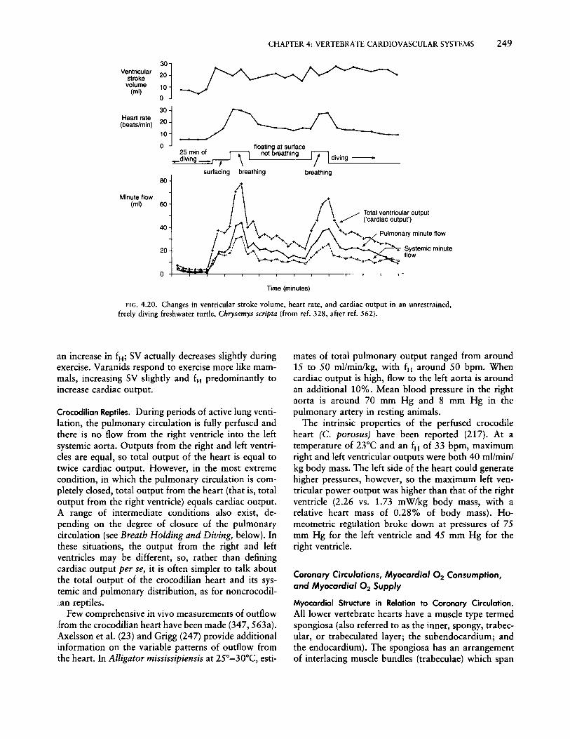
Ventricularstrokevolume
(ml)
Heart rate(beats/min)
CHAPTER 4: VERTEBRATE CARDIOVASCULAR SYSTEMS 249
:~j10
o
:~110
o floating at sur1ace25.min of ~\ not breathing 17l; ..diving . . diVing -- -, -~-----
sur1acing breathing breathing80
Minute flow(ml) 60
40
20
Time (minutes)
\+....+.......+.... / Pulmonary minute flow
~-...+....---
FIG. 4.20. Changes in ventricular stroke volume, heart rate, and cardiac output in an unrestrained,freely diving freshwater turtle, Chrysemys scripta (from ref. 328, after ref. 562).
an increase in fH ; SV actually decreases slightly duringexercise. Varanids respond to exercise more like mammals, increasing SV slightly and fH predominantly toincrease cardiac output.
Crocodilian Reptiles. During periods of active lung ventilation, the pulmonary circulation is fully perfused andthere is no flow from the right ventricle into the leftsystemic aorta. Outputs from the right and left ventricles are equal, so total output of the heart is equal totwice cardiac output. However, in the most extremecondition, in which the pulmonary circulation is completely closed, total output from the heart (that is, totaloutput from the right ventricle) equals cardiac output.A range of intermediate conditions also exist, depending on the degree of closure of the pulmonarycirculation (see Breath Holding and Diving, below). Inthese situations, the output from the right and leftventricles may be different, so, rather than definingcardiac output per se, it is often simpler to talk aboutthe total output of the crocodilian heart and its systemic and pulmonary distribution, as for noncrocodilian reptiles.
Few comprehensive in vivo measurements of outflowfrom the crocodilian heart have been made (347, 563a).Axelsson et al. (23) and Grigg (247) provide additionalinformation on the variable patterns of outflow fromthe heart. In Alligator mississipiensis at 25°-30°C, esti-
mates of total pulmonary output ranged from around15 to 50 ml/min/kg, with fH around 50 bpm. Whencardiac output is high, flow to the left aorta is aroundan additional 10%. Mean blood pressure in the rightaorta is around 70 mm Hg and 8 mm Hg in thepulmonary artery in resting animals.
The intrinsic properties of the perfused crocodileheart (c. porosus) have been reported (217). At atemperature of 23°C and an fH of 33 bpm, maximumright and left ventricular outputs were both 40 ml/min/kg body mass. The left side of the heart could generatehigher pressures, however, so the maximum left ventricular power output was higher than that of the rightventricle (2.26 vs. 1.73 mW/kg body mass, with arelative heart mass of 0.28% of body mass). Homeometric regulation broke down at pressures of 75mm Hg for the left ventricle and 45 mm Hg for theright ventricle.
Coronary Circulations, Myocardial O2 Consumption,and Myocardial O2 Supply
Myocardial Structure in Relation to Coronary Circulation.All lower vertebrate hearts have a muscle type termedspongiosa (also referred to as the inner, spongy, trabecular, or trabeculated layer; the subendocardium; andthe endocardium). The spongiosa has an arrangementof interlacing muscle bundles (trabeculae) which span

250 HANDBOOK OF PHYSIOLOGY~COMPARATIVE PHYSIOLOGY
the atrium and ventricle to form a sponge-like network.This network can create many small compartmentswithin the heart itself. The diameter of the trabeculaein various elasmobranchs and holocephalans typicallyranges between 30 and 250 J,Lm (539). In a species ofRana, the mean trabecular diameter is 40 J,Lm (60).Trabecular diameter is likely a compromise betweenminimizing the diffusion distance for O2 from luminalblood and maximizing tension development. The capillary separation in frog sartorius muscle is around 60J,Lm. Thus, the maximum O2 diffusion distance fromluminal blood for ventricular trabeculae is betweenone-quarter and twice the distance in frog sartoriusmuscle.
The spongiosa is the only myocardial tissue type inthe ventricles and atria of cyclostomes, most teleostfishes, and possibly all amphibians. In all other lowervertebrates (elasmobranchs, certain teleosts, and allreptiles), the ventricular spongiosa is encased by alayer of another myocardial tissue, the compacta. Theventricular compacta has a variable thickness. It mayaccount for as little as 5% of total ventricular mass,as in chimaerid fish, or as much as 66%, in skipjacktuna (Katsuwonus pelamis) (see Table I in ref. 190).However, the compacta typically represents 20%-40%of ventricular mass when present in lower vertebrates.In birds and mammals, almost the entire ventricle iscompacta. Muscle fibers of the compacta (also referredto as the outer, compact, or cortical layer; the subepicardium; and the epicardium) are more densely packedthan those of the spongiosa, and the various architectural arrangements of the layers of fiber bundles withinthe compacta are well documented for fishes (180, 190,536,539).
The compacta is always associated with a coronarycirculation. Thus, the hearts of cyclostomes, most teleosts, and amphibians are avascular; they lack a fullydeveloped coronary circulation with capillaries withinthe muscle. However, the coronary circulation, whenpresent, is not necessarily confined to the compacta.Coronary arterioles and capillaries clearly reach thespongiosa of the ventricle and atrium in all elasmobranchs and several active fishes-for example, tunaand marlins (138, 190,539, 611).
The presence of a coronary circulation is not associated exclusively with either air breathing or terrestriallife. Air-breathing fishes, such as the bowfin (Amiacalvens), have a coronary circulation, but amphibiansand the lungfish Neoceratodus do not. Instead, aquatichypoxia and a high myocardial power output appearto be important selection pressures favoring the development of a coronary circulation.
Because a greater proportion of compacta corresponds to a more developed coronary circulation, gener-
alizations can be made about the coronary circulationby inference based on the measurements of compacta.The percentage of compacta is related to scope foractivity and cardiac power output within, but notnecessarily between, phyletic groupings. Endothermicsharks have more compacta than ectothermic sharks,as do endothermic teleosts compared with ectothermicteleosts. However, endothermic sharks do not havemore compacta than all ectothermic teleosts (164,190). This association probably reflects the higher pressure work performed by the hearts of endothermic fishes rather than the higher cardiac output.Hemoglobin-free Antarctic fishes, for example, havea large ventricular mass and cardiac output but nocoronary circulation.
The compacta appears early in development in salmonids, increases disproportionately with growth injuveniles, and reaches a relatively fixed proportion ofventricular mass in mature fish (187, 363, 499). Thisdevelopment pattern is mirrored in mammals. Embryologically, mammalian hearts are trabecular and coronary vessels become incorporated as the heart is modified during development (245).
Coronary Vascular Arrangements. The following generalization can be made regarding the anatomy of thecoronary circulation in lower vertebrates: it has twopossible sites of origin and for both sites oxygen-richarterial blood enters the coronary circulation, bloodpressure being close to the highest possible. There isalways an anterior (cranial, cephalad) origin, meaningthat the origin is from postbranchial vessels in fishesand from the systemic aorta close to the heart inamphibians, reptiles, birds, and mammals. In somefishes and reptiles, there is an additional posterior(pectoral, caudal) origin that usually goes to the apexof the ventricle.
(Detailed information on the anatomy of the coronary circulation in lower vertebrates can be found inrefs. 138, 190,214,245,255,416,481,539, and 612.The following is therefore only a brief phylogeneticoverview.)
Coronaries in fishes. The majority of fish species haveavascular hearts, which have no capillaries in the myocardium (Type I) (612). All elasmobranchs (includingchimaerids, sharks, rays, and skates), active teleosts,chondrosteans, and some dipnoans have a coronarycirculation but with varying distribution patterns.
Anterior coronary circulation in fish has its origin atthe ventral ends of one or more efferent branchialarteries and reaches the ventricle on the outer surfaceof either the bulbus arteriosus (teleosts and chondrosteans) or the conus arteriosus (elasmobranchs). Theorigin of the posterior coronary circulation-for exam-

CHAPTER4: VERTEBRATE CARDIOVASCULAR SYSTEMS 251
pIe, in eels, marlins, rays, and skates-is associatedwith the coracoid branch of the subclavian artery andreaches the heart via ligaments between the pericardium and the ventricle. In both cases, coronary bloodpressure is roughly equivalent to that of the dorsalaorta. The dipnoan Protopterus is unique in that thecoronary circulation is derived from the afferent sideof the branchial circulation; in contrast, Neoceratodusdoes not have a coronary artery.
In fish such as the salmonids, coronary capillariesare located only in the compacta of the ventricle (TypeII) (612). In all elasmobranchs and in teleosts such astuna and marlin, coronary capillaries and arteriolesreach the spongiosa and atrium as well as the ventricular compacta. Typically, the elasmobranch version ofthis arrangement contains <40% compacta and istermed Type III; the teleost version has around 40%or more compacta and is termed Type IV (138, 612).The conus arteriosus of elasmobranchs also has compacta and its own coronary arteries and veins (190).
Circulation to the compacta has a discrete venoussystem, with large veins emptying into the atrium nearthe atrioventricular region. However, the coronary circulation in the muscle trabeculae of Types III and IVventricles has no venous system (473, 630). Instead,the terminal arterioles and the few capillaries opendirectly to the ventricular lumen, thereby forming arteriolacunary connections analogous to the Thebesiansystem of the mammalian heart (612). There is noestimate of either what proportion of coronary bloodflow enters the spongiosa or whether the vessels ofthe spongiosa are even continuously perfused. [TheThebesian system in the perfused turtle heart (c. elegans) accounted for less than 0.1% of the coronarydrainage emptied into the ventricular cavity (63).]
Coronaries in amphibians. Urodeles and anurans havea single coronary artery which arises either at thebase of the ventricle or near the right carotid arterysubdivision. However, coronary vessels are locatedonly in the outer connective tissue of the heart and notin the myocardium per se; no compacta is found in theventricle (245, 255,416, 473). The bulbus cordis hascompacta and coronaries (245). Apodans do not havea coronary artery (215). Thus, it appears that themixed venous blood in the lumen of the heart providesan adequate O2 supply for the low cardiac work (andmyocardial O2 demand) of the amphibian heart.
Coronaries in reptiles. The origin of the reptilian coronary circulation is thoroughly described by MacKinnonand Heatwole (416). Typically, all reptiles possess either one or two anterior coronary arteries derived fromthe right (or sometimes left) systemicocarotid trunk atvarious locations near the heart. In addition, somefamilies have a posterior supply from the coeliacomes-
enteric artery, which reaches the apical region of theventricle via the gubernaculum cordis; a gubernaculumcordis is found in most reptiles but does not alwayscontain a coronary artery.
Coronary vessels in reptiles are restricted mostly tothe ventricle. In the tortoise Emys orbicularis, coronaries are confined to the compacta (348), but whetherthis is true for other reptiles is not clear from theliterature. The extent of coronary development in reptiles appears to be related to the level of cardiac workand myocardial O2 demand. For example, "fine" coronary vessels are found on the ventricle of squamatelizards, whereas the ventricle of varanid lizards, whichgenerates higher aortic blood pressures, has larger coronary vessels and more compacta (416). Similarly,coronary circulation in the crocodile is more extensiveon the left side of the heart. The absence of atrialcoronaries may be directly related to the lower O2
demand of the atrium. In varanid lizards, coronaryvessels are located in the right but not the left atrium,a distribution pattern which suggests that the systemic,but not the pulmonary, venous return, may becomelimiting in terms of a luminal O2 supply. Coronarycirculation to the atria may become more importantwhen reptiles grow to a large size and the atrial wallthickness increases.
Coronary O2 Supply and Control of Coronary Blood Flow.Information on blood flow in, and vasoactivity of,coronary vessels is largely limited to studies on fish(see reviews in refs. 137, 180, 190) and a study onthe E. orbicularis coronaries (348). This informationclearly suggests that there are major differences in thecontrol, and the role, of the coronary circulation inlower vertebrates compared with mammals.
Cardiac function in the mammalian heart is dependent on a continuous coronary blood flow. Furthermore, hypoxia, adrenaline, and increased metabolism(cardiac work) all result in a coronary vasodilationthat significantly increases coronary blood flow. In thisway, there is tight matching between the coronary O2supply and the work performed by the heart. In contrast, the lower vertebrates studied so far show coronary vasoconstriction in response to adrenaline and thecontrol of coronary flow is less precisely matched toarterial blood pressure generated by the heart. Thismight be expected given the anatomical origin of thecoronary arteries in fish. The origin of the coronariesis postbranchial, so the driving pressure for coronaryblood flow is substantially lower than the pressuredeveloped by the ventricle since gill resistance reducesarterial pressure by 25%-40%.
Direct measurements of coronary blood flow in resting coho salmon yield values of 0.2 mllmin/g ventricu-

252 HANDBOOK OF PHYSIOLOGY-COMPARATIVE PHYSIOLOGY
lar mass (1.1% of cardiac output) (19). In burbot (LotaIota) and sucker (Catastomus catastomus), coronaryblood flow was estimated at 0.56%-0.65% of cardiacoutput using microspheres (106). Estimates of coronaryflow based on in vitro pressure-flow relationships yieldhigher values compared to in vivo values, perhapsbecause of a loss of vascular tone (for example, 0.38and 0.67 mllmin/g ventricular mass, 1.5% and 1.9%of resting cardiac output for rainbow trout and skipjack tuna, respectively) (177, 184). By comparison,coronary flow in mammalian hearts represents agreater proportion (about 4%) of resting cardiac output. Some of this difference may reflect the smallerproportion of the fish ventricle being perfused by thecoronary circulation (177).
Extravascular compression of coronary vessels during systole produces phasic flow in the coronary arteryof the coho salmon (Fig. 4.21). Even though ventricularcontraction attenuates flow in the main coronary artery, there is no flow reversal. In contrast, a briefreversal of coronary flow occurs in the anesthetizedschool shark (Galeorhinus australis) coincident withventricular systole (141). This difference may reflectthe different coronary distribution patterns to the compacta and spongiosa in sharks and salmonids (seeabove). Extravascular compression is significant inmammalian hearts (201). Moreover, because of thedifferent radii of curvature of the thick-walled mamma-
Iian ventricle, blood flow to the inner myocardial layersis affected to a much greater degree. The differentialwall tension between the inner and outer layers may,through vascular compression effects, limit wall thickness to some degree even though there is adequate vascularity.
Even though there is a resting level of blood flow tothe coronary circulation, there does not appear to bean absolute requirement for coronary blood flow insalmonids during routine cardiac function. Ligation ofthe coronary artery does not result in immediate deathof salmon ids (146, 189, 196). Furthermore, salmonidswith the coronary artery ligated can swim at speedsmore than 50% of their normal maximum prolongedswimming speed (Vetit ) . Therefore, the luminal O2 supply can at least partially compensate for whatever O2is normally supplied by the coronary circulation. Infact, in vitro perfused rainbow trout hearts can performmaximally while receiving aerated saline in the lumenand no coronary perfusion (191, 433). It is likely,however, that as fish grow the dependence upon thecoronary circulation increases with increasing ventricular size and thickness.
Calculations suggest that of the O2 normally available in luminal blood, the entire myocardial O2 demand would deplete luminal O2 content by only 1%10%, even during exercise (190). In view of this, it isunlikely that the O2 content of luminal blood limits
80U-~~_~~~~_~~~
160
140
t 120
~ 100
160
140
t 120
~... 100
83 250 8 6
po.... Q~ 200 qCor
~~
-'" 2 ~ 6-'" po....150
~
c -: '"·s c ~:§,
's 0
100<
:§, cC.
; 4u ·0'CT
50
0 0 2 250ms
80 U-~~_~~~_~_~~~
120.,. :I: Rqcor
Ie.... .,. •• Rt 100 .•~.. .,. '.
~ . ':.... " ..- ..~ 80 .......~...... .~•••ir...,. .................:...,...... .,.!J..l..LJ.l.l.II: 11l11ll!1111l1111
60-2 0 4 8 10 12 14 16
Time (min)
FIG. 4.21. Simultaneous measurements of cardiac output (Q), coronary blood flow(ilcM)' and dorsal aorta (PDA,,) in coho salmon. Heart rate ((H)' total systemic resistance(R,yJ, and coronary vascular resistance (Reo,) were derived from these measurements.A: Injection of isoproterenol into the dorsal aorta (at time zero) resulted in an increasein qCa, and coronary vasodilation without a significant change in driving pressure(PDA,,)' B: Phasic relationship between pressure and flows. Flow in the main coronaryartery is biphasic and continuous throughout the cardiac cycle (adapted from ref. 19with permission).

CHAPTER 4: VERTEBRATE CARDIOVASCULAR SYSTEMS 253
myocardial oz supply (137). [Note, however, thathemoglobin-free icefish are unusual in that they mayuse 43% of the luminal oz supply (138).]
Since the limitation on the venous myocardial ozsupply route is the venous POz, we can expect a greaterreliance on the coronary oz supply route (1) in situations where venous Poz decreases (that is, the pressuregradient for oz diffusion is reduced) and (2) in animalsin which the wall of the heart is particularly thick (thatis, the distance for oz diffusion is usually larger inanimals as they grow and in animals noted for a largerelative ventricular mass). There is some experimentalsupport for these ideas. During exercise and environmental hypoxia in fishes, venous Po, decreases andcoronary flow not only increases (19) but maximumcardiac performance is apparently tied to adequatecoronary perfusion (142, 189, 195, 196). Skipjack tunarepresent an extreme in terms of the high ventral aorticpressures generated by their large heart having a thickventricular wall. It appears that these high blood pressures are possible only if the heart is receiving oz viathe coronary supply route, perhaps making skipjacktuna, like mammals, obligately dependent upon coronary circulation (184).
Control of coronary blood flow is achieved throughchanges in perfusion pressure and coronary vasoactivity. In mammals, perfusion pressure is the primarydeterminant of coronary blood flow (201) and is closelytied to the pressure work performed by the left ventricle. In exercising mammals, coronary blood flow canincrease fivefold for a sixfold increase in cardiac work.A similar situation is likely to exist in the amphibianand reptilian coronary circulations. In rainbow trout,a roughly proportional increase in coronary blood flowaccompanies a change in Pda (19, 177). However, Pdais well regulated and rarely changes by more than 30%or so during either exercise or hypoxia. This meansthat pressure-dependent changes in coronary flow under these two conditions do occur but are not ofsufficient magnitude to account for the two-fold increases in coronary blood flow observed in cohosalmon during hypoxia and exercise (19). Consequently, a coronary vasodilatory reserve is of primaryimportance in bringing about changes in coronaryblood flow in salmon.
The basis for the coronary vasodilatory reserve inlower vertebrates is not fully understood. A number ofin vitro studies in fishes have identified a-adrenergic,j3-adrenergic, cholinergic, and purinergic vasoactivitiesin either the entire coronary circulation or the maincoronary arteries (38, 136, 177, 183, 184, 186, 569).The relative importance of arterial and arteriolar control has not been addressed. An a-adrenergic constriction and a j3-adrenergic dilation of the coronary circu-
lation have been confirmed for coho salmon in vivo(19). In the tortoise, there is a-adrenergic, cholinergic,and histaminergic vasoactivity (348). An importantdifference in coronary vasoactivity between mammalsand lower vertebrates is the coronary response toadrenaline. In the tortoise and salmonids, adrenalineincreases the overall coronary vascular resistance and,therefore, by itself would reduce coronary flow (19,177, 348). This is the opposite of the mammaliansituation. Thus, it appears that o-adrenoceprors predominate in the coronary circulation of lower vertebrates, whereas j3-adrenoceptors predominate inmammals.
Adrenergic-based vasoactivity is clearly not responsible for the entire vasodilatory reserve.in coho salmon.Isoproterenol produces only a 40% increase in coronary flow (19). The only other potent coronary vasodilators identified are the prostaglandins Iz and Ez (A. P.Farrell, unpublished observations). Consequently, thevasodilatory reserve is based on either the release of atonic vasoconstrictor mechanism or increased activityof vasodilators such as prostaglandins Iz and Ez. Tonica-adrenergic coronary vasoconstriction could be important in vasoconstriction, but so too could endothelin and prostaglandin Fza, which are very potentvasoconstrictors of fish coronary arteries in vitro (137,467). What appears to be emerging is a very importantrole for autocoids in coronary blood flow regulation.
Myocardial Oxygen Consumption. The hearts of lowervertebrates are aerobic, and most would cease to function without an adequate myocardial oz supply. ozsupply to the myocardium is provided by two circulations, the luminal and coronary. The luminal oz supply(the venous blood being pumped by the heart, alsotermed the lacunary or sinusoidal blood supply) bathesonly the inner surfaces of the cardiac chambers; thereare no blood vessels associated with it. Trabeculaeincrease the area of contact between cardiac muscleand venous blood, and, as noted above, the diameterof the trabeculae may reflect a compromise betweenminimizing diffusion distance for Oz and maximizingthe cross-sectional area for tension development. Thecoronary oz supply, in contrast, has arterioles andcapillaries which penetrate the myocardium.
All vertebrate hearts rely upon the luminal circulation to some degree, but not all lower vertebrateshave a coronary circulation. Thus, there is considerablevariability in the extent to which these two circulationsare utilized, and, as a consequence, there is variabilityin the anatomy and physiological role of the coronarycirculation. Ostadal et al. (472) have suggested thatdevelopment of the vertebrate coronary circulation isrelated to anatomical factors such as the extent of

254 HANDBOOK OF PHYSIOLOGY-COMPARATIVE PHYSIOLOGY
compacta myocardium and changes in heart size duringdevelopment, as well as physiological factors such asthe higher metabolic turnover of endotherms and theamount of cardiac work associated with activity of theanimal. In the following it will become clear that amajor difference between mammals and lower vertebrates is the relative myocardial O2 demand. Myocardial O2 demand is related to myocardial power output.
Studies with working, perfused fish hearts have provided the basis for our understanding of myocardial O2consumption (V02) in lower vertebrates. The generalfinding is that myocardial V02 is directly proportionalto myocardial power output under aerobic conditions(140, 190, 197, 243). The values for O2 cost per unitpower output range from 0.26 to 0.42 JLI 02/s per mWfor various fish species with differing cardiac anatomies(138). Mechanical efficiency increases slightly with thework load, being 15%-20%. Since mechanical efficiency is relatively similar among all vertebrate hearts,myocardial O2 demand can be easily compared amongvertebrates by comparing values for myocardial poweroutput. (It is much easier to measure flow and pressuregenerated by the heart and to estimate myocardial V02
than to try to measure myocardial V02 directly.) Thus,it should be fairly obvious that the high myocardialpower output of mammalian, avian, and tuna heartsresults in a higher O2 demand and greater reliance ona coronary 02 supply compared with other vertebrates.Knowing the relationship between power output andmyocardial V02, it is also possible to estimate the O2cost of cardiac pumping as a function of the animal'stotal V02 from measurements of myocardial poweroutput (190, 195). The O2 cost of cardiac pumping isrelatively small (between 0.5% and 5.0% of total V02 )
in fish species living in temperate water. However, itmay be as high as 23% of resting V02 in C. aceratusas a result of the high cardiac output value and lowO2 content of these hemoglobin-free Antarctic fishes.
In general, cardiac ATP demand greatly exceeds thepotential to generate ATP anaerobically; resting myocardial power output can range over three orders ofmagnitude (0.1-20 mWlg ventricular mass) and canincrease up to fivefold with exercise. However, theability of vertebrate hearts to generate ATP anaerobically does not vary nearly as much. Anaerobicallygenerated ATP can and does support myocardial poweroutput in animals in which it is either normally lowor suppressed during special conditions (for example,during either winter hibernation in fishes, amphibians,and reptiles or summer estivation in air-breathingfishes). To examine the importance of anaerobic metabolism in supporting cardiac work, the cardiac ATPrequirement has to be considered (515). To do this,myocardial V02 can be estimated from myocardial
power output and then converted to an ATP equivalent. In hagfish, for example, the resting myocardialoutput of around 1 mWig ventricular mass can bealmost completely supported by anaerobic metabolism(208, 209). In the hypoxic sea raven heart, with amyocardial power output of 0.8 mWig ventricularmass, anaerobic metabolism can contribute 25% of theATP requirement for resting power output (197). Apower output of 0.3-2.3 mW (ventricular mass notreported) was maintained for up to 15 h (providedplasma was present in the perfusion fluid) in the severely hypoxic (P02 < 2 mm Hg) perfused turtle heart(515). Farrell et al. (185) have shown in situ thatanoxic turtle hearts are capable of generating up to0.77 mWlg ventricular mass at l50 C. Likewise, anoxicrainbow trout hearts can generate 0.6 mWig ventricular mass at l50e in situ (11). Apparently, ATP generation from anaerobic metabolism can routinely fuel acardiac work level of around 0.1-0.2 mWig ventricularmass routinely but may not be able to fuel much morethan 1.0 mWlg ventricular mass, a level quite similarin both anoxia-tolerant and hypoxia-sensitive species.
Long-term cardiac survival during anoxia clearlymust be tied to two attributes (185); (1) the ability toreduce ATP demand to a level that can be supportedby anaerobic metabolism and (2) the ability to furtherreduce ATP demand to minimize waste product accumulation and fuel reserve depletion. In the overwintering anoxic turtle at 3°C, for example, myocardialpower output is around 0.03 mWlg ventricular mass[estimated from the data of Herbert and Jackson (273)and assuming cardiac output to be decreased in proportion with the 58-fold decrease in fH ] . This value is wellbelow the 0.17 mWig ventricular mass that can besupported by anaerobic ATP generation in anoxic perfused turtle hearts at 5°C (185). An additional adaptation for anoxic survival may be the intrinsic compensation for the negative inotropic effects of acidosis andhypoxia in anoxia-tolerant species (228, 321).
PERIPHERAL CIRCULAliON AND HEMODYNAMICS
Arterial Blood Pressure and Its Regulation
Levels of Arterial Pressure. Arterial pressure can be considered as the force required to overcome vascularresistance and to maintain blood flow to body tissues.The arterial pressure at any moment is related to thevolume and compliance of the vasculature, cardiacaction, and the resistance to blood flow attributableto the vessel architecture and properties of blood. Asophisticated understanding of pressure regulation involving the heart and systemic circulation has been

CHAPTER 4: VERTEBRATE CARDIOVASCULAR SYSTEMS 255
achieved, chiefly from mammalian studies. Control ofthe circulation is important for adaptation to stresses,and comparative investigations suggest that regulationof arterial pressures to a homeostatic level adequate toperfuse tissues is a basic requirement of all vertebrates.At the same time, nonsteady states are important for afuller appreciation of cardiovascular function, especially in ectothermic species.
Systemic arterial pressures range from a few to several hundred millimeters of mercury in various vertebrates (Fig. 4.22). There is, in the most general sense,an evolutionary trend toward increasing pressure asone progresses from primitive (agnathan) circulatorysystems to those of endothermic birds and mammals.Such generalizations about strictly phyletic progressions of pressure are complicated, however, by environmental influence, activity of animals, temperature, andother considerations. For example, arterial pressures inamphibians are lower than those in many fishes. Whiledevelopment of high arterial pressures is a function ofthe stressed filling volume, vessel compliance, cardiaccontractility, and peripheral resistance, combinationsof these features to produce high levels of pressure arenot limited to birds and mammals.
The lowest steady-state pressures recorded from vertebrate arterial systems are associated with the circulation of hagfish, which displays primitive features oftissue sinuses ("open" system) and accessory hearts to
assist circulation (324, 543). Low pressures also occurin other fishes, some amphibians, and aquatic snakesrepresenting taxa that have also evolved high-pressuresystems. Pulmonary pressures consistently are low relative to systemic pressures in air-breathing vertebrates(328), a consequence of low resistance of the pulmonary vasculature and the requirement that gas exchangers are not compromised by excessive filtrationof fluid. Given this generalization, although systemicpressures of birds and mammals are characteristicallyconsiderably higher than those of ectotherms, the pulmonary pressures of the latter animals tend to exceedthose of mammals (576). Such pressure differencesmay reflect intermittent breathing patterns and possiblygreater rates of lymph flow in the ectothermic species,notwithstanding possible differences in capillary structure and the filtration coefficient.
Pressures in the arterial system of the hagfish are onthe order of a few millimeters of mercury and aresupported by the independent actions of various accessory hearts on the venous side of the circulation (543).In other fishes, the highest central pressures are recorded in the ventral aorta. The ventral aorta is relatively short and, together with the bulbus arteriosus,accounts for the majority of compliance in the fisharterial system (543). Dorsal arterial pressures are lowered by gill resistance, which precedes in series that ofthe systemic tissues. The gills offer a significant resis-
~ branchial
osystemic
I pulmonary
C)
:J:EEW0:::::)tJ)tJ)W0::Q.
.J<I:i:2wto::<I:
120
100
80
60
40
20
o
co
~goE(;j/Jl
VI~
'tlo10..GIUoGIZ
VI~..GI-Q.o'0..e,
VI~Q.oCGI)(
VI>-EGIVI>-.co
(QUGIeClo'tl~..VIGI~
VI:cQ.ocE(Q~
~
(Qc(Q~
~
lit~c(Q..(Q
>
FIG. 4.22. Blood pressures in branchial (lined bar) v. systemic (open bar) and pulmonary (stippledbar) v. systemic (open bar) arteries of selected vertebrates. Height of vertical bar is equivalent to pulsepressure (from ref. 181).

256 HANDBOOK OF PHYSIOLOGY~COMPARATIVE PHYSIOLOGY
tance to blood flow, and systemic pressure reductionsof from 15% to 50% are commonly noted betweendorsal and ventral aortae (Fig. 4.22) (173, 176, 339,507, 646). Both pressure and flow exhibit pulsitility inthe dorsal aorta, though these features are considerablydampened by reduced elasticity of the dorsal vessel andhigh fH • In general, arterial pressures tend to be lowerin elasmobranchs than in teleosts (507).
The amphibian heart generally does not generatepressures as high as those in most fishes (Fig. 4.22).This may be related to the fact that the single ventricleserves both the pulmonary and systemic circuits. Eventhough ventricular work is essentially identical for thepulmonary and systemic circuits, a significant and variable resistance protects the pulmonary circuit downstream of the outflow tract (60, 190, 325, 560). As aconsequence, pulmonary arterial pressures are eitherlower than or equal to, but not higher than, systemicarterial pressures. Systemic arterial blood pressure andventricular mass are greater in terrestrial amphibians(for example, B. marinus) compared with more amphibious species (for example, R. catesbeiana).
Among reptiles, the chelonians have the lowest systemic arterial blood pressures, with terrestrial species(for example, Testudo pardalis) having slightly higherpressures than semiaquatic species (for example, C.scripta) (Fig. 4.22). Resting systemic arterial bloodpressure varies considerably among snakes, in partrelated to their diversification of exposure to gravitational forces (see below). An allometric relationshipexists in snakes between arterial blood pressure andbody mass (exponent =0.15), likely because ventricularmass increases with increasing body mass (553). Resting systemic arterial blood pressures in crocodiliansand varanid lizards are similar to those in mammals,and they are almost twice that in Iguana (Fig. 4.22).Therefore, the functional and anatomical divisions ofthe reptilian ventricle are associated with higher systemic arterial pressures.
Development of high arterial pressures is characteristic of, but not limited to, birds and mammals. Higharterial pressures are measured in species of fishadapted for rapid swimming and high physiologicalperformance, including endothermy. Ventricular orventral aortic pressures can exceed 100 mm Hg in tunaand salmon (184, 524, 646). Despite differences inmyocardial structure and body temperature, ventricular contractile force and tissue blood flow rates ofalbacore tuna approach those of mammals (646). Animportant conclusion from these data is that, exceptfor agnathans, diversification of cardiovascular designduring vertebrate evolution does not exert an overwhelming constraint on the evolution of high bloodpressures.
High levels of arterial pressure are often associatedwith large cardiac output values. This relationship doesnot always hold, however. For example, high levels ofblood flow are achieved in the hemoglobin-free antarctic icefish C. aceratus with lowered arterial pressuresbecause of decreased vascular resistance in the gills andvascular beds as compared with other teleosts (269,302). Both red-blooded and hemoglobin-free Antarcticfishes, by having conspicuously low ventral aortic pressures but relatively large ventricles, are exceptions tothe generalization that a higher aortic pressure is associated with a large ventricular mass (190).
High arterial blood pressures have also evolved inrelation to environmental or behavioral demands, irrespective of phylogeny or body mass. It is widely appreciated that the highest mammalian blood pressuresare measured in giraffes (235, 237, 258, 620, 621).Aortic pressures of 353/303 mm Hg have been recorded in upright adult giraffes, and such levels ofhypertension have been considered to be necessary forperfusion of cerebral vasculature, which may be 2-3m above the heart (reviewed in ref. 258). Whether thehigh pressures are required to overcome the gravitational column of the neck vessels or merely the flowresistance of the cephalic vasculature is unresolved (28,258,278,556). Cerebral vasculature aside, high levelsof arteriolar resistance and tight endothelial junctionshelp to prevent edema in dependent tissues of thegiraffe's limbs (258).
Systemic arterial pressures of snakes are correlatedwith the degree of arboreality or the gravitational stressto which a species is likely to be exposed (Fig. 4.23).Resting arterial pressures range generally between 15and 30 mm Hg in sea snakes, which live in a nearlyweightless environment, whereas arterial pressures inarboreal species can exceed 90 mm Hg (14, 394, 395,400, 557). Arboreal snakes have a comparatively shortheart-to-head distance (anterior heart) and are moreresistant to blood pooling and edema in the dependentvasculature. Superior adrenergic control of the vasculature in arboreal species probably reflects a high peripheral vascular resistance relative to nonclimbing species,but this hypothesis remains to be investigated. Pulmonary arterial pressures in these snakes remain ratheruniformly low despite species variation in the level ofsystemic pressures.
Generalizing from the available data for endotherms,the "average" mammal, with the exception of thegiraffe, has a mean arterial pressure of about 97 mmHg, while the "average" bird has a mean arterialpressure of about 133 mm Hg (250). Differences ofpressure between these taxa appear to be attributablemore to a greater cardiac output (relative to mass ormetabolic rate) in birds than to differences of periph-

CHAPTER4: VERTEBRATE CARDIOVASCULAR SYSTEMS 257
I
30..i
25
Venous pressure
··········f
i
10I
5
60
Time (min.)
Ci 50:cE.§. 40
FIG. 4.24. Changes in blood pressure in ventral aorta, dorsalaorta, and subintestinal vein during and after moderate swimmingactivity in rainbow trout (from ref. 587).
'":;en~ 30a."8 20 Dorsal aortic:iIi ....-.~, systolic
10 ..dJtjli~x-:SIOIiCI i I.?61Ag .4
i~·2 i i ~:; 0 5 10 0
f Arboreal
1=====:11 }Terrestrial
100
....... 90ClIEE 80.....~
Q)-c:: 70Q)o
?>-"0 600.0-Cll
50Q)~
::::Jl/ll/l 40Q)~
C.
"0 3000Zic:: 20CllQ)
~
10
Time (h)
CD 100 * * * **:;<II<IICD0.0 80"OJ:
8e:Os. 60"iii.~
:i 40
z "j" '25 :"<:; 20 A---- • • • • •J: •
15 ...
FIG. 4.25. Pattern of arterial pressure, hematocrit (Het), and bloodvolume in a single snake, Elaphe obsoleta, for an extended "recovery" period following 15 min of locomotion (stippled bar). Duringrecovery the snake was held within an acrylic tube, where it restedin an extended position but was free to move forward or backwardfor a distance of several centimeters. Note that the movements ofthe snake (stars in top panel) elevated arterial pressure and preventedblood volume from returning to control level measured before exercise (from ref. 404 with permission).
of stabilized arterial pressure levels compatible withthe morphology and activity of a species. The evolutionof stabilized arterial pressures has produced in vertebrates redundant feedback controllers involving reflexneural and neuroendocrine mechanisms affecting
o 10 2030405060708090
Tilt angle
Baroreflexes and Neurogenic Regulation of Arterial Pressure.Three conceptual considerations are important to understanding blood pressure regulation: (1) short-termregulation; (2) long-term regulation, for periods exceeding hours or days; and (3) evolutionary adaptation
eral resistance (250, 251). Relative to mammals, birdshave larger hearts, greater SVs, lower fH values, andgreater stroke work per gram of myocardium. In probably all vertebrate embryos, blood pressure increaseswhile peripheral resistance decreases with age and thedevelopment of a parallel system of vessels (121, 220a,306,590).
An increase in resting arterial blood pressure is possible in all species. Dramatic increases exceeding 50%of resting levels may occur in response to activity orexercise in fish, snakes, and mammals (Figs. 4.24,4.25). Relatively smaller increases in arterial bloodpressure are reported for exercising amphibians andlizards.
FIG. 4.23. Relationships between mean arterial pressures, measured at the body center (hydrostatic indifferent point, see text), andhead-up tilt angle determined in species of snakes from differentgravitational environments. Pressures at the body center are expectednot to change with tilt angle if the arterial column acts as a passivesystem contained within a rigid tube (224) (after ref. 557).

258 HANDBOOK OF PHYSIOLOGY-COMPARATIVE PHYSIOLOGY
moment-to-moment arterial pressure. Historically,studies of cardiovascular regulation, especially thoseprior to the 1970s, have tended to emphasize shortterm control of arterial pressure. Studies related specifically to the long-term control of pressure are relatively recent.
Two concepts are central to the current understanding of arterial pressure regulation: (1) shorter-termstabilization of pressure by means of cardiovascularneural reflexes and (2) long-term, slower adjustmentsof body fluid volumes mediated by physical and neuroendocrine mechanisms (130). Neurogenic reflex adjustments in cardiac output, total vascular resistance, andvenous tone are integrated with humoral, metabolic,and other tissue factors to maintain arterial perfusionpressure to all organs. The implication from mostmammalian literature is that these interacting processesare always homeostatic, in both design and action.Consideration of the biology of a range of vertebratespecies, however, illustrates the importance of nonsteady states in understanding the full integration ofcardiovascular controls.
A third important concept is that the net responseof interacting cardiovascular reflexes is not a simplelinear summation of the responses observed when eachreflex is activated alone (1). This is because thesereflexes interact, and when, for example, opposingreflexes are activated, it is difficult to predict the netresponse. Therefore, studies of intact organisms andresponses to several simultaneously changed sensoryinputs are important to a fuller appreciation of the roleof cardiovascular reflexes in controlling the circulation.
The principal elements involved in short-term control of arterial pressure are mechanoreceptors (usuallytermed baroreceptors), their sensory feedback afferents, central integrators of the sensory information,and the autonomic effectors comprising the efferentlimb of a barostatic reflex (Fig. 4.26). Mechanicalstretch receptors in the walls of blood vessels and inthe heart detect changes in wall tension and therebyprovide sensory information related indirectly to pressure and volume changes in the circulation. As judgedprimarily from studies of mammals, receptors in thearterial blood vessels are important for rapid adjustments of arterial pressure through changes in volumeoutput from the heart, whereas venous and cardiacreceptors are incapable of sensing arterial pressures butplay important roles in the regulation of blood volumeand cardiac output.
In mammals there are multiple baroreceptor centersinvolving several sites in the central circulation (154).Knowledge of baroreceptors and their characteristicsin non mammalian vertebrates is far less complete thanfor mammals. Not surprisingly, the search for barore-
ceptors in a range of vertebrates has been guided bycomparative anatomy and homology. Baroreceptorzones may extend from the heart and aortic arches todivisions of the arches or the truncus arteriosus (Fig.4.27). Whereas carotid baroreceptors have receivedemphasis in mammalian studies, predominant baroreceptive regions in other vertebrates are associated withthe aortic and pulmonary arteries (625, 645). It isimportant to suggest, however, that various vesselsmight contribute sensory information regarding theirstatus of distension, independent of organized "centers" of baroreceptors. In mammals, there is substantialinnervation of systemic vessels by unmyelinated sensory C-fibers having some degree of mechanoreceptivefunction (reviewed in ref. 229). The C-fiber afferentsarising from these vessels mediate spinal pressor reflexes in response to mechanical stimulation. Finally,arterial chemoreceptors often are near-mechanoreceptive segments of the vasculature and can influence cardiovascular control.
The functional characteristics of cardiovascularmechanoreceptors have been reviewed elsewhere (1,30, 46, 277, 476, 645). Arterial baroreceptors exhibitrate sensitivity and thereby produce an enhanced response to dynamic pressure stimuli. Although baroreceptors respond best to rapidly changing stimuli, slowlyadapting receptors also encode the mean level of arterial pressure. Baroreceptors exhibit sensory adaptationand thereby reset to new states of wall stress equilibria,so they are thought not to participate in the longterm regulation of pressure. Indeed, destruction ordenervation of the receptors may have little effect onthe long-term level of arterial pressure (130, 627). Ithas been proposed that denervation of both arterial andcardiopulmonary baroreceptors can lead to sustainedhypertension in mammals (488, 489), but this conclusion is unproven (225,563,564).
The distribution of baroreceptors in mammals includes the heart, the carotid sinus, several places alongthe common carotid arteries, the right subclavian region, the pulmonary arteries, and the intrarenal vessels(Fig. 4.27). Baroreceptor afferents consist of a heterogeneous population of neurons comprised of both myelinated and unmyelinated fibers (124, 202). Two distinct forms of mammalian baroreceptor may bedistinguished on the basis of functional and morphological characteristics. Myelinated fibers have relativelylow pressure thresholds and high discharge frequenciesand respond to a large range of stimulation frequencies(about 10-150 Hz). Unmyelinated baroreceptors havehigher thresholds and lower discharge frequencies andproduce depressor responses at relatively low stimulation frequencies of 1-50 Hz (462).
Unmyelinated baroreceptors have been identified in

CHAPTER 4: VERTEBRATE CARDIOVASCULAR SYSTEMS 259
Response to increase in • • Perturbation• Activity
baroreceptor activity I t HydrationI
HemorrhageI
... Heart liCEill~
Rate
Ventricular.? output•
Comparator Ventricular i..Volume(medulla) ,Change Aortic arterial ...
'Set Point',~ pressure
~+Error signal
Pulmocutaneous~(reset - arterial
during activity) ~ Systemicpressure
...Arterioles
Tone t0 Systemic IPeripheralResistance ? • Intrapulmonary
.0. ,, VascularExtrinsic ,... Pulmonary , Pressure
~ Pulmonary ResistanceArtery I
@;D ? IPulmonaryI
•Baroreceptor
neurogram6~ E 1111 1111 1111
, Fluid(IlV) @,
activity ·60 , Filtration
Pulmocutaneous arterial :~ Er-:»~ .-blood pressure (mm Hg)
20
Aortic arterial blood 60 /'---.;~ ..-pressure (mm Hg)
FIG. 4.26. Schematic diagram of mechanisms participating in baroreflex regulation of cardiovascular variables in Bufo. "Classical"representation of negarive feedback, which uses a hypothesized "setpoint" for arterial pressure and a medullary comparator. Responseof heart and systemic vasculature to baroreceptor stimulation isreduction in heart rate and systemic resistance, which opposes elevations in central arterial pressure. However, pulmocutaneous arterial
amphibians, reptiles, and mammals, whereas myelinated baroreceptors, with thresholds well below normalarterial pressures, have so far been identified only inmammals (624-627a). Differences in the function ofbaroreceptors between vertebrate classes may be related to corresponding differences in circulatory function. Unmyelinated receptors may reflect a widespreadneed to protect the circulation from potentially damaging increases in arterial pressure, including thegas-exchange vasculature. However, low-threshold myelinated baroreceptors may be important to pressureregulating systems of mammals which are relativelyintolerant of even transient hypotension.
There is a wealth of information concerning neural
(PCA) resistance increases, probably because of vagally mediatedconstriction of the extrinsic pulmonary artery. This opposes regulation of PCA pressure and ultimately central arterial pressure butprotects the pulmonary microcirculation from pressure elevations.Some relevant references are numbered (1-8): 1, 319; 2, 571; 3,579; 4, 624; 5, 626; 6, 625; 7, 627; 8, 644 (modified from ref. 645).
regulation of arterial pressure and the role of baroreceptors in cardiovascular control of mammals. Thisinformation has been summarized in numerous reviews, and space does not allow a thorough treatmenthere (see, for example, refs. 1, 30, 361, 362, 373, 374,476, 533). The following sections provide a comparative perspective by discussing neurogenic control of thecirculation in non-mammalian vertebrates.
Cyclostomes, dipnoans, and primitive fishes. Responsesof the heart of Myxine to drugs affecting the cardiovascular system are weak (see Cardiac Output and CardiacPerformance, above), and it is doubtful that circulatingcatecholamines play any role in cardiac control in theseprimitive vertebrates (455). It seems likely that the

260 HANDBOOK OF PHYSIOLOGY-COMPARATIVE PHYSIOLOGY
IX
IC
MAMMAL
TG
REPTILE(Chelonian)
BIRD
FISH AMPHIBIAN(Anuran)
IC
X EC ECVG
X XVG
FIG. 4.27. Schematic of baroreceptor zones and their innervationin five classes of vertebrate, drawn to illustrate developmental originsfrom visceral arch arteries and associations with corresponding visceral arch nerves. Amphibian and mammalian examples adaptedfrom reference 626; the remainder based on references in 30, 340,624-627. Drawing is from Van Vliet and West (627a). Abbreviations: A, aorta; AA, aortic arch; Abd A, abdominal aorta; CA, celiacartery; CL, carotid labyrinth, CN, carotid nerve; CS, carotid sinus;CSN, carotid sinus nerve; DA, dorsal aorta; DCA, dorsal carotidartery; EC, external carotid artery; IC, internal carotid artery; IN,innominate artery; ITN, inferior truncal nerve; LDA, ductus arterio-
sus or its ligament; NG, nodose ganglia; PA, pulmonary artery;PHI and PH2, pharyngeal branches of the vagus nerve; PCA,pulmocutaneous artery; RLN, recurrent laryngeal nerve; SC, subclavian artery; SLN, superior laryngeal nerve; STN, superior truncalnerve; TA, truncus arteriosus; TG, truncal ganglia; VA, ventralaorta; VCA, ventral carotid artery; VG, vagal ganglia; V, VII, IX,and X, cranial nerves 5, 7, 9 (glossopharyngeal), and 10 (vagus);1,2,3,4,5, and 6, visceral arch arteries 1-6. Stippled areas representfunctionally identified baroreceptive zones. Shaded region in uppermidline of each figure represents larynx and pharynx.
upper level for arterial blood pressure is set by thepressure-generating ability of the heart, which appearsto be quite limited (20). The chromaffin cells of dipnoans appear to be innervated by extrinsic, presumablycholinergic, fibers (547). The heart of the gar (Lepisosteus) is richly innervated by adrenergic fibers, and thereis evidence for adrenergic control attributable to bothadrenergic neurons and circulating catecholamines(454), as in more advanced fishes. Autonomic innerva-
tion of the vasculature of cyclostomes and dipnoansmay be comparatively poorly developed, and adrenergic control may be primarily by means of circulatingcatecholamines (4, 5). Little is known about reflexogenic control of the circulation in cyclostomes, ganoids,and dipnoans, but complex regulation of blood flowpatterns has obvious significance in relation to thebranchial and pulmonary circuits of the lungfishes.
Elasmobranchs. The cardiovascular system of elasmo-

CHAPTER 4: VERTEBRATE CARDIOVASCULAR SYSTEMS 261
branchs appears to represent a primitively organizedsystem with limited control and weak ability to withstand stress. In dogfish, baroreceptors are claimed tooccur on the afferent side of the branchial arches,where increased perfusion pressures induce bradycardia (413). Bradycardia is also elicited by mechanicalor electrical stimulation of several regions, includinggills, heart, and the cut central ends of the first tofourth branchial nerves (413,414). Nerve activity synchronous with cardiac systole can be recorded frombranches of the branchial nerves (316). Although cardiovascular responses are elicited by nerve stimulationand autonomically active drugs (470, 471), there islittle evidence of neuronally mediated control of theperipheral circulation. The cardiovascular systems ofstudied elasmobranchs are intolerant of tilting (464)and behave like simple pressure-volume systems inresponse to hemorrhage (591).
The heart of elasmobranchs is supplied by two pairsof cardiac branches of the vagus. Acetylcholine as wellas stimulation of the vagus reduce fH , and cholinergicvagal tone is considered the most important modeof nervous chronotropic regulation in elasmobranchs(455). It is generally accepted that the atrium andventricle of elasmobranchs lack a sympathetic innervation (12). Catecholamines generally have weak positivechronotropic and inotropic effects on the heart, butresponses are variable and there is evidence for a cholinergic element in catecholamine-induced inhibition(137, 386). The two naturally occurring catecholamines epinephrine and norepinephrine could conceivably mediate control of cardiac activity (111).
Most literature on the adrenergic control of elasmobranch vasculature suggests that circulating catecholamines are the most important factor in the control ofblood pressure (reviewed in refs. 30, 455). The majorsystemic arteries of the dogfish are adrenergically innervated and contracted by epinephrine via aadrenoceptor-mediated mechanisms (457, 458). Verylittle is known concerning the direct nervous controlof branchial vasculature, but regulation of branchialresistance may involve the release of catecholaminesfrom strategically located chromaffin tissue (223, 541).
The best documented cardiac reflex in elasmobranchs is the bradycardia that is induced by hypoxiain the dogfish (101, 540). Stroke volume increasesduring the response and is attributable to increasedcardiac filling (Starling's law of the heart) and possibleinotropic enhancement by circulating catecholamines(102, 103, 565, 594). Vagal cholinergic innervationcomprises the efferent cardioinhibitory pathway, thereflex originating with a diffuse system of oxygen receptors in the gills (99). The significance of hypoxic
bradycardia is that it maximizes respiratory gas exchange, though the mechanism is incompletely understood and appears to involve more than relative flowrates of water and blood (566, 593).
Teleosts. In teleosts, the cardiac vagi innervate thesinus venosus and atrium but do not reach the ventricle.The vagus is cardioinhibitory, as in all vertebratesexcept the cyclostomes, and a resting tonus has beendemonstrated in some species (107, 501, 587). Although the vagus does not directly influence the ventricle, it does affect cardiac output because atrial contraction in part sets ventricular filling (Fig. 4.12; 328,345).The importance of vagal tone in cardiac control canbe temperature-dependent, such that adrenergic mechanisms predominate in cardioacceleratory actions athigher temperatures (378, 501, 664). Adrenergic innervation has been demonstrated in all parts of theteleost heart [except flounder (12)], but the density andfunctional importance vary from species to species(455). Levels of circulating catecholamines are sufficient to produce an adrenergic tonus on the heart insome species of fish, but a neural adrenergic tonus maybe predominant (18). Comparative data suggest thatboth a cholinergic inhibitory tonus and an adrenergicexcitatory tonus are general features of the teleostheart, during rest and at moderate levels of swimming (18).
Chemoreceptors in the gill arches cause bradycardiaduring hypoxia (145, 221, 493, 573, 574) except in afew species (89, 112). In salmonids and Atlantic cod,the oxygen receptors are located in the region of afferent vessels associated with the first gill arch. Oxygenreceptors located elsewhere elicit a rapid increase inboth ventral and dorsal aortic blood pressures, attributable to an increase in systemic vascular resistance(221). Hypoxic bradycardia could be related in part toa barostatic reflex. Hypoxia typically increases vascularresistance (95).
Rapid increases and decreases in blood pressure arebrought under control by a barostatic reflex. Whereashypertension reduces cardiac output through bradycardia to minimize the pressure excursion, it is importantto note that there is a characteristic maximum pressurethat a heart can generate (see section on homeometricregulation Mechanical Properties of Cardiac Muscle,above). Thus, if vascular resistance increases beyondthe capabilities of the heart, cardiac output decreasesas pressure increases (a trade-off between pressure andflow). The decrease in cardiac output results froma smaller SV and not bradycardia. Perhaps the bestillustration of this phenomenon is in the Atlantic hagfish, where the efferent arm of the barostatic reflex isabsent. When doses of ACh greater than 100 nmol/kg
262 HANDBOOK OF PHYSIOLOGY~COMPARATIVE PHYSIOLOGY
were injected into the caudal vein, gill vasoconstrictionwas so great that cardiac output actually stopped asventral aortic pressure reached 1.6 kPa (20 mm Hg)(20).
Barostaric reflexes involving cardiac inhibition andlowering of blood pressure are elicited in various teleosts by electrical stimulation of cut branchial nervesor pressure perfusion of the gill arteries (30). Thesedepressor responses are abolished by vagotomy or atropine. The baroreceptors responsible for the reflex arelocated in various parts of the gills.
In the carp (Cyprinus carpio), reflex bradycardia iselicited by perfusion of one of the four gill arteries, butthere is a progressive decrease in sensitivity of the reflex(higher perfusion pressure required) from the first tothe fourth pair of gill arteries (521). This finding is ofinterest in view of the differential sensitivity of themammalian carotid and aortic sinuses, which are homologous to the third and fourth gill arches, respectively. In addition to the gill arteries, pressure-sensitivereceptors appear to be present in the afferent artery ofthe pseudobranch and possibly the carotid labyrinth(387,468).
Further evidence for functional baroreceptors in teleosts is demonstrated by cardiac responses to increasedloading of baroreceptors via either a pharmacologicallyor a physically induced rise in systemic blood pressureas well as to decreased loading attributable to hypovolemic lowering of arterial pressure. Injection of epinephrine elevates blood pressure by constriction ofsystemic vasculature in a variety of teleost species. Thisinduces a bradycardia due to a baroreceptor reflex(450,455,492,513,520,666). Vascular compressionof the systemic circulation has also been shown toelicit a barostatic reflex (176). In both instances, thebarostatic reflex is abolished by atropine pretreatment.Barostatic reflexes can be very potent in fishes andhave the capability to override chemoreceptive drives(667). However, as Wood and Shelton (667) point out,the cardioacceleration effector arm of the barostaticreflex in rainbow trout may be much weaker than thecardiodepressor arm.
Beyond the barostatic reflex, there is a significantvascular tonus in resting fish which contributes tothe overall pressure. Injection of the vascular smoothmuscle poison papaverine causes hypotension (176).There is evidence that adrenergic control of the cardiovascular system is primarily neuronal in some species(570,572,665) and primarily humoral in others (635).Systemic vasoconstriction involves an e-adrenoceptormediated mechanism, and there appears to be appreciable adrenergic innervation of the major arteries andarterioles (360, 636). Increased levels of circulatingcatecholamines are evident during stressfully ex-
hausting exercise but appear not to be important incirculatory adjustments during moderate exercise (314,522). In cod, hypertension noted during swimming isattributable to both an increase in cardiac output andthe maintenance of systemic vascular resistance by theactivity of adrenergic vasomotor fibers (25). Adrenergicfibers maintain a tonic influence on the vasculatureboth at rest (572) and during exercise (25). Isoprenalinedecreases blood pressure and vascular resistance, whichsuggests that e-adrenoceptors are also present in thesystemic vasculature (114, 135, 266, 492, 662). Cholinergic fibers are abundant in the spinal autonomiccontrol of various organs in teleosts, but their role invascular control is uncertain (455).
Further indirect evidence for reflexogenic controlis the observation of Mayer waves or low-frequencyoscillations of the arterial pressure independent of ventilatory frequency (661). Such oscillations in mammalsare considered to reflect feedback regulation of arterialpressure involving vascular smooth muscle activity inthe resistance vessels. Tolerance of bluefish (Pomatomus saltatrix) to head-up tilt with maintenance ofarterial pressure to angles of 60° from horizontal provide additional evidence for active regulation of vascular smooth muscle (464). Ogilvy and Dubois (464)speculate that the orthostatic tolerance of bluefish toposture change outside of water reflects adaptation tocirculatory stresses evoked during forward accelerationand rapid carangiform swimming (see also ref. 176).These observations and the demonstration of barostaticreflexes in fishes are significant because they emphasizethat reflexogenic mechanisms conferring orthostatictolerance did not evolve de novo solely in response tostrong gravitational fields experienced by terrestrialvertebrates (340).
The barostatic reflex in teleosts also shows shortterm accommodation. In the sea raven, the bradycardiaassociated with vascular compression decays after 3060s, with the exact period depending upon the initiallevel of bradycardia (176).
Amphibians. The amphibian heart is richly innervatedby cardiac branches of "vagosympathetic trunks" containing fibers arising from pre- and postganglionicparasympathetic and postganglionic sympathetic efferent nerves, as well as vagal sensory fibers (455). Thevagal fibers are cholinergic and produce negative chronotropic and inotropic effects in both urodeles andanurans. The intrinsic neurons (that is, postganglionicvagal neurons) of the heart of B. marinus contain andrelease both ACh and somatostatin (109). Cardiacadrenergic neurons release primarily epinephrine, as inother tissues, and produce positive chronotropic andinotropic effects on the heart of anurans. However,there is no evidence for an adrenergic innervation of

CHAPTER4: VERTEBRATE CARDIOVASCULAR SYSTEMS 263
the heart of urodeles (24). All chambers of the heart,including the sinus venosus and truncus arteriosus,contain mechanoreceptors, but their function has notbeen demonstrated (153, 367, 369, 370, 453). Thespontaneous activity of both atrial and ventricularmechanoreceptors peaks during systole, but some activity may be sustained for almost the entire cardiac cycle.The cardiac mechanoreceptor afferents include bothmyelinated and unmyelinated elements.
Mechanoreceptors have been identified in the carotidartery or labyrinth, the aortic arch, and the pulmocutaneous artery of anuran amphibians (Fig. 4.27) (317,318; reviewed in ref. 645). In B. marinus, the pulmocutaneous receptors provide the principal sensory feedback of arterial pressure to the cardiovascular centersof the medulla, with a smaller contribution arisingfrom the aortic arch. The physiological significanceof the carotid baroreceptors is at present uncertain.Pulmocutaneous mechanoreceptors and their attendantactions in anurans are presently the most extensivelycharacterized baroreflex in nonmammalian vertebrates(Fig. 4.26).
Pulmocutaneous baroreceptors are slowly adaptingand transmit their information via nonmyelinated afferents. The mean threshold pressure for baroreceptorfiring (30 mm Hg) coincides with normal systolic arterial pressures (30-32 mm Hg) of conscious toads (B.marinus). At pressures above threshold, feedback information from baroreceptors is increased by increasedfiring frequency as well as recruitment of receptorswhose threshold lies above the mean. The increase infiring frequency of receptors can be up to 1.7% oftheir maximum, which represents a sensitivity muchgreater than that of mammalian baroreceptors (625).Such high sensitivity is appropriate, however, becausethe receptors operate over a more limited range ofblood pressure than do those of mammals (645).
Feedback from the pulmocutaneous baroreceptorsinhibits the cardiovascular system and results in a fallin systemic pressure. A reduction of arterial pressurecan occur in the absence of bradycardia, indicatingthat the reflex also reduces systemic peripheral vascularresistance (644). However, pulmocutaneous vascularresistance is greatly increased in response to baroreceptor stimulation (624). Presumably, the site of increasedresistance is the pulmonary vasculature because 79%92% of pulmocutaneous blood flow passes to the pulmonary artery (329). The increase in pulmocutaneousvascular resistance concomitant with a decrease in systemic vascular resistance would tend to protect thepulmonary vasculature from excess pressures transmitted by the incompletely divided circulation wheneversystemic pressures are high (626, 645). When pulmocutaneous baroreceptors are denervated, increased pul-
mocutaneous artery pressures produce a tenfold increase in fluid filtration from the pulmonaryvasculature, as well as vascular lesions in the lungs(579, 627). Considering the available data, arterialbaroreflexes of anurans appear to be adapted to protectthe pulmonary microcirculation while regulating thecentral arterial blood pressure (645). Analyses of theopen-loop gain of baroreflexes studied in toads andmammals suggest that they are surprisingly similar(645). Probably the best way of comparing the barostatic reflex between vertebrates is to calculate the normalized gain (percentage change in fH per unit changein mean arterial pressure), even though this approachhas recognized limitations (340). In fact, the normalized gain for a variety of aquatic, semiaquatic, andterrestrial vertebrates is quite similar [1.9%-6.7% forsea raven (176), 1.3% for B. marinus (571), and 1%5% for the lizard and 2.7%-6.9% for dog, rabbit andhuman (44)].
The constrictive innervation of pulmonary vasculature is cholinergic, and, at least in some species ofanuran, the most important effector unit may be adistinct sphincter in the extrinsic pulmonary artery(535). Vascular resistance in the pulmonary vessels isalso varied in relation to lung ventilation, and perfusionmay be reflexly limited (right-to-left shunt) during periods of apnea (560).
Adrenergic fibers innervate both arterial and venoussystemic vasculature, though the innervation of majorarteries is somewhat less dense than that seen in reptilesand mammals (455). The systemic vasculature is constricted by epinephrine, which elevates blood pressureas a result. In addition to adrenergic vasoconstrictorinnervation, major arteries in the toad (B. marinus)may possess a cholinergic excitatory innervation (360,641). Vasodilation is mediated by f3-adrenoceptormechanisms (166, 448).
While the mechanisms of autonomic regulation ofblood pressure are quite extensively studied in anuranamphibians, there is little evidence that blood pressureis tightly regulated. Autonomic control of the circulation seems to be quite weak in resting Rana and Bufo(80, 634). In Bufo, there is no adrenergic pressor toneand cholinergic cardiac inhibition is modest. Of interestis that systolic arterial pressures of toads are oftencorrelated positively with fH such that increases inarterial pressure are accompanied by tachycardia ratherthan a bradycardia predicted solely on the basis ofbarostatic reflexes (627). This observation has beeninterpreted to indicate that cardiac interval in restinganimals reflects integration of many sensory inputsto the medullary cardiovascular centers and is notdetermined primarily by baroreflex feedback.
Activity in anurans increases both fH and arterial

264 HANDBOOK OF PHYSIOLOGY-COMPARATIVE PHYSIOLOGY
pressure, which are initially elevated by adrenergicefferents and sustained by circulating catecholamines(127,634). At least in toads (B. marinus) the cardiovascular response to exercise appears to be mediated by apsychogenic as well as an autonomic reflex component(634). In these circumstances, it is not known whetherbarostatic reflexes are reset or, in any case, how thereflexogenic feedback is integrated with other inputoutput information. There appears to be neither adrenergic innervation of the heart nor adrenergic tone onthe vasculature of the mudpuppy (24, 26). In the mudpuppy, the heart is controlled by a variable cholinergictonus combined with a variable adrenergic tonus,which is probably mediated by circulating catecholamines. During exercise, arterial pressure is elevated byincreased levels of circulating catecholamines, mainlynorepinephrine (26). Therefore, circulatory regulationmay be quite different between urodeles and anurans.
A number of studies suggest that control of gill, skin,and lung perfusion in amphibians may be, in part,reflexly controlled by peripheral or central chemoreceptor input (645). As in other vertebrates, however, theinteraction of chemo- and baroreceptors in controllingblood pressure is poorly understood. Based on available information, it seems conceivable that in amphibians, as in mammals, bradycardia can result from stimulation of peripheral chemoreceptors, receptors withinthe upper airways, or baroreceptors (645).
Denervation of pulmocutaneous baroreceptors hasdemonstrated that the pulmocutaneous baroreflex isnot essential for maintenance of resting arterial pressure in B. marinus (627). In the long term, endocrineand renal mechanisms may adjust arterial pressure viaeffects on vascular tone and fluid volumes independentof the baroreflexes.
Reptiles. For reasons of convenience, phylogeny, andanatomical diversity, reptiles have been the subjectof numerous studies of cardiovascular dynamics andcontrol. Like amphibians and fishes, reptiles displaywide variation in levels of blood pressure attributableto species differences, environmental stresses, and nonsteady states arising from a variety of causes. Nonetheless, like mammals, some reptilian species exhibit precise control of arterial pressure.
Resting blood pressures of reptiles are generally quitestable in the absence of external disturbance but mayvary with temperature, activity, or state of wakefulness(588). However, arterial pressure in resting tiger snakes(Notechis scutatus) does not vary significantly over arange of body temperatures at which this species isnormally active, implying adjustments of peripheralresistance and/or cardiac output as fH increases logarithmically over the same range (402). Arterial pressures usually increase with locomotor activity and may
remain elevated for long periods (1-2 h) after activityceases (404, 589) (Fig. 4.25). Activity-induced increasesof arterial pressure are partly attributable to autonomicactivation and partly to augmentation of venous return.
The potency of baroreflexes in squamate reptiles hasbeen well illustrated by studies of responses to tilt,hemorrhage, and pharmacological agents. Postural disturbance during head-up tilt displaces a significantproportion of the blood volume posteriad and immediately reduces cardiac output and central arterial pressure. Partial or full recovery of pressure is achievedby reflexogenic increases in both fH and peripheralresistance during a compensatory period in whichevents are basically similar to those seen in mammals(300, 400, 402).
Responses to tilt vary considerably among species ofsnake (Fig. 4.23). Aquatic and some ground-dwellingspecies are unable to maintain arterial pressure duringhead-up tilt, and cerebral blood flow diminishes as aresult (401, 405, 557). However, arboreal species aretolerant of tilting, and arterial pressures at head levelare maintained generally within 80%-100% of pretilt (horizontal) values (394; H. Lillywhite, unpublisheddata). Reflex activation of vascular smooth muscle ismore important than either cardiac chronotropic orinotropic effects in controlling arterial pressure; indeed,pressure is regulated even though cardiac output isreduced by 50% or more (400, 402). Increases inperipheral vascular resistance occur principally in visceral organs, posterior skin, and muscle and areblocked by o-adrenoceptor antagonists (400, 402).Control of this magnitude must involve venous constrictory mechanisms as well as arteriolar vasoconstrictory mechanisms. In the elapid snake N. scutatus, boththe magnitude and the rapidity of regulatory responsesvary with temperature and the apparent optimal response occurs at preferentially selected body temperatures (402).
Qualitatively similar autonomic adjustments of fH
and peripheral vascular resistance occur when reptilesare subjected to hemorrhage (300, 577, 578). Regulation of arterial pressure during hypovolemic states isdependent on barostatic reflexes. and compensatorytranscapillary shifts of extravascular fluid into the circulation. Remarkable abilities to regulate arterial pressure are again illustrated by arboreal snakes, fromwhich more than 100% of the initially measured bloodvolume (not subtracting compensatory fluid shift) canbe withdrawn before arterial pressure falls precipitously (403). Investigations have demonstrated extensive colocalization of neuropeptides in perivascularnerves innervating larger arteries and veins of ratsnakes (143). These data provide evidence for the importance of parasympathetic nerves in controlling cir-

CHAPTER4: VERTEBRATE CARDIOVASCULAR SYSTEMS 265
culation, particularly the regulation of venous capacitance.
Studies of squamates show that all parts of thesystemic vasculature are densely innervated by adrenergic fibers, which, in snakes, show considerable speciesand regional variation (149, 455). The density of adrenergic fibers innervating systemic vessels is especiallyhigh in posterior arteries and veins of arboreal snakes,which reflects the importance of adrenergic adjustments of vascular tone in postural contexts of hemodynamic regulation (149; J. Donald and H. Lillywhite,unpublished data). Catecholamines constrict vascularbeds, elevate arterial and venous pressures, and areantagonized by pharmacological blockade of aadrenoceptors (7, 43, 360, 394, 401, 402, 516). Thedense innervation of veins presumably reflects theirrole in active regulation of venous capacity. Centralvenous pressures of snakes remain constant duringprogressive isovolemic hemorrhage, suggesting thatvenomotor tone is actively regulated to maintain venous return, cardiac filling (403), and hence cardiacoutput.
Perivascular nerves innervating the vasculature ofsnakes contain a variety of neuropeptides apparentlyassociated with parasympathetic postganglionic fibers,adrenergic fibers, and afferent sensory fibers (143). Thedistribution of these neuropeptides suggests considerable functional specialization within the peripheral autonomic system controlling the circulation, especiallythe regulation of venous capacity.
The reptilian heart is innervated by vagal cholinergicinhibitory nerves that produce negative inotropic andchronotropic effects and an extensive sympathetic adrenergic excitatory innervation that promotes positiveinotropic and chronotropic responses (69, 151, 263,455). These elements innervate both atria and ventricles, as well as the adjoining inflow and outflow vessels.(Cholinergic inhibitory nerves are vagal in origin,whereas adrenergic fibers are of spinal autonomicorigin.) Adrenergic effects are mediated via {3adrenoceptors, and cholinergic receptors are muscarinic in character. The hearts of some reptiles appearto be, during resting states, under the tonic influenceof both cholinergic and adrenergic fibers (263, 402).Vagal stimulation can arrest the heart for very longperiods, and a "vagal escape" of the sort seen inmammals is not apparent in reptiles (435). Little isknown concerning the interplay of chemoreceptors inautonomic cardiovascular responses, but oxygensensitive receptors in the central circulation may playa role in modulating autonomic outflow to the heart(672).
As in amphibians, somatostatin is present in theheart of rat snakes (Elaphe obsoleta), probably in
postganglionic cell bodies and axons of intrinsic parasympathetic neurons (152). Additionally, immunohistochemical studies have demonstrated the presence ofsubstance P-like, calcitonin gene-related peptide-like,and neuropeptide Y-like immunoreactive axons in thesinus venosus, atria, and ventricles of E. obsoleta (152).The extensive distribution of peptides in the cardiacnerves suggests an as yet unknown cardiovascular role.
The reptilian heart is capable of enormous cardiacoutput variation and its respective distribution to systemic and pulmonary circuits. The most dramatic examples of such flow variation are related to intracardiac shunts that develop in relation to intermittentbreathing cycles of diving species (91, 399, 562, 654).Blood flow completely bypasses the lung for variableperiods when aquatic filesnakes (Acrochordus granulatus) are submerged and resting (399). Conversely, whenthe lung is ventilated during intermittent bouts of airbreathing, pulmonary blood flow increases dramatically and the blood pumped from the ventricle largelybypasses the systemic circulation. The reciprocatingpatterns of preferential cardiac outflow reflect inversechanges in vascular resistance in the two parallel (pulmonary and systemic) circuits, such that pulmonaryand systemic arterial pressures remain quite constantthroughout repetitive dive cycles.
Cholinergic excitation and adrenergic inhibition ofvascular smooth muscle is found in the pulmonarycirculation of turtles and squamates (151, 455). Theextrinsic pulmonary artery is the major site of vasoconstriction in the pulmonary circuit (68) except in snakes,in which the vasoconstrictive response occurs additionally in the extrinsic and intrinsic pulmonary vasculature(151, 399). Adrenergic and cholinergic innervation ispresent throughout the arterial and venous vasculaturein the lungs of snakes (150, 399). In addition, variouspulmonary vessels are innervated by vasoactive intestinal polypeptide-like, substance P-like, and calcitoningene-related peptide-like immunoreactive axons (151).However, there is no information on the role of thesepeptides in the neurogenic control of reptilian vasculature. Isolated perfusion studies found no evidence fornonadrenergic, noncholinergic (NANC) transmissionto the pulmonary vasculature in snakes (151).
Reptilian baroreceptor physiology has been summarized by Berger (42). Depressor responses are elicitedby stimulation of truncal nerves that terminate in theventricle and truncus arteriosus of squamates (59,356).Cardiac mechanoreceptors have not been studied inreptiles, but considerable attention has been paid tothe truncal region and central vessels as potential baroreceptive regions. Mechanoreceptors have been localized to the proximal pulmonary artery and bulbuscordis region of the pond turtle (171, 365) and the

266 HANDBOOK OF PHYSIOLOGY~COMPARATIVE PHYSIOLOGY
truncus arteriosus of lizards (44, 45) and snakes (27,376). Both anatomical and physiological evidence alsoindicate that baroreceptors are located in distal segments of the aorta and pulmonary arteries, as well astruncal regions, in two species of chelonian (317).Electron-microscopic investigations of snakes havedemonstrated the presence of baroreceptor-like elements in the entire circumferential wall of the truncusat locations close to the ventricle as well as the freepulmonary artery (J. Donald, unpublished data). Inbrief, congruences between morphological and physiological data point to the truncal region and centraloutflow vessels as important baroreceptive regions inthe reptilian vasculature (Fig. 4.27).
Other baroreceptive regions have also been proposed, however, and it is likely that multiple receptivefields provide sensory feedback concerning pressures inthe reptilian vasculature. Anatomical investigations ledBoyd (59) to conclude that cardiovascular mechanoreceptors were present in distal segments of the aorta andpulmonary artery. Seymour and Barker (555) inducedpressure changes in the necks of snakes using externalcuffs and suggested that baroreceptors might be diffusely distributed along the length of the carotid arteries. It remains significant, however, that physiologicalas well as anatomical investigations have failed to showthat reptiles possess reflexogenic sites homologous tothe mammalian carotid sinus.
There are few studies that attempt to characterizethe activity of reptilian baroreceptor afferents (27, 171,376). Arterial baroreceptor afferents of snakes exhibitlow conduction velocities (0.3-0.65 m/s) that are characteristic of unmyelinated afferent fibers (27; correcteddata in ref. 376). Single- and multi-unit activities increase with increasing pressure over a physiologicalrange of systolic pressures, and bursts of activity areessentially confined to systolic interval pressures. Kozubowski (376) suggested that while some receptors appeared to be rate-sensitive, others might provide information about the absolute level of arterial pressure (seealso ref. 171).
Birds. Very little is known concerning the neuralregulation of blood pressure in birds (reviews in refs.30, 340, 590). The avian heart is innervated by vagi ina direct sympathetic fashion (455). All parts of theheart are innervated, but fiber density is greatest at thesinoatrial and AV nodes. In contrast to the situation inreptiles and mammals, there is substantial cholinergicinnervation of the ventricles. The vagi mediate negativeinotropic and chronotropic effects, whereas the adrenergic nerves have positive effects via /3-adrenoceptormechanisms. Despite high fH relative to many othervertebrates, the avian heart is under substantial cholinergic (vagal) and adrenergic tonus (333, 616). Ventricu-
lar mechanoreceptors that discharge irregularly are sensitive to CO 2, which diminishes their discharge (167).
Much of the avian vasculature has dual adrenergicand cholinergic innervation, and the pressor and depressor effects of ACh and exogenous catecholaminesparallel closely those seen in mammals (455). Catecholamines produce a /3-adrenoceptor-mediated relaxation of vascular smooth muscle in addition to /3adrenoceptor-mediated constriction. Both cholinergicand NANC vasodilatory fibers have been implicated inthe reflexogenic control of blood flow in the avianfoot. Vasodilatory fibers may be dopaminergic (39).Variation of responses may reflect adaptation to environmental requirements. For example, the wall thickness and vasoconstrictive response of mesenteric arteries are considerably greater in the duck than in thechicken, features that are undoubtedly related to requirements of diving (238).
Arterial baroreceptors have been demonstrated inbirds, though various workers have established thatbirds lack a carotid reflexogenic zone homologous tothat of mammals (590). Nor is there evidence forcephalic receptors, as was sometimes postulated earlier(425). Arterial baroreceptors are present in the wallsof the ascending aorta, pulmonary artery, and commoncarotid artery (2, 3, 336, 359, 592, 602). Increases inatrial or ventricular diastolic pressure activate receptorsthat reflexly cause bradycardia and hypotension, butthe physiological significance of cardiac receptors isunknown (343). Injection of catecholamines transientlyraises systemic, but not pulmonary, arterial pressureand causes a concomitant bradycardia (336). Somestudies indicate that reflex changes in fH are mediatedby the vagus nerve and not cardioaccelerator nerves(159, 615, 668). Reflex vasoconstriction is elicited byhemorrhage, but the response varies greatly amongspecies (148,497).
Part of the diving bradycardia seen in ducks is attributable to a barostatic reflex in response to chemoreceptor-induced increases in peripheral resistance(341). However, studies of ducks with denervated baroceptors indicate that intact barostatic reflexes are nota requirement for most of the cardiovascular responseto forced submergence (336, 342, 392). Generally,studies of ducks have shown that activation of carotidbody chemoreceptors increases peripheral vascular resistance and induces bradycardia in the absence ofbreathing (8, 341, 344). Some two-thirds to threefourths of diving bradycardia is attributable to stimulation of carotid chemoreceptors by hypoxia and hypercapnia (337, 341).
Walking or treadmill exercise increases blood pressure in several species of bird, particularly at higherwork levels (590). In running turkeys, much of the

CHAPTER 4: VERTEBRATE CARDIOVASCULAR SYSTEMS 267
increased fH associated with exercise is attributable toincreased sympathetic tone, vagal control of the heartbeing less important (35). Hypoxia lowers blood pressure in chickens, apparently due to a direct effect onvessels (590). Flight may have little effect on bloodpressure (104).
Central Neural Control of Arterial Pressure. The CNS mediates in all vertebrates a wide range of cardiovascularresponses, including those that accompany arousal,emotions, and cognitive functions. Many of these responses are acute, such as defense reactions involvingtachycardia and elevation of blood pressure. Centrallymediated cardiovascular responses to a range of external sensory stimuli are probably universal among vertebrates. Central nervous mechanisms in mammals cansuppress baroreceptor reflex responses during exercise(411, 423), and there is some evidence that the braincan alter the long-term set-point around which arterialpressure is controlled or stabilized (40, 348a).
In a comparative context, few generalizations regarding central mechanisms in arterial pressure regulationare possible because of the paucity of comparativeliterature in this area. The CNS clearly is importantfor central integration and the provision of sustainedsympathetic output that is essential to the maintenanceof peripheral vascular tone and cardiac output. If vascular sympathetic tone is absent, reflexogenic controllers cannot effectively stabilize pressure on a momentto-moment basis. There is no evidence, however, thatthe CNS can detect changes of arterial pressure independent of baroreceptor mechanisms unless the centralpressure decreases to levels below the cerebral autoregulatory range, thereby inducing the ischemic sympathetic discharge demonstrated in mammals. However,bradycardia associated with spontaneous activity inlingcod slightly precedes the start of the activity, suggesting an anticipatory response (173).
Blood Volume and Its Regulation
Despite the importance of barostatic reflexes to shortterm regulation of arterial pressure, it is increasinglyclear from studies of various vertebrates that thesemechanisms may not be crucial to the long-term homeostasis of blood pressure (130, 392, 466, 627).While the nervous system is certainly involved withchronic adjustments of arterial pressure, the factor ofprevailing importance is the adjustment of body fluidvolumes that depend on the loss or intake of waterand osmolytes. There is thus a functional couplingbetween mechanisms of arterial pressure control andosmoregulation. Space does not permit a thoroughdiscussion of all factors that determine the volumes
and distribution of body fluids. However, in generalprinciple, the total fluid volume and its compartmentaldistribution remain within narrow limits relative todaily exchanges of water and solutes.
Measurements of whole blood volume in vertebratesrange typically from a few percent to slightly over10% of body mass. Few generalizations are possibleconcerning the variation. The smallest blood volumesare reported to occur in certain teleosts (1.2% ), whilethe largest occur in cyclostomes (16.9%) and somediving mammals (15%-20%) (181). Therefore, variation among fishes nearly equals that among vertebrates,generally. There is also wide variation of blood volumeamong marine snakes, with a range from 3.9% in asea snake to 13.3% in the file snake, A. granulatus(262,401,406). In other reptiles as well as amphibians,blood volume ranges generally from 5%-7%. Bloodvolumes in nondiving birds and mammals usually rangefrom 7%-10% and are somewhat smaller than thosefound in some diving species (580). All of these measurements are presumed to reflect steady-state conditions but are, of course, subject to variation dependingon hydration state, activity, hibernation, and otherfactors.
Regulation of the blood volume is dependent largelyon the transcapillary fluid equilibrium determined byprevailing gradients of osmotic, oncotic, and hydrostatic pressures. However, there are no known mechanisms to detect the blood volume per se. None of thebody fluid compartments is regulated in the sense thatreceptors exist to detect the absolute volume of fluidin these spaces (130). The mechanoreceptors at variouslocations in the circulation detect volume only in thesense that filling pressures stress the containing wallin relation to the ratio of filling volume to vascularcompliance. Thus, blood volume, vascular compliance,and arterial pressure are related and cannot be considered separately. Physiologically, the proportion of filling volume that does not stress the vascular wall isundetected by known vascular receptors and, thus, isinert to hemodynamic control systems.
A variety of receptors have been identified withinmammalian atria and veins whose afferent activityaffects numerous neural and endocrine mechanismsaffecting salt and water excretion. Receptors locatedin low-pressure regions on the venous side of the circulation are best suited to detect changes in the degreeof filling of the circulation. (The venous system, inaddition to low pressure, has vessels that are thinwalled and very compliant and contains the majorityof the blood volume.) Afferent fibers innervating thoseregions of mammals respond to relatively small changesin pressure on the order of 1-2 em of water (606).The responses of these receptors to volume expansion

268 HANDBOOK OF PHYSIOLOGY-COMPARATIVE PHYSIOLOGY
with consequent stimulation of sensory afferents include reductions in renal sympathetic nerve activity,secretion of arginine vasopressin, and reduced activityof the renin-angiotensin-aldosterone system. Thesemechanisms have been thoroughly described in mammals (130) but are less well characterized in othervertebrates, especially with respect to the afferent limbof the reflex.
Renal Sympathetic Nerve Responses. Limited publishedinformation suggests that vertebrate kidneys are underboth nervous and endocrine control. Innervation of thekidneys, which in mammals is exclusively sympathetic(33), provides a major link by which variations inblood volume and pressure can signal changes in renalexcretion. Increased efferent nerve activity can elicitrenal vasoconstriction, which in turn severely reducesrenal blood flow. These responses are mediated by a
adrenoceptors, which occur on both afferent and efferent arterioles in mammals. Despite the existence ofthese pathways, the relevance of barostatic reflexes inthe regulation of renal nerve activity and the physiological role of neurogenic renal control have been difficultto establish. Studies suggest that the principal functionof mammalian renal nerves is the achievement of rapid(reflexogenic) short-term adjustments of water and sodium excretion analogous to stabilization of arterialpressure by baroreceptors (130). This view is supportedby the fact that mammals, including humans, deprivedof renal nerves can survive without long-term impairment of sodium and water balance.
Neural regulation of glomerular filtration may be ofphysiological importance in amphibians, fishes, andreptiles (133). Pang et al. (478) suggested that suchneural influence is probably absent in fishes, achievedprominence in early tetrapods, then declined in importance as hormonal controls became more completein advanced tetrapods [for example, the evolution ofantidiuretic actions of arginine vasotocin (AVT)].
Endocrine and Other Factors Affecting Hemodynamics and
Blood Volume. Various endocrines are involved in complex controls of water and solute balance, coupled toblood volume through direct pressor-depressor actionsand baroreflexes. The afferent limbs of such reflexesare well studied only in mammals, where patterns ofaction are still subject to many unresolved questions.Low-pressure baroreceptors in atria and veins are almost totally unstudied in non mammalian vertebrates.
Neurohypophyseal and other peptides. Arginine vasopressin (AVP), the mammalian antidiuretic hormone,promotes retention of water at the level of the kidney,and its physiological importance to direct effects on
blood pressure is secondary. It is well established thatsecretion of AVP decreases in response to stretch ofthe atrium in dogs, in which reflex changes in plasmaAVP concentrations are highly correlated with leftatrial pressure (506). The release of AVP is less sensitiveto atrial stretch receptors than to changes in bloodosmolality, however, and may vary with species (130).
The octapeptide AVT, which is chemically similarto AVP, is the most primitive and ubiquitous of theneurohypophyseal peptides. It is found in the neurohypophysis of all nonmammalian vertebrates and fetalmammals (240). Except in the cyclostomes, other neutral peptides are present as well. AVT is vasoactive,and its primitive physiological role is thought to berelated to control of blood pressure (546). As in mammals, neurohypophyseal peptides of nonmammaliantetrapods promote water conservation, and AVT is themore potent factor relative to mesotocin (MST) oroxytocin (OXY). AVT has both vasopressor and antidiuretic effects in most tetrapods but has vasopressorand diuretic effects in teleosts and lungfishes (113,477). In fishes, osmoregulatory effects of AVT actingat the gills may also have importance for volume regulation.
AVT causes diuresis in freshwater fishes, thoughsome species exhibit an antidiuretic effect at low doses(270). The renal effects of this hormone are variableand dependent on dose. In general, the role of neurohypophyseal peptides in water balance and volume regulation of fishes is unclear. Other hormones, for exampleprolactin, are also important to osmoregulation offishes.
The actions of AVT are better studied in amphibians,in which the hormone causes pressor responses andantidiuresis (618). The renal effect is attributablelargely to a reduction in glomerular filtration rate(GFR) due to vasoconstriction of afferent arterioles,which are more sensitive to the peptide than to thevasculature peripheral to the kidneys. AVT also increases tubular reabsorption of water (41). The effectsof AVT are most pronounced in terrestrial amphibians,in which it also affects permeability of skin and bladder(564a). As a consequence of these several effects, dehydration and attendant decrease of body fluid volumeselicit in terrestrial species a "water balance response"(or Brunn effect) in which excretion of water is decreased while water is reabsorbed from the bladder (animportant water store) and taken up more rapidly fromthe environment. MST apparently causes vasodilationand produces depressor effects, the significance ofwhich is not understood. Other hormones, such asprolactin, insulin, catecholamines, aldosterone, andother peptides, also affect amphibian osmoregulation(58).

CHAPTER 4: VERTEBRATE CARDIOVASCULAR SYSTEMS 269
The neurohypophyseal octapeptides AVT and MSTare present in reptiles and birds and appear to havephysiological roles in regulating water reabsorption(518). Both OXY and MST have demonstrated depressor effects in crocodilians and squamates, whileOXY has a pressor effect in turtles. Both OXY andAVT are antidiuretic in birds, but the effects of AVTare more potent. OXY is the more effective depressoragent, however. The physiological importance of cardiovascular actions is questionable, and the role ofoctapeptide hormones is quite variable among avianspecies (461). In general, the renal effects of neurohypophyseal peptides in reptiles and birds are similar tothose of mammals in that antidiuresis results fromwater reabsorption in kidney tubules. An additionalaction of decreased GFR appears to be characteristicin reptiles (133).
Many teleost fishes have neurosecretory cells locatedin the spinal cord near the caudal vertebrae, withaxonal projections to a well-developed neurohemalorgan, the urophysis, located in the ventral midline.Such neurosecretory cells are diffuse in e1asmobranchsand lacking altogether in cyclostomes, dipnoan lungfishes, and tetrapods. The urophysis secretes a numberof peptides, called "urotensins," that appear to controlosmoregulation and have pharmacological actions thatincrease blood pressure.
The importance of neuropeptides (bioactive peptidesof about four to 40 amino acids and present in autonomic neurons as well as in neurons of the eNS) incardiovascular control is rapidly gaining acceptance.Immunohistochemical techniques have revealed a variety of peptides within autonomic and afferent sensoryperivascular nerves, often in coexistence with classicalautonomic transmitters (229). Particular combinationsof these neurotransmitter substances may "code" anatomically and functionally distinct populations of neurons in specific neural pathways, enabling a populationof neurons to be subdivided on the basis of theirtransmitter complement and innervation target (222).Despite increasingly broad and detailed knowledge ofthe existence and colocalization of neuropeptides, thereis little information regarding their functional roles incardiovascular regulation. Nilsson and Holmgren (457)have reviewed the known cardiovascular actions infishes. What emerges is that vasoactive intestinal peptides (VIP), bombesin, and substance P display vasoactivity in a variety of organs, but particularly mesentericones, in dogfish and several teleost fishes. Other studieshave suggested considerable functional specializationwithin peptidergic innervation of both the heart andperipheral autonomic system of amphibians and reptiles (see ref. 143 and references therein). The presentknowledge of the morphological distribution of neuro-
peptides suggests that they may be important in regulation of central cardiovascular shunts, blood flow distribution, arterial pressure, and venous capacitance.
Renin-angiotensin system. The renin-angiotensin system(RAS) is present in all vertebrates except cyclostomesand e1asmobranchs. Its role in maintenance of salt andwater balance has received considerable attention, butthe majority of experiments have been conducted inmammals. Numerous reviews of the structure, function, and evolution of the RAS are available (21, 160,416, 459, 460, 468, 484), and only salient featuresrelated to blood volume and pressures are given here.
The principal components of the RAS consist of aspecific proteinase, renin, which hydrolyzes the decapeptide angiotensin I (ANG I) from the globulin angiotensinogen. Angiotensin-converting enzyme then hydrolyzes the carboxyl terminal end of ANG I toproduce a dipeptide, ANG II, which has potent vascular, renal, adrenal, and other actions. ANG I is largelybiologically inactive, while a third angiotensin, ANGIII, sometimes produced in mammals, is less vasoactivethan ANG II. Angiotensins can be inactivated by avariety of peptidases. Renin circulates in plasma or isbound to membranes, where it may have endocrine,paracrine, or autocrine functions. Messenger RNAs forrenin and angiotensinogen have been identified inbrain, heart, vascular, adrenal, and other tissues inaddition to the kidney, which suggests the operationof local RASs (422).
The end product of renin secretion, ANG II, is apotent hormone involved not only with salt and waterbalance but also with the control of blood pressuresecondary to changes in volume of the fluid compartments. Additionally, ANG II is a potent vasoconstrictorin all groups of vertebrates and directly affects bloodpressure through its action on the vasculature. The roleof ANG II in the maintenance of blood pressure infish is demonstrated by drops in pressure followingadministration of angiotensin-converting enzyme inhibitors (466). In agnathans and e1asmobranchs, thepressor responses of ANG II are mediated by aphentolamine-sensitive catecholamine release, whereasin lungfishes and a variety of teleosts only a fractionof the pressor response to ANG II is mediated bycatecholamines (466). In turtles, pressor responses areshown to include a catecholamine-dependent component as well as direct vascular receptors sensitive toANG II (585, 674). ANG II has a biphasic action inbirds, wherein pressor actions appear to be related tosecondary release of catecholamines and the directeffect of ANG II may be depressor. ANG II can bevasodilatory in vitro in birds and mammals, but themechanisms appear to differ. Vascular relaxations mediated by ANG II involve cyclooxygenase/prostaglan-

270 HANDBOOK OF PHYSIOLOGY~COMPARATIVE PHYSIOLOGY
din pathways, whereas those of the fowl aorta involvenitric oxide release and are not dependent on prostanoids (670). Nonetheless, the RAS is primarily a vasoactive system, and some have suggested that it evolvedin response to a disturbance of homeostatic mechanisms regulating blood pressure.
In addition to direct vascular actions, ANG II has aphysiological role in regulating AVP release and produces central nervous effects on blood pressure inmammals. ANG II elicits drinking and salt intake,influences tubular sodium reabsorption, and acts directly on the adrenal gland to stimulate release ofaldosterone, an important mediator of sodium andwater balance in mammals. There is also strong evidence for a brain RAS and many central neurons thatrespond to ANG II in mammals. ANG II stimulatessecretion of adrenal corticosterone in turtles (538) andmay have a role in sodium balance in these reptiles.Amphibians are not stimulated to drink by ANG II,but the hormone regulates behavioral water absorptionresponses (299), which are a functional parallel tothe control of drinking in other vertebrates, includingfish (421).
Mammalian studies have established that plasmarenin activity and circulating ANG II are controlledpartly by volume changes in atria attributable to variations in central venous filling pressures. Cardiac reflexes involving atrial stretch receptors are shown tobe important in controlling renal sympathetic nerveactivity, but these do not regulate renin-ANG II levelsalone. Although high circulating levels of ANG II canreduce GFR, the role of the hormone in regulatingGFR remains to be clarified. ANG II can stimulateprostaglandin synthesis in isolated glomeruli (549), sothe constrictive actions of the peptide could be attenuated by the dilatative effects of the prostaglandins (orby nitric oxide release).
Hemorrhage and hypotension are potent stimulatorsof renin release in several mammals and in both birdsand fish (466). In freshwater turtles, however, severehypotension does not affect blood plasma levels ofrenin. Unlike other vertebrates studied, it may be inferred that turtles lack baroreceptors that control reninrelease, implying that the RAS does not function directly in the regulation of blood pressure in this animal (118).
Actions of the RAS are powerful, making this appearto be one of the more important hormonal systemsin the long-term modulation of renal function andhemodynamics in mammals. Given the inherent complexity of the system (and its interacting modulators),much further research is needed to understand its rolein the hemodynamics of other vertebrates which havebeen far less intensively studied than mammals.
Paracrine, Autocrine, and Other Factors Influencing Hemody
namics. Numerous factors other than classically studiedhormones affect the circulatory system and are potentially quite important to the control of pressure andvolume. In most cases, these have been discovered andstudied most intensively in mammals. The following isa brief account of some of the more important factorsthat are likely to prove significant in vertebrate circulatory function generally.
Atrial natriuretic ioctor. This family of peptides is secreted primarily by atrial myocytes in response to localwall stress (increased intraatrial volume) and evidentlyis present in all vertebrates (reviewed in refs. 64, 115).Atrial natriuretic factor (ANF) has combined actionson vasculature, kidney, and adrenals which serve bothacutely and chronically to reduce systemic arterial pressure and intravascular volume. Arterial pressure is reduced partly by direct relaxation of vascular smoothmuscle and partly by diminished cardiac output. Theactive peptide also increases renal papillary blood flow,inhibits sodium transport in tubular epithelium, suppresses renal sympathetic nerve activity and renin secretion, blocks renal actions of ANG II, and inhibitsbiosynthesis of aldosterone, thereby inducing both diuresis and natriuresis.
Investigation of ANF (cardiac peptides) in nonmammalian vertebrates is producing an emerging picture ofthese hormones as present in all vertebrate classes andas playing an important role in various aspects of fluidand cardiovascular homeostasis. ANF has been shownto be vasoactive and natriuretic in fish, which havebeen most intensively studied among the groups ofnonmammalian vertebrates (168, 169, 466). In rainbow trout, continuous infusion of ANP lowers bloodvolume and extracellular fluid volume to different degrees. Hypotension is the most common response toANP infusion in dogfish and various teleost species.ANP relaxes isolated vessels from hagfish, elasmobranchs, and teleosts. ANP does not, however, affectcardiac function in perfused trout hearts, so the observed tachycardia with ANP infusion may simply reflect a barostatic reflex. The mechanism of ANF releasefrom the perfused trout heart has been studied (A.Cousins and A. P. Farrell, unpublished observations).The majority of ANF is located in the atrium, and, asin mammals, the rate of release is dependent upon thedegree of filling of the atrium (as induced by increasedfilling pressure). The rate of ANF release can increasethreefold over the basal rate and be maintained at theelevated rate for several hours.
ANF has been characterized in both atria and theventricle of anurans and may have a role in the production and secretion of corticosteroids (390). ANF infreshwater turtles has vasodepressive effects similar to

CHAPTER 4: VERTEBRATE CARDIOVASCULAR SYSTEMS 271
those observed in mammals and birds but apparentlyno effect on renal function (117). These findings suggest that the ANF peptide in turtles is more importantin cardiovascular regulation than in controlling renalfunction. However, prolonged anoxic dives by freshwater turtles appear to induce a diuresis that might besecondary to increased ANF concentrations found inthe blood plasma (29).
Release of ANF from mammalian atria is transientand rapidly attenuated, but repetitive stimulation canlead to sustained elevations of plasma ANF (130).Thus, prolonged expansion of the blood volume withattendant increases in atrial filling pressures can produce sustained elevations of plasma ANF. As discussedabove, baroreceptor reflexes exhibit rapid resetting andtherefore are unlikely to provide a sustained mechanism effective in long-term control of blood pressure.ANF, however, may prove to be an important controller in the long-term regulation of blood volume andarterial pressure due to the apparent nonadaptive nature of stretch receptor mechanisms of cardiac myocytes (130). Several studies have suggested that ANFparticipates in the long-term regulation of arterial pressure (244, 435, 482). This involvement must, however,be through volume effects since ANF release is inresponse to atrial stretch rather than changes of arterial pressure.
Unidentified natriuretic factors are suspected to bereleased from mammalian atrial tissue. It can be expected that novel cardiac peptides will be isolated fromcardiac myocytes and endothelial and smooth musclecells and subject to release upon stretch of the tissue.The integrative roles of these factors, if any, need tobe investigated.
Eicosanoids. Prostaglandins and leukotrienes are derived from fatty acid substrates and produced in cellsand tissues rather than endocrine organs. These compounds have many physiological actions and affect thecardiovascular system of vertebrates. Various prostaglandins increase or decrease blood pressure in variousspecies and in various tissues. Prostaglandin 12 (PGI2 )
is a potent systematic vasodilator in many mammals,while PGF2a is the most potent vasoconstrictor in some,but not all, mammalian species. Whether prostaglandins increase or decrease blood pressure, they alwaysincrease fH in vivo. Studies of bullfrogs suggest thatsynthesis of prostaglandins differs depending on acclimation temperature (275). Moreover, the pressor anddepressive effects of PGF2a and PGI2 are weakened atlow temperatures, and neither factor increases fH incold-acclimated (5°C) frogs (275). These findings suggest that receptor sensitivity to prostaglandins is diminished at low temperatures. In fish, PGI2 enhances inotoropy of atria (6), while PGF2a is a very potent
vasoconstrictor of coronary arteries from rainbowtrout (569), long-nosed skate (Raja nasuta) (182), andmako shark (Isurus oxyrinchus) (183).
Sulfidopeptide leukotrienes decrease blood pressurewhile increasing fH in amphibians (274). As with prostaglandins, the cardiovascular effects of these eicosanoids are blunted at lower temperatures. Leukotrienesare capable of contracting vascular smooth muscle andincreasing vascular permeability in mammals.
Endothelium-derived factors. Mammalian studies havedemonstrated the release of vasoactive substances fromendothelial cells that diffuse to the underlying smoothmuscle (256). Such substances include numerous peptides and produce vasoconstrictive or vasodilatory effects. Endothelium-derived relaxing factors (EDRFs),including prostaglandins and nitric oxide, have beendemonstrated in all vertebrate classes and can be released by ACh or the calcium ionophore A23187 (432).Endothelium-derived contraction factors (EDCFs), including endothelins, also are probably ubiquitousamong the vertebrate classes (498). The localizationand physiological significance of these substances inthe regulation of vascular tone remain to be elucidatedin mammals, as well as in other vertebrates. Fish vasculature is sensitive to endothelins (ET-l) (467,498), butnonprostanoid EDRFs apparently are not present introut vessels (432, 469).
Parathyroid hormone. Parathyroid hormone (PTH) reduces blood pressure in all vertebrates, apparently dueto direct vasodilatory effects (479). Responses of various groups of vertebrates are not similar, however.PTH has positive inotropic and chronotropic effectson isolated atria of bullfrogs (274).
Other ioctors. Various autocrine or paracrine factorsare present in cells and affect the vertebrate cardiovascular system. Histamine is released from injured tissuesand has variable vasomotor actions in different taxaand species. It relaxes arterioles and constricts venuleswhen sympathetic tone is absent in skeletal muscle.Vasomotor actions vary with species, but these resultsmust be considered in relation to variable doses usedin the different investigations (for example, ref. 507).Serotonin produces vasoconstriction following vascularinjury but also dilates arterioles, affects adrenergicneurotransmission, and produces variable pressor anddepressive effects in vivo.
Coronary responses to histamine, bradykinin, andserotonin were either weak or absent in the rainbowtrout, skate, and mako shark (137, 138, 569). In contrast, serotonin produces marked constriction of thebranchial circulation in Atlantic cod and eel, resultingin increases and decreases, respectively, in ventral anddorsal aortic blood pressures (456). Constrictions ofcoronary arteries were commonly observed with vari-

272 HANDBOOK OF PHYSIOLOGY-COMPARATIVE PHYSIOLOGY
ous purinergic agents [adenosine, ATP, and adenosinediphosphate (ADP)] in teleosts and elasmobranchs(138), though high concentrations of ADP and ATPproduced relaxation in elasmobranchs. ATP producessystemic vasoconstriction in rainbow trout (663), butadenosine is without effect (125). Adenosine dilatesthe branchial vasculature of M. glutinosa (20) butconstricts the branchial vasculature of rainbow trout(125).
Numerous regulatory peptides having vascular orcardiac effects have been described from autonomicnerves or endocrine cells of all vertebrate groups (456).There is also evidence that plasma of all vertebrategroups contains components of the kallikrein-kininsystem found in mammals (126, 466). Bradykinin orkinin peptides have pressor or depressive actions invarious species and may have physiological importancein the regulation of cardiovascular functions. Theseconclusions are tentative, however, and further workis required to elucidate the significance of such peptidesin vertebrate circulatory systems. Evidence suggeststhat kinins formed in the mammalian kidney influencetubular reabsorption by modifying papillary bloodflow (525).
In mammals, a circulating factor, related to or identified as ouabain, increases peripheral resistance andelevates arterial pressure during acute blood volumeexpansion (130). Studies in rats indicate that the factoris independent of central neural control (including neurally mediated release of factors from the eNS) (296).Other new or novel factors no doubt remain to bediscovered and their physiological role or significanceelucidated in various vertebrates.
Integrative Responses to Blood Volume Changes. Responsesto hypo- or hypervolemia have been investigated in anumber of vertebrates. Hypervolemic states have beenuseful as a means to elucidate excretory mechanismsand are problematic for freshwater vertebrates, especially amphibians. However, depletion of blood andbody fluid volumes is the more common natural perturbance for most species in terrestrial (dehydrationthrough evaporation) and marine (dehydration throughosmotic imbalance) environments.
Acute hypovolemia is conveniently studied by experimental hemorrhage, which elicits a suite of physiological responses that arise in the interest of maintainingor restoring tissue perfusion. Short-term volume restitution is accomplished by absorption of fluid frominterstitial or intracellular compartments and is generally the major determinant of survival. Reflex adjustments of cardiac performance and vascular tone maintain perfusion pressure during transient hypovolemicstates. Long-term recovery involves adjustments in wa-
ter and osmotic balance and replacement of plasmacells and proteins.
Both tolerance and responses to hemorrhage varyconsiderably among vertebrates. Mammals generallyare less tolerant of blood loss than are other vertebrates. Fish (157), amphibians (288,429,658), reptiles(397, 403, 577, 578), and birds (148, 375, 548) canwithstand a greater degree of hemorrhage and mobilizecompensatory fluid more rapidly than mammals.Snakes, for example, are especially tolerant and canwithstand acute volume depletion exceeding their initial blood volume without significant loss of arterialpressure (403). Responses may show marked speciesvariation, however. Whereas trout withstand volumedepletion similarly to snakes (157), bluefish are morelike mammals and do not survive 27% blood loss(465).
The single most important factor to survival of hemorrhage is restoration of the circulating fluid volumeand, ultimately, blood pressure. The source of fluidsthat are mobilized may differ quantitatively in differentspecies. Thus, lymph mobilization is important in amphibians and venous and splenic blood stores are important in trout, both according to their volume andlability. Transcapillary absorption of interstitial fluidis probably an important route in all vertebrates. Itmay account for all of the fluid restoration duringacute recovery of large hemorrhaged volumes in reptiles which appear to have comparatively large interstitial compartments (577, 578). The contributions oflymph (which may be considered as part of the interstitial volume) to hypovolemic recovery in reptiles ispresently unknown but likely to be substantial. Intracellular water is mobilized in a variety of vertebratesand may be coupled to an increase in plasma glucose(some mammals, birds) or plasma ions (some fish, amphibians).
Recovery of plasma volume is assisted by the restoration of plasma protein, which can be accomplished bydirect absorption across the capillary wall or by lymphreturn. Posthemorrhage decreases in the reflection coefficient appear to assist volume recovery in the dog (32)and perhaps trout (157).
The hypovolemic unloading of baroreceptors activates sympathetic reflexes that are expressed as improved cardiac output, increased peripheral resistance,and constriction of the capacitance vessels. There isevidence that in trout blood flow decreases to less vitalorgans to insure adequate perfusion of more criticaltissues (157). There is also evidence in amphibians forperfusion redistribution with hypovolemic and dehydrational stress (287). These reflex cardiovascular adjustments affect not only arterial pressure but also theintracapillary pressures which can be regulated in the

CHAPTER 4: VERTEBRATE CARDIOVASCULAR SYSTEMS 273
interest of fluid reabsorption. In mammals, ducks, somefish, and probably snakes, reflex vasoconstriction tendsto maintain arterial pressure while increasing the preto postcapillary resistance ratio, which in turn lowersintracapillary pressures. Secondary activation of f3adrenoceptors interacts with a concomitant aadrenergic constrictor influence to relax precapillarysphincter vessels, leading to an increase in capillarysurface area available for fluid exchange (252, 281,412). The absorption process is further facilitated by arelatively more pronounced dilator interaction in postthan in precapillary resistance, which decreases capillary pressures. These mechanisms preferentially occurin skeletal muscle, which contains a large fluid reservoir, especially in the duck. The microvascular effectsin skeletal muscle evoked by f3radrenoceptors are tosome extent neurally mediated but largely accountableto catecholamines released from the adrenal medulla.The latter were shown to account for about 70% ofthe cumulative fluid absorption from skeletal muscleto blood (280, 282).
Reflex vasoconstriction coupled with f3-adrenergiccontrol of microvasculature favors fluid volume restoration with concomitant regulation of arterial pressure;that is, arterial pressure is maintained by vasomotortone in the face of decreased cardiac output. An alternative mechanism that allows rapid restoration ofblood volume independent of increased peripheral adrenergic control is seen in some species [for example,chicken (496), bluefish (465), probably some snakesand turtles (401, 577)]. The fall of arterial pressurewhich would occur with hemorrhage is argued to allowrapid volume absorption and restoration of cardiacoutput (157). It is not possible to say at present whichresponse to hemorrhage is the more effective, especiallyin view of species variability in capillary permeability.It has been argued that a fall in arterial pressure isusually "less efficient" because precapillary autoregulation tends to minimize the fall in capillary pressure ifthere is no reflex adjustment of the resistance vessels(148, 463). Evidently, considerable hemorrhage tolerance and blood volume regulation can be achieved byeither mechanism in different species.
Amphibians are interesting with respect to volumecontrol because rapid increases and decreases of bloodvolume are natural stresses in aquatic and terrestrialenvironments, respectively. Amphibians exhibit rapidresponses and interspecific variation in their capacityto compensate for volume stress, though generally theirtolerance for stress and abilities to regulate volume arevery great. Hypervolemic stress appears to be compensated primarily by transcapillary filtration into lymphatic spaces which are generally capacious, especiallyin anurans (564a). Renal compensation during acute
responses, mediated by pressure diuresis and hormones, is secondarily important. Of course, productionof dilute, voluminous urine is essential to long-termvolume control.
The primary mechanism important to restoration ofblood volume in hypovolemic states appears to belymphatic mobilization assisted by increased lymphheart activity, while transcapillary shifts may be secondary (37, 285). Dilute bladder water is also mobilized by hormonally induced changes of permeabilityand osmotic reabsorption. Data from anurans indicatethat loss of plasma volume during uncompensated dehydration stress is the principal factor diminishing cardiac output, while reflexogenic vasoconstriction mayreduce blood flow secondarily (285). Studies of amphibians suggest that cardiovascular transport is therate-limiting step to maximal oxygen consumption;therefore, blood volume regulation can be an importantphysiological adaptation affecting aerobic capacity(657). This is certainly thought to be the case in human athletes.
Environmental challenges and the activities of vertebrates produce variation in blood volume and a rathercommon occurrence of non-steady states. Activity, forexample, enhances transcapillary filtration of plasmafrom the vascular compartment to interstitial space(55, 404, 614). Measurements indicate that bloodvolume can be reduced 20%-25% during locomotoractivity in snakes (Fig. 4.25), which reinforces theinference that these animals possess a compliant extravascular space and a blood volume that is potentiallyquite labile (404). Postural edema due to gravity-drivencapillary filtration is another behavioral circumstancethat potentially leads to transient changes in circulatingblood volume (393). Clearly, variations in capillarypermeability and Starling forces affecting transcapillaryfluid equilibria may promote considerable variabilityin the moment-to-moment fluid distribution betweenthe vascular and extravascular fluid compartments oflower vertebrates.
Autoregulation. Stabilization of arterial pressure byrapid reflex mechanisms following acute changes inblood volume may result in temporarily inadequate orexcess perfusion of tissues. Regional tissue perfusion isadjusted, however, by local (autoregulatory) vasomotor adjustments appropriate to the local requirements(334,377). Adjustments of vascular arteriolar tone areattributable to either myogenic (pressure) or metabolicsignals. It appears that changes of vascular tone withreductions of blood volume are best explained by metabolic theory (131), though either mechanism may beoperative in different circumstances (130). Both largeand small arteries can exhibit increased active tone in

274 HANDBOOK OF PHYSIOLOGY-COMPARATIVE PHYSIOLOGY
response to elevations of transmural pressure. Variousvasodilatory or constrictive substances are producedby endothelial cells, including nitric oxide, adenosine,ammonia, EDRF (vasodilatory), eicosanoids, endothelin or other peptides, and serotonin (vasoconstrictive). Considerations of mechanisms for release of endothelial factors suggest that vascular smooth muscleand endothelial cells are sensitive to shear stress andmay possess stretch-activated or shear-stress-activatedion channels (383,449,551). The significance of theseconsiderations is the emerging concept that both pressure and flow can directly signal responses in vasculature.
The cumulative effects of local autoregulation havebeen termed "whole-body autoregulation" and theoretically lead to constancy of blood flow (cardiac output)in the face of changes in arterial pressure (129, 253).The systemic circulation of rats possesses considerableautoregulatory capacity during blood volume reductionand expansion (294, 295). While these local mechanisms help to ensure adequate perfusion of tissue inrelation to local requirements, their effects on arterialpressure and blood volume regulation must be considered together with other modulators, such as nervesand hormones.
The systemic vasculature also responds to long-termchanges in blood volume, pressure, and tissue perfusionby structural changes involving medial wall hypertrophy and/or reductions of microvessel density (rarefaction). There is evidence that such structural changesare mediated by local signals rather than by circulatingfactors (129).
Autoregulation has been preferentially studied andis best understood in mammals. The potential for autoregulatory phenomena in lower vertebrates clearly exists, but this topic has received very little attention.One clear and emerging distinction between fish andmammals is the role of endothelium-derived factors.Whereas endothelin is a potent vasoconstrictor in fish,no prostanoid EDRFs have been found to date in fish.Autoregulation in response to P02, pH, and myogenicstimuli were tested in the isolated tail muscle of theocean pout, Macrozoarces americanus (110). Changesin P02 and vascular compression to stimulate myogenicmechanisms were both without effect. However, acidosis produced vasodilation, a response consistent withthe predominantly white muscle mass that comprisesthe tail of this fish. Whether other autoregulatorymechanisms exist in the red muscle of fish and accountfor the profound increase in red muscle blood flowduring swimming is unknown.
The systemic vasculature also responds to long-termchanges in blood volume, pressure, and tissue perfusionby structural changes involving medial wall hypertro-
phy and/or reductions of microvessel density (rarefaction). There is evidence that such structural changesare mediated by local signals rather than by circulatingfactors (129).
long-Term Regulation of Blood Volume. Clearly, mechanisms exist for regulating blood volume in all vertebrates, and numerous studies have demonstrated shortterm compensatory adjustments of volume in a rangeof vertebrate species. To our knowledge, there is not asingle study that has been devoted to long-term bloodvolume regulation in nonmammalian vertebrates,though this topic has been a matter of intensive investigation in mammals.
Cowley (130) has provided an excellent review oflong-term control of arterial pressure and blood volume in mammals. He concludes that a growing body ofevidence supports the renal pressure-diuresis volumeregulation hypothesis for long-term control of arterialpressure, which reflects the coupling between renalexcretory function, blood volume, and arterial pressureregulation. The ability of the nervous system to controlarterial pressure is limited to the detection and reflexcorrection of rapid short-term changes of arterial pressure. Clearly, the nervous system is one of many factorsthat influence the long-term level of steady-state pressure, but there is no evidence that the CNS detectsinformation over sustained periods whereby it couldprovide adequate long-term normalization of relevanterror signals. However, "the integrating nature of apressure-driven volume control system endows it withthe capability of potentially serving as a continuouslyoperating, nonadaptive, long-term controller of arterialpressure" (130). Long-term control of arterial pressureis coupled to the regulation of body fluid volume, butthere is no direct long-term relationship between totalblood volume and arterial pressure. The level of arterialpressure is related to the stressed blood volume, therequirements for tissue perfusion, and the hydraulicrequirements of the kidney for maintenance of fluidbalance. Arterial pressure is influenced by the bloodvolume only in relation to cardiac output and theinteraction of regional autoregulatory responses andneural and hormonal control systems required to sustain adequate tissue perfusion and an optimal distribution of body fluids (Fig. 4.28).
Much further research is required before we canexpect to enhance our understanding of blood volumeregulation in nonmammalian vertebrates and the evolutionary development of control mechanisms present inmammals and humans. Increasing attention should bepaid to neural and endocrine control systems and theirinteractions with blood pressure-regulating systems,especially with regard to long-term homeostasis. The
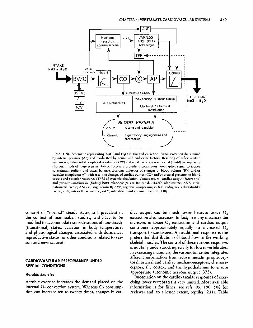
CHAPTER 4: VERTEBRATE CARDIOVASCULAR SYSTEMS 275
.----------~l ANF 11- - - ----,
Mechanoreceptors
atrium/arterial
AVPALDOANGII EDLF?Adrenergic
AlfTOREGULATJON
Kidney
lLEXCRETIONNaCI + H20
...... ,IIIIIIIIIIII
Electrical/ChemicalTransduction
Wall tension or shear stressOz / Metabolites
INTAKENaCI + H20 Atrial
4 BV/~"'""I rz rl...---r---J
BLOOD VESSELS!; tone and reactivity
hypertrophy, angiogenesis andrarefaction
FIG. 4.28. Schematic representing NaCl and H20 intake and excretion. Renal excretion determinedby arterial pressure (AP) and modulated by neural and endocrine factors. Resetting of reflex controlsystems regulating total peripheral resistance (TPR) and renal excretion is indicated (adapt) to emphasizeshort-term role of these systems. Arterial pressure provides a continuous nonadaptive signal to kidneyto maintain sodium and water balance. Bottom: Influence of changes of blood volume (BV) and/orvascular compliance (C) with resulting changes of cardiac output (CO) and/or arterial pressure on bloodvessels and vascular resistance (TPR) of systemic circulation. Venous return-cardiac output (Heart box)and pressure-natriuresis (Kidney box) relationships are indicated. ALDO, aldosterone; ANF, atrialnatriuretic factor; ANG II, angiotensin II; AVP, arginine vasopressin; EDLF, endogenous digitalis-likefactor; ICV, intracellular volume; ISFV, interstitial fluid volume (from ref. 130).
concept of "normal" steady states, still prevalent inthe context of mammalian studies, will have to bemodified to accommodate considerations of non-steady(transitional) states, variation in body temperature,and physiological changes associated with dormancy,reproductive status, or other conditions related to season and environment.
CARDIOVASCULAR PERFORMANCE UNDERSPECIAL CONDITIONS
Aerobic Exercise
Aerobic exercise increases the demand placed on theinternal O2 convection system. Whereas O2 consumption can increase ten to twenty times, changes in car-
diac output can be much lower because tissue O2extraction also increases. In fact, in many instances theincreases in tissue O2 extraction and cardiac outputcontribute approximately equally to increased O2transport to the tissues. An additional response is thepreferential distribution of blood flow to the workingskeletal muscles. The control of these various responsesis not fully understood, especially for lower vertebrates.In exercising mammals, the vasomotor center integratesafferent information from active muscle (proprioceptors), arterial and cardiac mechanoreceptors, chemoreceptors, the cortex, and the hypothalamus to ensureappropriate autonomic nervous output (373).
Information on the cardiovascular responses of exercising lower vertebrates is very limited. Most availableinformation is for fishes (see refs. 95, 190, 508 forreviews) and, to a lesser extent, reptiles (231). Table

276 HANDBOOK OF PHYSIOLOGY~COMPARATIVE PHYSIOLOGY
4.1 summarizes key cardiovascular data for selectedvertebrates at rest and at maximum exercise states. Anumber of general points emerge from these data. Themajority of vertebrates increase cardiac output duringexercise by between 1.3- and 3.3-fold over restingvalues. Known exceptions to this are trained humanathletes and thoroughbred horses (Table 4.1). Also,flying pigeons can increase fH sixfold with no changein SV (104). The mechanism by which cardiac outputis increased during exercise varies among vertebrates.Most amphibians, reptiles, birds, and mammals relyheavily on tachycardia (Fig. 4.29), with SV changingrelatively little. Training in mammals does, however,increase the role of increasing SV. Fish rely on increasesin both fH and SV, with the increase in SV predominating in most species. P. borchgrevinki and possiblytuna are unusual in this regard by relying substantially
more on tachycardia (16, 66). Tachycardia typicallyinvolves both vagal release and, where possible, adrenergic stimulation. Central arterial blood pressure increases with exercise. However, species with in-seriesrespiratory and systemic circulations (fishes and mammals) generally show somewhat larger increases insystemic arterial blood pressure than species with anin-parallel respiratory and systemic circulation (amphibians and noncrocodilian reptiles). The extent ofthe hypertension is reduced because total vascular resistance is reduced. More active mammals appear to becapable of a greater factorial reduction in vascularresistance. The net effect of increased cardiac outputand acute hypertension is to increase myocardial poweroutput by between 1.4- and S.7-fold (Table 4.1), andthis, of course, requires a concomitant increase in myocardial O2 consumption.
1ml kg-I beat-I
J- 20min- 1
Ivol%
Dog 20kg
Savannahmonitorl.Okg
Vs
........................................... . . . . . . . . . . . . . . . . . . . . . . . . . . . ,. . ...
FtG. 4.29. Cardiovascular adjustments to exercise in selected vertebrates. This three-dimensionaltreatment deals with stroke volume (ml/kg/beat), heart rate (bprn), and arterial-mixed venous oxygenextraction (vol %). Resting conditions are heavily shaded, while exercising conditions are lightly stippled(from ref. 231).

CHAPTER 4: VERTEBRATE CARDIOVASCULAR SYSTEMS 277
The change in regional blood flow distribution withexercise has been measured in several species of fish.Because aerobic swimming is powered largely by redmuscle, there is a dramatic increase in both the massspecific blood flow to red muscle and the proportionof cardiac output going to red muscle. In rainbow troutand large mouth sucker, red muscle blood flow is 9%and 0.57% of cardiac output at rest, increasing to 42%and 13.2% during exercise, respectively (368, 512). Atthe same time as blood flow to skeletal muscle increases, that to visceral organs is reduced (605). Thereduction in blood flow to the celiac, mesenteric, andintestinal arteries can be particularly pronounced, being around 30% in Atlantic cod (21) and 70% inchinook salmon (605). Because systemic resistance isreduced, the effect of skeletal muscle vasodilation mustbe greater than that of visceral vasoconstriction. Thecontrol of these vascular responses is poorly understood. Modulation of the systemic a-adrenergic tone isone possibility. The roles of locally produced metabolites (110) and nonadrenergic, noncholinergic fibers,especially in the gut circulation (457,458), have yet tobe thoroughly investigated.
With respect to amphibians and reptiles, changes inparticular aspects of cardiovascular performance, suchas fH and blood pressure during exercise, have beeninvestigated (56, 100,292,659). However, our understanding of how cardiac output is regionally distributedduring activity in amphibians and reptiles is very limited. One of the few such studies measured left pulmonary and left aortic blood flows during rest and swimming in the green sea turtle Chelonia mydas (642).During exercise, cardiac output was presumed to increase, primarily due to an increase in heart rate from24 bpm to 40 bpm (28°C). Both pulmonary and aorticflows increased markedly with exercise, but there waslittle evidence for a redistribution of cardiac outputother than that typically associated with periods ofintermittent lung ventilation and breath holding (seebelow).
Breath Holding and Diving
One of the fundamental tenets of effectivegas exchangeis that ventilation and perfusion of a gas-exchangeorgan be appropriately matched. Many animals, especially lower vertebrates, breathe only intermittently.This may be due to voluntary cessation of continuousbreathing while in air or may be a result of diving. Inany event, to prevent a relative hyperperfusion of gasexchange organs during apnea, as well as to conservelimited O2 stores during nonbreathing periods, profound changes occur in the cardiovascular system ofalmost every animal examined in this context. The
literature on the subject is relatively extensive, initiatedlargely by the pioneering work of Scholander and colleagues (for reviews, see refs. 54, 74, 97, 98, 163, 198,205, 305, 338, 371, 372, 554, 586). The literatureprovides conflicting data as to whether or not theclassical diving response as described by Scholanderand co-workers in laboratory experiments was indeedthe diving response that occurs in all animals in nature.The literature on the subject, reviewed below, sometimes fails to take into account the natural historyof the animal in question-that is, whether the divedurations used were those typically shown by animalsand whether forced diving mimics diving in the natural habitat.
Almost all intermittently breathing animals show asimilar suite of cardiovascular responses associatedwith periodic gas exchange. These include changes intotal cardiac output, achieved through changes in SVand/or fH , and redistribution (shunting) of cardiac output between and within tissues of the systemic andgas-exchange circuits. In addition, some diving animalsare now known to use adjustments in cardiac outputand blood flow to "meter out" oxygen during apneicperiods. The extent of this total suite of changes will beat different levels, however, depending on the duration,stress, and/or exercise levels associated with diving.
Cardiac Output Reduction. A reduction in cardiac outputduring apnea is one of the most universal cardiovascular responses of intermittently breathing animals. Whilemany species respond to the increased cardiac demandsof exercise by specificallyincreasing predominantly oneof either SV or fH (see Aerobic Exercise) during breathholding cardiac output reduction is often profound,and in most species examined both SV and fH aredecreased. The rate and extent of fall in cardiac outputduring breath holding is related to a complex interaction between the resting oxygen demand of the animal,the O2 stores available at the beginning of the breathhold, the duration of the breath hold, and the abilityof the animal to tolerate hypoxia and hypercapnia(98). In animals like humans that are poorly adaptedfor breath holding, cardiac output may decrease by amaximum of 20% during voluntary breath holds ofseveral minutes (305). Animals adapted for breathholding, most often in the form of diving, show muchmore profound cardiac output reduction. In experimentally submerged Weddell seals and spotted seals,cardiac output falls by about 90% during prolongeddives (162, 364, 673). Stroke volume decreases about30%, indicating that bradycardia is the biggest component of the cardiac response to diving in these seals.Cardiovascular responses to both involuntary and voluntary diving in diving birds (especially ducks) are

278 HANDBOOK OF PHYSIOLOGY-COMPARATIVE PHYSIOLOGY
FIG. 4.30. Changes in heart rate, stroke volume, and cardiacoutput in the domestic mallard before, during, and after a forceddive (from ref. 206).
similar to those for similar conditions in seals (forreviews, see refs. 98, 371, 586). During short forcedsubmersion in the domestic mallard, for example, SVdecreases by about 25% and fH by more than 90%,leading to a fall in cardiac output of more than 90%(Fig. 4.30). Voluntary submersion in the diving freshwater turtle C. scripta can lead to a reduction in cardiacoutput of more than 95%, contributed to by profoundreductions in both SV and fH (Fig. 4.20). Similar responses have been measured in diving crocodiles andalligators (247,491,649). In perhaps the most extremecase, forced submersion of Chrysemys picta in completely anoxic water at 3°C for up to 6 months (anannoyance rather than a lethal condition for theseanimals) results in an fH of about 0.4 bpm (273).
Estimates of cardiac output during diving in amphibians are few. Experiments based on microsphere distribution suggest that cardiac output falls during diving(447). Amphibians, like reptiles, show considerablereductions in fH during diving (58, 86,495). Measurements of systemic and pulmonary blood flows duringintermittent breathing indicate that SV also falls greatlyduring apnea (560). Collectively, these data suggest alarge fall in cardiac output associated with breathholding.
Indicating both the physiological utility and phyleticantiquity of the reduction in cardiac output duringbreath holding, air-breathing fishes also show this response. A decrease in fH associated with the intervalsbetween air breaths occurs in air-breathing fish such
Shunting. Decreased cardiac output during breath holding is an important physiological response to diminished oxygen availability. However, redistribution ofcardiac output between vascular beds is also an essential component of maintaining O2 delivery to thosetissues that most need it. Redistribution is of two majortypes: (1) between systemic and gas-exchange circuits(except for birds and mammals) and (2) between organs within the systemic circulation.
In animals with either an incompletely divided heartor non-intracardiac, central arterial shunting capabilities (crocodilians), cardiac output can be preferentiallyand variably distributed, or "shunted," to either thesystemic circulation or the gas-exchange organ(s). Borrowing nomenclature derived from more bilaterallydivided mammalian circulations, shunts are classifiedas either "left-to-right" (L-R) or "right-to-left" (R-L).L-R shunt is a measure of the percentage of the oxygenrich blood returning from the gas-exchange circuit viathe left atrium, which is then shunted within the ventricle (or in the central arterial circulation in the case ofcrocodiles) directly back into the gas-exchange circuitfor a second, immediate pass through the gills, lungs,or other exchange organs. R-L shunt represents a bypass of the gas-exchange circuit in that oxygen-poorblood returning from the systemic circulation via theright atrium is shunted within the ventricle directlyback into the systemic circuit without an opportunityfor gas exchange. A further distinction needs to bemade between a gross shunt and a net shunt. A grossshunt reflects the total quantity of blood (usually expressed as a percentage of right or left atrial flow) thatis shunted in a particular direction. L-R and R-L shuntscan occur simultaneously, so a net shunt and its direction are calculated from the difference between the twoflows. Thus, a net R-L shunt occurs when the bloodflow to the pulmonary circuit (Qpul) is less than theblood flow to the systemic circuit (Qsys)-that is,Qpul/Qsys < 1.0-while a net L-R occurs when Qpul/Qsys> 1.0.
Central blood shunting is a carefully regulated process in all animals in which it occurs. Fortunately,the comparison of lower vertebrates with intracardiac
as the bowfin Amia and the gar pike Lepisosteus (331,510). Most attention in this regard has been focusedon the lungfishes (for reviews, see refs. 84, 85, 204,205,327, 328), where fH similarly decreases. Althoughcardiac output has not been measured directly, bloodflow in individual major vessels has been measured andindicates a reduction of stroke flow and cardiac outputbetween air breaths. However, as discussed earlier,there is no change in cardiac output during apnea inthe jeju, an air-breathing fish (172).
ml (t£ART RATE 13- 14IminSTROKE VOLUt.E 5 mI
I 234 5 6 789
Postdive period (min)
lt~ 3536 ml {I-£ART RATE 390 Imin\STROKE VOLUME 9.1 ml
l!\f .•...
ttf
Prediveperiod
MUH STROKEVOlUME. 6. 7 mI
3000
500
2500
1000
3500
gS 20009-s:.l 1500
io

CHAPTER4: VERTEBRATE CARDIOVASCULAR SYSTEMS 279
shunts to humans with intraventricular septal defects,where shunting is an unfortunate consequence of apathological situation, is seen as highly flawed. Indeed,central vascular shunting in intermittently breathinganimals provides a very useful way of matching perfusion and ventilation in the gas-exchange organs.
The magnitude and direction of intracardiac shuntsassociated with various ventilatory states is measuredusing blood gas contents in central arteries and veins,blood flow, or microsphere distribution. R-L and L-Rshunts have been documented in air-breathing fishes(for reviews, see refs. 84, 205), amphibians (598, 610),and reptiles (90, 264, 399, 554, 562, 563a, 652, 654).For example, in the freshwater turtle C. scripta, duringlung ventilation 60%-65% of cardiac output perfusesthe pulmonary circulation, indicating a net L-R shuntof 10%-15% (90, 562). During apnea, the net L-Rshunt reverses to a net R-L shunt, with about 55% ofcardiac output flowing into the systemic circulation.
What purpose does central vascular shuntingachieve? Several answers are possible (73). The direction and magnitude of the shunt tends to vary with theminute-by-minute potential of the aerial gas-exchangeorgan for O2 uptake and CO 2 elimination. During theactual periods of air breathing and shortly thereafter,P02 in the exchanger is highest and Pcoj lowest, whileat the end of a prolonged period of apnea, the oppositeis the case and a net R-L shunt prevails. By not perfusing the gas-exchange organ when its potential for gasexchange is low, the metabolic cost of perfusing thegas-exchange circuit is reduced. In turtles (c. scripta),the metabolic saving due to R-L shunting during apneais about 0.5% of total aerobic metabolism (73). Whilethis seems very small, our knowledge of what constitutes a significant energetic saving in a physiologicalprocess is fragmentary at best. It may be that a smallsaving applied over the thousands of apneic periods ina turtle's lifetime amounts to large energetic savings.R-L shunting during apnea may also reduce plasmafiltration into the lungs, which appears to correlatewith level of pulmonary blood flow, at least in turtles(71) and toads (576, 579). Fewer explanations havebeen offered' as a rationale for L-R shunting duringapnea, but this may be because shunting is often examined in the context of its effects on blood O2 transport.While the repeated recirculation of fully oxygensaturated blood through the gas-exchange circuitmakes little energetic sense vis-a-vis O2 transport, itdoes in terms of CO 2 elimination. With every circuitthrough the lung, additional CO 2 is removed (653).Indeed, the gas-exchange ratio of the lungs of bothfreshwater turtles (90) and African clawed toads (57)rises progressively to values well above 1 during longbouts of breathing, suggesting that CO 2 elimination
increases disproportionately over O2 uptake when anL-R shunt is occurring.
Shunting may also have nonrespiratory effects, mostpredominantly on heat exchange with the environment.By recirculating blood to the non-ventilated lung (L-Rshunt), blood is kept within the core and away fromthe peripheral systemic tissues, where it could pick upor lose heat, depending on the gradients between theanimal and the environment.
Central vascular shunting in air-breathing fishes, amphibians, and reptiles is controlled by adjusting therelative peripheral resistances of the systemic and gasexchange circuits. The lack of anatomical ventriculardivision (or, in the case of crocodilians, the presenceof a left aorta arising from the right ventricle) essentially places the systemic and gas-exchange circuits inparallel rather than in series as in birds and mammals.Consequently, a rise in pulmonary relative to systemicarterial resistance, for example, will result in a redistribution of cardiac output from the lungs to the systemictissues. The mechanism(s) by which this is achieved issomewhat species-specific but usually involves vasoconstriction and vasodilation of vascular smooth muscle (for reviews, see refs. 73, 76, 178, 205). Both largecentral and small peripheral vessels may be involved.In air-breathing fishes like the lungfish Protopterus,redistribution of cardiac output between the gills,lungs, and systemic tissues is achieved by a combination of vasodilation/vasoconstriction in both large central vessels and the microvasculature of the gills (205).Freshwater turtles, as well as crocodilians, have asmooth muscle sphincter at the base of the pulmonaryartery that can regulate pulmonary vascular resistanceand, thus, the degree of blood shunting (68). At leastin turtles and tortoises, the direction and magnitude ofthe intracardiac shunt is regulated in part by the temporal and spatial pattern of cardiac depolarization andactivation, which occurs in one of two states associatedwith periods of breathing and periods of apnea (69).Regulation of cardiac contraction patterns, and thusof intracardiac shunting, is under vagal control.
While mechanistically we understand how intracardiac shunting is achieved in many lower vertebrates,we know very little about the factors that regulate thisactive process. Given that we have yet to identify thefactors that initiate and terminate the much more easilystudied process of ventilation of intermittently ventilated gas-exchange organs, it will no doubt take considerable additional study to identify the role of the CNSin controlling shunting.
O2 Metering. Oxygen stores at the beginning of a periodof apnea (typically a dive) can be distributed in theblood (predominantly bound to hemoglobin), in the

280 HANDBOOK OF PHYSIOLOGY-COMPARATIVE PHYSIOLOGY
Reduced Metabolism
Certain birds and mammals reduce O2 consumptionovernight (torpor) or over the winter (hibernation) byregulating body temperature at a lower than normalvalue (50_15°C). In addition, a self-imposed state ofhypoxia accompanies breath-hold diving under water.The cardiovascular responses to reduced metabolicstates in birds and mammals are well documented (48,96, 128, 163, 298, 371, 655). There are a variety ofconditions under which lower vertebrates may reducetheir metabolic state, but the physiological responsesin general, and the cardiovascular responses in particular, are less well understood. Furthermore, more thanone condition may influence the overall cardiovascularresponse. For example, the overwintering of frogs involves reduced temperature, submergence, and hypoxia(495). Ectotherrns may either conform or acclimate toa decrease in environmental temperature (315, 528).Thus, rate functions can have a QlO of around 2.0 inconformers or much larger in animals that downregulate metabolic activity (for example, eels overwintering on the bottom of rivers). Semiaquatic vertebrates, such as Protopterus and Bufo, burrow oversummer to survive drought conditions (estivation).During burrowing these animals take advantage of alower environmental temperature and down-regulate
gas, lung gas, and pulmonary blood flow measurementsin the diving turtle Chelodina longicollis (91). Pulmonary blood flow appears to be carefully regulated to"meter out" oxygen during diving, maintaining arterialO2 saturation at 90%-95% as lung P0 2 progressivelyfalls, during all but the longest dives. Sharp increasesin blood flow during diving result in actual increasesin blood saturation (Fig. 4.32).
Little is known about how O2 metering is regulatedin amphibians or reptiles. The simple notion that thereis some threshold level of blood or lung gas PaZ, Pcoy,or pH that might stimulate pulmonary perfusion (orany other cardiorespiratory) process is antiquated (643,645). Additional studies are needed on amphibians andreptiles to identify what will probably be a suite offactors that modulate CNS control over shunt patternsand pulmonary perfusion.
The phenomenon of O2 metering during apnea inair-breathing fishes has not been observed to ourknowledge. The smaller size of many air-breathingfishes combined with the need to monitor constantlyeither blood flow or arterial O2 levels has conspiredagainst the type of physiological measurements thatwould lead to 02 metering. However, the physiologicalrationale for this phenomenon applies equally well tofish intermittently breathing air.
IBLOOD AND
TISSUES
~~~~~~~~~~porpoise~ Weddell Seal
MAMMALS ~~~~~~~~~~BladdernoseSealt Sperm Whale
~§§§§§~~~~Tufted DUCk] Lung &"'allard Air Sacs
BIRDSTufted DUCk] Lung"'allard only
FIG. 4.31. Distribution of O2 stores available at the beginning ofa dive in a selection of amphibians, reptiles, birds, and mammals.Data are approximations derived from primary data from differentstudies on the same species (from ref. 74).
t::~~~~~~~~BUllfrOgAMPHIBIANS Congo EelClawed Toad
o 20 40 60 80 100
02 STORES (% OF TOT AU
~:~~~~~~~~FreShwaterTurtleREPTILES ~ File Snake
tissues (predominantly bound to myoglobin), or in lunggas. Different species tend to distribute their oxygenstores in different sites (Fig. 4.31). Diving mammalsand, to a lesser extent, diving birds tend to store oxygenprimarily in blood and tissues; lung gas oxygen storesin these animals are relatively small (74, 98, 163, 371,372). Birds and mammals tend to dive to much greaterdepths than lower vertebrates, and by minimizing lunggas stores so, too, the risk of the bends may be reduced.Reptiles and amphibians, however, tend to dive withlarge, fully inflated lungs, and consequently a muchgreater proportion of their oxygen is stored in the formof lung gas (74).
A pulmonary gas O2 store is of benefit to animalsduring diving only if they can effectively transfer O2to the blood from these stores and distribute it tothe least anaerobic tissues. Thus, while L-R shuntsproducing a partial pulmonary bypass are useful mechanisms for conserving oxygen during certain circumstances (see above), the ability to minimize or evenreverse the shunt during apnea, enabling the temporarytransfer of O2 from pulmonary to blood stores, iscrucial. Indirect evidence for such a transfer duringapnea in diving frogs (Xenopus) and turtles (Chrysemys) has been recorded (53, 74) and consists of aslow decline in lung gas P0 2 that quickly turns into amore sharp decrease as arterial P0 2 actually increases.Direct evidence for transfer of O2 from lung gas toarterial blood exists in the form of simultaneous blood

CHAPTER 4: VERTEBRATE CARDIOVASCULAR SYSTEMS 281
insp.
expo-H....--.-LungVentilation
Lung 10]~Pressure 5]
(ern H20) 0
---v~--~------~-~-
Pulmonary~Flow
(Impedance)
Oxygen
Partial
Pressure
(mmHg)
130
120
110100 Lung Gas
90
!~~Arterial Blood
Lung Gas
02 Sat.
____9_5%.,..~90% Arterial Blood
''''!!lu',,,,,!!,,,,I!!!!!!!,,,,I!!''.,,,,,,I,, l!IlIll!!!ll'''''''I!!!!I'l!!!!I'lIIllllllll""lll'lIw".lWI'!!l!!'''!l'"J!
Time (Min)
FIG. 4.32. Pulmonary oxygen metering in the turtle Chelodina longicollis. Left panel shows recordsof lung ventilation, intrapulmonary lung pressure, pulmonary flow, POz of lung gas, and POz and Ozsaturation of systemic arterial blood at termination of a dive and during two brief lung ventilationbouts. Right panel shows same records taken between 9 and 16 min into a voluntary dive (from ref. 91).
their metabolism. Amphibians, reptiles, and airbreathing fishes perform breath-hold dives. Aquaticvertebrates also experience environmental hypoxia.
As with reduced metabolic states in mammals, lowervertebrates typically reduce cardiac output through avagally mediated bradycardia. This is true whetherdiving is performed by crocodiles, turtles, amphibians,lungfishes, or air-breathing fishes (178, 510); whetherfishes are exposed to severe aquatic hypoxia (95);whether lungfishes estivate (147); and whether turtlesoverwinter under water (272). The extent to whichbradycardia develops depends in part on (1) the duration and severity of the change and (2) other confounding inputs to the vasomotor center, such as afferentinput associated with exercise during diving. Estivatinglungfishes reduce fH and arterial blood pressure overthe first 70 days to 12 bpm and 14 mm Hg, respectively, and these levels are maintained for the next 180days of estivation (147). Perhaps the most extremeexample of cardiac depression during a reduced metabolic state is that of a 129-day submersion in anoxicwater at 3°e by Chrysemys: fH was reduced to 0.1bpm-1.0 bpm and arterial blood pressure to 10 mmHg (273). However, frogs that freeze over the wintermust have a complete cardiac arrest.
Vascular changes also accompany reduced metabolicstates in lower vertebrates. Systemic vascular resistance
is reported to increase, decrease, or remain unchangedduring aquatic hypoxia in fish (95). These varied responses may reflect differences between species, in thelevel and length of hypoxic exposure, and in the extentof accompanying motor activity. During prolongeddiving by amphibians and reptiles, the most profoundvascular change is the progressive reduction in pulmonary flow (559, 562). This is primarily brought aboutby a vagally mediated constriction of the pulmonaryartery. The extent of pulmonary vasoconstriction isagain a function of (I) the duration of the dive and (2)other confounding inputs to the vasomotor center.Perhaps the best example of this confounding effect isthe response of the green sea turtle swimming underwater; pulmonary flow is maintained even though theanimal is "diving" (100, 642).
Digestive State
That metabolic rate increases following ingestion offood, a phenomenon termed specific dynamic action(SDA), has long been appreciated by both medicallyoriented and comparative physiologists. Metabolic increases in the postprandial period can range from 10%to up to tenfold depending on the species in questionand the size of the meal. The metabolic consequencesof SDA have been examined by comparative physiolo-

282 HANDBOOK OF PHYSIOLOGY~COMPARATIVE PHYSIOLOGY
gists in both vertebrates and invertebrates (for a selection of references, see ref. 87). However, far less attention has been paid to the cardiovascular changes thatpresumably support SDA-associated increases in metabolic rate. Presumably, a large increase in metabolismassociated with SDA must be supported by an increasein cardiovascular transport of nutrients to tissues experiencing the elevated metabolic rate. Indeed, circumstantial evidence for this appears in the toad B. marinus, where both fH and myocardial O2 consumptionquickly double following injection of peptone into thestomach (158, 637). Feeding causes redistribution ofblood flow from other systemic tissues preferentiallytoward the gut in mammals (170), alligators (22), andfishes (17, 21). This response is generally produced byvasodilation of mesenteric vessels; in some species, thiscan produce a several-fold increase in blood flow tothe gut.
Unlike ambush predators and humans, some animalseat while they exercise. Thorarensen and co-workers(604, 605) have examined the cardiovascular changesrelated to intestinal blood flow in exercising salmonids.At rest, intestinal blood flow was 12-18 ml/min/kgbody mass, and this accounted for 34% of cardiacoutput. Postprandially, intestinal blood flow increasedby 81%, peaking 23 h after feeding. In unfed fishswimming maximally, although cardiac output wasincreased by 86%, intestinal blood flow was reducedby 60%-70% because of an increase in splanchnicvascular resistance. A negative linear relationship wasobtained between intestinal blood flow and oxygenconsumption, suggesting an association between theregulation of intestinal blood flow and the oxygendemand of locomotory muscles. The apparent tradeoff of blood flow between the intestine and workingskeletal muscle was calculated to provide 14% of thetotal oxygen transported to the skeletal muscle duringswimming. Thorarensen et al. (604, 605) further propose that an additional and perhaps more importantbenefit of increasing splanchnic vascular resistance isblood pressure homeostasis.
Much of the cardiovascular physiology that we know(and that is discussed in this chapter) is based onrelatively steady-state conditions, often measured inanimals in a fasting or otherwise steady nutritionalstate. Yet, nonsteady-state conditions clearly have profound effects on cardiovascular function as well as gasexchange and metabolism and deserve to be the focusof additional studies.
Responses to Gravity
Gravity affects blood circulation primarily by inducingshifts of blood volume whenever posture departs fromthe horizontal (396a). The effect is especially important
in long or tall animals. Pooling of blood in dependentvasculature and the tendency to edema in dependenttissues are both related to the hydrostatic pressure,which in turn depends directly on the length of thevertical blood column. Gravity-dependent shifts ofblood volume when posture departs from the horizontal initiate a sequence of hemodynamic changes andreflexogenic adjustments that have been thoroughlystudied by numerous investigators (reviewed in refs. 49,224, 529a, 575). Depending on the height of arterial orvenous blood columns and the associated changes inhydrostatic pressure, the total pressure across bloodvessel walls (that is, transmural pressure approximatesthe sum of blood pressure plus hydrostatic pressure)increases passively in the lower vasculature and decreases in the upper vasculature ("lower" and "upper"defined with reference to a central or near-central hydrostatic indifferent point, HIP, where pressure doesnot change) (224). Pooling of blood reduces cardiacoutflow and thereby tends to reduce arterial pressures,which shifts the effective HIP downward in the absenceof compensatory responses. However, cardiovascularreflexes which elevate arterial pressures shift the HIPupward such that the passive initial decrease of pressure in elevated vasculature tends to be compensated.
Regional pressure changes in the arterial system conform closely to expected hydrostatic pressures relatedto vertical distance. For example, pressures in mammalian appendages vary with respect to central pressuresdepending on whether they are elevated or loweredwith reference to the heart (258, 350). In veins, however, postural changes can introduce significant nonlinearities in the distribution of intravenous pressures,partly attributable to the presence of valves. Pressuresin human leg veins can be modified by venous collapseabove the heart when the body is upright or by partialcollapse within the legs when the head is down (49).Jugular veins in the neck and upper part of the superiorvena cava may collapse in upright humans (366) andgiraffes (236). However, intracranial vessels are maintained in a patent state by periosteal adhesions andhydrostatic pressure gradients in the cerebrospinalfluid.
Both neural and endocrine mechanisms interact tocounter gravitational disturbance of the cardiovascularsystem. The principal interplay of responses entailsadjustments in peripheral resistance, fH , and venouscapacity. It is generally assumed that arterial baroreceptors play the major role in regulating the acuteresponses to vertical posture, though cardiac and lowpressure mechanoreceptors may also be involved. Thegeneralized vasomotor response to head-up postureentails substantial vasoconstriction mediated by decreased arterial and cardiac mechanoreceptor stimulation, coupled to reflex a-adrenergic stimulation.

CHAPTER 4: VERTEBRATE CARDIOVASCULAR SYSTEMS 283
Sympathetically mediated vasoconstriction affects peripheral resistance nonuniformly and is quantitativelymore important than adjustments of cardiac rate orcontractility. Thus, central arterial blood pressure (atheart level) is usually maintained despite significantreductions of cardiac output. Vasoconstriction affectsprincipally splanchnic, skin, and muscle vasculature.Vasoconstriction in skin and muscle of snakes duringhead-up tilt is limited largely to dependent (posterior)regions in which vessels are densely innervated compared with cervical regions of the body (149, 400).
Adaptive responses elicited by head-up tilt are similarin snakes, lizards, humans, and other mammals. Central arterial pressure falls transiently immediately following head-up tilt but recovers toward or exceedsthe pre-tilt level as compensatory mechanisms regulatearterial pressure. As a consequence, the band of arterialpulse pressure narrows due largely to elevation ofdiastolic pressure attributable to increases in peripheralresistance, while fH increases. Progressive blood pooling, with attendant reductions in SV, secondarily reduces arterial pressure, which promotes compensatorytachycardia and increases cardiac contractility and peripheral resistance due to reduced sensory outflow fromcardiovascular mechanoreceptors. The effects on fH
reflect combined f3-adrenergic stimulation in combination with vagal withdrawal. Arterial pressures at headlevel drop by 20-30 mm Hg during standing in humans(271, 483), and such changes are comparable to theproportional reductions of arterial pressure measuredduring vertical posture in arboreal snakes (394). Driving pressures at the cephalic vasculature may be relatively unaffected, however, due to reductions in venousas well as arterial pressures at head level. The juxtaposition of vascular and cerebrospinal fluid columns isprobably important in protecting against excessivechanges in transmural pressures that are counterbalanced at all points in the cranium and spine, therebypreventing venous distension or collapse.
Endocrine responses contribute importantly to regulatory responses to gravity but have been studied onlyin mammals. Sympathetic stimulation and decreasedrenal blood flow stimulate the release of renin, whichinitiates a cascade leading to increases in plasma angiotensin and aldosterone. The release of renin duringhead-up posture can also be augmented by increases inintra renal vascular pressures and reductions of circulating levels of ANF (132, 254). Postural changes canalso produce small increases in plasma levels of AVP,which is a potent vasoconstrictor in humans (144, 537,552). Finally, sympathetic activation tends to promoteparallel increases in plasma catecholamines, which sustain the pressor response elicited by arterial baroreceptors (407, 523).
A range of possible adaptive responses to head-up
posture is nicely illustrated by the behavior of centralarterial pressures in various species of snake (Fig. 4.23).Arterial pressures at the body's center (approximatelythe HIP) either increase, show little change, or decreasedepending on the species' adaptations to counteractgravitational blood pooling and to elevate arterial pressure (400, 401, 405, 557). Considering a range ofspecies, the ability to control disturbances to arterialpressure appears to be at least somewhat graded according to the degree of gravitational disturbance normally encountered (394). Studies of arboreal snakesindicate that hemodynamic adjustments in central vessels bias systemic cardiac output in the cephalic direction and assist the regulation of carotid arterial bloodflow during head-up tilt (398). Similar data for otherterrestrial snakes demonstrate that carotid blood flowis not well regulated (during tilt) in nonclimbing species, suggesting adaptation of the control mechanismsin the arboreal forms (396, 405).
Circulation to the brain of giraffes (Giraffa camelopardalis) has attracted the interest of physiologists sincethe early 1950s. Various investigations have demonstrated that systemic arterial pressures near the giraffeheart are about twice that in humans, sufficient to raiseblood against the arterial gravitational column andto supply additional pressure sufficient to perfuse thecranial tissues (235-237, 619-621). An average-sizedgiraffe stands about 5 m, and the heart is at roughly thecenter of this distance. Central arterial blood pressureaverages about 200 mm Hg in a 3-4 m giraffe andsupplies "normal" perfusion pressures (ca. 100 mmHg) at the cranium, which is 1.2 m above the heart(258). Systolic blood pressures are reported to reach360 mm Hg at heart level in standing giraffes (236).When the head of the giraffe is lowered, as duringdrinking, blood pressure measured in the carotid arterynear the base of the brain may actually decrease (from200 to 175 mm Hg) (235). It has been suggested thata rete mirabile and voluminous jugular vein both helpregulate cerebral hemodynamics during head-downtilt (236).
Development of Cardiovascular Systems
The study of the ontogeny of cardiovascular systemphysiology has been heavily biased toward (1) themammalian fetus and (2) the final third or quarter ofgestation. The reasons for these biases are understandable. Apart from the obvious focus of most biomedicalresearch on systems that can further our understandingof human development, large domesticated mammalshave large fetuses that allow invasive measurements.Although the vast majority of studies on the development of cardiovascular systems have been made onmammalian fetuses, the positioning of the fetus within
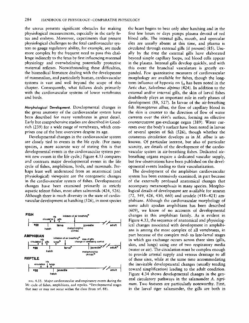
284 HANDBOOK OF PHYSIOLOGY-COMPARATIVE PHYSIOLOGY
FIG. 4.33. Major cardiovascular and respiratory events during thelife cycle of fishes, amphibians, and reptiles. "Developmental stagesthat mayor may not occur within the class (from ref. 88).
Hb
I cirCUjion ll,ngs • L--Jegg ........TI~Ju-ve-n""'lle--------~/radUItl
hatcl1
the uterus presents significant obstacles for makingphysiological measurements, especially in the early fetus and embryo. Moreover, experiments that presentphysiological challenges to the fetal cardiovascular system to gauge regulatory ability, for example, are mademore complex by the frequent need to pass this challenge indirectly to the fetus by first influencing maternalphysiology and overwheiming potentially protectivematernal reflexes. Notwithstanding these difficulties,the biomedical literature dealing with the developmentof mammalian, and particularly human, cardiovascularsystems is vast and well beyond the scope of thischapter. Consequently, what follows deals primarilywith the cardiovascular systems of lower vertebratesand birds.
the heart begins to beat only after hatching and in thefirst few hours or days pumps plasma devoid of redblood cells. The internal gills, mouth, and opercularslits are usually absent at this time, and plasma iscirculated through external gills (if present) (85). Usually by the time the external gills have elaboratedbeyond simple capillary loops, red blood cells appearin the plasma. Internal gills develop quickly, and withthis event the branchial vasculature is greatly expanded. Few quantitative measures of cardiovascularmorphology are available for fishes, though the longterm influence of hypoxia on fH has been noted in theArtie char, Salvelinus a/pinus (424). In addition to theexternal and/or internal gills, the skin of larval fishesdoubtlessly plays an important role in early phases ofdevelopment (88, 527). In larvae of the air-breathingfish Monopterus albus, the flow of capillary blood inthe skin is counter to the direction of flow of watercurrents over the skin's surface, forming an effectivecountercurrent gas-exchange organ (389). Water currents over the body's surface have been noted in larvaeof several species of fish (526), though whether thecutaneous circulation develops as in M. albus is unknown. Of particular interest, but also of particularscarcity, are details of the development of the cardiovascular system in air-breathing fishes. Dedicated airbreathing organs require a dedicated vascular supply,but few observations have been published on the developmental events leading to their vascularization.
The development of the amphibian cardiovascularsystem has been extensively examined, in part becauseof the externally profound anatomical changes thataccompany metamorphosis in many species. Morphological details of development are available for anuran(72, 349, 426, 430, 660) and urodele (418-421) amphibians. Although the cardiovascular morphology ofsome adult apodan amphibians has been described(609), we know of no accounts of developmentalchanges in this amphibian family. As is evident inFigure 4.33, the sequence of anatomical and physiological changes associated with development in amphibians is among the most complex of all vertebrates, inpart because of the complex mid- to late-larval stagesin which gas exchange occurs across three sites (gills,skin, and lungs) using one of two respiratory media(water or air). The circulation must be complex enoughto provide arterial supply and venous drainage to allof these sites, while at the same time accommodatingthe inevitable developmental changes (usually tendingtoward simplification) leading to the adult condition.Figure 4.34 shows developmental changes in the general circulatory pathways in the salamander A. tigrinum. Two features are particularly noteworthy. First,in the larval tiger salamander, the gills are both in
inniation01air breathing* loss 01gillslungs* change in ventilatorycontrol
hatcl1
Circuta~on1bg:s innia1ion 01r_jr_br_ea1_h_ing_* ~/ /
I larva t Juvenile 7 " adulthatcl1 metamorphic
climax
FISH
Morphologicol Development. Developmental changes inthe gross anatomy of the cardiovascular system havebeen described for many vertebrates in great detail.Early but comprehensive studies are described in Goodrich (239) for a wide range of vertebrates, which comprises one of the best overviews despite its age.
Developmental changes in the cardiovascular systemare closely tied to events in the life cycle. (For manyspecies, a more accurate way of stating this is thatdevelopmental events in the cardiovascular system permit new events in the life cycle.) Figure 4.33 comparesand contrasts major developmental events in the lifecycle of fishes, amphibians, birds, and mammals. Perhaps least well understood from an anatomical (andphysiological) viewpoint are the ontogenetic changesin the cardiovascular system of fishes. Developmentalchanges have been examined primarily in strictlyaquatic teleost fishes, most often salmonids (424,526).Although there is much diversity in the state of cardiovascular development at hatching (526), in most species
REPTILE

CHAPTER4: VERTEBRATE CARDIOVASCULAR SYSTEMS 285
FIG. 4.34. Gross morphology of central venous circulation oflarval and adult tiger salamanders (Ambystoma tigrinum) (fromref. 417).
series and in parallel due to the derivation of thepulmonary arch directly from the bulbus arteriosus.Urodeles are also the only amniotes to retain the fifthaortic arch as adults. This differs from the pattern inlarval bullfrogs, where the lungs and that portion ofthe skin served by the cutaneous artery are in series anddownstream from gill arch VI. The second noteworthyfeature, true for both urodeles and anurans, is that theadult condition is substantially less complex than thatof the larvae. This is contrary to a prevailing view thatcontinuing development leads to continuing anatomical complexity.
In reptiles, as in birds and mammals, all structuralelements of the adult cardiovascular system are essentially in place and operational at birth or hatching,with no major developmental events occurring in thecirculation after assuming air breathing at birth/hatching (Fig. 4.33). The developmental cardiovascular anatomy of reptiles is scant (88) compared to that ofamphibians, a surprising finding given the large literature on both the anatomy and physiology of the cardiovascular system of adult reptiles (see references in thediscussion of reptiles under Cardiovascular Patterns inVertebrates, above). Most general accounts of cardiovascular development refer to a (fictitious) general amniote or tetrapod condition. Very little is known aboutthe developmental morphology of the cardiovascularsystem of reptiles, especially of varanid lizards andthe crocodilians.
LargeAdult
Eleutherodactylus coqui
H,AB
140
120
"M"
100
"7c 80E~coOJ
60£
s40 A8
20
0
Onset ofHeart Beat
FIG. 4.35. Developmental changes in heart rate (20°-25°C) duringdevelopment in four anuran amphibians. Data have been "normalized" with respect to development such that the earliest points arefrom when the heart first begins to beat and the last points are fromthe largest adults for which data are available. This distorts theabscissa such that a single vertical line at a given point on thedevelopmental scale is not the same stage of development of allspecies. Major developmental landmarks indicated; H, hatching; AB,onset of air breathing; M, metamorphosis (from ref. 77, whichprovides original sources of data).
DEVELOPMENT -..
Physiological Development
Heart. The study of developmental changes in heartphysiology in fishes has been restricted largely to thecategorization of ontogenetic changes in fH• Heart ratesare, of course, temperature-dependent and vary between species. Heart rates decline, sometimes after aninitial increase, during larval development in the browntrout Salmo trutta (249), the rainbow trout O. mykiss(303), the Arctic char S. a/pinus (424), and the littleskate Raja erinacea (485). While developmentalchanges in the membrane permeability of the cardiacpacemaker and/or the onset of sympathetic and parasympathetic tone could explain these developmentalchanges in fH , little is known about the ontogeny ofcardiac regulatory mechanisms in larval fishes.
Developmental changes in fH in amphibians havebeen well documented, especially for anurans (77, 86,93). As is evident in Figure 4.35, there is great variation
The development of the cardiovascular system ofbirds has been documented extensively, due to the roleof the chick embryo as the major model for vertebratedevelopment. The reader is referred to basic embryology textbooks for further information on developmentof the heart and blood vessels.
ADULTAfferentBronchialArteries
LARVA

286 HANDBOOK OF PHYSIOLOGY-COMPARATIVE PHYSIOLOGY
in the pattern of fH change accompanying developmentin anurans (and presumably in urodeles). In some species, like the bullfrog R. catesbeiana, fH has been measured over a body mass span of 40 mg shortly afterhatching (about 135 bpm at 20°C) to 400 g as adults(about 20-25 bpm at 20°C). When analyzed for anallometric relationship, fH is found to scale to bodymass to the exponent - 0.23, compared with a valueof about - 0.25 based on interspecific comparisons ofadult vertebrates (88). That is, almost all of the fH
changes in Rana during development can be accountedfor by changes in body mass rather than qualitativechanges due to development. In the coqui Eleutherodactylus coqui, a direct-developing neotropical frog, fH
rises very rapidly from about 50 bpm to about 100bpm (24°-25°C) in the first few days of development,then increases much more slowly during developmentto the adult morph within the egg, and then fromhatching to maximum adult size decreases from about120 bpm to 80 bpm. The changes prior to hatchingare completely the opposite of those that would bepredicted by allometric effects, suggesting that "pure"developmental effects, such as the opening of newvascular beds and the expansion of blood volume,primarily affect heart performance. After hatching,however, when presumably little additional developmental effects occur, the decline in fH can be explainedlargely (but not entirely) by increasing body mass andcholinergic control.
The mechanisms behind fH regulation in anuran
amphibians (and, to a much lesser extent, urodeles)have similarly been studied in detail. f3-Adrenergic andcholinergic receptors, cholinergic and adrenergic innervation, and vagal tone occur relatively early in larvaldevelopment (65, 82, 357, 358, 502). Consequently,although metamorphosis results in additional modification of the central arterial circulation, there is verylittle additional change in cardiac regulatory mechanisms at this time. It is beyond the scope of this chapterto review these data in detail, so the reader is referredto reviews of the subject (77, 86-88). Figure 4.36summarizes what is known of the development ofcardiac regulation in the bullfrog R. catesbeiana, forwhich most is known. Qualitative and quantitativedifferences between the ontogeny of cardiac regulationof the bullfrog and other anurans, urodeles, and apodans doubtlessly exist and deserve further study.
Perhaps due to the small size of reptilian embryosand the fact that most are either hidden within a uterus(some snakes and lizards), enclosed in an opaque shell(virtually all egg-laying reptiles), or guarded by a parentweighing in excess of 100 kg (crocodilians), we knowextremely little of the regulation of the heart of reptilian embryos. Embryos of the California black kingsnake (Lampropeltis getulus nigritis) and the Americanalligator (A. mississippiensis) at 40% of their embryonic development respond to short-term exposure to12% ambient oxygen by changes in fH (5. Warburton,unpublished data). However, California black kingsnake embryos show a reversible tachycardia (mean fH
Ventilationtachycardia
Vagal tone atrest disappears
Resting fH = intrinsic fH(no vagal/sympathetic tone)
Exercise tachycardia due toI!-adrenergic receptors and
increased venous return
Vagal toneat rest
Exercise tachycardiadue to increased
venous return
No exercisetachycardia
No hypoxicbradycardia
Adrenergic Non-cholinergic.fH not extrinsically excitatory non-adrenergic
regulated nerves exercise tachycardia
Ch u./" No hypoxico mergic bradycardia
inhibitorynerves
Spontaneous
I!-adrenergicreceptors
III IV-VII
LARVA
X-XIV XVI-XIX XX·XXIV
Hatching Ventilation ofinternal gills
Well developedrear legs
Metamorphicclimax
large increasein body mass
Air breathing large increasein body mass
DEVELOPMENTAL LANDMARKS
FIG. 4.36. Major developmental events in ontogeny of heart rate regulation in the bullfrog Rana catesbeiana (from ref. 77).

CHAPTER4: VERTEBRATE CARDIOVASCULAR SYSTEMS 287
increases from 64 bpm to 71 bpm), while Americanalligators show a reversible bradycardia (fH decreasesfrom 85 bpm to 70 bpm).
Ontogenetic changes in fH have been studied extensively in precocial birds, especially the domestic chicken(105, 121, 122, 230, 309, 312, 384, 596, 597, 600,622, 633). Differences exist between species in thedevelopmental patterns of fH • Heart rate temporarilyincreases during the last half of the incubation periodin the Japanese quail (Coturnix coturnix [aponica) andchicken and then decreases again as pipping approaches. Variations on this general theme are observed in the turkey, duck, and goose. In the peafowl(Pavo cristatus), fH increases rather than decreasestoward the end of incubation. In the semiprecocialbrown noddy (Anous stolidus), fH is constant duringthe last several days of prepipping development butsubsequently changes during the prolonged paranatalperiod (596).
Heart rate has been studied much less in altricialbirds, where differences in developmental trajectorieshave been identified for O2 consumption, thyroid development, and thermoregulatory capacity. In the pigeonColumbia domestica and the bank swallow Ripariariparia, fH increases slowly until the final 3-6 daysbefore external pipping, when it increases more sharplyuntil hatching (601). Interestingly, there seems to bemuch more inter- than intraclutch variability in termsof daily fH, as well as onset and length of externalpipping (92). That is, siblings show greater similarityin specific patterns of fH change during developmentthan do nonsiblings. The reasons for this are unclearbut could be due to genetic effects (that is, very specificaspects of the pattern of fH change are heritable) orto maternal effects, in which maternal environmentalexperiences influence development of the embryos produced by that female.
The ontogeny of mechanisms for fH regulation indomestic chicken embryos has been considered in somedetail (119, 165, 230, 388; see for review, ref. 595).The main protocol in these experiments has been toapply autonomic agonists and antagonists and to notechanges in cardiac performance. Cholinergic receptorsinfluencing cardiac function are in place at the end ofthe first week (the first third of incubation), but theabsence of any effect by atropine indicates that thereis no vagal tone in embryos around days 13-16 (595).The absence of vagal tone does not mean that the CNSdoes not exert control over the heart but rather thatthere was no tone under the circumstances of theexperiment. The lack of both parasympathetic andsympathetic neural tone is suggested by the fact thatup to day 16 of incubation, destruction of the CNScauses no change in fH (52). However, propranolol
often causes a decrease in fH in 13- to 16-day-oldembryos, suggesting that circulating catecholaminesmay elevate fH over its intrinsic rate by this pointin development (595). Adrenergic sensitivity in thedomestic chicken appears by the end of the first weekof incubation (230), and both a- and (3-receptors increase fH , blood pressure, and peripheral resistance inday 13-18 embryos. The relative roles of circulatingcatecholamines and sympathetic innervation in regulating fH remain to be determined. One weakness ofstudies using pharmacological probes is that they showthe presence of receptors, not of postganglionic neurons, However, to our knowledge, stimulation of sympathetic or parasympathetic trunks to determine theireffects on heart performance has not been attemptedin bird embryos.
Peripheral vasculature. The two nonmammalian groupsthat have been studied for ontogenetic changes in peripheral vasculature regulation are the amphibiansand birds.
In amphibians, detailed studies have been made ofthe regulation of blood flow through the elaboratebranchial/pulmonary vasculature illustrated in Figure4.34 (86, 203, 417-419, 534). In A. tigrinum, AChand vagal stimulation induce vasoconstriction in thebranchial vessels and the pulmonary artery, while carecholamines dilate the respiratory vessels in the gills.Catecholamines, however, have little effect on theshunt vessels that allow blood to bypass the gills.Collectively, these data indicated that cholinergic andadrenergic stimuli cause reciprocal changes in bloodflow between the vascular beds of the lungs and gills.
Studies on the peripheral vasculature of bird embryoshave centered around observations of the in situ effectsof intravascular injection of sympathetic and parasympathetic agonists and antagonists (see refs. 120, 595for reviews). The systemic vascular bed of the domesticchicken embryo responds to the (3-adrenergic agonistisoproterenol by the end of the third day (stage 21)of the 21-day incubation period. This drug inducesarteriolar constriction, reflected in increased arterialpressure, decreased cardiac output, and increased peripheral resistance (121). By days 13-16, blood pressure is increased by catecholamines (epinephrine andnorepinephrine) via o-adrenoreceptors, an effectblocked by phentolamine, while {3-receptors stimulatedby isoprenaline mediate vasodilation (Fig. 4.37; 595).Acetylcholine induces bradycardia and hypotension.Atrial natriuretic peptide has been identified in chickembryos as early as day 2 (stages 15-21) (451), andthis hormone decreases cardiac output and blood pressure through changes in cardiac SV (121).
Central hemodynamics. The measurement of centralhemodynamics (cardiac output, arterial blood pres-

288 HANDBOOK OF PHYSIOLOGY~COMPARATIVE PHYSIOLOGY
40 r----,--.:.=,.;:;.;-r"iT--,--,-rIT'T1r-rT""f""FFfflfift--:T!
3001
~ 20 +:+----:-t-c:i-+--!::::::!::: -]t'::::b,-f=t~
E 10 ""-j'I'-N .'C,,~
oL~~=ttti::tillttm~~~TItld
01:I:EE
30 sec 2 min
40 ""'~~,,~IT""T'l-r;~~-r-,-,----r-~-7""'~~""
3001
~ 20
E 10
0..L..J.-L-..L....L"-L-.JL.l..--"L.-'J...a..LL-L-L---'--L....:..~l-!...--l-----'--'---'---"-.l....-'.-'-'---'--'--J
FIG. 4.37. Adrenergic and cholinergic effects on arterial blood pressure and heart rate in I5-day-oldembryo of the domestic chicken. Top panels show influence of IJLg of epinephrine in 10 JLl of saline(first arrow) followed by 50 JLI of saline (second arrow) on central arterial blood pressure and heartrate. Effects at 30 sand 1, 2, 3, 5, and 10 min are shown. Bottom panel shows influence of IJLg ofacetylcholine in 10 ILl of saline (first arrow) followed by 50 ILl of saline (second arrow) on centralarterial blood pressure and heart rate (from ref. 595).
sures, peripheral resistance) in embryonic/fetal circulations has been advanced greatly by the use of servonull micropressure systems, Doppler crystal technology, and microvideo recording of heart dimensions(82a). Once again, hemodynamic measurements duringearly development have been made primarily in embryos of the domestic chicken and in embryos andlarvae of anuran amphibians.
Central arterial blood pressures in embryos of X.laevis increase sharply with development (307). Meanblood pressure in the truncus increases from only about3.5 mm Hg at a body mass of 2 mg to about 10 mmHg at a body mass of about 1 g. Further growth to anadult body mass of about 100 g results in a furtherrise in mean arterial blood pressure to about 20 mmHg. Intraventricular and central arterial blood pressures have been measured in the anuran R. catesbeiana(486,487). Strong seasonal effects independent of temperature were found in the larvae of this species, whichin its northernmost ranges may overwinter twice beforemetamorphosis). In spring/summer larvae, blood pressure at 20°-22°C ranged between 10 mm Hg and 12mm Hg and was not significantly affected by develop-
ment from stages II to XIV (Taylor-Kollros stages).In fall/winter larvae measured at 20°-22°C, however,blood pressure in stage II was only 2 mm Hg, increasingsignificantly to 11-12 mm Hg by stage X. Centralhemodynamics similarly showed intertwined seasonaland developmental influences. In spring/summer larvaeof all developmental stages, "adult-like" hemodynamics were evident, with the ventricle generating systolicpressures that slightly exceeded those in the conusarteriosus and the truncus arteriosus. In fall/winterlarvae of stage III and beyond, a similar pattern wasevident. In the earliest fall/winter larvae (stage II),however, higher systolic pressures were generated bythe contractile conus arteriosus rather than by theventricle (Fig. 4.38). Ventricular systolic pressuresnever reached those in the conus or truncus, indicatingthat in these larvae the ventricle was functioning as apriming antechamber for the conus, which was at leastan equivalent "cardiac" pump to the ventricle in termsof the net development of kinetic energy. The mechanisms accounting for these seasonal shifts in ventricularcontractility are currently unknown.
Developmental changes in arterial blood pressure

CHAPTER4: VERTEBRATE CARDIOVASCULAR SYSTEMS 289
- Stage II Stage IIIen
~lC.a. T.a.
I ~/ .: / " :fftJVtJ't' \r \
- - ,-,- -.,- ,-- -r'-._~ ~E . , , ,
E. \ , \\, \- 0
IQ) sec sec...:Jf/)f/)Q)...
Q.. Adult"'0 Stage XIII
30I00
10
1fblftrt 20<, .
CD 5 \' \ \ 10I \ ... \
0 0
sec sec
FIG. 4.38. Superimposed recordings of blood pressure from the ventricle (continuous line), conusarteriosus (broken line), and truncus arteriosus (broken and dotted line) from three larval stages andsmall adult bullfrogs (Rana catesbeiana). Taylor-Kollros developmental stages (from ref. 486).
have also been measured in the paradoxical frogPseudis paradoxsus (79). This anuran is highly unusualbecause, in addition to having the largest known tadpole (>150 g), it paradoxically shrinks to just 5-10g immediately before metamorphosis. Blood pressureseems to be largely unaffected by large body masschanges either before or after metamorphosis, but a
doubling of mean arterial pressure from around 15mm Hg to 30 mm Hg accompanies the actual processof metamorphosis (Fig. 4.39). Heart rate changes littleduring larval development and metamorphosis.
Cardiac output has been measured using visual techniques in the embryos of X. laevis weighing as little as2 mg (82a, 220a, 306, 471). By videotaping the beating
504020 30Body Mass (g)
10
.-:.:-:-..... . Mature Adults
:::::::~:}::::::::::~::::::::::::::-:':-"""Juveniles. '.:::~" .:-:.:-:.:-:-:.:.:.:.:.:.:.:.:-:.:.:.: -:-:-:.~..-:-:.:-:-:.;.; ~ ..
i:i::·t.Si~ .:~•.•::••J.••:.]~].~.~ ••:.:.... "-:-:.:-:-:-:-:-:-:-:.;.'~-:~~~"l1Ws~ .
~~j0i!i!!!~J.:'!'i:f'··. '.'-:'" " Larvae st 37-40
Larvae st 42-45 4::::::::::::::::::::::::::::::::::::::::::::~..~."'..~..~;.~.~:~;.':;~::~:::::J:~~~~~~~~~~: ..:-
"'~"":":'~ ...... '. ". _.--r-: ' .., .......~ ......, "......~.. -:... ....-.;..---.
-40enIEE
~30:JenenQ)...
Q.."'020oo
CD(ij.~
s 10...c(
c:a:sQ)
~O+---.----r--~-~--,-----.-----.------r-----.-~o
FIG. 4.39. Mean arterial blood pressure as a function of body mass and development in intact,undisturbed larvae and adults of the paradoxical frog, Pseudis paradoxsus. Gosner developmental stages(from ref. 79).

290 HANDBOOK OF PHYSIOLOGY~COMPARATIVE PHYSIOLOGY
heart and assuming its shape to be that of a prolatespheroid, changes in two dimensions could be usedto calculate cardiac output and its changes throughdevelopment. Along with simultaneous measurementsof central arterial pressures (306), peripheral resistancecould be calculated (307). Peripheral vascular resistance decreased greatly during development and wasinversely correlated with increasing body mass, suggesting a progressive rather than an intermittentgrowth of tissue and an attendant increase in totalcross-sectional area of the arterial vasculature. Massspecific values of SV, cardiac output, and cardiac workincreased continuously during larval development, indicating the increasing demands of larval growth onthe cardiovascular system.
In the domestic chick, the qualitative aspects ofthe hemodynamics of the simple circulation of thedeveloping embryo are surprisingly similar to those ofimmediate posthatch birds (122, 123). Interestingly,even in the early stages, when there are no patentcardiac valves, the endocardial cushions at the atrioventricular sulcus and at the proximal conotruncusproduce the same effects as the valves that develop
250
200
later since there is a distinct dicrotic notch. Cardiacoutput increases with increasing embryonic mass, whilevascular resistance decreases as new vascular beds areadded (121; Fig. 4.40). As Clark (121) indicates, peripheral resistance as the embryonic circulation develops is influenced by a multitude of opposing factors,including changes in cardiac output and vascular reactivity, addition of new resistance vessels, increases incross-sectional area of the systemic vascular bed, andcritical closing pressures of the microcirculation.
The role of cardiac output in supporting oxygenconsumption in the very early vertebrate embryo, whenconvective blood flow is just beginning, has been investigated. Counter to expectations, the elimination ofconvective oxygen transport by exposure of embryosto carbon monoxide (blocking hemoglobin oxygentransport), surgical removal of the heart primordia inthe embryo, and use of genetic mutations producingheartless animals had little or no effect on oxygenconsumption (92a, 486a). These findings indicate thatheartbeat and blood flow begin well before the absoluteneed for blood oxygen transport, during a period whenoxygen delivery by diffusion suffices. Why the heartbegins to beat earlier than is absolutely required (thatis, before blood flow contributes to oxygen consumption) is not yet clear. One possibility is that the heartbegins to beat to generate pressurized blood that helpsto force open newly forming peripheral vessels.
, 50
FIG. 4.40. Mean dorsal aortic blood flow (top) and vascularresistance (bottom) in stages 12-29 of chick embryo. Mean ± 1SEM, n> 15 at each stage (from ref. 121).
CONCLUSIONS AND FUTURE DIRECTIONS
Much is known about nonmammalian vertebrate cardiovascular systems, and yet many even quite basicaspects of their physiology remain but poorly known.As we assembled material for this review, each of usgenerated a list of new, unanswered questions and wasreminded of numerous long-standing questions thatbeg investigation. These questions were naturallygrouped around mechanistic, adaptive, integrative, anddevelopmental themes. We finish this review by presenting these questions in the hope that they will stimulate additional research on vertebrate cardiovascularsystems.
1. What are the mechanisms for long-term regulation of blood pressure and blood volume?
2. How is intracardiac blood shunting controlled,and what are the stimuli for changes in shunt patterns?
3. To what extent does blood distribution changebetween systemic tissues, as distinct from between thesystemic and respiratory circuits?
Mechanistic Unknowns
30
30
••26
•22
•1814
i
•• •• •
lL. 18 22 26
1l
• •
~ 050zc
'"II>~ 000 :--:O-"-----:r-__----.:--__--.. --.- -,
o
2600
124001
~
EE 100
~'"
~ 600MEE
¥ 400EE
~ 2,00c
'"Viin
~ 000 +----,.--------,------,.------,-----,oStage

CHAPTER 4: VERTEBRATE CARDIOVASCULAR SYSTEMS 291
4. What is the role of afferent neural informationvs. central pattern generators in the regulation of thecardiovascular system? Where are these receptors?
5. How extensive is the peptidergic innervation ofvasculature in vertebrates, and what is its importancerelative to cholinergic and adrenergic innervation?
Adaptive Unknowns
1. How do cardiovascular systems function in animals at low temperature-for example hibernation,overwintering? How do physiological processes resumeafter their suspension during freezing or torpor?
2. How does the cardiovascular system perform during avian flight?
3. Are the functions of hormones and local chemicalfactors, such as ANF, prostaglandins, and endothelium-derived factors, the same in lower vertebratesas in mammals? Has their physiological role changedduring evolution?
Integrative Unknowns
1. What factors/mechanisms influence the performance of the cardiovascular system when confrontedwith simultaneous multiple demands (for example, nutrient absorption and exercise, gas exchange, and thermoregulation) ?
2. How are conflicting functional demands on thecardiovascular system (for example, water conservation vs. thermoregulation) prioritized by animals?
3. What are the roles of the lymphatic system influid balance, blood volume regulation, and nutrienttransport? How, and to what extent, are lymphaticfunctions regulated?
4. What is the phyletic distribution of the secondarycirculation described in fishes, and what are its physiological roles?
Developmental Unknowns
1. When do the various elements of the cardiovascular system first come under neural and hormonal control, and how does this control change with further development?
2. What are the developmental "critical windows"for cardiovascular morphology, and do they coincidewith those for cardiovascular physiology?
3. What factors, in addition to hypoxia, stimulateangiogenesis and other aspects of cardiovascular development? How labile is the cardiovascular system ofadults compared with immature developmental stages?
The preceding lists reflect our own biases and research interests and represent but a tiny fraction of
important unanswered questions about the vertebratecardiovascular system. As mentioned at the outset, thecardiovascular system is in many respects rate-limitingto other physiological processes. Answering fundamental questions about the cardiovascular system of vertebrates will doubtlessly pave the way for a greaterunderstanding of vertebrate physiology as a whole.
REFERENCES
1. Abboud, F. M., and M. D. Thames. Interaction of cardiovascularreflexes in circulatory control. In: Handbook ofPhysiology. TheCardiovascular System. Peripheral Circulation and Organ BloodFlow, edited by J. T. Shepherd and F. M. Abboud. Bethesda,MD: Am. Physiol. Soc., 1983, sect. 2, vol. III, pt. 2, chapt, 19,p.675-753.
2. Abdel-Magied, E.T.M. Encapsulated sensory receptors in thewall of the avian common carotid artery. J. Anat. 127: 196,1987.
3. Abraham, A. Microscopic Innervation of the Heart and BloodVessels in Vertebrates Including Man. Oxford: Pergamon, 1969.
4. Abrahamsson, T., S. Holmgren, S. Nilsson, and K. Pettersson.On the chromaffin system of the African lungfish, Protopterusaetbiopicus. Acta Physiol. Scand. 107: 135-139, 1979.
5. Abrahamsson, T., S. Holmgren, S. Nilsson, and K. Pettersson.Adrenergic and cholinergic effects on the heart, the lung andthe spleen of the African lungfish, Protopterus aetbiopicus. ActaPhysiol. Scand. 107: 141-147,1979.
6. Acierno, R. C, C Agnisola, R. Venzi, and B. Tota. Performanceof the isolated and perfused working heart of the teleost Congerconger: study of the inotropic effect of prostacydin. J. CompoPhysiol. fBi 160: 365-371, 1990.
7. Akers, T. K., and C N. Peiss. Comparative study of effect ofepinephrine on cardiovascular system of turtle, alligator, chickenand opossum. Proc. Soc. Exp. BioI. Med. 112: 396-399, 1963.
8. Anderson, H. T., and A. S. Blix, Pharmacological exposure ofcomponents in the autonomic control of the diving reflex. ActaPhysiol. Scand. 90: 381-386, 1974.
9. Anderson, M. J. Initiation and reversal of heart beat in Ciona.J. Exp. Bioi. 49: 363-385, 1968.
10. Arlock, P. Electrical activity and mechanical response in thesystemic heart and the portal vein heart of Myxine glutinosa.Compo Biochem. Physiol. A 51: 521-522, 1975.
11. Arthur, P. C, J. E. Keen, P. W. Hochachka, and A. P. Farrell.Metabolic state of the in situ perfused trout heart during severehypoxia. Am. [. Physiol. 263 (Regulatory Integrative CompoPhysiol. 34): R798-R804, 1992.
ll. Ask, J.A. Comparative aspects of adrenergic receptors in heartsof lower vertebrates. Compo Biochem. Physiol. A 76: 543552, 1983.
13. Ask, J. A., G. Stene-Larsen, and K. B. Helle. Temperatureeffects on the betaj-adrenoceptors of the trout atrium. J. CompoPhysiol. fBi 143: 161-168, 1981.
14. Avolio, A. P., M. F. O'Rourke, B. T. Bulliman, M. E. Webster,and K. Mang. Systemic arterial hemodynamics in the diamondpython Morelia spilotes. Am. J. Physiol. 243 (Regulatory Integrative Compo Physiol. 14): R205-R2ll, 1982.
15. Axelsson, M. The importance of nervous and humoral mechanisms in the control of cardiac performance in the Atlantic cod,Gadus morhua, at rest and during non-exhaustive exercise. J.Exp. Bioi. 137: 287-303, 1988.
16. Axelsson, M., W. Davison, M. E. Forster, and A. P. Farrell.Cardiovascular responses of the red-blooded Antarctic fishes,

292 HANDBOOK OF PHYSIOLOGY~COMPARATIVE PHYSIOLOGY
Pagothenia bernacchii and P. borchgrevinki. J. Exp. Bioi. 167:179-201,1992.
17. Axelsson, M., W. R. Driedzic, A. P. Farrell, and S. Nilsson.Regulation of cardiac output and gut blood flow in the searaven, Hemitripterus americanus. Fish Physiol. Biochem. 6:315-326,1989.
18. Axelsson, M., F. Ehrenstrom, and S. Nilsson. Cholinergic andadrenergic influence on the teleost heart in vivo. Exp. Bioi. 46:179-186,1987.
19. Axelsson, M., and A. P. Farrell. Coronary blood flow in vivoin the coho salmon (Oncorhynchus kisutch). Am. J. Physiol.264 (Regulatory Integrative Camp. Pbysiol. 35): R963-R971,1993.
20. Axelsson, M., A. P. Farrell, and S. Nilsson. Effects of hypoxiaand drugs on the cardiovascular dynamics of the Atlantic hagfishMyxine glutinosa. J. Exp. Bioi. 151: 297-316, 1990.
21. Axelsson, M., and R. Fritsche. Effects of exercise, hypoxia andfeeding on the gastrointestinal blood flow in the Atlantic codGadus morhua. J. Exp. Bioi. 158: 181-198, 1991.
22. Axelsson, M., R. Fritsche, S. Holmgren, D. Grove, and S.Nilsson. Effects of diving, feeding and drugs on the gastrointestinal blood flow in the estuarin crocodile, Crocodylus porosus.Acta Pbysiol. Scand. 142: 509-516, 1991.
23. Axelsson, M., S. Holmgren, and S. Nilsson. Flow dynamics ofthe Crocodilian heart. Am. J. Pbysiol. 256 (Regulatory Integrative Camp. Physiol. 27): R875-R879, 1989.
24. Axelsson, M., and S. Nilsson. Control of the heart in themudpuppy, Necturus maculosus. Exp, Bioi. 44: 229-239, 1985.
25. Axelsson, M., and S. Nilsson. Blood pressure control duringexercise in the Atlantic cod, Gadus morhua. J. Exp. BioI. 126:225-236,1986.
26. Axelsson, M., I. Wahlqvist, and F. Ehrenstrom. Cardiovascularregulation in the mudpuppy Necturus maculosus at rest andduring short term exercise. Exp. Bioi. 48: 253-259, 1989.
27. Backhouse, S. S., A. A. Harper, B. N. van Vliet, and N. H.West. Localization and conduction velocities of arterial baroreceptors in the anesthetized snake. J. Physiol. (Lond.) 418: 143.
28. Badeer, H. S. Does gravitational pressure of blood hinder flowto the brain of the giraffe? Camp. Biocbem. Physiol. A 83:207-211,1986.
29. Baeyens, D. A., E. Price, C. J. Winters, and D. L. Vesely. Divingincreases atrial natriuretic factor-like peptide in freshwater diving turtles. Camp. Biochem. Phystol. A94: 515-518, 1989.
30. Bagshaw, R. J.Evolution of cardiovascular baroreceptor control.Bioi. Rev. 60: 121-162, 1985.
31. Bailey, J. R., and W. R. Driedzic. Enhanced inotropic andchronotropic responsiveness of fish hearts following temperatureacclimation. J. Exp. BioI. 149: 239-254, 1990.
32. Baker, C. H., and J. W. Remington. Fluid shifts after hemorrhage. Am. J. Physiol. 201: 910-914, 1961.
33. Barajas, L., and P. Wang. Demonstration of acetylcholinesterasein the adrenergic nerves of the renal glomerular arterioles. J.Ultrastruct. Res. 53: 244-253, 1975.
34. Barnes, R. D. Invertebrate Zoology (4th ed.), Philadelphia:Saunders, 1987.
35. Baudinette, R. V., A. L. Tonkin, J. Orbach, R. S. Seymour,and J. F. Wheeldrake. Cardiovascular function during treadmillexercise in the turkey. Camp. Biochem. Physiol. A 72: 327332, 1982.
36. Baumgarten, C. M., and H. A. Fozzard. The resting and pacemaker potentials. In: The Heart and Cardiovascular System,edited by H. A. Fozzard, E. Haber, R. B. jennings, A. M. Katz,and H. E. Morgan. New York: Raven, 1986, chapt. 30, p.601-626.
37. Baustian, M. The contribution of lymphatic pathways duringrecovery from hemorrhage in the toad, Bufo marinus. I'hysiol.Zool. 61: 555-563, 1988.
38. Belaud, A., and C. Peyraud. Etude preliminaire du debit coronaire sur coeur perfuse de poisson. J. Physiol. Paris 63: 165A,1971.
39. Bell, c., and C. Rome. Pharmacological investigations of thevasodilator nerves supplying the duck's foot. Br. J. Pharmacol.82: 801-808, 1984.
40. Benson, H. D., B. Shapiro, B. Tursky, and G. E. Schwartz.Decreased systolic pressure through operant conditioning techniques in patients with essential hypertension. Science 173: 740742, 1971.
41. Bentley, P. J. Actions of neurohypophyseal hormones in amphibians, reptiles, and birds. In: Handbook of Physiology. ThePituitary Gland and Its Neuroendocrine Control, edited by E.Knobil and W. H. Sawyer. Bethesda, MD: Am. Physiol. Soc.,1974, sect. 7, vol. IV, pt. 1, p. 545-563.
42. Berger, P. J.The reptilian baroreceptor and its role in cardiovascular control. Am. Zool. 27: 111-120, 1987.
43. Berger, P. J., and G. Burnstock. Autonomic nervous system. In:Biology of the Reptilia, edited by C. Gans. New York: Academic,1979, vol. X, p. 1-57.
44. Berger, P. J., B. K., Evans, and D. G. Smith. Localization ofbaroreceptors and gain of the baroreceptor-heart rate reflex inthe lizard Trachydosaurus rugosus. J. Exp. Bioi. 86: 197209,1980.
45. Berger, P. J., I. L. Gibbins, D. K. Hards, and L. J. Crosby.The distribution and ultrastructure of sensory elements in thebaroreceptor region of the truncus arteriosus of the lizard Tracbydosaurus rugosus. Cell Tissue Res. 226: 389-406, 1982.
46. Bishop, V. S., A. Malliani, and P. Thoren. Cardiac mechanoreceptors. In: Handbook of Physiology. The Cardiovascular System. Peripheral Circulation and Organ Blood Flow, edited byJ. T. Shepherd and F. M. Abboud. Bethesda, MD: Am. Physiol.Soc., 1983, sect. 2, vol. III, pt. 2, chapt. 15, p. 497-555.
47. Bjerring, H. C. Facts and thoughts on pixcine phylogeny. In:Evolutionary Biology of Primitive Fishes, edited by R. E. Foreman, A. Gorbman, J. M. Dodd, and R. Olsson. New York:Plenum, 1985, p. 31-57.
48. Blix, A. S., and B. Folkow. Cardiovascular adjustments to divingin mammals and birds. In: Handbook of Physiology. The Cardiovascular System. Peripheral Circulation and Organ BloodFlow, edited by J. T. Shepherd and F. M. Abboud. Bethesda,MD: Am. Physiol. Soc., 1983, sect. 2, vol. III, pt. 2, p. 917-945.
49. Blomqvist, C. G., and H. L. Stone. Cardiovascular adjustmentsto gravitational stress. In: Handbook of Physiology. The Cardiovascular System. Peripheral Circulation and Organ Blood Flow,edited by J. T. Shepherd and F. M. Abboud. Bethesda, MD:Am. Physiol. Soc., 1983, sect. 2, vol. III, pt. 2, p. 1025-1063.
50. Bloom, G., E. Ostlund, and R. Fange. Functional aspects ofcyclosome hearts in relation to recent structural findings. In:The Biology of Myxine, edited by A. Brodal and R. Fange. Oslo:Univcrsitetsforlaget, 1963, p. 316-339.
51. Bloom, G., E. Ostlund, U. S. von Euler, F. Lishajko, M. Ritzen,and J. Adams-Ray. Studies on catecholamine-containing granules of specific cells in cyclostome hearts. Acta Physiol. Scand.53 (Suppl. 185): 1-34, 1961.
52. Bogdanov, O. V. Effect of damage to different parts of thecentral nervous system on the cardiac activity of the chickembryo. Bull. Exp. Bioi. Med. 50: 889-891, 1961.
53. Boutilier, R. G. Control of arrhythmic breathing in bimodalbreathers: Amphibia. Can. J. Zool. 66: 6-19, 1988.
54. Boutilier, R. G. Diving physiology: amphibians. In: Comparative

CHAPTER 4: VERTEBRATE CARDIOVASCULARSYSTEMS 293
Pulmonary Physiology: Current Concepts, edited by S. C. Wood.New York: Marcel Dekker, 1989, p. 677-695.
55. Boutilier, R. G., M. G. Emilio, and G. Shelton. The effectsof mechanical work on electrolyte and water distribution inamphibian skeletal muscle. J. Exp. Bioi. 120: 333-350, 1986.
56. Boutilier, R. G., D. G. McDonald, and D. P. Toews. The effectsof enforced activity on ventilation, circulation, and acid-basebalance in the aquatic gill-less urodele, Cryptobranchus alleganiensis: a comparison with the semiterrestrial anuran, Bufomarinus. J. Exp. Bioi. 84: 289-302, 1980.
57. Boutilier, R. G., and G. Shelton. Gas exchange, storage andtransport in voluntarily diving Xenopus laevis. J. Exp. Bioi.126: 133-155,1986.
58. Boutilier, R. G., D. F. Stiffler, and D. P. Toews. Exchange ofrespiratory gases, ions, and water in amphibious and aquaticamphibians. In: Environmental Physiology of the Amphibians,edited by M. E. Feder and W. W. Burggren. Chicago: Universityof Chicago Press, 1992, p. 81-124.
59. Boyd, J. D. The nerve supply to the branchial arch arteries inVipera berus. J. Anat. 76: 248-258, 1942.
60. Brady, A. J. Physiology of the amphibian heart. In: Physiologyof Amphibia, edited by J. A. Moore. New York: Academic,1964, p. 211-246.
61. Brady, A. J. Electrophysiology of cardiac muscle. In: The Mammalian Myocardium, edited by G. A. Langer and A. J. Brady.1974, p. 135-161.
62. Brady, A. J. Mechanics of the myocardium. In: The MammalianMyocardium, edited by G. A. Langer and A. J. Brady. 1974, p.163-192.
63. Brady, A. j., and C. Dubkin. Coronary circulation in the turtleventricle. Biochem. Pbysiol. 13: 119-128, 1964.
64. Brenner, B. M., B. J. Ballerman, M. E. Gunning, and M. L.Zeidel. Diverse biological actions of atrial natriuretic peptide.Physiol. Rev. 70: 665-699, 1990.
65. Bride, M. Establissement de I'innervation dans Ie coeur duretard de Xenope et ses repercussions sur Ie fonctionnement del'organe, C. R. Soc. Bioi. Paris 169: 1265-1271, 1975.
66. Brill, R. W., and P. G. Bushnell. Metabolic and cardiac scopeof high energy demand teleosts-the tunas. Can. J. ZooI. 69:2002-2009, 1991.
67. Burggren, W. W. Circulation during intermittent lung ventilationin the garter snake Thamnophis. Can. J. ZooI. 55: 720-725,1977.
68. Burggren, W. W. The pulmonary circulation of the chelonianreptile: morphology, pharmacology and haemodynamics. J.Compo Physiol. fBI 116: 303-324, 1977.
69. Burggren, W. W. Influenceof intermittent breathing on ventricular depolarization patterns in chelonian reptiles. j. Physiol.(Lond.) 278: 349-364, 1978.
70. Burggren, W. W. Bimodal gas exchange during variationin environmental oxygen and carbon dioxide in the air breathing fish Trichogaster trichopterus. J. Exp. Bioi. 82: 197-214,1979.
71. Burggren, W. W. Pulmonary plasma filtration in the turtle; awet vertebrate lung? Science 215: 77-78, 1982.
72. Burggren, W. W. Transition of respiratory processes duringamphibian metamorphosis: from egg to adult. In: Respirationand Metabolism of Embryonic Vertebrates, edited by R. S.Seymour. Dordrech, the Netherlands: Junk, 1984, p. 31-53.
73. Burggren, W. W. Form and function in reptilian circulations.Am. Zool. 27: 5-19, 1987.
74. Burggren, W. W. Cardiovascular responses to diving and theirrelation to lung and blood oxygen stores in vertebrates. Can. J.Zoo I. 66: 20-28, 1988.
75. Burggren, W. W. Role of the central circulation in regulationof cutaneous gas exchange. Am. Zool. 28: 985-998, 1988.
76. Burggren, W. W. Lung structure and function. In: ComparativePulmonary Physiology; Current Concepts, edited by S. C.Wood. New York: Marcel Dekker, 1989, p. 153-192.
77. Burggren, W. W. Central cardiovascular function in amphibians: qualitative influences of phylogeny, ontogeny and seasonality. In: Mechanisms of Systemic Regulation: Respiration andCirculation, edited by N. Heisler. Berlin: Springer-Verlag,1995, vol. I, p. 175-197.
78. Burggren, W. W., and W. E. Bemis. Studying physiologicalevolution; paradigms and pitfalls. In: Evolutionary Innovations: Patterns and Processes, edited by M. H. Nitecki. Oxford:Oxford University Press, 1990, p. 191-228.
79. Burggren, W. W., J. E. Bicudo, M. L. Glass, and A. S. Abe.Development of blood pressure and cardiac reflexes in thefrog Pseudis paradoxsus. Am. J. Physiol. 263 (RegulatoryIntegrative Compo Physiol. 34): R602-R608, 1992.
80. Burggren, W. W., and M. Doyle. Ontogeny of heart rateregulation in the bullfrog, Rana catesbeiana. Am. J. Physiol.251 (Regulatory Integrative Compo PhysioI22): R231-R239,1986.
81. Burggren, W. W., and M. Doyle. Ontogeny of regulation ofgill and lung ventilation in the bullfrog, Rana catesbeiana.Respir. Physiol. 66: 279-291, 1986.
82. Burggren, W. W., and M. Doyle. The action of acetylcholineupon heart rate changes markedly with development in thebullfrog. J. Exp. ZooI. 240: 137-140,1986.
82a. Burggren, W. W., and Fritsche, R. Cardiovascular measurements in animals in the milligram body mass range. Brazil JMed. Bioi. Res. 28: 1291-1305, 1995.
83. Burggren, W. W., and K. Johansen. Ventricular hemodynamics in the monitor lizard, Varanus exanthematicus, pulmonaryand systemic pressure separation. J. Exp, Bioi. 96: 343-353,1982.
84. Burggren, W. W., and K.Johansen. Circulation and respirationin lungfishes. In: The Biology and Evolution of Lungfishes,edited by W. Bemis, W. Burggren, and N. Kemp. New York:Alan R. Liss, 1986, p. 217-236.
85. Burggren, W. W., K. Johansen, and B. R. McMahon. Respiration in primitive fishes. In: The Biology of Primitive Fishes,edited by R. E. Forman, A. Gorbman, J. M. Dodd, and R.Olsson. New York: Plenum, 1985, p. 217-252.
86. Burggren, W. W., and J. J. Just. Developmental changes inphysiological systems. In: Environmental Physiology of theAmphibians, edited by M. E. Feder and W. W. Burggren.Chicago: University of Chicago Press, 1992, p. 467-530.
87. Burggren, W. W., G. S. Moreira, and M. C. F. Santos. Specificdynamic action and the metabolism of the brachyuran landcrabs Ocypode quadrata (Fabricius, 1787), Goniopsis cruentata (Latreille, 1803) and Cardisoma guanhumi (Latreille,1825). J. Exp. Mar. Bioi. Ecol. 169: 117-130, 1993.
88. Burggren, W. W., and A. W. Pinder. Ontogeny of cardiovascular and respiratory physiology in lower vertebrates. Annu. Rev.Physiol. 53: 107-135,1991.
89. Burggren, W. W., and D. J. Randall. Oxygen uptake andtransport during hypoxic exposure in the sturgeon Acipensertransmontanus, Respir. Physiol. 34: 171-183, 1978.
90. Burggren, W. W., and G. Shelton. Gas exchange and transportduring intermittent breathing in chelonian reptiles. J. Exp.Bioi. 82: 75-92, 1979.
91. Burggren, W. W., A. W. Smits, and B. Evans. Arterial oxygenhomeostasis during diving in the turtle Chelodina longicol/is.Physiol. Zool. 62: 668-686, 1989.

294 HANDBOOK OF PHYSIOLOGY~COMPARATIVE PHYSIOLOGY
92. Burggren, W. W., H. Tazawa, and D. P. Thompson. Geneticand maternal environmental influences on embryonic physiology: intraspecific variability in avian embryonic heart rates.Isr. j. ZooI. 40: 351-362, 1994.
92a. Burggren, W. W., and P. Territo. Early development of bloodoxygen transport. In: Hypoxia and Brain, edited by J. Houstonand J. Coates. Burlington, VT: Queen City Printer, 1995, p.45-56.
93. Burggren, W. W., and S. Warburton. Patterns of form andfunction in developing hearts: Contributions from nonmammalian vertebrates. Cardioscience 5: 183-191,1994.
94. Burton, A. C. Physiology and Biophysics of the Circulation.Chicago: Year Book Medical Publishers, 1972.
95. Bushnell, P. G., D. R. Jones, and A. P. Farrell. The arterialsystem. In: Fish Physiology, edited by W. W. Hoar, D. ].Randall and A. P. Farrell. San Diego: Academic, 1992, vol.XIIA, p. 89-139.
96. Butler, P. J. Respiratory cardiovascular control during divingin birds and mammals. j. Exp. Bioi. 100: 95-221, 1986.
97. Butler, P. J. The exercise response and the "classical" divingresponse during natural submersion in birds and mammals.Can. J. Zoo/. 66: 29-39, 1988.
98. Butler, P. J. Respiratory adaptations to limited oxygen supplyduring diving in birds and mammals. In: Physiological Strategies for Gas Exchange and Metabolism. Society for Experimental Biology Seminar Series 41, edited by A. ]. Woakes, M.K. Grieshaber, and C. R. Bridges. Cambridge: CambridgeUniversity Press, 1991, p. 235-257.
99. Butler, P. J., and]. D. Metcalfe. Cardiovascular and respiratorysystems. In: Physiology of Elasmobranch Fishes, edited by T.J. Shuttleworth. Berlin: Springer-Verlag, 1988, p. 1-47.
100. Butler, P. J., W. K. Milsom, and A. ]. Woakes. Respiratory,cardiovascular and metabolic adjustments during steady stateswimming in the green turtle, Chelodina mydas. j. CompoPhysiol. rBf 154: 167-174,1984.
10 I. Butler, P. J., and E. W. Taylor. Response of the dogfish (Scyliorhinus canicula L.) to slowly induced and rapidly induced hypoxia. Compo Biochem. Physiol. A 39: 307-323, 1971.
102. Butler, P. J., E. W. Taylor, M. F. Capra, and W. Davison. Theeffect of hypoxia on the levels of circulating catecholamines inthe dogfish (Scyliorhinus canicula) j. Compo Physiol. [B] 127:325-330,1978.
103. Butler, P. J., E. W. Taylor, and W. Davison. The effect oflong term, moderate hypoxia on acid-base balance, plasmacatecholamines and possible anaerobic end products in theunrestrained dogfish: Scyliorbinus canicula. j. Compo Physiol.rBf 132: 297-303, 1979.
104. Butler, P. ]., N. H. West, and D. R. Jones. Respiratory andcardiovascular responses of the pigeon to sustained level flightin a wind tunnel. j. Exp. Bioi. 71: 7-26, 1977.
105. Cain,]. R., U. K. Abbott, and V. L. Rogallo. Heart rate of thedeveloping chick embryo. Proc. Soc. Exp, Bioi. Med. 126:507-510, 1967.
106. Cameron,]. N. Morphometric and flow indicator studies ofthe teleost heart. Can. j. Zool. 53: 691-698, 1975.
107. Cameron, ]. S. Autonomic nervous tone and regulation ofheart rate in the goldfish, Carassius auratus. Compo Biochem.Physiol. tci 63: 341-349, 1979.
108. Campbell, D. L., R. L. Rasmusson, and H. C. Strauss. Ioniccurrent mechanisms generating vertebrate primary cardiacpacemaker activity at the single cell level: an integrative view.Annu. Rev. Physiol. 54: 279-302, 1992.
109. Campbell, G., I. L. Gibbins, ]. L. Morris, J. B. Furness, M.Costa, A. M. Oliver, A. M. Beardsley, and R. Murphy. Somato-
statin is contained in and released from cholinergic nerves inthe heart of the toad Bufo marinus. Neuroscience 7: 20132023, 1982.
110. Canty, A. A., and A. P. Farrell. Intrinsic regulation of flow inan isolated tail preparation of the ocean pout (Macrozoarcesamericanus). Can. j. Zool. 63: 2013-2020, 1985.
111. Capra, M. F., and G. H. Satchell. The differential hemodynamic responses of the elasmobranch, Squalus acanthias, tothe naturally occurring catecholamines, adrenaline and noradrenaline. Compo Biochem. Physiol. [C] 58: 41-47, 1977.
112. Cech, J. ]., D. M. Rowell, and ]. S. Glasgow. Cardiovascularresponses of the winter flounder Pseudopleuronectes americanus to hypoxia. Compo Biochem. Physiol. A 57: 123-125,1977.
113. Chan, D. K. O. Comparative physiology of the vasomotoreffects of neurohypophyseal peptides in the vertebrates. Am.Zool. 17: 751-762, 1977.
114. Chan, D. K. 0., and N. Y. S. Chow. The effects of acetylcholine, biogenic amines and other vasoactive agents on the cardiovascular functions of the eel, Anguilla japonica. j. Exp. Bioi.96: 13-26, 1976.
115. Chapeau, c., ]. Gutkowska, P. W. Schiller, R. W. Milne, G.Thibault, R. Garcia, ]. Genest, and M. Cantin. Localization ofimmunoreactive synthetic atrial natriuretic factor (ANF) in theheart of various animal species. j. Histochem. Cytochem. 33:541-550,1985.
116. Chapman, C. B., D. Jensen, and K. Wildenthal. On circulatorycontrol mechanisms in the Pacific hagfish. Circ. Res. 12: 427440, 1963.
117. Cho, K. W., S. H. Kim, G. Y. Koh, and K. H. Seul. Renal andhormonal responses to atrial natriuretic peptide and turtleatrial extract in the freshwater turtle, Amyda japonica. J. Exp,Zool. 247: 139-145, 1988.
118. Cipolle, M. D., and]. E. Zehr. Renin release in turtles: effectsof volume depletion and furosemide administration. Am. j.Physiol. 249 (Regulatory Integrative Compo Physiol. 20):RI00-RI05,1985.
119. Clark, E. B. Functional aspects of cardiac development. In:Growth of the Heart in Health and Disease, edited by R. Zak.New York: Raven, 1984, p. 377-386.
120. Clark, E. B. Growth, morphogenesis and function: the dynamics of cardiac development. In: Fetal, Neonatal and InfantHeart Disease, edited by J. H. Moller, W. Neal and J. Lock.New York: Appleton-Century-Crofts, 1989, p. 1-17.
121. Clark, E. B. Functional characteristics of the embryonic circulation. In: The Development of the Vascular System, edited byR. N. Feinberg, G. K. Sherer, and R. Auerbach. Basel: Karger,1991, p. 125-135.
122. Clark, E. B., and N. Hu. Developmental hemodynamic changesin the chick embryo stages 18 to 27. Circ. Res. 51:810815, 1982.
123. Clark, E. B., N. Hu,]. L. Durnrnett, G. K. Vandekiefr, C. Olson,and R. J. Tamanek. Ventricular function and morphology inthe chick embryo stage 18 to 29. Am. j. Physio/. 250 (HeartCirc. Physiol. 21): H407-H413, 1986.
124. Coleridge, H. M., ]. C. G. Coleridge, and H. D. Schultz.Characteristic of C fiber baroreceptors in the carotid sinus ofdogs. j. Physiol. (Lond.) 394: 291-313, 1987.
125. Colin, D. A., and C. Leray. Interaction of adenosine and itsphosphorylated derivatives with putative purinergic receptorsin the gill vascular bed of rainbow trout. Pflugers Arch. 383:35-40,1979.
126. Comeau, S., V. A. Lance, ]. W. Hicks, and J. M. Conlon.Purification and biological activity of alligator bradykinin. Am.

CHAPTER 4: VERTEBRATECARDIOVASCULARSYSTEMS 295
J. Physiol. 263 (Regulatory Integrative Compo Pbysiol. 34):R400-R404, 1992.
127. Cordeiro de Sousa, M. B., and A. Hoffmann. Autonomicadjustments during avoidance and orienting responses inducedby electrical stimulation of the central nervous system in toads(Bufo paracnemis}. J. Compo Pbysiol. fBi 155: 381-386, 1985.
128. Cossins, A. R., and K. Bowler. Temperature Biology of Animals. London: Chapman and Hall, 1987.
129. Cowley, A. W., Jr. The concept of autoregulation of totalblood flow and its role in hypertension. Am. J. Med. 68: 906916, 1980.
130. Cowley, A. W., Jr. Long-term control of arterial blood pressure. Physiol. Rev. 72: 231-300, 1992.
131. Cowley, A. W., Jr., C Hinojosa-Laborde, B. J. Barber, D.R. Harder, J. H. Lombard, and A. S. Greene. Short-termautoregulation of systemic blood flow and cardiac output.News Physiol. Sci. 4: 219-225, 1989.
132. Cuneo, R. C, E. A. Espiner, M. G. Nicholls, T. G. Yandle, S. L.Joyce, and N. L. Gilchrist. Renal, hemodynamic, and hormonalresponses to atrial natriuretic peptide infusions in normal man,and effect of sodium intake. J. Clin. Endocrinol. Metab. 63:946-953, 1986.
133. Dantzler, W. H. Comparative Physiology of the VertebrateKidney. Berlin: Springer-Verlag, 1988.
134. Datta Munshi, J. S., and G. M. Hughes. Air-breathing Fishesof India. Rotterdam: A. A. Balkema, 1992, p. 338.
135. Davie, P. S. Vascular responses of an eel tail preparation: alphaconstriction and beta dilation. J. Exp. Bioi. 90: 65-84, 1981.
136. Davie, P. S., and C Daxboeck. Anatomy and adrenergic pharmacology of the coronary vascular bed of the Pacific bluemarlin (Makaira nigricans}. Can. j. Zoo/. 62: 1886-1888,1984.
137. Davie, P. S., and A. P. Farrell. Cardiac performance of anisolated heart preparation from the dogfish (Squalus acanthias}:the effects of hypoxia and coronary artery perfusion. Can. J.Zoo/. 69: 1822-1828,1991.
138. Davie, P. S., and A. P. Farrell. The coronary and luminalcirculations of the myocardium of fishes. Can. J. Zool. 69:1993-2001, 1991.
139. Davie, P. S., M. E. Forster, B. Davison, and G. H. Satchell.Cardiac function in the New Zealand hagfish, Eptatretus cirrhatus. Pbysiol. Zoo/. 60: 233-240, 1987.
140. Davie, P. S., and C E. Franklin. Myocardial oxygen consumption and mechanical efficiency of a perfused dogfish heartpreparation. J. Compo Physiol. fBi 162: 256-262, 1992.
141. Davie, P. S., and C E. Franklin. Preliminary observationson blood flow in the coronary artery of two school sharks(Galeorhinus australis). Can.]. Zool. 71: 1238-1241, 1993.
142. Davie, P. S., C E. Franklin, and A. P. Farrell. The effect ofhypoxia and coronary perfusion on the maximum performanceof the hypoxic eel heart. J. Exp, Zool. 262: 113-121, 1992.
143. Davies, P.J., and J. A. Donald. The distribution and colocalization of neuropeptides in perivascular nerves innervating thelarge arteries and veins of the snake, Elaphe obsoleta. CellTissue Res. 269: 495-504, 1992.
144. Davies, R., and M. L. Forsling. Vasopressin release after postural changes and syncope. J. Endocrinol. 65: 59P-60P, 1975.
145. Daxboeck, C, and G. F. Holeton. Oxygen receptors in therainbow trout, Salmo gairdneri. Can. J. Zool. 56: 12541259, 1978.
146. Daxboeck, C R. Effect of coronary artery ablation on exerciseperformance in Salmo gairdneri. Can. J. Zool. 60: 375-381,1982.
147. Delaney, R. G., S. Lahiri, and A. P. Fishman. Aestivation of
the African lungfish, Protopterus aethiopicus: Cardiovascularand respiratory functions.]. Exp, Bioi. 61: 111-118, 1974.
148. Djojosugito, A. M., B. Folkow, and A.G.B. Kovach. The mechanism behind the rapid blood restoration after hemorrhage inbirds. Acta Physiol. Scand. 74: 114-122, 1968.
149. Donald, J. A., and H. B. Lillywhite. Adrenergic innervation ofthe large arteries and veins of the semi-arboreal ratsnake,Elaphe obsoleta. J. Morpho/. 198: 25-31, 1988.
150. Donald, J. A., and H. B. Lillywhite. Adrenergic nerves and 5hydroxytryptamine-containing cells in the pulmonary vasculature of the aquatic file snake Acrochordus granulatus. CellTissue Res. 256: 113-118,1989.
151. Donald, J. A., J. E. O'Shea, and H. B. Lillywhite. Neuralregulation of the pulmonary vasculature in a semi-arborealsnake, Elaphe obsoleta. J. Compo Physiol. [B] 159: 677685, 1990.
152. Donald, J. A., J. E. O'Shea, and H. B. Lillywhite. Somatostatinand innervation of the heart of the snake Elaphe obsoleta. Am.J. Pbysiol. 258 (Regulatory Integrative Compo Pbysiol. 29):Rl00l-RlO07, 1990.
153. Downing, S. E., and R. W. Torrance. Vagal baroreceptors ofthe bullfrog. [, Physiol. (Lond.) 156: 13, 1961.
154. Downing, S. W. Baroreceptor regulation of the heart. In:Handbook of Physiology. The Heart, edited by R. M. Berneand N. Sperelakis. Bethesda, MD: Am. Physiol. Soc., 1979,sect. 2, vol. I, p. 621-652.
155. Driedzic, W. R., and H. Gesser. Ca2 + protection from thenegative inotropic effect of contraction frequency on teleosthearts. J. Compo Pbysiol. fBi 156: 135-142, 1985.
156. Driedzic, W. R., and H. Gesser. Differences in force-frequencyrelationships and calcium dependency between elasmobranchand teleost hearts.]. Exp. Bioi. 140: 227-241, 1988.
157. Duff, D. W., and K. R. Olson. Response of rainbow trout toconstant-pressure and constant-volume hemorrhage. Am. ].Physiol. 257 (Regulatory Integrative Compo Pbysiol. 28):R1307-R1314,1989.
158. Dumsday, B. Resting heart rate of the toad Bufo marinus: Along-term study of individual differences and environmentalinfluences. Physiol. Zool. 63: 420-431, 1990.
159. Durfee, W. K. Cardiovascular Reflex Mechanisms in the Fowl.New Brunswick, NJ: Rutgers University, 1963. Ph.D. Thesis.
160. Dzau, V. ]., D. W. Burt, and R. E. Pratt. Molecular biology ofthe renin-angiotensin system. Am. J. Pbysiol. 255 (Renal FluidElectrolyte Physiol. 26): F563-F573, 1988.
161. Elger, M. The branchial circulation and the gill epithelia in theAtlantic hagfish, Myxine glutinosa L. Anat. Embryol. 175:489-504, 1987.
162. Elsner, R., A. S. Blix, and J. K. Kjekshus. Tissue perfusion andischemia in diving seals. Physiologist 21: 33, 1978.
163. Elsner, R., and B. Gooden. Diving and Asphyxia: A Comparative Study of Animals and Man. Cambridge: Cambridge University Press, 1983.
164. Emery, S. H., C Mangano, and V. Randazzo. Ventricle morphology in pelagic elasmobranch fishes. Compo Biochem. Physiol. A 82: 635-643, 1985.
165. Enermar, A., B. Flack, and R. Hakanson. Observations onthe appearance of norepinephrine in the sympathetic nervoussystem of the chick embryo. Dev. Bioi. 11: 268-283, 1965.
166. Erlij, D., R. Centrangolo, and R. Valadez. Adrenotropic receptors in the frog. J. Pharmacol. Exp. Ther. 149: 65-70, 1965.
167. Estavillo,]., and R. E. Burger. Avian cardiac receptors: activitychanges by blood pressure, carbon dioxide, and pH. Am. J.Pbysiol. 225: 1067-1071,1973.
168. Evans, D. H. An emerging role for a cardiac peptide hormone

296 HANDBOOK OF PHYSIOLOGY-COMPARATIVE PHYSIOLOGY
in fish osmoregulation. Annu. Rev. Physiol. 52: 43-60, 1990.169. Evans, D. H., and Y. Takei. A putative role for natriuretic
peptides in fish osmoregulation. News Physiol. Sci. 7: 1519,1992.
170. Fara, J. W. Postprandial mesenteric hyperemia. In: Physiologyof the Intestinal Circulation, edited by A. P. Shepherd and D.N. Granger. New York: Raven, 1984, p. 99-106.
171. Faraci, F. M., H. W. Shirer, J. A. Orr, and J. W. Trank.Circulatory mechanoreceptors in the pond turtle Pseudemysscripta. Am. J. Physiol. 242 (Regulatory Integrative Camp.Physiol. 13): R216-R219, 1982.
172. Farrell, A. P. Cardiovascular events associated with air breathing in two teleosts, Hoplerythrinus unitaenialus and Arapaimagigas. Can. J. Zool. 56: 953-958, 1978.
173. Farrell, A. P. Cardiovascular changes in the lingcod (Ophiodonelongatus) following adrenergic and cholinergic drug infusions.J. Exp. Bioi. 91: 293-305,1981.
174. Farrell, A. P. A review of cardiac performance in the teleostheart: intrinsic and humoral regulation. Can. J. ZooI.62: 523536, 1984.
175. Farrell, A. P. Cardiovascular and hemodynamic energetics offishes. In: Circulation, Respiration and Metabolism, edited byR. Gilles. Berlin: Springer-Verlag, 1985, p. 377-385.
176. Farrell, A. P. Cardiovascular responses in the sea raven, Hemitripterus americanus, elicited by vascular compression. J. Exp,Bioi. 122: 65-80, 1986.
177. Farrell, A. P. Coronary flow in a perfused rainbow trout heart.J. Exp. Bioi. 129: 107-123,1987.
178. Farrell, A. P. Comparative biology of the pulmonary circulation. In: The Pulmonary Circulation: Normal and Abnormal,edited by A. P. Fishman. Philadelphia: University of Pennsylvania Press, 1990, p. 17-30.
179. Farrell, A. P. Circulation of body fluids. In: ComparativeAnimal Physiology, Environmental and Metabolic AnimalPhysiology (4th ed.), edited by C. L. Prosser. New York: WileyLiss, 1991, p. 509-558.
180. Farrell, A. P. From hagfish to tuna: a perspective on cardiacfunction in fish. Physiol. ZooI. 64: 1137-1164,1991.
181. Farrell, A. P. Introduction to cardiac scope in lower vertebrates.Can. J. Zool. 69: 1981-1984,1991.
182. Farrell, A. P., and P. S. Davie. Coronary artery reactivity inthe mako shark, Isurus oxyrinchus. Can. J. Zool. 69: 375379, 1991.
183. Farrell, A. P., and P. S. Davie. Coronary artery reactivity inthe rough skate, Raja nasuta. Camp. Biochem. Physiol. [Cj99: 555-560, 1991.
184. Farrell, A. P., P. S. Davie, C. E. Franklin, J. A. Johansen, andR. W. Brill. Cardiac physiology in tunas: I. In vitro perfusedheart preparations from yellowfin and skipjack tunas. Can. J.Zoo I. 70: 1200-1210, 1992.
185. Farrell, A. P., C. E. Franklin, P. G. Arthur, H. Thorarensen,and K. L. Cousins. Mechanical performance of an in situperfused heart from the turtle Chrysemys scripta during normoxia and anoxia at 5° and 15°C. J. Exp, Bioi. 191: 207229, 1994.
186. Farrell, A. P., and M. S. Graham. Effects of adrenergic drugson the coronary circulation of Atlantic salmon (Salmo salar).Can. J. Zool. 64: 481-484, 1986.
187. Farrell, A. P., A. M. Hammons, M. S. Graham, and G. F.Tibbits. Cardiac growth in rainbow trout, Salmo gairdneri.Can. J. Zoo I. 66: 2368-2373, 1988.
188. Farrell, A. P., J. A. Johansen, and M. S. Graham. The role ofthe pericardium in cardiac performance of the trout (Salmogairdneri). Physiol. Zool. 61: 213-221, 1988.
189. Farrell, A. P., J. A. Johansen, and R. K. Suarez. Effects ofexercise-training on cardiac performance and muscle enzymesin rainbow trout, Oncorhynchus mykiss. Fish Physiol. Biochem, 9: 303-312, 1991.
190. Farrell, A. P., and D. R. Jones. The heart. In: Fish Physiology,edited by W. W. Hoar, D. J. Randall, and A. P. Farrell. NewYork: Academic, 1992, vol. XII, pt. A, p. 1-88.
191. Farrell, A. P., K. R. Macleod, and B. Chancey. Intrinsic mechanical properties of the perfused rainbow trout heart andthe effects of catecholamines and extracellular calcium undercontrol and acidotic conditions. J. Exp, Bioi. 125: 319-345,1986.
192. Farrell, A. P., K. R. Macleod, and C. Scott. Cardiac performance of the trout (Salmo gairdneri) heart during acidosis:effects of low bicarbonate, lactate and cortisol, Camp. Biochem. Physiol. A 91: 271-277,1988.
193. Farrell, A. P., R. L. Saunders, H. C. Freeman, and T. P.Mommsen. Arteriosclerosis in Atlantic salmon: effects of dietary cholesterol and maturation. Arteriosclerosis 6: 453461,1986.
194. Farrell, A. P., S. Small, and M. S. Graham. Effect of heart rateand hypoxia on the performance of a perfused trout heart.Can. J. Zool. 67: 274-280, 1989.
195. Farrell, A. P., and J. F. Steffensen. An analysis of the energeticcost of the branchial and cardiac pumps during sustainedswimming in trout. Fish Physiol. Biochem. 4: 73-79, 1987.
196. Farrell, A. P., and J. F. Steffensen. Coronary ligation reducesmaximum sustained swimming speed in chinook salmon, Oncorhynchus tshawytscha. Camp. Biochem. Physio/. A 87: 3537, 1987.
197. Farrell, A. P., S. Wood, T. Hart, and W. R. Driedzic. Myocardial oxygen consumption in the sea raven, Hemitripterus americanus: the effects of volume loading, pressure loading andprogressive hypoxia. J. Exp. Bioi. 117: 237-250, 1985.
198. Fedak, M. A., M. R. Pullen, and J. Kanwisher. Circulatoryresponses of seals to periodic breathing: heart rate and breathing during exercise and diving in the laboratory and open sea.Can.]. Zool. 66: 53-60, 1988.
199. Feder, M. E., and W. W. Burggren. Cutaneous gas exchangein vertebrates: designs, patterns, control and implications. Bioi.Rev. 60: 1-45, 1985.
200. Feder, M. E., and W. W. Burggren. Skin breathing in vertebrates. Sci. Am. 235: 503-514, 1985.
201. Feigl, E. O. Coronary physiology. Physiol. Rev. 63: 1-205,1983.
202. Fidone, S. J., and A. Saro, A study of chemoreceptor andbaroreceptor A and C-fibers in the cat carotid nerve. J. Physiol.(Lond.) 205: 527-548, 1969.
203. Figge, F. H. J. The differential reaction of the blood vessels ofa branchial arch of Amblystoma tigrinum (Colorado axolotl) I.The reaction to adrenalin, oxygen and carbon dioxide. Physiol.Zool. 9: 79-101, 1936.
204. Fishman, A. P., R. G. Delaney, and P. Laurent. Circulatoryadaptation to bimodal respiration in the dipnoan lungfish. J.Appl. Physiol.: Respir. Environ. Exerc. Physiol. 59: 285294, 1985.
205. Fishman, A. P., R. J. Galante, and A. I. Pack. Diving physiology: lungfish. In: Comparative Pulmonary Physiology; CurrentConcepts, edited by S. C. Wood. New York: Marcel Dekker,1989, p. 645-676.
206. Folkow, B., N. J. Nilsson, and L. R. Yonce. Effects of divingon cardiac output in ducks. Acta Physiol. Scand. 70: 347361, 1967.
207. Forster, M. E. Performance of the heart of the hagfish, Epta-

CHAPTER 4: VERTEBRATE CARDIOVASCULAR SYSTEMS 297
tretus cirrbatus. Fish Physio/. Biochem. 6: 327-331, 1989.208. Forster, M. E. Myocardial oxygen consumption and lactate
release by the hypoxic hagfish heart. J. Exp. BioI. 156: 583590, 1991.
209. Forster, M. E., M. Axelsson, A. P. Farrell, and S. Nilsson.Cardiac function and circulation in hagfishes. Can. J. Zoo/.69: 1985-1992, 1991.
210. Forster, M. E., P. S. Davie, W. Davison, G. H. Satchell, andR. M. G. Wells. Blood pressures and heart rates in swimminghagfish. Compo Biochem. Physiol. A 89: 247-250, 1988.
211. Forster, M. E., W. Davison, M. Axelsson, and A. P. H. Farrell.Cardiovascular responses to hypoxia in the New Zealand hagfish, Eptatretus cirrbatus. Respir. Physio/. 88: 373-386, 1992.
212. Forster, M. E., W. Davison, G. H. Satchell, and H. H. Taylor.The subcutaneous sinus of the hagfish Eptatretus cirrhatus andits relation to the central circulating blood volume. CompoBiochem. Physiol A 93: 607-612, 1989.
213. Forster, M. E., and A. P. Farrell. The volumes of the chambersof the trout heart. Compo Biochem. Physiol A 109: 127132, 1994.
214. Foxon, G. E. H. A description of the coronary arteries indipnoan fishes and some remarks on their importance fromthe evolutionary standpoint.]. Anat. 84: 121-131, 1950.
215. Foxon, G. E. H. Blood and respiration. In: Physiology of theAmphibia, edited by J. A. Moore. New York: Academic, 1964,p.151-209.
216. Fozzard, H. A., and M. F. Arnsdorf. Cardiac electrophysiology.In: The Heart and Cardiovascular System, edited by H. A.Fozzard, R. B. Jennings, A. M. Katz, and H. E. Morgan. NewYork: Raven, 1986, chapt. 1, p. 1-30.
217. Franklin, C. E. Intrinsic properties of an in situ turtle heart(Emydura signata) preparation perfused via both atria. CompoBiochem. Physiol. A 107: 501-507, 1994.
218. Franklin, C. E., and M. Axelsson. The intrinsic properties ofan in situ perfused crocodile heart. J. Exp, BioI. 186: 269288, 1994.
219. Franklin, C. E., and P. S. Davie. The pericardium facilitatespressure work in the eel heart. J.Fish BioI. 39: 559-564, 1991.
220. Franklin, C. E., and P. S. Davie. Myocardial power output ofan isolated eel (Anguilla dieffenbachii) heart preparation inresponse to adrenaline. Compo Biochem. Physio/. [C] 101:293-298, 1992.
220a. Fritsche, R., and W. W. Burggren. Development of cardiovascular responses to hypoxia in larvae of the frog Xenopus laevis.Am. J. Physio/. (Regulatory Integrative Compo Physiol.) (inpress), 1996.
221. Fritsche, R., and S. Nilsson. Cardiovascular responses to hypoxia in the Atlantic cod, Gadus morhua. Exp. Bioi. 48: 153160, 1989.
222. Furness, J. B., J. L. Morris, I. L. Gibbins, and M. Costa.Chemical transmissions of neurons and plurichemical transmission. Annu. Rev. Pharmacal. Toxical. 29: 289-306, 1989.
223. Gannon, B.J.,G. D. Campbell, and G. H. Satchell. Monoaminestorage in relation to cardiac regulation in the Port Jacksonshark Heterodontus portusjacksoni. Z. Zellforsch. 131: 437450, 1972.
224. Gauer, O. H., and H. L. Thron. Postural changes in thecirculation. In: Handbook of Physiology. The CardiovascularSystem. Peripheral Circulation and Organ Blood Flow, editedby J. T. Shepherd and F. M. Abboud. Washington, DC: Am.Physiol. Soc., 1965, sect. 2, vol. III, p. 2409-2439.
225. Geer, P. G., B. C. Wang, and K. L. Goetz. Blood pressure ofsinoaortic-denervated dogs is not increased by cardiac denervation. Proc. Soc. Exp. BioI. Med. 181: 33-40, 1986.
226. Gesser, H. Effects of hypoxia and acidosis on fish heart performance. In: Circulation, Respiration and Metabolism, edited byR. Gilles. Berlin: Springer-Verlag, 1985, p. 402-411.
227. Gesser, H., P. Andresen, P. Brams, and ], Sund-Laursen. Inotropic effects of adrenaline on the anoxic or hypercapnic myocardium of rainbow trout and eel. J. Compo Physiol. [B] 147:123-128,1982.
228. Gesser, H., and O. Poupa. Acidosis and cardiac muscle contractility: comparative aspects. Compo Biochem. Physio/. A 76:559-566, 1983.
229. Gibbins, I. L., J. L. Morris, J. B. Furness, and M. Costa.Innervation of systemic blood vessels. In: Nonadrenergic Innervation of Blood Vessels, Regional Innervation, edited by G.Burnstock and S. G. Griffith. Boca Raton, FL: CRC, 1988,vol. II, p. 1-35.
230. Girard, H. Arterial pressure in the chick embryo. Am. J.Physio/. 224: 454-460, 1973.
231. Gleeson, T. T. Patterns of metabolic recovery from exercise inamphibians and reptiles. J. Exp. Bioi. 160: 187-207, 1991.
232. Gleeson, T. T., and K. M. Baldwin. Cardiovascular responseto treadmill exercise in untrained rats. J.Appl. Physio/.: Respir.Environ. Exerc. Physiol. 50: 1206-1211,1981.
233. Gleeson, T. T., and A. F. Bennett. Respiratory and cardiovascular adjustments to exercise in reptiles. In: Circulation, Respiration and Metabolism, edited by R. Giles. Berlin: SpringerVerlag, 1985, p. 23-38.
234. Gleeson, T. T., G. S. Mitchell, and A. F. Bennett. Cardiovascular responses to graded activity in the lizards Varanus andIguana. Am. J. Physiol. 239 (Regulatory Integrative CompoPhysio/. 10): RI74-RI79, 1980.
235. Goetz, R. H., and O. Budtz-Olsen. Scientificsafari-the circulation of the giraffe. S. Afr. Med. J. 29: 773-776, 1955.
236. Goetz, R. H., and E. N. Keen. Some aspects of the cardiovascular system in the giraffe. Angiology 8: 542-564, 1957.
237. Goetz, R. H., J. V. Warren, O. H. Gauer, J. L. Patterson, j-;J.T. Doyle, E. N. Keen, and M. McGregor. Circulation of thegiraffe. Circ. Res. 8: 1049-1058, 1960.
238. Gooden, B. A. The effects of hypoxia on the vasoconstrictorresponse of isolated mesenteric arterial vasculature from chickens and ducklings. Compo Biochem. Physio/. [C] 67: 219222, 1980.
239. Goodrich, E. S. Studies on the Structure and Function ofVertebrates. London: Macmillan, 1930.
240. Gorbman, A., W. W. Dickhoff, S. R. Vigna, N. B. Clark,and C. L. Ralph. Comparative Endocrinology. New York:Wiley, 1983.
241. Graham, J. B. Ecological and evolutionary aspects of integumentary respiration: body size, diffusion, and the invertebrata.Am. Zoo/. 28: 1031-1045, 1988.
242. Graham, M. S., and A. P. Farrell. The effect of temperatureacclimation and adrenaline on the performance of a perfusedtrout heart. Physio/. Zoo I. 62: 38-61, 1989.
243. Graham, M. S., and A. P. Farrell. Myocardial oxygen consumption in trout acclimated to 5°C and 15°C. Physio/. Zoo I. 63:536-554, 1990.
244. Granger, J. P., T. J. Opgenorth, J. Salazar, J. C. Romero, andJ. C. Burnett. Chronic hypotensive and renal effects of atrialnatriuretic peptide. Hypertension 8 (Suppl. II): 11-112-11-116,1986.
245. Grant, R. T., and M. Regnier. The comparative anatomy ofthe cardiac coronary vessels. Heart 14: 285-317, 1926.
246. Greenfield, L. J., and A. G. Morrow. The cardiovascular hemodynamics of Crocodilia. J. Surg. Res. 1: 97-103, 1961.
247. Grigg, G. Central cardiovascular anatomy and function in

298 HANDBOOK OF PHYSIOLOGY~COMPARATIVE PHYSIOLOGY
Crocodilia. In: Physiological Adaptations in Vertebrates: Respiration, Circulation and Metabolism, edited by S. C. Wood,R. E. Weber, A. R. Hargens, and R. W. Millard. New York:Marcel Dekker, 1992, p. 339-354.
248. Grigg, G. c., and K. Johansen. Cardiovascular dynamics inCrocodylus porosus breathing air and during voluntary aerobicdives. I Compo Physiol. {Bj 157: 381-392, 1987.
249. Grodzinsky, Z. Susceptibility of the heart in the sea troutembryo Salmo trutta L. to small changes in temperature. Bull.Acad. Pol. Sci. BII 173-182, 1950.
250. Grubb, B. R. Allometric relations of cardiovascular functionin birds. Am. I Physiol. 245 (Heart Circ. Physiol. 16): H567H572,1983.
251. Grubb, B. R., D. D. Jorgensen, and M. Conner. Cardiovascularchanges in the exercising emu. I Exp. Bioi. 104: 193-201,1983.
252. Gustafsson, D., J. Hillman, and J. Lundvall. Influences oncentral hemodynamics in hemorrhage of B2-adrenergic vascularcontrol mechanisms. Acta Physiol. Scand. 116: 181-188,1982.
253. Guyton, A. c., H. J. Granger, and T. G. Coleman. Autoregulation of the total systemic circulation and its relation to controlof cardiac output and arterial pressure. Cire. Res. 28(Suppl.I): 93-97, 1971.
254. Haller, B. G. D., H. Zust, S. Shaw, M. P. Gnadinger, D. E.Uehlinger, and P. Weidmannu. Effects of posture and ageingon circulating atrial natriuretic peptide levels in man. I Hypertens. 5: 551-556, 1987.
255. Halpern, M. H., and M. M. May. Phylogenetic study of theextracardiac arteries to the heart. Am. I Anat. 102: 469480, 1958.
256. Halpern, W., B. L. Pegrani, J. E. Brayden, K. Mackey, M. K.McLaughlin, and G. 0501. Review of endothelial release ofvasoactive substances. In: Resistance Arteries, edited by W.Halpern, B. L. Pegram, J. E. Grayden, K. Mackey, M. K.McLaughlin, and G. Osol. Ithaca, NY: Perinatology, 1988, p.3-187.
257. Hansen, C. A., and B. D. Sidell. Atlantic hagfish cardiac muscle:metabolic basis of tolerance to anoxia. Am. J. Physiol. 244(Regulatory Integrative Compo Physiol. 15): R356-R362,1983.
258. Hargens, A. R., R. W. Millard, K. Pettersson, and K. Johansen.Gravitational haemodynamics and oedema prevention in thegiraffe. Nature 329: 59-60, 1987.
259. Harrison, S. M., and D. M. Bers. Influence of temperature onthe calcium sensitivity of the myofilaments of skinned ventricular muscle from the rabbit. J. Gen. Physiol. 93: 411-428, 1989.
260. Harrison, S. M., and D. M. Bers. Modification of temperaturedependence of myofilament Ca sensitivity by troponin C replacement. Am. J. Physiol. 258 (Cell Physiol. 21): C282C289, 1990.
261. Harrison, S. M., and D. M. Bers. Temperature dependence ofmyofilament Ca sensitivity of rat, guinea pig and frog ventricular muscle. Am. I Physioi. 258 (Cell Physiol. 21): C274C281, 1990.
262. Heatwole, H., and W. A. Dunson. Hematological parametersof some marine snakes. I Exp. Mar. Bioi. Ecol. 113: 289300, 1987.
263. Hedberg, A., and S. Nilsson. Vago-sympathetic innervation ofthe heart of the puff-adder, Bitis arietans. Compo Biochem.Physiol. {C] 53: 3-8, 1975.
264. Heisler, N., and M. Glass. Mechanisms and regulation ofcentral vascular shunts in reptiles. In: Cardiovascular Shunts:Ontogenetic, Phylogenetic and Clinical Aspects. Alfred Benzon
Symposium 21, edited by K. Johansen and W. W. Burggren.Copenhagen: Munksgaard, 1985, p. 163-178.
265. Heisler, N., P. Neumann, and G. M. O. Maloiy. The mechanism of intra cardiac shunting in the lizard Varanus exanthematicus. J. Exp. Bioi. 105: 15-32, 1983.
266. Helgason, S. S., and S. Nilsson. Drug effects on pre- and postbranchial blood pressure and heart rate in a free-swimmingmarine teleost, Gadus morhua. Acta Physiol. Scand. 88: 533540, 1973.
267. Helle, K. B. Structures of functional interest in the myocardiumof lower vertebrates. Compo Biochem. Physiol. A 76: 447452, 1983.
268. Hemmingsen, E. A., and E. L. Douglas. Respiratory and circulatory adaptations to the absence of hemoglobin in Chaenichthyid fishes. In: Adaptations within Antarctic Ecosystems, edited by G. A. Llano. Washington, DC: Smithsonian, 1977, p.479-487.
269. Hemmingsen, E. A., E. L. Douglas, K. Johansen, and R. W.Millard. Aortic blood flow and cardiac output in thehemoglobin-free fish Chaenocephalus aceratus. Compo Biochem. Physiol. {A] 43: 1045-1051, 1972.
270. Henderson, I. W., N. Hazan, and K. Hughes. Hormones, ionicregulation and kidney function in fishes. Symp. Soc. Exp, Bioi.39: 245-265, 1985.
271. Henry, J., O. Gauer, S. Kety, and K. Kramer. Factors maintaining cerebral circulation during gravitational stress. I Clin.Invest. 30: 292-301, 1951.
272. Herbert, C. V., and D. C. Jackson. Temperature effects on theresponses to prolonged submergence in the turtle Chrysemyspicta bellii. I. Blood acid-base and ionic changes during andfollowing anoxic submergence. Physiol. Zool. 58: 655-669,1985.
273. Herbert, C. V., and D. C. Jackson. Temperature effects on theresponses to prolonged submergence in the turtle Chrysemyspicta bellii. II. Metabolic rate, blood acid-base and ionicchanges, and cardiovascular function in aerated and anoxicwater. Physiol. Zooi. 58: 670-681, 1985.
274. Herman, C. A. Endocrinology. In: Environmental Physiologyof the Amphibians, edited by M. E. Feder and W. W. Burggren.Chicago: Chicago University Press, 1992, p. 40-54.
275. Herman, C. A., D. O. Robleto, P. L. Mara, and M. D. Lujan.Attenuated cardiovascular effects of prostaglandin 12 and prostaglandin F2 in cold-acclimated American bullfrogs, Ranacatesbeiana. I Exp. Zool. 238: 167-174,1986.
276. Heron, A. C. Advantages of heart reversal in pelagic tunicates.I Mar. Bioi. Assoc. U.K. 55: 959-963, 1975.
277. Heymans, c.,and E. Neil. Reflexogenic Areas of the Cardiovascular System. Boston: Little, Brown, 1958.
278. Hicks, J. W., and H. S. Badeer. Siphon mechanism in collapsible tubes: application to circulation of the giraffe head. Am.I Physiol. 256 (Regulatory Integrative Compo Physiol. 27):R567-R571, 1989.
279. Hicks, J. W., and S. C. Wood. Oxygen homeostasis in lowervertebrates: the impact of external and internal hypoxia.In: Comparative Pulmonary Physiology; Current Concepts,edited by S. C. Wood. New York: Marcel Dekker, 1989, p.311-342.
280. Hillman, J. Further studies on beta-adrenergic control of transcapillary fluid absorption from skeletal muscle to blood duringhemorrhage. Acta Physiol. Scand. 112: 281-286, 1981.
281. Hillman, J., D. Gusrafsson, and J. Lundvall. J3rAdrenergiccontrol of plasma volume in hemorrhage. Acta Physiol. Scand.116: 175-180,1982.
282. Hillman, J., and J. Lundvall. Classification of J3-adrenoceptors

CHAPTER 4: VERTEBRATE CARDIOVASCULAR SYSTEMS 299
in the microcirculation of skeletal muscle. Acta Physiol. Scand.113: 67-71, 1981.
283. Hillman, S. S. Cardiovascular correlates of maximal oxygenconsumption rates in Anuran amphibians. J. Compo Physiol.fBi 109: 199-207, 1976.
284. Hillman, S. S. Inotropic influences of dehydration and hyperosmolal solutions on amphibian cardiac muscle. J. Compo Physiol. fBi 154: 325-328, 1984.
285. Hillman, S. S. Dehydrational effects on cardiovascular andmetabolic capacity in two amphibians. Pbysiol. Zool. 60: 608613, 1987.
286. Hillman, S. S. Cardiac scope in amphibians: transition to
terrestrial life. Can. J. Zool. 69: 2010-2013, 1991.287. Hillman, S. S., and R. W. Sommerfeldt. Microsphere srudies
on amphibian systemic blood flow redistribution during dehydration, hypovolemia, and salt load. J. Exp, Zool. 218: 305308, 1981.
288. Hillman, S. S., and P. C. Withers. The hemodynamic consequences of hemorrhage and hypernatremia in two amphibians.J. Compo Physiol. fBi 157: 807-812, 1988.
289-290. Reference numbers not used.291. Hillman, S. 5., P. C. Withers, M. S. Hedrick, and P. B. Kimmel.
The effects of erythrocythemia on blood viscosity, systemicoxygen transport and maximal rates of oxygen consumptionin an amphibian. J. Compo Pbysiol. [B] 155: 577-581, 1985.
292. Hillman, S. S., P. C. Withers, W. B. Palioca, and J. A. Ruben.Cardiovascular consequences of hypercalcemia during activityin two species of amphibian. J. Exp. ZooI. 242: 303-308,1987.
2.93. Hillman, S. 5., A. Zygmunt, and M. Baustian. Transcapillaryfluid forces during dehydration in two amphibians. Physiol.Zool. 60: 339-345, 1987.
294. Hinojosa-Laborde, c., A. S. Greene, and A. W. Cowley, Jr.Autoregulation of the systemic circulation in conscious rats.Hypertension 11: 685-691, 1988.
2.95. Hinojosa-Laborde, c., A. S. Greene, and A. W. Cowley, Jr.Whole-body autoregulation in conscious areflexic rats duringhypoxia and hyperoxia. Am. J. Physiol. 256 (Heart Circ.Physiol. 27): HI023-HI029, 1989.
2.96. Hinojosa-Laborde, c., R. L. Thunhorst, and A. W. Cowley,Jr. Vasoconstriction during volume expansion is independentof central control. Hypertension 15: 712-717, 1990.
2.97. Hirst, G. D. S., F. R. Edwards, N. J. Bramich, and M. F.Kiemm. Neural control of cardiac pacemaker potentials. NewsPhysiol. Sci. 6: 185-190, 1991.
298. Hochachka, P. W. Living without Oxygen. Cambridge, MA:Harvard University Press, 1980.
299. Hoff, K. S., and S. D. Hillyard. Angiotensin II stimulatescutaneous drinking in a toad, Bufo punctatus: a new perspective on the evolution of thirst in terrestrial vertebrates. Physiol.Zoo/. 64: 1165-1172,1991.
300. Hohnke, L. A. Regulation of arterial pressure in the commongreen iguana. Am. J. Physiol. 228: 386-391, 1975.
301. Hoi, R., and K. Johansen. A cineradiographic study of thecentral circulation in the hagfish, Myxine glutinosa L. J. Exp.Bioi. 37: 469-473, 1960.
302. Holeton, G. F. Oxygen uptake and circulation by a hemoglobinless Antarctic fish (Chaenocephalus aceratus Lonnberg)compared with three red-blooded Antarctic fish. Compo Biochem. Physiol. 34: 457-471, 1970.
303. Holeton, G. F. Respiratory and circulatory responses of rainbow trout larvae to carbon monoxide and to hypoxia. J. Exp,Bio/. 55: 683-694, 1971.
304. Holmgren, S. Regulation of the heart of a teleost, Gadus
morhua, by autonomic nerves and circulating catecholamines.Acta Physiol. Scand. 99: 62-74, 1977.
305. Hong, S. K. Diving physiology: man. In: Comparative Pulmonary Physiology; Current Concepts, edited by S. C. Wood.New York: Marcel Dekker, 1989, p. 787-802.
306. Hou, Pi-C, L., and W. W. Burggren. Blood pressures andheart rate during larval development in the anuran amphibianXenopus laeuis. Am. J. Physiol. 267 (Regulatory IntegrativeCompo Physiol. 38): Rl120-Rl125, 1994.
307. Hou, Pi-C, L., and W. W. Burggren. Cardiac output andperipheral resistance during larval development in the anuranamphibian Xenopus laeuis. Am. J. Physiol. 267 (RegulatoryIntegrative Compo Physiol. 38): R1126-Rl132, 1994.
308. Hove-Madsen, L., and H. Gesser. Force frequency relation inthe myocardium of rainbow trout. J. Compo Physiol. [B] 159:61-69, 1989.
309. Howe, R. 5., W. W. Burggren, and S. J. Warburton. Fixedpatterns of bradycardia during late embryonic development indomestic fowl with C locus pleiotropic mutations. Am. J.Physiol. 268: (Heart Circ. Physiol. 39): H56-H60, 1995.
310. Hoyt, R. W., M. Eldridge, and S. C. Wood. Noninvasive pulsedDoppler determination of cardiac output in an unanesthetizedneotenic salamander, Ambystoma tigrinum. J. Exp. Zool. 230:491-493, 1984.
311. Huang, T. F. The action potential of the myocardial cells ofthe golden carp. Jpn. J. Pbysiol. 23: 529-540, 1973.
312. Hughes, A. F. W. The heart output of the chick embryo. J. R.Microsc. Soc. 69: 145-152,1949.
313. Hughes, G. M. General anatomy of the gills. In: Fish Physiology. Anatomy, Gas Transfer, and Acid-Base Regulation, editedby W. W. Hoar and D. J. Randall. New York: Academic,1984, vol. X, pt. A, p. 1-72.
314. Hughes, G. M., Y. L. Bras-Pennec, and J.-P. Pennec. Relationships between swimming speed, oxygen consumption, plasmacatecholamines and heart performance in rainbow trout (S.gairdneri R.). Exp. Bioi. 48: 45-49, 1988.
315. Hutchison, V. H., and R. K. Dupre. Thermoregulation. In:Environmental Physiology of the Amphibians, edited by M. E.Feder and W. W. Burggren. Chicago: University of ChicagoPress, 1992, p. 206-224.
316. Irving, L., D. Y. Solandt, and O. M. Solandt, Nerve impulsesfrom branchial pressure receptors in the dogfish. J. Physiol.(Lond.) 84: 187-190, 1935.
317. Ishii, K., and K. Ishii. Glossopharyngeal innervation of chernoand baroreceptors in the dorsal carotid artery of the tortoise
Testudo hermanii. Respir. Physiol. 65: 295-302, 1986.318. Ishii, K., K. Ishii, and T. Kusakabe. Chemo- and baroreceptor
innervation of the aortic trunk of the toad Bufo vulgaris.Respir. Physiol. 60: 365-375, 1985.
319. Ishii, K., K. Ishii, and T. Kusakabe. Electrophysiological aspectsof reflexogenic area in the chelonian, Geoclemmys reevesii.Respir. Pbysiol. 59: 45-54, 1985.
320. Ishimatsu, A., G. K. Iwama, T. B. Bentley, and N. Heisler.Contribution of the secondary circulatory system to acid-baseregulation during hypercapnia in rainbow trout (Oncorhynchus mykiss). J. Exp. Bioi. 170: 43-56, 1992.
321. Jackson, D. C. Cardiovascular function in turtles during anoxiaand acidosis: in vivo and in vitro studies. Am. Zool. 27: 4958, 1987.
322. Jamieson, B. G. Fish Evolution and Systematics: Evidence fromSpermatozoa. Cambridge: Cambridge University Press, 1991,p.319.
323. Jensen, D. Cardioregulation in an aneural heart. Compo Biochem. Physiol. 2: 181-201,1961.

300 HANDBOOK OF PHYSIOLOGY-COMPARATIVE PHYSIOLOGY
324. Johansen, K. Circulation in the hagfish, Myxine glutinosa L.Bioi. Bull. 118: 289-295, 1960.
325. Johansen, K. Cardiovascular dynamics in the amphibian, Amphiuma tridactylum Cuvier. Acta Physiol. Scand. 60(Suppl.217): 1-82,1963.
326. Johansen, K. The cardiovascular system of Myxine glutinosaL. In: The Biology of Myxine, edited by A. Brodal and R.Fange, Oslo: Universitetsforlaget, 1963, p. 289-316.
327. Johansen, K. Air breathing in fishes. In: Fish Physiology. TheNervous System, Circulation and Respiration, edited by W. W.Hoar and D. J. Randall. New York: Academic, 1970, vol. IV,p.411.
328. Johansen, K., and W. W. Burggren. Cardiovascular functionin lower vertebrates. In: Hearts and Heart-like Organs, editedby G. Bourne. New York: Academic, 1980, p. 61-117.
329. Johansen, K., and W. W. Burggren. Venous return and cardiacfilling in varanid lizards. J. Exp. Bioi. 113: 389-399, 1984.
330. Johansen, K., R. Fange, and M. W. Johannessen. Relationsbetween blood, sinus fluid and lymph in Myxine glutinosa L.Compo Biochem. Physiol. 7: 23-28, 1962.
331. Johansen, K., D. Hanson, and C. Lenfant. Respiration in aprimitive air breather, Amia calva. Respir. Physiol. 9: 157186, 1970.
332. Johansen, K., C. Lenfant, and D. Hanson. Cardiovasculardynamics in the lungfish. Z. Vergl. Physiol. 59: 157-186,1968.
333. Johansen, K., and O. B. Reite. Cardiovascular responses tovagal stimulation and cardioacceleration blockade in birds,duck (Anas boscas) and seagull (Larus argentatus). CompoBioehem. Pbysiol. 12: 479-488, 1964.
334. Johnson, P. C. Autoregulation of blood flow. Cire. Res. 59:483-495,1986.
335. jolie, M. Chordate Morphology. Huntington, NY: Krieger,1973.
336. Jones, D. R. Systemic arterial baroreceptors in ducks andthe consequences of their denervation on some cardiovascularresponses to diving. J. Physiol. 234: 499-518, 1973.
337. Jones, D. R., and P. ]. Butler. On the cardioinhibitory role ofwater immersion per se in ducklings during submersion. Can.J. Zool. 60: 830-832, 1982.
338. Jones, D. R., R. A. Furilla, M. R. A. Heieis, G. R. J. Gabbott,and F. M. Smith. Forced and voluntary diving in ducks; cardiovascular adjustments and their control. Can. J. Zool. 66: 7583, 1988.
339. Jones, D. R., B. L. Langille, D. ]. Randall, and G. Shelton.Blood flow in dorsal and ventral aortas of the cod, Gadusmorhua. Am. J. Physiol. 226: 90-95, 1974.
340. Jones, D. R., and W. K. Milsom. Peripheral receptors affectingbreathing and cardiovascular function in non-mammalian vertebrates. J. Exp. Bioi. 100: 59-91, 1982.
341. Jones, D. R., W. K. Milsom, and G. R. ]. Gabbert, Role ofcentral and peripheral chemoreceptors in diving responses ofducks. Am. J. Physiol. 243. (Regulatory Integrative CompoPhysiol. 14): R537-R545, 1982.
342. Jones, D. R., W. K. Milsom, F. M. Smith, N. H. West, and O.S. Bamford. Diving responses in ducks after acute barodenervation. Am. J. Physiol. 245 (Regulatory Integrative Compo Physiol. 16): R222-R229, 1983.
343. Jones, D. R., W. K. Milsom, and N. H. West. Cardiac receptorsin ducks; the effect of their stimulation and blockade ondiving bradycardia. Am. J. Physiol. 238 (Regulatory IntegrativeCompo Physiol. 9): R50-R56, 1980.
344. Jones, D. R., and M. ]. Purves. The carotid body in theduck and the consequence of its denervation upon the cardiac
responses to immersion. J. Physiol. (Lond.) 211: 279-294,1970.
345. Jones, D. R., and D.]. Randall. The respiratory and circulatorysystems during exercise. In: Fish Physiology. Locomotion. edited by W. S. Hoar and D. J. Randall. New York: Academic,1978, vol. III, p. 425-501.
346. Jones, D. R., and G. Shelton. Factors affecting diastolic bloodpressures in the systemic and pulmocutaneous arches of anuranAmphibia. J. Exp. Bioi. 57: 789-803, 1972.
347. Jones, D. R., and G. Shelton. The physiology of the alligatorheart: left aortic flow patterns and right-to-left shunts. J. Exp.Bioi. 176: 247-269, 1993.
348. juhasz-Nagy, A., M. Szentnanyi, M. Szabo, and B. Vamosi.Coronary circulation of the tortoise heart. Acta Physiol. Hung.23: 33-48, 1963.
348a. Julius, S. The blood pressure seeking properties of the centralnervous system. J. Hypertens. 6: 177-185,1988.
349. Just, ]. J., R. N. Gatz, and E. C. Crawford. Changes inrespiratory functions during metamorphosis of the bullfrog,Rana catesbeiana. Respir. Physiol. 17: 276-282, 1973.
350. Katkov, V. E., and V. V. Chestukhin. Blood pressures andoxygenation in different cardiovascular compartments of anormal man during postural exposures. Aviat. Space Environ.Med. 51: 1234-1242,1980.
351. Katz, A. M. Physiology of the Heart (2nd ed.). New York:Raven, 1992, p. 687.
352. Keen, ]. E. Thermal Acclimation, Cardiac Performance andAdrenergic Sensitivity in Rainbow Trout. Burnaby, Canada:Simon Fraser University, 1992. Dissertation.
353. Keen, ]. E., A. P. Farrell, G. F. Tibbits, and R. W. Brill.Cardiac dynamics in tunas. II. Effect of ryanodine, calciumand adrenaline on force-frequency relationships in arterialstrips from skipjack tuna tKatsuwonus pelamis). Can. J. Zool.70: 1211-1217,1992.
354. Keen,]. E., D. M. Vianzon, A. P. Farrell, and G. F. Tibbits.Thermal acclimation alters both adrenergic sensitivity and adrenoceptor density in cardiac tissue of rainbow trout. J. Exp,Bioi. 181: 27-47, 1993.
355. Keen, J. E., D. M. Vianzon, A. P. Farrell, and G. F. Tibbits.Effect of temperature and temperature acclimation on ryanodine sensitivity of the trout myocardium. J. Compo Physiol. [B]164: 438-443, 1995.
356. Khalil, F., and S. Malek. The anatomy of the vagosympatheticsystem of Uromastyx aegyptia (Forskal) and the significanceof its union on the heart beat. J. Compo Neurol. 96: 497517,1952.
357. Kimmel, P. Ontogeny of Cardiovascular Control Mechanismsin the Bullfrog, Rana catesbeiana. Amherst: University of Massachusetts, 1990. Dissertation.
358. Kimmel, P. Adrenergic receptors and the regulation of vascularresistance in bullfrog tadpoles (Rana catesbeianai. ]. CompoPhysiol. [Bi 162: 455-462, 1992.
359. King, D. Z., and A. S. King. A possible aortic vasoreceptorzone in the domestic fowl. J. Anat. 127: 195-196, 1978.
360. Kirby, S., and G. Burnstock. Comparative pharmacologicalstudies of isolated spiral strips of large arteries from lowervertebrates. Compo Biochem. Physiol. 28: 307-320, 1969.
361. Kirchheim, H. S. Systemic arterial baroreceptor reflexes. Physiol. Rev. 56: 100-176,1976.
362. Kirchheim, H. S. Cardiopulmonary-arterial baroreceptor interaction in the control of blood pressure. News Physiol. Sci. 4:56-59, 1989.
363. Kitoh, K., and M. Oguri. Differentiation of the compact layer

CHAPTER 4: VERTEBRATECARDIOVASCULAR SYSTEMS 301
in the heart ventricle of rainbow trout. Bull. Jpn. Soc. Sci. Fish51: 539-542, 1985.
364. Kjekshus,]. K., A. S. Blix, R. Elsner, R. Hoi, and E. Amundse.Myocardial blood flow and metabolism in the diving seal. Am.I Physiol. 242 (Regulatory Integrative Compo Physiol. 13):
R79-RI04, 1982.365. Klotz, M. A. Cardiovascular Regulation in the Pond Turtles
Pseudemys scripta elegans and Chrysemys picta: Presence ofCardiac Synchronous Activity in the Vagus Nerve. Lawrence:University of Kansas, 1979. M.A. Thesis.
366. Knebel, von, R., and T. Ockenga. Das Verhalten des zentralenVenendrucks beim Kipptischversuch. Z. Kreislaufforsch. 49:235-250, 1960.
367. Kolatat, T., K. Kramer, and N. Muhl. Uber die Aktivitarsensibler Herzherven des Frosches und ihre Beziehungen zurHerzdynamik. Pflugers Arch. 264: 127-144, 1957.
368. Kolok, A. M., M. Spooner, and A. P. Farrell. The effect ofexercise on the cardiac output and blood flow distribution ofthe largescale sucker Catostomus macrocheilus. I Exp. Bioi.183: 301-321, 1993.
369. Kontani, H., and R. Koshiura. Action of divalent cations on themechanoreceptor of isolated frog heart. Nippon YakurigakuZasshi 77: 177-185, 1981.
370. Kontani, H., and R. Koshiura. Effects of local anesthetics,tetrodotoxin, aconitine, and veraparnil on the mechanoreceptors of isolated frog heart. Jpn. J. Pharmacol. 33: 503-513,1983.
371. Kooyman, G. L. Diverse Divers. Berlin: Springer-Verlag, 1989.372. Kooyman, G. L. Diving physiology: Marine mammals. In:
Comparative Pulmonary Physiology: Current Concepts, editedby S. C. Wood. New York: Marcel Dekker, 1989, p. 721-734.
373. Korner, P. I. Integrative neural cardiovascular control. Physiol.Rev. 51: 312-367, 1971.
374. Korner, P. I. Central nervous control of autonomic cardiovascular function. In Handbook of Physiology. The Heart, editedby R. M. Berne. Washington, DC: Am. Physiol. Soc, 1979,sect. 2, vol. I, p. 691-739.
375. Kovach, A. G. B., E. Szasz, and N. Pilmayer. Mortality ofvarious avian and mammalian species following blood loss.Acta Physiol. Acad. Sci. Hung. 35: 109-116, 1969.
376. Kozubowski, M. M. Cardiovascular Mechanoreceptors in theRat Snake (Elaphe obsoleta) and the Rattlesnake (Crotalusviridis helleri): Location and Characteristics. Lawrence: University of Kansas, 1985. Dissertation.
3·77. Krogh, A. The supply of oxygen to the tissues and the regulation of the capillary circulation. J. Physiol. (Lond.) 52: 457474, 1918.
378. Laffont, J., and R. Labat. Action de l'adrenaline sur la frequence cardiaque de Ie carpe commune. Effet de la temperaturedu milieu sur l'inrensite de la reaction. I Physiol. (Paris) 58:351-355, 1966.
379. Lagerstrand, G., A. Martisson, and O. Poupa. Studies onthe calcium paradox phenomenon in cardiac muscle strips ofpoikilotherms. Compo Biochem. Physiol. A 75: 601-614,1983.
380. Lai, N. c., J. B. Graham, and L. Burnett. Blood respiratoryproperties and the effect of swimming on blood gas transportin the leopard shark Triakis semifasciata. I Exp. Bioi. 151:161-173,1990.
381. Lai, N. c.,]. B. Graham, and W. R. Lowell. Elevated pericardial pressure and cardiac output in the leopard shark Triakissemifasciata during exercise: the role of the pericardioperitoneal canal. J. Exp. Bioi. 147: 263-277, 1989.
382. Lai, N. c., R. Shabetai, ]. B. Graham, B. D. Hoit, K. S.Sunnerhagen, and V. Bhargava. Cardiac function of the leopardshark, Triakis semifaserata. J. Compo Physiol. [Bi 160: 259268, 1990.
383. Lansman, J. B., T. ]. Hallaim, and T. ]. Rink. Single stretchactivated ion channels in vascular endothelial cells as mechanotransducers? Nature 325: 8111-8113, 1987.
384. Laughline, K. F., H. Lundy, and]. A. Tair, Chick embryo heartrate during the last week of incubation: population studies.Br. Poult. Sci. 17: 293-310, 1976.
385. Laurent, P. Gill internal morphology. In: Fish Physiology,Anatomy, Gas Transfer, and Acid-Base Regulation, edited byW. W. Hoar and D. ]. Randall. New York: Academic, 1984,vol. X, Pt. A, p. 73-184.
386. Laurent, P., S. Holmgren, and S. Nilsson. Nervous and humoralcontrol of the fish heart: structure and function. Compo Biochem. Physiol. A 76: 525-542, 1983.
387. Laurent, P., and J. D. Rouzeau. Afferent neural activity frompseudobranch of teleosts. Effects of POz, pH, osmotic pressureand Na " ions. Respir. Physiol. 14: 307-331, 1972.
388. LeGrande, M. c., G. H. Paff, and R. J., Boueck. Initiation ofvagal control of heart rate in the embryonic chick. Anat. Rec.155: 163-166.
389. Liem, K. Larvae of air-breathing fishes as counter-current flowdevices in hypoxic environments. Science 211: 1177-1179,1981.
390. Lihrmann, I., P. Netchitailo, M. Feuilloley, M. Cantin, C.Delarue, F. Leboulenger, A. Delean, and H. Vaudry. Effect ofatrial natriuretic factor on corticosteroid production by perfused frog interrenal studies. Gen. Compo Endocrinol. 71: 5562,1988.
391. Lillo, R. S. Heart rate and blood pressure in bullfrogs duringprolonged maintenance in water at low temperature. CompoBiochem. Physiol. A 65: 251-253, 1980.
392. Lillo, R. S., and D. R. Jones. Control of diving responses bycarotid bodies and baroreceptors in ducks. Am. I Physiol. 242(Regulatory Integrative Compo Physiol. 13): R105-RI08,1982.
393. Lillywhite, H. B. Postural edema and blood pooling in snakes.Physiol. Zool. 58: 159-165, 1985.
394. Lillywhite, H. B. Circulatory adaptations of snakes to gravity.Am. Zool. 27: 81-95, 1987.
395. Lillywhite, H. B. Snakes, blood circulation and gravity. Sci.Am. 256: 92-98, 1988.
396. Lillywhite, H. B. Orthostatic intolerance in viperid snakes.Physiol. Zool. 66: 1000-1014, 1993.
396a. Lillywhite, H. B. Evolution of cardiovascular adaptation togravity. J. Gravitation Physiol. 2: 1-4, 1995.
397. Lillywhite, H. B., R. A. Ackerman, and L. Palacios. Cardiorespiratory responses of snakes to experimental hemorrhage. ICompo Physiol. [Bi 152: 59-65, 1983.
398. Lillywhite, H. B., and J. A. Donald. Anterograde bias of bloodflow in arboreal snakes. Am. Zool. 28: 86A, 1988.
399. Lillywhite, H. B., and ]. A. Donald. Pulmonary bloodflow regulation in an aquatic snake. Science 245: 293-295,1989.
400. Lillywhite, H. B., and K. P. Gallagher. Hemodynamic adjustments to head-up posture in the partly arboreal snake, Elapheobsoleta. I Exp. Zool. 235: 325-334, 1985.
401. Lillywhite, H. B., and F. H. Pough. Control of arterial pressurein aquatic sea snakes. Am. J. Physiol. 244 (Regulatory Integrative Compo Physiol. 15): R66-R73, 1983.
402. Lillywhite, H. B., and R. S. Seymour. Regulation of arterial

302 HANDBOOK OF PHYSIOLOGY~COMPARATIVE PHYSIOLOGY
blood pressure in Australian tiger snakes. J. Exp. Bioi. 75: 6579, 1978.
403. Lillywhite, H. B., and L. H. Smith. Haemodynamic adjustmentsto head-up posture in the partly arboreal snake, Elaphe obsoleta. J. Exp. Bioi. 94: 275-283, 1981.
404. Lillywhite, H. B., and A. W. Smits. Lability of blood volumein snakes and its relation to activity and hypertension. J. Exp.Bioi. 110: 267-274, 1984.
405. Lillywhite, H. B., and A. W. Smits. Cardiovascular adaptationsof viperid snakes. In: Biology of the Pitvipers, edited by J. A.Campbell and E. D. Brodie, Jr. Tyler, TX: Selva, 1992, p.143-153.
406. Lillywhite, H. B., A. W. Smits, and M. E. Feder. Body fluidvolumes in the aquatic snake Acrochordus granulatus. J. Herpetol. 22: 434-438, 1988.
407. Linde, B., and P. Hjemdahl. Effect of tilting on adipose vasculartissue resistance and sympathetic activity in humans. Am. J.Physiol. 242 (Heart Circ. Physiol. 13): HI61-HI67, 1982.
408. Little, C. The Colonisation of Land. Cambridge: CambridgeUniversity Press, 1983.
409. Little, C. The Terrestrial Invasion. Cambridge: CambridgeUniversity Press, 1990.
410. Lu, H. H., G. Lange, and C. McC. Brooks. Factors controllingactivity of pacemaker cells in the mammalian sino-atrial node.In: Electrophysiology and Ultrastructure of the Heart, editedby T. Saro, V. Mizuhira, and K. Matsuda. New York: Gruneand Stratton, 1967, p. 117-126.
411. Ludbrook, J., and W. F. Graham. Circulatory responses toonset of exercise: role of arterial and cardiac baroreflexes. Am.J. Physiol. 248 (Heart Circ. Physiol. 19): H457-H467, 1985.
412. Lundvall, J., and J.Hillman. Fluid transfer from skeletal muscleto blood during hemorrhage. Importance of beta-adrenergicvascular mechanisms. Acta Physiol. Scand. 102: 450-458,1978.
413. Lutz, B. R. Reflex cardiac and respiratory inhibition in theelasmobranch, Scyllium canicula. Bioi. Bull. 59: 170-178,1930.
414. Lutz, B. R., and L. C. Wyman. Reflex cardiac inhibition ofbranchiovascular origin in the elasmobranch, Squalus acanthias. Bioi. Bull. 62: 10-16, 1932.
415. MacDonald, T. F., A. Cavalie, W. Trautwein, and D. Pelzier.Voltage-dependent properties of macroscopic and elementalcalcium channel currents in guinea pig ventricular myocytes.Pilugers Arch. 406: 437-448, 1986.
416. MacKinnon, M. R., and H. Heatwole. Comparative cardiacanatomy of the Reptilia. The coronary arterial circulation. [,Morphol. 170: 1-27, 1981.
417. Malvin, G. M. Adrenoceptor types in the respiratory vasculature of the salamander gill. J. Compo Physiol. [BliSS: 591596, 1985.
418. Malvin, G. M. Cardiovascular shunting during amphibianmetamorphosis. In: Cardiovascular Shunts: Ontogenetic, Phylogenetic and Clinical Aspects. Alfred Benzon Symposium 21,edited by K. Johansen and W. W. Burggren. Copenhagen:Munksgaard, 1985, p. 163-178.
419. Malvin, G. M. Vascular resistance and vasoactivity of gills andpulmonary artery of the salamander, Ambytoma tigrinum. J.Compo Physiol. [BI 155: 241-249, 1985.
420. Malvin, G. M. Gill structure and function: amphibian larvae.In: Comparative Pulmonary Physiology: Current Concepts.Lung Biology in Health and Disease, edited by S. C. Wood.New York: Marcel Dekker, 1989, vol. 39, p. 121-151.
421. Malvin, R. L. Some comparative aspects of the renmangiotensin system. Physiologist 27: 367-373, 1984.
422. Manwen, J., M. J.Wilhelm, R. E. Lang, T. Unger, K. Lindpaintner, and D. Ganten. Endogenous tissue renin-angiotensin systems, from molecular biology to therapy. Am. J. Med. 84: 2834, 1988.
423. Mcallen, R. M. The inhibition of the baroreceptor input tothe medulla by stimulation of the hypothalamic defence area[Abstract]. J. Physiol. (Lond.) 257: 45P, 1976.
424. McDonald, G., and B. R. McMahon. Respiratory developmentin Arctic char Salvelinus alpinus under conditions of normoxiaand chronic hypoxia. Can. J. Zool. 55: 1461-1467,1977.
425. McGinnis, C. H., and R. K. Ringer. Carotid sinus reflex in thechicken. Poult. Sci. 45: 402-404, 1966.
426. Mcindoe, R., and D. G. Smith. Functional morphology ofgills in larval amphibians. In: Respiration and Metabolism ofEmbryonic Vertebrates, edited by R. S. Seymour. Dordrecht,the Netherlands: Junk, 1984, p. 55-69.
427. McMahon, B. R., and L. E. Burnett. The crustacean opencirculatory system: A reexamination. Physiol. Zool. 63: 3571, 1990.
428. Mendes, E. G. Contribuicao para a fisiologia dos sistemasrespiratorio e circulatorio de Siphonops annulatus (AmphibiaGymnophiona). Bol. Fac. Filos. Cienc. Letras Univ. San PauloSer. ZooI. 5: 283-304, 1945.
429. Middler, S. A., C. R. Kleeman, and E. Edwards. Lymph mobilization following acute blood loss in the toad, Bufo marinus.Compo Biochem. Physiol. 24: 343-353, 1968.
430. Millard, N. The development of the arterial system of Xenopuslaeuis, including experiments on the destruction of larval aorticarches. Trans. R. Soc. S. Afr. 30: 217-234, 1945.
431. Miller, R. R., W. H. Fennel, Y. B. Young, A. R. Palomo, andM. A. Quinones. Differential systemic arterial and venousactions and consequent cardiac effects of vasodilator drugs.Prog. Cardiovasc. Dis. 24: 353-369, 1982.
432. Miller, V. M., and P. M. Vanhoutte. Endothelium-dependentresponses in isolated blood vessels of lower vertebrates. BloodVessels 23: 225-235, 1986.
433. Milligan, C. L., and A. P. Farrell. Lactate utilization by an insitu perfused trout heart: effects of workload and blockers oflactate transport. J. Exp. Bioi. 155: 357-373, 1991.
434. Mills, T. W. The innervation of the heart of the slider terrapin (Pseudemys rugosa). j. Physiol. (Lond.) 6: 248-286,1885.
435. Mizelle, H. L., D. A. Hildebrandt, C. A. Gaillard, M. W.Brands, J.-P. Montani, M. J. Smith, Jr., and J. E. Hall. Atrialnatriuretic peptide induces sustained natriuresis in consciousdogs. Am. j. Physiol. 258 (Regulatory Integrative Compo Pbysiol. 29): RI445-RI452, 1990.
436-446. Reference numbers not used.447. Moalli, R., R. S. Meyers, D. C. Jackson, and R. W. Millard.
Skin circulation in the frog, Rana catesbeiana: distribution anddynamics. Respir. Physiol. 40: 137-148, 1980.
448. Morris, C. E., and W. J. Sigurdson. Stretch-inactivated ionchannels coexist with stretch-activated ion channels. Science243: 807-809, 1989.
449. Morris, J. L. Seasonal variation in responses of the toad renalvasculature to adrenaline. Naunyn Schmiedebergs Arch. Pharmacal. 320: 246-254, 1982.
450. Mott, J. c. Some factors affecting the blood circulation in thecommon eel (Anguilla anguilla). j. Physiol. (Lond.) 114: 387398, 1951.
451. Nakazawa, M., E. B. Clark, and N. Hu. Effect of environmental hypothermia in the stage 18, 21 and 24 chick embryo.Pediatr. Res. 20: 1213-1215, 1986.
452. Navaratnam, V. Anatomy of the mammalian heart. In: Hearts

CHAPTER 4: VERTEBRATE CARDIOVASCULAR SYSTEMS 303
and Heart-like Organs, edited by G. H. Bourne. New York:Academic, 1980, vol. I, p. 349-374.
453. Niijima, A. Experimental studies on the afferent innervationof the toad's heart. [pn. J. Physiol. 20: 527-539, 1970.
454. Nilsson, S. On the adrenergic system of ganoid fish: the Floridaspotted gar, Lepisosteus platyrhincus (Holostei). Acta Physio/.Scand. 111: 447-454, 1981.
455. Nilsson, S. Autonomic Nerve Function in the Vertebrates. NewYork: Springer-Verlag, 1983.
456. Nilsson, S., and S. Holmgren. Novel neurotransmitters in theautonomic nervous systems of nonmammalian vertebrates.Pharmacol. Ther. 41: 257-457, 1989.
457. Nilsson, S., and S. Holmgren. Cardiovascular control by purines, 5-hydroxytryptamine, and neuropeptides. In: Fish Physiology, edited by D. J. Randall and W. W. Hoar. New York:Academic, 1992, vol. XIIA, p. 89-139.
458. Nilsson, S., S. Holmgren, and D. J. Grove. Effects of drugsand nerve stimulation on the spleen and arteries of two speciesof dogfish, Scyliorhinus canicula and Squalus acantbias. ActaPhysiol. Scand. 95: 219-230, 1975.
459. Nishimura, H. Physiological evolution of the renin-angiotensinsystem. ]pn. Heart J. 19: 806-822, 1978.
460. Nishimura, H. Evolution of the renin-angiotensin system. In:The Renin-Angiotensin System, edited by J. A. Johnson and R.R. Anderson. New York: Plenum, 1980, p. 29-77.
461. Norris, D. O. Vertebrate Endocrinology (2nd ed.). Philadelphia: Lea & Febiger, 1985.
462. Numao, Y., M. Siato, N. Terui, and M. Kumada. The aorticnerve-sympathetic reflex in the rat. J. Autonom. Nerv. Syst.13: 65-79, 1985.
463. Oberg, B. Effects of cardiovascular reflexes on net capillaryfluid transfer. Acta Physio/. Scand. 62 (Suppl. 229): 1-98,1964.
464. Ogilvy, C. S., and A. B. Dubois. Effect of tilting on bloodpressure and interstitial fluid pressures of blue fish and smoothdogfish. Am. ]. Physio/. 242 (Regulatory Integrative CompoPhysiol. 13): R70-R76, 1982.
465. Ogilvy, C. S., P. G. Tremml, and A. B. Dubois. Pressor andhemodilution responses compensate for acute hemorrhage inbluefish. Compo Biochem. Physiol. A 91: 807-813, 1988.
466. Olson, K. R. Blood and extracellular fluid volume regulation:role of the renin angiotensin system, kallikrein-kinin systemand atrial natriuretic peptides. In: Fish Physiology, edited byW. S. Hoar, D. J. Randall, and A. P. Farrell. San Diego:Academic, 1992, vol. XII, pt. B, p. 136-232.
467. Olson, K. R., D. W. Duff, A. P. Farrell, J. Keen, M. D. Kellogg,D. Kullman, and J. Villa. Cardiovascular effects of endothelinin trout. Am. J. Physio/. 260 (Heart Circ. Physio/. 31): H1214H1223, 1991.
468. Olson, K. R., K. B. Flint, and R. B. Budde. Vascular corrosionreplicas of cherno-baroreceptors in fish: the carotid labyrinthin Ictaluridae and Clariidae. Cell Tissue Res. 219: 535-541,1981.
4059. Olson, K. R., and J. Villa. Evidence against nonprostanoidendothelium-derived relaxing factor(s) in trout vessels. Am. ].Physio/. 260 (Regulatory Integrative Compo Physio/. 31):R925-R933, 1991.
470. Opdyke, D. F., J. R. McGreenhan, S. Messing, and N. E.Opdyke. Cardiovascular responses to spinal cord stimulationand autonomically active drugs in Squalus acanthias. CompoBiochem. Physio/. A 42: 611-620, 1972.
471. Orlando, K., and A. Pinder. Larval cardiorespiratory ontogenyand allometry in Xenopus laevis. Physio/. Zoo/. 68: 63-75,1995.
472. Ostadal, B., Z. Rychter, and o. Poupa, Comparative aspectsof the development of the terminal vascular bed in the myocardium. Physio/. Bohemoslov. 19: 1-7, 1970.
473. Ostadal, B., and T. H. Schiebler. Dber die terminale Strombannin Fishherzen. Z. Anat. Entwickl. 134: 101-110, 1971.
474. Owen, R. On the structure of the heart in the perennibranchiateBatrachia. Trans. Zoo I. Soc. Lond. 1: 213-220, 1834.
475. Page, S. G., and R. Niedergerke. Structures of physiologicalinterest in the frog heart ventricle. [. Cell. Sci. 11: 179203, 1972.
476. Paintal, A. S. Cardiovascular receptors. In: Handbook of Sensory Physiology. Ill. Enteroreceptors, edited by E. Neil. Berlin:Springer-Verlag, 1972, p. 1-46.
477. Pang, P. K. T. Osmoregulatory functions of neurohypophysealhormones in fishes and amphibians. Am. Zoo/. 17: 739749, 1977.
478. Pang, P. K. T., M. Uchiyama, and W. H. Sawyer. Endocrineand neural control of amphibian renal functions. FederationProc. 41: 2366-2370, 1982.
479. Pang, P. K. T., M. Yang, C. Oguro, J. G. Phillips, and J. A.Yee. Hypotensive actions of parathyroid hormone preparationsin vertebrates. Gen. Compo Endocrino/. 41: 135-138, 1980.
480. Panizza, B. Sulla structura del cuore e sulla circolazione delsangue del Crocodilus luciua. Bibl. Ital. 87: 87-91, 1833.
481. Parker, G. H., and F. K. Davis. The blood vessels of the heartin Carcbarias, Raja, Amia. Proc. Boston Soc. Nat. History 29:163-178,1899.
482. Parkes, D. G., J. P. Coghlan, J. G. McDougall, and B. A.Scoggins. Long-term hemodynamic actions of atrial natriureticfactor (99-126) in conscious sheep. Am. J. Physiol. 254 (HeartCirc. Physiol. 25): H811-H815, 1988.
483. Patterson, J. L., Jr., and J. L. Cannon. Postural changes inthe cerebral circulation, studied by continuous oximetric andpressure-recording techniques [Abstract]. J. Clin. Invest. 30:664, 1951.
484. Peach, M. J. Renin-angiotensin: biochemistry and mechanismsof action. Physio/. Rev. 57: 313-370, 1979.
485. Pelster, B., and W. E. Bemis. Ontogeny of heart functionin the little skate, Raja erinacea. J. Exp. Bioi. 156: 387398, 1991.
486. Pelster, B., and W. W. Burggren. Central arterial hemodynamics in larval bullfrogs (Rana catesbeiana): developmental andseasonal effects. Am. J. Physio/. 260 (Regulatory IntegrativeCompo Physiol. 31): R240-R246, 1991.
486a. Pelster, B. and W. W. Burggren. Disruption of hemoglobinoxygen transport does not impact oxygen-dependent physiological processes in developing embryos of zebrafish (Daniorerio). Circ. Res., 79: 358-362, 1996.
487. Pelster, B., W. W. Burggren, S. Petrou, and I. Wahlqvist.Developmental changes in the acetylcholine influence on heartmuscle of Rana catesbeiana: in situ and in vitro effects. J. Exp.Zoo/. 267: 1-8, 1993.
488. Persson, P., H. Ehmke, H. Kirchheim, and H. Seller. Effectof sinoaortic denervation in comparison to cardiopulmonarydeafferentation on long-term blood pressure in conscious dogs.Pflugers Arch. 411: 160-166, 1988.
489. Persson, P., H. Ehmke, and H. Kirchheim. Cardiopulmonaryarterial baroreceptor interaction in the control of blood pressure. News Physiol. Sci. 4: 56-59, 1989.
490. Peters, R. H. The Ecological Implications of Body Size. Cambridge: Cambridge University Press, 1983.
491. Pettersson, K., M. Axelsson, and S. Nilsson. Shunting ofblood flow in the caiman. In: Physiological Adaptations inVertebrates: Respiration, Circulation and Metabolism, edited

304 HANDBOOK OF PHYSIOLOGY-COMPARATIVE PHYSIOLOGY
by S. C Wood, R. E. Weber, A. R. Hargens, and R. W.Millard. New York: Marcel Dekker, 1992, p. 355-362.
492. Petterson, K., and S. Nilsson. Drug induced changes in cardiovascular parameters in the Atlantic cod, Gadus morhua. J.Compo Physiol. 137131-138,1980.
493. Peyraud-Waitzenegger, M., and P. Soulier Ventilatory andcirculatory adjustments in the European eel (Anguilla anguillaL.) exposed to short term hypoxia. Exp. Bioi. 48: 107-122,1989.
494. Piiper, ]., M. Meyer, H. Worth, and H. Willmer. Respirationand circulation during swimming activity in the dogfish Scyliorhinus stellaris. Respir. Physiol. 30: 221-239, 1977.
495. Pinder, A. W., K. B. Storey, and G. R. Ultsch. Estivation andhibernation. In: Environmental Physiology of the Amphibians,edited by M. E. Feder and W. W. Burggren. Chicago: Universityof Chicago Press, 1992, p. 250-274.
496. Ploucha, J. M., and G. D. Fink. Hemodynamics of hemorrhagein the conscious rat and chicken. Am. J. Physiol. 251 (Regulatory Integrative Compo Physiol. 22): R846-R850, 1986.
497. Ploucha, J. M., J. B. Scott, and R. K. Ringer. Vascular andhematologic effects of hemorrhage in the chicken. Am. ].Physiol. 240 (Heart Circ. Physiol. 11): H9-H17, 1981.
498. Poder, T. C, S. D. Silberberg, and D. Rampe. Contraction ofreptile, amphibian, and fish blood vessels by endothelin-1. Can.J. Physiol. Pharmacol. 69: 215-217, 1991.
499. Poupa, 0., J. A. Ask, and K. B. Helle. Absence of calciumparadox in the cardiac ventricle of the Atlantic hagfish (Myxineglutinosa). Compo Biochem. Physiol. A 78: 181-183, 1984.
500. Poupa, 0., K. B. Helle, and L. M. Lomsky. Calcium paradoxfrom cyclostome to man; a comparative study. Compo Biochem. Physiol. A. 81: 801-805, 1985.
501. Priede, I. G. The effect of swimming activity and section of thevagus nerves on heart rate in rainbow trout. 1- Exp, Bioi. 60:305-319,1974.
502. Protas, L., and G. R. Leontieva. Ontogeny of cholinergic andadrenergic mechanisms in the frog (Rana temporaria) heart.Am. 1- Physiol. 262 (Regulatory Integrative Compo Physiol33): R150-R161. 1992.
503. Putnam, J. L. Septation in the ventricle of the heart of Sirenintermedia, Copeia 1975: 773-774, 1975.
504. Putnam, ]. L., and ]. F. Dunn. Septation in the ventricle ofthe heart of Necturus maculosus. Herpetologica 34: 292297, 1978.
505. Putnam, J. L. and J. B. Parkerson. Anatomy of the heart of theAmphibia. II. Cryptobranchus alleganiesis. Herpetologica 41:287-298, 1985.
506. Quillen, E. W., ]r., and A. W. Cowley,]r. Influence of volumechanges on osmolality-vasopressin relationships in consciousdogs. Am. J. Physiol. 244 (Heart Cire. Physiol. 15): H73H79, 1983.
507. Randall, D. J. The circulatory system. In: Fish Physiology,edited by W. S. Hoar and D.]. Randall. New York: Academic,1970, vol. IV. p. 133-172.
508. Randall, D. J., and C Brauner. Effects of environmental factorson exercise in fish. J. Exp. Bioi. 160: 113-126,1991.
509. Randall, D. J., W. W. Burggren, A. P. Farrell, and M. S.Haswell. Evolution of Air Breathing in Vertebrates. Cambridge: Cambridge University Press, 1980.
510. Randall, D. j., J. N. Cameron, C Daxboeck, and N. Smatersk.Aspects of bimodal gas exchange in the bowfin, Amia calvaL. (Actinopterygii; Amiiformes). Respir. Physiol. 43: 339348, 1981.
511. Randall, D. J., and P. S. Davie. The hearts of urochordates andcephalochordates, In: Hearts and Heart-like Organs, edited by
G. H. Bourne. New York: Academic, 1980, vol. 1. P- 41-59.512. Randall, D. J., and C Daxboeck. Cardiovascular changes in
the rainbow trout (Salmo gairdneri, Richardson) during exercise. Can. J. ZooI. 60: 1135-1140, 1992.
513. Randall, D. ]., and E. D. Stevens. The role of adrenergicreceptors in cardiovascular changes associated with exercise insalmon. Compo Biochem. Physiol. A 21: 415-424, 1967.
514. Rasmusson, R. L., J. W. Clark, W. R. Giles, K. Robinson, andR. B. Clark. A mathematical model of electrophysiologicalactivity in bullfrog atrial cells. Am. J. Physiol. 259 (HeartCirc. Physiol. 30): H370-H389, 1990.
515. Reeves, R. B. Energy cost of work in aerobic and anaerobicturtle heart muscle. Am. J. Physiol. 205: 17-22, 1963.
516. Reite, O. B. The evolution of vascular smooth muscle responsesto histamine and 5-hydroxytryptamine. III. Manifestation ofdual actions of either amine in reptiles. Acta Physiol. Scand.78:213-231, 1970.
517. Reuter, H. Calcium channel modulation by neurotransmitters,enzymes and drugs. Nature 301:569-574, 1983.
518. Rice, G. E. Plasma arginine vasotocin concentrations in thelizard Varanus gouldi (Gray) following water loading, saltloading and dehydration. Gen. Compo Endocrinol. 47: 16, 1982.
519. Ringer, S. A further contribution regarding the influence of thedifferent constituents of the blood on the contraction of theheart. 1- Physiol. (Lond.) 4: 29-42, 1883.
520. Ristori, M. T. Reflexe de barosensibilite chez un poisson telcosteen (Cyprinus carpio L). C R. Seances Soc. Bioi. Fil. 164:1512-1516,1970.
521. Ristori, M. T., and G. Dessaux. Sur l'existence d'un gradientde sensibilire dans les recepteurs branchiaux de Cyprinus carpioL. C R. Seances Soc. Bioi. Fil. 164: 1517-1519,1970.
522. Ristori, M. T., and P. Laurent. Plasma catecholamines andglucose during moderate exercise in the trout: comparison withbursts of violent activity. Exp. BioI. 44: 247-253, 1985.
523. Robertson, D., G. A. Johnson, R. M. Robertson, A. S. Nies,D. G. Shand, and J. A. Oates. Comparative assessment ofstimuli that release neuronal and adrenomedullary catecholamines in man. Circulation 59: 637-643, 1979.
524. Robertson, O. H., M. A. Krupp, N. Thompson, S. F. Thomas,and S. Hane. Blood pressure and heart weight in immatureand spawning Pacific salmon. Am.]. Physiol. 210: 957-964,1966.
525. Roman, R. ]., M. L. Kaldunski, A. G. Scicli, and O. A.Carretero. Influence of kinins, angiotensin II on the regulationof papillary blood flow. Am. J. Physiol. 255 (Renal FluidElectrolyte Physiol. 26): F690-F698, 1988.
526. Rombough, P. J. Respiratory gas exchange, aerobic metabolism, and effects of hypoxia during early life. In: Fish Physiology, edited by W. W. Hoar and D. J. Randall. New York:Academic, 1988, vol. XIA, p. 59-161.
527. Rombough, P. j., and B. M. Moroz. The scaling and potentialimportance of cutaneous and branchial surfaces in respiratorygas exchange in young Chinook salmon Oncorhynchus tshawytscha.]. Exp. Bioi. 154: 1-12, 1990.
528. Rome, L. C, E. D. Stevens, and H. B. John-Alder. The influenceof temperature and thermal acclimation on physiological function. In: Environmental Physiology of the Amphibians, editedby M. E. Feder and W. W. Burggren. Chicago: University ofChicago Press, 1992, p. 183-205.
529. Romer, A. S. The Vertebrate Body. Philadelphia: Saunders,1955.
529a. Rowell, L. B. Human Cardiovascular Control. New York:Oxford University Press, 1993.

CHAPTER 4: VERTEBRATE CARDIOVASCULAR SYSTEMS 305
530. Rowlatt, U. Functional and nonfunctional determinants ofmammalian cardiac anatomy, parts I and II. In: Hearts andHeart-like Organs, edited by G.H. Bourne. New York: Academic, 1980, vol. I, p. 259-300.
531. Ruben, J. A., and A. F. Bennett. Intense exercise, bone structureand blood calcium levels in vertebrates. Nature 291: 411413, 1981.
532. Sabatier, A. Etudier sur Ie coeur et la circulation centrale dansla series des vertebres. Ann. Sci. Nat. Zool. Paleontol. Ser. 518: 1-89, 1873.
533. Sagawa, K. Baroreflex control of systemic arterial pressure andvascular bed. In: Handbook ofPhysiology. The CardiovascularSystem. Peripheral Circulation and Organ Blood Flow, editedby J. T. Shepherd and F. M. Abboud. Washington, DC: Am.Physiol. Soc., 1983, sect. 2, vol. III, pt. 2, chapt. 14, p.453-496.
534. Saint-Aubain de, M. L. The morphology of amphibian skinbefore and after metamorphosis. Zoomorphology 100: 5563, 1982.
535. Saint-Aubain de, M. L., and K. G. Wingstrand. A sphincter inthe pulmonary artery of the frog Rana temporaria and itsinfluence on blood flow in skin and lungs. Acta Zool. (Stockh.)60: 163-172, 1979.
536. Sanchez-Quintana, D., and J. M. Hurle. Ventricular myocardialarchitecture in marine fishes. Anat. Rec. 217: 263-273, 1987.
537. Sander-Jensen, K., N. H. Secher, A. Astrup, N. J. Christensen,J. Giese, T. W. Schwartz, J. Warberg, and P. Bie. Hypotensioninduced by passive head-up tilt: endocrine and circulatorymechanisms. Am. J. Physiol. 251 (Regulatory IntegrativeCompo Physiol. 22): R742-R748, 1986.
538. Sanford, B., and G. A. Stephens. The effects of adrenocorticotropin hormone and angiotensin II on adrenal corticosteroidsecretions in the freshwater turtle Pseudemys scripta. Gen.Compo Endocrinol. 72: 107-114, 1988.
539. Santer, R. M. Morphology and innervation of the fish heart. In:Advances in Anatomy, Embryology and Cell Biology. Berlin:Springer-Verlag, 1985, p. 1-102.
540. Satchell, G. H. The response of the dogfish to anoxia. ]. Exp.Bioi. 38: 531-543, 1961.
541. Satchell, G. H. Sites of cholinergic vasoconstriction in trout(Salmo gairdnerii. Can. J. Zoo I. 56: 1678-1683, 1971.
542. Satchell, G. H. The circulatory system of air-breathing fish.In: Respiration of Amphibious Vertebrates, edited by G. M.Hughes. London: Academic, 1976, p. 105-123.
543. Satchell, G. H. Physiology and Form of Fish Circulation.Cambridge: Cambridge University Press, 1991.
544. Satchell, G. H. The venous system. In: Fish Physiology, editedby W. W. Hoar, D. J. Randall, and A. P. Farrell. New York:Academic, 1992, vol. XII, pt A., p. 141-183.
545. Sawaya, P. Sobre 0 veneo das glandulas cutaneas, a secrecao eo coracao de Sipbonops annulatus. Bol. Fac. Filos. Cienc.Letras Uniu. San Paulo Ser. Zoo I. 4: 206-270, 1940.
546. Sawyer, W. H. Evolution of active neurohypophysial principlesamong the vertebrates. Am. Zool. 17: 727-737, 1977.
547. Scheuermann, D. W. Untersuchungen hinsichtlich der Innervation des Sinus venosus and des Aurikels von Protopterus annectens. Acta Morphol. Neerl. Scand. 17: 231-232, 1979.
548. Schindler, S. L., and R. P. Gildersleeve. Comparison of recoveryfrom hemorrhage in birds and mammals. Compo Biochem.Physiol. 87: 533-542, 1987.
549. Schlondorff, D., S. Roczniak, J.A. Satriano, and V. W. Folkert.Prostaglandin synthesis by isolated rat glomeruli: effect ofangiotensin II. Am. [, Physiol. 238 (Renal Fluid ElectrolytePhysiol. 9): F486-F495, 1980.
550. Schmidt-Nielsen, K. Scaling: Why Is Animal Size So Important?Cambridge: Cambridge University Press, 1984.
551. Schultz, S. G. Volume preservation: then and now. NewsPhysiol. Sci. 4: 169-172, 1989.
552. Segar, W. E., and W. W. Moore. The regulation of antidiuretichormone release in man. I. Effects of change in position andambient temperature on blood ADH levels. J. Clin. Invest. 47:2143-2151, 1968.
553. Seymour, R. S. Scaling of cardiovascular physiology in snakes.Am. Zool. 27: 97-109, 1987.
554. Seymour, R. S. Diving physiology: reptiles. In: ComparativePulmonary Physiology; Current Concepts, edited by S. C.Wood. New York: Marcel Dekker, 1989, p. 697-720.
555. Seymour, R. S., and S. J. Barker. Evolution of blood pressureregulation in snakes [Abstract]. Proc. Int. Union Physiol. Sci.15: 312, 1983.
556. Seymour, R. S., and K. Johansen. Blood flow uphill and downhill: does a siphon facilitate circulation above the heart? CompoBiochem. Physiol. fA] 88: 167-170, 1987.
557. Seymour, R. S., and H. B. Lillywhite. Blood pressure in snakesfrom different habitats. Nature 264: 664-666, 1976.
558. Shabetai, R., D. C. Abel, J. B. Graham, V. Bhargava, R. S.Keyes, and K. Witztum. Function of the pericardium andpericardioperitoneal canal in elasmobranch fishes. Am. J. Pbysiol. 248 (Heart Circ. Physiol. 19): H198-H207, 1985.
559. Shelton, G. The effect of lung ventilation on blood flow to thelungs and body of the amphibian, Xenopus laevis. Respir.Physiol. 9: 183-196, 1970.
560. Shelton, G. The regulation of breathing. In: Fish Physiology,edited by W. S. Hoar and D. J. Randall. New York: Academic,1970, p. 293-359.
561. Shelton, G. Gas exchange, pulmonary blood supply, and thepartially divided amphibian heart. In: Perspectives in Experimental Biology, edited by P. S. Davies. Oxford: Pergamon,1976, p. 247-259.
562. Shelton, G., and W. Burggren. Cardiovascular dynamics of thechelonia during apnoea and lung ventilation. J. Exp, BioI. 64:323-343, 1976.
563. Shelton, G., and D. R. Jones. Pressure and volume relationshipsin the ventricle, conus and arterial arches of the frog heart. ].Exp. Bioi. 43: 479-488, 1965.
563a. Shelton, G., and D. R. Jones. The physiology of the alligatorheart: the cardiac cycle. J. Exp. Bioi. 158: 539-564, 1991.
564. Shen, Y.-T., A. W. Cowley, jr., and S. F. Vatner. Relative rolesof cardiac and arterial baroreceptors in vasopressin regulationduring hemorrhage in conscious dogs. Circ. Res. 68: 14221436, 1990.
564a. Shoemaker, V. H., S. S. Hillman, S. D. Hillyard, D. C. Jackson,L. L. McCanahan, P. C. Withers, and M. L. Wygoda. Exchangeof water, ions, and respiratory gases in terrestrial amphibians.In: Environmental Physiology of the Amphibians, edited byM. E. Feder and W. W. Burggren. Chicago: University ofChicago Press, 1992, p. 125-150.
565. Short, S., P. J. Butler, and E. W. Taylor. The relative importance of nervous humoral and intrinsic mechanisms in theregulation of heart rate and stroke volume in the dogfish(Scyliorhinus canicula L.). J. Exp. Bioi. 70: 77-92, 1977.
566. Short, S., E. W. Taylor, and P. J. Butler. The effectivenessof oxygen transfer during normoxia and hypoxia before andafter cardiac vagotomy. J. Compo Physiol. fB] 132: 289-295,1979.
567. Shumway, S. E. Respiration, pumping activity and heart ratein Ciona intestinalis exposed to fluctuating salinities. MarineBioi. 48: 235-242, 1978.

306 HANDBOOK OF PHYSIOLOGY~COMPARATIVE PHYSIOLOGY
568. Siebert, H. Thermal adaptation of heart rate and its parasympathetic control in the european eel Anguilla (L.) Compo Biochem.Physiol. [C] 64: 275-278, 1979.
569. Small, S. A., C. MacDonald, and A. P. Farrell. Vascular reactivity of the coronary artery in rainbow trout (Oncorhynchusmykiss). Am. J. Physiol. 258 (Regulatory Integrative CompoPhysiol. 29): R1402-R1410, 1990.
570. Smith, D. G. Neural regulation of blood pressure in rainbowtrout (Salmo gairdneri). Can. J. Zool. 56: 1678-1683, 1978.
571. Smith, D. G., P. J. Berger, and B. K. Evans. Baroreceptorcontrol of heart rate in the conscious toad, Bufo marinus. Am.J. Physiol. 241 (Regulatory Integrative Compo Physiol. 12):R307-R311,1981.
572. Smith, D. G., S. Nilsson, I. Wahlqvist, and B. M. Eriksson.Nervous control of blood pressure in the Atlantic cod, Gadusmorhua. J. Exp. Bioi. 117: 335-347, 1985.
573. Smith, F. M., and P. S. Davie. Effects of sectioning cranialnerves IX and X on the cardiac response to hypoxia in thecoho salmon. Can. J. Zool. 62: 766-769, 1984.
574. Smith, F. M., and D. R. Jones. Localization of receptors causinghypoxic bradycardia in trout iSa'mo gairdneri). Can. J. Zool.56: 1260-1265, 1978.
575. Smith, J.J. (Ed). Circulatory Response to the Upright Posture.Boca Raton, FL: CRC, 1990.
576. Smits, A. W. Fluid balance in vertebrate lungs. In: ComparativePulmonary Physiology; Current Concepts, edited by S. C.Wood. New York: Marcel Dekker, 1989, p. 503-537.
577. Smits, A. W., and M. M. Kozubowski. Partitioning of bodyfluids and cardiovascular responses to circulatory hypovolaemia in the turtle, Pseudemys scripta elegans.], Exp. Bioi. 116:237-250,1985.
578. Smits, A. W., and H. B. Lillywhite. Maintenance of bloodvolume in snakes: transcapillary shifts of extravascular fluidsduring acute hemorrhage. J. Compo Physiol. [BJ 155: 305310,1985.
579. Smits, A. W., N. H. West, and W. W. Burggren, Pulmonaryfluid balance following pulmocutaneous baroreceptor denervation in the toad. J. Appl. Physiol.: Respir. Environ. Exerc.Physiol. 61: 331-337, 1986.
580. Snyder, G. K. Respiratory adaptations in diving mammals.Respir. Physiol. 54: 269-294, 1983.
581. Sommer, J. c., and E. A. Johnson. Ultrastructure of cardiacmuscle. In: Handbook of Physiology. The Cardiovascular System. The Heart, edited by R. M. Berne. Washington, DC: Am.Physiol. Soc., 1979, sect. 2, vol. 1, p. 113-186.
582. Steffensen, J. F., K. Johansen, C. D. Sindberg, J. H. Soernsen,and J. L. Moller. Ventilation and oxygen consumption in thehagfish, Myxine glutinosa L. J. Exp. Mar. Bioi. Ecol. 84: 173178, 1984.
583. Steffensen, J. F., and J. P. Lomholt. The secondary vascularsystem. In: Fish Physiology, edited by W. W. Hoar, D. J.Randall, and A. P. Farrell. New York: Academic, 1992, vol.XII, A, p. 185-217.
584. Steffensen, J. F., J. P. Lomholt, and W. O. P. Vogel. In vivoobserations on a specialized microvasculature, the primary andsecondary vessels in fishes. Acta Zool. (Stockh.)) 67: 193200, 1986.
585. Stephens, G. A. Blockade of angiotensin pressor activity inthe freshwater turtle. Gen. Compo Endocrinol. 45: 364-371,1981.
586. Stephenson, R., and D. R. Jones. Diving physiology: birds. In:Comparative Pulmonary Physiology; Current Concepts, editedby S. C. Wood. New York: Marcel Dekker, 1989, p. 735-786.
587. Stevens, E. D., and D. J. Randall. Changes in blood pressure,
heart rate and breathing rate during moderate swimming activity in rainbow trout. J. Exp. Bioi. 46: 307-315, 1967.
588. Stinner, J. N. Cardiovascular and metabolic responses to temperature in Coluber constrictor. Am. }. Pbysiol. 253 (Regulatory Integrative Compo Physiol. 24): R222-R227, 1987.
589. Stinner, J. N. Blood pressure during routine activity, stress,and feeding in black racer snakes (Coluber constrictor). Am.J. Physiol. 264 (Regulatory Integrative Compo Physiol. 35):R79-R84,1993.
590. Sturkie, P. D. Heart and circulation: anatomy, hemodynamics,blood pressure, blood flow. In: Avian Physiology (4th ed.),edited by P. D. Sturkie. New York: Springer-Verlag, 1986, p.130-166.
591. Sudak, F. N., and C. G. Wilber. Cardiovascular responses tohemorrhage in the dogfish. Bioi. Bull. 119: 342, 1960.
592. Taha, A.A.M., E. M. Abdel-Magied, and A. S. King. Ultrastructure of aortic and pulmonary baroreceptors in the domesticfowl. J. Anat. 137: 197-207, 1983.
593. Taylor, E. W., and D. J. Barrett. Evidence of a respiratory rolefor the hypoxic bradycardia in the dogfish Scyliorhinus caniculaL. Compo Biochem. Physiol. [AJ 80: 99-102, 1985.
594. Taylor, E. W., S. Short, and P. J. Butler. The role of the cardiacvagus in the response of the dogfish (Scyliorhinus canicula) tohypoxia. J. Exp. Bioi. 70: 57-75, 1977.
595. Tazawa, H., Y. Hashimoto, and K. Doi. Blood pressure andheart rate of the chick embryo (Gallus domesticus) within theegg: responses to autonomic drugs. In: Phylogenetic Models inFunctional Coupling of the CNS and the Cardiovascular System, edited by R. B. Hill, K. Kuwasawa, B. R. McMahon, andT. Kuramoto. Basel: Karger, 1992, p. 86-96.
596. Tazawa, H., T. Hiraguchi, O. Kuroda, S. G. Tullett, and D.C. Deeming. Embryonic heart rate during development ofdomesticated birds. Physiol. Zool. 64: 1002-1022, 1991.
597. Tazawa, H., O. Kuroda, and G. C. Whittow. Noninvasivedetermination of embryonic heart rate during hatching in thebrown noddy (Anous stolidus). Auk 108: 594-601, 1991.
598. Tazawa, H., M. Mochizuki, and J. Piiper. Respiratory gastransport by the incompletely separated double circulationin the bullfrog, Rana catesbeiana. Respir. Physiol. 36: 7795, 1979.
599. Reference number not used.600. Tazawa, H., and S. Nakagawa. Response of egg temperature,
heart rate and blood pressure in the chick embryo to hypothermal stress. J. Compo Physiol. [B] 155: 195-200, 1985.
601. Tazawa, H., W. Watanabe, and W. W. Burggren. Embryonicheart rate in altricial birds, the pigeon (Columbia domestica)and the bank swallow (Riparia riparia). Physiol. Zool. 67:1448-1460,1994.
602. Tcheng, K.-T., S.-K. Fu, and T.-Y. Chen. Intravagal receptorsand sensory neurons in birds. Sci. Sinica 14: 1323-1331,1965.
603. Thomas, D. P. and G. F. Fregin. Cardiorespiratory and metabolic responses to treadmill exercise in the horse. J. Appl.Physiol.: Respir. Environ. Exerc. Physiol. 50: 864-868, 1981.
604. Thorarensen, H. P. Gastrointestinal Blood Flow in ChinookSalmon (Oncorhynchus tshaurytscha), Burnaby, Canada: Simon Fraser University, 1994. Dissertation.
605. Thorarensen, H. P., P. Gallagher, A. Kiessling, and A. P.Farrell. Intestinal blood flow in swimming Chinook salmon,Oncorhynchus kisutch: the effects of hematocrit on blood flowdistribution. J. Exp. Bioi. 179: 115-129,1993.
606. Thoren, P. Role of cardiac vagal-C-fibers in cardiovascularcontrol. Rev. Physiol. Biochem. Pharmacol. 86: 1-94, 1979.
607. Tibbits, G. F., L. Hove-Madsen, and D. M. Bers. Ca2 + transport and the regulation of cardiac contractility in teleosts: a

CHAPTER 4: VERTEBRATE CARDIOVASCULAR SYSTEMS 307
comparison with higher vertebrates. Can. J. Zoo/. 69: 20142019, 1991.
608. Tibbits, G. F., H. Kashihara, M. J. Thomas, J. E. Keen, andA. P. Farrell. Ca2 + transport in myocardial sarcolemma fromrainbow trout. Am. J. Physiol. 259 (Regulatory IntegrativeCompo Physiol. 30): R453-R460, 1990.
609. Toews, D. P., and D. MacIntyre. Respiration and circulationin an apodan amphibian. Can. J. Zool. 56: 998-1004, 1978.
610. Toews, D. P., G. Shelton, and D. J. Randall. Gas tensions inthe lungs and major blood vessels of the urodele amphibian,Amphiuma tridactylum. J. Exp. BioI. 55: 47-61, 1971.
611. Tota, B. Vascular and metabolic zonation in the ventricularmyocardium of mammals and fishes. Compo Biocbem. Physiol.fAj76: 423-437, 1983.
612. Tota, B. Myoarchitecture and vascularization of the elasmobranch heart ventricle. J. Exp. Zool. (Suppl. 2): 122-135,1989.
613. Tota, B., R. Acierno, and C. Agnisola. Mechanical performanceof the isolated and perfused heart of the haemoglobin lessAntarctic icefish Cbionodraco hamatus (Lonnberg): effects ofloading conditions and temperature. Phil. Trans. R. Soc. Lond.B 332: 191-198, 1991.
614. Tufts, B. 1., D. C. Mense, and D. G. Randall. The effects offorced activity on circulating catecholamines and pH and watercontent of erythrocytes in the toad. J. Exp. BioI. 128: 411418, 1987.
615. Tummons, J. 1. Nervous control of heart rate in domestic fowl.New Brunswick, NJ: Rutgers University, 1970. Dissertation.
616. Tummons, J. 1., and P. D. Sturkie. Nervous control of heartrate during excitement in the adult White Leghorn cock. Am.]. Physiol. 216: 1437-1440,1969.
617. Uchiyama, K., Y. Tsumuraya, A. Ishibiki, and K Okada. Electrophysiology and Ultrastructure of the Heart, edited by T.Saro, V. Mizuhira, and K. Matsuda. New York: Grune andStratton, 1967, p. 109-115.
618. Uchiyama, M., and P.K.T. Pang. Renal and vascular responsesto AVT and its antagonistic analog (KB IV-24) in the lungfish,mudpuppy, and bullfrog. In: Current Trends in ComparativeEndocrinology, edited by B. Lofts and W. N. Holmes. HongKong: Hong Kong University Press, 1985, p. 929-932.
619. Van Citters, R. 1., D. 1. Franklin, S. F. Varner, T. Patrick,and J.V. Warren. Cerebral hemodynamics in the giraffe. Trans.Assoc. Am. Physicians 82: 293-304, 1969.
620. Van Citters, R. 1., S. Kemper, and D. 1. Franklin. Bloodpressure responses of wild giraffes studied by radio telemetry.Science 152: 384-386, 1966.
621. Van Citters, R. 1., S. Kemper, and D. 1. Franklin. Blood flowand pressure in the giraffe carotid artery. Compo Biochem.Physiol. A 24: 1035-1042, 1968.
622. Van Mierop, L.H.S., and C. J. Bertuch, Jr. Development ofarterial blood pressure in the chick embryo. Am. J. Physiol.212: 43-48, 1967.
623. Van Mierop, L.H.S., and 1. M. Kutsche. Some aspects ofcomparative anatomy of the heart. In: Cardiovascular Shunts:Phylogenetic, Ontogenetic and Clinical Aspects, edited by K.Johansen and W. W. Burggren. Copenhagen: Munksgaard,1985, p. 38-53.
624. Van Vlier, B. N., and N. H. West. Cardiovascular responsesto electrical stimulation of the recurrent laryngeal nerve inconscious toads (Bufo marinus). J. Compo Pbysiol. fBj 156:363-375, 1986.
625. Van Vliet, B. N., and N. H. West. Response characteristicsof pulmocutaneous arterial baroreceptors in the toad, Bufomarinus. J. Physiol. (Lond.) 388:55-70, 1987.
626. Van Vliet, B. N., and N. H. West. Responses to circulatorypressures, and conduction velocity of pulmocutaneous baroreceptors in Bufo marinus. J. Physiol. (Lond.) 388: 41-53,1987.
627. Van Vliet, B. N., and N. H. West. Cardiovascular responsesto denervation of pulmocutaneous baroreceptors in toads. Am.J. Physiol. 256 (Regulatory Integrative Compo Physio/. 27):R946-R954, 1989.
627a. Van Vliet, B. N., and N. H. West. Phylogenetic trends in thebaroreceptor control of arterial blood pressure. Physiol. Zool.67: 1284-1304,1994.
628. Vassort, G., O. Rougier, D. Garnier, M. P. Sauviat, E. Corabeouf, and Y. M. Gargouil. Effects of adrenaline on membraneinward currents during the cardiac action potential. PflugersArch. 309: 70-81, 1969.
629. Versteeg, P.G.A., M.I.M. Noble, J. Stubbs, and G. Elzinga.The effect of cardiac denervation and beta blockade on controlof cardiac output in exercising dogs. Eur. J. Appl. Physiol. 52:62-68, 1983.
630. Voboril, A., and T. H. Schiebler. Zur GefaBBversorgung vonfischherzen. Z. Anat. Entwicklungsgesch 130: 1-8, 1970.
631. Vogel, W.O.P. Systemic vascular anastomoses, primary andsecondary vessels in fish, and the phylogeny of lymphatics. In:Cardiovascular Shunts: Phylogenetic, Ontogenetic and ClinicalAspects, edited by K. Johansen and W. W. Burggren. Copenhagen: Munksgaard, 1985, p. 143-151.
632. Vornanen, M. Regulation of contractility of the fish (Carassiuscarassius 1.) heart ventricle. Compo Biochem. Physiol. {Cj 94:477-483,1989.
633. Wagman, A. J., and E. B. Clark. Frank-Starling relationshipin the developing cardiovascular system. In: CardiovascularMorphogenesis, edited by V. Ferrans, G. Rosenquist, and C.Weinstein. New York: Elsevier, 1985, p. 245-252.
634. Wahlqvist, I., and G. Campbell. Autonomic influences on heartrate and blood pressure in the toad, Bufo marinus, at rest andduring exercise. J. Exp. BioI. 134: 377-396, 1988.
635. Wahlqvist, I., and S. Nilsson. The role of sympathetic fibres andcirculating catecholamines in controlling the blood pressure inthe cod, Gadus morbua. Compo Biochem. Physiol. {Cj 57: 6567,1977.
636. Wahlqvist, I., and S. Nilsson. Sympathetic nervous control ofthe vasculature in the tail of the Atlantic cod, Gadus morhua.J. Compo Physiol. {Bj144: 153-156, 1981.
637. Wang, T., W. W. Burggren, and E. Nobrega. Metabolic, ventilatory and acid-base responses associated with specificdynamicaction in the toad, Bufo marinus. Physiol. Zool. 68: 192205, 1995.
638. Weathers, W. W. Influence of temperature acclimation onoxygen consumption, haemodynamics and oxygen transport inbullfrogs. Aust.]. Zool. 24: 321-330, 1976.
639. Webb, G.J.W. Comparative cardiac morphology of the Reptilia. III. The heart of crocodilians and a hypothesis on thecompletion of the intraventricular septum of crocodilians andbirds. J. Morphol. 161: 221-240, 1979.
640. Webb, G.J.W., H. Heatwole, and J. DeBavay. Comparativecardiac anatomy of the Reptilia: I. The chambers and septa ofthe varanid ventricle. J. Morphol. 134: 335-350, 1971.
641. West, N. H., and W. W. Burggren. Control of pulmonary andcutaneous blood flow in the toad, Bufo marinus. Am. J. Physiol. 247 (Regulatory Integrative Compo Physio/. 18): R884R894,1984.
642. West, N. H., P. J. Butler, and R. M. Bevan. Pulmonary bloodflow at rest and during swimming in the green turtle, Cheloniamydas. Physiol. Zool. 65: 287-310, 1992.
643. West, N. H., A. W. Smits, and W. W. Burggren. Factors

308 HANDBOOK OF PHYSIOLOGY-COMPARATIVE PHYSIOLOGY
terminating nonventilatory periods in the turtle, Chelydra longicollis. Physiol. Zool. 62: 668-686, 1989.
644. West, N. H., and B. N. Van Vliet. Open-loop analysis of thepulmocutaneous baroreflex in the toad (Bufo marinus). Am. J.Physiol. 245: R642-R650, 1983.
645. West, N. H., and B. N. Van Vliet. Sensory mechanisms regulating cardiovascular and respiratory systems. In: EnvironmentalPhysiology of the Amphibians, edited by M. E. Feder and W.W. Burggren. Chicago: University of Chicago Press, 1992, p.151-182.
646. White, F. c., R. Kelly, S. Kemper, P. T. Schumacker, K. R.Gallagher, and R. M. Laurs. Organ blood flow haemodynamicsand metabolism of the albacore tuna Thunnus alalunga (Bonnaterre). Exp. Bioi. 47: 161-169, 1988.
647. White, F. N. Circulation in the reptilian heart (Caiman sclerops). Anat. Rec. 125: 417-432, 1956.
648. White, F. N. Circulation in the reptilian heart (Squamata).Anat. Rec. 135: 129-134,1959.
649. White, F. N. Redistribution of cardiac output in the divingalligator. Copeia 3: 567-570, 1969.
650. White, F. N. Central vascular shunts and their control inreptiles. Federation Proc. 29: 1149-1153, 1970.
651. White, F. N. Circulation. In: Biology of the Reptilia, edited byC. Gans. New York: Academic, 1976, vol. 5, p. 275-334.
652. White, F. N. Role of intracardiac shunts in pulmonary gasexchange in chelonian reptiles. In: Cardiovascular Shunts: Ontogenetic, Phylogenetic and Clinical Aspects. Alfred BenzonSymposium 21, edited by K. Johansen and W. W. Burggren.Copenhagen: Munksgaard, 1985, p. 296-309.
653. White, F. N. Carbon dioxide homeostasis. In: ComparativePulmonary Physiology; Current Concepts, edited by S. C.Wood, New York: Marcel Dekker, 1989, p. 503-537.
654. White, F. N., and G. Ross. Circulatory changes during experimental diving in the turtle. Am. J. Physiol. 211: 15-18,1966.
655. Whittow, G. C. (Ed). Comparative Physiology Of Thermoregulation. Mammals. New York: Academic, 1971, vol. II.
656. Withers, P. C. Comparative Animal Physiology. Philadelphia:Saunders, 1992.
657. Withers, P. c., and S. S. Hillman. A steady-state model ofmaximal oxygen and carbon dioxide transport in anuran amphibians. J. Appl. Physiol.: Respir. Environ. Exerc. Physiol.64: 860-868, 1988.
658. Withers, P. C, S. S. Hillman, and P. B. Kimmel. Effects ofactivity, hemorrhage, and dehydration on plasma catecholamine levels in the marine toad Bufo marin us. Gen. Camp.Endocrinol. 72: 63-71, 1988.
659. Withers, P. c., S. S. Hillman, L. A. Simmons, and A. C.Zygmunt. Cardiovascular adjustments to enforced activity inthe anuran amphibian, Bufo marinus. Compo Biochem. Physiol. A. 89: 45-49, 1988.
660. Witschi, E. Development of Vertebrates. Philadelphia: Saunders, 1956.
661. Wood, C. M. Mayer waves in the circulation of a teleost fish.J. Exp, Zool. 189: 267-274, 1974.
662. Wood, C. M. Pharmacological properties of the adrenergicreceptors regulating systemic vascular resistance in the rainbowtrout. J. Compo Physiol. [B] 107: 211-228, 1976.
663. Wood, C. M. Cholinergic mechanisms and the response toATP in the systemic vasculature of the rainbow trout. ]. Camp.Physiol. [Bf 122: 325-345, 1977.
664. Wood, C. M., P. Pieprzak, and J. N. Trott. The influence oftemperature and anaemia on the adrenergic and cholinergicmechanisms controlling heart rate in the rainbow trout. Can.J. Zool. 57: 2440-2447, 1979.
665. Wood, C. M., and G. Shelton. Physical and adrenergic factorsaffecting systemic vascular resistance in the rainbow trout: acomparison with branchial vascular resistance. J. Exp. BioI.63: 505-523, 1975.
666. Wood, C. M., and G. Shelton. Cardiovascular dynamics andadrenergic responses of the rainbow trout in vivo. J. Exp, Bioi.87: 247-270, 1980.
667. Wood, C. M., and G. Shelton. The reflex control of heart rateand cardiac output in the rainbow trout: interactive influencesof hypoxia, haemorrhage and systemic vasomotor tone. J. Exp,Bioi. 87: 271-284, 1980.
668. Woods, J. J. Studies on the Distribution and Action of Serotonin in the Avian Cardiovascular System. New Brunswick, NJ:Rutgers University, 1971. Dissertation.
669. Wright, G. M. Structure of the conus arteriosus and ventralaorta in the sea lamprey, Petromyzon marinus, and the Atlantichagfish, Myxine glutinosa: microfibrils, a major component.Can. J. Zool. 62: 2445-2456, 1984.
670. Yamaguchi, K., and H. Nishimura. Angiotensin Il-inducedrelaxation of fowl aorta. Am. J. Physiol. 255 (RegulatoryIntegrative Compo Physiol. 26): R591-R599, 1988.
671. Yamamoto, T. Observations on the fine structure of the cardiamuscle cells in goldfish (Carassius auratus). In: Electrophysiology and Ultrastructure of the Heart, edited by T. Saro, V.Mizuhira, and K. Matsuda. New York: Grune and Stratton,1967, p. 1-13.
672. Yamauchi, A. Comparisons of the fine structure of receptorend-organs in the heart and aorta. In: Cardiac Receptors, editedby R. Hainsworth, C. Kidd, and R. J. Linden. Cambridge:Cambridge University Press, 1979, p. 51-70.
673. Zapol, W. M., G. C. Liggins, R. C. Schneider, J. Qvist, M. T.Snider, R. K. Creasy, and P. W. Hochachka. Regional bloodflow during simulated diving in the conscious Weddell seal. J.Appl. Physiol.: Respir. Environ. Exerc. Physiol. 47: 968973, 1979.
674. Zehr, J. E., D. J. Standen, and M. D. Cipolle. Characterizationof angiotensin pressor responses in the turtle Pseudemysscripta. Am. J. Physiol. 240 (Regulatory Integrative CompoPhysiol. 11): R276-R281, 1981.