Embed Size (px)
Citation preview
Also available at NCCN.org/patients
VersioN 1.2014
NCCN Guidelines for Patients®
Lung
Can
cer S
cree
ning
Presented with the generous support of the Lung Cancer Alliance.Dedicated to the At-Risk Public.
2NCCN Guidelines for Patients®: Lung Cancer Screening Version 1.2014
About this bookletIts purposeShould you be screened for lung cancer? Do you feel scared to start or scared that cancer might be found? Cancer screening is testing for cancer before signs of cancer appear. This booklet describes who should be screened and the test used for screening. It also has a special guide to the screening process recommended by experts in lung cancer.
Supported by the NCCN FoundationThe NCCN Foundation supports the mission of the National Comprehensive Cancer Network® (NCCN®) to improve the care of patients with cancer. One of its aims is to raise funds to create a library of booklets for patients. Learn more about the NCCN Foundation at NCCN.org/foundation.
The source of the informationNCCN is a not-for-profit network of 23 of the world’s leading cancer centers. Experts from NCCN have written screening guidelines for doctors. These guidelines suggest what the best practice is for lung cancer screening. The information in this booklet is based on these guidelines.
For more informationThis booklet focuses on the screening of lung cancer. NCCN also offers a booklet on non-small cell lung cancer. Visit NCCN.org/patients for the full library of booklets.
© 2013 National Comprehensive Cancer Network, Inc. All rights reserved. The NCCN Guidelines for Patients® and illustrations herein may not be reproduced in any form for any purpose without the express written permission of NCCN.
3NCCN Guidelines for Patients®: Lung Cancer Screening Version 1.2014
Part 1: About lung cancerTable of contentsPart 1 – Why get screened?Presents the dangers of lung cancer.
Part 2 – Am I at risk? Describes what increases your chances for lung cancer.
Part 3 – Should I start now? Describes who should start screening.
Part 4 – The screening processPresents a guide to care after starting screening.
Part 5 – Testing for lung cancer Describes testing of lung tissue for cancer.
Part 6 – ToolsGives links to helpful webpages and lists questions to ask your doctors.
Part 7 – DictionaryLists the definitions of medical and other terms.
Credits
Index
46
10152530363942

4
why getscreened?
5NCCN Guidelines for Patients®: Lung Cancer Screening Version 1.2014
Part 1: About lung cancer Part
1Pa
rt 2
Part
3Pa
rt 4
Part
5Pa
rt 6
Part
7
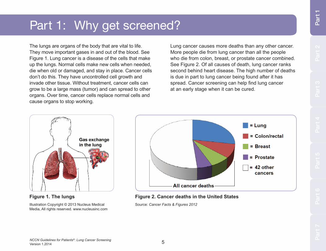
Part 1: Why get screened?The lungs are organs of the body that are vital to life. They move important gases in and out of the blood. See Figure 1. Lung cancer is a disease of the cells that make up the lungs. Normal cells make new cells when needed, die when old or damaged, and stay in place. Cancer cells don’t do this. They have uncontrolled cell growth and invade other tissue. Without treatment, cancer cells can grow to be a large mass (tumor) and can spread to other organs. Over time, cancer cells replace normal cells and cause organs to stop working.
Lung cancer causes more deaths than any other cancer. More people die from lung cancer than all the people who die from colon, breast, or prostate cancer combined. See Figure 2. Of all causes of death, lung cancer ranks second behind heart disease. The high number of deaths is due in part to lung cancer being found after it has spread. Cancer screening can help find lung cancer at an early stage when it can be cured.
Figure 1. The lungsIllustration Copyright © 2013 Nucleus Medical Media, All rights reserved. www.nucleusinc.com
Figure 2. Cancer deaths in the United StatesSource: Cancer Facts & Figures 2012

6NCCN Guidelines for Patients®: Lung Cancer Screening Version 1.2014
am I at risk?
7NCCN Guidelines for Patients®: Lung Cancer Screening Version 1.2014
Part
3Pa
rt 4
Part
5Pa
rt 6
Part
7Pa
rt 2
Part
1
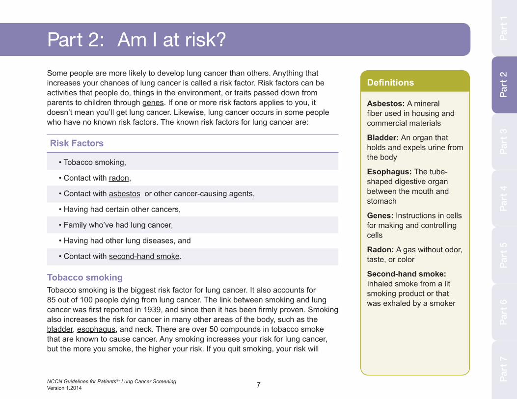
Part 2: Am i at risk?Some people are more likely to develop lung cancer than others. Anything that increases your chances of lung cancer is called a risk factor. Risk factors can be activities that people do, things in the environment, or traits passed down from parents to children through genes. If one or more risk factors applies to you, it doesn’t mean you’ll get lung cancer. Likewise, lung cancer occurs in some people who have no known risk factors. The known risk factors for lung cancer are:
Risk Factors
• Tobacco smoking,
• Contact with radon,
• Contact with asbestos or other cancer-causing agents,
• Having had certain other cancers,
• Family who’ve had lung cancer,
• Having had other lung diseases, and
• Contact with second-hand smoke.
Tobacco smokingTobacco smoking is the biggest risk factor for lung cancer. It also accounts for 85 out of 100 people dying from lung cancer. The link between smoking and lung cancer was first reported in 1939, and since then it has been firmly proven. Smoking also increases the risk for cancer in many other areas of the body, such as the bladder, esophagus, and neck. There are over 50 compounds in tobacco smoke that are known to cause cancer. Any smoking increases your risk for lung cancer, but the more you smoke, the higher your risk. If you quit smoking, your risk will
Asbestos: A mineral fiber used in housing and commercial materials
Bladder: An organ that holds and expels urine from the body
Esophagus: The tube-shaped digestive organ between the mouth and stomach
Genes: Instructions in cells for making and controlling cells
Radon: A gas without odor, taste, or color
Second-hand smoke: Inhaled smoke from a lit smoking product or that was exhaled by a smoker
Definitions

8NCCN Guidelines for Patients®: Lung Cancer Screening Version 1.2014
decrease. However, the risk for lung cancer is higher for former smokers than people who never smoked. Thus, current or past tobacco smoking is a risk factor for lung cancer.
If you smoke tobacco, ask your doctor about counseling and drugs to help you quit.
Radon Uranium is a metallic chemical found in rocks and soil. As it decays, radon is made and gets into air and water. Miners of uranium have a high risk for developing lung cancer. Some studies of radon in the home have linked radon to lung cancer while other studies have not. The risk for lung cancer may depend on how much radon is in the home. For people who’ve had contact with radon, such as uranium miners, the risk for lung cancer is higher for those who smoke than for those who don’t smoke.
Other cancer-causing agentsBesides radon, 10 other agents are known to cause lung cancer. Five are metallic chemicals: arsenic, beryllium, cadmium, chromium, and nickel. The others are asbestos, coal smoke, soot, silica, and diesel fumes. Among people who’ve had contact with these agents, the risk for lung cancer is higher for those who’ve smoked than for those who’ve never smoked.
History of other cancersYour risk for lung cancer may be increased if you’ve had other cancers. Having had small cell lung cancer increases your risk of developing cancer in other types of lung cells. Likewise, if you’ve had another smoking-related cancer, like head and neck cancer, your risk for lung cancer is increased. The risk for lung cancer increases after receiving radiation therapy in the chest for other cancers, especially if you smoke. Treatment of Hodgkin’s lymphoma with alkylating agents—a type of cancer drug—increases the risk for lung cancer too.
Part 2: Am i at risk?
1 in 14 people will develop lung cancer
9NCCN Guidelines for Patients®: Lung Cancer Screening Version 1.2014
Part 1: About lung cancer
Part
3Pa
rt 4
Part
5Pa
rt 6
Part
7Pa
rt 2
Part
1
Definitions
Diesel fumes: Gases from thick, heavy fuel made from crude oil
Hodgkin’s lymphoma: Cancer of white blood cells
Mucus: A sticky, thick liquid that moisturizes or lubricates
Silica: A natural mineral mostly found in sand
Small cell lung cancer: Cancer that started in the small, round cells of the lung
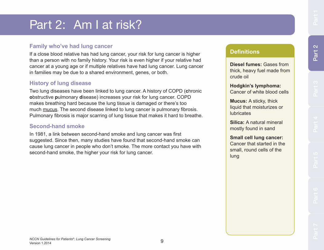
Part 2: Am i at risk?Family who’ve had lung cancerIf a close blood relative has had lung cancer, your risk for lung cancer is higher than a person with no family history. Your risk is even higher if your relative had cancer at a young age or if multiple relatives have had lung cancer. Lung cancer in families may be due to a shared environment, genes, or both.
History of lung diseaseTwo lung diseases have been linked to lung cancer. A history of COPD (chronic obstructive pulmonary disease) increases your risk for lung cancer. COPD makes breathing hard because the lung tissue is damaged or there’s too much mucus. The second disease linked to lung cancer is pulmonary fibrosis. Pulmonary fibrosis is major scarring of lung tissue that makes it hard to breathe.
Second-hand smokeIn 1981, a link between second-hand smoke and lung cancer was first suggested. Since then, many studies have found that second-hand smoke can cause lung cancer in people who don’t smoke. The more contact you have with second-hand smoke, the higher your risk for lung cancer.

10
should Istart now?
11NCCN Guidelines for Patients®: Lung Cancer Screening Version 1.2014
Part 1: About lung cancer
Part
3Pa
rt 4
Part
5Pa
rt 6
Part
7Pa
rt 3
Part
1
Definitions
Mucus: A sticky, thick liquid that moisturizes or lubricates
Pneumonia: An infection causing the lungs to fill up with pus
Wheezing: A coarse, whistling sound while breathing
Part 3: should i start now?The goal of cancer screening is to find lung cancer when treatments will work best. Treatments usually work best before there are symptoms of cancer. However, at this time, most lung cancer is found after symptoms appear. Common symptoms of lung cancer are:
Symptoms of lung cancer
• Coughing that lasts,
• Blood in lung mucus,
• Shortness of breath,
• Wheezing,
• Pain in chest area,
• Tiredness that lasts,
• Pneumonia,
• Hoarse voice,
• Pain when swallowing, and
• High-pitched sound when breathing.
See your doctor if you have these symptoms. Most often, they are caused by health problems other than lung cancer. If they are caused by lung cancer, talk with your doctor about treatment options. If you have no symptoms of lung cancer, a screening program may be right for you.
Part
2
12NCCN Guidelines for Patients®: Lung Cancer Screening Version 1.2014
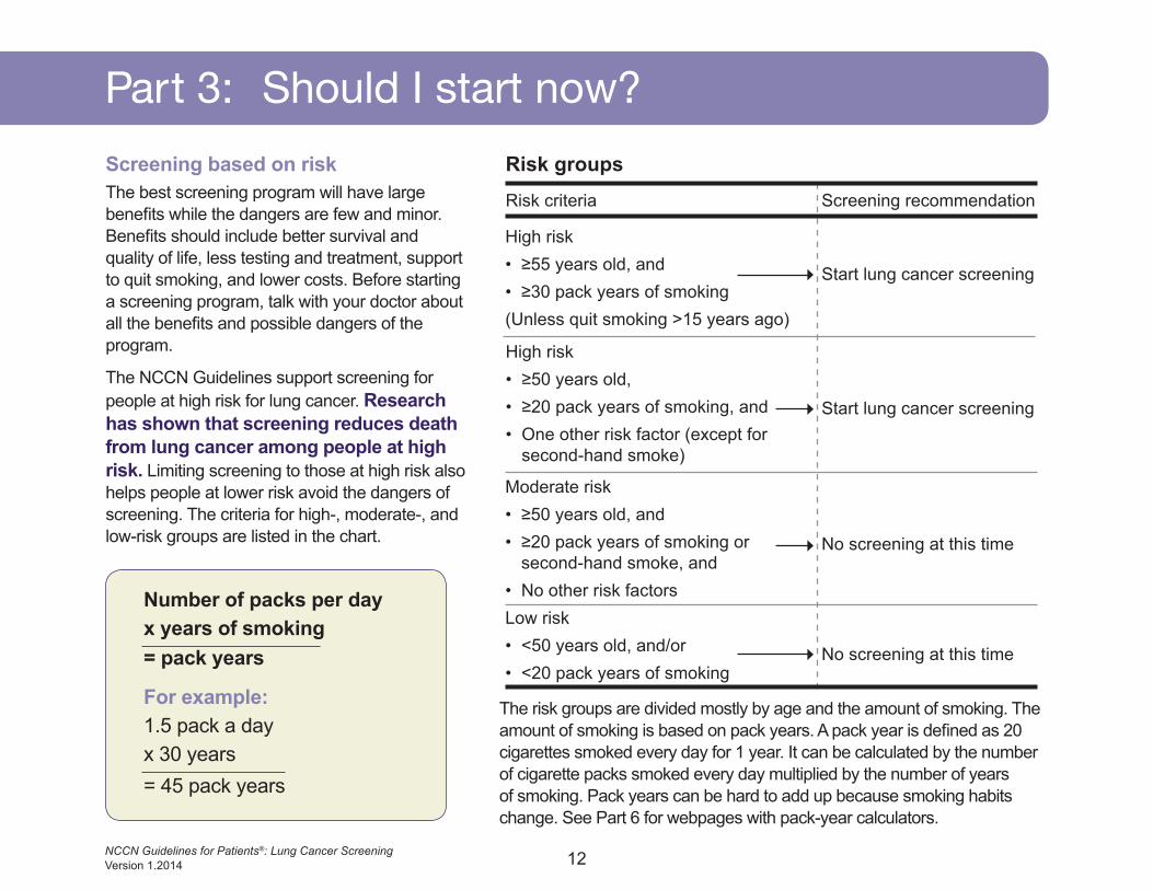
For example:1.5 pack a dayx 30 years= 45 pack years
Part 3: should i start now?Screening based on riskThe best screening program will have large benefits while the dangers are few and minor. Benefits should include better survival and quality of life, less testing and treatment, support to quit smoking, and lower costs. Before starting a screening program, talk with your doctor about all the benefits and possible dangers of the program.
The NCCN Guidelines support screening for people at high risk for lung cancer. Research has shown that screening reduces death from lung cancer among people at high risk. Limiting screening to those at high risk also helps people at lower risk avoid the dangers of screening. The criteria for high-, moderate-, and low-risk groups are listed in the chart.
The risk groups are divided mostly by age and the amount of smoking. The amount of smoking is based on pack years. A pack year is defined as 20 cigarettes smoked every day for 1 year. It can be calculated by the number of cigarette packs smoked every day multiplied by the number of years of smoking. Pack years can be hard to add up because smoking habits change. See Part 6 for webpages with pack-year calculators.
Number of packs per dayx years of smoking= pack years
Risk groups
Start lung cancer screening
Risk criteria Screening recommendation
High risk• ≥55 years old, and• ≥30 pack years of smoking(Unless quit smoking >15 years ago)
Moderate risk• ≥50 years old, and• ≥20 pack years of smoking or
second-hand smoke, and• No other risk factorsLow risk• <50 years old, and/or• <20 pack years of smoking
No screening at this time
No screening at this time
Start lung cancer screening
High risk• ≥50 years old,• ≥20 pack years of smoking, and• One other risk factor (except for
second-hand smoke)
13NCCN Guidelines for Patients®: Lung Cancer Screening Version 1.2014
Part 1: About lung cancer
Part
3Pa
rt 4
Part
5Pa
rt 6
Part
7Pa
rt 3
Part
1
Part 3: should i start now?
Screening programsKnown benefits
• Screening programs can reduce the number of deaths from lung cancer and other causes.
• Lung cancer found by screening is often an earlier stage of disease than cancer found because of symptoms.
• Patients whose cancer was found with screening more often can have minimally invasive surgery and have less lung tissue removed.
Known dangers• Screening programs don’t always find cancer early enough to be cured.
• Some people get treated even though the cancer grows so slowly that it won’t cause death.
• Some people get unneeded tests, treatment, or both because screening results were unclear or wrong.
Screening for lung cancer is recommended for the two high-risk groups. The first high-risk group consists of people 55 years old and older who have smoked for 30 or more pack years. People who quit smoking more than 15 years ago are excluded. The second high-risk group consists of people 50 years old and older who have smoked for 20 or more pack years and have at least one more risk factor other than second-hand smoke. Risk factors are described in Part 2 on page 7. Screening isn’t recommended for high-risk people with poor health, who if diagnosed with cancer would not be able to receive curative treatment.
Definitions
Curative treatment: A medicine that cures disease or symptoms
Risk factor: Something that increases the chance of getting a disease
Second-hand smoke: Inhaled smoke from a lit smoking product or that was exhaled by a smoker
Part
2

14NCCN Guidelines for Patients®: Lung Cancer Screening Version 1.2014
Which test for screening?Research supports using spiral LDCT (low-dose computed tomography) of the chest for lung cancer screening. This test takes many pictures of the inside of your body from different angles using x-rays. The amount of radiation used is much lower than standard doses of a CT (computed tomography) scan.
Getting an LDCT is easy. Before the test, you may be asked to stop eating or drinking for several hours. You also should remove any metal objects that are on your body. The machine is large and has a tunnel in the middle. See Figure 3. During the test, you will need to lie on a table that moves through the tunnel. Pillows or straps may be used to keep you still during the test. You will be alone, but a technician will operate the machine in a nearby room. He or she will be able to see, hear, and speak with you at all times.
As the machine takes pictures, you may hear buzzing, clicking, or whirring sounds. Earplugs are sometimes worn to block these sounds. A computer combines all pictures into one detailed picture. The test can be done in a few minutes, but the whole process takes about 30 minutes. You may not learn of the results for a few days since a radiologist needs to see and interpret the pictures. A radiologist is a doctor who’s an expert in reading LDCT scans.
Part 3: should i start now?
Figure 3. Computed tomography
15
thescreening process
16NCCN Guidelines for Patients®: Lung Cancer Screening Version 1.2014
Screening with LDCT is used to find nodules in the lungs. Nodules are small, round masses of tissue. Many people have nodules. Nodules can be caused by cancer, infections, scar tissue, or other conditions. Most nodules are benign—not cancer.
Nodules caused by cancer have specific traits. First, they aren’t likely to have calcium buildup. Second, they often have rough edges and odd shapes. Third, they often grow faster and are larger in size than nodules without cancer. Nodules are measured in mm (millimeters). This letter “o” is about 1 mm long.
Doctors also assess the density of a nodule to tell if it may be cancer. Density refers to how well the x-rays from the LDCT go through the lung. Think of a flashlight shining in the dark. If the light doesn’t hit an object, it is dark a few feet away from the flashlight. If the light does hit an object, the object reflects the light and can be seen. Nodules are divided into three groups based on density:
• Solid nodules have high density. They look evenly white on an LDCT scan.
• Non-solid nodules have low density. They look like hazy clouds on an LDCT scan. Your doctors
may call this type of nodule a “pure ground-glass opacity” or a “pure ground-glass nodule.”
• Part-solid nodules have both high and low areas of density. These nodules have both solid white and hazy parts. Your doctors may call this type of nodule a “mixed ground-glass nodule,” “semi-solid nodule,” or “subsolid nodule.”
Part 4 describes the recommended screening process after the first LDCT. Often, the use of one LDCT detects a nodule but isn’t clear whether the nodule is lung cancer. Thus, the first LDCT—the baseline test—is compared to follow-up LDCTs. Your doctors will look for increases in size or density. Such changes are likely signs of cancer.
No nodulesIf no lung nodules are found, your next LDCT should be in 1 year. Screening with LDCT should occur every year for at least 2 years. After 2 years, your doctors may want you to continue yearly screening. However, screening isn’t recommended for people with poor health, who if diagnosed with cancer would not be able to receive curative treatment.
Part 4: The screening process
17NCCN Guidelines for Patients®: Lung Cancer Screening Version 1.2014
Part
2Pa
rt 3
Part
4Pa
rt 5
Part
6Pa
rt 7
Definitions:
Calcium: A mineral found in body tissue
Curative treatment: Medicine that cures disease or symptoms
Scar tissue: Supportive fibers that form to heal a wound
! Read page 14 for a description of LDCT.
Part
1
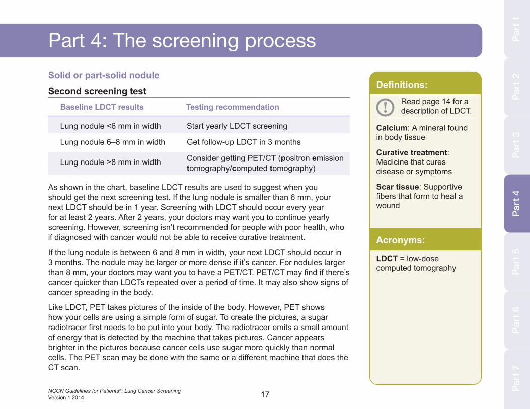
Solid or part-solid noduleSecond screening test
Baseline LDCT results Testing recommendation
Lung nodule <6 mm in width Start yearly LDCT screening
Lung nodule 6–8 mm in width Get follow-up LDCT in 3 months
Lung nodule >8 mm in width Consider getting PET/CT (positron emission tomography/computed tomography)
As shown in the chart, baseline LDCT results are used to suggest when you should get the next screening test. If the lung nodule is smaller than 6 mm, your next LDCT should be in 1 year. Screening with LDCT should occur every year for at least 2 years. After 2 years, your doctors may want you to continue yearly screening. However, screening isn’t recommended for people with poor health, who if diagnosed with cancer would not be able to receive curative treatment.
If the lung nodule is between 6 and 8 mm in width, your next LDCT should occur in 3 months. The nodule may be larger or more dense if it’s cancer. For nodules larger than 8 mm, your doctors may want you to have a PET/CT. PET/CT may find if there’s cancer quicker than LDCTs repeated over a period of time. It may also show signs of cancer spreading in the body.
Like LDCT, PET takes pictures of the inside of the body. However, PET shows how your cells are using a simple form of sugar. To create the pictures, a sugar radiotracer first needs to be put into your body. The radiotracer emits a small amount of energy that is detected by the machine that takes pictures. Cancer appears brighter in the pictures because cancer cells use sugar more quickly than normal cells. The PET scan may be done with the same or a different machine that does the CT scan.
Part 4: The screening process
Part
4
Acronyms:
LDCT = low-dose computed tomography
18NCCN Guidelines for Patients®: Lung Cancer Screening Version 1.2014
Part 4: The screening process
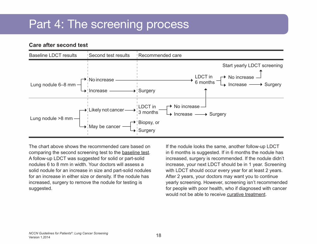
The chart above shows the recommended care based on comparing the second screening test to the baseline test. A follow-up LDCT was suggested for solid or part-solid nodules 6 to 8 mm in width. Your doctors will assess a solid nodule for an increase in size and part-solid nodules for an increase in either size or density. If the nodule has increased, surgery to remove the nodule for testing is suggested.
If the nodule looks the same, another follow-up LDCT in 6 months is suggested. If in 6 months the nodule has increased, surgery is recommended. If the nodule didn’t increase, your next LDCT should be in 1 year. Screening with LDCT should occur every year for at least 2 years. After 2 years, your doctors may want you to continue yearly screening. However, screening isn’t recommended for people with poor health, who if diagnosed with cancer would not be able to receive curative treatment.
Care after second test
Baseline LDCT results
Lung nodule 6–8 mm
Start yearly LDCT screening
No increase
Likely not cancer
Increase Surgery
No increase
No increase
Increase
Increase
Surgery
Surgery
SurgeryBiopsy, or
May be cancer
LDCT in 6 months
LDCT in 3 months
Second test results Recommended care
Lung nodule >8 mm
19NCCN Guidelines for Patients®: Lung Cancer Screening Version 1.2014
Part
2Pa
rt 3
Part
4Pa
rt 5
Part
6Pa
rt 7
Part
1
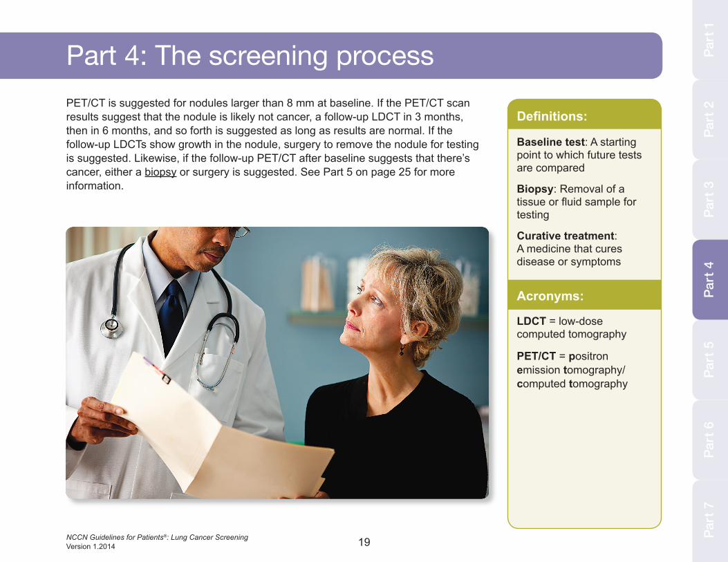
Part 4: The screening processPET/CT is suggested for nodules larger than 8 mm at baseline. If the PET/CT scan results suggest that the nodule is likely not cancer, a follow-up LDCT in 3 months, then in 6 months, and so forth is suggested as long as results are normal. If the follow-up LDCTs show growth in the nodule, surgery to remove the nodule for testing is suggested. Likewise, if the follow-up PET/CT after baseline suggests that there’s cancer, either a biopsy or surgery is suggested. See Part 5 on page 25 for more information.
Part
4
Definitions:
Baseline test: A starting point to which future tests are compared
Biopsy: Removal of a tissue or fluid sample for testing
Curative treatment: A medicine that cures disease or symptoms
Acronyms:
LDCT = low-dose computed tomography
PET/CT = positron emission tomography/computed tomography
20NCCN Guidelines for Patients®: Lung Cancer Screening Version 1.2014
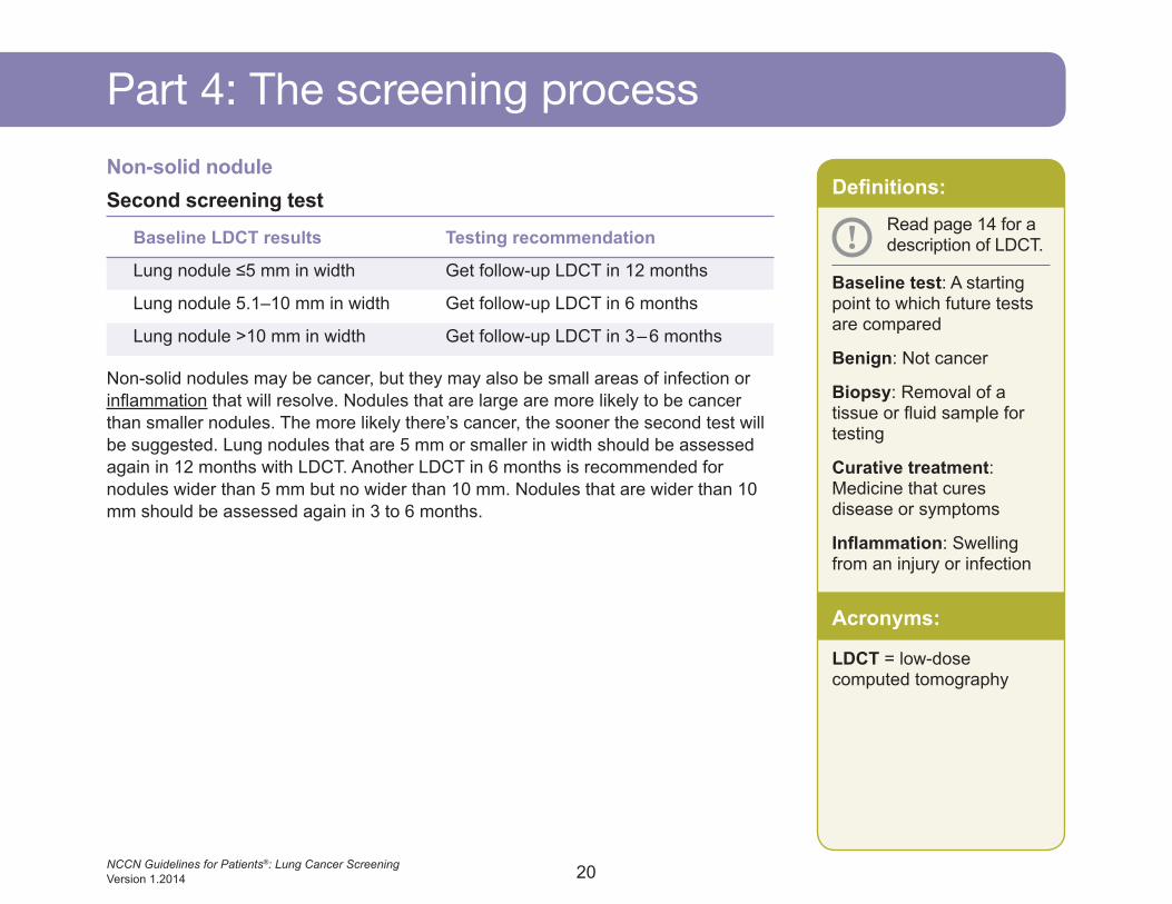
Part 4: The screening processNon-solid noduleSecond screening test
Baseline LDCT results Testing recommendation
Lung nodule ≤5 mm in width Get follow-up LDCT in 12 months
Lung nodule 5.1–10 mm in width Get follow-up LDCT in 6 months
Lung nodule >10 mm in width Get follow-up LDCT in 3 – 6 months
Non-solid nodules may be cancer, but they may also be small areas of infection or inflammation that will resolve. Nodules that are large are more likely to be cancer than smaller nodules. The more likely there’s cancer, the sooner the second test will be suggested. Lung nodules that are 5 mm or smaller in width should be assessed again in 12 months with LDCT. Another LDCT in 6 months is recommended for nodules wider than 5 mm but no wider than 10 mm. Nodules that are wider than 10 mm should be assessed again in 3 to 6 months.
Definitions:
Baseline test: A starting point to which future tests are compared
Benign: Not cancer
Biopsy: Removal of a tissue or fluid sample for testing
Curative treatment: Medicine that cures disease or symptoms
Inflammation: Swelling from an injury or infection
! Read page 14 for a description of LDCT.
Acronyms:
LDCT = low-dose computed tomography
21NCCN Guidelines for Patients®: Lung Cancer Screening Version 1.2014
Part 7: Dictionary Part
1Pa
rt 2
Part
3Pa
rt 4
Part
5Pa
rt 6
Part
7Pa
rt 2
Part
3Pa
rt 4
Part
5Pa
rt 6
Part
7Pa
rt 1
Part 4: The screening process
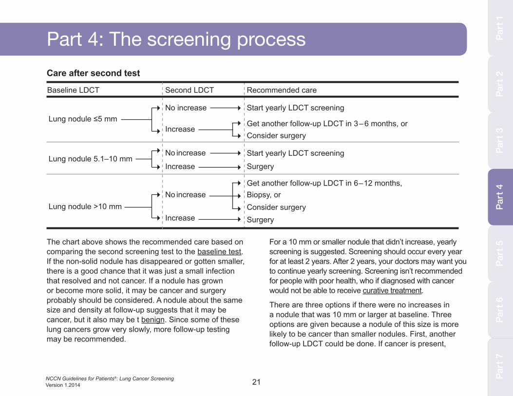
The chart above shows the recommended care based on comparing the second screening test to the baseline test. If the non-solid nodule has disappeared or gotten smaller, there is a good chance that it was just a small infection that resolved and not cancer. If a nodule has grown or become more solid, it may be cancer and surgery probably should be considered. A nodule about the same size and density at follow-up suggests that it may be cancer, but it also may be t benign. Since some of these lung cancers grow very slowly, more follow-up testing may be recommended.
For a 10 mm or smaller nodule that didn’t increase, yearly screening is suggested. Screening should occur every year for at least 2 years. After 2 years, your doctors may want you to continue yearly screening. Screening isn’t recommended for people with poor health, who if diagnosed with cancer would not be able to receive curative treatment.
There are three options if there were no increases in a nodule that was 10 mm or larger at baseline. Three options are given because a nodule of this size is more likely to be cancer than smaller nodules. First, another follow-up LDCT could be done. If cancer is present,
Consider surgeryBiopsy, or
Baseline LDCT
Lung nodule ≤5 mmStart yearly LDCT screening
Get another follow-up LDCT in 6 – 12 months,
No increase
No increase
Increase
Increase
Get another follow-up LDCT in 3 – 6 months, orConsider surgery
Surgery
Second LDCT Recommended care
Lung nodule 5.1–10 mmStart yearly LDCT screeningNo increase
Increase Surgery
Part
4
Lung nodule >10 mm
Care after second test
22NCCN Guidelines for Patients®: Lung Cancer Screening Version 1.2014
Part 4: The screening processthe nodule will likely be larger or denser in 6 to 12 months. Instead of waiting, other options are a biopsy or surgery—both of which can confirm if cancer is present. See Part 5 on page 25 for more information.
Nodules that are larger or denser at follow-up may be cancer. Two options are given for a nodule that was smaller than 5 mm at baseline but increased in size or density. First, another follow-up LDCT could be done. If cancer is present, the nodule will most likely be even larger or denser in 3 to 6 months. The second option is surgery to remove the nodule and test for cancer. For nodules that were 5 mm or larger at baseline and have increased in size or density, surgery to remove the nodule for testing is suggested. See Part 5 on page 25 for more information.
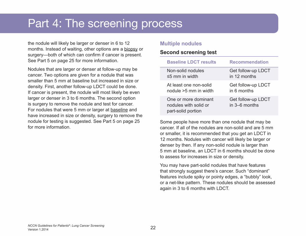
Multiple nodulesSecond screening test
Baseline LDCT results Recommendation
Non-solid nodules Get follow-up LDCT ≤5 mm in width in 12 months
At least one non-solid Get follow-up LDCT nodule >5 mm in width in 6 months
One or more dominant Get follow-up LDCT nodules with solid or in 3–6 months part-solid portion
Some people have more than one nodule that may be cancer. If all of the nodules are non-solid and are 5 mm or smaller, it is recommended that you get an LDCT in 12 months. Nodules with cancer will likely be larger or denser by then. If any non-solid nodule is larger than 5 mm at baseline, an LDCT in 6 months should be done to assess for increases in size or density.
You may have part-solid nodules that have features that strongly suggest there’s cancer. Such “dominant” features include spiky or pointy edges, a “bubbly” look, or a net-like pattern. These nodules should be assessed again in 3 to 6 months with LDCT.
23NCCN Guidelines for Patients®: Lung Cancer Screening Version 1.2014
Part
1Pa
rt 2
Part
3Pa
rt 4
Part
5Pa
rt 6
Part 4: The screening process
Part
7Pa
rt 1
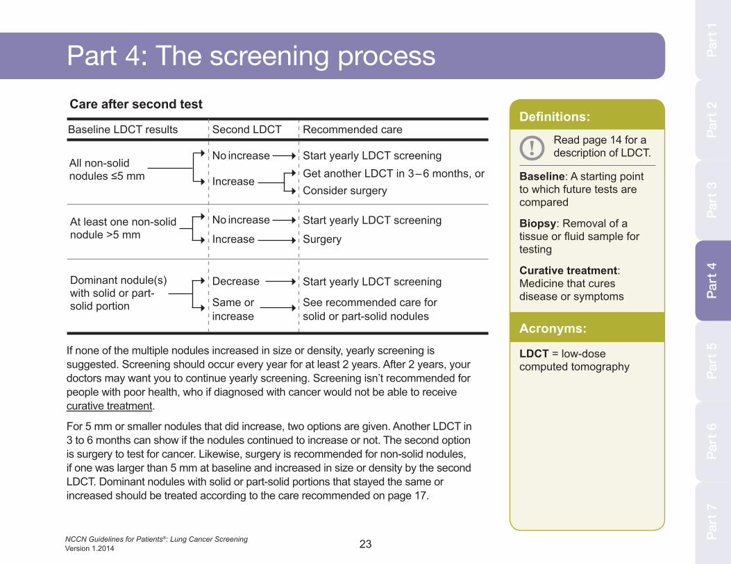
Care after second test
Baseline LDCT results
All non-solid nodules ≤5 mm
Dominant nodule(s) with solid or part-solid portion
Start yearly LDCT screeningNo increase
IncreaseGet another LDCT in 3 – 6 months, orConsider surgery
Second LDCT Recommended care
At least one non-solid nodule >5 mm
Start yearly LDCT screening
Start yearly LDCT screening
No increase
Decrease
Increase
Same or increase
Surgery
See recommended care for solid or part-solid nodules
Part
4
If none of the multiple nodules increased in size or density, yearly screening is suggested. Screening should occur every year for at least 2 years. After 2 years, your doctors may want you to continue yearly screening. Screening isn’t recommended for people with poor health, who if diagnosed with cancer would not be able to receive curative treatment.
For 5 mm or smaller nodules that did increase, two options are given. Another LDCT in 3 to 6 months can show if the nodules continued to increase or not. The second option is surgery to test for cancer. Likewise, surgery is recommended for non-solid nodules, if one was larger than 5 mm at baseline and increased in size or density by the second LDCT. Dominant nodules with solid or part-solid portions that stayed the same or increased should be treated according to the care recommended on page 17.
Definitions:
Baseline: A starting point to which future tests are compared
Biopsy: Removal of a tissue or fluid sample for testing
Curative treatment: Medicine that cures disease or symptoms
! Read page 14 for a description of LDCT.
Acronyms:
LDCT = low-dose computed tomography
24NCCN Guidelines for Patients®: Lung Cancer Screening Version 1.2014
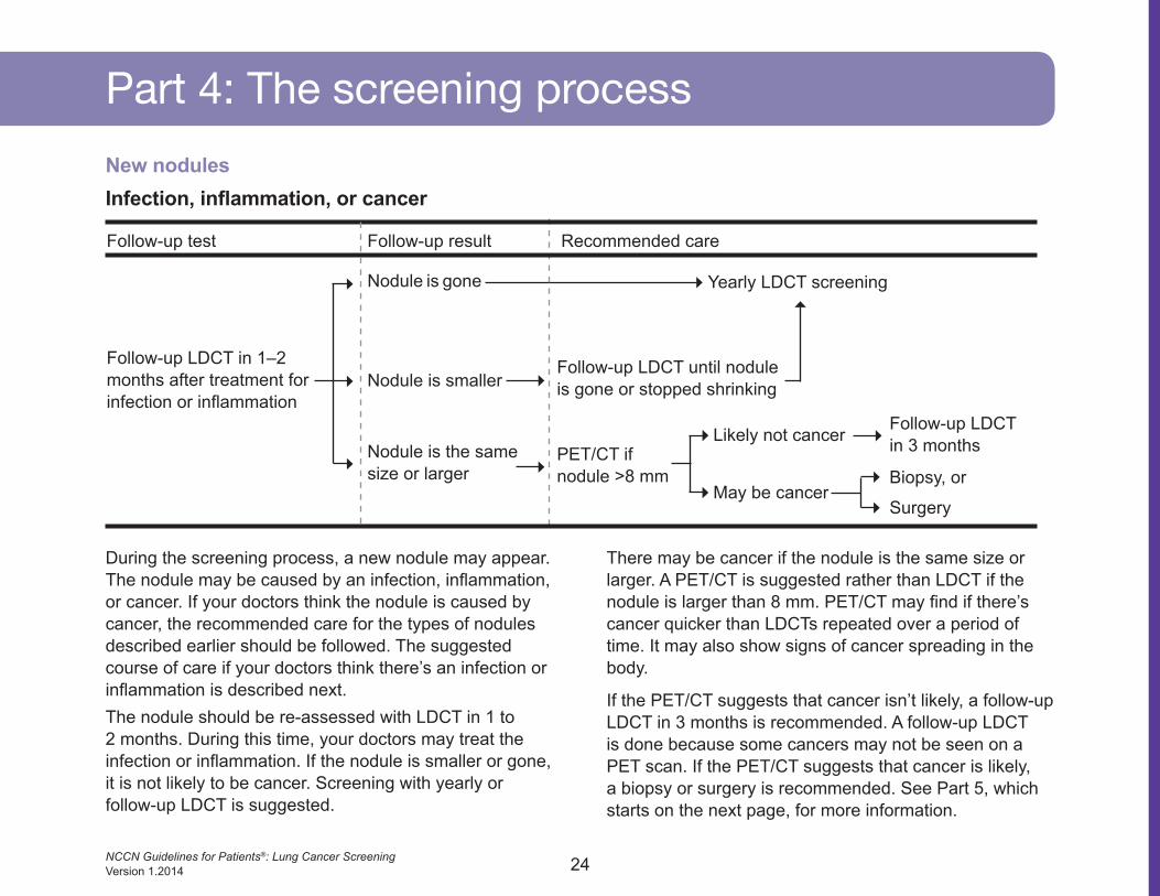
Part 4: The screening processNew nodulesInfection, inflammation, or cancer
Follow-up test
Nodule is gone Yearly LDCT screening
Follow-up LDCT until nodule is gone or stopped shrinking
Biopsy, or
Surgery
Nodule is smaller
Nodule is the same size or larger
PET/CT if nodule >8 mm
Likely not cancer
May be cancer
Recommended care
Follow-up LDCT in 1–2 months after treatment for infection or inflammation
Follow-up result
During the screening process, a new nodule may appear. The nodule may be caused by an infection, inflammation, or cancer. If your doctors think the nodule is caused by cancer, the recommended care for the types of nodules described earlier should be followed. The suggested course of care if your doctors think there’s an infection or inflammation is described next.The nodule should be re-assessed with LDCT in 1 to 2 months. During this time, your doctors may treat the infection or inflammation. If the nodule is smaller or gone, it is not likely to be cancer. Screening with yearly or follow-up LDCT is suggested.
There may be cancer if the nodule is the same size or larger. A PET/CT is suggested rather than LDCT if the nodule is larger than 8 mm. PET/CT may find if there’s cancer quicker than LDCTs repeated over a period of time. It may also show signs of cancer spreading in the body.
If the PET/CT suggests that cancer isn’t likely, a follow-up LDCT in 3 months is recommended. A follow-up LDCT is done because some cancers may not be seen on a PET scan. If the PET/CT suggests that cancer is likely, a biopsy or surgery is recommended. See Part 5, which starts on the next page, for more information.
Follow-up LDCT in 3 months
25
testing for
lung cancer
26NCCN Guidelines for Patients®: Lung Cancer Screening Version 1.2014
To test for cancer, tissue from the nodule must be removed from your body. The tissue will then be sent to a lab and examined with a microscope to look for cancer cells. A biopsy removes small samples of tissue. Surgery removes the entire nodule for testing.
BiopsySince a biopsy only removes a very small piece of the nodule, the results could be wrong. There may be cancer cells in another part of the nodule. Thus, your doctors may suggest surgery instead of a biopsy if your risk for cancer is high. Likewise, your doctors may suggest another biopsy or surgery if the first biopsy shows no cancer.
There are two types of biopsies used for lung nodules. Before either biopsy, you may be asked to stop eating, stop taking some medicines, or stop smoking. A sedative, local anesthesia, or both may be used. A biopsy is generally a safe test and takes about 30 to 60 minutes to complete.
Percutaneous needle biopsyThis biopsy uses a very thin needle. Before or during the biopsy, CT may be used to find the right spot. Your skin will be cleaned and your doctors will make a small cut after numbing the area with local anesthesia. The needle will be inserted through the cut and into the nodule. During the biopsy, you may be asked to keep still and hold your breath at times. After the biopsy, you will be given a chest x–ray to check the results.
BronchoscopyA bronchoscopy allows your doctor to biopsy a nodule using a bronchoscope. A standard bronchoscope is a thin, long tube about as thick as a pencil. It has a very small light, camera, and open channel for taking biopsies. The light and camera allow your doctor to guide the bronchoscope down your mouth into your lungs. A small tool is used to remove tissue from the nodule.
The airways of the lungs get smaller as they extend toward the side of the body. Standard bronchoscopes are often too large to travel through these small airways. A navigational bronchoscopy can be done instead to guide a probe and biopsy instrument to the site of the nodule.
For a navigational bronchoscopy, your doctor will plan how to reach the nodule using a picture made by CT. During the biopsy, you will lie on an electromagnetic plate. The bronchoscope will be fitted with another very small tube through which a sensor probe will be inserted. The electromagnetic plate allows your doctor to see and guide the sensor probe. When the nodule is in reach, the sensor probe will be removed and a small tool will be inserted to collect tissue.
SurgerySurgery removes the nodule as well as a rim of normal-looking tissue around the nodule. The normal tissue is called the surgical margin. The whole nodule and the surgical margin will be examined for cancer cells.
Part 5: Testing for lung cancer
27NCCN Guidelines for Patients®: Lung Cancer Screening Version 1.2014
Part
3Pa
rt 4
Part
5Pa
rt 6
Part
7Pa
rt 1
Part
5
Part 5: Testing for lung cancerSurgery types
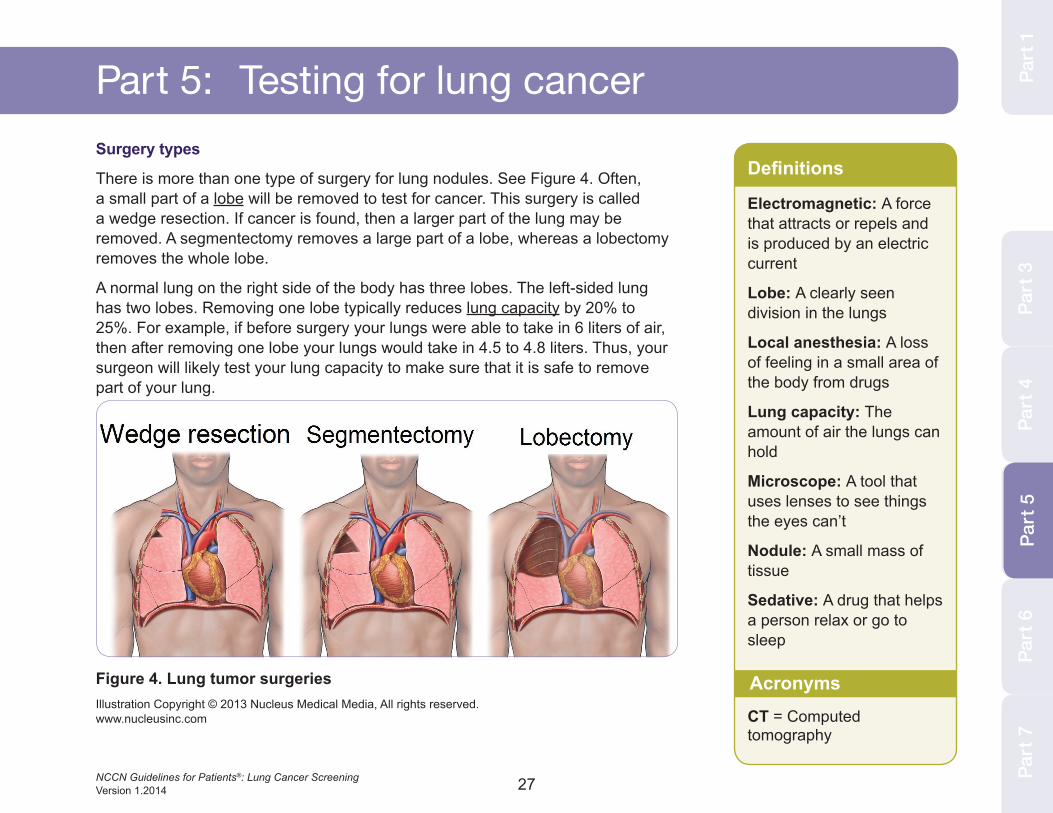
There is more than one type of surgery for lung nodules. See Figure 4. Often, a small part of a lobe will be removed to test for cancer. This surgery is called a wedge resection. If cancer is found, then a larger part of the lung may be removed. A segmentectomy removes a large part of a lobe, whereas a lobectomy removes the whole lobe.
A normal lung on the right side of the body has three lobes. The left-sided lung has two lobes. Removing one lobe typically reduces lung capacity by 20% to 25%. For example, if before surgery your lungs were able to take in 6 liters of air, then after removing one lobe your lungs would take in 4.5 to 4.8 liters. Thus, your surgeon will likely test your lung capacity to make sure that it is safe to remove part of your lung.
Definitions
Electromagnetic: A force that attracts or repels and is produced by an electric current
Lobe: A clearly seen division in the lungs
Local anesthesia: A loss of feeling in a small area of the body from drugs
Lung capacity: The amount of air the lungs can hold
Microscope: A tool that uses lenses to see things the eyes can’t
Nodule: A small mass of tissue
Sedative: A drug that helps a person relax or go to sleep
CT = Computed tomography
AcronymsFigure 4. Lung tumor surgeriesIllustration Copyright © 2013 Nucleus Medical Media, All rights reserved. www.nucleusinc.com
28NCCN Guidelines for Patients®: Lung Cancer Screening Version 1.2014
Part 5: Testing for lung cancerSurgery methodsSurgery may be done with one of two methods. The classic method is thoracotomy. VATS (video-assisted thoracic surgery) is a newer method. VATS is often preferred for a small nodule, but a thoracotomy is sometimes preferred because of nodule size, nodule location, or other reasons.
Before either surgery, you will be asked to stop eating, drinking, and taking some medicines for a short period of time. If you smoke, it is important to stop to get the best results possible. General anesthesia is used for both surgeries.
With thoracotomy, a cut is made in the side of the chest passing under the armpit and shoulder blade. The cut is made between the ribs and through the chest wall. The ribs are spread apart with retractors to allow the surgeon to work. Sometimes, a part of the rib is removed. During surgery, the lung with the nodule is deflated and a breathing tube is used to help you breathe with the other lung. After surgery the cut is sewn closed, but tubes are left in for a few days to drain fluid and air. The surgery can take 2 to 3 hours. You may stay in the hospital for a few days to recover.
With VATS, 3 to 4 small cuts are made between the ribs on the side of the chest. A camera and surgical tools are inserted through the cuts. Video from the camera is shown on a computer so that the surgeon can see your organs. Tissue is removed through the small cuts rather
than a large opening in the chest wall. During surgery, the lung with the nodule is deflated and a breathing tube is used to help you breathe with the other lung. After surgery the cuts are sewn closed, but tubes are left in for a few days to drain fluid and air. The surgery can take 2 to 3 hours. You may stay in the hospital for 1 to 3 days to recover.
29NCCN Guidelines for Patients®: Lung Cancer Screening Version 1.2014
Part
3Pa
rt 4
Part
5Pa
rt 6
Part
7Pa
rt 5
Part
1
Part 5: Testing for lung cancer
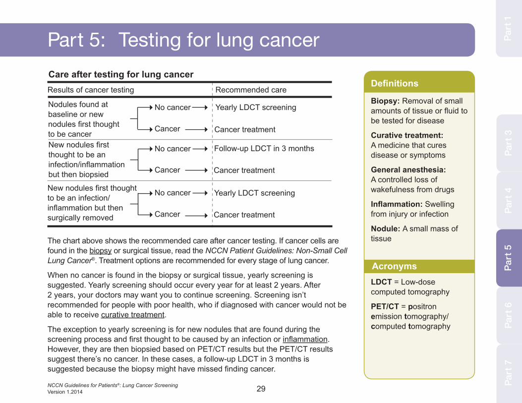
The chart above shows the recommended care after cancer testing. If cancer cells are found in the biopsy or surgical tissue, read the NCCN Patient Guidelines: Non-Small Cell Lung Cancer®. Treatment options are recommended for every stage of lung cancer.
When no cancer is found in the biopsy or surgical tissue, yearly screening is suggested. Yearly screening should occur every year for at least 2 years. After 2 years, your doctors may want you to continue screening. Screening isn’t recommended for people with poor health, who if diagnosed with cancer would not be able to receive curative treatment.
The exception to yearly screening is for new nodules that are found during the screening process and first thought to be caused by an infection or inflammation. However, they are then biopsied based on PET/CT results but the PET/CT results suggest there’s no cancer. In these cases, a follow-up LDCT in 3 months is suggested because the biopsy might have missed finding cancer.
Results of cancer testing
Care after testing for lung cancer
Nodules found at baseline or new nodules first thought to be cancer
Yearly LDCT screening
Cancer treatment
Recommended care
No cancer
Cancer
New nodules first thought to be an infection/inflammation but then biopsied
Follow-up LDCT in 3 months
Cancer treatment
No cancer
Cancer
New nodules first thought to be an infection/inflammation but then surgically removed
Yearly LDCT screening
Cancer treatment
No cancer
Cancer
Definitions
Biopsy: Removal of small amounts of tissue or fluid to be tested for disease
Curative treatment: A medicine that cures disease or symptoms
General anesthesia: A controlled loss of wakefulness from drugs
Inflammation: Swelling from injury or infection
Nodule: A small mass of tissue
LDCT = Low-dose computed tomography
PET/CT = positron emission tomography/computed tomography
Acronyms

30NCCN Guidelines for Patients®: Lung Cancer Screening Version 1.2014
Tools
31NCCN Guidelines for Patients®: Lung Cancer Screening Version 1.2014
Part
1Pa
rt 2
Part
3Pa
rt 4
Part
5Pa
rt 7
Part
6
Part 6: Tools
Part
6Pa
rt 1
• Should I be screened for lung cancer?
• What screening plan do you recommend for me?
• What are the benefits and possible dangers of this screening plan?
• Do you use low-dose computed tomography for screening?
• Where will the screening take place? Will I have to go to the hospital?
• Do you have a team of experts who are dedicated to lung cancer screening? Do they include pulmonologists, thoracic surgeons, and specialists in chest radiology?
• Are the surgeons board certified in thoracic surgery? Do they have a major part of their practice dedicated to lung cancer surgery? Do they do VATS surgery?
• Do I have to do anything to prepare for screening?
• Should I bring someone with me?
• How long will screening take?
• What are the risks?
• How soon will I know the results and who will explain them to me?
• Who will talk with me about the next steps? When?
Questions about screening to ask your doctor
32NCCN Guidelines for Patients®: Lung Cancer Screening Version 1.2014
Part 6: Tools
• What type of biopsy will I have?
• Where will it take place? Will I have to go to the hospital?
• How long will it take? Will I be awake?
• Will it hurt? Will I need anesthesia?
• What are the risks? What are the chances of lung collapse, infection, or bleeding afterward?
• How do I prepare for the biopsy? Should I not take aspirin or eat beforehand?
• Should I bring a list of my medications?
• Should I bring someone with me?
• How long will it take for me to recover? Will I be given an antibiotic or another drug afterward?
• How soon will I know the results and who will explain them to me?
• Will I get a copy of the results?
• Who will talk with me about the next steps? When?
Questions about a biopsy to ask your doctor
33NCCN Guidelines for Patients®: Lung Cancer Screening Version 1.2014
Part
3Pa
rt 4
Part
5Pa
rt 6
Part
7
Part 6: Tools Part
1
• What type of surgery will I have?
• What are the benefits and possible dangers of the surgery?
• What should I do to prepare for surgery? Should I stop taking my medications? Should I store my blood in case I need a transfusion?
• Are you board certified in thoracic surgery?
• Is lung surgery a major part of your practice? How many lung surgeries do you do per year? What other types of surgery do you do? General surgery? Heart surgery?
• How much will the surgery cost? How can I find out how much my insurance company will cover?
• How long does the surgery last?
• Do you test any lymph nodes before surgery? During surgery?
• What will my lung capacity be after surgery? Will it change my life?
• When will I be able to return to my normal activities?
• How soon will I know the results and who will explain them to me?
• If I have cancer, how likely is it that I’ll be cancer-free after surgery? Will I need any other treatment?
Questions about surgery to ask your doctor
Part
6Pa
rt 6
Part
2
34NCCN Guidelines for Patients®: Lung Cancer Screening Version 1.2014
Part 6: ToolsWhere to go for screeningYour primary care doctor can help you decide whether to start cancer screening. This decision should take into account your chance for developing lung cancer and your health history. Since your doctor knows this information, he or she can make a good suggestion and help guide you to the right screening site.
Some sites require a doctor’s prescription before the visit. Other sites will talk to you without a prescription to decide if you should be screened. They will ask questions about your health history and risk for lung cancer.
You should only go to a screening site that:
• Follows an organized plan—a proven protocol—that is updated to include new technology and knowledge like that from NCCN,
• Has a high-quality screening program with enough staff and resources,
• Is accredited to do CT scans by a certifying organization, such as the American College of Radiology,
• Has scans read by an American Board of Radiology board-certified radiologist who’s an expert in lung cancer screening,
• Has modern multislice CT equipment that does high-quality, low-dose, and non-contrast spiral CT scans, and
• Is partnered with a health center that has: 1) experience and excellence in biopsy methods; 2) board-certified pulmonologists; and 3) board-certified thoracic surgeons who are experts in lung cancer.
Definitions
Biopsy: Removal of small amounts of tissue or fluid to be tested for disease
Board-certified: A status to identify doctors who finished training in a specialized field of medicine
Pulmonologist: A doctor who’s an expert in lung diseases
Radiologist: A doctor who’s an expert in reading imaging tests
Thoracic surgeon: A doctor who’s an expert in surgery within the chest
CT = Computed tomography
NCCN = National Comprehensive Cancer Network
Acronyms
35NCCN Guidelines for Patients®: Lung Cancer Screening Version 1.2014
Part
3Pa
rt 4
Part
5Pa
rt 6
Part
7
Part 6: Tools Part
1Pa
rt 6
WebpagesLung Cancer Alliancewww.screenforlungcancer.org
www.lungcanceralliance.org
NCCNwww.nccn.org/patients
www.nccn.org/patients/guidelines/cancers.aspx#nscll
Smoking Pack Yearssmokingpackyears.com/calculate
Part
2
36NCCN Guidelines for Patients®: Lung Cancer Screening Version 1.2014
Part 7: DictionaryAlkylating agent A type of cancer-killing drug.
Arsenic A very toxic metallic chemical.
Asbestos A mineral fiber used in housing and commercial materials.
Baseline test A starting point to which future tests are compared.
Benign Tissue without cancer cells.
Beryllium A hard, gray metallic chemical.
Biopsy Removal of small amounts of tissue or fluid to be tested for disease.
Bladder An organ that holds and expels urine from the body.
Board certified A status to identify doctors who finished training in a specialized field of medicine.
Bronchi The two airways extending from the windpipe into the lungs.
Bronchoscope A thin, long tube fitted with tools that is guided down the mouth.
Bronchoscopy Use of a thin tool guided down the mouth into the lungs.
Cadmium A heavy metallic chemical.
Calcium A mineral found in body tissues.
Cancer screening The use of tests to find cancer before signs of cancer appear.
Chromium A hard, semi-gray metallic chemical.
Chronic obstructive pulmonary disease (COPD) Trouble with breathing due to lung damage or too much mucus.
Colon An organ that changes eaten food from a liquid into a solid form.
Computed tomography (CT) A test that combines many x-rays to make pictures of the inside of the body.
Curative treatment A medicine that cures disease or symptoms.
Diesel fumes Gases from fuel that is thick, heavy, and made from crude oil.
Early stage Cancer that has had little or no growth into nearby tissues.
Electromagnetic A force that attracts or repels and is produced by an electric current.
Esophagus The tube-shaped digestive organ between the mouth and stomach.
Follow-up testing A close watch by your doctors of possible cancer using tests.
37NCCN Guidelines for Patients®: Lung Cancer Screening Version 1.2014
Part 7: Dictionary Part
1Pa
rt 2
Part
3Pa
rt 4
Part
5Pa
rt 6
Part
7
General anesthesia A controlled loss of wakefulness from drugs.
Genes Instructions in cells for making and controlling cells.
Ground-glass opacity A small mass of lung cells with low density.
Hodgkin’s lymphoma A cancer of white blood cells.
Inflammation Redness, heat, pain, and swelling from injury or infection.
Lobe A clearly seen division in the lungs.
Lobectomy The removal of an entire lobe of the lung.
Local anesthesia A loss of feeling in a small area of the body from drugs.
Low-dose computed tomography (LDCT) A test that uses little amounts of radiation to make pictures of the insides of the body.
Lung An organ in the body made of airways and air sacs.
Lung capacity The amount of air the lungs can hold.
Microscope A tool that uses lenses to see things the eyes can’t.
Mucus A sticky, thick liquid that moisturizes or lubricates.
Navigational bronchoscopy Use of a thin tool guided down the mouth into the smallest airways of the lung.
Nickel A silvery-white metal.
Nodule A small mass of tissue.
Non-solid nodule A small mass of tissue of low density.
Pack years The number of cigarette packs smoked every day multiplied by the number of years of smoking.
Part-solid nodule A small mass of tissue with areas of low and high density.
Percutaneous needle biopsy Insertion of a needle through the skin into a mass to remove tissue for testing.
Pneumonia An infection causing the lungs to fill up with pus.
Positron emission tomography (PET) A test that uses radioactive material to see the shape and function of body parts.
Prostate A male gland that makes fluid for protecting sperm from acid in the vagina.
38NCCN Guidelines for Patients®: Lung Cancer Screening Version 1.2014
Part 7: DictionaryPulmonary fibrosis Major scarring of lung tissue.
Pulmonologist A doctor who’s an expert in lung diseases.
Radiation therapy Treatment with radiation.
Radiologist A doctor who’s an expert in reading imaging tests.
Radiotracer Radioactive material used to make images of the body.
Radon A gas without odor, taste, or color that is made from uranium as it decays.
Retractors A tool that holds back the edges of a surgical cut.
Risk factor Something that increases the chance of getting a disease.
Scar tissue Supportive fibers formed to heal a wound.
Second-hand smoke Inhaled smoke from a lit smoking product or that was exhaled from a smoker.
Sedative A drug that helps a person relax or go to sleep.
Segmentectomy Surgical removal of a large part of a lobe.
Silica A natural mineral mostly found in sand.
Small-cell lung cancer Lung cancer of small, round cells.
Solid nodule A small mass of tissue of high density.
Surgery An operation to remove or repair tissue.
Surgical margin The normal tissue around the tumor removed during surgery.
Thoracic surgeon A doctor who’s an expert in surgery within the chest.
Thoracotomy Surgery done through a large cut to remove all or part of the lungs.
Tumor A mass of cells.
Uranium A silvery-white metallic chemical.
Video-assisted thoracic surgery (VATS) Use of thin tools inserted between the ribs to do work in the chest.
Wedge resection Surgical removal of a small part of a lobe.
Wheezing A coarse, whistling sound while breathing.
39NCCN Guidelines for Patients®: Lung Cancer Screening Version 1.2014
CreditsNCCN aims to improve the care given to patients with cancer. NCCN staff work with experts to create helpful programs and resources for many stakeholders. Stakeholders include health providers, patients, businesses, and others. One resource is the series of booklets for patients called the NCCN Patient Guidelines. Each booklet presents the standard of care for a type of cancer.
NCCN abbreviations and acronymsNCCN® National Comprehensive Cancer Network®
NCCN Patient Guidelines® NCCN Guidelines for Patients®
NCCN Guidelines® NCCN Clinical Practice Guidelines in Oncology®
The patient booklets are based on guidelines written for doctors. These guidelines are called the NCCN Guidelines. They give a step-by-step course of care that many cancer doctors follow. Panels of experts create the NCCN Guidelines. Most of the experts are from the 23 NCCN Member Institutions. Panelists may include surgeons, radiation oncologists, medical oncologists, and patient advocates. Recommendations in the NCCN Guidelines are based on clinical trials and the experience of the panelists.
The people involved in the making of the guidelines for patients and doctors are listed next, starting with NCCN staff:
NCCN Patient GuidelinesDorothy A. Shead, MS Director Patient and Clinical Information Operations
Laura J. Hanisch, PsyD Medical Writer/ Patient Information Specialist
Lacey Meyer Associate Medical Writer
NCCN GuidelinesKristina M. Gregory, RN, MSN, OCN Vice President Clinical Information Operations
Miranda Hughes, PhD Oncology Scientist/ Senior Medical Writer
NCCN MarketingSusan KidneyGraphic Design Specialist
NCCN Drugs and Biologics ProgramsRachael ClarkeMedical Copyeditor
40NCCN Guidelines for Patients®: Lung Cancer Screening Version 1.2014
NCCN Panel Members for Lung Cancer screeningDouglas E. Wood, MD/ChairUniversity of Washington/ Seattle Cancer Care Alliance
Ella Kazerooni, MD/Vice ChairUniversity of Michigan Comprehensive Cancer Center
Scott L. Baum, MDUniversity of Tennessee Cancer Institute
Mark M. Dransfield, MDUniversity of Alabama at Birmingham Comprehensive Cancer Center
George A. Eapen, MDThe University of Texas MD Anderson Cancer Center
David S. Ettinger, MDThe Sidney Kimmel Comprehensive Cancer Center at Johns Hopkins
Lifang Hou, MD, PhD Robert H. Lurie Comprehensive Cancer Center of Northwestern University
David M. Jackman, MDDana-Farber/Brigham and Women’s Cancer Center
Donald Klippenstein, MDMoffitt Cancer Center
Rudy P. Lackner, MD Fred & Pamela Buffett Cancer Center at The Nebraska Medical Center
Lorriana E. Leard, MDUCSF Helen Diller Family Comprehensive Cancer Center
Ann N. C. Leung, MD Stanford Cancer Institute
Pierre P. Massion, MD Vanderbilt-Ingram Cancer Center
Bryan F. Meyers, MD, MPHSiteman Cancer Center at Barnes-Jewish Hospital and Washington University School of Medicine
Reginald F. Munden, MD, DMD, MBA The University of Texas MD Anderson Cancer Center
Gregory A. Otterson, MD The Ohio State University Comprehensive Cancer Center - James Cancer Hospital and Solove Research Institute
Kimberly Peairs, MD The Sidney Kimmel Comprehensive Cancer Center at Johns Hopkins
Sudhakar Pipavath, MDUniversity of Washington/ Seattle Cancer Care Alliance
Christie Pratt-Pozo, MA, DHSc Moffitt Cancer Center
Chakravarthy Reddy, MD Huntsman Cancer Institute at the University of Utah
Mary E. Reid, PhDRoswell Park Cancer Institute
Arnold J. Rotter, MDCity of Hope Comprehensive Cancer Center
Matthew B. Schabath, PhDMoffitt Cancer Center
Lecia V. Sequist, MD, MPH Massachusetts General Hospital Cancer Center
Betty C. Tong, MD, MHSDuke Cancer Institute
William D. Travis, MDMemorial Sloan-Kettering Cancer Center
Michael Unger, MDFox Chase Cancer Center
Stephen C. Yang, MDThe Sidney Kimmel Comprehensive Cancer Center at Johns Hopkins
41NCCN Guidelines for Patients®: Lung Cancer Screening Version 1.2014
NCCN Member institutionsFred & Pamela Buffett Cancer Center at The Nebraska Medical Center Omaha, Nebraska 800.999.5465 unmc.edu/cancercenter
City of Hope Comprehensive Cancer Center Los Angeles, California 800.826.4673 cityofhope.org
Dana-Farber/Brigham and Women’s Cancer Center Massachusetts General Hospital Cancer Center Boston, Massachusetts 877.332.4294 • dfbwcc.org 877.789.6100 • massgeneral.org/cancer Duke Cancer Institute Durham, North Carolina 888.275.3853 dukecancerinstitute.org
Fox Chase Cancer Center Philadelphia, Pennsylvania 888.369.2427 foxchase.org
Huntsman Cancer Institute at the University of Utah Salt Lake City, Utah 877.585.0303 huntsmancancer.org
Fred Hutchinson Cancer Research Center/ Seattle Cancer Care Alliance Seattle, Washington 206.667.5000 • fhcrc.org 206.288.7222 • seattlecca.org
The Sidney Kimmel Comprehensive Cancer Center at Johns Hopkins Baltimore, Maryland 410.955.8964 hopkinskimmelcancercenter.org
Robert H. Lurie Comprehensive Cancer Center of Northwestern University Chicago, Illinois 866.587.4322 cancer.northwestern.edu
Memorial Sloan-Kettering Cancer Center New York, New York 800.525.2225 mskcc.org
Moffitt Cancer Center Tampa, Florida 800.456.3434 moffitt.org
The Ohio State University Comprehensive Cancer Center - James Cancer Hospital and Solove Research Institute Columbus, Ohio 800.293.5066 cancer.osu.edu Roswell Park Cancer Institute Buffalo, New York 877.275.7724 roswellpark.org
Siteman Cancer Center at Barnes-Jewish Hospital and Washington University School of Medicine St. Louis, Missouri 800.600.3606 siteman.wustl.edu
St. Jude Children’s Research Hospital/ The University of Tennessee Health Science Center Memphis, Tennessee 901.595.4055 • stjude.org 901.683.0055 • westclinic.com
Stanford Cancer Institute Stanford, California 877.668.7535 cancer.stanford.edu
42NCCN Guidelines for Patients®: Lung Cancer Screening Version 1.2014
University of Alabama at Birmingham Comprehensive Cancer Center Birmingham, Alabama 800.822.0933 ccc.uab.edu
UC San Diego Moores Cancer Center La Jolla, California 858.657.7000 cancer.ucsd.edu
UCSF Helen Diller Family Comprehensive Cancer Center San Francisco, California 800.689.8273 cancer.ucsf.edu
University of Colorado Cancer Center Aurora, Colorado 720.848.0300 coloradocancercenter.org
University of Michigan Comprehensive Cancer Center Ann Arbor, Michigan 800.865.1125 mcancer.org
The University of Texas MD Anderson Cancer Center Houston, Texas 877.632.6789 mdanderson.org
Vanderbilt-Ingram Cancer Center Nashville, Tennessee 800.811.8480 vicc.org
NCCN Members institutions indexBaseline 16–23, 29Biopsy 18–24, 26, 29, 34Chronic obstructive pulmonary disease 9Computed tomography 14, 17, 26, 34Lobectomy 27Low-dose computed tomography 14, 16–24, 29Member Institutions 41–42National Comprehensive Cancer Network 2, 39
Nodule 16–24 , 26–29Panel members 40
Positron emission tomography/computed tomography 17, 19, 24, 29Pulmonary fibrosis 9Radon 7–8Risk factors 7–9 , 12–13Risk groups 12–13Screening process 15–24, 29Second-hand smoke 7, 9, 12–13Segmentectomy 27Smoking 7–8, 12–13Surgery 26–33Wedge resection 27
NCCN Guidelines for Patients®
NCCN.org/patients – For Patients | NCCN.org – For Clinicians
Breast CancerCaring for Adolescents and Young AdultsChronic Myelogenous Leukemia
Colon CancerEsophageal CancerLung CancerMelanomaMesothelioma
Multiple MyelomaOvarian CancerPancreatic CancerProstate Cancer
The same authoritative sources referenced by physicians and other health care professionals are available for patients.
To request a printed copy, visit: [email protected]
Available at NCCN.org/patients

Lung
Can
cer S
cree
ning
275 Commerce Drive, Suite 300, Fort Washington, PA 19034 • 215.690.0300
NCCN.org – For Clinicians | NCCN.org/patients – For Patients
NCCN.com-N-0065-0813