Embed Size (px)
Citation preview
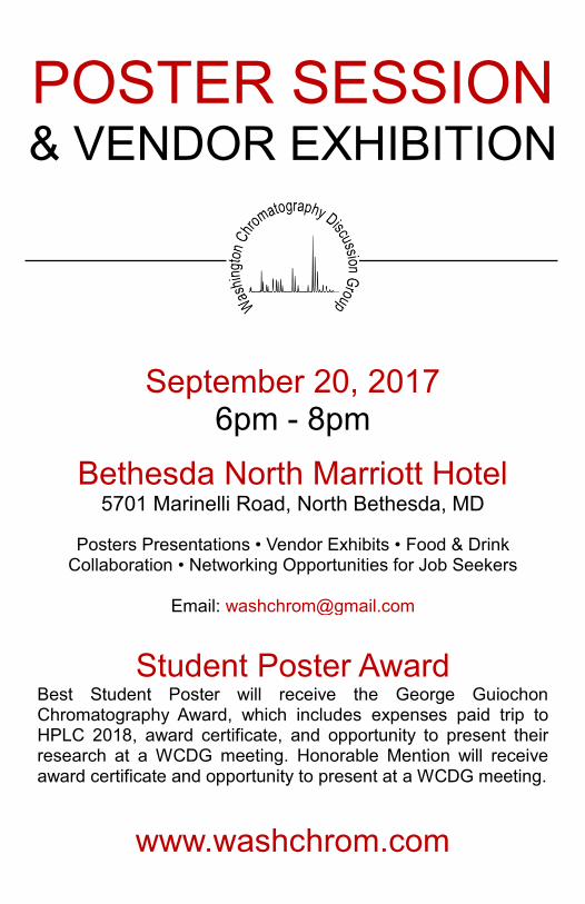
POSTER SESSION & VENDOR EXHIBITION
September 20, 2017
6pm - 8pm
Bethesda North Marriott Hotel
5701 Marinelli Road, North Bethesda, MD
Posters Presentations • Vendor Exhibits • Food & Drink Collaboration • Networking Opportunities for Job Seekers
Email: [email protected]
Student Poster Award
Best Student Poster will receive the George Guiochon Chromatography Award, which includes expenses paid trip to HPLC 2018, award certificate, and opportunity to present their research at a WCDG meeting. Honorable Mention will receive award certificate and opportunity to present at a WCDG meeting.
www.washchrom.com
1
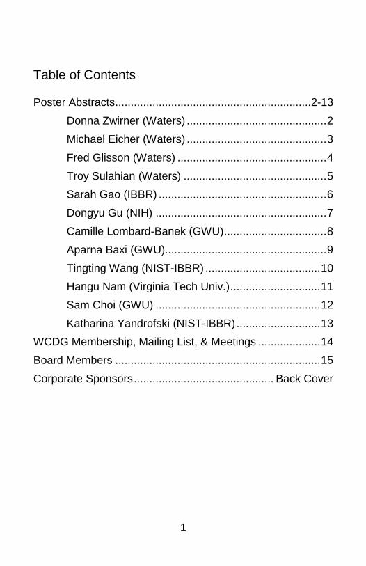
Table of Contents
Poster Abstracts ...............................................................2-13
Donna Zwirner (Waters) ............................................. 2
Michael Eicher (Waters) ............................................. 3
Fred Glisson (Waters) ................................................ 4
Troy Sulahian (Waters) .............................................. 5
Sarah Gao (IBBR) ...................................................... 6
Dongyu Gu (NIH) ....................................................... 7
Camille Lombard-Banek (GWU) ................................. 8
Aparna Baxi (GWU).................................................... 9
Tingting Wang (NIST-IBBR) ..................................... 10
Hangu Nam (Virginia Tech Univ.) ............................. 11
Sam Choi (GWU) ..................................................... 12
Katharina Yandrofski (NIST-IBBR) ........................... 13
WCDG Membership, Mailing List, & Meetings .................... 14
Board Members .................................................................. 15
Corporate Sponsors ............................................. Back Cover
2
Tracking and Reporting Synthetic Peptide Impurities with a Cost-Effective Single Quadrupole Mass Detector for Improved Confidence in Analysis Donna Zwirner, Brooke M. Koshel, Robert Birdsall, Ximo Zhang, William Alley, Jing Fang, Asish Chakraborty and Ying Qing Yu Waters, Milford, MA There has recently been a renewed interest in peptide-based drugs due in part to advancements in overcoming short half-lives and the potential for new modalities. Therapeutic peptides can be made through both recombinant and synthetic strategies, but synthetic peptides can offer manufacturers the advantage of a regulatory approval process that follows that of a small molecule. When following a synthetic approach, process-related impurities can arise from the raw materials as well as the manufacturing process, and thus can differ from batch to batch or between manufacturing sites. LC-UV-based methods are most commonly used to determine product purity, but by incorporating a single quadrupole mass detector into the analysis, method development and optimization can be streamlined. In this study, eledoisin, a vasodilator, is used as a clinically relevant sample to demonstrate how a single quadrupole mass detector can be used to streamline the method development process for synthetic peptide impurity monitoring. A general gradient method was used to screen multiple column chemistries in mobile phase containing 0.1% (v/v) trifluoroacetic acid or 0.1% (v/v) formic acid. This allows for critical attributes to be readily monitored, but can also help determine an initial set of chromatographic conditions. After selecting a column chemistry and mobile phase, a focused gradient can be used to separate additional impurities. Once a method is optimized, the added mass detection can identify co-elutions, which again is useful when a particular impurity compromises patient safety. This work demonstrates that by incorporating UPLC and orthogonal mass detection into a single workflow, there is an added level of assurance in product quality.
3
Identification of Prohibited Skin Lightening Agents in Cosmetic Products Using UHPLC with PDA and Mass Detection Michael Eicher, Marian Twohig, Paula Hong Helene Boiteux and Chris Stumpf Waters, Milford, MA Skin whitening products are used to lighten and produce a more even skin tone. The use of pharmaceutical active ingredients such as corticosteroids is prohibited in cosmetics due to the potential side effects. In the EU, hydroquinone, is prohibited in cosmetics while several other countries either ban or limit the amount allowed in cosmetic products. Despite the regulations placed on these components in cosmetics, they can still be found in formulated cosmetic products. In this study, skin lightening products were obtained from online vendors to assess their composition. The samples were extracted and analysed using UHPLC with PDA and mass detection. UHPLC separation of eight whitening agents ranging in polarity was performed at a flow-rate of 0.80 mL/min using 0.1% formic acid in water as the aqueous mobile phase and methanol as the organic modifier. A column designed to provide exceptional retention for both polar and non-polar analytes even at 100% aqueous conditions was used for the analysis. The column was maintained at a temperature of 35 0C and had dimensions of 3.0 x 100 mm and a particle size of 2.7 μm. Detection was by photodiode array (PDA) and mass detection. Several samples in this study were found to contain prohibited skin whitening agents. In some cases packaging labels omitted the presence of the skin lightening agent on the enclosed product information thus increasing the likelihood of improper long-term use and adverse side effects to consumers.
4
Development and Comparison of Orthogonal Chromatographic Techniques for the Analysis of Potential Mutagenic Impurities Fred Glisson, Jennifer Simeone, Paula Hong Waters, Milford, MA There are many steps during the manufacturing process of an active pharmaceutical ingredient (API) where impurities can be introduced, whether as reagents, byproducts, intermediates, etc. Some of these impurities may be mutagenic, or those that have the potential to interact with DNA and ultimately cause carcinogenicity. Methodologies associated with monitoring API purity levels are often HPLC-UV based. This detection technique frequently does not provide the sensitivity levels needed to detect potential mutagenic impurities at the levels required by regulatory agencies. However, the use of tandem quadrupole mass spectrometry can provide both high sensitivity and specificity for these analytical methods. Additionally, mass spectrometry is known to be suitable for use with both RPLC and SFC methodologies. To evaluate the use of mass spectrometry with both liquid and supercritical fluid chromatography for the analysis of potential mutagenic impurities, an API and five related impurities specified in the USP monograph were analyzed. Ondansetron is a pharmaceutical used in the prevention of nausea and vomiting, and contains two process impurities that are potentially mutagenic, imidazole and 2-methyl imidazole. Quantitative methods for the analysis of ondansetron and five process impurities, including the two potential mutagenic impurities, imidazole and 2-methyl imidazole, were developed using two orthogonal chromatographic methods- reversed phase liquid chromatography and supercritical fluid chromatography, yet both methods employed tandem quadrupole mass detection. Method parameters, such as limit of quantitation, linearity, and run time will be compared between the two orthogonal chromatographic methods to determine the benefits of each technique in the analysis of ondansetron and its potentially mutagenic impurities.
5
Fraction Collection for Isolating Impurities in Forced Degradation Studies Troy Sulahian, Paula Hong and Patricia R. McConville Waters, Milford, MA Forced degradation studies are typically performed to understand the degradation pathway of pharmaceuticals. Analysis is most often conducted using HPLC and UV detectors. In these studies, performing mass balance, or assessing the conservation of mass, is critical to ensuring all impurities are accounted. However, given the range of impurities and their chemical and physical properties, mass balance studies can be challenging. One of the specific challenges includes determining the response factor of impurities relative to the active pharmaceutical ingredient (API). Incorrectly identifying the relative response factors (RRFs) could lead to over or under quantification of the impurity, which can in turn lead to mass imbalance. In this presentation we will evaluate mass balance using a triple detection system consisting of a photodiode array (PDA), evaporative light scattering detector (ELSD) and a mass detector. The triple detection system provides three unique detection modes to ensure that all of the impurities are detected and quantified. These studies will include evaluation of the relative response factors of impurities and the calculation of mass balance using these experimentally obtained values. The RRFs will be evaluated initially by established methodologies, specifically comparison of the calibration curve of both the API and impurity standard. Next, in place of a standard, the impurity will be collected from the forced degradation analysis by small scale fraction collection and subsequently used to determine RRFs. Using these experimentally obtained values, mass balance of a degraded sample will be evaluated. The degradation pathway will then be confirmed through the identification of impurities and their by-products using a mass detector.
6
Structure and function analysis of four novel Klebsiella pneumoniae phage endolysins Sarah Gao, Sara B. Linden, Daniel C. Nelson Institute for Bioscience and Biotechnology Research, Rockville, MD The overuse and misuse of antibiotics has led to the selection of antibiotic-resistant bacteria, which cause infections that are very difficult to treat or cannot be cured. A recently rediscovered concept called phage therapy uses bacteriophages (phages) as an alternative to antibiotics. In particular, the sole use of bacteriophage endolysins is a promising option for the treatment of multi-drug resistant bacterial infections. Previous studies show that endolysins isolated from phages targeting Gram-positive bacteria can directly lyse the peptidoglycan cell wall of those bacteria upon contact from the outside. However, for Gram-negative bacteria that have an outer membrane, it is more difficult for endolysins to access the peptidoglycan. These bacteria are also becoming increasingly difficult to treat with available antibiotics, as they have developed various resistance mechanisms associated with their outer membranes. Thus, there is a need to study novel endolysins isolated from phages targeting Gram-negative bacteria to optimize them for use against Gram-negative pathogens. We isolated three novel phages, SopranoGao, MezzoGao, and AltoGao, from a sewage treatment plant, which target multi-drug resistant Gram-negative bacterium Klebsiella pneumoniae. We sequenced and annotated the genomes of these phages, and identified four putative endolysins within them. We purified recombinant proteins of these endolysins and analyzed their activities in various assays. The results showed that all of the endolysins actively killed multi-drug resistant K. pneumoniae, even without the presence of outer membrane permeablizers, suggesting that they can directly penetrate the outer membrane and lyse the peptidoglycan. These endolysins represent potential therapeutics for Gram-negative bacterial infections.
7
Purification of R-phycoerythrin from Gracilaria lemaneiformis using Centrifugal Precipitation Chromatography Dongyu Gu, Rodrigo Lazo-Portugal, Martha Knight , Zhantong Wang, Ying Ma, Yi Yang, Yoichiro Ito NIH, Bethesda, MD Phycoerythrin is a main light-harvesting proteins in red alga and commonly used as fluorescent label in many biochemical technique. Centrifugal Precipitation Chromatography was successfully applied for the purification of R-Phycoerythrin from the red alga Gracilaria lemaneiformis for the first time. The purified R- Phycoerythrin exhibited a typical “three peak” spectrum with absorption maximum at 497, 538, and 565 nm. The absorbance ratio A565/A280, a criterion for purity, was up to 6.5. The purified R- Phycoerythrin showed one single protein band in Native-PAGE, and SDS-PAGE demenstrated the presence of one 20 kDa subunits (α and β) and 33 kDa subunits (γ) which are consistent with the (αβ)6γ subunit composition of R- Phycoerythrin. The results indicated that Centrifugal Precipitation Chromatography is an effiecient method to obtain the high purity R-Phycoerythrin from Gracilaria lemaneiformis.
8
Single-cell Temporal Proteomic Analysis in the Developing Frog (Xenopus) Embryo Camille Lombard-Banek, Aparna Baxi, Sally A. Moody, and Peter Nemes George Washington University, Washington, DC Protein analysis at the single-cell level holds great potential to better understand how differential gene expression coordinates cell differentiation during embryonic development. Traditional mass spectrometry based techniques average across large number of cells, limiting access to important information on cell-to-cell heterogeneity. Recently, we developed a highly sensitive capillary electrophoresis (CE) high-resolution mass spectrometer (HRMS), enabling the detection of differential protein expression across individual neighboring cells of the 16-cell frog embryo. At this stage, cells have been found to form specific tissue in the developed tadpole. Here we extend this technology to measure protein content of progressively smaller cells in later stages of development by integrating in-situ microsampling with CE-ESI-HRMS. For in-situ microsampling, we used a tapered borosilicate capillary to collect ~10 nL of cytoplasm from identified cells in the frog embryo. Sampled proteins were treated using a bottom-up proteomic workflow, downscaled to the total protein content available in the collected material. After evaluating three solvent mixtures to aid digestion, we successfully identified ~350 unique protein groups (PGs) on average from microaspirated cells. This compared well to the number of protein groups identified from whole dissected cells (~250 PGs). Next, we used the approach to measure protein production in smaller cells across four developmental stages: 16-, 32, 64, and 128-cell stage embryos. We identified 470 PGs and quantified ~450, among which 175 were quantifiable across all stages and all biological replicates. These measurements demonstrated differential protein production over time when compared to the 16-cell stage. Moreover, using cluster analysis, we uncovered three characteristic temporal trends across the four developmental stages. Our microanalytical single-cell proteomics approach creates new opportunities to understand how cell specific gene translation induces cell differentiation and organ development. We anticipate this technology to be applicable to other models in cell and developmental biology.
9
High-pH Fractionation for Enhancing the Proteome Coverage in Tissues Dissected from the Early Frog (Xenopus laevis) Embryo Aparna Baxi, Camille Lombard-Banek, Sally A. Moody, and Peter Nemes George Washington University, Washington, DC Cellular reorganization events during early embryonic development are induced by gradients of signaling molecules like transcripts and proteins and guide the formation of essential embryonic features like body axes and germ layers. However, the complete suite of proteins involved in these key developmental events are not fully understood due to a lack of highly sensitive technologies. To address this knowledge gap, we characterized the production of thousands of proteins during early embryonic development using nano-flow liquid chromatography (nanoLC) and high-resolution mass spectrometry (HRMS). Here, we enhanced protein identification by bottom-up proteomics in embryonic tissues from the South African clawed frog (Xenopus laevis), a powerful model in developmental biology and health studies. We performed fractionation of peptide samples using offline high-pH reversed phase fractionation prior to low-pH nanoLC separation to identify ~1,400 different protein groups from Xenopus embryonic tissues as compared to ~900 proteins identified without peptide sample fractionation. Next, we depleted abundant yolk proteins that are known to cause interferences during mass spectrometry analysis and identified ~1,700 different proteins. The newly identified proteins covered low and medium intensity domains of the dynamic concentration range including gene products involved in essential signaling pathways during embryonic development. By complementing already existing knowledge of transcripts produced in the embryo, deep coverage of the proteome will aid our understanding of molecular events unfolding during normal vertebrate development.
10
Assessment of extracellular vesicles purity using proteomic standards Tingting Wang, Kyle W. Anderson, Illarion V. Turko Institute for Bioscience and Biotechnology Research, Rockville, MD National Institute of Standards and Technology, Gaithersburg, MD The increasing interest in extracellular vesicles (EVs) research is fueled by reports indicating their unique role in intercellular communication and potential connection to the development of common human diseases. The unique role assumes unique protein and nucleic acid cargo. Unfortunately, accurate analysis of EVs cargo faces a challenge of EVs isolation. Generally used isolation techniques do not separate different subtypes of EVs and even more, poorly separate EVs from non-EVs contaminants. Further development of EVs isolation protocols urgently needs a quantitative method of EVs purity assessment. We report here that multiple reaction monitoring (MRM) assay using 15N-labeled quantification concatamer (QconCAT) to quantify a pattern of targeted EVs and non-EVs proteins is a suitable approach to assess purity of EVs preparations. These quantifications were then used to compare purity of EVs preparations obtained from human serum by two protocols; the first one includes ultracentrifugation and size exclusion chromatography (SEC), the second one includes polymer-based precipitation techniques in-front of ultracentrifugation. The rationale for such comparison was (i) to demonstrate that MRM with QconCAT can be used for evaluation of EVs purity and (ii) to address the question whether precipitation techniques offer advantages for proteomic downstream applications.
11
Application of DoE for Development of a High Throughput Size Exclusion Chromatography Hangu Nam,1 Adrian Man,2 Sheau-Chiann Wang,3 and Sophia V. Levitskaya3 1Biochemistry, Virginia Tech University; 2Purification Process Sciences, MedImmune; 3Analytical Sciences, MedImmune Size exclusion chromatography has been the one of the most important and commonly used analytical assays for protein purity evaluation. The current platform method with TSK G3000 column can separate most of protein samples, but this platform method requires 20 minutes of runtime and is not suitable for a large number of samples. Also, there are growing demands for more efficient SEC methods for analysis of bispecific antibodies (Bis-mAb). Due to unnatural molecular structures of Bis-mAbs with additional binding sites that challenge platform analytical method. In this project, we tested short SEC columns packed with smaller particles that increase resolving capability of SEC column and allows faster analysis. Application of Design of Experiment (DoE) concept and Nexera Liquid Chromatography (LC) method scouting system from Shimadzu significantly enhanced development of an HTSEC method for a bispecific antibody product. BioRad protein mix was used to evaluate column performance and select columns for a Bis-mAb HTSEC method. Best performing columns were assessed using Bis-mAb in-process sample with low purity. The 3-parameters (flow rate, pH, and ionic strength), working window was defined for a better aggregate resolution at a fastest run time using JMP software. Based on the analysis of interactions of three method parameters, the JMP prediction model and the 5-minutes HTSEC method for Bis-mAb have been developed.
12
Ultrasensitive Bottom-up Proteomic Characterization of Mouse Hippocampal Neurons by CE-nanoESI-HRMS Microanalytical Platform Sam Choi, Marta Zamarbide, M. Chiara Manzini, and Peter Nemes George Washington University, Washington, DC Discovery proteomic characterization of neurons is informative of molecular processes involved in normal development of the nervous system. However, sensitive detection technology is needed to uncover cell-to-cell differences, specifically those capable of measuring trace amounts of proteins in single cells or small populations of neurons. Currently, high-resolution mass spectrometry (HRMS) is technology of choice to identify proteins from large population of cells, usually millions of cells. Here, we report a HRMS-based microanalytical platform that allows ultrasensitive characterization of proteins from limited populations of neurons in the mouse hippocampus. Our approach integrates a custom-built capillary electrophoresis (CE) nanoelectrospray ionization (nanoESI) interface for HRMS to achieve trace-level sensitivity. Using peptide standards, the CE-nanoESI-HRMS platform was able to achieve a ~260-zmol (156,000 copies) lower limit of detection with high separation power (~330,000 theoretical plates). Furthermore, the instrument was able to detect ~15 amol (~1 pg) of bovine serum albumin and cytochrome c in a bottom-up approach, raising sufficient sensitivity to measure proteins in small neuron populations. Finally, we utilized this platform to analyze ~500 pg of protein digest from cultured hippocampal neurons isolated from pup mouse (embryonic day 16). The platform was able identify over 1,000 different peptides corresponding to 361 nonredundant protein groups (<1 % FDR). The label-free quantitation intensities calculated for these proteins suggested a 4-log-order dynamic concentration range. Identified proteins were enriched in many genes that are classical neuron markers. Ultrasensitive characterization of proteins by CE-nanoESI-HRMS raises new potentials to investigate how differential gene expression establishes neuron heterogeneity for normal brain function.

13
Size heterogeneity of NISTmAb RM 8671 by SEC Katharina Yandrofski, Alan Heckert, Jim Fillibin, John Schiel Institute for Bioscience and Biotechnology Research, Rockville, MD National Institute of Standards and Technology, Gaithersburg, MD The NISTmAb RM 8671 IgG1κ is intended to provide a well characterized, longitudinally available test material that is expected to greatly facilitate development of originator and follow-on biologics for the foreseeable future. Aggregation is a critical metric to establishing monoclonal antibody consistency and quality due to potential immunogenicity concerns. Therefore, a monomeric purity assay was optimized to evaluate and quantify the presence of aggregates using size exclusion chromatography (SEC). A central composite design optimization was conducted, resulting in a highly robust SEC assay. The optimized SEC method was used to (I) evaluate the homogeneity and stability of RM 8671; (II) assign monomeric purity reference values, and (III) establish the appropriate storage and handling conditions for the material.

14
WCDG Membership
Interested in becoming a member of the WCDG? Membership dues are $10 per year (September-May) and
$50 for a lifetime membership. Dues can be paid at a meeting to Al Del-Grosso or online at
www.wcdgdues.ezregister.com using the “Pay Membership Dues” link on the right menu.
WCDG Mailing List
To be added to our email list, visit our website www.washchrom.com and use the “Subscribe” link on the right menu.
WCDG Meetings
WCDG holds regular meetings, generally on the third Wednesday of each month from September through May at the US Pharmacopeia in Rockville, MD (unless otherwise
noted). A dinner and social hour begins at 6:00 pm, followed by a featured speaker at 7:00 pm.
We welcome you to join our discussions!
October 18 John Hanover (NIH-NIDDK) at ACS HQ with Chemical Society of Washington
November 15 Gary Mallard (NIST) at USP
December 6 Joseph Zaia (Boston Univ.) at USP
January 17 David Muddiman (NCSU) at USP
Visit www.washchrom.com for more information.

15
WCDG Board
President Kyle Anderson [email protected]
Program Chair M. Lorna De Leoz [email protected] Secretary Ravi Ravichandran [email protected] Treasurer Alfred Del-Grosso [email protected] Board Members Lois Ann Beaver [email protected] Carolyn Burdette [email protected] Ashraf Khan [email protected] Morgan Richardson [email protected] Robert Swart [email protected] Walter Wilson [email protected]
16
WCDG Corporate Sponsors
GOLD Level Sponsor
Interested in becoming a corporate sponsor of the WCDG? We offer many levels of corporate sponsorship to fit the needs of your chromatography company. Visit our sponsorship site at www.wcdgsponsorship.ezregister.com for more information about the sponsorship levels and to join our growing corporate sponsorship program. For questions about sponsorship, contact our president, Kyle Anderson, at [email protected].
BRONZE Level Sponsors