Embed Size (px)
Citation preview

CHAPTER 4
Vectors of Gene Therapy
KATHERINE PARKER PONDER, M.D.
INTRODUCTION
Currently, gene therapy refers to the transfer of a gene that encodes a functionalprotein into a cell or the transfer of an entity that will alter the expression of anendogenous gene in a cell. The efficient transfer of the genetic material into a cellis necessary to achieve the desired therapeutic effect. For gene transfer, either amessenger ribonucleic acid (mRNA) or genetic material that codes for mRNAneeds to be transferred into the appropriate cell and expressed at sufficient levels.In most cases, a relatively large piece of genetic material (>1kb) is required thatincludes the promoter sequences that activate expression of the gene, the codingsequences that direct production of a protein, and signaling sequences that directRNA processing such as polyadenylation. A second class of gene therapy involvesaltering the expression of an endogenous gene in a cell. This can be achieved bytransferring a relatively short piece of genetic material (20 to 50bp) that is com-plementary to the mRNA. This transfer would affect gene expression by any of avariety of mechanisms through blocking translational initiation, mRNA processing,or leading to destruction of the mRNA. Alternatively, a gene that encodes antisenseRNA that is complementary to a cellular RNA can function in a similar fashion.
Facilitating the transfer of genetic information into a cell are vehicles calledvectors. Vectors can be divided into viral and nonviral delivery systems. The mostcommonly used viral vectors are derived from retrovirus, adenovirus, and adeno-associated virus (AAV). Other viral vectors that have been less extensively used arederived from herpes simplex virus 1 (HSV-1), vaccinia virus, or baculovirus. Nonvi-ral vectors can be either plasmid deoxyribonucleic acid (DNA), which is a circle ofdouble-stranded DNA that replicates in bacteria or chemicaly synthesized compounds that are or resemble oligodeoxynucleotides. Major considerations indetermining the optimal vector and delivery system are (1) the target cells and itscharacteristics, that is, the ability to be virally transduced ex vivo and reinfused tothe patient, (2) the longevity of expression required, and (3) the size of the geneticmaterial to be transferred.
77
An Introduction to Molecular Medicine and Gene Therapy. Edited by Thomas F. Kresina, PhDCopyright © 2001 by Wiley-Liss, Inc.
ISBNs: 0-471-39188-3 (Hardback); 0-471-22387-5 (Electronic)
VIRAL VECTORS USED FOR GENE THERAPY
Based on the virus life cycle, infectious virions are very efficient at transferringgenetic information. Most gene therapy experiments have used viral vectors com-prising elements of a virus that result in a replication-incompetent virus. In initialstudies, immediate or immediate early genes were deleted. These vectors couldpotentially undergo recombination to produce a wild-type virus capable of multi-ple rounds of replication. These viral vectors replaced one or more viral genes witha promoter and coding sequence of interest. Competent replicating viral vectorswere produced using packaging cells that provided deleted viral genes in trans. Forthese viruses, protein(s) normally present on the surface of the wild-type virus werealso present in the viral vector particle. Thus, the species and the cell types infectedby these viral vectors remained the same as the wild-type virus from which theywere derived. In specific cases, the tropism of the virus was modified by the surfaceexpression of a protein from another virus, thus allowing it to bind and infect othercell types. The use of a protein from another virus to alter the tropism for a viralvector is referred to as pseudotyping.
A number of viruses have been used to generate viral vectors for use in genetherapy. The characteristics of these viruses and their virulence are shown in Table4.1. Characteristics of viral vectors that have been generated from these viruses areshown in Table 4.2. Important features that distinguish the different viral vectorsinclude the size of the gene insert accepted, the duration of expression, target cellinfectivity, and integration of the vector into the genome.
RETROVIRAL VECTORS
Retroviruses are comprised of two copies of a positive single-stranded RNAgenome of 7 to 10kb. Their RNA genome is copied into double-stranded DNA,which integrates into the host cell chromosome and is stably maintained. A prop-erty that allowed for the initial isolation was the rapid induction of tumors in susceptible animals by the transfer of cellular oncogenes into cells. However, retro-viruses can also cause delayed malignancy due to insertional activation of a down-stream oncogene or inactivation of a tumor suppressor gene. Specific retroviruses,such as the human immunodeficiency virus (HIV), can cause the immune deficiencyassociated with the acquired immunodeficiency syndrome (AIDS) see Chapter 12.Retroviruses are classified into seven distinct genera based on features such as envelope nucleotide structure, nucleocapsid morphology, virion assembly mode, andnucleotide sequence.
Retroviruses are ~100nm in diameter and contain a membrane envelope. Theenvelope contains a virus-encoded glycoprotein that specifies the host range ortypes of cells that can be infected by binding to a cellular receptor. The envelopeprotein promotes fusion with a cellular membrane on either the cell surface or inan endosomal compartment. The ecotropic Moloney murine leukemia virus (MLV)receptor is a basic amino acid transporter that is present on murine cells but notcells from other species. The amphotropic MLV receptor is a phosphate transporterthat is present on most cell types from a variety of species including human cells.There are co-HIV receptors, CD4, and a chemokine receptor. After binding to the
78 VECTORS OF GENE THERAPY

RETROVIRAL VECTORS 79
cellular receptor, the viral RNA enters the cytoplasm and is copied into double-stranded DNA via reverse transcriptase (RT) contained within the virion. Thedouble-stranded DNA is transferred to the nucleus, where it integrates into the hostcell genome by a mechanism involving the virus-encoded enzyme integrase. Thisactivity is specific for each retrovirus. For MLV, infection is only productive in divid-ing cells, as transfer of the DNA to the nucleus only occurs during breakdown ofthe nuclear membrane during mitosis. For HIV, infection can occur in nondividingcells, as the matrix protein and the vpr-encoded protein have nuclear localizationsignals that allow transfer of the DNA into the nucleus to occur.
Moloney Murine Leukemia Virus: MLV Proteins
Retroviral proteins are important in the manipulation of the system to develop avector. MLV is a relatively simple virus with four viral genes: gag, pro, pol, and env(Fig. 4.1). The gag gene encodes the group specific antigens that make up the viralcore. The Gag precursor is cleaved into four polypeptides (10, 12, 15, and 30kD) bythe retroviral protease (PR). The 15-kD matrix protein associates closely with themembrane and is essential for budding of the viral particle from the membrane.The12-kD phosphoprotein (pp12) is of unresolved function. The 30-kD capsid protein
TABLE 4.1 Characteristics of Viruses That Have Been Used to Generate Viral Vectors
Virus Size and Type Viral Proteins Physical Disease in Animalsof genome Properties
Retrovirus 7–10kb of Gag, Pro, Pol, 100nm Rapid or slowsingle- Env diameter; induction of stranded RNA enveloped tumors; acquired
immunodeficiencysyndrome (AIDS)
Adenovirus 36-kb double- Over 25 70–100nm in Cold; conjunctivitis;stranded proteins diameter; gastroenteritislinear DNA nonenveloped
Adenovirus- 4.7-kb single- Rep and Cap 18–26nm in No known diseaseassociated stranded diameter;virus linear DNA nonenveloped
Herpes 152kb of Over 81 110nm in Mouth ulcers andsimplex virus double- proteins diameter genital warts;1 (HSV-1) stranded encephalitis
linear DNA
Vaccinia 190kb of Over 198 350 by Attenuated virus virus double- open reading 270nm that was used to
stranded frames rectangles; vaccinate againstlinear DNA enveloped smallpox
Baculovirus 130kb of Over 60 270 by 45 nm None in mammals;double- proteins rectangles; insect pathogenstranded envelopedcircular DNA

80 VECTORS OF GENE THERAPY
forms the virion core while the 10-kD nucleocapsid protein binds to the RNAgenome in a viral particle.The PR and polymerase (Pol) proteins are produced froma Gag/Pro/Pol precursor. This precursor is only 5% as abundant as the Gag pre-cursor and is produced by translational read-through of the gag termination codon.The number of infectious particles produced by a cell decreases dramatically if PRand Pol are as abundant as the Gag-derived proteins. PR cleaves a Gag/Pro/Pol precursor into the active polypeptides, although it is unclear how the first PR getsreleased from the precursor. The pol gene product is cleaved into 2 proteins, theamino terminal 80-kD reverse transcriptase (RT) and the carboxy terminal 46-kDintegrase (IN). The RT has both reverse transcriptase activity (which functions in RNA- or DNA-directed DNA polymerization) and RNase H activity (whichdegrades the RNA component of an RNA:DNA hybrid). The IN protein binds todouble-stranded DNA at the viral att sites located at the ends of each long termi-nal repeat and mediates integration into the host cell chromosome.
The env gene is translated from a subgenomic RNA that is generated by splic-ing between the 5¢ splice site in the 5¢ untranslated region and the 3¢ splice sitepresent just upstream of the env coding sequence. The env precursor is processed
TABLE 4.2 Summary of Relative Advantages and Disadvantages of Vectors Used forGene Therapy
Vector Infects Maximum Stability of TiterNondividing Size of Expression
Cells? Insert
Retroviral No £8kb Stable (random 1 ¥ 106 cfu/mlvectors (yes for DNA insertion) unconcentrated;
lentiviral 1 ¥ 108 cfu/mlvectors) concentrated
Adenovirus Yes 8kb for Expression lost in 1 ¥ 1012 pfu/mlE1/E3 3–4 weeks in normaldeleted animals; expressionvectors; can last weeks to 35kb for months with“gutless” immunosuppression.vectors No integration
Adenoassociated Yes <4.5kb Stable; it is unclear 1 ¥ 106 infectiousvirus (AAV) if DNA integrates particles/ml
in vivo unconcentrated;1 ¥ 1010 infectiousparticles/mlconcentrated
Herpes simplex Yes >25kb Stable; maintained 1 ¥ 1010 pfu/mlvirus (HSV)-1 as episome
Vaccinia Yes >25kb Expression transient 1 ¥ 108 pfu/mldue to an immune response; replicates in cytoplasm
Baculovirus Yes >20kb Unstable 1 ¥ 1010 pfu/ml

RETROVIRAL VECTORS 81
into 3 proteins: SU, transmembrane (TM; or p15E), and p2. The 70-kD SU proteinbinds to a cell surface receptor. Neutralizing antibodies directed against SU canblock infection. The 15-kD TM plays a role in fusion of the virus and cellular mem-brane. In many retroviruses, the association between the SU and TM proteins israther tenuous and SU is rapidly lost from virions. This contributes to poor infec-tivity of viral preparations and instability to manipulations such as concentration byultracentrifugation. Envelope proteins from different retroviruses, or even fromviruses of other families, can be used to produce infectious particles with alteredtropism and/or greater stability.
Sequences Required in cis for Replication and Packaging
The term provirus refers to the form of the virus that is integrated as double-stranded DNA into the host cell chromosome. Genetic sequences are needed in cisto develop a provirus that can transfer genetic information into a target cell. Fourimportant sequences are required in cis for replication and infection in the contextof gene therapy. They are (1) the long terminal repeats (LTRs), (2) the primerbinding site (PBS), (3) the polypurine (PP) tract, and (4) the packaging sequence.These sequences and their function are shown in Figure 4.2. LTRs are approximately600 nucleotide sequences present at both the 5¢ and the 3¢ end of the provirus. Theyinitiate transcription at the 5¢ end, perform polyadenylation at the 3¢ end, and inte-grate a precise viral genome into a random site of the host cell chromosome byvirtue of the att sites at either end. The LTR-initiated transcripts serve as an mRNAfor the production of viral proteins and as the RNA genome for producing addi-tional virus. The PBS is located just downstream of the 5¢ LTR. It binds to a cellu-lar transfer RNA (tRNA), which serves as a primer for the polymerization of thefirst DNA strand. The PP tract contains at least nine purine nucleotides and islocated upstream of the U3 region in the 3¢ LTR. The RNA within this sequence isresistant to degradation by RNase H when hybridized with the first DNA strand.
FIGURE 4.1 Diagram of a Moloney murine leukemia retrovirus (MLV). The proviral formwith two complete long terminal repeats (LTRs) and the genomic RNA that is expressedfrom the provirus are shown at the top. The genomic RNA can be translated to produce theGag gene products, or produce a Gag/Pro/Pol precursor by reading through the translationalstop codon at the 3¢ end of the Gag gene. The genomic RNA can also be spliced to generatea smaller subgenomic RNA, which is translated into the Env protein. The regions that aretranslated are shown as black boxes, while the untranslated regions of the RNA appear as ablack line.

FIGURE 4.2 Mechanism ofreverse transcription andintegration of the genomicRNA into the host cell chro-mosome. (a) Genomic RNAwith a tRNA primer. Thegenomic RNA has a 60-nt Rregion (for redundant) atboth the 5¢ and the 3¢ end.The5¢ end has the 75-nt U5 region(for unique to 5¢ end) and the3¢ end has the 500-nt U3region (for unique to 3¢ end).The PBS of the genomicRNA (shown in black)hybridizes to the terminal 18nt at the 3¢ end of a tRNA. (b)Reverse transcription of the5¢ end of the genomic RNA.The tRNA primer enables theRT to copy the 5¢ end of thegenomic RNA, to generate aportion of the first DNAstrand. (c) Degradation of theRNA portion of an RNA :DNA hybrid by RNase H.RNase H degrades the RNAportion that was used as atemplate for synthesis of thefirst DNA strand. Althoughshown as a separate step here,this occurs ~18 nt down-stream of where polymeriza-tion is occurring. (d) Firststrand transfer. The portionof the first strand that repre-
sents the R region hybridizes with the R region in the 3¢ end of the genomic RNA. (e) Reversetranscription of the remainder of the genomic RNA. The RT copies the genomic RNA up tothe PBS. As elongation occurs, RNase H continues to degrade the RNA portion of the RNA:DNA hybrid. The RNA in the PP tract (shown in black) is resistant to cleavage by RNaseH and remains associated with the first DNA strand. ( f) Initiation of second strand synthe-sis. The primer at the PP tract initiates polymerization of the second strand. Polymerizationup to the 3¢ end of the PBS continues. Additional sequences in the tRNA are not copied, asthe 19th nucleotide is blocked by a methyl group in the base pairing region of the tRNA. (g)RNase H digestion of the tRNA. The RNase H degrades the tRNA, which is present in anRNA :DNA hybrid. (h) Second strand transfer. The second DNA strand hybridizes to thefirst DNA strand in the PBS region. (i) Completion of the first and second strands. RT copiesthe remainder of the first and the second DNA strands, to generate a double-stranded linearDNA with intact LTRs at both the 5¢ and the 3¢ end. The integrase binds to the att sequenceat the 5¢ end of the 5¢ LTR and at the 3¢ end of the 3¢ LTR (not shown) and mediates inte-gration into the host cell chromosome. Upon integration, the viral DNA is usually shortenedby two bases at each end, while 4 to 6 nt of cellular DNA is duplicated. Although integrationis a highly specific process for viral sequences, integration into the host chromosome appearsto be random.
tRNA
tRNA
tRNA
tRNA
tRNA
3�
3�3�
3� 3�
3�
3�
5�
5�
5�
3�
3�5�
5�
R U5 PBS Gag PR Pol Env PP U3 R
R U5 PBS Gag PR Pol Env PP U3 R
PBS
Env PP U3 R
Env PP U3 R
PBS
PBS
Gag PR Pol
Pol PR Gag
Pol PR Gag
Env PP U3 R
A.
B.
C.
D.
E.
(a)
(b)
(c)
(d)
(e)
Env PP U3 R U5
Env
Env
PP
PP
U3
U3U3
R
RR
U5
U5U5
PBS
PBS
PBS
LTR LTR
tRNA
PBS
PBS
PBS
3�
3� 3�
5�
5�5�
Env PP U3 R U5 PBS3�
3�
3�3�
5�
5�5�
5�
Pol PR Gag
Pol
Pol
PR
PR
Gag
Gag
F.
G.
H.
I.
( f )
(g)
(h)
(i)

The PP tract therefore serves as the primer for synthesis of the second DNA strand.The packaging signal binds to the nucleocapsid protein of a retroviral particle allow-ing the genomic RNA to be selectively packaged. Although the encapsidationsequence was initially mapped to the region of the virus between the 5¢ LTR andthe gag gene, vectors that only contained this sequence were packaged inefficiently,resulting in low titers of viral vector produced. Subsequent studies demonstratedthat inclusion of some gag sequences (the extended packaging signal) greatlyincreased the titer of the vector produced. Most vectors that are currently in useutilize the extended packaging signal.
Use of Retroviral Sequences for Gene Transfer
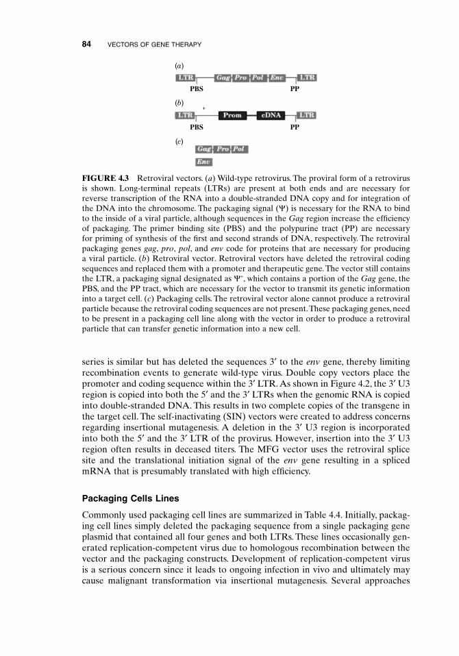
All of the genomic sequences that are necessary in cis for transcription and pack-aging of RNA, for reverse transcription of the RNA into DNA and for integrationof the DNA into the host cell chromosome need to be present in the retroviralvector. It is, however, possible to remove the coding sequences from the retroviralgenome and replace them with a therapeutic gene to create a retroviral vector. Thedeletion of viral coding sequences from the retroviral vector makes it necessary toexpress these genes in trans in a packaging cell line. Packaging cell lines that sta-billy express the gag, pro, pol, and env genes have been generated. The transfer ofa plasmid encoding the retroviral vector sequence into packaging cell results in aretroviral particle capable of transferring genetic information into a cell (assumingappropriate tropism). However, upon transfer of the retroviral vector into a cell,infectious particles are not produced because the packaging genes necessary for syn-thesizing the viral proteins are not present. These vectors are therefore referred toas replication incompetent. Figure 4.3 diagrams how retroviral vectors and packag-ing cells are generated.
Commonly used retroviral vectors and their salient features are summarized inTable 4.3. Plasmid constructs that resemble the provirus and contain a bacterialorigin of replication (see Chapter 1) outside of the LTRs can be propagated in bacteria. The therapeutic gene is cloned into a vector using standard molecularbiology techniques. Upon transfection into mammalian cells, the 5¢ LTR of thevector DNA initiates transcription of an RNA that can be packed into a viral par-ticle. Although a packaging cell line can be directly transfected with plasmid DNA,the integrated concatemers are unstable and are often deleted during large-scalepreparation of vector. To circumvent this problem, most cell lines used in animalsare infected with the vector rather than transfected. This involves transfection intoone packaging cell line, which produces a vector that can infect a packaging cell linewith a different envelope gene. The infected packaging cell line generally containsa few copies of the retroviral vector integrated into different sites as a provirus.
Most vectors have genomic RNAs that are less than 10 kb, to allow for efficientpackaging. N2 was the first vector using an extended packaging signal that, as notedearlier, greatly increased the titer of vector produced. In LNL6, the AUG at thetranslational initiation site was mutated to UAG, which does not support transla-tional initiation.This mutation prevents potentially immunogenic gag peptides frombeing expressed on the surface of a transduced cell. In addition, it decreases the pos-sibility that a recombination event would result in replication-competent virus sincethe recombinant mutant would not translate the gag gene into a protein. The LN
RETROVIRAL VECTORS 83
84 VECTORS OF GENE THERAPY
series is similar but has deleted the sequences 3¢ to the env gene, thereby limitingrecombination events to generate wild-type virus. Double copy vectors place thepromoter and coding sequence within the 3¢ LTR. As shown in Figure 4.2, the 3¢ U3region is copied into both the 5¢ and the 3¢ LTRs when the genomic RNA is copiedinto double-stranded DNA. This results in two complete copies of the transgene inthe target cell. The self-inactivating (SIN) vectors were created to address concernsregarding insertional mutagenesis. A deletion in the 3¢ U3 region is incorporatedinto both the 5¢ and the 3¢ LTR of the provirus. However, insertion into the 3¢ U3region often results in deceased titers. The MFG vector uses the retroviral splicesite and the translational initiation signal of the env gene resulting in a splicedmRNA that is presumably translated with high efficiency.
Packaging Cells Lines
Commonly used packaging cell lines are summarized in Table 4.4. Initially, packag-ing cell lines simply deleted the packaging sequence from a single packaging geneplasmid that contained all four genes and both LTRs. These lines occasionally gen-erated replication-competent virus due to homologous recombination between thevector and the packaging constructs. Development of replication-competent virusis a serious concern since it leads to ongoing infection in vivo and ultimately maycause malignant transformation via insertional mutagenesis. Several approaches
+
PP
PPPBS
PBS
(a)
(b)
(c)
FIGURE 4.3 Retroviral vectors. (a) Wild-type retrovirus. The proviral form of a retrovirusis shown. Long-terminal repeats (LTRs) are present at both ends and are necessary forreverse transcription of the RNA into a double-stranded DNA copy and for integration ofthe DNA into the chromosome. The packaging signal (Y) is necessary for the RNA to bindto the inside of a viral particle, although sequences in the Gag region increase the efficiencyof packaging. The primer binding site (PBS) and the polypurine tract (PP) are necessary for priming of synthesis of the first and second strands of DNA, respectively. The retroviralpackaging genes gag, pro, pol, and env code for proteins that are necessary for producing a viral particle. (b) Retroviral vector. Retroviral vectors have deleted the retroviral codingsequences and replaced them with a promoter and therapeutic gene. The vector still containsthe LTR, a packaging signal designated as Y+, which contains a portion of the Gag gene, thePBS, and the PP tract, which are necessary for the vector to transmit its genetic informationinto a target cell. (c) Packaging cells. The retroviral vector alone cannot produce a retroviralparticle because the retroviral coding sequences are not present.These packaging genes, needto be present in a packaging cell line along with the vector in order to produce a retroviralparticle that can transfer genetic information into a new cell.

RETROVIRAL VECTORS 85
have been taken to reduce the generation of replication-competent virus. One strat-egy is to separate the packaging genes into two plasmids integrated into differentchromosomal locations. Examples of this approach include the GP + E86, GP +envAM12, Y-CRIP, and Y-CRE packaging cell lines. For these cell lines, thegag/pro/pol genes are expressed from one piece of DNA while the env gene isexpressed from a second piece of DNA. Then each DNA piece is introduced intothe cell independently. Another strategy is to minimize homology between thevector and packaging sequences. Some packaging systems use transient transfectionto produce high titers of retroviral vector for a relatively short period of time foruse in animal experimentation.
Recently developed packaging cell lines are of human origin and are advanta-geous. The presence of human antibodies in human serum results in rapid lysis ofretroviral vectors packaged in murine cell lines. The antibodies are directed againstthe a-galactosyl carbohydrate moiety present on the glycoproteins of murine butnot human cells. This murine carbohydrate moiety is absent from retroviral vectorsthat are produced by human cells, which lack the enzyme a1-3-galactosyl transferase.Human or primate-derived packaging cell lines will likely be necessary to produceretroviral vectors for in vivo administration to humans. To this point, the produc-
TABLE 4.3 Summary of Retroviral Vectors Used for Gene Therapy in Animals or Humans
Name Salient Features
N2 Contains an intact 5¢ and 3¢ LTR, an extended packaging signal with 418nt of coding sequence of the gag gene, and an intact translational startcodon (AUG) of the gag gene. Can recombine to generate wild-type virus.
LNL6 Contains intact 5¢ and 3¢ LTRs, an extended packaging signal with 418 ntof coding sequence of the gag gene, a mutation in the translational start codon (AUG) of the gag gene to the inactive UAG, and the 3¢ portion ofthe env gene.
LN series Similar to LNL6 except all env sequences are deleted to decrease the chance of recombination with the packaging genes. This series includes LNSX, LNCX, and LXSN, where L stands for LTR promoter, N for neomycin resistance gene, S for SV40 promoter, C for CMV promoter,and X for polylinker sequences for insertion of a therapeutic gene.
Double copy Places the promoter and the therapeutic gene in the U3 region of the 3¢LTR. This results in two copies of the therapeutic gene within the 5¢ and 3¢ LTRs after transduction.
Self- Deletes the enhancer and part of the promoter from the U3 region of theinactivating 3¢ LTR. This deletion is present in both the 5¢ and the 3¢ LTRs after(SIN) transduction. This decreases the chance of transcriptional activation of a
downstream oncogene after transduction of a cell.
MFG Contains an intact 5¢ and 3¢ LTR, an extended packaging signal with anintact 5¢ splice site, a 380-nt sequence with the 3¢ end of the pol gene andthe 3¢ splice site, and 100nt of the 3¢ end of the env gene. The therapeuticgene is translated from a spliced RNA and uses the env gene translationalstart site.

86 VECTORS OF GENE THERAPY
tion of retroviral vectors for clinical use is simple but not without challenges. A suitable stable packaging cell line containing both the packaging genes and thevector sequences is prepared and tested for the presence of infectious agents andreplication-competent virus. This packaging cell line can then be amplified and used to produce large amounts of vector in tissue culture. Most retroviral vectorswill produce ~1 ¥ 105 to 1 ¥ 106 colony forming units (cfu)/ml, although unconcen-trated titers as high as 1 ¥ 107 cfu/ml have been reported. The original vector prepa-ration can be concentrated by a variety of techniques including centrifugation andultrafiltration. Vectors with retroviral envelope proteins are less stable to these con-centration procedures than are pseudotyped vectors with envelope proteins fromother viruses. The preparations can be frozen until use with some loss of titer onthawing.
TABLE 4.4 Summary of Retroviral Packaging Cell Lines Used for Animal and Human Studies
Line Plasmids That Contain Packaging Envelope Detection ofGenes Protein Wild-Type
Virus?
Y-2, Y-Am, All contain a 5¢ LTR, a deletion in Variable Yesand PA12 the packaging signal, the gag, pro,
pol, and env genes, and the 3¢ LTR.
PA317 The 5¢ LTR has a deletion 5¢ to PA317: Some PE501 the enhancers, the Y sequence is amphotropic; detected with
deleted, gag, pro, pol, and env PE501: N2; none withgenes are present on one plasmid ecotropic LN-basedwith intact splice signals, the PBS vectorsis deleted, and the 3¢ LTR is replaced with the SV40 poly A site.
Y-CRE One plasmid contains a 5¢ LTR, Y-CRE: Not reportedY-CRIP has a deletion of Y, expression of ecotropic;
gag-pro-pol from a construct that Y-CRIP:also contains an inactive env gene, amphotropicand has an SV40 polyadenylation site. The second plasmid has a 5¢LTR, deletion of Y, expression of env from a construct that also contains inactive gag, pro, and polgenes, and an SV40 polyadenylation site.
GP + E-86 One plasmid has an intact 5¢ LTR, GP + E-86: Reported butGP + envAM the 5¢ splice site, a deletion in the ecotropic; not verified12 packaging signal Y, the gag-pro- GP + envAM12:
pol gene with a small amount of amphotropicthe env gene, and the SV40 polyadenylation site. A second plasmid has an intact 5¢ LTR, the 5¢ splice site, the 3¢ splice site, and the env gene.

Use of Retroviral Vectors for Gene Therapy
Retroviral vectors have been extensively used in animals and substantially used inhumans to determine the efficacy of gene therapy. They are the major vector thathas been used for ex vivo gene therapy. Cells that have been modified ex vivo witha retroviral vector include hematopoietic stem cells, lymphocytes, hepatocytes,fibroblasts, keratinocytes, myoblasts, endothelial cells, and smooth muscle cells.Retroviral vectors have also been used for in vivo delivery. For many organs, therequirement of cellular replication for transduction poses a problem since termi-nally differentiated cells in organs are not proliferative.Thus, retroviral organ-basedgene therapy approaches necessitate the induction of cell replication for in vivotransfer into cell types such as hepatocytes, endothelial cells, or smooth muscle cells.Alternatively, the use of viral vectors that do not require cellular replication couldbe used to transfer genes into nondividing cells in vivo. Studies using HIV have been initiated since that virus does not require replicating cells for transduction.Retroviral vectors have been directly injected into malignant cells in various locations, as malignant cells are highly proliferative. Efficient in vivo delivery willlikely require human or primate-derived packaging cell lines or pseudotyping toprevent complement-mediated lysis in all clinical applications of retroviral genetherapy.
After transfer into a replicating cell, the expression of the retroviral vector is crit-ical to achieve a therapeutic effect. In the application of retroviral vectors for genetherapy, the relatively low levels of gene expression achieved in animals are prob-lematic. For currently selected genes used for gene therapy, the level of expressionof the gene product does not need to be tightly regulated for clinical effectiveness.However, for diseases such as diabetes mellitus or thalassemia, the level of expres-sion of insulin or b-globin, respectively, requires precise control.Thus, a specific clin-ical condition may not only require a threshold level for therapeutic effectivenessbut may also require a narrow window of concentration for physiological effect.There is a paucity of quantitative data in animals regarding the levels of expressionper copy from different vectors, particularly in the context of organ-specific geneexpression. This is a major challenge for the field of gene therapy. The difficulties inthis area are many. First, current delivery systems make the experimental determi-nation of surviving transduced cells in situ difficult. Accurate determation of thecopy number present in vivo is necessary since overall protein expression is a func-tion of both the number of transduced cells and the gene expression per cell. Second,direct comparison of expression levels of different proteins cannot be determinedfor current delivery systems because of the marked differences in mRNA half-life,protein translation, and protein half-life for different genes.Third, the genomic inte-gration site can dramatically influence the expression level. For delivery systems that modify a small number of stem cells, such as in bone marrow stem-cell-directedgene therapy (see Chapter 7), considerable variation in expression occurs based onanimal species. This variation makes it essential to quantitate expression in a largenumber of animals and report the average results. Thus, an improved understand-ing of the regulatory controls of gene expression from retroviral vectors remainsessential for the clinical application of gene therapy in humans. Unfortunately,expression of vectors in differentiated cell types in vitro does not accurately predictexpression levels that can be achieved in vivo. In vitro screening for expression
RETROVIRAL VECTORS 87

levels provides only limited information on different retroviral vector systems in thecontext of human application.
An important genetic sequence or element in the gene expression from a retro-viral vector is the LTR. The in vivo transcriptional activity of the LTR in bone-marrow-derived cells, liver, and muscle often attenuates over the first few weeksafter transfer. However, long-term expression in some cases has been achieved. Theattenuation of the LTR reflects the absence of transcription factors that are essen-tial for expression of the LTR promoter in nondividing cells, the presence ofinhibitory proteins that shut off the LTR, methylation of the LTR, or deacetylationof the associated histones. Retroviral sequences from the U3 region and the PBScan inhibit expression of the LTR in embryonic carcinoma cells by binding to pro-teins that inhibit transcription. These inhibitory sequences may contribute to thepoor expression observed from the LTR in vivo. Retroviral vectors that alter theseinhibitory sequences are expressed in vitro in embryonic carcinoma cells and mayalso be expressed in vivo. Methylation of the LTR is associated with loss of pro-moter activity. It is unclear, however, whether methylation per se is responsible for inactivation of the promoter or if methylation is a by-product of binding to thepromoter.
Retroviral vectors can include an internal promoter located immediatelyupstream of the therapeutic gene. These “internal promoters” can be viral promot-ers, housekeeping promoters, or organ-specific promoters. Viral promoters werecomponents of many first-generation vectors because they are active in most celltypes in vitro. However, many of the viral promoters, such as the cytomegalovirus(CMV) promoter, are attenuated or completely shut-off in vivo in organs such asthe liver. This loss of function could reflect the absence of transcription factors thatare essential for expression of the promoter or the presence of inhibitory proteinsthat terminate viral promoter activity in nonreplicating cells. Internal promotersmay also comprise the ubiquitously expressed housekeeping promoters that directthe expression of proteins required by all cells. However, housekeeping genes areoften expressed at relatively low levels, and their promoters have been shown to berelatively weak in vitro and in vivo in retroviral vectors constructs. Alternatively,organ-specific promoters have two major advantages: (1) allowing limited expres-sion to specific cell types or tissues and (2) directing high levels of gene expression.Muscle- or liver-specific enhancers and/or promoters, in comparison to housekeep-ing or viral promoters, direct higher levels of expression in vivo. Gene expression,in these studies, has been stable for over one year. In other studies, however,organ-specific promoters have been inactivated in vivo in transgenic mice or in aretroviral vector by the presence of adjacent retroviral sequences. These inhibi-tory sequences play a role in attenuation of the LTR promoter. It is also possiblethat these inhibitory sequences can decrease expression from adjacent internal promoters.
The control of gene expression in vivo may be an appropriate mechanism todecrease variability in expression as well as decrease the chance that the therapeu-tic gene is overexpressed. In clinical situations, variability or overexpression wouldhave adverse therapeutic effects. Inducible expression systems have been developedto tightly regulate expression from a retroviral vector through responsivness to anorally administered drug. A tetracycline-responsive system can modify expression>200-fold from a retroviral vector in muscle cells in the presence of a drug when
88 VECTORS OF GENE THERAPY

compared to the absence of a drug in vivo. However, this system requires the all-important introduction of a drug-responsive transcription factor. This is an addi-tional burden to the individual cell, which needs to receive and express two separategenes.
Other factors, in addition to the choice of the promoter, can influence geneexpression from a retroviral vector. For some genes and through an unknown mech-anism, the presence of a splice site dramatically increases the level of expression ofthe protein. Inclusion of genomic splice sites from the therapeutic gene is techni-cally difficult. An intron would be efficiently removed from the RNA genome if thegene were inserted in the forward orientation. However, the gene can sometimesbe packaged in the backwards orientation. In this case the mRNA for the thera-peutic gene is transcribed from the opposite strand and these constructs are oftenunstable. Some retroviral vectors such as the MFG vector have used the retroviralsplice signals that direct partial splicing of the genomic retroviral RNA.
Co-expression of two genes has many potential advantages. Through the use ofa selectable marker gene and a therapeutic gene, it is possible to eliminate cells notexpressing the therapeutic gene by either in vitro or in vivo selection methods. Manyfirst-generation vector constructs express one gene from the LTR promoter and asecond gene from an internal promoter. Using these vectors, however, cells selectedby virtue of expression of one gene product have a lower level of expression of thesecond gene product. This observation was due to the phenomenon of promoterinterference. An improved approach that obtains co-expression of two genes uti-lizes a bicistronic mRNA with an internal ribosome entry site (IRES). This enablesthe downstream gene to be translated in a Cap-independent fashion.
Risks of Retroviral Vectors
There are two major concerns in the use of retroviral vectors for gene therapy inhumans: (1) insertional mutagenesis and (2) generation of wild-type virus. Inser-tional mutagenesis occurs when a retroviral vector inserts within or adjacent to acellular gene. This insertion could result in the development of malignancy throughthe inactivation of a tumor suppressor gene or by activation of a proto-oncogene.The risk of developing a malignancy through the process of receiving a single copyof a retroviral vector appears to be minimal. The induction of malignancy has notbeen observed in animals receiving replication-incompetent retroviral vectors. Thisobserved low incidence of mutagenesis indicates that the retroviral vector is unlikelyto integrate into a genomic site that will modify cellular growth properties such ascyclins- or cyclin-dependent kinases (see Chapter 10). However, if the vector insertsinto a growth-sensitive site, this would represent only the first step in a multistepprocess. Thus, procedures that introduce multiple retroviral vector integrations intoa single cell will only increase the risk of the development of malignancy. A secondsafety concern regarding retroviral vectors in human use is viral recombination.Viral recombination may result in the development of replication-competent virus.This event can clearly result in the slow onset of malignancy in animals. Tech-nical refinements in vector development have lowered the risk of generating a replication-competent virus. These include elimination of homology between thepackaging genes and the vector as well as separation of the packaging genes into two or more separate pieces of DNA. However, if recombination occurs,
RETROVIRAL VECTORS 89

the extensive testing performed prior to administration of vectors to humans is an added safety measure that identifies recombinant(s). Thus, it is unlikely that replication-competent virus will be administered to humans when the appropri-ate safety controls are observed. It remains possible, however, that a replication-incompetent retroviral vector could recombine with endogenous viruses in vivo.Endogenous viruses are present in vivo and recombination in the human genomecan generate additional pathogenic replication-competent virus(es).The occurrencecan only be determined by monitoring individual gene therapy recipients for theappearance of replication-competent virus.
Summary: Retroviral Vectors
Replication-incompetent retroviral vectors can be easily generated by deletingretroviral genes and adding gene(s) of interest. Vectors can be produced in pack-aging cell lines that express packaging genes. The major advantage of retroviralvectors is the precise integration into a random site in the host cell chromosome.This can result in long-term survival of the gene in the transduced cell. The majordisadvantage is the need to transduce dividing cells. This characteristic poses diffi-culties for the in vivo delivery to quiescent cells. Gene expression at therapeuticlevels has been achieved from a retroviral vector in vivo in some studies for overone year, but expression has been problematic in other studies.
Lentiviral Vectors
The lentiviruses are a family of retroviruses comprising seven subgenera with spe-cific biological properties. One such property is an advantage for its use in genetherapy, that is, the ability to transduce nondividing cells. The matrix protein andthe vpr gene product of the lentivirus contain nuclear localization signals that allowthe DNA to be transported to the nucleus without breakdown of the nuclear mem-brane. These gene products facilitate the infection of nondividing cells. Lentivirusescontain a number of proteins exclusive of the MLV genome (see also Chapter 11).The tat gene encodes a protein that stimulates expression via the tat responseelement (TAR) located in the HIV LTR. The rev gene encodes a protein that bindsto the rev response element (RRE) and facilitates the transfer of unspliced RNAsto the cytoplasm.The nef gene encodes a protein that is localized to the inner surfaceof the cell membrane and can decrease the amount of the HIV cell surface recep-tors, such as CD4. The nef gene protein is important for virulence in vivo throughas yet undefined mechanisms. The function of the vif gene is unclear. The productof the vpu gene appears to play a role in processing of the env gene product and inthe efficient budding and release of virions.The vpr gene product contains a nuclearlocalization signal and may play a role in transporting HIV to the nucleus of nondividing cells. The role of the vpx gene product is unclear.
Several replication-defective HIV-based vectors and packaging system has beenused to deliver genes to nondividing neurons, muscle, lung, endothelial cells, hemato-pioetic stem cells, and liver cells in vivo. One HIV packaging system contains avector with the HIV LTRs at either end (including the TAR), an extended packag-ing signal, the RRE, and a reporter gene whose expression was directed by the CMVpromoter. The packaging construct deleted the packaging signal and mutated the
90 VECTORS OF GENE THERAPY

env gene. The VSV-G envelope was expressed from a third construct. The super-natant of cells that were transfected simultaneously with all three plasmids con-tained retroviral particles that infected nondividing cells in vitro and in vivo. Morerecently, all of the accessory genes except for tat and rev have been mutated in thepackaging construct, and the particles still transduced nondividing cells at the siteof injection allowing for multiple exposures. Also, a new series of lentiviral vectorsbased on HIV-1 have been developed as a self-inactivating vector. Here, the U3 region of the 5¢ LTR was replaced by the CMV promotor, resulting in tat-independent transcription. The self-inactivating vector was constructed by deleting133bp in the U3 region of the 3¢ LTR including the TATA box and the binding sitesfor specific transcription factors. This deletion is transferred to the 5¢ LTR afterreverse transcription and integration into the genome of infected cells resulting intranscriptional inactivation of the LTR of the provirus. Such a self-inactivating virustransfected brain cells at a comparable level to wild-type virus.
Transduction of nondividing cells is a major advance for retroviral vector tech-nology. Furthermore, lentivirus vectors pseudotyped with vesticular stomatitis virusG glycoprotein can transduce a wide range of nondividing cells. In addition, noinflammation is observed at the site of injection allowing for multiple exposures. Itis possible that the multiple added properities of nonvirulent HIV-based vectors as described above will revolutionize human gene therapy procedures for non-replicating cells in vivo. Three major concerns regarding these vectors remain,however. The first is the absolute assurence that recombination to generate wild-type HIV that causes immunodeficiency syndrome in a patient will not occur. Manyof the HIV accessory genes can be mutated to prevent production of a functionalprotein. But, the complicated nature of the HIV genome and the high mutagenicrate currently made it impossible to completely assure that these accessory geneswill remain nonpathogenic. Stringent tests regarding the generation of wild-typevirus will be necessary prior to human use. A second concern regards the possibil-ity of promiscuous transduction of all cell types in vivo. This may cause the unnec-essary transduction of cell types where expression of the vector does not have atherapeutic effect. As noted above pseudotyping of the viral vector may limit orbroaden the spectrum of cells infected. The third concern is the production of suf-ficient quantities of these vectors for in vivo delivery. The packaging cells currentlyusing a transient expression system need to be enhanced.
ADENOVIRAL VECTORS
The adenovirus is a 36-kb double-stranded linear DNA virus that replicates extra-chromosomally in the nucleus. The virus was first isolated from the adenoids ofpatients with acute respiratory infections, although it can also cause epidemic con-junctivitis and infantile gastroenteritis in humans. In patients with an intact immunesystem, infections are mild and self-limited. In immunosuppressed patients, how-ever, infections can result in dissemination to the lung, liver, bladder, and kidneyand can be life-threatening. Although human adenovirus type 12 can induce malig-nant transformation after inoculation into newborn hamsters, adenoviral DNA hasnot been associated with human tumors.
Adenoviral particles are 70 to 100nm in diameter and do not contain membrane.
ADENOVIRAL VECTORS 91

Over 100 different adenoviruses have been identified that infect a wide range ofmammalian and avian hosts. Initial attachment of adenoviruses to cells is mediatedby the fiber protein that binds to a cellular receptor. The cellular receptor has yetto be identified and may be different for different serotypes. Type-specific viral neu-tralization results from antibody binding to epitopes on the fiber protein and thevirion hexon protein. Subsequent to initial binding, the penton base protein bindsto members of a family of heterodimeric cell surface receptors known as integrins.The adenovirus:receptor complex then enters the cell via coated pits and is releasedinto the cytoplasm from an endosomal compartment. The viral particles are trans-ported to the nucleus via nuclear localization signals embedded in the capsid pro-teins. There the DNA is released in part by proteolytic degradation of the particle.The viral DNA persists during an active infection and for long periods of time in lymphocytes as a nonintegrated episome, although integration can occur duringthe process of transformation. Adenoviruses can transfer genetic information to avariety of cell types from many species, although they only replicate in human cells.For wild-type adenovirus, DNA replication begins ~5h after infection and is com-pleted at 20 to 24h in HeLa cells, a human cervical carcinoma-derive cell line. Eachcell produces 10,000 progeny virus and is lysed by their release. The production oflarge numbers of adenoviral particles facilitates the preparation of very high titersof adenoviral vectors.
Adenoviral Genes and Sequences Required in cis for Replication
Adenoviral genes can be transcribed from either strand of DNA and have a complexsplicing pattern. There are five early transcription units, E1A, E1B, E2, E3, and E4,all of which are transcribed shortly after infection and encode several differentpolypeptides. Two delayed early units and the major late unit generate five familiesof late mRNAs.Adenoviruses also contain one or two VA genes that are transcribedby RNA polymerase III and serve to block host cell translation.
The E1A region codes for two E1A polypeptides. E1A polypeptides can activatetranscription by binding to a variety of different cellular transcription factors andregulatory proteins, including the retinoblastoma gene product Rb. E1A induces thecell to enter the cell cycle, which is necessary for replication of adenoviral DNA.The E1B 55-kD protein binds to p53 and prevents p53 from blocking progressionthrough the cell cycle or inducing apoptosis. The E1B 19-kD protein blocks apop-tosis by an as yet unknown mechanism. The E2 region encodes three different pro-teins, all of which function directly in DNA replication. The E2-encoded terminalprotein is an 80-kD polypeptide that is active in initiation of DNA replication. It isfound covalently attached to the 5¢ ends of the viral DNA. The other E2-encodedproteins include a 140-kD DNA polymerase and a 72-kD single-stranded DNAbinding protein. The E3 region encodes proteins that modify the response of thehost to the adenovirus. The E3-gp 19-kD protein binds to the peptide-bindingdomain of MHC class I antigens and causes retention of class I antigen in the endo-plasmic reticulum. The E3 14.7-kD protein, or the complex of E3 14.5-kD/E3 10.4-kD proteins prevent cytolysis by tumor necrosis factor.The E4 unit encodes proteinsthat regulate transcription, mRNA transport, and DNA replication. Of the 11 virionproteins, 7 are located in the outer shell and 4 are present in the core of the virion.These are primarily encoded by the late genes.
92 VECTORS OF GENE THERAPY

ADENOVIRAL VECTORS 93
There are two sequences that need to be supplied in cis for viral replication: (1)the 100- to 140-bp inverted terminal repeats at either end of the linear genome and(2) the packaging signal, which is adjacent to one of the inverted terminal repeats.The 5¢ ends of the viral DNA have a terminal protein of 80 kD covalently attachedvia a phosphodiester bond to the 5¢ hydroxyl group of the terminal deoxycytosineresidue. The terminal protein serves as a primer for DNA replication and mediatesattachment of the viral genome to the nuclear matrix in cells. Inverted repeatsenable single strands of viral DNA to circularize by base pairing of their terminalsequences. The resulting base-paired panhandles are thought to be important forreplication of the viral DNA. The packaging sequence, located at nucleotide 194 to358 at the left end of the chromosome, directs the interaction of the viral DNA withthe encapsidating proteins.
Use of Adenoviral Sequences for Gene Transfer
The observation that E1A- and E1B-deficient adenoviruses are propagated in 293cells paved the way for the development of adenoviral vectors. The 293 cells are ahuman embryonic kidney cell line that contains and expresses the Ad5 E1A andE1B genes. Early first-generation adenoviral vectors replaced a 3-kb sequence from the E1 region with a promoter and a gene of interest, as shown in Figure 4.4.In addition to providing space for the therapeutic gene, deletion of the E1 regionremoved oncogenes that might contribute to malignancy. Although the early
FIGURE 4.4 Adenoviral vectors. (a) Wild-type adenovirus.Adenoviruses contain a double-stranded linear DNA genome of ~36kb. The inverted terminal redundancies (ITRs) of ~100base pairs at either end are necessary for replicating the DNA. The packaging signal (P) isnecessary for the viral DNA to get packaged into a viral particle. Multiple early (E) and late(L) genes code for proteins that are necessary for replicating the DNA and producing aninfectious adenoviral particle. (b) Adenoviral vector. Most adenoviral vectors have deletedthe E1 gene and replaced it with a promoter and therapeutic gene. This results in a vectorthat still contains most of the adenoviral genes. Other adenoviral vectors that are not shownhere have deleted additional adenoviral genes from the E2, E3, or E4 region. (c) Packagingcells. The adenoviral vector alone cannot produce adenoviral particles because it does notcontain the E1 gene. Packaging cells that express E1 and contain the adenoviral vectorsequences are necessary for producing adenoviral particles that can transmit information toa new cell. E2 or E4 also need to be expressed in packaging cells that are used to produceE2- or E4-deleted adenoviral vectors.

adenoviral vectors resulted in high levels of expression in a variety of organs at earlytime points in animals, expression was transient. The transient expression was primarily a result of an immune response targeted to cells that express the residualadenoviral vector proteins. This observation led to further manipulations of the adenoviral vector genome in an attempt to stabilize the vector in vivo and reducethe inflammatory response.
Later generations of adenoviral vectors have deleted E2, E3, or E4 in additionto E1 in an attempt to decrease the expression of late genes and the subsequentimmune response. An added advantage of the manipulation is the additional spacefor the therapeutic gene. E2- or E4-deleted adenoviral vectors require cell lines thatexpress E2 or E4 in addition to E1. The E3-deleted adenoviral vectors can still beproduced in 293 cells, since the E3 region does not encode any genes that are essen-tial for replication in vitro. The products of the E2 gene include a 72-kD single-stranded DNA binding protein, which plays a role in both DNA replication andviral gene expression. An adenoviral vector that contained a mutation in the E2Agene has resulted in the generation of a temperature-sensitive single-stranded DNAbinding protein. Use of this vector construct results in prolonged expression of thetherapeutic gene, decreased expression of the late adenoviral vector genes, and adelayed inflammatory response. However, even in the latter case expression still did not extend beyond 100 days. Deletion of the E4 region has led to increased stability of the adenoviral DNA in vivo, with a loss of expression from the CMVpromoter in the liver. Deletion of the E3 region has decreased the stability of theadenoviral vector in vivo.This E3 region helps the virus to avoid the immune systemof the host by blocking class I MHC presentation of viral antigens, and thus dele-tion of this region promotes antigen presentation and host immunity.
The removal of all adenoviral proteins creates a so-called gutless adenoviralvector. The purpose of this line of investigation is to eliminate the expression of theadenoviral proteins in vivo in order to prevent a host immune response. Gutlessadenoviral vectors have been generated in which the inverted terminal repeats and the packaging signal remains, but all adenoviral coding sequences have beenremoved and replaced with the therapeutic gene. Unfortunately, these vectors havenot resulted in prolonged expression in vivo. It is possible that the adenovirus con-tains other sequences that are necessary for long-term extrachromosomal mainte-nance of the DNA in cells.
Preparation of recombinant adenoviral vectors for clinical use is somewhat morecomplicated than is the production of retroviral vectors. The 293 cells are a humanembryonal kidney cell line that expresses the E1 genes and are commonly used topropagate E1-deficient adenoviral vectors. The large size of the adenovirus (~36kb)makes cloning by standard methods difficult due to the paucity of unique restric-tion sites. Most genes are inserted into the adenoviral vector by homologous re-combination between a transfer vector and the helper vector in cells that expressany necessary proteins in trans. The transfer vector contains the therapeutic geneflanked by adenoviral sequences on a plasmid that contains a bacterial origin ofreplication, and this can be propagated in bacteria. The helper virus contains all ofadenoviral genes except those that are supplied in trans by the packaging cells. Insome cases, the helper virus can be propagated in 293 cells and therefore must berestricted prior to co-transfection with the transfer vector to decrease the number
94 VECTORS OF GENE THERAPY

ADENOVIRAL VECTORS 95
of nonrecombinants that are obtained. For other helper vectors, such as pJM17, thehelper vector is present on a plasmid with a bacterial origin of replication insertedin the E1 region. This can be propagated in bacteria but is too large to be packagedinto an adenoviral particle. After co-transfection of the transfer vector and thehelper vector into 293 cells, homologous recombination results in the insertion ofthe therapeutic gene and deletion of the bacterial origin of replication. The result-ing vector can be packaged. Recombinants that replicate in 293 cells result in celldeath that appears as a plaque on a lawn of viable cells. Plaques are screened forthe presence of the therapeutic gene and the absence of the helper vector. A ther-apeutic gene of up to 8kb can be inserted into an adenoviral vector.
To produce large amounts of the adenoviral vector, packaging cells are infectedwith the plaque-purified adenoviral vector. When a cytopathic effect is observed,the cells are broken up and the adenoviral vector is purified from the cellular debrisusing a variety of techniques including CsCl2 gradients and column chromatogra-phy.Titers of up to 1012 plaque forming units (pfu)/ml can be obtained and are stableto freezing. Preparations must be tested for the presence of wild-type adenovirusor other pathogens prior to use in humans.
Use of Adenoviral Vectors for Gene Therapy
Adenoviral vectors have been used to transfer genes in vivo into the lung, liver,muscle, blood vessel, synovium, eye, peritoneum, brain, and tumors in animals. Thetiters that can be achieved enable a high percentage of the cells to be transducedas well as express elevated levels of the transgene. A major limitation of adenovi-ral vectors is the transgene expression for less than one month primarily due to animmune response to the remaining viral proteins. This targeted specific immuneresponse rapidly eliminates the transduced cells. This immune response can alsoresult in severe inflammation at the site of delivery and organ dysfunction. Fur-thermore, the vigorous host immune response to the surface proteins of the aden-ovirus diminishes the efficacy of repeat administration.
A strategy to prolong gene express is to inhibit the immune response to the adenoviral vector. Studies in immunodeficient mice have demonstrated that in theabsence of antigen-specific immunity, gene expression is prolonged and secondarygene transfer is possible. MHC class I-restricted CD8+ cytotoxic T lymphocytes are the primary effector cells for the destruction of adenoviral infected cells in the mouse. The use of immunosuppressive therapy could provide persistent geneexpression following adenovirus-mediated gene transfer and allow secondary genetransfer.A variety of approaches to suppress the immune response have been taken.These include immunosuppression with drugs such as cytoxan or cyclosporine, orinhibition of the CD28:B7 costimulatory response using a soluble form of murineCTLA4Ig. Injection of adenoviral vector into neonates or into the thymus, result-ing in tolerization, allows subsequent injection of an adenoviral vector into adultswithout immune rejection.
Evaluation of gene expression from adenoviral vectors has been complicated byits instability. Many studies have not differentiated between loss of DNA and lossof gene expression. Some studies have demonstrated relatively long-term expres-sion from the CMV promoter of an adenoviral vector in the liver in vivo. These

96 VECTORS OF GENE THERAPY
studies contradict the results obtained using a retroviral vector, in which the CMVpromoter was rapidly shut-off. However, it was subsequently demonstrated thatdeletion of the E4 region of the adenovirus led to loss of expression from a CMVpromoter in an adenoviral vector in the liver in vivo. It is therefore likely that thedeletion of other early genes might modulate expression of an adenoviral vector invivo. Studies have demonstrated that the housekeeping promoter elongation factor1 was more active than the CMV promoter. The CMV-enhancer–b-actin-promotercombination was more active than the SRa promoter. Additional experiments inwhich transgene expression is followed over time and normalized to the adenoviralvector copy number in various organs will be necessary to optimize expression levelsin vivo.
Risks of Adenoviral Vectors
There are three potential risks of adenoviral vectors: (1) the development of organinflammation and dysfunction due to the immune response to adenoviral vector-transduced cells, (2) the development of tolerance to an adenoviral vector that could result in fulminant disease upon infection with wild-type virus, and (3) thedevelopment of wild-type virus. Early generation adenoviral vectors were toxicwhen administered at high doses. For example, one patient with cystic fibrosis whoreceived an adenoviral vector to the lung had a severe inflammatory response. It islikely that decreasing the immunogenicity of adenoviral vector-transduced cells ormodulating the immune response will decrease this inflammation. Whether limitedorgan-based inflammation will be acceptable is an open question. The risks of mod-ulating the immune response to an adenoviral vector have not been adequatelystudied. It is possible that immunomodulation will predispose to fulminant diseaseupon infection with wild-type adenovirus of the same serotype. These risks cannotbe assessed in animal models where the adenovirus does not replicate. The thirdrisk of using adenoviral vectors is the generation of wild-type virus in vivo. This alsocould lead to fulminant infection if immunomodulation has led to tolerance. It isless likely that development of wild-type adenovirus would contribute to malig-nancy since the virus does not integrate.
Summary: Adenoviral Vectors
In summary, adenoviral vectors result in high-level expression in the majority ofcells of many organs for 1 to 2 weeks after transfer. Gene transfer occurs in nondi-viding cells, a major advantage over most retroviral vectors. However, expression istransient in most studies. This is due primarily to an immune response. The insta-bility of expression is a serious impediment to the use of adenoviral vectors in thetreatment of monogenic deficiencies. It is less of a problem for gene therapyapproaches for cancer that require short-term expression. The immune response toadenoviral-transduced cells can lead to organ damage and has resulted in death insome animals. Any preexisting or induced antiadenovirus neutralizing antibodiescould prevent an initial or subsequent response to adenoviral treatment. Modifica-tion of the adenoviral vector to decrease its immunogenicity or suppression of therecipient’s immune response may prolong expression and/or allow repeated deliv-ery to patients.

ADENOVIRUS-ASSOCIATED VIRUS 97
ADENOVIRUS-ASSOCIATED VIRUS
Adenovirus-associated virus (AAV) is a 4.7-kb single-stranded DNA virus thatreplicates in the nucleus in the presence of adenovirus and integrates into the chro-mosome to establish a latent state. It was first discovered as a satellite contaminantin human and simian cell cultures infected with adenovirus.AAV has not been asso-ciated with disease in humans, although up to 90% of all humans have evidence ofprior infection with some serotypes of AAV. Humans are frequently seropositivefor AAV2 and AAV3, while evidence of prior AAV5 infection is infrequent. AAVparticles are 18 to 26 nm in diameter and do not contain membrane. They enter thecell by receptor-mediated endocytosis and are transported to the nucleus. Althoughthe receptor has not yet been cloned, entry occurs in a wide range of mammalianspecies. Wild-type AAV integrates as double-stranded DNA into a specific regionof chromosome 19. AAV can also be maintained in an extrachromosomal form foran undefined period of time.
AAV Genes
The AAV genome has two major open reading frames, as shown in Figure 4.5. Theleft open reading frame extends from map position 5 to 40 and encodes the Repproteins. The right open reading frame extends from map position 50 to 90 andencodes the AAV coat proteins. The rep gene was so named because its products
FIGURE 4.5 Map of the AAV genome. The AAV2 genome of 4.8 kb has 100 map units.AAV has inverted terminal repeats (ITRs) of 145 nt at either end, which contain sequencesnecessary for DNA replication and packaging into virions. There are 3 promoters at mapposition 5, 19, and 40, which are designated p5, p19, and p40, respectively. These is an intronat map position 42 to 46, which may or may not be utilized, resulting in 2 transcripts thatderive from each promoter. There is a polyadenylation site at map position 96, which is usedby all transcripts. The p5-initiated proteins Rep 68 and Rep 78 are necessary for replicationand for transcriptional regulation of AAV and heterologous viral and cellular promoters. Thep19-derived proteins Rep 40 and Rep 52 are required for accumulation of single-strandedDNA. For the p5- and p19-derived transcripts, the unspliced species is the major mRNA. TheAAV cap gene encodes the structural AAV capsid proteins, which are transcribed from thep40 promoter. VP-1 is derived from an alternatively spliced mRNA that uses an AUG fortranslational initiation. VP-2 is derived from the more common splice product and utilizesthe nonconsensus ACG as the translational initiation site. VP-3 is derived from the mostcommon splice product and uses the consensus AUG for translational initiation. The size ofeach RNA is shown on the right.

are required in trans for DNA replication to occur. Rep 68/78 is an ATPase, heli-case, site-specific endonuclease and transcription factor. Rep 68/78 plays a criticalregulatory role in several phases of the AAV life cycle. It is necessary for site-specific integration into the host cell chromosome and to establish a latent infec-tion. Rep 68/78 binds to a dodecamer sequence (GCTC)3 in the stem of the ITRand causes a nick in the DNA. The latter is essential for replication of the DNA. Aregion of chromosome 19 also contains the AAV Rep protein binding sequence(GCTC)3 responsible for region-specific integration. Integration can occur withinseveral hundred nucleotides of this recognition site. In the presence of helper virus,Rep 68/78 is a transactivator at all three AAV promoters, p5, p19, and p40. In theabsence of co-infection with a helper virus, Rep68/78 negatively regulates AAV geneexpression. Although the functions of the smaller 52- and 40-kD Rep proteins arenot totally clear, each are necessary for the accumulation of single-stranded genomicDNA. The cap gene codes for the capsized proteins, VP-1 of 87 kD, VP-2 of 73kD,and VP-3 of 62kD. VP-2 and VP-3 are initiated from different transnational startcodons of the same mRNA, while VP-1 is translated from an alternatively splicedmRNA. Although VP-3 is the most abundant protein, VP1, 2, and 3 are required forinfectivity.
Sequences Required in cis for Replication
AAV has an inverted terminal repeat of 145nt at both ends that is required in cisfor DNA replication, encapsidation, and integration. The first 125 bases contains apalindromic sequence that forms a T-shaped structure, as shown in Figure 4.6.Replication begins in the ITR where a stable hairpin is formed, leading to self-priming from the 3¢ end and replication using a cellular DNA polymerase. Rep 68/78nicks the parental strand in the ITR as shown in Figure 4.6c, which allows filling inof the bottom strand. When capsid proteins are expressed, capsid assembly leads todisplacement and sequestration of single-stranded AAV genomes. Single stands ofeither polarity can be packaged into AAV particles.
Helper Functions of Other Viruses
AAV are unique in that they usually require co-infection with another virus for productive infection. The helper (co-infection) virus is usually adenovirus or herpessimplex virus. Cytomegalovirus and pseudoradies virus can also function as a helpervirus. Treatment of cells with genotoxic agents such as ultraviolet irradiation, cyclo-heximide, hydroxyurea, and chemical carcinogens can also induce production ofAAV, albeit at low levels. The helper functions of adenovirus requires the early butnot late genes. E1A is required for AAV transcripts to be detected and presumablyactivates transcription of the AAV genes. The E4 35-kD protein forms a complexwith the E1B 55-kD protein and may regulate transcript transport. The E2A 72-kDsingle-stranded DNA binding protein stimulates transcription of AAV promotersand increases AAV DNA replication, but it is not absolutely required for AAV replication. The adenovirus VAI RNA facilitates the initiation of AAV protein synthesis. The helper functions provided by HSV-1 have been less clearly defined.Two studies indicate that the ICP-8 single-stranded DNA protein is required.
98 VECTORS OF GENE THERAPY

ADENOVIRUS-ASSOCIATED VIRUS 99
B BA A�
A� D D�
D A� C� C B� B A
D� A C C� B B� A�
C� C�ACC
B�
BA
A�
A� C� C B� B A
A�
A�
C�
C�
C�
C�C� CB� B
BB
C
C
C
CAA
B�
B�
B�
B�B�
B A
A
A
A
A C C� B
B
B
B� A�
A�
A�
A�C�CA�
D
D�
D
D�
D
DD
D�
D�
D�
C�C
B�
B�
3�
3�
5�
5�
5�3�
D A� C� C B� B A
D� A C C� B B� A�
3�5�
D A� C� C B� B A
D� A C C� B B� A�
3�
3�
5�
5�D A� C� C B� B A
D� A C C� B B� A�
3�5�
5�3�D A� C� C B� B A
D� A C C� B B� A�
3�5�
(a)
(b)
(c)
(d)
(e)
( f )
FIGURE 4.6 Mechanism of replication of AAV DNA. AAV has a single-stranded DNAgenome (shown in black) with inverted terminal repeats (ITRs) at either end. (a) Structureof the single-stranded genomic DNA. The ITRs are palindromic and form a T-shaped struc-ture at either end. The 3¢ end is double stranded and thus can serve as a primer for the ini-tiation of DNA synthesis. (b) Elongation of the 3¢ end. A cellular DNA polymerase initiatesDNA synthesis at the 3¢ end and copies the DNA up until the 5¢ end of the genomic DNA.The arrow designates the site at which Rep will cleave the DNA. (c) Endonucleolytic cleav-age of the genomic DNA. The viral protein Rep performs an endonucleolytic cleavage of theDNA. The T-shaped structure can be unfolded to result in the structure shown. (d) Elonga-tion of the DNA to generate a double-stranded unit length intermediate. DNA polymeraseinitiates polymerization at the free 3¢ end, resulting in the synthesis of a full-length double-stranded intermediate. Note that the B and C sequences have become inverted relative totheir initial orientation. This is designated as the “flop” orientation, while the initial structureshown in (a) in which the B sequence was closer to the terminus is designated as the “flip”orientation. Either orientation can be packaged into a viral particle. (e) Isomerization. Theleft end of the double-stranded intermediate can isomerize to form the structure shown.Alternatively, the right end of the double-stranded intermediate could isomerize to form asimilar structure (not shown here). (f ) Continued DNA synthesis to release a single-strandedgenomic DNA and a covalently linked double-stranded intermediate. The free 3¢ end primessynthesis of new DNA. This results in the release of a single-stranded genomic DNA that canbe packaged into a viral particle. The double-stranded DNA intermediate shown here ishomologous to the intermediate shown in (b) and can be cleaved by Rep to generate a free3¢ end and undergo the subsequent steps shown in (c) through ( f ). These steps would returnthe DNA to the original “flip” orientation.

100 VECTORS OF GENE THERAPY
There are discrepancies as to the function of the ICP4 transactivator, the DNA polymerase, and various submits of the helicase–primase complex.
Use of AAV Sequences for Gene Transfer
AAV vectors, like retroviral vectors, can be deleted of all coding sequences andreplaced with a promoter and coding sequence of interest, as shown in Figure 4.7.This process eliminates the immune response to residual viral proteins. The mostcommon method for packaging AAV vectors involves co-transfection of an ITR-flanked vector-containing plasmid and a rep-cap expression plasmid into adenoviral-infected 293 cells. A cloned duplex forms containing ITRs and results inthe production of the single-stranded DNA genome. Rep and cap genes areexpressed from a packaging plasmid not containing ITRs and thus cannot replicateor be packaged into a viral particle.
Wild-type AAV integrates within a specific region of several hundred nucleotideson chromosome 19. AAV vectors do not integrate specifically because they do notexpress the Rep protein. Upon integration, the viral termini are extremely hetero-geneous, and significant deletions are common. AAV vectors can also integrate asa tandem head-to-tail array. Episomal forms of AAV have been found after up to10 passages.
The production of large quantities of AAV vector for clinical use has been prob-lematic. Large-scale preparation of the ITR-containing plasmids in bacteria is dif-ficult since the palindromic sequences are subject to deletion. The toxicity of the
FIGURE 4.7 Adenovirus-associated virus (AAV) vectors. (a) Wild-type AAV.AAV containa single-stranded DNA genome of 4.7kb. The inverted terminal repeats (ITRs) are neces-sary for conversion of the single-stranded genome to double-stranded DNA, packaging, andfor integration into the chromosome. The protein products of the rep and cap genes are necessary for replicating the AAV genome and for producing an AAV particle. (b) AAVVector. AAV vectors have deleted the AAV coding sequences and replaced them with a pro-moter and therapeutic gene. They still contain the ITRs which are necessary for the vectorto transmit its genetic information into a target cell. (c) Packaging Cells. The AAV vectoralone cannot produce an AAV particle because the rep and cap genes are not present. TheseAAV genes need to be present in a packaging cell line along with the AAV vector in orderto produce an AAV particle that can transfer genetic information into a cell. In addition,another virus such as an adenovirus needs to be present for the production of infectious particles.

Rep proteins limits the generation of stable mammalian packaging lines that can beused to propagate the vector.To produce AAV vectors, most investigators have usedtransient transfections with two plasmids in combination with infection with an adenoviral vector. However, the number of recombinant AAV vector particles pro-duced by packaging cells is lower than the amount of wild-type AAV that can beproduced. The lack of production may reflect the fact that Rep and Cap proteinsare limiting since their plasmid does not contain ITRs and is not amplified. Afterrecombinant AAV particles are produced, they must be separated from adenovirusand cellular components for the isolation of a nontoxic vector. Methods for sepa-ration of AAV vector from adenovirus include heat inactivation of adenovirus, CsCl2
banding, and ion-exchange chromatography. AAV vector preparations are stable tofreezing and must be tested for wild-type AAV, adenovirus, and other pathogensprior to use.
Use of AAV Vectors for Gene Therapy
A major advantageous characteristic of AAV vectors is their ability to transducenondividing cells. AAV vectors have been used to transfer genes into a variety ofcell types including hematopoietic stem cells in vitro and hepatocytes, brain, retina,lung, skeletal, and cardiac muscle in vivo. Stable expression has been observed forup to one year in several organs. It is not yet clear if the AAV vectors integrate intothe host cell chromosome or are maintained episomally. Studies in a variety ofanimal models indicate that AAV-transduced cells do not elicit an inflammatoryreaction or a cytotoxic immune response.
Some studies have suggested that AAV transduction efficiency increases whencells are replicating, or treated with cytotoxic agents, or co-infected with an aden-oviral vector. However, such procedures did not increase the copy number of theAAV vector in experimental studies. The data indicate the techniques increase thenumber of cells that expressed the reporter gene through activation of the viral pro-moter of the AAV vector rather than increasing the transfer of genetic material intothe cells.
Little information is available regarding the level of expression per copy from anAAV vector in various cell types in vivo. ITRs have transcriptional activity and havebeen utilized to direct expression of the cystic fibrosis transmembrane receptor.Most AAV vectors utilize an internal promoter to direct expression of the thera-peutic gene. The CMV promoter functions at levels sufficient to produce detectableprotein product in muscle and brain. But it is poorly functional in the liver in vivo.Use of the LTR promoter from the MFG retroviral vector resulted in a high-levelexpression in the liver. However, an LTR promoter in another context was muchless active. It is possible that the presence of a splice site in the MFG-derived vectoraccounts for this discrepancy. These studies indicate that it will be necessary toempirically test different constructs in vivo for their relative efficacy.
It is possible that residual AAV sequences will not have the inhibitory effect thatoccurs for some internal promoters of a retroviral vector. However, expression froman internal promoter of an AAV vector can attenuate in vitro by a process thatinvolves histone deacetylation. In addition, the ITRs have transcriptional activityand may be subject to inhibitory factors. Recently, a protein has been identified asthe single-stranded D-sequence-binding protein whose phosphorylation and ITR-
ADENOVIRUS-ASSOCIATED VIRUS 101

binding activity is modulated by the cell cycle. Binding of the phosphorylatedprotein to the ITR inhibited replication of the DNA and might influence tran-scription. It is therefore possible that AAV vector sequences will attenuate expres-sion from some internal promoters.
A noteworthy feature of AAV vectors is the slow increase of gene expressionover several weeks after delivery to the liver or the muscle. Such an increase mayrepresent conversion of the single-stranded DNA genome to double-strandedDNA. The DNA is maintained as a concatemer in an episomal or integrated state.Expression has been stable for up to one year in liver and muscle, implying that theDNA is not lost or inactive. Longer-term evaluation and determination of the statusof the DNA is required in future studies.
Risks of AAV Vectors
There are three potential risks of AAV vectors: (1) insertional mutagenesis, (2) generation of wild-type AAV, and (3) administration of contaminating adenovirus.It is theoretically possible that AAV vectors could activate a proto-oncogene orinactivate a tumor suppressor gene by integration into the chromosome in vivo.However, AAV vectors have not been reported to result in malignancy. Wild-typeAAV could be produced when recombination between the vector and the packag-ing plasmid occurred. However, since AAV has not been shown to be pathogenicand is not capable of efficient replication in the absence of a helper virus, the gen-eration of wild-type AAV may not be a serious concern in human gene therapy. Afinal potential problem is a helper virus contaminating preparations of AAV vectorand causing adverse effects. Careful testing of AAV vectors for the presence of the helper virus would reduce this risk. It therefore appears that AAV vectors canbe considered relatively safe, although further long-term studies in animals are necessary.
Summary: AAV Vectors
AAV vectors can be generated by removing viral genes and replacing them with apromoter and therapeutic gene. They can be produced in cells expressing the AAVrep and cap genes and that have been co-infected with a helper virus such as ade-novirus. Production of large amounts of AAV vector is difficult. The major advan-tage of AAV in gene therapy is the ability to transfer genetic information intonondividing cells in vivo. In addition, expression has been maintain for up to oneyear. Further experiments to determine if AAV vectors integrate or are maintainedin an episomal state are necessary. A current major limitation of AAV vectors isthat they cannot accommodate more than 4.5kb of exogenous genetic material.
HERPES SIMPLEX VIRUS 1
Herpes simplex virus 1 (HSV-1) has a 152-kb double-stranded linear DNA genomethat can be maintained episomally in the nucleus of cells. It can cause mucocuta-neous lesions of the mouth face, and eyes and can spread to the nervous system and cause meningitis or encephalitis. The related HSV-2 can cause lesions in the
102 VECTORS OF GENE THERAPY
genitalia. HSV can establish a lifelong latent infection in neurons without integrat-ing into the host cell chromosome.
The HSV-1 virion is enveloped and ~110nm in diameter. Viral infection is initi-ated in epithelial cells of the skin or mucosal membranes by binding of the viralenvelope glycoproteins to heparin sulfate moieties on the plasma membrane.Specific attachment can be mediated by a novel member of the tumor necrosis factor(TNF)/nerve growth factor (NGF) receptor family, which triggers fusion of the virusenvelope with the plasma membrane.After the initial rounds of replication, the virusis taken up into the axon terminals of neurons innervating the site of primary infec-tion. The viral capsid is transported to the nucleus via a process that probablyinvolves the cytoskeleton. For neurons, this process results in the retrograde trans-port of viral particles long distances within the axon. Upon entering a cell, the viruscan enter a lytic cycle, resulting in cell death within 10h, or can enter a latent phasein the nucleus.
HSV Genes
The viral genome consists of a long and short unique region, designated UL and US,respectively, each flanked by inverted repeats. Transcription of early genes is initi-ated by VP16, a potent transcription factor present in the virion. These early geneproducts lead to replication of the viral DNA, followed by expression of the lategenes. HSV-1 has at least 81 gene products, 43 of which are not essential for repli-cation in vitro but contribute to the virus life cycle in vivo. During the latent state,however, no HSV proteins are detected. Instead, a family of RNAs, the latency-associated transcripts (LAT), are present in the nucleus. The roles of these tran-scripts are unknown. The virus can establish latency without the LATs.
Sequences Required in cis for Replication
HSV-1 contains three origins of replication (see Chapter 2). One is located in themiddle of the UL region (OriL) and two are within the inverted repeats flanking theUS region (OriS). Only one replication origin needs to be present on a circular pieceof double-stranded DNA to support replication. The viral DNA is packaged via thepackaging signal, a sequence which is located in the genomic termini. Both originof replication and the packaging signal are sufficient to allow a circular piece ofDNA to be replicated and to be packaged by cells that express the remainder ofthe essential HSV-1 proteins in trans.
Use of HSV Sequences for Gene Transfer
Most vectors based upon HSV-1 have deleted one or more genes necessary for replication. Genes coding for proteins necessary for replication such as infected cell polypeptide (ICP)4 can be deleted. HSV-1 particles are produced in cells thatexpress these proteins in trans. HSV-1 vectors can accommodate up to 25kb offoreign DNA sequences and can establish latency. However, these viral vectors aretoxic for some cells in vitro and can cause encephalitis when administered to thebrain at high doses.
An alternative type of HSV-1-based vector is an amplicon. Amplicons contain
HERPES SIMPLEX VIRUS 1 103
bacterial and HSV origins of replication, as well as the packaging sequence. If anamplicon is present in cells that also contain wild-type HSV, the amplicon will bereplicated along with the wild-type virus and then packaged into viral capsids. It isdifficult, however, to separate the amplicon from the wild-type virus. One approachto circumvent this problem is to co-transfect cells with an amplicon and a series ofcosmids that contained all the HSV-1 coding sequences, except for the packagingsignals. Amplicons have been used to express genes for up to 1 month in the brain(see Chapter 9).
The insertion of a therapeutic gene into HSV-1 vectors requires homologousrecombination, using procedures that are similar to those described for adenoviralvectors. HSV-1 vectors that have deleted HSV genes are produced in cell lines thatexpress the deficient protein in trans. HSV-1 amplicons genes are expressed in cells that are co-infected with a replication-competent HSV-1 genome or have the HSV-1 genes introduced on multiple cosmids.
Use of HSV Vectors for Gene Therapy
HSV vectors have been used to transfer genes into the brain, spinal cord, and muscle but have not been used in humans for gene therapy. Delivery into the central nervous system has utilized stereotactic injection. Transduced cells havebeen limited to a relatively small region because the virus does not readily diffuse.Delivery of HSV-1-based vectors to the muscle has resulted in only short-termexpression due to the cytopathic effects and/or the immune response to the resid-ual HSV-1 proteins.These results with HSV-1 in muscle are similar to what has beenobserved with the adenoviral vectors in many organs.
Expression from an HSV Vector in vivo
A number of promoters are active in vivo when lytic infection occurs. However,stable expression during latency from an HSV-1-based vector has only beendetected in the brain using the LAT promoter. A variety of others including viral,RNA polymerase III-activated, housekeeping, and neuronal promoters are shutdown in vivo.The LTR, LAP, or a neuronal-specific promoter have resulted in stableexpression in dorsal root ganglion neurons of the spinal cord.
Risks of HSV Vectors
There are two major risks of HSV-1-based vectors: (1) toxicity due to the cytopathiceffect of relatively unattenuated virus and (2) the development of wild-type virus.Administration of high doses of HSV-1 vectors with only a single gene deleted hada considerable cytopathic effect. HSV-1 vectors with deletion of four genes had lesstoxicity. The development of wild-type HSV-1, which can cause serious infectionssuch as encephalitis, is a concern.
Summary: HSV Vectors
HSV-1 vectors can be generated by deleting genes that are essential for replication,inserting a therapeutic gene into a nonessential region, and transferring the DNAinto cells that supply the essential HSV-1 protein(s) in trans. HSV-1 amplicons can
104 VECTORS OF GENE THERAPY
NONVIRAL VECTORS 105
be generated by placing the therapeutic gene on a plasmid with the HSV-1 originof replication and packaging signal and transferring the DNA into a cell along withthe essential wild-type HSV-1 genes. HSV-1 vectors have resulted in stable expres-sion in the brain with the LAT promoter. Toxicity due to the HSV-1 vector and thegeneration of wild-type virus are a concern.
OTHER VIRAL VECTORS
There are other viruses that have been used as vehicles for gene transfer. The bac-ulovirus is an 80- to 230-kb double-stranded circular DNA virus that replicates ininsect cells in vitro or in vivo. The vaccinia virus is a 191-kb double-stranded DNAthat was used in the past to vaccinate humans against the related smallpox virus. Ithas over 198 open reading frames and ~50kb of the genome is not essential for repli-cation in vitro. Genes can be inserted into a vaccinia genome by homologous recom-bination. Recombinant vaccinia has been used for immunization against proteinsthat play an important role in the pathogenesis of encephalitis, rabies, and otherinfectious diseases. It has also been used to express cytokine genes in animals in an attempt to boost the immune response to a cancer. An advantage of vaccinia-derived vectors is their ability to accommodate a large amount of exogenous geneticmaterial. Disadvantages include the fact that an immune response to the vector will preclude gene transfer in some patients and will limit the duration of geneexpression.
Baculoviruses can transfer genetic information into hepatocytes but do notexpress the baculoviral genes, which require transcription factors that are onlypresent in insect cells. A mammalian promoter and gene of interest can beexpressed, however. Baculoviral vectors have been used to express genes in hepa-tocytes in vitro and have been delivered to intact livers using an isolated perfusionsystem. Advantages of baculoviral vectors include the ability to accept largeamounts of genetic material and the absence of expression of baculoviral proteinsin mammalian cells. Disadvantages include the transient gene expression and thesensitivity of the vector to complement.
NONVIRAL VECTORS
Nonviral vectors include any method of gene transfer that does not involve pro-duction of a viral particle. They can be divided into two classes: (1) RNA or DNAthat can be amplified in bacteria or eukaryotic cells, and whose transfer into a celldoes not involve a viral particle, or (2) oligodeoxynucleotides or related moleculessynthesized chemically. Nonviral vectors amplified in cells generally encode a genethat expresses the therapeutic protein, although they can encode antisense RNAthat acts by blocking expression of an endogenous gene. Oligonucleotides act byaltering expression of endogenous genes in cells by a variety of mechanisms (seeFigure 4.8).
There are three important factors regarding nonviral vectors that can be ampli-fied in prokaryotic or eukaryotic cells for gene therapy: (1) the size of the insertaccepted, (2) how to get the genetic material into cells efficiently, and (3) how to maintain the genetic material inside the cell in order to achieve long-term expression.

106 VECTORS OF GENE THERAPY
Size of Insert
The size of the insert accepted varies considerably among the different nonviralvectors that replicate in cells. Bacteria can amplify plasmids, bacteriophage, cosmids,or bacterial artificial chromosomes. All can be purified from cells as nucleic aciddevoid of proteins. Plasmids are double-stranded circular DNA molecules thatcontain a bacterial origin of replication. They can accommodate up to 15kb ofexogenous genetic information. Bacteriophage is a double-stranded linear DNAvirus that can accommodate up to 20kb of foreign DNA. Cosmids are modified plasmids that carry a copy of the DNA sequences needed for packaging the DNAinto a bacteriophage particle. They can accommodate up to 45kb of genetic infor-mation. Bacterial artificial chromosomes (BACs) contain elements from a normalchromosome that allow it to replicate and to segregate appropriately and canaccommodate up to 100kb of exogenous genetic material. Yeast artificial chromo-somes (YACs) contain telomeres, replication origins, and sequences that ensureappropriate segregation in yeast cells. They can accommodate up to 1000kb ofexogenous genetic material. They do not replicate in mammalian cells. Morerecently, the production of a human artificial minichromosome was reported,although its transfer into cells was very inefficient. The most successful use of arti-ficial chromosomes is the recent report of the generation of transgenic mice(Chapter 3) via germline transmission of a mammalian artificial chromosome using
Promoter
CDNA
mRNA
3�3�5�
5�Oligodeoxynucleotide
(a)
(b)
FIGURE 4.8 Nonviral vectors for gene therapy. Nonviral vectors are any type of vectorthat does not involve a viral particle that can alter gene expression in a cell. (a) Plasmid DNA.Plasmids are double-stranded circles of DNA that replicate efficiently in bacteria. They cancontain up to 15 kb of exogenous genetic information.They generally contain a promoter andcoding sequence that results in expression of a therapeutic protein. Although plasmid DNAdoes not enter cells efficiently because of its large size, cationic liposomes, or receptor-mediated targeting can be used to facilitate its entry into cells. (b) Oligonucleotide vectors.Oligonucleotides, or more stable analogs such as phosphorothioates, contain 10 to 25 bases.An oligonucleotide is shown hybridized with RNA in this panel, which can affect the pro-cessing, translation, or stability of the RNA. Oligonucleotides can also form a triple helix withDNA and alter transcription, or serve as a decoy by binding to transcription factors andprevent them from binding to endogenous genes.

nuclear microinjection (Chapter 2).Thus, artificial chromosomes could theoreticallybe used for gene therapy. To date, most studies have used plasmid DNA for genetransfer using nonviral vectors because they are easily amplified to a high copynumber in bacteria, and their smaller size makes them easier to insert into cells.
Transfer of Nonviral Vectors into Cells
A major problem of nonviral vectors is the difficulty to efficiently transfer the highlycharged DNA molecule into a cell.Transfer of nonviral vectors into cells can be per-formed ex vivo or in vivo. For ex vivo transfer, genes are usually transferred intothe cell by using calcium phosphate co-precipitation, electroporation, cationic lipids,or liposomes. For most cell types, 5 to 10% of the cells can be modified, and trans-fected cells can often be selected by virtue of a selectable marker that is also presenton the piece of DNA. Larger pieces of DNA are transferred less efficiently thansmaller pieces of DNA.
Efficient in vivo transfer is somewhat more difficult to achieve than ex vivo genetransfer. Many investigators have utilized liposomes, cationic lipids, or anionic lipidsthat promote entry of the DNA into the cell. A variety of such molecules have been synthesized. Another effective method for promoting entry into the cell is tocomplex the DNA with an inactivated viral particle containing plasma membranefusions proteins. For example, association of DNA with heat-inactivated Sendaivirus [also known as the hemagglutinating virus of Japan (HVJ)] dramaticallyincreases the expression of the DNA in vivo. Similarly, inactivated adenovirusgreatly potentiates the entry of DNA into a cell. A third approach is to attach theDNA to a small particle delivered to the inside of the cell using a ballistic devicereferred to as a DNA gun (see Chapter 5).
Selective delivery (targeting) of a nonviral vector to a specific organ or cell typewould be desirable for some applications. For example, DNA has been targeted to the asialoglycoprotein receptor of hepatocytes by complexing the DNA withpolylysine-conjugated asialoglycoprotein or targeted for cells that express a trans-ferrin or folate receptor (see Chapter 7).
Stabilization of Nonviral Vectors in Cells
A major problem with nonviral vectors is transient gene expression, since thegenetic material transferred into the cell is unstable. Methods for stabilizing theDNA in the cell would prolong the clinical effect in vivo. Some investigators haveplaced origins of replication derived from viruses into nonviral vectors. Plasmidsmust be engineered to express any proteins necessary to activate the origin of repli-cation. The human papilloma virus (HPV) E1 protein supports replication of theHPV origin of replication, while the Ebstein–Barr virus nuclear antigen 1 (EBNA-1) supports replication of an EBV origin of replication. Plasmids containing thesereplication origins and relevent appropriate proteins activating origins are main-tained longer in cells in vitro and in vivo than plasmids that do not contain thesesequences. Artificial chromosomes have elements that stabilize genetic material ina cell and should not have problems of instability. If difficulties in amplifying andtransferring artificial chromosomes into cells can be overcome, such vectors shouldbe maintained stably in a cell.
NONVIRAL VECTORS 107

Use on Nonviral Vectors for Gene Therapy
Plasmid DNA has been delivered into muscle in vivo as naked DNA, into a varietyof organs complexed with cationic lipids, with HJV liposomes, or by using a DNAgun. Expression has been detected in several organs, although it is usually both tran-sient and at a relatively low level because the DNA is not stable in cells. There islittle quantitative data regarding the efficacy of expression from different promot-ers in vivo. Gene therapy with plasmid vectors has been used to attempt to treatcystic fibrosis (see Chapter 3) and cancer in humans (see Chapter 10).
Risks of Nonviral Vectors for Gene Therapy
There are two major risks of using nonviral vectors for gene therapy: (1) insertionalmutagenesis could activate oncogenes or inhibit tumor suppressor genes if theplasmid integrates and (2) the compounds that are used to facilitate the entry ofDNA into a cell might have some toxicity. A major advantage of using nonviralvectors is the lack of risk of generating a wild-type virus via recombination. In addi-tion, episomal plasmids do not pose the risk of insertional mutagenesis since theydo not integrate into the chromosome. However, some plasmids can integrate intothe genome particularly when a procedure is used to select clones exhibiting long-term expression. This is often done with ex vivo gene therapy procedures. Indeed,transplantation of myoblasts transfected with a plasmid DNA and selected in vitrohas led to the development of tumors in the muscle. It, therefore, appears that selec-tion of cells with an integrated plasmid vector poses some risks in animals, althoughmaintenance of episomal DNA should be relatively safe.A second potential risk fornonviral vectors is that certain compounds can facilitate entry into a cell and exerta toxic effect in vivo. For example, many cationic lipids have considerable toxicitywhen administered at high doses to cells in vitro. These could be toxic at high dosesin vivo as well. It will be necessary to assess the toxicity of such compounds care-fully in vivo.
Summary: Nonviral Vectors
Nonviral vectors can be amplified to high copy numbers in bacterial cells as well asreadily engineered to express a therapeutic gene from a mammalian promoter.These plasmids can be efficiently introduced into cells ex vivo and introduced some-what less efficiently into cells in vivo. Their major advantages are the ease of production and that they cannot recombine to generate replication-competent virus. They can, however, integrate at a low frequency into the chromosome and,therefore, do pose some risk of insertional mutagenesis.A major disadvantage is thetransient nature of gene expression that is observed.
OLIGONUCLEOTIDES
The second major class of nonviral vectors are oligodeoxynucleotides and relatedpolymers of nucleotides that have different backbones. Oligodeoxynucleotides are15 to 25nt long pieces of DNA that can modulate gene expression in cells in a
108 VECTORS OF GENE THERAPY

variety of ways including: (1) formation of triplex DNA, (2) acting as an antisensemolecule to block processing or expression of mRNA or to promote its degrada-tion, and (3) forming a transcription factor binding site that serves as a decoy.
Triplex DNA is the colinear association of three deoxynucleotides strands andusually involves binding of an oligodeoxynucleotide in the major groove of a DNAdouble helix. This binding can block access of transcription factors, thus inhibitingtranscription of a gene. The triplex-forming oligodeoxynucleotide binds to thepurine-rich strand of the double helix via Hoogsteen hydrogen bonds. Potentialtarget sites for triplex formation are limited to regions that contain homopurine onone strand. The relatively weak binding affinity and the instability of oligodeoxynu-cleotides in cells results in a transient effect.
A second mechanism by which oligodeoxynucleotides alter gene expressioninvolves binding to an mRNA via standard Watson–Crick base pairing. This canblock splicing by binding to a pre-mRNA splice signal or block translational initia-tion by binding to the 5¢ Cap region or the translational initiation codon region.They can also result in degradation of the mRNA by RNase H, an enzyme thatdegrades the RNA portion of an RNA:DNA hybrid. A third mechanism by whicholigodeoxynucleotides can alter gene expression is to bind transcription factors,which prevents them from associating with endogenous genes.
Natural antisense oligodeoxynucleotides consist of phosphodiester oligomers, aresensitive to nucleases, and have a half-life in serum of 15 to 60 min. Modificationsto the backbone have increased the stability of oligonucleotides to allow a pro-longed biological effect on targeted cells in vivo. Substitution of a nonbridge oxygenin the phosphodiester backbone with a sulfur molecule results in phosphorothioatenucleotides, which are resistant to nucleases. Substitution of a nonbridge oxygenwith a methyl group results in methylphosphonate nucleotides. These are also resis-tant to nucleases, although they do not allow RNase H to act upon hybridized RNA.Peptide nucleic acids have an achiral amide-linked backbone homologous to thephosphodiester backbone that can form standard Watson–Crick base pairs withRNA. Modified oligonucleotides are stable in culture and serum and have resultedin prolonged biological effects.
For oligonucleotides to exert a biological effect, they must enter the cell. Oligonu-cleotides appear to enter the cell via receptor-mediated endocytosis. Permeabiliza-tion of the cell membrane can potentiate entry. In vivo delivery of oligonucleotidescan be increased by HVJ liposome complexes. Improved delivery to cells shouldresult in a biological effect at lower doses.
Use and Safety of Oligonucleotides for Gene Therapy
Oligonucleotides have been administered in vivo for gene therapy. They have suc-cessfully inhibited intimal hyperplasia of arteries. Oligonucleotides that served as adecoy for a transcription factor have been used to inhibit proliferation of smoothmuscle cells in blood vessels in vivo. Antisense oligonucleotides have blockedexpression of oncogenes, slowed replication in cells in vitro, and had a modest buttransient effect upon growth of tumor cells in vivo.
The major toxicity of oligonucleotides relates to the administration of large doses to achieve a clinical effect. Administration of high doses of phosphorothioateoligonucleotides resulted in cardiovascular toxicity and death in some primates.
OLIGONUCLEOTIDES 109
Mechanisms to promote the entry of oligonucleotides into cells should decreasetheir toxicity. Oligonucleotides are unlikely to have any long-term adverse effectssince they do not integrate into the chromosome.
Summary: Oligonucleotides
In summary, oligodeoxynucleotides can be used to alter expression of an endoge-nous gene by blocking transcription, blocking mRNA processing or translation,potentiating mRNA degradation, or through serving as a decoy for a transcriptionfactor. Modified oligonucleotides can function in a similar fashion and are morestable. Oligonucleotides can alter gene expression in vitro and to a lesser extent in vivo. Their effects are short-lived due to their instability in cells and in blood.Their use for gene therapy will probably be limited to diseases where transientexpression is sufficient.
KEY CONCEPTS
• Viral vectors can be produced by removing some or all of the genes that encodeviral proteins, and replacing them with a therapeutic gene. These vectors areproduced by cells that also express any proteins that are necessary for produc-ing a viral particle. A risk of all viral vectors is that they might recombine togenerate replication-competent virus that could cause disease in humans.
• Nonviral vectors are plasmids that can be propagated in bacteria or oligonu-cleotides that can be synthesized chemically. Plasmids can transfer a therapeu-tic gene into a cell, while oligonucleotides inhibit the expression of endogenousgenes. Transfer of nonviral vectors into cells is inefficient and the effect is gen-erally transient. These vectors do not carry the risk of recombining to generatewild-type virus.
• Retroviral vectors are devoid of any retroviral genes and result in long-termexpression due to their ability to integrate into the chromosome. Their majordisadvantage is the fact that they only transduce dividing cells. Recently devel-oped lentiviral vectors do transduce nondividing cells, but there are concernsregarding the safety of these vectors.
• Adenoviral vectors generally contain many adenoviral genes, although“gutless” vectors in which all coding sequences have been deleted have beendeveloped recently. Adenoviral vectors transduce nonreplicating cells very effi-ciently, although expression is short-lived. This transient expression is primar-ily due to the immune response to residual adenoviral genes or the transgenein early generation vectors and may be due to the deletion of sequences thatstabilize the DNA in cells for the gutless vectors.
• AAV vectors are devoid of any AAV genes and can transduce nondividing cells.They have resulted in long-term expression, although it is unclear if they remainepisomal or integrate into the chromosome in nondividing cells. Production oflarge amounts of AAV vector is problematic.
110 VECTORS OF GENE THERAPY

SUGGESTED READINGS
Adenovirus
Armentano D, Zabner J, Sacks C, Sookdeo CC, Smith MP, St. George JA, Wadsworth SC,Smith AE, Gregory RJ. Effect of the E4 region on the persistence of transgene expres-sion from adenovirus vectors. J Virol 71:2408–2416, 1997.
Christ M, Lusky M, Stoeckel F, Dreyer D, Dieterle A, Michou AI, Pavirani A, Mehtali M.Gene therapy with recombinant adenovirus vectors: Evaluation of the immune response.Immunol Lett 57:19–25, 1997.
Ilan Y, Droguett G, Chowdhury NR, Li Y, Sengupta K, Thummala NR, Davidson A, Chowd-hury JR, Horwitz MS. Insertion of the adenoviral E3 region into a recombinant viral vectorprevents antiviral humoral and cellular immune responses and permits long-term geneexpression. Proc Natl Acad Sci USA 94:2587–2592, 1997.
Kiwaki K, Kanegae Y, Saito I, Komaki S, Nakamura K, Miyazaki JI, Endo F, Matsuda I.Correction of ornithine transcarbamylase deficiency in adult spf(ash) mice and in OTC-deficient human hepatocytes with recombinant adenoviruses bearing the CAG promoter.Hum Gene Therapy 7(7):821–830, 1996.
Adeno-Associated Virus
Qing KY, Wang XS, Kube DM, Ponnazhagen S, Bajpai A, Srivastava A. Role of tyrosine phosphorylation of a cellular protein in adeno-associated virus 2-mediated transgeneexpression. Proc Natl Acad Sci USA 94:10879–10884, 1997.
Snyder RO, Miao C, Patijn GA, Spratt SK, Danos O, Nagy D, Gown AM, Winther B, MeuseL, Cohen LK, Thompson AR, Kay MA. Persistent and therapeutic concentrations ofhuman factor IX in mice after hepatic gene transfer of recombinant AAV vectors. NatGenet 16:270–275, 1997.
Epstein-Barr Virus
Kieff E. Epstein-Barr virus and its replication. In Fields BN, Knipe DM, Howley PM (Eds.),Fundamentals of Virology, 3rd ed. Lippincott-Raven, New York, 1996.
Herpes Simplex Virus
Glorioso JC, DeLuca NA, Fink DJ. Development and application of herpes simplex virusvectors for human gene therapy. Annu Rev Microbiol 49:675–710, 1995.
Huard J, Krisky D, Oligini T, Marconi P, Day CS, Watkins SC, Glorioso JC. Gene transfer tomuscle using herpes simplex virus-based vectors. Neuromusc Disord 7:299–313, 1997.
Lachmann RH, Efstathiou S. The use of herpes simplex virus-based vectors for gene delivery to the nervous system. Mol Med Today 3:404–411, 1997.
Lentivirus Vectors
Kafri T, Blomer U, Peterson DA, Gage FH, Verma IM. Sustained expression of genes delivered directly into liver and muscle by lentiviral vectors. Nat Genet 17:314–317, 1997.
Naldini L, Blomer U, Gallay P, Ory D, Mulligan R, Gage FH, Verma IM, Trono D. In vivo
SUGGESTED READINGS 111

gene delivery and stable transduction of nondividing cells by a lentiviral vector. Science272:263–267, 1996.
Zufferey R, Nagy D, Mandel RJ, Naldini L, Trono D. Multiply attenuated lentiviral vectorachieves efficient gene delivery in vivo. Nat Biotech 15:871–875, 1997.
Baculovirus Vectors
Sandig V, Hofmann C, Steinert S, Jennings G, Schlag P, Strauss M. Gene transfer into hepatocytes and human liver tissue by baculovirus vectors. Hum Gene Therapy7:1937–1945, 1996.
Oligonucleotides
Scanlon KJ, Ohtat Y, Ishida H, Kijima H, Ohkawa T, Kaminshi A, Tsai J, Horng G, Kashani-Sabet M. Oligonucleotide-mediated modulation of mammalian gene expression. FASEBJ 9:1288–1296, 1995.
Wolff JA. Naked DNA transport and expression in mammalian cells. Neuromusc Disord7:314–318, 1997.
Gene Therapy and Transfer
Bohl D, Naffakh N, Heard JM. Long-term control of erythropoietin secretion by doxycyclinein mice transplanted with engineered primary myoblasts. Nat Med 3:299–305, 1997.
Burns KI. Parvoviridae: The viruses and their replication. In Fields BN, Knipe DM, HowleyPM (Eds.), Fundamentals of Virology, 3rd ed. Lippincott-Raven, New York, 1996.
Chen WY, Bailey EC, McCune SL, Dong JY, Townes TM. Reactivation of silenced, virallytransduced genes by inhibitors of histone deacetylase. Proc Natl Acad Sci USA94:5798–5803, 1997.
Kay MA, Liu D, Hoogerbrugge PM. Gene therapy. Proc Nat Acad Sci USA 94:12747–12748,1997.
Kessler PD, Podsakoff GM, Chen X, McQuiston SA, Colosi PC, Matelis LA, Kurtzman GJ,Byrne BJ. Gene delivery to skeletal muscle results in sustained expression and systemicdelivery of a therapeutic protein. Proc Natl Acad Sci USA 93:14082–14087, 1996.
Lee RJ, Huang L. Lipidic vector systems for gene transfer. Crit Rev Therapeut Drug CarrierSys 14:173–206, 1997.
Limbach KJ, Paoletti E. Non-replicating expression vectors: Applications in vaccine devel-opment and gene therapy. Epidemiol Infect 116:241–256, 1996.
Smith AE. Viral vectors in gene therapy. Annu Rev Microbiol 49:807–838, 1995.
Artificial Chromosomes
Co DO, Borowski AH, Leung JD et al. Generation of transgenic mice and germline trans-mission of mammalian artificial chromosome introduced into embryos by pronuclearmicroinjection. Chrom Res 8:183–191, 2000.
Harrington JJ, van Bokkelen G, Mays RW, Gustashaw K, Williard H. Formation of de novocentromeres and construction of first-generation human artificial minichromosomes. NatGenet 15:345–355, 1997.
Kumar-Singh R, Chamberlain JS. Encapsidated adenovirus minichromosomes allow deliveryand expression of a 14kb dystrophin cDNA to muscle cells. Hum Mol Genet 5:913–921,1996.
112 VECTORS OF GENE THERAPY


![[38] Infectious Epstein-Barr Virus Vectors for Episomal Gene Therapy · 2019-11-29 · [38] INFECTIOUS EBV VECTORS FOR EPISOMAL GENE THERAPY 649 [38] Infectious Epstein-Barr Virus](https://img.pdfslide.us/doc/110x75/5f07f5127e708231d41f9c3a/38-infectious-epstein-barr-virus-vectors-for-episomal-gene-2019-11-29-38-infectious.jpg)