Embed Size (px)
Citation preview
V(D)J recombination activates a .p53-dependent DNA damage checkpoint m s c l d lymphocyte precursors Cynthia J. Guidos, 1,3,s Christine J. Will iams, 1'2'3 Ildiko Grandal, 1'2 Gisele Knowles, 1 Manley T.F. Huang, 4'6 and Jayne S. Danska 1"2'a
~Division of Immunology and Cancer and ZDivision of Surgical Research, Hospital for Sick Children Research Institute, Toronto, Ontario, Canada; "~Department of Immunology, University of Toronto, Toronto, Ontario, Canada; 4GenPharm International, Mountain View, California, USA
Double-stranded DNA breaks (DSBs) trigger p53-mediated cell cycle arrest or apoptosis pathways that limit the oncogenic consequences of exposure to genotoxic agents, but p53-mediated responses to DSB generated by normal physiologic events have not been documented. "Broken" V(D)J coding ends accumulate in scid lymphocyte precursors as a consequence of a mutation in DNA-dependent protein kinase (DNA-PK). The ensuing failure to rearrange efficiently antigen receptors arrests lymphoid development. Here we show that scid thymocytes express high levels of p53 protein, attributable to recombinase activating gene (RAG)-dependent generation of DSB adjacent to V, D, and J gene segments. To examine the functional importance of p53 expression in vivo, we bred p53- / - scid mice. The absence of p53 facilitated production of in-frame V(D)JI3 coding joints and developmental progression of scid thymocytes, in addition to a dramatic accumulation of pro-B cells. All mice developed disseminated pro-B or immature T cell lymphoma/leukemia by 7-12 weeks of age. We present evidence that p53 deficiency prolongs the survival of scid lymphocyte precursors harboring broken V(D)J coding ends, allowing the accumulation of aneuploid cells. These results demonstrate that a p53-mediated DNA damage checkpoint contributes to the immune deficiency characteristic of the scid mutation and limits the oncogenic potential of DSBs generated during V(D)J recombination.
[Key Words: DNA repair; scid; V(D)J recombination; protein kinase; p53 protein]
Received April 10, 1996; revised version accepted July 8, 1996.
The p53 tumor suppressor gene is a tetrameric transcrip- tion factor that plays a critical role in transducing a sig- nal from damaged DNA to genes that control the cell cycle and apoptosis. Treatment of cells with DNA-dam- aging agents, such as ionizing or UV radiation, causes rapid accumulation of p53 by a post-transcriptional mechanism involving stabilization of p53 protein (for re- view, see Cox and Lane 1995). In addition to DNA dam- age, both types of radiation activate a wide spectrum of stress response pathways. However, recent studies show that small numbers of double-stranded DNA breaks (DSBs) provide a sufficient signal for p53 induction (Lu and Lane 1993; Nelson and Kastan 1994; Huang et al. 1996). DNA damage-induced accumulation of p53 pro- tein can cause cell-cycle arrest through p53-mediated transcriptional activation of the cyclin-dependent kinase inhibitor p21 waf-l/cip-1 (for review, see Ko and Prives
SCorresponding author. 6present address: Alza Corporation, 1454 Page Mill Rd., Palo Alto, CA 94304 USA.
1996). In addition, DNA damage can induce p53-depen- dent apoptosis by transcriptional activation of death genes such as BAX, as well as by transcription-indepen- dent mechanisms (for review, see White 1996).
The importance of p53 functions in regulating the cell cycle and apoptosis in response to DNA damage arising from normal physiological processes, rather than from treatment with genotoxic mediators, has not been estab- lished. Embryonic and postnatal development proceeds normally in p53- / - mice (Donehower et al. 1992; Jacks et al. 1994), suggesting that p53 does not perform essen- tial functions during DNA replication and cell differen- tiation. However, the crucial role of p53 in regulating DNA damage-induced cell cycle checkpoints and apop- tosis is underscored by the observation that the p53 gene is mutated or deleted in the majority of spontaneous hu- man malignancies, as well as in the inherited Li-Frau- meni cancer syndrome (Malkin et al. 1990). In addition, mice lacking functional p53, because of overexpression of a dominant negative p53 transgene or to germ-line disruption of both p53 alleles, are highly tumor-prone
2038 GENES & DEVELOPMENT 10:2038-2054 © 1996 by Cold Spring Harbor Laboratory Press ISSN 0890-9369/96 $5.00
Cold Spring Harbor Laboratory Press on October 31, 2018 - Published by genesdev.cshlp.orgDownloaded from
V(D)J recombination activates a p53 checkpoint
(Lavigueur et al. 1989; Donehower et al. 1992; Jacks et al. 1994). The vast majority (70--90%) of spontaneous ma- lignancies in p53- / - mice are lymphomas (Donehower et al. 1992; Harvey et al. 1994, 1995; Jacks et al. 1994), suggesting that absence of p53 tumor suppressor func- tion is particularly devastating in the lymphoid lineage.
A unique feature of lymphocyte development that may explain the sensitivity to p53 action is the site- specific DNA recombination process that assembles an- tigen receptor variable genes from dispersed coding seg- ments. During this process, two recombinase-activating genes, RAG-1 and RAG-2, generate DSB at conserved recombination signal sequences (RSS) that flank V, D, or J coding segments (Mombaerts et al. 1992b; Shinkai et al. 1992; McBlane et al. 1995). Accumulating evidence sug- gests that lymphocyte precursors undergoing V(D)J re- combination are arrested in the Go/G1 phase of the cell cycle (Lin and Desiderio 1995), presumably to minimize the chance of aberrant V(D)J joining events during the proliferative burst that occurs during lymphocyte devel- opment. It is not known, however, whether this cell cy- cle regulation occurs as a consequence of p53-mediated detection of V(D)J-specific DSB. Chromosomal translo- cations that juxtapose antigen receptor gene segments and proto-oncogenes are a common feature of lymphoid malignancies, attesting to the oncogenic potential of ab- normal V(D)J recombination (Rabbitts 1994).
The DNA-dependent protein kinase (DNA-PK) regu- lates a DSB repair activity required for site-specific V(D)J recombination. Recent studies have shown that DNA- PK belongs to a subfamily of PI-3 kinase-related genes (Hartley et al. 1995) known to regulate DNA damage- induced cell cycle checkpoints (Keith and Schreiber 1995; Zakian 1995). These include RAD3 and MEC1 in yeast, mei-41 in Drosophila, and the ataxia telangiecta- sia (ATM) gene in humans. DNA-PK was first identified biochemically as a nuclear serine/threonine kinase with the unusual property of requiring the presence of DNA ends for catalytic activity (Lees-Miller and Anderson 1989; Carter et al. 1990; Lees-Miller et al. 1990). The large (-460 kD) catalytic subunit, DNA-PKcs, is targeted to DNA ends by association with the Ku70/Ku80 het- erodimer (Gottlieb and Jackson 1993). Recent studies have mapped the murine scid mutation (Bosma and Car- roll 1991) to the DNA-PKcs gene (Blunt et al. 1995; Kirchgessner et al. 1995; Peterson et al. 1995). This mu- tation confers a generalized DSB repair defect that ren- ders all cell lineages more sensitive to the effects of DSB- inducing agents (Fulop and Phillips 1990; Biedermann et al. 1991; Hendrickson et al. 1991) and also impairs V(D)J recombination (Lieber et al. 1988; Malynn et al. 1988).
In contrast to normal lymphocyte precursors, V(D)J coding ends accumulate in scid lymphocyte precursors (Roth et al. 1992a, b; Zhu and Roth 1995) and are rejoined inefficiently ILieber et al. 1988; Malynn et al. 1988; Blackwell et al. 1989). The profound immunodeficiency in scid mice (Bosma and Carroll 1991) is thought to re- flect the absence of immunoglobulin ~ (Ig~) and T cell receptor ~ (TCR[~) containing pre-B and pre-T cell recep- tor complexes that transmit essential signals for devel-
opmental progression of B or T cell progenitors {von Boemer 1994). This is consistent with the demonstration that genetic disruption of either RAG-1 or RAG-2 arrests T and B cell development at the same stage as the arrest caused by the scid mutation (Mombaerts et al. 1992b; Shinkai et al. 1992). Introduction of functionally rear- ranged TCRf~ transgenes, however, completely restores normal lymphocyte development in RAG-I- or RAG-2- deficient mice (Mombaerts et al. 1992a; Shinkai et al. 1993; Spanopoulou et al. 1994; Young et al. 1994), whereas introduction of receptor transgenes into scid mice only partially restores lymphocyte development (Reichman-Fried et al. 1990; Kishi et al. 1991; Shores et al. 1993). A recent study suggests that transgene-medi- ated rescue of lymphocyte development is incomplete in scid mice because of the potentially lethal effects of con- tinuing attempts to rearrange endogenous receptor loci (Chang et al. 1995).
These considerations prompted us to investigate whether p53 might be operative in detection and man- agement of DSB that arise during the process of V(D)J recombination, as this site-specific DNA damage poten- tially could contribute to oncogenesis. We have ad- dressed the role of p53 in the context of V(D)J recombi- nation in normal and scid lymphocyte precursors, which differ in their ability to resolve rapidly V(D)J-specific DSB into coding joints. We report that p53 protein is undetectable in normal thymocytes undergoing V(D)J re- combination, and is dispensable for the cell cycle arrest that occurs in these cells. In contrast, we show that de- fective DNA-PK function in scid thymocytes results in high levels of p53 protein expression as a consequence of V{D)J-specific DSB. To examine the functional impor- tance of p53 expression in scid lymphocyte precursors, we bred p 5 3 - / - scid mice. Analysis of these double- mutant mice indicates that a p53-mediated DNA dam- age checkpoint can be activated during site-specific V(D)J recombination to limit survival of precursors with unresolved V(D)J breaks, thus minimizing the oncogenic potential of abnormal V(D)J recombination.
R e s u l t s
RSS-specific DSB induce p53 expression in scid lymphocyte precursors
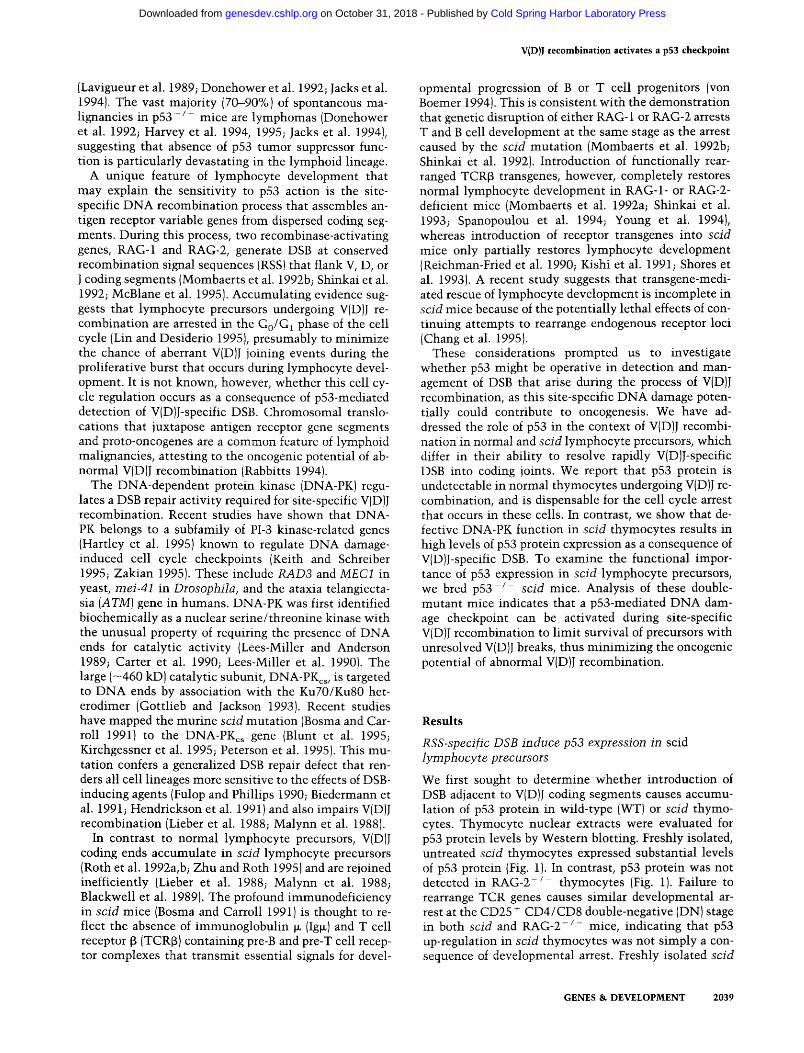
We first sought to determine whether introduction of DSB adjacent to V(D)J coding segments causes accumu- lation of p53 protein in wild-type (WT) or scid thymo- cytes. Thymocyte nuclear extracts were evaluated for p53 protein levels by Western blotting. Freshly isolated, untreated scid thymocytes expressed substantial levels of p53 protein (Fig. 1). In contrast, p53 protein was not detected in R A G - 2 - / - thymocytes (Fig. 1). Failure to rearrange TCR genes causes similar developmental ar- rest at the CD25 + CD4/CD8 double-negative (DN) stage in both scid and RAG-2- / - mice, indicating that p53 up-regulation in scid thymocytes was not simply a con- sequence of developmental arrest. Freshly isolated scid
GENES & DEVELOPMENT 2039
Cold Spring Harbor Laboratory Press on October 31, 2018 - Published by genesdev.cshlp.orgDownloaded from
Guidos et al.
A "~
i
69 kD
/ ~ 46 kD
Z Z r~ r~
B BALB/c TCR~-scid
f..G_y3 0 100 3OO 0 100 300
69 kD -~ I 46 kD--.l, ' - - - - - - - - ~
69 kD-~
46 kD.-~
B6Thy ~ "~ TCR~scid
0 500 0 0 0 100
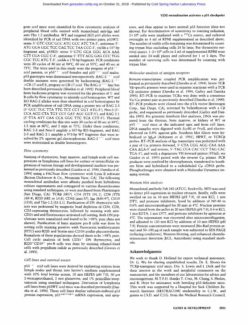
Figure 1. Western blot analysis of p53 protein expression in thymocyte nuclear extracts. Fifty micrograms of nuclear pro- tein from each sample were resolved by SDS-PAGE, transferred to nitrocellulose filters and incubated with anti-p53 antibody, followed by anti-mouse horseradish peroxidase (HRP), and vi- sualized by enhanced chemiluminescence (ECL). (A) Nuclear extracts prepared from fresh ex vivo thymocytes from the indi- cated mouse strains were analyzed for p53 protein expression by Western blotting. The last two lanes contain nuclear extract protein from purified WT (B6) CD25 + and CD25- DN thymo- cytes. (B,C) p53 protein Western blot analysis of nuclear ex- tracts prepared from fresh ex vivo thymocytes (lanes marked 0), or from thymocytes exposed to 100, 300, or 500 cGy from a ~'WCs source and cultured cells for 90 rain at 37°C. Note that the TCR[3 transgene results in greater thymus cellularity because of development and expansion of DP thymocytes, thereby provid- ing a more similar populations for comparison with WT thy- mocytes, which are 75-85% DP. After evaluation with anti-p53 antibody, all membranes were stained with amido black to con- firm equal protein loading (not shown).
splenocytes, which contain mostly myeloid cells and are largely devoid of V(D)J-specific DSB, did not express de- tectable amounts of p53 (data not shown), demonstrating that the scid mutation does not cause a generalized in- crease in p53 protein levels. To examine whether p53 expression in scid thymocytes was dependent on the generation of V(D)J-specific DSB, we bred RAG-2 - / - scid double-mutant mice in which RAG-2 deficiency prevents cleavage at RSS adjacent to V, D, and J coding ends. In contrast to scid thymocytes, RAG-2-deficient scid thymocytes lacked detectable p53 protein expres- sion (Fig. 1A, lane 3), demonstrating that up-regulation of p53 in scid thymocytes requires RAG-2 gene function. Purified immature CD25 + DN thymocytes from WT mice, however, which are actively engaged in TCR rear-
rangement (Pearse et al. 1989; Godfrey et al. 1994), did not express detectable p53 (Fig. 1A, lane 5), suggesting that V(D)J recombination is not accompanied by high- level p53 protein expression in a high frequency of WT thymocytes.
In scid thymocytes, rearrangements are attempted at the TCR~, TCRT, and TCR~, whereas TCRoL remains in germ-line configuration, presumably because of the de- velopmental arrest (Schuler et al. 1988; Carroll and Bosma 1991; Roth et al. 1992b). Introduction of a TCR~3 transgene into scid mice prevents rearrangement of en- dogenous TCRI3 genes and promotes development to the CD4/CD8 double positive {DP) stage, where TCRa rear- rangement begins {Kishi et al. 1991; Shores et al. 1993; Livak et al. 1996). We also observed high levels of p53 protein in TCR[3-transgenic scid thymocytes (Fig. 1B, lane 4; 1 C, lane 5), suggesting that attempted rearrange- ments at TCR~/, TCRS, or TCRot can also provoke p53 accumulation in scid thymocytes. In contrast, p53 pro- tein was undetectable by Western blot analysis of freshly isolated WT thymocytes (Fig. 1B, C), although they con- sist predominantly of DP thymocytes known to be en- gaged actively in TCRe~ rearrangement. However, p53 protein was up-regulated 90 min after exposure of WT and TCRt3-transgenic scid thymocytes to low doses of 7-irradition (Fig. 1B, C). Collectively, these results dem- onstrate that detection of V(D)J-specific DSB in vivo in- duces p53 up-regulation in scid thymocytes, and that radiation can induce further up-regulation of p53 in these cells.
Absence of p53 partially restores TCR~ rearrangement and T cell development in scid mice
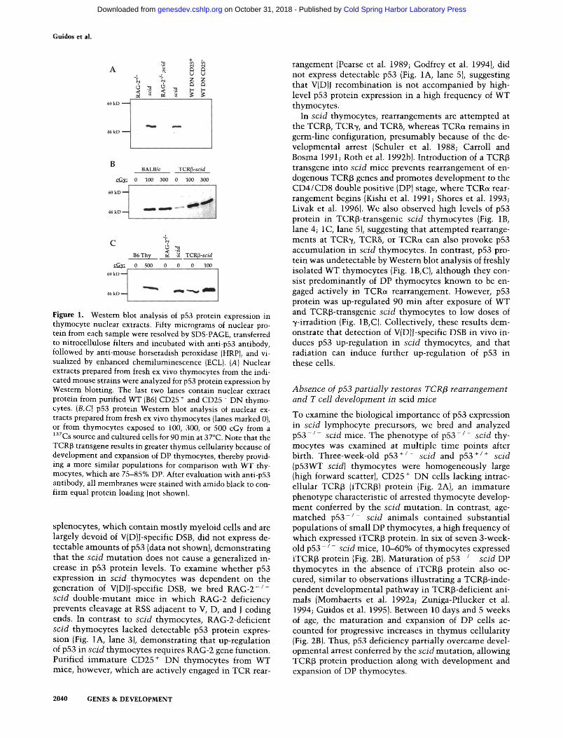
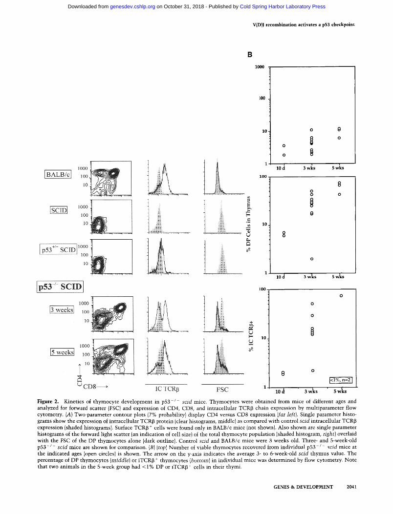
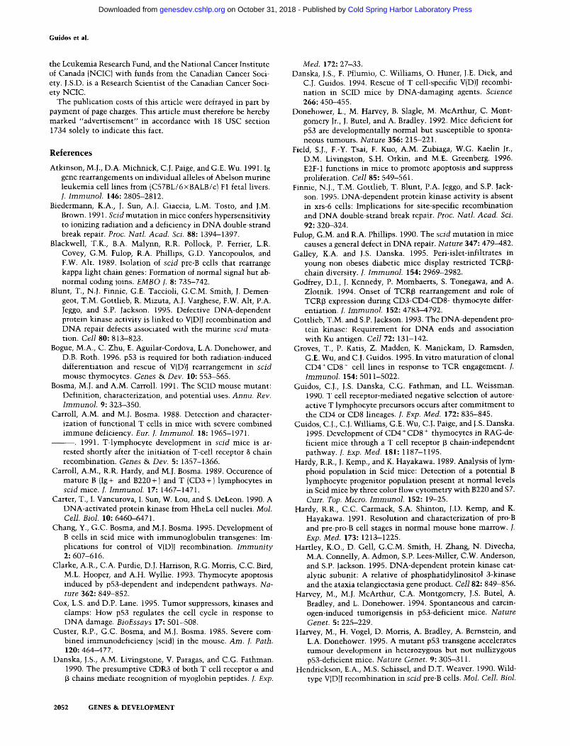
To examine the biological importance of p53 expression in scid lymphocyte precursors, we bred and analyzed p 5 3 - / - scid mice. The phenotype of p 5 3 - / - scid thy- mocytes was examined at multiple time points after birth. Three-week-old p53 + / - scid and p53 + / + s c i d (p53WT scid) thymocytes were homogeneously large (high forward scatter), CD25 + DN cells lacking intrac- ellular TCR~3 (iTCRI3) protein (Fig. 2A), an immature phenotype characteristic of arrested thymocyte develop- ment conferred by the scid mutation. In contrast, age- matched p53 - / - scid animals contained substantial populations of small DP thymocytes, a high frequency of which expressed iTCR~3 protein. In six of seven 3-week- old p 5 3 - / - scid mice, 10-60% of thymocytes expressed iTCR~ protein IFig. 2B). Maturation of p 5 3 - / - scid DP thymocytes in the absence of iTCR[3 protein also oc- cured, similar to observations illustrating a TCR~-inde- pendent developmental pathway in TCR~-deficient ani- mals (Mombaerts et al. 1992a; Zuniga-Pflucker et al. 1994; Guidos et al. 1995). Between 10 days and 5 weeks of age, the maturation and expansion of DP cells ac- counted for progressive increases in thymus cellularity (Fig. 2B). Thus, p53 deficiency partially overcame devel- opmental arrest conferred by the scid mutation, allowing TCR[3 protein production along with development and expansion of DP thymocytes.
2040 G E N E S & D E V E L O P M E N T
Cold Spring Harbor Laboratory Press on October 31, 2018 - Published by genesdev.cshlp.orgDownloaded from
V(D)I recombinat ion activates a p53 checkpoint
B
1000
A
lOO0 [BALB/c] 100
10
1000
100
10 B 1000
p53 /- SCID loo
10
i . . . . . - I . . . . . . i . . . . . . n . . . . . .
p53 /- SCID I
1000
13 weeksl loo
10
]5 w e e k s 1000
lOO
I , o
C D 8 ,
O
.¢,
[-,
5.
......... ~;.~%14 ..........
......... :~. ~. .~, ..........
100
lO
o
o
1 10 ci
1 0 0
10
8
1 10 d
o 0 o
3 wl~s s wks
3 wks s wks
L [--
100
10
[<1%, n=2 ] 1
IC TCRI5 FSC 10 d 3 wks 5 w k s
Figure 2. Kinetics of thymocyte development in p53-/ sc id mice. Thymocytes were obtained from mice of different ages and analyzed for forward scatter (FSC) and expression of CD4, CD8, and intracellular TCR[3 chain expression by multiparameter flow cytometry. (A) Two-parameter contour plots (7% probability) display CD4 versus CD8 expression (far left). Single parameter histo- grams show the expression of intracellular TCR~ protein (clear histograms, middle) as compared with control sc id intracellular TCR[3 expression (shaded histograms). Surface TCR~ + cells were found only in BALB/c mice (not shown). Also shown are single parameter histograms of the forward light scatter (an indication of cell size) of the total thymocyte population (shaded histogram, right) overlaid with the FSC of the DP thymocytes alone (dark outline). Control sc id and BALB/c mice were 3 weeks old. Three- and 5-week-old p53- / - s c i d mice are shown for comparison. (B) (top) Number of viable thymocytes recovered from individual p 5 3 - / - sc id mice at the indicated ages (open circles) is shown. The arrow on the y-axis indicates the average 3- to 6-week-old sc id thymus value. The percentage of DP thymocytes (middle) or iTCR[3 + thymocytes (bo t tom) in individual mice was determined by flow cytometry. Note that two animals in the 5-week group had < 1% DP or iTCR[3 + cells in their thymi.
GENES & DEVELOPMENT 2041
Cold Spring Harbor Laboratory Press on October 31, 2018 - Published by genesdev.cshlp.orgDownloaded from
Guidos et al.
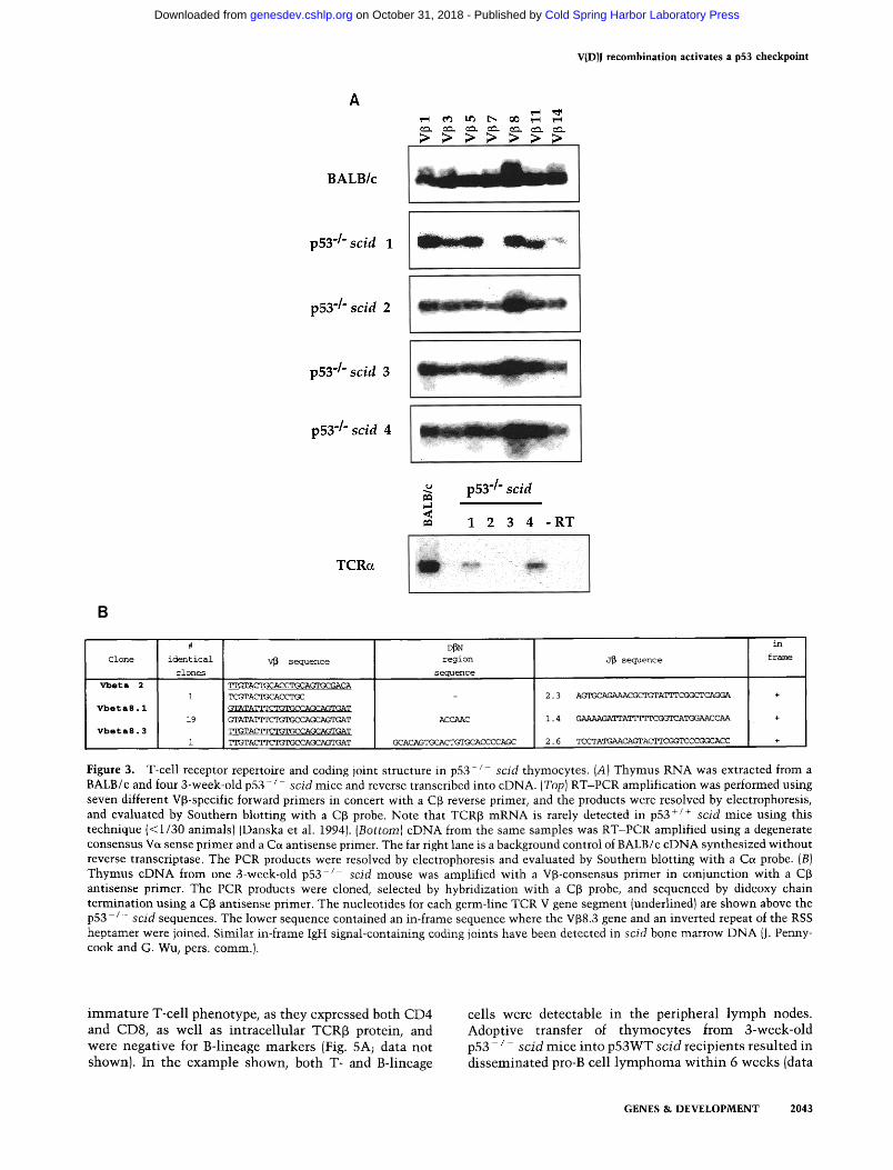
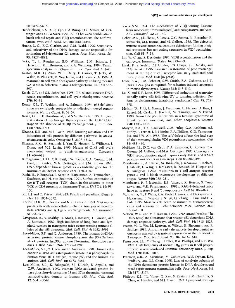
TCR~ protein or mRNA is detected rarely in p53WT scid animals (Bosma and Carroll 1991; Danska et al. 1994), owing to impaired V(D)J recombination. Intracel- lular TCR~ protein expression in p 5 3 - / - scid thymo- cytes could result from diverse V(D)J rearrangements in many precursors, or the expansion of relatively few, oli- goclonal precursors. To examine the heterogeneity of these rearrangements in individual animals, reverse transcriptase coupled-polymerase chain reactions (RT- PCR) were performed with a series of seven TCRV~-spe- cific primers in conjunction with an antisense C~ primer (Danska et al. 1994). In BALB/c thymocytes, each V~- specific reaction yielded TCR~ products, reflecting the use of multiple V-region gene segments in the WT thy- mocyte repertoire (Fig. 3A, top). At 3 weeks of age, tran- scripts containing all seven V~-regions were detected in three of four p53 - / - s c id littermates, suggesting the het- erogeneous use of V~ gene segments. One 3 week-old p53 - / - scid thymus lacked both V~7- and V~314-con- taining transcripts, suggesting more limited TCRf3 diver- sity in this animal (Fig. 3A, second row). Using a Ve~- consensus/Ce~ primer set for RT-PCR amplification (Danska et al. 1990), we found that two of four 3-week- old littermates contained low level TCR~ transcripts of normal length (Fig. 3A, bottom). These results demon- strate that productive TCR~ rearrangement and DP thy- mocyte maturation in p 5 3 - / - scid animals was some- times associated with progression to TCRe~ rearrange- ment and transcription. However, we did not detect surface TCR~ + thymocytes in p 5 3 - / - scid mice (data not shown). Because efficient TCR~ transport to the cell surface is TCRe-dependent (Mombaerts et al. 1992a; Philpott et al. 1992), this observation suggests that few, if any, thymocytes harbored in-frame rearrangements at both TCR~ and TCRa loci. Accordingly, cloning and se- quence analysis of TCRoL transcripts from double mutant animals demonstrated a low frequency of normal, pro- ductive VJ~ coding joints (data not shown).
Previous studies have shown that rare V(D)J coding joints can be isolated from scid cells, often with charac- teristic structural features, including extensive P-nucle- otide addition, homology-directed joining, and extensive coding sequence deletions (for review, see Lewis 1994). To examine the diversity and fidelity of individual V(D)J~ coding joints from p53 - / - scid mice, TCRf~ tran- scripts from a 3-week-old p53 - / - scid thymus were cloned by RT-PCR amplification (Danska et al. 1994). Only 3 of 21 sequences analyzed were unique, and the structure of some TCRf~ coding joints revealed potential signatures of the scid V(D)J recombinase machinery (Fig. 3B and legend). Collectively, these phenotypic and mo- lecular analyses suggest that p53 deficiency permits scid thymocytes to progress through multiple contingent steps characteristic of normal maturation: productive TCR~ rearrangement, expression of CD4 and CD8 core- ceptors, and onset of TCRe rearrangement and transcrip- tion. However, the limited diversity and unusual struc- tural features of TCR transcripts suggested that p53 de- ficiency did not elicit WT V(D)J recombination in scid thymocytes.
Impact of p53 deficiency on B lineage development in scid mice
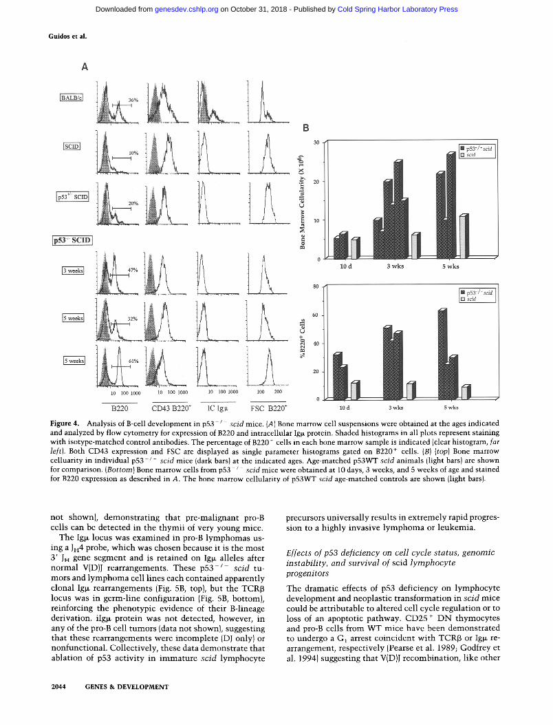
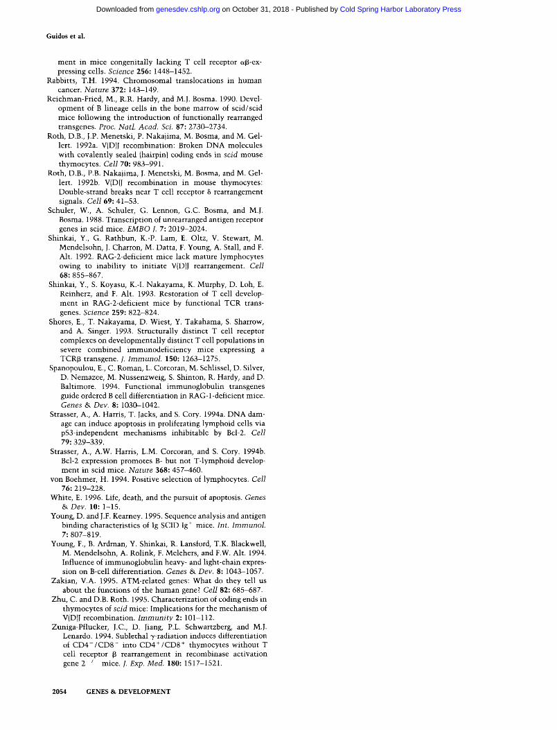
In agreement with a previous report (Hardy et al. 1989), only 5-10% of p53WT scid bone marrow cells expressed the B lineage marker B220, about one-third the frequency in normal mice, and all scid B220 + cells were large, CD43 +, and iIg~- (Fig. 4). In contrast, bone marrow from p53- j - scid mice was enriched in B220 + cells as early as ten days of age, and the B220 + frequency rose to 20-65% by 5 weeks of age (Fig. 4B). Bone marrow cellu- larity increased significantly during this time period, in- dicating an expansion of absolute numbers of B220 + cells (Fig. 4B). Despite the increase in the frequency and number of B220 + cells in the double mutant mice, p53 deficiency did not appear to cause significant develop- mental progression of pro-B cell blasts in scid mice. Al- though we sometimes observed slightly decreased ex- pression of CD43, this was not accompanied by expres- sion of detectable intracellular Ig~ protein in B220 + cells (Fig. 4A). To examine Ig~ transcription, we performed RT-PCR of bone marrow from 5- to 10-day-old p 5 3 - / - scid animals. C~ germ-line transcripts characteristic of the pro-B cell stage were present, but we were unable to detect full-length Ig~ mRNA in the p53 - / - scid samples using a 5' VHau consensus primer paired with a 3' C~ primer (data not shown). Thus, in contrast to the promo- tion of T cell development, p53 deficiency promoted ac- cumulation of B-cell precursors but did not appear to circumvent the block in B-cell maturation conferred by the scid mutation.
p53-deficiency results in rapid onset of lymphoma/leukemia in scid mice
At 7-12 weeks of age, all p53- / - scid animals appeared moribund, and on necropsy, 16 of 16 animals were found to have variable degrees of thymic hyperplasia, lymph- adenopathy, and splenomegaly, attributable to infiltra- tion of large numbers of lymphoblastoid cells. Histolog- ical examination of infiltrated tissues revealed that nor- mal architecture was replaced by densely packed lymphoblasts with abundant mitotic figures (data not shown). These findings are indicative of a significant lymphoma or leukemia burden. Tumors were observed in mice as young as 6 weeks of age, and the median age of onset was 8 weeks. In most animals, the lymphoma/ leukemia was disseminated to multiple sites, including perithymic and inguinal lymph nodes, and spleen. Cell lines were readily derived from explanted thymic or lymph node tumor tissue of multiple animals.
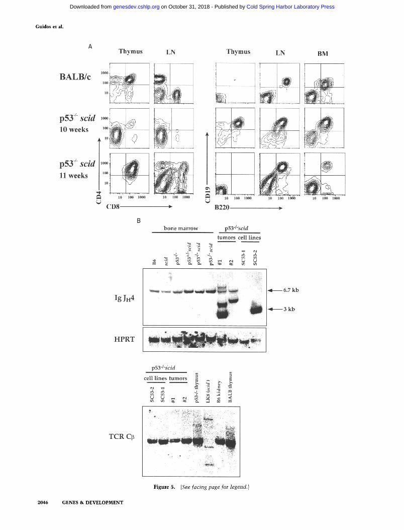
Flow cytometric analyses of tumor phenotype demon- strated that both lymphoid lineages are highly prone to malignant transformation in p53-deficient scid mice. Fourteen of 16 tumors explanted from thymus and lymph nodes did not express T lineage markers (CD4, CD8, Thy-1; data not shown) but were comprised of uni- formly large CD19 + B220 + CD43 + cells (Fig. 5A) that closely resembled normal pro-B cells (Hardy et al. 1991). In contrast, 2 of 16 animals had thymic tumors of an
2042 GENES & D E V E L O P M E N T
Cold Spring Harbor Laboratory Press on October 31, 2018 - Published by genesdev.cshlp.orgDownloaded from
A
BALB/c
> > >> >>>
V(D)J recombination activates a p53 checkpoint
p53 "/" scid 1
p53 "/" scid 2
p53 -/" scid 3
p53 "/" scid 4
B
TCRo~
p53 "/" scid
= 1 2 3 4 - R T .,,~:. ,..:::: . ,.. . ,:. :
Clone
V b e t a 2
Vbet a8.1
V b e t a 8 . 3
#
identical
clones
V~ sequence
TTGTA~CC~GXP_42GACA
TCGTACXP_42ACCXX~C
GTATATIq~TGTGC CAGCAGTGAT
GTATATTTCTGTGC CAGCAGTGAT
TTGTA~TGTGC CAGCAGTGAT
TTGTACTTCTGTC~ CACK/AGTGAT
D~N
region
sequence
ACCAAC
GCACAGTGCACTG~CC CCAGC
J~ sequence
2.3 AGTGCXKGAAACGCTGTATTTCGGCPCAGGA +
i. 4 GAAAAGATTATTTTTCC~TCATGGAACCAA +
2 . 6 TCCTATGAACAGTACTTCGGTCCCGC~ACC +
in
frame
Figure 3. T-cell receptor repertoire and coding joint structure in p53 - / - scid thymocytes. (A) Thymus RNA was extracted from a BALB/c and four 3-week-old p53- / - scid mice and reverse transcribed into cDNA. (Top) RT-PCR amplification was performed using seven different V~3-specific forward primers in concert with a C~ reverse primer, and the products were resolved by electrophoresis, and evaluated by Southern blotting with a C~ probe. Note that TCR~ mRNA is rarely detected in p53 +/+ scid mice using this technique (< 1/30 animals) (Danska et al. 1994). (Bottom) cDNA from the same samples was RT-PCR amplified using a degenerate consensus Va sense primer and a Ca antisense primer. The far right lane is a background control of BALB/c cDNA synthesized without reverse transcriptase. The PCR products were resolved by electrophoresis and evaluated by Southern blotting with a Ca probe. (B) Thymus cDNA from one 3-week-old p53-/ scid mouse was amplified with a V~-consensus primer in conjunction with a C~ antisense primer. The PCR products were cloned, selected by hybridization with a Cf3 probe, and sequenced by dideoxy chain termination using a C~ antisense primer. The nucleotides for each germ-line TCR V gene segment (underlined) are shown above the p53- / - scid sequences. The lower sequence contained an in-frame sequence where the V~8.3 gene and an inverted repeat of the RSS heptamer were joined. Similar in-frame IgH signal-containing coding joints have been detected in scid bone marrow DNA (J. Penny- cook and G. Wu, pers. comm.).
i m m a t u r e T-cell phenotype , as they expressed both CD4 and CD8, as wel l as in t race l lu la r T C R ~ protein, and were negat ive for B-lineage markers (Fig. 5A; data no t shown). In the example shown, both T- and B-lineage
cells were detectable in the per ipheral l y m p h nodes. Adopt ive t ransfer of t h y m o c y t e s f rom 3-week-old p53 - / - scid mice in to p53WT scid rec ip ients resul ted in d i s semina ted pro-B cell l y m p h o m a w i t h i n 6 weeks (data
GENES & DEVELOPMENT 2043
Cold Spring Harbor Laboratory Press on October 31, 2018 - Published by genesdev.cshlp.orgDownloaded from
Guidos et ai.
A
Ip53*' sc l
I pS3 "- s c m I
A
O
l 0 1 0 0 1 0 0 0 10 100 1 0 0 0
. . . . . ~ , . - ~ . . . . ~ .._~ . . . . . . . . . . . . . .
10 1 0 0 1 ~ 101) 2110
10 d 3 w k s 5 w k s
B220 CD43 B220 + IC Igl~ FSC B220 ÷ lOd 3wks 5wks
[] p53-/- sc/d [3 scid J
Figure 4. Analysis of B-cell development in p53- / scid mice. {A) Bone marrow cell suspensions were obtained at the ages indicated and analyzed by flow cytometry for expression of B220 and intracellular Ig~ protein. Shaded histograms in all plots represent staining with isotype-matched control antibodies. The percentage of B220 ÷ cells in each bone marrow sample is indicated (clear histogram, far left). Both CD43 expression and FSC are displayed as single parameter histograms gated on B220 + cells. (B) (top) Bone marrow celluarity in individual p53 /- scid mice {dark bars) at the indicated ages. Age-matched p53WT scid animals {light bars) are shown for comparison. (Bottom) Bone marrow cells from p53 - / scid mice were obtained at 10 days, 3 weeks, and 5 weeks of age and stained for B220 expression as described in A. The bone marrow cellularity of p53WT scid age-matched controls are shown (light bars).
not shown), demonstrat ing that pre-malignant pro-B cells can be detected in the thymii of very young mice.
The Iglx locus was examined in pro-B lymphomas us- ing a JH 4 probe, which was chosen because it is the most 3' JH gene segment and is retained on Ig~ alleles after normal V(D)J rearrangements. These p53 - / - scid tu- mors and lymphoma cell lines each contained apparently clonal Igl~ rearrangements (Fig. 5B, top), but the TCR[3 locus was in germ-line configuration (Fig. 5B, bottom), reinforcing the phenotypic evidence of their B-lineage derivation, iIg~ protein was not detected, however, in any of the pro-B cell tumors (data not shown), suggesting that these rearrangements were incomplete (DJ only) or nonfunct ional . Collectively, these data demonstrate that ablation of p53 act ivi ty in immature scid lymphocyte
precursors universally results in extremely rapid progres- sion to a highly invasive lymphoma or leukemia.
Effects of p53 deficiency on cell cycle status, genomic instability, and survival of scid lymphocyte progenitors
The dramatic effects of p53 deficiency on lymphocyte development and neoplastic t ransformation in scid mice could be attributable to altered cell cycle regulation or to loss of an apoptotic pathway. CD25 ÷ DN thymocytes and pro-B cells from WT mice have been demonstrated to undergo a G~ arrest coincident wi th TCRf3 or Iglx re- arrangement, respectively (Pearse et al. 1989; Godfrey et al. 1994) suggesting that V(D)J recombination, l ike other
2044 GENES & D E V E L O P M E N T
Cold Spring Harbor Laboratory Press on October 31, 2018 - Published by genesdev.cshlp.orgDownloaded from
V(D)J recombination activates a p53 checkpoint
forms of DSB repair, is t ightly coordinated wi th the cell cycle (Lin and Desiderio 1995). Because we did not detect p53 up-regulation in purified CD25 + DN WT thymo- cytes (see Fig. 1A), we reasoned that the G1 arrest at this maturat ional stage in WT mice may be p53-independent. To examine this issue, we compared the cell cycle status of CD25 + DN thymocytes purified from WT B6 and p 5 3 - / - (WT at the scid locus) mice, using propidium iodide staining and flow cytometric analysis (Table 1). These data showed that 390% of both WT and p53-de- ficient CD25 + DN thymocytes were arrested in the G~ phase of the cell cycle. Similarly, CD25 + DN thymo- cytes from RAG-2- / - and scid mice consist primari ly of cells in Go/G1 (Table 1). Loss of p53 did not decrease the proportion of scid CD25 + DN thymocytes (Table 1) or bone marrow pro-B cells (Table 2) in Go/G ~. Thus, the G1 arrest accompanying V(D)J recombinat ion in WT and scid lymphocyte precursors occurs independently of p53 accumulat ion.
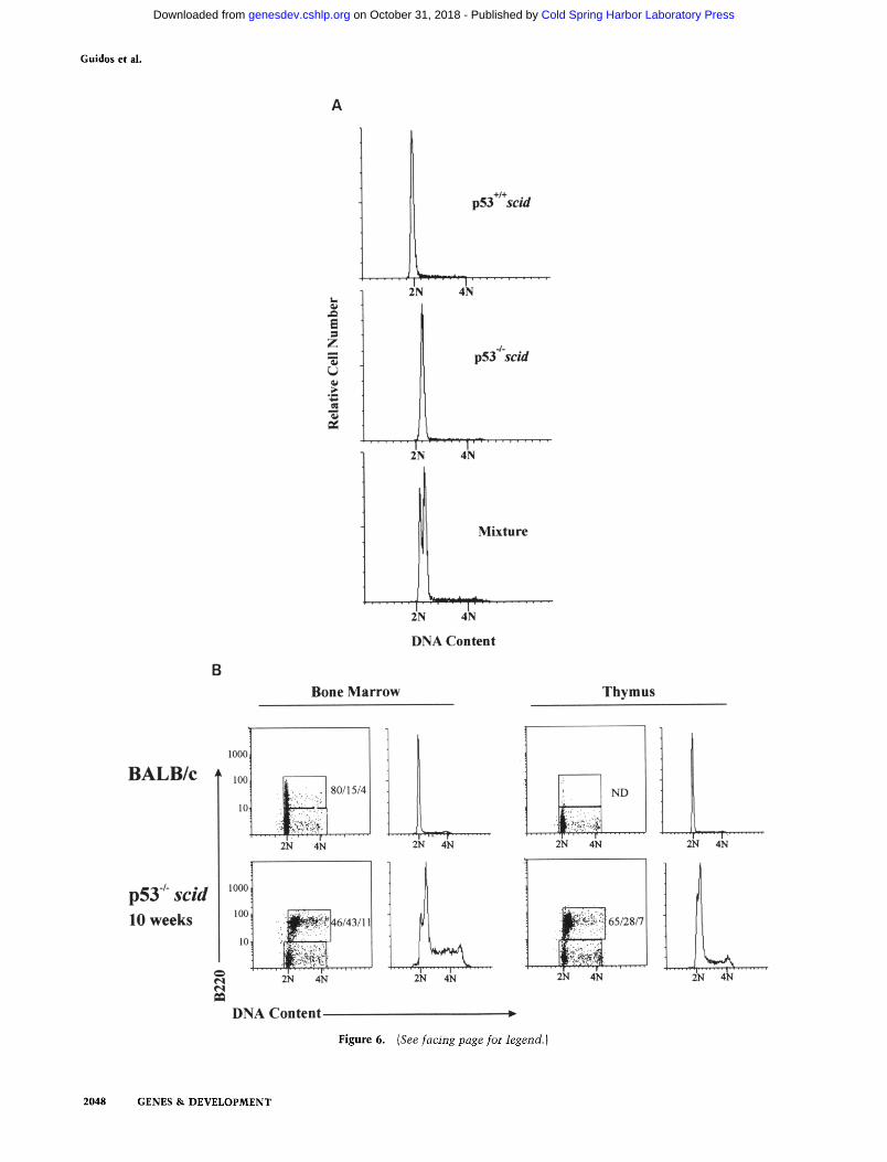
Although p53 deficiency did not promote abnormal cell cycle progression of scid lymphocyte precursors, we noted that pro-B cells and thymocytes harvested from the double mutan t animals during the premalignant phase displayed a significant degree of aneuploidy. Figure 6A shows that pro-B cells purified from p53- / - scid an- imals have ~20% greater Go/G ~ DNA content than those isolated from p53 + / + s c i d animals. This was best demonstrated when the two purified populations were mixed together and then stained for DNA content (Fig. 6A, bottom). Hyperdiploidy was evident in purified pro-B cells from 75% of double mutan t animals sacrified at 5-6 weeks of age, but was not detected in bone marrow my- eloid cells from the same animals (data not shown). We also examined primary tumor cell populations for evi- dence of aneuploidy. D N A content histograms showed bimodal Go/G~ peaks in bone marrow cells and thymo- cytes from a 10-week-old moribund p53 - / - scid mouse, but not from an age-matched BALB/c mouse (Fig. 6B). The B220 versus D N A content dot plot demonstrates that the B220 + cells are hyperdiploid in the double mu-
Table 1. Effect of p53 deficiency on cell cycle status of CD25 + DN wild-type and scid thymocytes
Genotype % G0/G 1 N ~
WT 90 + 2 2 p53 - / - 91 + 2 2 RAG-2-/- 94 + 2 3 scid 95 -+ 1 3 p53 +/- scid 96 -+ 2 5 p53 /- scid 94 + 2 4
CD25 + DN thymocytes were purified from 6-8-week-old WT (B6) and p53-/- mice, fixed, and stained with propidium iodide. Unfractionated thymocytes from the other genotypes were stained with FITC-anti-CD25 and fixed prior to staining with propidium iodide. Cells were analyzed by flow cytometry to determine the cell cycle distribution of the CD25 + subset. All mice were analyzed at 3-6 weeks after birth. aNumber of individual mice analyzed.
tant, but not in BALB/c animals. However, in contrast to the premalignant aneuploid populations, the mal ignant pro-B cells were actively cycling (50% in S/G2/M). Sim- ilarly, we observed active cycling of hyperdiploid cells in CD4/CD8 DP thymic l ymphoma (data not shown). These data suggest that the absence of p53 promotes ge- nomic instabi l i ty in scid lymphocyte progenitors.
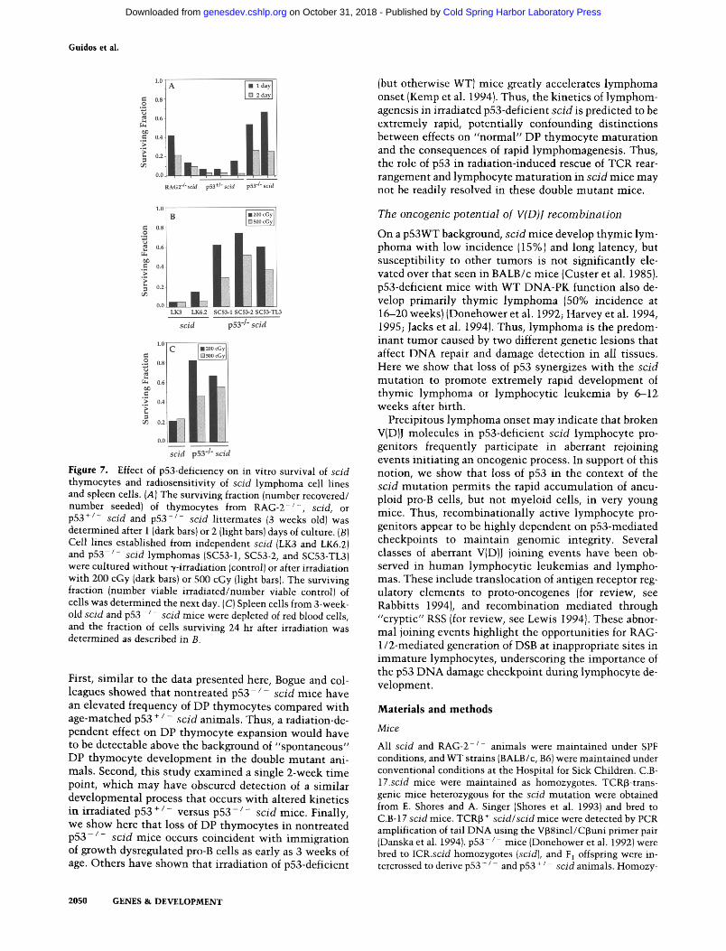
The accumulat ion of noncycling, hyperdiploid lym- phoid progenitors in p53-deficient scid mice also sug- gests that p53 may l imi t the survival of cells harboring broken V(D)J molecules. To assess the role of p53 in the survival of scid cells wi th DSB, we compared the relative survival of thymocytes from RAG-2 - / - , p53WTscid, p53 + / - scid, and p 5 3 - / - scid mice after 1 or 2 days of culture. Mult iple genetic studies have shown this assay to correlate well wi th survival of lymphocyte progeni- tors in vivo (Strasser et al. 1994b; Motoyama et al. 1995; Field et al. 1996). The surviving fraction (number of vi- able cells recovered/number of viable cells seeded) of p53WT scid and p53 + / - scid thymocytes was <0.2 (Fig. 7A). In contrast, the survival of R A G - 2 - / - or p 5 3 - / - scid thymocytes was respectively two- to fourfold greater or three- to sevenfold greater than scid thymo- cytes, p53-deficiency also enhanced dramatical ly the abili ty of scid l ymphoma cell l ines to survive DNA dam- age induced exogenously by low-dose ~/-irradiation (Fig. 7B). As expected, the l ymphoma cell l ines derived from p53WT scid mice were extremely radiosensitive and few cells survived for 24 hr after t reatment wi th 200 and 500 cGy. Lymphoma lines adapted from p 5 3 - / - scid mice, however, were 4--14 t imes more radioresistant. Irradi- ated spleen cells from p53-deficient scid mice also showed substantial ly greater survival than those from p53WT scid mice, demonstrat ing that this effect was not l imited to transformed cells, or to the lymphoid lineage (Fig. 7C). In accord wi th previous studies on p53-defi- cient cells (Strasser et al. 1994a), p53-deficient scid cells also displayed some p53-independent, radiation-induced cell death. Collectively, these observations support the notion that loss of the p53-dependent D N A damage checkpoint in scid lymphocytes confers protection from cell death associated wi th both RSS-dependent and radi-
Table 2. Effect of p53 deficiency on cell cycle status of scid pro-B cells
Genotype % G0/G 1 N a
scid 86 -+ 3 2 p53 +/ scid 87 - 5 4 p53 - / - scid 89 + 2 4
1 x l0 s to 2 x l0 s CD19 + B220 + pro-B cells from individual mice (4-6 weeks old) of each genotype were purified by cell sorting, fixed, stained with propidium iodide, and analyzed by flow cytometry for DNA content. The double mutant mice used for this experiment appeared healthy and had only moderate accumulations of pro-B cells (33 --- 4%), and no lymphadenopa- thy, thymic hyperplasia, or splenomegaly. aNumber of individual mice analyzed.
GENES & DEVELOPMENT .2045
Cold Spring Harbor Laboratory Press on October 31, 2018 - Published by genesdev.cshlp.orgDownloaded from
Guidos et al.
Thymus
BALB/c ~oi
p53 -# scid , 0 ~
o weeks '" ~ @
p53 # scid ~ ~ 11 weeks ~oo
10
10 I00 1000
CD8
Ig JFI4
LN Thymus
10 100 1000 10 100 1000 ~J
= B220
LN BM
10 100 1000 10 100 1000
bone marrow p53"/'scid tumors cell l ines
~ Ln
,: i • :il,;ii!i :
ii:i:i::
. ~ - - - - 6.7 kb
"~---- 3 kb
HPRT
TCR C~
2046 GENES & DEVELOPMENT
p53-/-scid
cell l ines tumors ~ ~ ~
• " ' • ,£.'.:., ::i
F i g u r e 5 . (See facing page for legend.)
Cold Spring Harbor Laboratory Press on October 31, 2018 - Published by genesdev.cshlp.orgDownloaded from
V(D)J recombination activates a p53 checkpoint
ation-induced DSB, directly promoting genomic instabil- ity of these recombinat ional ly active progenitor cells.
D i s c u s s i o n
V(D)J recombination activates a DNA damage checkpoint in scid lymphocytes
Numerous studies have demonstrated that imposi t ion of DNA breaks by exogenous agents are sufficient to initi- ate a p53-mediated cell cycle arrest or apoptosis, but nor- mal physiological conditions that activate p53 pathways have not been identified previously. Here we provide the first demonstrat ion that this DNA damage checkpoint is activated physiologically by V(D)J recombinat ion in scid mice. Examinat ion of p53 protein levels in scid versus RAG-2-deficient scid thymocytes demonstrated that RAG-mediated introduction of DSB in scid thymocytes provides a necessary signal to cause p53 accumulat ion (Fig. 1A). Given that (1) TCR~, TCR% and TCRg are all recombinat ional ly active in this cell population, (2) that V to D joining is not usual ly at tempted before successful D to J joining, and (3) rearrangement of both alleles can be at tempted s imultaneously, the number of broken V(D)J ends present in an individual CD25 + DN thymo- cyte is l ikely between 2 and 16. This implies that the DNA damage-sensing mechan i sm is highly sensitive, in accord wi th a recent study suggesting that a single DSB can activate the p53 checkpoint (Huang et al. 1996). We could not detect p53 accumulation, however, in recom- binat ional ly active WT lymphocyte precursors. We sug- gest that the differential accumulat ion of p53 in WT ver- sus scid lymphocyte progenitors reflects the abnormal persistence of V(D)J coding ends caused by the scid mu- tation in DNA-PK (Roth et al. 1992a, b; Zhu and Roth 1995). Nonetheless, rare WT precursors in which V(D)J coding ends persist may also activate the p53-dependent checkpoint.
Several properties of DNA-PK suggest that it may play a direct role in activation of the p53 checkpoint. It be- longs to a subfamily of PI-3 kinase-related genes impli- cated in yeast, Drosophila, and humans that regulate D N A damage checkpoints (Zakian 1995), and defects in
the h u m a n ATM family member cause abnormali t ies in DNA damage-induced p53 accumulat ion, trans-activa- t ion function, and cell cycle arrest (Kastan et al. 1992; Khanna and Lavin 1993; Lu and Lane 1993; Khanna et al. 1995). Intriguingly, the enzymat ic activity of DNA-PK requires association wi th D N A breaks, and serine resi- dues in the p53 amino- terminal trans-activation domain are targets of phosphorylation by h u m a n and rodent DNA-PK in vitro (Lees-Miller et al. 1992; Finnie et al. 1995). Although scid DNA-PK fails to phosphorylate this site in vitro (Blunt et al. 1995), the biological significance of phosphorylation at this site is unclear (Ko and Prives 1996). We show here that p53 protein is up-regulated in scid thymocytes in vivo as a consequence of at tempted V(D)J recombination. Moreover, scid and WT thymo- cytes accumulated s imilar levels of p53 in response to ionizing radiation. We also observed radiation-induced p21 war-I/tip-1 m R N A expression in scid cells (data not shown), indicative of normal p53-mediated trans-activa- tion. Thus, in contrast to muta t ions in ATM, the scid mutat ion does not compromise the D N A damage-in- duced accumulat ion and funct ion of p53.
Effects of p53 deficiency on survival, V(D)J recombination, and developmental progression of scid lymphocyte precursors
Our results show that p53 deficiency enhances survival of T and B cell precursors in scid mice, as evidenced by prolonged in vitro survival of thymocytes, and by accu- mula t ion of noncycling pro-B ceils in vivo. We observed both productive TCR~ rearrangements and progression to the DP stage, in thymocytes, whereas productive Ig~ rearrangements and developmental progression of pro-B cells were not seen. Previously, we observed a s imil iar lineage dichotomy in restoration of V(D)J recombinat ion and development in newborn scid mice given low doses of ionizing radiation (Danska et al. 1994). Productive TCRg rearrangements were also documented in the lat- ter system (Bogue et al. 1996; Livak et al. 1996), which can facilitate development of DP thymocytes in the ab- sence of TCRB (Mombaerts et al. 1992a; Kersh et al.
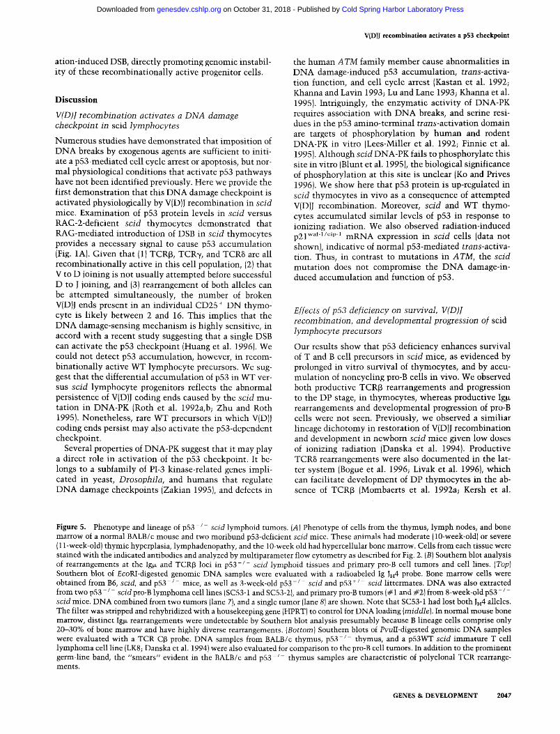
Figure 5. Phenotype and lineage of p53 /- scid lymphoid tumors. (A) Phenotype of cells from the thymus, lymph nodes, and bone marrow of a normal BALB/c mouse and two moribund p53-deficient scid mice. These animals had moderate (10-week-old) or severe (11-week-old) thymic hyperplasia, lymphadenopathy, and the 10-week old had hypercellular bone marrow. Cells from each tissue were stained with the indicated antibodies and analyzed by multiparameter flow cytometry as described for Fig. 2. {B) Southern blot analysis of rearrangements at the Ig~ and TCR~ loci in p53 - / - scid lymphoid tissues and primary pro-B cell tumors and cell lines. (Top) Southern blot of EcoRI-digested genomic DNA samples were evaluated with a radioabeled Ig JH 4 probe. Bone marrow cells were obtained from B6, scid, and p53 /- mice, as well as 3-week-old p53- / - scid and p53 + / scid littermates. DNA was also extracted from two p53- / - scid pro-B lymphoma cell lines (SC53-1 and SC53-2}, and primary pro-B tumors (# 1 and #2) from 8-week-old p53- / - scid mice. DNA combined from two tumors (lane 7), and a single tumor (lane 8) are shown. Note that SC53-1 had lost both JH4 alleles. The filter was stripped and rehybridized with a housekeeping gene (HPRT) to control for DNA loading (middle). In normal mouse bone marrow, distinct Ig~ rearrangements were undetectable by Southern blot analysis presumably because B lineage cells comprise only 20-30% of bone marrow and have highly diverse rearrangements. (Bottom) Southern blots of PvuII-digested genomic DNA samples were evaluated with a TCR CB probe. DNA samples from BALB/c thymus, p53-/- thymus, and a p53WT scid immature T cell lymphoma cell line (LK8; Danska et al. 1994) were also evaluated for comparison to the pro-B cell tumors. In addition to the prominent germ-line band, the "smears" evident in the BALB/c and p53-/- thymus samples are characteristic of polyclonal TCR rearrange- ments.
GENES & DEVELOPMENT 2047
Cold Spring Harbor Laboratory Press on October 31, 2018 - Published by genesdev.cshlp.orgDownloaded from
Guidos et al.
p53+/+sc/d
z i
• u i
~e
I . . . . . . I . . . . . . . . . . . 2N 4N
p53"/'scid
/
2N 4N
2N 4N
DNA Content
Bone Marrow Thymus
BALB/c
p53 # scid 10 weeks
1000
100
10
1000
100
10
, . . . . , ' ' ' i ' , " , I
2N 4N
. / ,
-.]:.~,!,~'. ...... ' '~.~ 614311
' . . . . ' " T ' ' . . . . I ' . . . . ' ~ . . . .
2N 4N
DNA Content
Figure 6.
. . . . . . . . ~ : . ~ . . . . . . . . .
2N 4N
2N 4N
2N 4N
I" . " •
2N 4N
(See facing page for legend. )
. . . . . . . . . . . . . . ~ . . . . . . . . . . .
2N 4N
. . . . . . . I . . . .
2048 GENES & DEVELOPMENT
Cold Spring Harbor Laboratory Press on October 31, 2018 - Published by genesdev.cshlp.orgDownloaded from
V(D)J recombination activates a p53 checkpoint
1995). Thus, we suggest that productive TCR~ or TCR3 rearrangements promoted DP development in p 5 3 - / - scid mice, and that the absence of pro-B cell maturat ion in these animals reflects the failure to generate produc- tive Ig~ rearrangements. It remains unclear whether the differential effects of p53 deficiency and irradiation on promoting successful TCR versus Ig rearrangements in scid pro-B cells reflect unique, Ig~-specific constraints on V(D)J recombination, or owe to different properties of the thymus versus bone marrow microenvironments .
We observed oligoclonal, in-frame TCR~ transcripts, suggesting expansion of a l imited cohort of p53- / - scid precursors wi th productive coding joints (Fig. 3). An in- teresting impl icat ion of these results is that scid DNA- PK has a higher capacity to mediate coding joint forma- tion than has been appreciated previously. The fre- quency of coding joint formation in scid cells has been measured as 10- to 1000-fold below WT levels (Lieber et al. 1988; Malynn et al. 1988; Pennycook et al. 1993), and normal V(D)J rearrangements have been seen in rare lym- phocytes in aged scid mice (Carroll and Bosma 1988; Blackwell et al. 1989; Carroll et al. 1989; Hendrickson et al. 1990; Kotloff et al. 1993; Young and Kearney 1995). Our results help to rationalize this " leakiness" by sug- gesting that the scid mutat ion causes an inefficiency in the V(D)J recombinat ion process that l imits cell survival. However, the l imited diversity and "scid-like" TCR cod- ing joints we observed suggest that scid DNA-PK still functions relatively ineffectively to coordinate DSB re- pair when thymocyte survival is enhanced by p53 abla- tion.
Independent support for the notion that at tempted V(D)J recombinat ion is deleterious for scid lymphocyte precursors has been reported. Transgenic expression of Bcl-2, which can inhibi t DNA damage/p53-mediated cell death (for review, see White 1996J, improved sur- vival and developmental progression of scid pro-B cells, but this study did not evalaute whether this was associ- ated wi th productive Ig~ recombinat ion (Strasser et al. 1994b). In another study, restoration of B-cell production by transgenic expression of Igp~ and Ig light chains in scid mice was dependent on the capacity of different trans- genes to inhibi t endogeneous light chain rearrangement (Chang et al. 1995). We posit that p53-mediated apopto- sis e l iminated pre-B cells at tempting light chain gene rearrangement, but this pathway was not invoked in pre-
cursors bearing a transgene that suppressed endogenous rearrangements. From this perspective, the profound im- mune deficiency conferred by the scid muta t ion does not result exclusively from failure to generate positive selec- tion signals through pre-T and pre-B cell surface antigen receptors. Rather, scid lymphocyte survival is l imited by V(DIJ coding breaks that trigger p53-mediated apoptosis.
Effects of p53 deficiency vs. low-dose irradiation on T-cell development and V(D)I recombination in scid mice
Previously, we have shown that low-dose irradiation of scid newborns provokes a rapid, t ransient burst of di- verse TCR~ and TCR8 rearrangements coincident wi th DP thymocyte maturat ion and expansion (Danska et al. 1994; Livak et al. 1996). However, mul t ip le features dis- t inguish the irradiated scid and p53-deficient scid mod- els. Irradiation of p53WT scid mice provokes rapid gen- eration of thymocytes wi th normal, polyclonal TCR~ and TCRS, but not TCRa, coding joints, suggesting a transient induct ion of repair activities that compensate for the scid mutat ion (Danska et al. 1994; Livak et al. 1996). In contrast, V(D)J~ coding joints in p53-deficient scid mice are oligoclonal, include nonstandard coding joints, and accumulate slowly over a 5- to 8-week post- natal period, consistent wi th enhanced precursor sur- vival revealing the leakiness of the scid mutat ion. These differences in efficiency, kinetics, and fidelity of coding joint formation suggest that mechan i sms operative in promoting radiation-induced maturat ion and survival of scid thymocytes are l ikely to be distinct from those op- erative in p53-deficient scid mice.
A recent study concluded that p53 is required for ra- diation-induced T-cell development in scid mice (Bogue et al. 1996). This conclusion is surprising, given that low-dose radiation further elevates p53 levels in scid thymocytes (Fig. 1), and that p53 activates an apoptotic pathway in thymocytes (Clarke et al. 1993; Lowe et al. 1993), and it provides an apparent paradox wi th our dem- onstration that p53 deficiency promotes survival and V(D)J recombinat ion in scid thymocytes. However, sev- eral variables not considered in the Bogue study may impact the conclusion that p53 plays an obligatory role in radiation-induced development of scid thymocytes.
Figure 6. Presence of hyperdiploid premalignant and malignant pro-B cells in p53-deficient scid mice. (A) Bone marrow-derived CD19 + B220 + pro-B cells were purified by cell sorting from 6-week-old p53 +/+, p53 + / , and p53 /- scid mice. The latter appeared healthy and tumor free (no thymic hypeplasia or lymphadenopathy} at the time of sacrifice. Cells were then fixed, stained with propidium iodide, and analyzed for DNA content by flow cytometry. The Go/G ~ DNA content of pro-B cells from p53 ÷ / ÷ scid and p53 +/- scid mice was identical, therefore only the former cells are shown. To ensure that the apparent hyperdiploid shift of the p53-/- scid cells was not attributable to minor differences in dye or cell concentration, approximately equal numbers of pro-B cells from p53 ÷ / + scid and p53- / - scid were mixed before propidium iodide staining. The bimodal G o/G1 peak of the mixture confirms the unequal DNA content of the two populations. (B) Bone marrow cells and thymocytes from the indicated mice were stained for surface expression of B220 as described for Fig. 3, followed by fixation and propidium iodide staining for DNA content. The B220 versus DNA content dot plot demonstrates a hyperdiploid shift of the B220 + cells from both tissues of double mutant mice. Numbers on each plot refer to the percentage of B220 + cells in G O or G~/S/G 2 or M phases. Note that in normal thymocytes, a significant B220 + population is not detectable (ND), and that the majority of B220 + bone marrow cells in normal mice are in Go/G~.
GENES & DEVELOPMENT 2049
Cold Spring Harbor Laboratory Press on October 31, 2018 - Published by genesdev.cshlp.orgDownloaded from
Guidos et al.
"°tA l O2da tL '~ 0.6
:!. RAG2 -1" sc/d p53 +/" scid p53 "/" sc/d
1.0
o.s
~-~ 0.6
~1~ 0.4
0.2 ~t3
0.0
1.0[ (
~O 0.8
£'~ 0.6
~ 0.2
0.0
LK3 LK6.2 SC53-1 SC53.-2 SC53-TL3
scid p53 °# scid
scid p53 "/" scid
Figure 7. Effect of p53-deficiency on in vitro survival of scid thymocytes and radiosensitivity of scid lymphoma cell lines and spleen cells. (A) The surviving fraction (number recovered/ number seeded) of thymocytes from RAG-2 - / - , scid, or p53 +/- scid and p53 - / - scid littermates {3 weeks old) was determined after 1 (dark bars) or 2 (light bars) days of culture. (B) Cell lines established from independent scid (LK3 and LK6.2) and p53 - / - scid lymphomas (SC53-1, SC53-2, and SC53-TL3) were cultured without ~/-irradiation (control) or after irradiation with 200 cGy (dark bars} or 500 cGy (light bars). The surviving fraction (number viable irradiated/number viable control) of cells was determined the next day. (C) Spleen cells from 3-week- old scid and p53 - / - scid mice were depleted of red blood cells, and the fraction of cells surviving 24 hr after irradiation was determined as described in B.
First, s imilar to the data presented here, Bogue and col- leagues showed that nontreated p53- / - scid mice have an elevated frequency of DP thymocytes compared with age-matched p53 + / - scicl animals. Thus, a radiation-de- pendent effect on DP thymocyte expansion would have to be detectable above the background of "spontaneous" DP thymocyte development in the double mutan t ani- mals. Second, this study examined a single 2-week t ime point, which m a y have obscured detection of a s imilar developmental process that occurs wi th altered kinetics in irradiated p53 + / - versus p53 - / - scid mice. Finally, we show here that loss of DP thymocytes in nontreated p 5 3 - / - scid mice occurs coincident wi th immigrat ion of growth dysregulated pro-B cells as early as 3 weeks of age. Others have shown that irradiation of p53-deficient
2050 GENES & DEVELOPMENT
(but otherwise WT) mice greatly accelerates lymphoma onset (Kemp et al. 1994). Thus, the kinetics of lymphom- agenesis in irradiated p53-deficient scid is predicted to be extremely rapid, potentially confounding dist inctions between effects on "normal" DP thymocyte maturat ion and the consequences of rapid lymphomagenesis . Thus, the role of p53 in radiation-induced rescue of TCR rear- rangement and lymphocyte matura t ion in scid mice may not be readily resolved in these double mutan t mice.
The oncogenic potential of V(D)J recombinat ion
On a p53WT background, scid mice develop thymic lym- phoma wi th low incidence (15%) and long latency, but susceptibili ty to other tumors is not significantly ele- vated over that seen in BALB/c mice (Custer et al. 1985). p53-deficient mice wi th WT DNA-PK funct ion also de- velop primari ly thymic l ymphoma (50% incidence at 16-20 weeks) {Donehower et al. 1992; Harvey et al. 1994, 1995; Jacks et al. 1994). Thus, l ymphoma is the predom- inant tumor caused by two different genetic lesions that affect DNA repair and damage detection in all tissues. Here we show that loss of p53 synergizes wi th the scid mutat ion to promote extremely rapid development of thymic lymphoma or lymphocyt ic leukemia by 6-12 weeks after birth.
Precipitous lymphoma onset m a y indicate that broken VID)J molecules in p53-deficient scid lymphocyte pro- genitors frequently participate in aberrant rejoining events init iat ing an oncogenic process. In support of this notion, we show that loss of p53 in the context of the scid mutat ion permits the rapid accumulat ion of aneu- ploid pro-B cells, but not myeloid cells, in very young mice. Thus, recombinat ional ly active lymphocyte pro- genitors appear to be highly dependent on p53-mediated checkpoints to ma in ta in genomic integrity. Several classes of aberrant V(D}J joining events have been ob- served in human lymphocyt ic leukemias and lympho- mas. These include translocation of antigen receptor reg- ulatory elements to proto-oncogenes (for review, see Rabbitts 1994), and recombinat ion mediated through "cryptic" RSS (for review, see Lewis 1994). These abnor- mal joining events highlight the opportunities for RAG- 1/2-mediated generation of DSB at inappropriate sites in immature lymphocytes, underscoring the importance of the p53 DNA damage checkpoint during lymphocyte de- velopment.
M a t e r i a l s a n d m e t h o d s
Mice
All scid and RAG-2 /- animals were maintained under SPF conditions, and WT strains (BALB/c, B6) were maintained under conventional conditions at the Hospital for Sick Children. C.B- 17.scid mice were maintained as homozygotes. TCRJ3-trans- genie mice heterozygous for the scid mutation were obtained from E. Shores and A. Singer (Shores et al. 1993) and bred to C.B-17 scid mice. TCR[3 + scid/scid mice were detected by PCR amplification of tail DNA using the VI38incl/Cf3uni primer pair (Danska et al. 1994). p53- / mice (Donehower et al. 1992) were bred to ICR.scid homozygotes (scid), and F 1 offspring were in- tercrossed to derive p53- / - and p53 + / scid animals. Homozy-
Cold Spring Harbor Laboratory Press on October 31, 2018 - Published by genesdev.cshlp.orgDownloaded from
V(D)J recombination activates a p53 checkpoint
gous scid mice were identified by flow cytometric analysis of peripheral blood cells stained with monoclonal anti-Ig~ and anti-Thy 1.2 antibodies. WT and targeted (KO) p53 alleles were identified by PCR of tail DNA with two primer pairs; p53WT: sense 5'-GTC CGC GCC ATG GCC ATC TA-3', antisense 5'- ATG GGA GGC TGC CAG TCC TAA CCC-3', yields a 137-bp fragment and, p53KO: sense 5'-GTG GGA GGG ACA AAA GTT CGA GGC C-3', antisense 5'-TTT ACG GAG CCC TGG CGC TCG ATG T-3', yields a 170-bp fragment. PCR conditions were 30 cycles of 30 sec at 94°C, 60 sec at 55°C, and 40 sec at 72°C. The mice used in this study were the progeny of p53 +/- scid parents, or p53+/- scid females and p53- / - scid males. p53 genotypes were determined retrospectively. RAG-2- / scid double mutants were generated by backcrossing (RAG-2 - / - xCB-17.scid) F~ progeny to CB-17.scid. The RAG-2 - / - line has been described previously (Shinkai et al. 1992). Peripheral blood from backcross progeny was screened for the presence of T- and B-cells by flow cytometry to identify scid homzygotes. WT and KO RAG-2 alleles were then identified in scid homozygotes by PCR amplification of tail DNA using a primer trio of RAG 2-3 (5'-GCC TGC TTA TTG TCT CCT GGT ATG-3'), Neo-3 (5'- CCA ACG CTA TGT CCT GAT AGC GGT-3'), and RAG 2-1 (5'-TTA ATT CAA CCA GGC TTC TCA CTT-3'). Thermal cycling conditions for this trio were 30 cycles of 30 sec at 94°C, 1.5 min at 60°C, and 2 rain at 72°C. Under these conditions, RAG 2-3 and Neo-3 amplify a 937-bp KO fragment, and RAG 2-3 and RAG 2-1 amplify a 973-bp WT fragment that were re- solved by 2% agarose gel electrophoresis. RAG-2-/- scid mice were maintained as double homozygotes.
Flow cytometry
Staining of thymocyte, bone marrow, and lymph node cell sus- pensions or lymphoma cell lines for surface or intracellular ex- pression of various lineage and developmental markers was per- formed as previously described (Guidos et al. 1990; Danska et al. 19941 using a FACScan flow cytometer with Lysis II software (Becton Dickinson & Co., Mountain View, CA). The following monoclonal antibodies were affinity purified from hybridoma culture supernatants and conjugated to various fluorochromes using standard techniques, or were purchased from Pharmingen [San Diego, CA): TCR~ (H57), CD4 (YTS 191.1), CD8 (YTS 169.4), B220 (6B2 or 14.8), CD43 (anti-S7), Ig~ (R40-97), CD19 (1D3), and Thy-l.2 (53-2.1). Purification of DN thymocyte sub- sets was performed by magnetic bead depletion of CD4, CDS, CD3-expressing thymocytes, followed by staining with anti- CD25 and and fluorescence-activated cell sorting. Both DN pop- ulations were reanalyzed and found to be >98% pure (data not shown). Purification of bone marrow pro-B cells was done by sorting cells staining positive with fluorescein isothiocyanate [FITC)-anti-B220 and biotin-anti-CD19/avidin-phycoerytherin. Reanalysis of these populations showed them to be >98% pure. Cell cycle analysis of both CD25 + DN thymocytes, and B220+CD19 + pro-B cells was done by staining ethanol-fixed cells with propidium iodide as previously described (Groves et al. 1995).
Cell lines and survival assays
p53- / - scid cell lines were derived by explanting tumors from lymph nodes and thymi into Iscove's medium supplemented with 10% fetal bovine serum, 10 mM HEPES (pH 7.0), 50 ~M 2-mercaptoethanol, 2 mM glutamine, and 1% penicillin/strep- tomycin using standard techniques. Derivation of lymphoma cell lines from p53WT scid mice was described previously (Dan- ska et al. 1994). These cell lines display radiation-induced p53 protein expression, p2 l"~f-~/cip- 1 mRNA expression, and apop-
tosis, and thus appear to have normal p53 function (data not shown). For determination of sensitivity to ionizing radiation, 2x 10⁶ cells were irradiated with a ~37Cs source, and cultured overnight in 4 ml of RPMI supplemented as described above. The number of viable cells remaining was determined by count- ing trypan blue excluding cells 24 hr later. For thymocyte sur- vival assays, 1-2x 10⁶ cells in 2 ml of supplemented RPMI were seeded into 24-we!l plates and cultured for 1 or 2 days. The number of surviving cells was determined by counting with trypan blue.
Molecular analysis of antigen receptors
Reverse-transcriptase coupled PCR amplification was per- formed as previously described (Danska et al. 1994). Seven TCR V[~-specific primers were used in separate reactions with a TCR CB antisense primer (Danska et al. 1994; Galley and Danska 1995). RT-PCR to examine TCR~ was done with a consensus Va/C~ primer pair as previously described {Danska et al. 1990). RT-PCR products were cloned into the pTA vector (Invitrogen Corp., San Diego, CA), screened by hybridization with a CB probe, and sequenced as previously described (Galley and Dan- ska 1995). For genomic Southern blot analyses, DNA was pre- pared from the thymus, bone marrow, or kidney of WT or p53 - / - scid mice at the indicated ages and from cell lines. DNA samples were digested with EcoRI or PvuII, and electro- phoresed on 0.8% agarose gels. Southern blot filters were hy- bridized to IgJH4 (Atkinson et al. 1991), HPRT, or TCRCB probes. RT-PCR analysis of Ig~z transcripts was performed with a pair of C~ primers (forward, 5'-CTA CGG AGG CAA AAA CAA AGA-3' and reverse, 5'-TAG CCA CAC CCT TAG CAC TGA-3'), and with a degenerate VH forward primer (VHall; see Guidos et al. 1995) paired with the reverse C~ primer. PCR products were resolved by electrophoresis, transferred to South- ern blots and hybridized to a radiolabeled C~ cDNA probe. PhosphorImages were obtained with a Molecular Dynamics im- aging system.
Western blot analysis
Monoclonal antibody Pab 242 (ATCC, Rockville, MD) was used to detect p53 expression in nuclear extracts. Briefly, cells were swelled on ice in 10 mM HEPES (pH 7.9), 10 mM KC1, 1 mM DTT, and protease inhibitors, lysed by addition of NP-40 to 0.6% and microcentrifuged for 30 min at 4°C. Nuclear proteins were eluted from the pellet in 20 mM HEPES {pH 7.9), 0.4 M KC1, 1 mM EDTA, 1 mM DTT, and protease inhibitors by agitation at 4°C. The supernatant was recovered after microcentrifugation, and adjusted to 120 mM KC1 by addition of 10 mM HEPES (pH 7.9). Protein concentrations were measured (Bio-Rad protein as- say) and 50-100 ~g of each sample was subjected to SDS-PAGE (reducing conditions), Western blotting, and enhanced chemilu- minescence detection (ECL, Amersham) using standard meth- ods.
A c k n o w l e d g m e n t s
We wish to thank D. Holland for expert technical assistance, Dr. G. Wu for sharing unpublished results, Dr. E. Shores for TCR[3-transgenic scid mice, Drs. S. Lewis and J. Dick and for their interest in the work and insightful comments on the manuscript, and the members of our laboratories for advice and encouragement. M.T.F.H. thanks T. Cruz, M. Chang, S. Phelan, and R. Hoyt for assistance with breeding p53-deficient mice. This work was supported by a Hospital for Sick Children Re- search Institute (RESTRACOM) studentship to C.J.W., and grants to J.S.D. and C.J.G. from the Medical Research Council,
GENES & DEVELOPMENT 2051
Cold Spring Harbor Laboratory Press on October 31, 2018 - Published by genesdev.cshlp.orgDownloaded from
Guidos et al.
the Leukemia Research Fund, and the National Cancer Institute of Canada (NCIC) with funds from the Canadian Cancer Soci- ety. J.S.D. is a Research Scientist of the Canadian Cancer Soci- ety NCIC.
The publication costs of this article were defrayed in part by payment of page charges. This article must therefore be hereby marked "advertisement" in accordance with 18 USC section 1734 solely to indicate this fact.
R e f e r e n c e s
Atkinson, M.J., D.A. Michnick, C.J. Paige, and G.E. Wu. 1991. Ig gene rearrangements on individual alleles of Abelson murine leukemia cell lines from (C57BL/6xBALB/c) F1 fetal livers. I. Immunol. 146: 2805-2812.
Biedermann, K.A., J. Sun, A.J. Giaccia, L.M. Tosto, and J.M. Brown. 1991. Scid mutation in mice confers hypersensitivity to ionizing radiation and a deficiency in DNA double-strand break repair. Proc. Natl. Acad. Sci. 88: 1394--1397.
Blackwell, T.K., B.A. Malynn, R.R. Pollock, P. Ferrier, L.R. Covey, G.M. Fulop, R.A. Phillips, G.D. Yancopoulos, and F.W. Alt. 1989. Isolation of scid pre-B cells that rearrange kappa light chain genes: Formation of normal signal but ab- normal coding joins. EMBO ]. 8: 735-742.
Blunt, T., N.J. Finnie, G.E. Taccioli, G.C.M. Smith, J. Demen- geot, T.M. Gottlieb, R. Mizuta, A.J. Varghese, F.W. Alt, P.A. Jeggo, and S.P. Jackson. 1995. Defective DNA-dependent protein kinase activity is linked to V{D)J recombination and DNA repair defects associated with the murine scid muta- tion. Cell 80: 813-823.
Bogue, M.A., C. Zhu, E. Aguilar-Cordova, L.A. Donehower, and D.B. Roth. 1996. p53 is required for both radiation-induced differentiation and rescue of VID)J rearrangement in scid mouse thymocytes. Genes & Dev. 10: 553-565.
Bosma, M.J. and A.M. Carroll. 1991. The SCID mouse mutant: Definition, characterization, and potential uses. Annu. Rev. Immunol. 9: 323-350.
Carroll, A.M. and M.J. Bosma. 1988. Detection and character- ization of functional T cells in mice with severe combined immune deficiency. Eur. J. Immunol. 18: 1965-1971.
1991. T-lymphocyte development in scid mice is ar- rested shortly after the initiation of T-cell receptor 8 chain recombination. Genes & Dev. 5: 1357-1366.
Carroll, A.M., R.R. Hardy, and M.J. Bosma. 1989. Occurence of mature B {Ig + and B220 +) and T (CD3 +) lymphocytes in scid mice. I. ImmunoL 17: 1467-1471.
Carter, T., I. Vancurova, I. Sun, W. Lou, and S. DeLeon. 1990. A DNA-activated protein kinase from HheLa cell nuclei. Mol. Cell. Biol. 10: 6460--6471.
Chang, Y., G.C. Bosma, and M.J. Bosma. 1995. Development of B cells in scid mice with immunoglobulin transgenes: Im- plications for control of V(D)J recombination. Immuni t y 2: 607-616.
Clarke, A.R., C.A. Purdie, D.J. Harrison, R.G. Morris, C.C. Bird, M.L. Hooper, and A.H. Wyllie. 1993. Thymocyte apoptosis induced by p53-dependent and independent pathways. Na- ture 362: 849-852.
Cox, L.S. and D.P. Lane. 1995. Tumor suppressors, kinases and clamps: How p53 regulates the cell cycle in response to DNA damage. BioEssays 17: 501-508.
Custer, R.P., G.C. Bosma, and M.J. Bosma. 1985. Severe com- bined immunodeficiency (scid) in the mouse. Am. J. Path. 120: 464-477.
Danska, J.S., A.M. Livingstone, V. Paragas, and C.G. Fathman. 1990. The presumptive CDR3 of both T cell receptor a and
chains mediate recognition of myoglobin peptides. I. Exp.
Med. 172: 27-33. Danska, J.S., F. Pflumio, C. Williams, O. Huner, J.E. Dick, and
C.J. Guidos. 1994. Rescue of T cell-specific V{D)J recombi- nation in SCID mice by DNA-damaging agents. Science 266: 450--455.
Donehower, L., M. Harvey, B. Slagle, M. McArthur, C. Mont- gomery Jr., J. Butel, and A. Bradley. 1992. Mice deficient for p53 are developmentally normal but susceptible to sponta- neous tumours. Nature 356: 215-221.
Field, S.J., F.-Y. Tsai, F. Kuo, A.M. Zubiaga, W.G. Kaelin Jr., D.M. Livingston, S.H. Orkin, and M.E. Greenberg. 1996. E2F-1 functions in mice to promote apoptosis and suppress proliferation. Cell 85: 549-561.
Finnie, N.J., T.M. Gottlieb, T. Blunt, P.A. Jeggo, and S.P. Jack- son. 1995. DNA-dependent protein kinase activity is absent in xrs-6 cells: Implications for site-specific recombination and DNA double-strand break repair. Proc. Natl. Acad. Sci. 92: 320-324.
Fulop, G.M. and R.A. Phillips. 1990. The scid mutation in mice causes a general defect in DNA repair. Nature 347: 479-482.
Galley, K.A. and J.S. Danska. 1995. Peri-islet-infiltrates in young non obeses diabetic mice display restricted TCRS- chain diversity. ]. Immunol. 154: 2969-2982.
Godfrey, D.I., J. Kennedy, P. Mombaerts, S. Tonegawa, and A. Zlotnik. 1994. Onset of TCR[3 rearrangement and role of TCRf~ expression during CD3-CD4-CD8- thymocyte differ- entiation. J. Immunol. 152: 4783-4792.
Gottlieb, T.M. and S.P. Jackson. 1993. The DNA-dependent pro- tein kinase: Requirement for DNA ends and association with Ku antigen. Cell 72: 131-142.
Groves, T., P. Katis, Z. Madden, K. Manickam, D. Ramsden, G.E. Wu, and C.J. Guidos. 1995. In vitro maturation of clonal CD4 + CD8 ÷ cell lines in response to TCR engagement. I. Immunol. 154: 5011-5022.
Guidos, C.J., J.S. Danska, C.G. Fathman, and I.L. Weissman. 1990. T cell receptor-mediated negative selection of autore- active T lymphocyte precursors occurs after commitment to the CD4 or CD8 lineages. 1. Exp. Med. 172: 835-845.
Guidos, C.J., C.J. Williams, G.E. Wu, C.J. Paige, and J.S. Danska. 1995. Development of CD4 + CD8 + thymocytes in RAG-de- ficient mice through a T cell receptor [3 chain-independent pathway. 1. Exp. Med. 181: 1187-1195.
Hardy, R.R., J. Kemp., and K. Hayakawa. 1989. Analysis of lym- phoid population in Scid mice: Detection of a potential B lymphocyte progenitor population present at normal levels in Scid mice by three color flow cytometry with B220 and $7. Curr. Top. Micro. Immunol. 152: 19-25.
Hardy, R.R., C.C. Carmack, S.A. Shinton, J.D. Kemp, and K. Hayakawa. 1991. Resolution and characterization of pro-B and pre-pro-B cell stages in normal mouse bone marrow. I. Exp. Med. 173: 1213-1225.
Hartley, K.O., D. Gell, G.C.M. Smith, H. Zhang, N. Divecha, M.A. Connelly, A. Admon, S.P. Lees-Miller, C.W. Anderson, and S.P. Jackson. 1995. DNA-dependent protein kinase cat- alytic subunit: A relative of phosphatidylinositol 3-kinase and the ataxia telangiectasia gene product. Cell 82: 849-856.
Harvey, M., M.J. McArthur, C.A. Montgomery, J.S. Butel, A. Bradley, and L. Donehower. 1994. Spontaneous and carcin- ogen-induced tumorigensis in p53-deficient mice. Nature Genet. 5: 225-229.
Harvey, M., H. Vogel, D. Morris, A. Bradley, A. Bernstein, and L.A. Donehower. 1995. A mutant p53 transgene accelerates tumour development in heterozygous but not nullizygous p53-deficient mice. Nature Genet. 9: 305-31t.
Hendrickson, E.A., M.S. Schissel, and D.T. Weaver. 1990. Wild- type V{D)J recombination in scid pre-B cells. Mol. Cell. Biol.
2052 GENES & DEVELOPMENT
Cold Spring Harbor Laboratory Press on October 31, 2018 - Published by genesdev.cshlp.orgDownloaded from
V(D)J recombination activates a p53 checkpoint
10: 5397-5407. Hendrickson, E.A., X.-Q. Qin, E.A. Bump, D.G. Schatz, M. Oet-
tinger, and D.T. Weaver. 1991. A link between double-strand break-related repair and V(D)J recombination: The scid mu- tation. Proc. Natl. Acad. Sci. 88: 4061-4065.
Huang, L.-C., K.C. Clarkin, and G.M. Wahl. 1996. Sensitivity and selectivity of the DNA damage sensor responsible for activating p53-dependent G1 arrest. Proc. Natl. Acad. Sci. 93: 4827-4832.
Jacks, T., L. Remington, B.O. Williams, E.M. Schmitt, S. Halachmi, R.T. Bronson, and R.A. Weinberg. 1994. Tumor spectrum analysis in p53-mutant mice. Curr. Biol. 4: 1-7.
Kastan, M.B., Q. Zhan, W. E1-Deiry, F. Carrier, T. Jacks, W. Walsh, B. Plunkett, B. Vogelstein, and J. Fornace, A. 1992. A mammalian cell cycle checkpoint pathway utilizing p53 and GADD45 is defective in ataxia-telangiectasia. Cell 71: 587- 597.
Keith, C.T. and S.L. Schreiber. 1995. PIK-related kinases: DNA repair, recombination, and cell cycle checkpoints. Science 2 7 0 : 5 0 - 5 1 .
Kemp, C.J., T. Weldon, and A. Balmain. 1994. p53-deficient mice are extremely susceptible to radiation-induced tumor- igenesis. Nature Genet. 8: 66-69.
Kersh, G.J., F.F. Hooshmand, and S.M. Hedrick. 1995. Efficient maturation of c,f~ lineage thymocytes to the CD4+CD8 + stage in the absence of TCR~3 rearrangement. J. Immunol . 154: 5706-5714.
Khanna, K.K. and M.F. Lavin. 1993. Ionizing radiation and UV induction of p53 protein by different pathways in ataxia- telangiectasia cells. On cogene 8: 3307-3312.
Khanna, K.K., H. Beamish, J. Yan, K. Hobson, R. Williams, I. Dunn, and M.F. Lavin. 1995. Nature of G1/S cell cycle checkpoint defect in ataxia-telangiectasia. Oncogene 11: 609-618.
Kirchgessner, C.U., C.K. Patil, J.W. Evans, C.A. Cuomo, L.M. Fried, T. Carter, M.A. Oettinger, and J.M. Brown. 1995. DNA-dependent kinase (p350) as a candidate gene for the murine SCID defect. Science 267:1178-1182.
Kishi, H., P. Borgulya, B. Scott, K. Karjalainen, A. Traunecker, J. Kaufman, and H. yon Boehmer. 1991. Surface expression of the f3 T cell receptor (TCR) chain in the absence of other TCR or CD3 proteins on immature T cells. EMBO J. 10: 93- 100.
Ko, L.J. and C. Prives. 1996. p53: Puzzle and paradigm. Genes & Dev. 10: 1054-1072.
Kotloff, D.B., M.J. Bosma, and N.R. Ruetsch. 1993. Scid mouse pre-B cells with intracellular ~ chains: Analysis of recombi- nase activity and lgH gene rearrangements. Int. Immunol . 5: 383-391.
Lavigueur, A., V. Maltby, D. Mock, J. Rossant, T. Pawson, and A. Bernstein. 1989. High incidence of lung, bone and lym- phoid tumors in transgenic mice overexpressing mutant al- leles of the p53 oncogene. Mol. Cell. Biol. 9: 3982-3991.
Lees-Miller, S.P. and C. Anderson. 1989. The human ds-DNA- activated protein kinase phosphorylates the 90-kDa heat shock protein, hsp90~, at two N-terminal threonine resi- dues. J. Biol. Chem. 264: 17275-17280.
Lees-Miller, S.P., Y. Chen, and C. Anderson. 1990. Human cells contain a DNA-activated protein kinase that phosphorylates Simian virus 40 T antigen, mouse p53 and the human Ku antigen. Moi. Cell. Biol. 10: 6472-6481.
Lees-Miller, S.P., K. Sakaguchi, S.J. Ullrich, E. Appella, and C.W. Anderson. 1992. Human DNA-activated protein ki- nase phosphorylates serines 15 and 37 in the amino-terminal transactivation domain in human p53. Mol. Cell. Biol. 12: 5041-5049.
Lewis, S.M. 1994. The mechanism of V(D)J joining: Lessons from molecular, immunological, and comparative analyses. Adv. Immunol . 56: 27-150.
Lieber, M.R., J.E. Hesse, S. Lewis, G.C. Bosma, N. Rosenber, K. Mizuuchi, M.J. Bosma, and M. Gellert. 1988. The defect in murine severe combined immune deficiency: Joining of sig- nal sequences but not coding segments in V(D)J recombina- tion. Cell 55: 7-16.
Lin, W.-C. and S. Desiderio. 1995. V{D)J recombination and the cell cycle. ImmunoI. Today 16: 279-289.
Livak, F., S. Welsh, C.J. Guidos, I.N. Crispe, J.S. Danska, and D.G. Schatz. 1996. Transient restoration of gene rearrange- ment at multiple T cell receptor loci in ~/ irradiated scid mice. J. Exp. Med. 184: (in press).
Lowe, S.W., E.M. Schmitt, S.W. Smith, B.A. Osborne, and T. Jacks. 1993. p53 is required for radiation-induced apoptosis in mouse thymocytes. Nature 362: 847-849.
Lu, X. and D.P. Lane. 1993. Differential induction of transcrip- tionally active p53 following UV or ionizing radiation: De- fects in chromosome instability syndromes? Cell 75: 765- 778.
Malkin, D., F. Li, L. Strong, J. Fraumeni, C. Nelson, D. Kim, J. Kassel, M. Gryka, F. Bischoff, M. Tainsky, and S. Friend. 1990. Germ line p53 mutations in a familial syndrome of breast cancer, sarcomas, and other neoplasms. Science 250: 1233-1238.
Malynn, B.A., T.K. Blackwell, G.M. Fulop, G.A. Rathbun, A.J. Furley, P. Ferrier, L.B. Heinke, R.A. Phillips, G.D. Yancopou- los, and F.W. Alt. 1988. The scid defect affects the final step of the immunoglobulin VDJ recombinase mechanism. Cell 54: 453-460.
McBlane, J.F., D.C. van Gent, D.A. Ramsden, C. Romeo, C.A. Cuomo, M. Gellert, and M.A. Oettinger. 1995. Cleavage at a V(D}J recombination signal requires only RAG1 and RAG2 proteins and occurs in two steps. Cell 83: 387-395.
Mombaerts, P., A. Clarke, M. Rudnicki, J. Iacomini, S. Itohara, J. Lafaille, L. Wang, Y. Ichikawa, R. Jaenisch, M. Hooper, and S. Tonegawa. 1992a. Mutations in T-cell antigen receptor genes ~ and ~ block thymocyte development at different stages. Nature 360: 225-231.
Mombaerts, P., J. Iacomini, R.S. Johnson, K. Herrup, S. Tone- gawa, and V.E. Papaioannou. 1992b. RAG-l-deficient mice have no mature B and T lymphocytes. Cell 68: 869-877.
Motoyama, N., F. Wang, K.A. Roth, H. Sawa, K.-I. Nakayama, K. Nakayama, I. Negishi, S. Senju, Q. Zhang, S. Fuji, and D.Y. Loh. 1995. Massive cell death of immature hematopoietic cells and neurons in Bcl-x-deficient mice. Science 267: 1506-1510.
Nelson, W.G. and M.B. Kastan. 1994. DNA strand breaks: The DNA template alterations that trigger p53-dependent DNA damage response pathways. Mol. Cell. Biol. 14: 1815-1823.
Pearse, M., L. Wu, M. Egerton, A. Wilson, K. Shortman, and R. Scollay. 1989. A murine early thymocyte developmental se- quence is marked by transient expression of the interleukin 2 receptor. Proc. Natl. Acad. Sci. 86: 1614-1618.
Pennycook, J.L., Y. Chang, J. Celler, R.A. Phillips, and G.E. Wu. 1993. High frequency of normal DJH joints in B cell progen- itors in severe combined immune deficiency mice. L Exp. Med. 178: 1007-1016.
Peterson, S.R., A. Kurimasa, M. Oshimura, W.S. Dynan, E.M. Bradbury, and D.J. Chen. 1995. Loss of catalytic subunit of the DNA-dependent protein kinase in DNA double-strand break repair mutant mammalian cells. Proc. Natl. Acad. Sci. 92: 3171-3174.
Philpott, K.L., J.L. Viney, G. Kay, S. Rastan, E.M. Gardiner, S. Chae, A. Hayday, and M.J. Owen. 1992. Lymphoid develop-
GENES & DEVELOPMENT 2053
Cold Spring Harbor Laboratory Press on October 31, 2018 - Published by genesdev.cshlp.orgDownloaded from
Guidos et al.
ment in mice congenitally lacking T cell receptor aft-ex- pressing cells. Science 256- 1448-1452.
Rabbitts, T.H. 1994. Chromosomal translocations in human cancer. Nature 372: 143-149.
Reichman-Fried, M., R.R. Hardy, and M.J. Bosma. 1990. Devel- opment of B lineage cells in the bone marrow of scid/scid mice following the introduction of functionally rearranged transgenes. Proc. Natl. Acad. Sci. 87: 2730-2734.
Roth, D.B., J.P. Menetski, P. Nakajima, M. Bosma, and M. Gel- left. 1992a. V(D)J recombination: Broken DNA molecules with covalently sealed (hairpinl coding ends in scid mouse thymocytes. Cell 70: 983-991.
Roth, D.B., P.B. Nakajima, J. Menetski, M. Bosma, and M. Gel- lert. 1992b. V(D)J recombination in mouse thymocytes: Double-strand breaks near T cell receptor 8 rearrangement signals. Cell 69: 41-53.
Schuler, W., A. Schuler, G. Lennon, G.C. Bosma, and M.J. Bosma. 1988. Transcription of unrearranged antigen receptor genes in scid mice. EMBO I. 7: 2019-2024.
Shinkai, Y., G. Rathbun, K.-P. Lam, E. Oltz, V. Stewart, M. Mendelsohn, J. Charron, M. Datta, F. Young, A. Stall, and F. Alt. 1992. RAG-2-deficient mice lack mature lymphocytes owing to inability to initiate V(D)J rearrangement. Cell 68: 855-867.
Shinkai, Y., S. Koyasu, K.-I. Nakayama, K. Murphy, D. Loh, E. Reinherz, and F. Alt. 1993. Restoration of T cell develop- ment in RAG-2-deficient mice by functional TCR trans- genes. Science 259: 822-824.
Shores, E., T. Nakayama, D. Wiest, Y. Takahama, S. Sharrow, and A. Singer. 1993. Structurally distinct T cell receptor complexes on developmentally distinct T cell populations in severe combined immunodeficiency mice expressing a TCR[3 transgene. ]. Immunol . 150: 1263-1275.
Spanopoulou, E., C. Roman, L. Corcoran, M. Schlissel, D. Silver, D. Nemazee, M. Nussenzweig, S. Shinton, R. Hardy, and D. Baltimore. 1994. Functional immunoglobulin transgenes guide ordered B cell differentiation in RAG-1-deficient mice. Genes & Dev. 8: 1030-1042.
Strasser, A., A. Harris, T. Jacks, and S. Cory. 1994a. DNA dam- age can induce apoptosis in proliferating lymphoid cells via p53-independent mechanisms inhibitable by Bcl-2. Cell 79: 329-339.
Strasser, A., A.W. Harris, L.M. Corcoran, and S. Cory. 1994b. Bcl-2 expression promotes B- but not T-lymphoid develop- ment in scid mice. Nature 368: 457-460.
von Boehmer, H. 1994. Positive selection of lymphocytes. Cell 76: 219-228.
White, E. 1996. Life, death, and the pursuit of apoptosis. Genes & Dev. 10: 1-15.
Young, D. and J.F. Kearney. 1995. Sequence analysis and antigen binding characteristics of Ig SCID Ig + mice. Int. ImmunoI . 7: 807-819.
Young, F., B. Ardman, Y. Shinkai, R. Lansford, T.K. Blackwell, M. Mendelsohn, A. Rolink, F. Melchers, and F.W. Alt. 1994. Influence of immunoglobulin heavy- and light-chain expres- sion on B-cell differentiation. Genes & Dev. 8: 1043-1057.
Zakian, V.A. 1995. ATM-related genes: What do they tell us about the functions of the human gene? Cell 82: 685-687.
Zhu, C. and D.B. Roth. 1995. Characterization of coding ends in thymocytes of scid mice: Implications for the mechanism of V(D)J recombination. I m m u n i t y 2:101-112.
Zuniga-Pflucker, J.C., D. Jiang, P.L. Schwartzberg, and M.J. Lenardo. 1994. Sublethal ~-radiation induces differentiation of CD4- /CDS- into CD4 +/CD8 + thymocytes without T cell receptor ~3 rearrangement in recombinase activation gene 2 - / - mice. J. Exp. Med. 180: 1517-1521.
2054 GENES & DEVELOPMENT
Cold Spring Harbor Laboratory Press on October 31, 2018 - Published by genesdev.cshlp.orgDownloaded from
10.1101/gad.10.16.2038Access the most recent version at doi: 10:1996, Genes Dev.
C J Guidos, C J Williams, I Grandal, et al. checkpoint in scid lymphocyte precursors.V(D)J recombination activates a p53-dependent DNA damage
References
http://genesdev.cshlp.org/content/10/16/2038.full.html#ref-list-1
This article cites 83 articles, 38 of which can be accessed free at:
License
ServiceEmail Alerting
click here.right corner of the article or
Receive free email alerts when new articles cite this article - sign up in the box at the top
Copyright © Cold Spring Harbor Laboratory Press
Cold Spring Harbor Laboratory Press on October 31, 2018 - Published by genesdev.cshlp.orgDownloaded from