Embed Size (px)
Citation preview
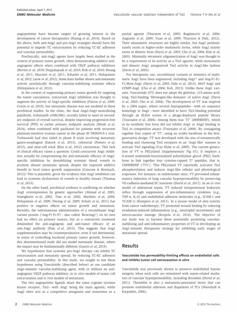
Research Article
Vasculotide reduces endothelial permeability andtumor cell extravasation in the absence of bindingto or agonistic activation of Tie2Florence T H Wu1,2, Christina R Lee2, Elena Bogdanovic2, Aaron Prodeus1,3, Jean Gariépy1,3 &
Robert S Kerbel1,2,*
Abstract
Angiopoietin-1 (Ang1) activation of Tie2 receptors on endothelialcells (ECs) reduces adhesion by tumor cells (TCs) and limits junc-tional permeability to TC diapedesis. We hypothesized thatsystemic therapy with Vasculotide (VT)—a purported Ang1mimetic,Tie2 agonist—can reduce the extravasation of potentially meta-static circulating TCs by similarly stabilizing the host vasculature.In vitro, VT and Ang1 treatments impeded endothelial hypermeabilityand the transendothelial migration of MDA-MB-231∙LM2-4 (breast),HT29 (colon), or SN12 (renal) cancer cells to varying degrees. Inmice, VT treatment inhibited the transit of TCs through the pulmo-nary endothelium, but not the hepatic or lymphatic endothelium.In the in vivo LM2-4 model, VT monotherapy had no effect onprimary tumors, but significantly delayed distant metastaticdissemination to the lungs. In the post-surgical adjuvant treatmentsetting, VT therapeutically complemented sunitinib therapy, ananti-angiogenic tyrosine kinase inhibitor which limited the localgrowth of residual disease. Unexpectedly, detailed investigationsinto the putative mechanism of action of VT revealed no evidenceof Tie2 agonism or Tie2 binding; alternative mechanisms have yetto be determined.
Keywords Angiopoietin; Metastasis; Tie2; tumor cell extravasation; vascular
permeability
Subject Categories Cancer; Vascular Biology & Angiogenesis
DOI 10.15252/emmm.201404193 | Received 29 April 2014 | Revised 3 March
2015 | Accepted 9 March 2015
Introduction
Metastatic disease—as opposed to primary tumors—accounts for
90% of cancer-related mortality (Steeg, 2012). Most cancer drugs
are selected from preclinical studies based on their potency at inhib-
iting primary tumor growth, and brought into clinical trials with the
rationale that they will similarly inhibit the growth of metastases
(Francia et al, 2011; Guerin et al, 2013). The inadequacy of this
approach has been reviewed and editorials have highlighted the
need for new anti-metastatic therapies that block not just the growth
(progression) but also the spread or formation (incidence) of metas-
tases (Steeg, 2012).
The metastatic process comprises a cascade of events (Talmadge
& Fidler, 2010; Hanahan & Weinberg, 2011): stromal invasion from
a localized tumor; intravasation of tumor cells (TCs); their systemic
circulation and arrest in distant capillary beds; TC extravasation into
the host organ parenchyma; and overt colonization as micrometas-
tases grow into macrometastases. In clinically relevant settings, the
temporal window of TC extravasation is asynchronous and wide:
Primary tumors shed millions of TCs per gram of tumor into the
blood circulation every day (Bockhorn et al, 2007), and metastases
themselves can metastasize to tertiary sites or ‘self-seed’ back to
primary sites (Comen et al, 2011). Surgical trauma associated with
primary tumor resections can also sometimes paradoxically fuel
metastatic spread—for example, by mechanically dislodging tumor
cells into the circulation and by inducing the production/release of
inflammatory and angiogenic cytokines that promote metastatic
seeding and progression (Goldfarb & Ben-Eliyahu, 2006).
Tumor cell extravasation is regulated by many cytokines, includ-
ing angiopoietin-1 (Ang1), a guardian of EC quiescence (Augustin
et al, 2009; Huang et al, 2010), as well as angiopoietin-2 (Ang2)
and vascular endothelial growth factor (VEGF), the cooperative
initiator and driver of angiogenesis (Huang et al, 2010; Felcht et al,
2012). It was found that VEGF stimulates, Ang2 potentiates, while
Ang1 counteracts the TNF-a/NF-jB-mediated EC surface expression
of ICAM-1, VCAM-1, and E-selectin—which facilitate the adhesion
and migration of TCs across the endothelium (Kim et al, 2001b;
Fiedler et al, 2006; Miles et al, 2008; Huang et al, 2010). VEGF also
stimulates, while Ang1 counteracts Src-mediated destabilization of
paracellular VE-cadherin junctions and IP3/eNOS-mediated calcium
influxes in ECs—which together cause vascular hyperpermeability
and lowered resistance to TC diapedesis (Gamble et al, 2000;
Gavard et al, 2008; Le Guelte et al, 2011; Koh, 2013). The
1 Department of Medical Biophysics, University of Toronto, Toronto, ON, Canada2 Biological Sciences Platform, Sunnybrook Research Institute, Toronto, ON, Canada3 Physical Sciences Platform, Sunnybrook Research Institute, Toronto, ON, Canada
*Corresponding author. Tel: +1 416-480-5711; E-mail: [email protected]
ª 2015 The Authors. Published under the terms of the CC BY 4.0 license EMBO Molecular Medicine 1
Published online: April 7, 2015
angiopoietins have become targets of growing interest in the
development of cancer therapeutics (Huang et al, 2010). Based on
the above, both anti-Ang2 and pro-Ang1 strategies should have the
potential to impede TC extravasation by reducing TC-EC adhesion
and vascular permeability.
Preclinically, anti-Ang2 agents have largely been studied in the
context of primary tumor growth, often demonstrating additive anti-
angiogenic effects when combined with VEGF pathway inhibitors
(Brown et al, 2010; Doppalapudi et al, 2010; Koh et al, 2010; Huang
et al, 2011; Mazzieri et al, 2011; Schaefer et al, 2011; Holopainen
et al, 2012; Leow et al, 2012). Some have further shown anti-metastatic
activity preclinically through vascular-stabilizing systemic effects
(Holopainen et al, 2012).
In the context of suppressing primary tumor growth by targeting
the tumor vasculature, concurrent Ang1 inhibition was thought to
augment the activity of Ang2-specific inhibition (Falcon et al, 2009;
Coxon et al, 2010), but metastatic disease was not modeled in these
preclinical studies. In the clinic, the dual Ang2/Ang1-neutralizing
peptibody, trebananib (AMG386), recently failed to meet its second-
ary endpoint of overall survival, despite improving progression-free
survival (PFS) in earlier primary endpoint analysis (Monk et al,
2014), when combined with paclitaxel for patients with recurrent
platinum-sensitive ovarian cancer in the phase III TRINOVA-1 trial.
Trebananib had also failed in phase II trials involving metastatic
gastro-esophageal (Eatock et al, 2013), colorectal (Peeters et al,
2013), and clear-cell renal (Rini et al, 2012) carcinomas. This lack
of clinical efficacy raises a question: Could concurrent Ang1 inhibi-
tion actually be compromising the anti-metastatic efficacy of Ang2-
specific inhibition by destabilizing systemic blood vessels to
promote distant metastatic spread, despite the expected additive
benefit in local tumor growth suppression (Cascone & Heymach,
2012)? This is plausible, given the evidence that Ang1 inhibition can
lead to systemic dysfunction of vessels in healthy tissues (Thomas
et al, 2013).
On the other hand, preclinical evidence is conflicting on whether
Ang1 overexpression by genetic approaches (Ahmad et al, 2001;
Hawighorst et al, 2002; Tian et al, 2002; Machein et al, 2004;
Holopainen et al, 2009; Hwang et al, 2009; Schulz et al, 2011) has
positive or negative effects on tumor growth and metastasis.
Recently, the subcutaneous administration of a recombinant Ang1
variant protein (‘Ang-F1-Fc-F1’, also called ‘BowAng1’) on its own
had no effect on primary tumors, but as a concurrent treatment
diminished the anti-angiogenic and anti-tumor efficacy of an
anti-Ang2 antibody (Daly et al, 2013). This suggests that Ang1
supplementation may be counterproductive, even if not detrimental,
in terms of controlling localized primary tumor growth; however,
this aforementioned study did not model metastatic disease, where
the impact may be fundamentally different (Guerin et al, 2013).
We hypothesize that systemic pro-Ang1 therapy can inhibit TC
extravasation and metastatic spread, by reducing TC-EC adhesion
and vascular permeability. In this study, we sought to test these
hypotheses using Vasculotide (described below) as our candidate
Ang1-mimetic vascular-stabilizing agent, with or without an anti-
angiogenic VEGF pathway inhibitor, in in vitro models of tumor cell
extravasation and in vivo models of metastasis.
The two angiopoietin ligands share the same cognate tyrosine
kinase receptor, Tie2—with Ang1 being the main agonist, while
Ang2 often acts as a competitive antagonist and sometimes as a
partial agonist (Thurston et al, 2005; Bogdanovic et al, 2006;
Augustin et al, 2009; Yuan et al, 2009; Thurston & Daly, 2012).
Their monomeric structures are highly similar, but Ang1 predomi-
nantly exists in higher-order multimeric forms, while Ang2 mainly
exists in dimeric form (Davis et al, 2003; Cho et al, 2004; Kim et al,
2005). Minimally tetrameric oligomerization of Ang1 was thought to
be a requirement of its activity as a Tie2 agonist, while monomeric
and dimeric Ang1 antagonized Tie2 activity in Ang2-like fashion
(Davis et al, 2003).
For therapeutic use, recombinant variants or mimetics of multi-
meric Ang1 have been engineered, including Ang1* and Ang-F1-Fc-
F1/Bow-Ang1 (Davis et al, 2003; Daly et al, 2013), MAT-Ang1 and
COMP-Ang1 (Cho et al, 2004; Koh, 2013). Unlike these Ang1 vari-
ants, Vasculotide (VT) does not adopt the globular, 215-amino-acid-
long, Tie2-binding ‘fibrinogen-like domains’ of native Ang1 (Davis
et al, 2003; Cho et al, 2004). The development of VT was inspired
by a 2004 paper, where several heptapeptides—with no sequence
homology to Ang1—were identified to have Tie2-binding potential
through an ELISA screen of a phage-displayed peptide library
(Tournaire et al, 2004). Among them was ‘T7’ (HHHRHSF), which
in its synthetic free form did not inhibit Ang1 or Ang2 binding to
Tie2 in competition assays (Tournaire et al, 2004). By conjugating
together four copies of ‘T7’, using an avidin backbone in the first-
generation design, VT was developed with the aim of tetramerically
binding and clustering Tie2 receptors in an ‘Ang1-like’ manner to
activate Tie2 signaling (Van Slyke et al, 2009). The current genera-
tion of VT is PEGylated (Supplementary Fig S1): It employs a
4-armed maleimide-functionalized polyethylene glycol (PEG) back-
bone to link together four cysteine-capped T7 peptides, that is,
CHHHRHSF (‘T7c’). This PEGylated VT reportedly activates Tie2
phosphorylation and induces Ang1-like cellular and physiological
responses. For instance, in endotoxemic mice, VT prevented inflam-
matory induction of lung vascular hyperpermeability by preserving
VE-cadherin-mediated EC junctions (David et al, 2011). In an in vivo
model of abdominal sepsis, VT reduced intraperitoneal leukocyte
influx through suppression of pro-inflammatory cytokines (e.g.,
TNF-a, IL-6) and endothelial adhesion molecules (e.g., ICAM-1 and
VCAM-1) (Kumpers et al, 2011). In a mouse model of skin toxicity
from cancer radiotherapy, VT promoted wound healing by reducing
irradiation-induced inflammation (e.g., neutrophil recruitment) and
microvascular damage (Korpela et al, 2014). The objective of
our study was to harness these potentially promising vascular-
stabilizing and anti-inflammatory properties of VT in developing an
Ang1-mimetic therapeutic strategy for inhibiting early stages of
metastatic spread.
Results
Vasculotide has permeability-limiting effects on endothelial cellsand inhibits tumor cell extravasation in vitro
Vasculotide was previously shown to preserve endothelial barrier
integrity when such cells are stimulated with sepsis-related media-
tors of vascular hyperpermeability, including thrombin (David et al,
2011). Thrombin is also a metastasis-associated factor that can
promote endothelial adhesion and diapedesis of TCs (Nierodzik &
Karpatkin, 2006).
EMBO Molecular Medicine ª 2015 The Authors
EMBO Molecular Medicine Vasculotide: vascular stabilizer independent of Tie2 Florence T H Wu et al
2
Published online: April 7, 2015
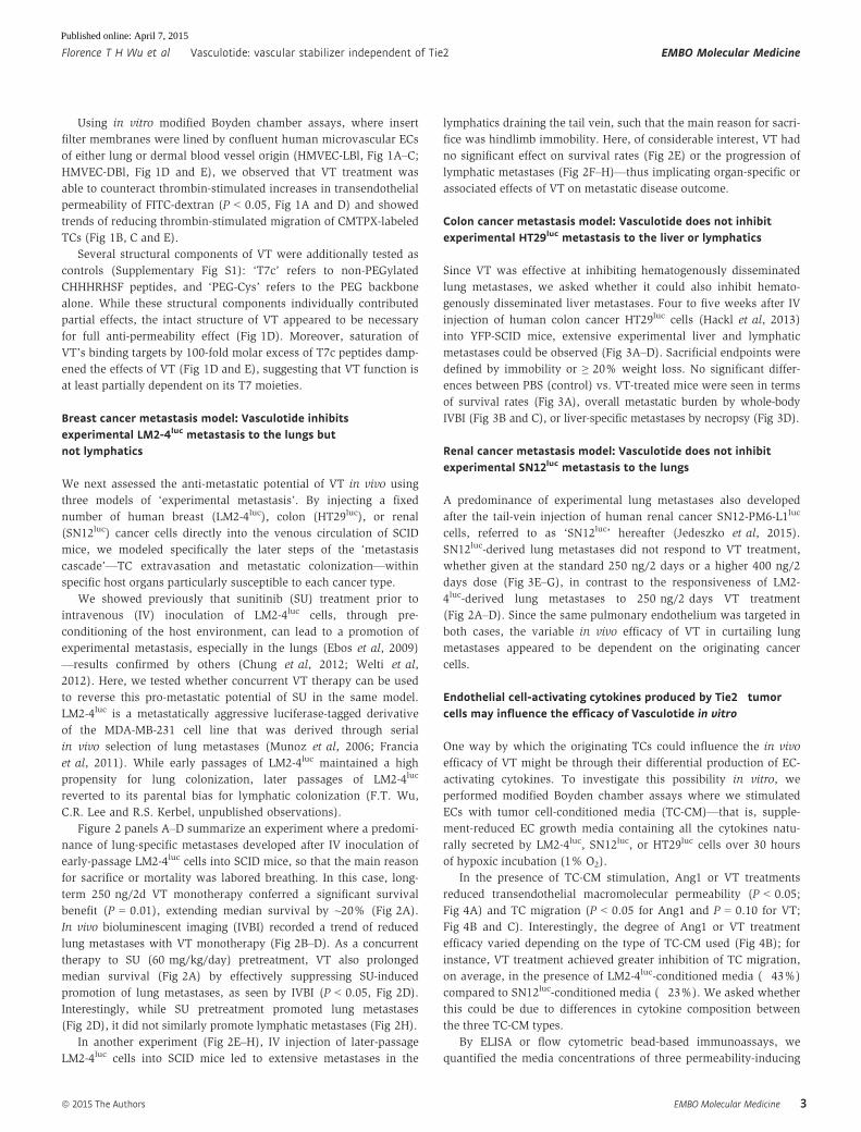
Using in vitro modified Boyden chamber assays, where insert
filter membranes were lined by confluent human microvascular ECs
of either lung or dermal blood vessel origin (HMVEC-LBl, Fig 1A–C;
HMVEC-DBl, Fig 1D and E), we observed that VT treatment was
able to counteract thrombin-stimulated increases in transendothelial
permeability of FITC-dextran (P < 0.05, Fig 1A and D) and showed
trends of reducing thrombin-stimulated migration of CMTPX-labeled
TCs (Fig 1B, C and E).
Several structural components of VT were additionally tested as
controls (Supplementary Fig S1): ‘T7c’ refers to non-PEGylated
CHHHRHSF peptides, and ‘PEG-Cys’ refers to the PEG backbone
alone. While these structural components individually contributed
partial effects, the intact structure of VT appeared to be necessary
for full anti-permeability effect (Fig 1D). Moreover, saturation of
VT’s binding targets by 100-fold molar excess of T7c peptides damp-
ened the effects of VT (Fig 1D and E), suggesting that VT function is
at least partially dependent on its T7 moieties.
Breast cancer metastasis model: Vasculotide inhibitsexperimental LM2-4luc metastasis to the lungs butnot lymphatics
We next assessed the anti-metastatic potential of VT in vivo using
three models of ‘experimental metastasis’. By injecting a fixed
number of human breast (LM2-4luc), colon (HT29luc), or renal
(SN12luc) cancer cells directly into the venous circulation of SCID
mice, we modeled specifically the later steps of the ‘metastasis
cascade’—TC extravasation and metastatic colonization—within
specific host organs particularly susceptible to each cancer type.
We showed previously that sunitinib (SU) treatment prior to
intravenous (IV) inoculation of LM2-4luc cells, through pre-
conditioning of the host environment, can lead to a promotion of
experimental metastasis, especially in the lungs (Ebos et al, 2009)
—results confirmed by others (Chung et al, 2012; Welti et al,
2012). Here, we tested whether concurrent VT therapy can be used
to reverse this pro-metastatic potential of SU in the same model.
LM2-4luc is a metastatically aggressive luciferase-tagged derivative
of the MDA-MB-231 cell line that was derived through serial
in vivo selection of lung metastases (Munoz et al, 2006; Francia
et al, 2011). While early passages of LM2-4luc maintained a high
propensity for lung colonization, later passages of LM2-4luc
reverted to its parental bias for lymphatic colonization (F.T. Wu,
C.R. Lee and R.S. Kerbel, unpublished observations).
Figure 2 panels A–D summarize an experiment where a predomi-
nance of lung-specific metastases developed after IV inoculation of
early-passage LM2-4luc cells into SCID mice, so that the main reason
for sacrifice or mortality was labored breathing. In this case, long-
term 250 ng/2d VT monotherapy conferred a significant survival
benefit (P = 0.01), extending median survival by ~20% (Fig 2A).
In vivo bioluminescent imaging (IVBI) recorded a trend of reduced
lung metastases with VT monotherapy (Fig 2B–D). As a concurrent
therapy to SU (60 mg/kg/day) pretreatment, VT also prolonged
median survival (Fig 2A) by effectively suppressing SU-induced
promotion of lung metastases, as seen by IVBI (P < 0.05, Fig 2D).
Interestingly, while SU pretreatment promoted lung metastases
(Fig 2D), it did not similarly promote lymphatic metastases (Fig 2H).
In another experiment (Fig 2E–H), IV injection of later-passage
LM2-4luc cells into SCID mice led to extensive metastases in the
lymphatics draining the tail vein, such that the main reason for sacri-
fice was hindlimb immobility. Here, of considerable interest, VT had
no significant effect on survival rates (Fig 2E) or the progression of
lymphatic metastases (Fig 2F–H)—thus implicating organ-specific or
associated effects of VT on metastatic disease outcome.
Colon cancer metastasis model: Vasculotide does not inhibitexperimental HT29luc metastasis to the liver or lymphatics
Since VT was effective at inhibiting hematogenously disseminated
lung metastases, we asked whether it could also inhibit hemato-
genously disseminated liver metastases. Four to five weeks after IV
injection of human colon cancer HT29luc cells (Hackl et al, 2013)
into YFP-SCID mice, extensive experimental liver and lymphatic
metastases could be observed (Fig 3A–D). Sacrificial endpoints were
defined by immobility or ≥ 20% weight loss. No significant differ-
ences between PBS (control) vs. VT-treated mice were seen in terms
of survival rates (Fig 3A), overall metastatic burden by whole-body
IVBI (Fig 3B and C), or liver-specific metastases by necropsy (Fig 3D).
Renal cancer metastasis model: Vasculotide does not inhibitexperimental SN12luc metastasis to the lungs
A predominance of experimental lung metastases also developed
after the tail-vein injection of human renal cancer SN12-PM6-L1luc
cells, referred to as ‘SN12luc’ hereafter (Jedeszko et al, 2015).
SN12luc-derived lung metastases did not respond to VT treatment,
whether given at the standard 250 ng/2 days or a higher 400 ng/2
days dose (Fig 3E–G), in contrast to the responsiveness of LM2-
4luc-derived lung metastases to 250 ng/2 days VT treatment
(Fig 2A–D). Since the same pulmonary endothelium was targeted in
both cases, the variable in vivo efficacy of VT in curtailing lung
metastases appeared to be dependent on the originating cancer
cells.
Endothelial cell-activating cytokines produced by Tie2� tumorcells may influence the efficacy of Vasculotide in vitro
One way by which the originating TCs could influence the in vivo
efficacy of VT might be through their differential production of EC-
activating cytokines. To investigate this possibility in vitro, we
performed modified Boyden chamber assays where we stimulated
ECs with tumor cell-conditioned media (TC-CM)—that is, supple-
ment-reduced EC growth media containing all the cytokines natu-
rally secreted by LM2-4luc, SN12luc, or HT29luc cells over 30 hours
of hypoxic incubation (1% O2).
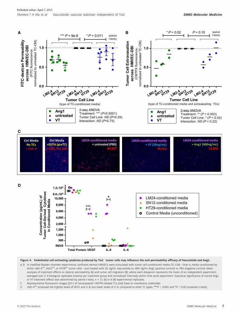
In the presence of TC-CM stimulation, Ang1 or VT treatments
reduced transendothelial macromolecular permeability (P < 0.05;
Fig 4A) and TC migration (P < 0.05 for Ang1 and P = 0.10 for VT;
Fig 4B and C). Interestingly, the degree of Ang1 or VT treatment
efficacy varied depending on the type of TC-CM used (Fig 4B); for
instance, VT treatment achieved greater inhibition of TC migration,
on average, in the presence of LM2-4luc-conditioned media (�43%)
compared to SN12luc-conditioned media (�23%). We asked whether
this could be due to differences in cytokine composition between
the three TC-CM types.
By ELISA or flow cytometric bead-based immunoassays, we
quantified the media concentrations of three permeability-inducing
ª 2015 The Authors EMBO Molecular Medicine
Florence T H Wu et al Vasculotide: vascular stabilizer independent of Tie2 EMBO Molecular Medicine
3
Published online: April 7, 2015
Tumor Cell (LM2-4CMTPX) Extravasation across HMVEC-LBl
0
1
2
3 * P = 0.02 P = 0.053
-
-
VT 10ng/mL
-
-
T 2U/mL
VT 10ng/mL
T 2U/mL
A1 200ng/mL
T 2U/mL
-
-
no TCcontrolTe
xasR
ed F
luor
esce
nce
(nor
mal
ized
to P
BS)
FITC-Dextran Permeability across HMVEC-LBl
0.0
0.5
1.0
1.5
2.0P = 0.0053 ** 0.003 ***
P = 0.02 *
Pre-Treatment:
Stimulation:
-
-
VT 10ng/mL
-
-
T 2U/mL
VT 10ng/mL
T 2U/mL
A1 200ng/mL
T 2U/mL
FITC
Flu
ores
cenc
e(n
orm
aliz
ed to
PBS
)
PBS+ 0.1% BSANo tumor cells0
PBS+ 0.1% BSA50,628
PBS+ Thrombin 2U/mL93,730
VT 10ng/mL+ Thrombin 2U/mL73,044
Ang1 200ng/mL+ Thrombin 2U/mL76,590
VT 10ng/mL+ 0.1% BSA59,539
A
ED
C
B
Tumor Cell (LM2-4CMTPX) Extravasation across HMVEC-DBl
0
1
2
3
4
5
20 P = 0.09
Pre-Treatment:
EDTA50mM
(pre-TC)
VT1.4nM
Thrombin(U/mL): - -
-
- 1.4 1.4 1.4 1.4 1.4 1.4
- - -VT
1.4nM+
T7c140nM
Ang1~1.4nM
(400ng/mL)
PEG-Cys1.4nM
T7c1.4nM
VT1.4nM
(20ng/mL)
noTCctrl
Texa
sRed
Flu
ores
cenc
e(n
orm
aliz
ed to
PBS
)
FITC-Dextran Permeability across HMVEC-DBl
0
2
4
6P = 0.0007 *** P = 0.011 *
P = 0.04 *
Pre-Treatment:
EDTA50mM
VT1.4nM
Thrombin(U/mL): -
-
- 1.4 1.4 1.4 1.4 1.4 1.4
- -VT
1.4nM+
T7c140nM
Ang1~1.4nM
(400ng/mL)
PEG-Cys1.4nM
T7c1.4nM
VT1.4nM
(20ng/mL)
FITC
Flu
ores
cenc
e(n
orm
aliz
ed to
PBS
)
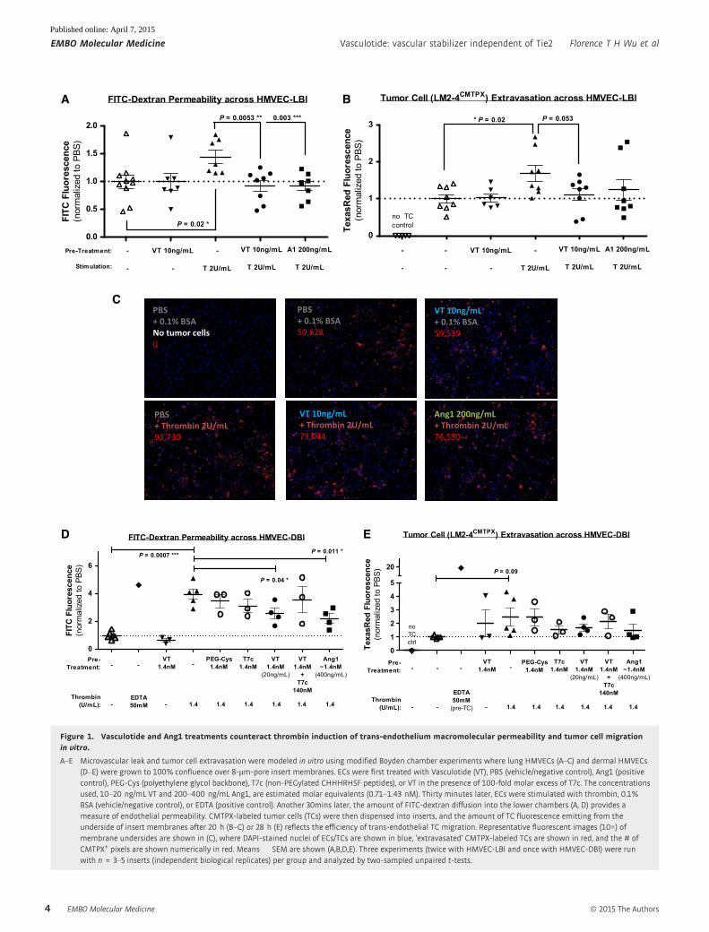
Figure 1. Vasculotide and Ang1 treatments counteract thrombin induction of trans-endothelium macromolecular permeability and tumor cell migrationin vitro.
A–E Microvascular leak and tumor cell extravasation were modeled in vitro using modified Boyden chamber experiments where lung HMVECs (A–C) and dermal HMVECs(D–E) were grown to 100% confluence over 8-lm-pore insert membranes. ECs were first treated with Vasculotide (VT), PBS (vehicle/negative control), Ang1 (positivecontrol), PEG-Cys (polyethylene glycol backbone), T7c (non-PEGylated CHHHRHSF peptides), or VT in the presence of 100-fold molar excess of T7c. The concentrationsused, 10–20 ng/mL VT and 200–400 ng/mL Ang1, are estimated molar equivalents (0.71–1.43 nM). Thirty minutes later, ECs were stimulated with thrombin, 0.1%BSA (vehicle/negative control), or EDTA (positive control). Another 30mins later, the amount of FITC-dextran diffusion into the lower chambers (A, D) provides ameasure of endothelial permeability. CMTPX-labeled tumor cells (TCs) were then dispensed into inserts, and the amount of TC fluorescence emitting from theunderside of insert membranes after 20 h (B–C) or 28 h (E) reflects the efficiency of trans-endothelial TC migration. Representative fluorescent images (10×) ofmembrane undersides are shown in (C), where DAPI-stained nuclei of ECs/TCs are shown in blue, ‘extravasated’ CMTPX-labeled TCs are shown in red, and the # ofCMTPX+ pixels are shown numerically in red. Means � SEM are shown (A,B,D,E). Three experiments (twice with HMVEC-LBl and once with HMVEC-DBl) were runwith n = 3–5 inserts (independent biological replicates) per group and analyzed by two-sampled unpaired t-tests.
EMBO Molecular Medicine ª 2015 The Authors
EMBO Molecular Medicine Vasculotide: vascular stabilizer independent of Tie2 Florence T H Wu et al
4
Published online: April 7, 2015
and/or pro-inflammatory cytokines, whose elevated levels in
cancer patients often correlate with disease progression and poor
prognosis: VEGF, IL-8, and IL-6 (Ferrara, 2005; Kut et al, 2007;
Lippitz, 2013). Both VEGF and IL-8 can directly activate ECs, via
VEGFR2 and CXCR1/CXCR2 receptors, respectively (Kim et al,
2001a; Le Guelte et al, 2011). In contrast, IL-6 primarily targets
IL-6R+ leukocytes/TCs and only indirectly affects IL-6R� ECs
(Romano et al, 1997; Lo et al, 2011). We found that the media
concentrations of VEGF and IL-8, but not IL-6, were considerably
higher in LM2-4-conditioned media compared to other TC-CM types
(Fig 4D)—suggesting that the contribution of LM2-4luc-derived
VEGF/IL-8 to endothelial gap formation (Fig 4A) and TC diapedesis
(Fig 4B) may be particularly amenable to counteraction by Ang1 or
VT.
Spontaneous breast cancer metastasis model: Vasculotidetreatment does not suppress orthotopic primary tumor growthbut inhibits metastasis to the lungs
Orthotopic implantation experiments were also performed with the
LM2-4luc cells, to further explore the effects of VT on primary tumor
growth vs. spontaneous metastatic dissemination.
A E
B
C D
F
G H
-7 0 7 14 21 28 35 42 490
10
20
30
40
50
60
70
80
90
100
Vehicle
Sunitinib (SU)
Vasculotide (VT)
Plog-rank = 0.025 *Hazard ratio = 0.19 (95% CI, 0.04 to 0.81)
Plog-rank = 0.012 *Hazard ratio = 0.13 (95% CI, 0.03 to 0.63)
SU+VT
250ng, i.p., q2d60mg/kg, p.o., ODSUVT
Days Post-Implantation (DPI) of LM2-4luc, i.v.
% S
urvi
val
-7 0 7 14 21 28 35 42 49 560
10
20
30
40
60
70
80
90
100
VechicleVasculotide (VT)Sunitinib (SU)SU+VT
All comparisonsPlog-rank > 0.05.
50
250ng, i.p., q2d60mg/kg, p.o., ODSUVT
Days Post-Implantation (DPI) of LM2-4luc, i.v.
% S
urvi
val
24 D
PI
Vehicle SU+VT
Color Bar: Min = 1e+04, Max = 5e+04 (p/sec/cm2/sr)1
DPI
Color Bar: Min = 5e+04, Max = 1e+07 (p/sec/cm2/sr)
27 D
PI
Vehicle SU+VTSunitinib (SU)Vasculotide (VT)Sunitinib (SU)Vasculotide (VT)
24 D
PI
Color Bar: Min = 5e+04, Max = 1e+07 (p/sec/cm2/sr)
Color Bar: Min = 1e+04, Max = 1e+07 (p/sec/cm2/sr)
1 D
PI
104
105
106
107
108
109
All comparisons P > 0.05.Cau
dal
Lym
phat
icM
etas
tase
s(p
hoto
ns/s
ec)
27 D
PI
105
106
107
108
109 P < 0.05 *
P < 0.05 *
Lung
Met
asta
ses
(pho
tons
/sec
)
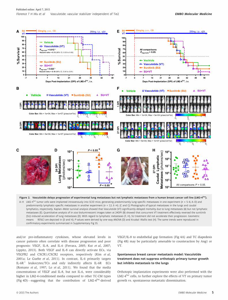
Figure 2. Vasculotide delays progression of experimental lung metastases but not lymphatic metastases from a human breast cancer cell line (LM2-4luc).
A–H LM2-4luc tumor cells were implanted intravenously into SCID mice, generating predominantly lung-specific metastases in one experiment (n = 5–6; A–D) andpredominantly lymphatic-specific metastases in another experiment (n = 12; E–H). (C and G) Photographs of typical metastases in the lungs and caudallymphatics, respectively. Kaplan–Meier survival analysis showed that Vasculotide (VT) significantly delayed mortality due to lung metastases (A) but not lymphaticmetastases (E). Quantitative analysis of in vivo bioluminescent images taken at 24DPI (B) showed that concurrent VT treatment effectively reversed the sunitinib(SU)-induced acceleration of lung metastases (D). With regard to lymphatic metastases (F, H), SU treatment did not accelerate their progression. Geometricmeans � 95%CI are depicted in (D and H); P values were derived by one-way ANOVA (D) and Kruskal–Wallis test (H). The same trends were reproduced inconfirmatory experiments summarized in Supplementary Fig S5.
ª 2015 The Authors EMBO Molecular Medicine
Florence T H Wu et al Vasculotide: vascular stabilizer independent of Tie2 EMBO Molecular Medicine
5
Published online: April 7, 2015
The first experiment involved VT and SU treatments in the pres-
ence of an unresected primary tumor (Fig 5). At 10 days post-
implantation (DPI), when primary tumors reached an average size
of ~175 mm3, mice were randomized into groups and chronic VT
therapy was initiated. At 17DPI, when tumors reached an average
size (~400 mm3) where tumor debulking is typically required for
long-term metastasis experiments, a 7-day anti-angiogenic SU treat-
ment was initiated in lieu of surgical resection. SU effectively stabi-
lized primary tumor growth (Fig 5A–C). In contrast, VT did not
alter the growth kinetics of primary tumors, as measured by volume
or bioluminescence (Fig 5A–C). This was consistent with in vitro
observations from an MTS cell viability assay (Supplementary
Fig S2), where VT had no direct cytotoxic effect on Tie2� TCs and
did not interfere with SU inhibition of Tie2+ ECs. Histological
analysis of primary tumors showed that the extent of tumor cell
viability (assessed by H&E staining; Fig 5D) and tumor hypoxia
(assessed by IHC staining of CAIX, a target of the hypoxia-inducible
transcription factor, HIF-1; Fig 5E) also remained unchanged after
22 days of VT treatment.
However, as detected by organ-specific ex vivo bioluminescent
imaging (EVBI) upon sacrifice, there were trends of reduced
incidences of metastatic involvement in the lungs, but no change in
C
-7 7 21 35 49 63 770
25
50
75
100
PBS, male (n=10)VT, male (n=10)PBS, female (n=5)VT, female (n=5)
Plog-rank
0.89
0.37
VT 250ng, i.p., q2d
Days Post-Implantation (DPI) of HT29luc, i.v.
% S
urvi
val
Male PBS, 35 DPI
Male VT, 35 DPI
104
2
46105
2
46106
2
46107
Col
or B
ar (p
/sec
/cm
2 /sr)
A
B
-7 7 21 35 49 63 770
25
50
75
100
PBSVT-250ng/2dVT-400ng/2d
Plog rank > 0.05.
VT
0
Days Post-Implantation (DPI) of SN12luc, i.v.
% S
urvi
val
VT 250ng/2d
VT 400ng/2d
50 DPI
PBS
104
2
4
68105
2
4
68106G
E
F
PBS#5, 49 DPI
VT#5, 53 DPI
D
Female (28DPI of HT29luc)
PBS VT105
106
107 P = 0.55
Tota
l Flu
x (p
hoto
ns/s
ec)
Male (35DPI of HT29luc)
PBS VT106
107
108P = 0.24
Tota
l Flu
x (p
hoto
ns/s
ec)
Col
or B
ar (p
/sec
/cm
2 /sr)
Control VT 250ng/2d VT 400ng/2d105
106
107
108P = 0.63
Tota
l Flu
x (p
hoto
ns/s
ec)
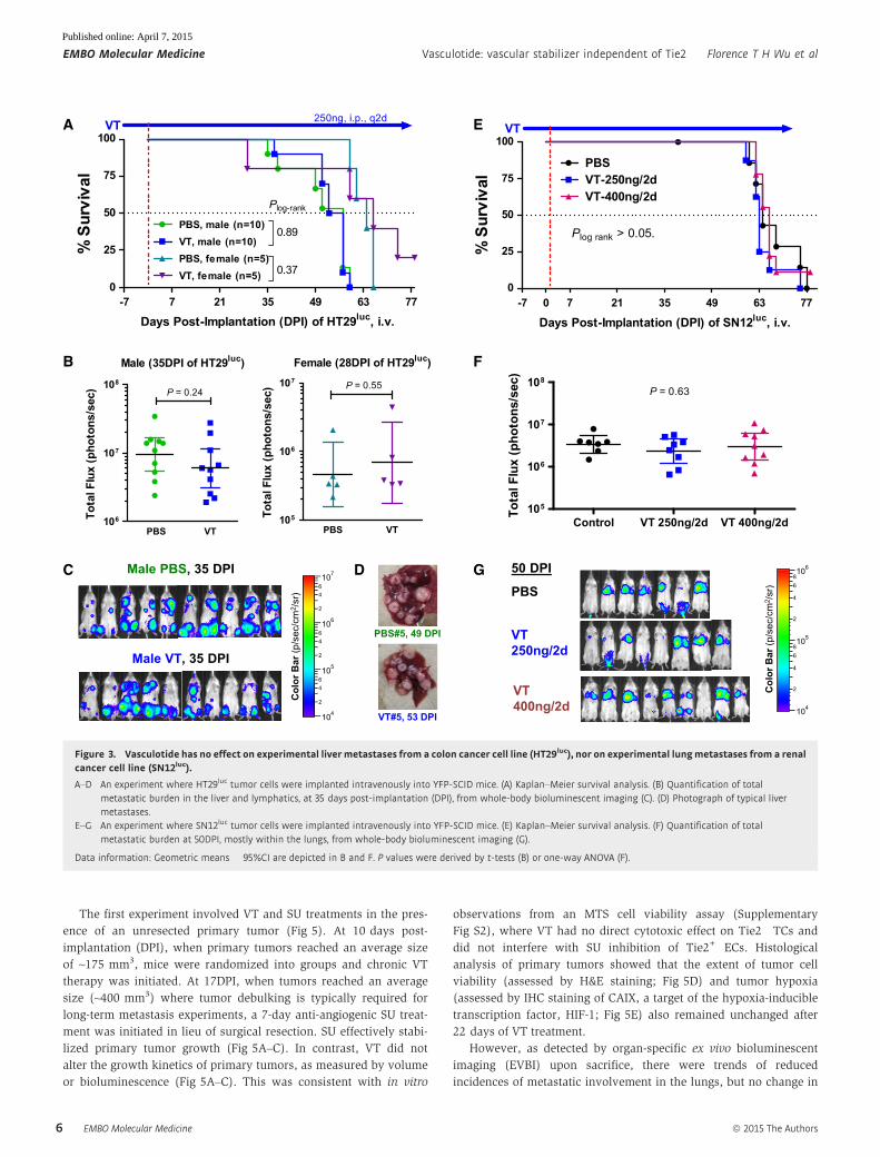
Figure 3. Vasculotide has no effect on experimental livermetastases from a colon cancer cell line (HT29luc), nor on experimental lungmetastases from a renalcancer cell line (SN12luc).
A–D An experiment where HT29luc tumor cells were implanted intravenously into YFP-SCID mice. (A) Kaplan–Meier survival analysis. (B) Quantification of totalmetastatic burden in the liver and lymphatics, at 35 days post-implantation (DPI), from whole-body bioluminescent imaging (C). (D) Photograph of typical livermetastases.
E–G An experiment where SN12luc tumor cells were implanted intravenously into YFP-SCID mice. (E) Kaplan–Meier survival analysis. (F) Quantification of totalmetastatic burden at 50DPI, mostly within the lungs, from whole-body bioluminescent imaging (G).
Data information: Geometric means � 95%CI are depicted in B and F. P values were derived by t-tests (B) or one-way ANOVA (F).
EMBO Molecular Medicine ª 2015 The Authors
EMBO Molecular Medicine Vasculotide: vascular stabilizer independent of Tie2 Florence T H Wu et al
6
Published online: April 7, 2015
A
D
B
LM24-condi�oned media+ untreated (PBS)
80,607
LM24-condi�oned media+ Ang1 (400ng/mL)
22,890
LM24-condi�oned media+ VT (20ng/mL)
40,062
Ctrl MediaNo TCs− Ctrl: 0
Ctrl Media+EDTA (preTC)+ Ctrl: 761,328
C
Total Protein VEGF IL-8 IL-6
500
1,000
1,500
2,000
4,000
6,000
8,000
10,0008.0×108
1.2×10⁹
IND/0
****
Con
cent
ratio
n (p
g/m
L) o
fTu
mor
Cel
l-Der
ived
Cyt
okin
esin
Con
ditio
ned
Med
ia
LM2-4SN12
HT29LM2-4
SN12HT29
LM2-4SN12
HT290.0
0.5
1.0
2-way ANOVATreatment: ** (P = 0.003)Tumor Cell Line: * (P = 0.02)Interaction: NS (P = 0.22)
untreatedVT
Ang1
P = 0.10* P = 0.02 (pairedt-tests)
Tumor Cell Line(type of TC-conditioned media and extravasating TCs)
Tum
or C
ell E
xtra
vasa
tion
acro
ss H
MVE
C-D
Bl
(CM
TPX
fluor
esce
nce,
norm
aliz
ed to
unt
reat
ed T
C-C
M)
LM2-4SN12
HT29LM2-4
SN12HT29
LM2-4SN12
HT290.0
0.5
1.0
2-way ANOVATreatment: *** (P<0.0001)Tumor Cell Line: NS (P=0.29)Interaction: NS (P=0.75)
untreatedVT
Ang1
* P = 0.011*** P = 9e-6
Tumor Cell Line(type of TC-conditioned media)
FITC
-dex
tran
Per
mea
bilit
yac
ross
HM
VEC
-DB
l(F
ITC
fluo
resc
ence
norm
aliz
ed to
unt
reat
ed T
C-C
M)
(pairedt-tests)
LM24-conditioned media
HT29-conditioned mediaControl Media (unconditioned)
SN12-conditioned media
Figure 4. Endothelial cell-activating cytokines produced by Tie2� tumor cells may influence the anti-permeability efficacy of Vasculotide and Ang1.
A, B In modified Boyden chamber experiments, confluent dermal HMVECs were stimulated with tumor cell-conditioned media (TC-CM)—that is, media conditioned byeither LM2-4luc, SN12luc, or HT29luc tumor cells—and treated with 20 ng/mL Vasculotide vs. 400 ng/mL Ang1 (positive control) vs. PBS (negative control). Meta-analyses of treatment effects on dextran permeability (A) and tumor cell migration (B), where each datapoint represents the mean of an independent experiment,averaged over 2–3 biological replicates (inserts) per treatment group and normalized internally within that same experiment. Statistical significance of overall Ang1or VT treatment effects was determined by paired t-tests, n = 11 (A) or 6 (B) experimental replicates.
C Representative fluorescent images (10×) of ‘extravasated’ CMTPX-labeled TCs (red) fixed on membrane undersides.D LM2-4luc produced the highest levels of VEGF and IL-8, but lower levels of IL-6, compared to other TC types; ***P < 0.001 and *P < 0.05 (unpaired t-tests).
ª 2015 The Authors EMBO Molecular Medicine
Florence T H Wu et al Vasculotide: vascular stabilizer independent of Tie2 EMBO Molecular Medicine
7
Published online: April 7, 2015
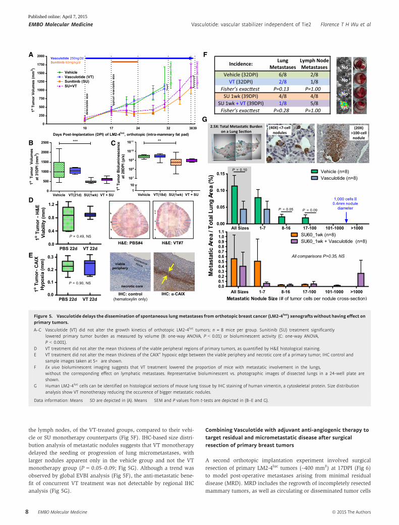
the lymph nodes, of the VT-treated groups, compared to their vehi-
cle or SU monotherapy counterparts (Fig 5F). IHC-based size distri-
bution analysis of metastatic nodules suggests that VT monotherapy
delayed the seeding or progression of lung micrometastases, with
larger nodules apparent only in the vehicle group and not the VT
monotherapy group (P = 0.05–0.09; Fig 5G). Although a trend was
observed by global EVBI analysis (Fig 5F), the anti-metastatic bene-
fit of concurrent VT treatment was not detectable by regional IHC
analysis (Fig 5G).
Combining Vasculotide with adjuvant anti-angiogenic therapy totarget residual and micrometastatic disease after surgicalresection of primary breast tumors
A second orthotopic implantation experiment involved surgical
resection of primary LM2-4luc tumors (~400 mm3) at 17DPI (Fig 6)
to model post-operative metastases arising from minimal residual
disease (MRD). MRD includes the regrowth of incompletely resected
mammary tumors, as well as circulating or disseminated tumor cells
PBS 22d VT 22d0.0
0.4
0.8
1.2
P = 0.49, NS1o Tum
or -
H&
EVi
abili
ty (m
m)
A
B
2.5X: Total Metasta�c Burdenon a Lung Sec�on
(40X) <7-cellnodules
(20X)>100-cellnodule
F
G
All Sizes 1-7 8-16 17-100 101-1000 >10000.00.10.20.30.40.50.60.70.80.91.01.1 SU60_1wk (n=8)
SU60_1wk + Vasculotide (n=8)
All comparisons P>0.35, NS
Met
asta
tic A
rea
/ Tot
al L
ung
Area
(%)
Metastatic Nodule Size (# of tumor cells per nodule cross-section)
All Sizes 1-7 8-16 17-100 101-1000 >10000.00
0.05
0.10
0.15 Vehicle (n=8)Vasculotide (n=8)
P = 0.10
P = 0.05 P = 0.09
1,000 cells �0.4mm nodule
diameter
PBS 22d VT 22d0.0
0.1
0.2
0.3
P = 0.90, NS
1o Tum
or- C
AIX
Hyp
oxia
(mm
)
IHC: control (hematoxylin only)
necrotic core
viable periphery
IHC: α-CAIX
H&E: PBS#4 H&E: VT#7
D
E
C
0
250
500
750
1000
1250
1500
1750
2000
Vehicle
Sunitinib (SU)SU+VT
Vasculotide (VT)
10 17 24 32 3839
Sunitinib 60mg/kg/dVasculotide 250ng/2d
dete
ctab
le s
ize
typi
cal r
esec
tabl
e si
ze
endp
oint
(sac
rific
e)
} endp
oint
(sac
rific
e)
Days Post-Implantation (DPI) of LM2-4luc, orthotopic (intra-mammary fat pad)
1oTu
mor
Vol
umes
(mm
3 )
Incidence: LungMetastases
Lymph NodeMetastases
Vehicle (32DPI) 6/8 2/8VT (32DPI) 2/8 1/8
Fisher’s exac�est P=0.13 P=1.00SU 1wk (39DPI) 4/8 4/8
SU 1wk + VT (39DPI) 1/8 5/8Fisher’s exac�est P=0.28 P=1.00
Vehicle VT(18d) SU(1wk) VT + SU1
10
107
108
109
1010
1011 **
1o Tum
or B
iolu
min
esce
nce
at 2
8DPI
(p/s
)
Vehicle VT(21d) SU(1wk) VT + SU0
500
1000
1500
2000
2500 ***
1o Tum
or V
olum
esat
31D
PI (m
m3 )
Figure 5. Vasculotide delays the dissemination of spontaneous lungmetastases from orthotopic breast cancer (LM2-4luc) xenografts without having effect onprimary tumors.
A–C Vasculotide (VT) did not alter the growth kinetics of orthotopic LM2-4luc tumors; n = 8 mice per group. Sunitinib (SU) treatment significantlylowered primary tumor burden as measured by volume (B: one-way ANOVA, P < 0.01) or bioluminescent activity (C: one-way ANOVA,P < 0.001).
D VT treatment did not alter the mean thickness of the viable peripheral regions of primary tumors, as quantified by H&E histological staining.E VT treatment did not alter the mean thickness of the CAIX+ hypoxic edge between the viable periphery and necrotic core of a primary tumor; IHC control and
sample images taken at 5× are shown.F Ex vivo bioluminescent imaging suggests that VT treatment lowered the proportion of mice with metastatic involvement in the lungs,
without the corresponding effect on lymphatic metastases. Representative bioluminescent vs. photographic images of dissected lungs in a 24-well plate areshown.
G Human LM2-4luc cells can be identified on histological sections of mouse lung tissue by IHC staining of human vimentin, a cytoskeletal protein. Size distributionanalysis show VT monotherapy reducing the occurence of bigger metastatic nodules.
Data information: Means � SD are depicted in (A). Means � SEM and P values from t-tests are depicted in (B–E and G).
EMBO Molecular Medicine ª 2015 The Authors
EMBO Molecular Medicine Vasculotide: vascular stabilizer independent of Tie2 Florence T H Wu et al
8
Published online: April 7, 2015
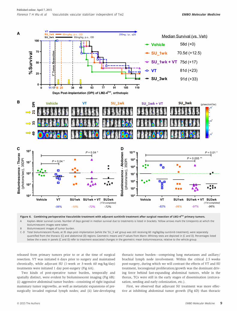
released from primary tumors prior to or at the time of surgical
resection. VT was initiated 6 days prior to surgery and maintained
chronically, while adjuvant SU (1-week or 3-week 60 mg/kg/day)
treatments were initiated 1 day post-surgery (Fig 6A).
Two kinds of post-operative tumor burden, temporally and
spatially distinct, were evident by bioluminescent imaging (Fig 6B):
(i) aggressive abdominal tumor burden—consisting of right inguinal
mammary tumor regrowths, as well as metastatic expansions of pre-
surgically invaded regional lymph nodes; and (ii) late-developing
thoracic tumor burden—comprising lung metastases and axillary/
brachial lymph node involvement. Within the critical 2.5 weeks
post-surgery, during which we will contrast the effects of VT and SU
treatment, locoregional proliferation/growth was the dominant driv-
ing force behind fast-expanding abdominal tumors, while in the
thorax, TCs were still in the early stages of dissemination (extrava-
sation, seeding and early colonization, etc.).
First, we observed that adjuvant SU treatment was more effec-
tive at inhibiting abdominal tumor growth (Fig 6D) than thoracic
49 63 77 91 105 1190
25
50
75
100Vehicle
VT
SU_1wk
SU_1wk + VT
SU_3wk
VTSU_1wk
0 11 17 25 18 39
SU_3wk
1o Tum
or R
esec
tion
250ng, i.p., q2d60mg/kg, p.o., OD60mg/kg, p.o., OD
58d (+0)
Median Survival (vs. Veh)
81d (+23)
70.5d (+12.5)
75d (+17)
91d (+33)
Days Post-Implantation (DPI) of LM2-4luc, orthotopic
% S
urvi
val
A
DC
B(p/sec/cm2/sr)
Vehicle VT SU1wk SU1wk + VT SU3wk104
105
106
107
108
109
1010 P = 0.01 *
P = 0.005 **
-82% -86% -97% -96%(17d completed)
Bio
lum
ines
cenc
e - A
bdom
en(p
hoto
ns/s
ec),
35D
PI
Vehicle VT SU1wk SU1wk + VT SU3wk104
105
106
107
108
P = 0.04 *
-56% -10% -72% -72%(17d completed)
P = 0.04 *
Bio
lum
ines
cenc
e - T
hora
x(p
hoto
ns/s
ec),
35D
PI
Figure 6. Combining perioperative Vasculotide treatment with adjuvant sunitinib treatment after surgical resection of LM2-4luc primary tumors.
A Kaplan–Meier survival curves. Number of days gained in median survival due to treatments is listed in brackets. Yellow arrows mark the timepoints at which thebioluminescent images were taken.
B Bioluminescent images of tumor burden.C–D Total bioluminescent fluxes, at 35 days post-implantation (while the ‘SU_3 wk’ group was still receiving 60 mg/kg/day sunitinib treatment), were separately
quantified from the thoracic (C) and abdominal (D) regions. Geometric means and P values from Mann–Whitney tests are depicted in (C and D). Percentages listedbelow the x-axes in panels (C and D) refer to treatment-associated changes in the geometric mean bioluminescence, relative to the vehicle group.
ª 2015 The Authors EMBO Molecular Medicine
Florence T H Wu et al Vasculotide: vascular stabilizer independent of Tie2 EMBO Molecular Medicine
9
Published online: April 7, 2015
metastatic dissemination (Fig 6C). We also noted the brevity
of SU treatment efficacy—abdominal tumors in the ‘SU_3 wk’
group appeared to be stabilized so long as SU treatment was
maintained (Fig 6B at 35DPI), but aggressively rebounded after
cessation of SU treatment (Fig 6B at 49DPI). This transiency in
tumor growth suppression by SU accounts for the longer median
survivals associated with longer treatment duration (Fig 6A):
1-wk and 3-wk SU treatments extended median survival by
12.5 days (PGehan-Breslow-Wilcoxon = 0.17) and 33 days (PGBW = 0.08),
respectively. At 10 days after discontinuation of 1-week SU
therapy, its transient inhibitory effect on abdominal tumor growths
has greatly diminished (Fig 6D), while no benefit was observed
with respect to thoracic metastases (Fig 6C).
In contrast, long-term VT monotherapy was more effective at
inhibiting thoracic metastatic dissemination (Fig 6C, P = 0.04) than
abdominal tumor growth (Fig 6D, P > 0.05). The survival advantage
associated with VT monotherapy was not statistically significant
despite an extension in median survival of 23 days (Fig 6A,
Plog-rank = 0.42). However, concurrent VT therapy conferred additive
benefits—combining VT with 1-wk SU therapy, it lowered tumor
bioluminescence to a similar extent as 3-wk SU monotherapy (Fig 6C
and D).
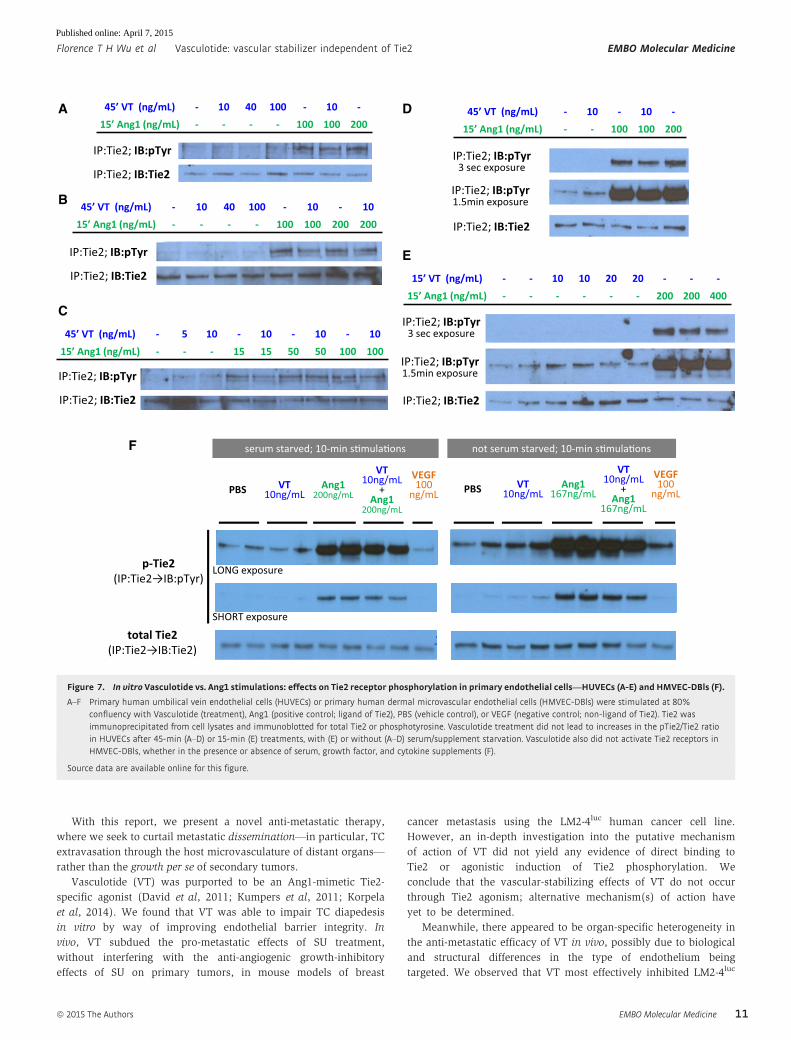
Vasculotide does not induce Tie2 phosphorylation in vitro orin vivo
To follow-up on previous characterizations of VT as a Tie2-specific
agonist, we stimulated primary human endothelial cells in vitro to
directly assess the effects of VT on Tie2 phosphorylation status
(Fig 7). Ang1, as our positive control, strongly and consistently
activated Tie2 phosphorylation across the concentrations tested
(15–400 ng/mL or 0.05–1.43 nM). VEGF, which is not a ligand of
Tie2, was used as a negative control. Unexpectedly, VT treatment
did not lead to any appreciable induction of Tie2 phosphorylation
under any conditions tested in this study—whether given at concen-
trations of 5, 10, 20, 40, or 100 ng/mL (0.36–7.1 nM); at different
timepoints (10, 15, or 45 min); with or without serum and supple-
ment starvation; in the absence or presence of concurrent Ang1
stimulation; and in ECs of venous (HUVEC) or microvascular
(HMVEC-DBl) origin (Fig 7). Importantly, even though VT (10–20
ng/mL) and Ang1 (200–400 ng/mL) both functionally inhibited
endothelial permeability in Boyden chamber assays where HMVEC-
DBl was used (Figs 1 and 4), the same concentration of VT (10 ng/mL)
did not have similar activity as equimolar Ang1 (200 ng/mL or
0.7 nM) at the upstream level of Tie2 phosphorylation in the same
dermal microvascular ECs (Fig 7F).
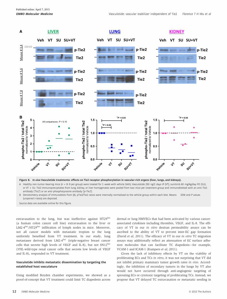
Since VT did not appear to be a direct Tie2 agonist within a
reductionistic in vitro system of cultured ECs (Fig 7), we asked
whether VT treatment may still indirectly lead to increased Tie2
phosphorylation within a physiological in vivo context. Tumor-free
mice were treated for 1 week with either vehicle, VT (250 ng/2
days), SU (60 mg/kg/day), or VT+SU and then sacrificed 5 h after
their last doses of treatment. Tissue homogenates derived from EC-
rich organs (livers, lungs, kidneys) were subjected to Tie2 immuno-
precipitation and then immunoblotted for phosphorylated Tie2
(Fig 8: ‘p-Tie2’) or total Tie2 (Fig 8: ‘Tie2’). Overall, in vivo VT treat-
ment—whether as a monotherapy or when combined with SU—
did not increase the ratio of phosphorylated Tie2 relative to total
Tie2; in fact, lung-specific pTie2/Tie2 was significantly lower in the
‘VT’ group vs. ‘vehicle’ group (Fig 8).
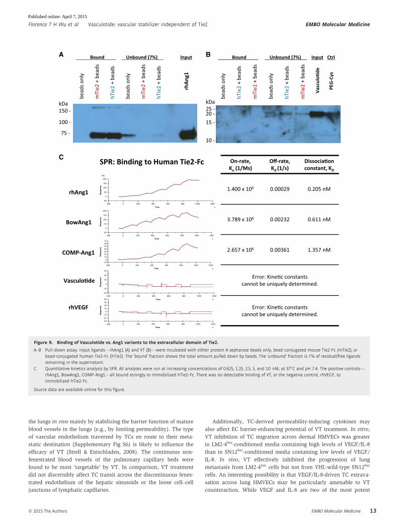
Vasculotide does not bind the extracellular domain of Tie2
To better understand the difficulties we had in observing Tie2
activation by VT, we sought to characterize the binding affinity and
kinetics of VT to Tie2. Purified human or mouse Tie2-Fc (i.e.,
Fc-conjugated extracellular domains of Tie2) were first coupled to
protein A sepharose beads for pull-down experiments (Fig 9A and B)
or immobilized on a Biacore CM5 sensor chip for surface plasmon
resonance (SPR) experiments (Fig 9C and Supplementary Fig S3). In
the pull-down assays, human and mouse Tie2-Fc effectively pulled
down the majority of available recombinant human Ang1 (rhAng1),
but did not pull down any appreciable levels of VT (Fig 9A and B).
Likewise, SPR experiments—performed at physiological conditions
of 37°C and pH 7.4—showed rhAng1, BowAng1, and COMP-Ang1 to
be high-affinity binders (dissociation constants, KD, of 0.2–1.4 nM)
to human and mouse Tie2-Fc (Fig 9C and Supplementary Fig S3B),
but no binding to Tie2-Fc was detectable for VT, VEGF, PEG-Cys, or
T7c (Fig 9C and Supplementary Fig S3).
Discussion
We report here five new findings relevant to therapeutic manipula-
tion of the vasculature for cancer treatment, especially of metastatic
disease. First, VT inhibits vascular permeability to the extravasation
of tumor cells in vitro. Second, consistent with the aforementioned
findings, VT inhibits the early stages of the metastatic process in vivo
and thus may be potentially effective as an adjuvant therapy; in
contrast, it does not inhibit tumor growth per se, for example, of
established primary tumors. Third, the anti-metastatic effects of VT
appear to be organ and tumor dependent, for example, inhibition of
lung metastasis of breast cancer was observed but not of liver
metastasis by colorectal cancer cells nor of lung metastasis by renal
cell carcinoma cells. Fourth, these aforementioned effects appear to
be independent of binding to or activation of Tie2 and thus may
work through a different and thus potentially novel mechanism.
Fifth, a potentially important ‘off-target’ effect of a commonly used
anti-angiogenic TKI, sunitinib, was observed—inhibition of Tie2
phosphorylation.
This study addresses a gap in cancer therapy research, where
efforts have largely focused on identifying drugs that limit the local
malignant growth of tumors, resulting in a paucity of treatments
that limit the distant metastatic spreading of cancer (Steeg, 2012).
For instance, sunitinib (SU), an anti-angiogenic TKI that target the
VEGF receptors (VEGFRs), repeatedly failed in multiple phase III
clinical trials and preclinical models of advanced metastatic breast
cancer, whereas, in contrast, it demonstrated potency in inhibiting
angiogenesis-dependent growth of primary breast tumors in
preclinical models (Abrams et al, 2003; Guerin et al, 2013). Recent
preclinical studies suggest that many anti-angiogenic TKIs may
inadvertently destabilize the normal microvasculature of distant
host organs to facilitate TC extravasation and promote paradoxical
pro-metastatic side effects, for example, by targeting pericytes
(Ebos et al, 2009; Chung et al, 2012; Cooke et al, 2012; Welti et al,
2012).
EMBO Molecular Medicine ª 2015 The Authors
EMBO Molecular Medicine Vasculotide: vascular stabilizer independent of Tie2 Florence T H Wu et al
10
Published online: April 7, 2015
With this report, we present a novel anti-metastatic therapy,
where we seek to curtail metastatic dissemination—in particular, TC
extravasation through the host microvasculature of distant organs—
rather than the growth per se of secondary tumors.
Vasculotide (VT) was purported to be an Ang1-mimetic Tie2-
specific agonist (David et al, 2011; Kumpers et al, 2011; Korpela
et al, 2014). We found that VT was able to impair TC diapedesis
in vitro by way of improving endothelial barrier integrity. In
vivo, VT subdued the pro-metastatic effects of SU treatment,
without interfering with the anti-angiogenic growth-inhibitory
effects of SU on primary tumors, in mouse models of breast
cancer metastasis using the LM2-4luc human cancer cell line.
However, an in-depth investigation into the putative mechanism
of action of VT did not yield any evidence of direct binding to
Tie2 or agonistic induction of Tie2 phosphorylation. We
conclude that the vascular-stabilizing effects of VT do not occur
through Tie2 agonism; alternative mechanism(s) of action have
yet to be determined.
Meanwhile, there appeared to be organ-specific heterogeneity in
the anti-metastatic efficacy of VT in vivo, possibly due to biological
and structural differences in the type of endothelium being
targeted. We observed that VT most effectively inhibited LM2-4luc
PBS VT10ng/mL
Ang1200ng/mL
VT10ng/mL
+Ang1
200ng/mL
VEGF100
ng/mL
A
B
C
D
E
F
IP:Tie2; IB:pTyr
IP:Tie2; IB:Tie2
45’ VT (ng/mL) - 10 40 100 - 10 -15’ Ang1 (ng/mL) - - - - 100 100 200
IP:Tie2; IB:pTyr1.5min exposure
IP:Tie2; IB:Tie2
45’ VT (ng/mL) - 10 - 10 -15’ Ang1 (ng/mL) - - 100 100 200
IP:Tie2; IB:pTyr3 sec exposure
IP:Tie2; IB:pTyr
IP:Tie2; IB:Tie2
45’ VT (ng/mL) - 10 40 100 - 10 - 1015’ Ang1 (ng/mL) - - - - 100 100 200 200
15’ VT (ng/mL) - - 10 10 20 20 - - -15’ Ang1 (ng/mL) - - - - - - 200 200 400
IP:Tie2; IB:pTyr1.5min exposure
IP:Tie2; IB:Tie2
IP:Tie2; IB:pTyr3 sec exposure45’ VT (ng/mL) - 5 10 - 10 - 10 - 10
15’ Ang1 (ng/mL) - - - 15 15 50 50 100 100
IP:Tie2; IB:pTyr
IP:Tie2; IB:Tie2
not serum starved; 10-min s�mula�onsserum starved; 10-min s�mula�ons
p-Tie2(IP:Tie2→IB:pTyr)
total Tie2(IP:Tie2→IB:Tie2)
LONG exposure
SHORT exposure
PBS VT10ng/mL
Ang1167ng/mL
VT10ng/mL
+Ang1
167ng/mL
VEGF100
ng/mL
Figure 7. In vitro Vasculotide vs. Ang1 stimulations: effects on Tie2 receptor phosphorylation in primary endothelial cells—HUVECs (A-E) and HMVEC-DBls (F).
A–F Primary human umbilical vein endothelial cells (HUVECs) or primary human dermal microvascular endothelial cells (HMVEC-DBls) were stimulated at 80%confluency with Vasculotide (treatment), Ang1 (positive control; ligand of Tie2), PBS (vehicle control), or VEGF (negative control; non-ligand of Tie2). Tie2 wasimmunoprecipitated from cell lysates and immunoblotted for total Tie2 or phosphotyrosine. Vasculotide treatment did not lead to increases in the pTie2/Tie2 ratioin HUVECs after 45-min (A–D) or 15-min (E) treatments, with (E) or without (A–D) serum/supplement starvation. Vasculotide also did not activate Tie2 receptors inHMVEC-DBls, whether in the presence or absence of serum, growth factor, and cytokine supplements (F).
Source data are available online for this figure.
ª 2015 The Authors EMBO Molecular Medicine
Florence T H Wu et al Vasculotide: vascular stabilizer independent of Tie2 EMBO Molecular Medicine
11
Published online: April 7, 2015
extravasation to the lung, but was ineffective against HT29luc
(a human colon cancer cell line) extravasation to the liver or
LM2-4luc/HT29luc infiltration of lymph nodes in mice. Moreover,
not all cancer models with metastatic tropism to the lung
uniformly benefited from VT treatment. In our study, lung
metastases derived from LM2-4luc (triple-negative breast cancer
cells that secrete high levels of VEGF and IL-8), but not SN12luc
(VHL-wild-type renal cancer cells that secrete low levels of VEGF
and IL-8), responded to VT treatment.
Vasculotide inhibits metastatic dissemination by targeting theestablished host vasculature
Using modified Boyden chamber experiments, we showed as a
proof-of-concept that VT treatment could limit TC diapedesis across
dermal or lung HMVECs that had been activated by various cancer-
associated cytokines including thrombin, VEGF, and IL-8. The effi-
cacy of VT in our in vitro dextran permeability assays can be
ascribed to the ability of VT to prevent inter-EC gap formation
(David et al, 2011). The efficacy of VT in our in vitro TC migration
assays may additionally reflect an attenuation of EC surface adhe-
sion molecules that can facilitate TC diapedesis—for example,
VCAM-1 and ICAM-1 (Kumpers et al, 2011).
Given the lack of inhibitory effects by VT on the viability of
proliferating ECs and TCs in vitro, it was not surprising that VT did
not inhibit primary mammary tumor growth rates in vivo. Accord-
ingly, the inhibition of secondary tumors in the lungs by VT also
would not have occurred through anti-angiogenic targeting of
sprouting ECs or cytotoxic targeting of proliferating TCs. Instead, we
propose that VT delayed TC extravasation or metastatic seeding in
LIVER
p-Tie2
Tie2
p-Tie2
Tie2
Veh VT SU SU+VT
Mou
se #
5,6
Mou
se #
3,4
LUNG KIDNEY
p-Tie2
Tie2
p-Tie2
Tie2
Mou
se #
7,8 p-Tie2
Tie2
Veh VT SU SU+VT Veh VT SU SU+VT
p-Tie2
Tie2
150 kD
Vehicl
e VT SU
SU+VT0.0
0.5
1.0
1.5
phos
pho-
Tie2
/ to
tal T
ie2
norm
aliz
ed to
Veh
icle
*P = 0.04
Vehicl
e VT SU
SU+VT0.0
0.5
1.0
1.5
phos
pho-
Tie2
/ to
tal T
ie2
norm
aliz
ed to
Veh
icle
*P = 0.02
*P = 0.03
Vehicl
e VT SU
SU+VT0
1
2
3
4
5All comparisons: P > 0.10
phos
pho-
Tie2
/ to
tal T
ie2
norm
aliz
ed to
Veh
icle
A
B
p-Tie2
Tie2
Tie2
p-Tie2
Figure 8. In vivo Vasculotide treatments: effects on Tie2 receptor phosphorylation in vascular-rich organs (liver, lungs, and kidneys).
A Healthy non-tumor-bearing mice (n = 8–9 per group) were treated for 1 week with vehicle (Veh); Vasculotide 250 ng/2 days IP (VT); sunitinib 60 mg/kg/day PO (SU);or VT + SU. Tie2 immunoprecipitates from lung, kidney, or liver homogenates were pooled from two mice per treatment group and immunoblotted with an anti-Tie2antibody (‘Tie2’) or an anti-phosphotyrosine antibody (‘p-Tie2’).
B Densitometry analysis of immunoblots from (A). pTie2/Tie2 ratios were internally normalized to the vehicle group within each blot. Means � SEM and P values(unpaired t-tests) are depicted.
Source data are available online for this figure.
EMBO Molecular Medicine ª 2015 The Authors
EMBO Molecular Medicine Vasculotide: vascular stabilizer independent of Tie2 Florence T H Wu et al
12
Published online: April 7, 2015
the lungs in vivo mainly by stabilizing the barrier function of mature
blood vessels in the lungs (e.g., by limiting permeability). The type
of vascular endothelium traversed by TCs en route to their meta-
static destination (Supplementary Fig S6) is likely to influence the
efficacy of VT (Strell & Entschladen, 2008). The continuous non-
fenestrated blood vessels of the pulmonary capillary beds were
found to be most ‘targetable’ by VT. In comparison, VT treatment
did not discernibly affect TC transit across the discontinuous fenes-
trated endothelium of the hepatic sinusoids or the loose cell–cell
junctions of lymphatic capillaries.
Additionally, TC-derived permeability-inducing cytokines may
also affect EC barrier-enhancing potential of VT treatment. In vitro,
VT inhibition of TC migration across dermal HMVECs was greater
in LM2-4luc-conditioned media containing high levels of VEGF/IL-8
than in SN12luc-conditioned media containing low levels of VEGF/
IL-8. In vivo, VT effectively inhibited the progression of lung
metastasis from LM2-4luc cells but not from VHL-wild-type SN12luc
cells. An interesting possibility is that VEGF/IL-8-driven TC extrava-
sation across lung HMVECs may be particularly amenable to VT
counteraction. While VEGF and IL-8 are two of the most potent
kDa25 -20 -15 -
10 -
Bound . Unbound (7%) . Input
bead
sonl
y
mTi
e2 +
bea
ds
hTie
2 +
bead
s
bead
sonl
y
mTi
e2 +
bea
ds
hTie
2 +
bead
s
rhAn
g1
100 -
kDa150 -
75 -
BA Bound . Unbound (7%) . Input Ctrl
bead
sonl
y
hTie
2 +
bead
s
mTi
e2 +
bea
ds
bead
sonl
y
hTie
2 +
bead
s
mTi
e2 +
bea
ds
Vasc
ulo�
de
PEG
-Cys
BowAng1
COMP-Ang1
rhAng1
Vasculo�de
rhVEGF
-30-20-10
010203040
-200 0 200 400 600 800 1000 1200
Res
pons
e
Time s
-20
-10
0
10
20
30
-200 0 200 400 600 800 1000 1200
Res
pons
e
Time s
-100
10203040506070
-200 0 200 400 600 800 1000 1200
Res
pons
e
Time s
-50
0
50
100
150
200
-200 0 200 400 600 800 1000 1200
Res
pons
e
Time s
-50
0
50
100
150
200
-200 0 200 400 600 800 1000 1200
RU
Res
pons
e
Time s
On-rate,Ka (1/Ms)
Off-rate,Kd (1/s)
Dissocia�on constant, KD
1.400 x 10⁶ 0.00029 0.205 nM
3.789 x 10⁶ 0.00232 0.611 nM
2.657 x 10⁶ 0.00361 1.357 nM
Error: Kine�c constants cannot be uniquely determined.
Error: Kine�c constants cannot be uniquely determined.
SPR: Binding to Human Tie2-FcC
Figure 9. Binding of Vasculotide vs. Ang1 variants to the extracellular domain of Tie2.
A–B Pull-down assay. Input ligands—rhAng1 (A) and VT (B)—were incubated with either protein A sepharose beads only, bead-conjugated mouse Tie2-Fc (mTie2), orbead-conjugated human Tie2-Fc (hTie2). The ‘bound’ fraction shows the total amount pulled down by beads. The ‘unbound’ fraction is 7% of residual/free ligandsremaining in the supernatant.
C Quantitative kinetics analysis by SPR. All analytes were run at increasing concentrations of 0.625, 1.25, 2.5, 5, and 10 nM, at 37°C and pH 7.4. The positive controls—rhAng1, BowAng1, COMP-Ang1—all bound strongly to immobilized hTie2-Fc. There was no detectable binding of VT, or the negative control, rhVEGF, toimmobilized hTie2-Fc.
Source data are available online for this figure.
ª 2015 The Authors EMBO Molecular Medicine
Florence T H Wu et al Vasculotide: vascular stabilizer independent of Tie2 EMBO Molecular Medicine
13
Published online: April 7, 2015
and well-studied inducers of vascular permeability, there are
potentially other factors that could also significantly modulate the
efficacy of VT.
Is Vasculotide a Tie2 agonist or an Ang1 mimetic?
By definition, a Tie2 agonist is a ligand that (a) directly binds to
Tie2 receptors and (b) activates the tyrosine kinase activity and
autophosphorylation of Tie2. In the assays performed for this study,
we found no evidence of VT fulfilling either of these necessary
requirements.
To our knowledge, the direct binding of the full VT to Tie2 has
not been formally described or quantified. In their first paper, Van
Slyke et al observed a faint Western blot signal when 20 nM of
single-armed VT (i.e., one T7c peptide conjugated to a linear
maleimide–PEG–biotin) was used to pull-down Tie2 from whole-cell
lysates of EA.hy926 cells (Van Slyke et al, 2009). In contrast, the
direct binding of tetravalent VT—whether avidin-conjugated (Van
Slyke et al, 2009) or PEG-conjugated (David et al, 2011; Kumpers
et al, 2011; Korpela et al, 2014)—to purified Tie2 has not been
experimentally confirmed to our knowledge. The two direct binding
assays used in this study, namely a pull-down assay and SPR analy-
sis using purified Tie2-Fc, both revealed no detectable or quantifi-
able binding of PEGylated VT to the extracellular domain of Tie2.
Also unexpected was the undetectable binding of T7c peptides to
Tie2-Fc by SPR. The main focus of the original paper that discovered
T7 (Tournaire et al, 2004) was actually on a different peptide called
‘T4’ (NLLMAAS) and its potential use as a Tie2 inhibitor/ antagonist.
When they initially screened phage-displayed heptapeptides by
ELISA for Tie2-binding potential, T4 and T7 were among four candi-
dates selected for further study. These candidate peptides, in their
synthetic free form, were then subjected to competition assays by
ELISA and SPR to show that T4 was able to competitively inhibit
Ang1 or Ang2 binding to Tie2 (Ki ~ 0.3 mM), while T7 was not
(tested up to 1 mM). In essence, these competition assays confirmed
Tie2 binding for T4, but not for T7.
Our repeated investigations into the induction of Tie2 phosphory-
lation in vitro (analyzing primary venous and microvascular ECs
treated directly with VT) and in vivo (analyzing lung, kidney, and
liver tissue from mice treated with VT) also did not confirm the
anticipated agonistic activity of PEGylated VT on Tie2. Of the three
published papers on PEGylated VT, only the most recent study
showed direct testing of VT on ECs in vitro, albeit on hTERT-
immortalized ECs (Korpela et al, 2014). To show Tie2 phosphoryla-
tion or dependency, the older studies relied on in vivo systems,
where indirect dependency and sample heterogeneity (e.g., variabil-
ity in EC content) are inherently more difficult to exclude or account
for, particularly when crucial controls are absent.
Altogether, Tie2 agonism is unlikely to be the mechanism of
VT’s vascular-stabilizing effects. Since many other signaling path-
ways (e.g., VEGF, thrombin, IL-8, Ca2+) crosstalk and converge to
govern the same biological responses that are regulated by Ang-Tie2
signaling (Le Guelte et al, 2011; Koh, 2013), it would be premature
to categorize VT as an Ang1 mimetic based on similarity in down-
stream function alone.
Further studies will be needed to determine whether VT has any
other molecular binding partners, and if so, the binding affinity,
kinetics, specificity, and biological significance of such interactions.
Studies are in progress to elucidate possible mechanisms by which
VT can cause the functional effects we have observed.
Does sunitinib inhibit tumor growth, by targeting thedeveloping tumor neovasculature, while potentially exacerbatingtumor spread?
Many anti-angiogenic TKI therapies have underperformed clinically
for patients with advanced metastatic breast cancer (MBC)—
especially with sunitinib (SU) treatment, which when given as a
monotherapy or combined with conventional chemotherapies has
repeatedly failed in multiple phase III clinical trials (Ebos & Kerbel,
2011; Mackey et al, 2012). These clinical results were recently repli-
cated in a preclinical model of postsurgical advanced MBC (Guerin
et al, 2013). As discussed previously (Guerin et al, 2013), primary
and secondary tumors may have divergent responses to anti-
angiogenic therapies due to differences in their relative dependency
on angiogenesis.
Another possible explanation for the apparent insensitivity of
MBC to TKI treatments is that the growth of secondary tumors post-
metastatic colonization of the lung/liver may still be susceptible to
the intended anti-angiogenic inhibition, but that these growth-
inhibitory benefits may be diluted by concurrent unintended drug
effects that promote tumor spread. This appears to be the case in
our resected orthotopic LM2-4luc experiment, where adjuvant SU
treatment transiently suppressed the locoregional growth of residual
abdominal tumors, but was ineffective against the distal dissemina-
tion of thoracic tumors. Counterproductive dissemination-promoting
side effects may explain the clinical observations from a phase II
study of advanced MBC where response rates to sunitinib differed
between patients with locoregionally growing superficial metastatic
disease (20%) and patients with distally disseminated visceral meta-
static disease (9%) (Yardley et al, 2012).
An interesting and unexpected finding from our study is that
Tie2 dephosphorylation in the host vasculature may be yet another
unintended consequence of TKI treatments which could promote
metastatic seeding. This adds to other mechanisms previously
proposed: therapy-induced tumor hypoxia could activate HIF-1 and
HGF/Met pathways that increase tumor invasiveness and TC intrava-
sation (Paez-Ribes et al, 2009; Cooke et al, 2012); host responses
that upregulate circulating pro-angiogenic factors (Ebos et al, 2007)
could enhance vascular permeability and TC diapedesis; and the
destabilization of inter-EC junctions and pericytes in host organs
could promote TC arrest or extravasation (Chung et al, 2012; Welti
et al, 2012).
Also, organ specificity in relation to the pro-metastatic potential
of SU was noted in this study—SU pretreatment preferentially accel-
erated experimental lung metastasis, but not lymphatic metastasis,
from IV-injected breast cancer cells. This finding adds to a prior
report where SU treatment of pancreatic neuroendocrine tumors
increased metastasis to the liver but not the lymph nodes (LNs)
(Paez-Ribes et al, 2009). There are at least two plausible explana-
tions for this differential impact of SU on hematogenous metastasis
(to lung/liver) vs. lymphogenous metastasis (to LNs). Paez-Ribes
et al (2009) hypothesized that the concomitant disruption of
lymphatic EC signaling via VEGFR3 inhibition by SU, a broad-
spectrum TKI, may have prevented its general pro-metastatic poten-
tial from actualizing through the lymphogenous route. Moreover,
EMBO Molecular Medicine ª 2015 The Authors
EMBO Molecular Medicine Vasculotide: vascular stabilizer independent of Tie2 Florence T H Wu et al
14
Published online: April 7, 2015
PDGFR inhibition and pericyte depletion by SU had been implicated
in its augmentation of lung metastasis (Cooke et al, 2012; Welti
et al, 2012); since lymphatic vessels are not covered by pericytes,
one would not expect a similar enhancement of LN metastasis
through this mechanism.
Complementary anti-metastatic strategies: combining host-targeting vascular stabilization with tumor-targeting anti-angiogenic therapy
In summary, our results suggest that the mechanisms of action and
therapeutic effects of sunitinib (SU) treatment were distinct from—and
perhaps complementary to—that of VT treatment. Anti-angiogenic SU
therapy inhibits tumor growth, at least in part by targeting the tumor
neovasculature to suppress the ‘angiogenic switch’ (Bagri et al, 2010),
but may inadvertently promote metastatic dissemination. EC-
stabilizing VT therapy targets the normal vasculature of host organs,
especially in the lungs, to prevent metastatic extravasation, while
having no effect on tumor growth per se. As such, a potentially
promising application for anti-metastatic VT therapy to consider would
be in the post-surgical micrometastatic disease setting, where VT could
reduce the potential disadvantages associated with discontinuous
adjuvant anti-angiogenic sunitinib therapy, or surgery-induced spread-
ing of residual tumor cells (Goldfarb & Ben-Eliyahu, 2006).
Materials and Methods
In vitro modified Boyden chamber experiments
Endothelial cells (ECs) were grown on cell culture inserts with
uncoated 8-lm-pore PET filter membranes to 100% confluency. See
Supplementary Fig S4 for detailed timelines. Experiments began
with a media change, introducing EC-stabilizing treatments (VT
(Bachem); or rhAng1 (R&D Systems)) and EC-destabilizing stimu-
lants (thrombin; EDTA; or tumor cell (TC)-conditioned media) into
both upper and lower chambers. Permeability assays: A total of
100 lg of FITC-dextran (Sigma FD-20S) was dispensed into the upper
chamber media. After 30 min of thrombin stimulation or 4–6 h of
TC-CM stimulation, FITC fluorescence (ex/em = 490/520 nm) was
measured from a 50 lL sample of the lower chamber media. Tumor
cell extravasation assays: A total of 4 × 10⁴ TCs freshly labeled with
CellTrackerTM Red CMPTX (Invitrogen C34552: ex/em = 577/
602 nm) were seeded into each insert. Where EDTA was used in the
permeability assay, the assay media was changed to remove EDTA
before introducing TCs. After a 15- to 30-h incubation, the ECs and
non-migrated TCs above insert membranes were removed with a
cotton swab, whereas migrated/‘extravasated’ TCs and ECs below
insert membranes were fixed in 4% PFA. Fixed membranes were
mounted onto slides with DAPI stain. CMPTX fluorescence from
migrated TCs was then quantified from 10× microscopy images using
a MATLAB (MathWorks, Natick, MA, USA) script; five images (tech-
nical replicates) were analyzed per insert.
Animal experiments
In vivo experiments were performed in strict accordance with proto-
cols approved by the Sunnybrook Research Institute Animal Care
Committee, accredited by the Canadian Council of Animal Care. All
surgical, imaging, and euthanasia procedures were performed under
inhaled isoflurane anesthesia. Experimental metastasis models
involved injections of 106 tumor cells, suspended in 200 lL serum-
free DMEM, into the mouse tail vein. LM2-4luc implantations were
performed on 6 to 8-week-old female CB-17 SCID mice (Charles
River Canada). HT29luc implantations were performed on 19- to 20-
week-old male and 11- to 12-week-old female YFP-SCID mice (bred
in-house). SN12luc implantations were performed on 9-week-old
male YFP-SCID mice. Spontaneous metastasis models involved
orthotopic implantations of 2 × 10⁶ LM2-4luc cells/50 lL serum-free
DMEM in the right inguinal mammary fat pads of 6-week-old female
CB-17 SCID mice.
Drug preparation and in vivo dosing
Sunitinib malate (SU), from Pfizer or LC Laboratories, and its vehicle
were formulated as described before (Ebos et al, 2009). SU was
administered by oral gavage (PO), once daily, at a dose of 60 mg/kg
mouse weight. Vasculotide (VT): VT synthesized at the Sunnybrook
Research Institute (Toronto, ON, Canada) was used in experiments
described in Fig 2(A–D) and Supplementary Fig S5(A and B). VT
synthesized by American Peptide (Sunnyvale, CA, USA) was used in
experiments described in Fig 5. VT synthesized by Bachem UK Ltd
was used in experiments described in Figs 1, 2(E–H), 3, 4, 6–9,
Supplementary Figs S2, and S5(C and D). Lyophilized product was
reconstituted in PBS to 500 lg/500 lL stock aliquots and stored at
The paper explained
Problem‘Anti-angiogenic cancer therapies’ seek to inhibit the sprouting growthof new blood vessels that feed growing/expanding tumors. Approvedanti-angiogenic drugs that target the VEGF pathway have had modestclinical success in many types of advanced metastatic cancer, andearly clinical trial results in the post-surgical adjuvant treatmentsetting have been disappointing. Preclinical studies have uncoveredmechanisms whereby treatments such as VEGF pathway inhibitionand surgical resections can counterintuitively aggravate metastaticdissemination from treated tumors, despite obvious utility of the sametreatments in debulking or stabilizing localized tumor growth. Vascul-otide (VT)—a synthetic compound presumed to be a specific activatorof the Tie2 signaling pathway—was reported to have vascular-stabilizing effects. In this study, we tested whether VT can reducemetastasis by limiting the permeability of systemic blood vessels totumor cell extravasation.
ResultsUnexpectedly, extensive mechanistic analyses revealed no evidence ofdirect binding to Tie2, nor direct activation of Tie2 phosphorylation.Nonetheless, VT reduced trans-endothelial permeability and tumor cellmigration in cell culture. In a mouse model of breast cancer, wefound that VT did not affect primary tumor growth but delayed meta-static spread to the lungs.
ImpactThis study presents a novel combinatorial approach to anti-metastatictherapy, whereby a VEGF receptor-targeting anti-angiogenic drug(sunitinib) that inhibits local tumor growth may be complemented bya Tie2-independent vascular-stabilizing agent (VT) that inhibits distantmetastatic spread.
ª 2015 The Authors EMBO Molecular Medicine
Florence T H Wu et al Vasculotide: vascular stabilizer independent of Tie2 EMBO Molecular Medicine
15
Published online: April 7, 2015
�80°C. As needed, stock aliquots were diluted with PBS to a 250 ng/
50 lL working concentration and stored in 0.4–1 mL aliquots at
�80°C. VT (at a standard dose of 250 ng/mouse) or its vehicle (PBS)
was administered to mice by intraperitoneal (IP) injections every
other day.
Statistical analysis
GraphPad Prism software (San Diego, CA, USA) and Microsoft Excel
were used for statistical analysis. Kaplan–Meier survival curves were
compared by either the log-rank test (Plog-rank) when evaluating
chronic therapies or the Gehan–Breslow–Wilcoxon test (PGBW) when
evaluating the long-term effects of transient/terminated therapies.
Bioluminescent fluxes were log-transformed prior to comparison by
Mann–Whitney tests (two-sampled) or Kruskal–Wallis tests (with
Dunn’s post-tests on planned comparisons) for non-Gaussian distribu-
tions, and t-tests (two-sampled) or one-way ANOVA (with Bonferroni’s
post-tests on planned comparisons) for Gaussian distributions. For all
other results, see figure legends for specific statistical tests used.
Supplementary information for this article is available online:
http://embomolmed.embopress.org
AcknowledgementsWe thank Drs Daniel J. Dumont (DJD) and Paul Van Slyke, co-inventors of Vascul-
otide, Scientific Advisor and Chief Scientific Officer, respectively, of Vasomune
Therapeutics, for initiating and guiding the study and for providing Vasculotide,
PEG-Cys, and T7c. We thank Dr Urban Emmenegger, Dr Jorge Filmus, Dr Jim R.
Woodgett, and Dr Bradly G. Wouters for critical input throughout the project.
We also thank Dr Gou Young Koh (KAIST) for providing a COMP-Ang1-expressing
CHO cell line and Dr Gavin Thurston (Renegeron Pharmaceuticals) for providing
BowAng1. We further thank Dr Karl H. Plate (Edinger Institute, Goethe Univer-
sity, Frankfurt, Germany) and Dr Dietmar Vestweber (Max-Plank-Institute for
Molecular Biomedicine, Münster, Germany) for their critical reading of the
manuscript. This work was supported by research grants from the Canadian
Institutes of Health Research (CIHR MOP 119499; to RSK), Canadian Breast
Cancer Foundation (CBCF; to RSK), Canadian Breast Cancer Foundation (CBCF;
to JG), and Cancer Research Society/CURE FOUNDATION (CRS 1011391; to DJD
and RSK). FTHW was supported by a Vanier Canada Graduate Scholarship and
MD/PhD studentship awards from the CIHR and McLaughlin Centre.
Author contributionsRSK supervised the entire study; JG supervised the SPR experiments. All authors
contributed to the design and interpretation of experiments. FTHW and CRL
performed the in vivo experiments. FTHW performed the histological and
Boyden chamber assays. FTHW and EB performed the Tie2 phosphorylation
and Tie2-binding pull-down experiments. EB performed the COMP-Ang1 puri-
fication. AP performed the Tie2-binding SPR experiments. FTHW and RSK
wrote the manuscript. All authors critically reviewed this manuscript.
Conflict of interestThe authors declare that they have no conflict of interest.
References
Abrams TJ, Murray LJ, Pesenti E, Holway VW, Colombo T, Lee LB, Cherrington
JM, Pryer NK (2003) Preclinical evaluation of the tyrosine kinase inhibitor
SU11248 as a single agent and in combination with “standard of care”
therapeutic agents for the treatment of breast cancer. Mol Cancer Ther 2:
1011 – 1021
Ahmad SA, Liu W, Jung YD, Fan F, Wilson M, Reinmuth N, Shaheen RM,
Bucana CD, Ellis LM (2001) The effects of angiopoietin-1 and -2 on tumor
growth and angiogenesis in human colon cancer. Cancer Res 61:
1255 – 1259
Augustin HG, Koh GY, Thurston G, Alitalo K (2009) Control of vascular
morphogenesis and homeostasis through the angiopoietin-Tie system. Nat
Rev Mol Cell Biol 10: 165 – 177
Bagri A, Kouros-Mehr H, Leong KG, Plowman GD (2010) Use of anti-VEGF
adjuvant therapy in cancer: challenges and rationale. Trends Mol Med 16:
122 – 132
Bockhorn M, Jain RK, Munn LL (2007) Active versus passive mechanisms in
metastasis: do cancer cells crawl into vessels, or are they pushed? Lancet
Oncol 8: 444 – 448
Bogdanovic E, Nguyen VP, Dumont DJ (2006) Activation of Tie2 by
angiopoietin-1 and angiopoietin-2 results in their release and receptor
internalization. J Cell Sci 119: 3551 – 3560
Brown JL, Cao ZA, Pinzon-Ortiz M, Kendrew J, Reimer C, Wen S, Zhou JQ,
Tabrizi M, Emery S, McDermott B et al (2010) A human monoclonal anti-
ANG2 antibody leads to broad antitumor activity in combination with
VEGF inhibitors and chemotherapy agents in preclinical models. Mol
Cancer Ther 9: 145 – 156
Cascone T, Heymach JV (2012) Targeting the angiopoietin/Tie2 pathway:
cutting tumor vessels with a double-edged sword? J Clin Oncol 30:
441 – 444
Cho CH, Kammerer RA, Lee HJ, Steinmetz MO, Ryu YS, Lee SH, Yasunaga K,
Kim KT, Kim I, Choi HH et al (2004) COMP-Ang1: a designed angiopoietin-1
variant with nonleaky angiogenic activity. Proc Natl Acad Sci USA 101:
5547 – 5552
Chung AS, Kowanetz M, Wu X, Zhuang G, Ngu H, Finkle D, Komuves L, Peale
F, Ferrara N (2012) Differential drug class-specific metastatic effects
following treatment with a panel of angiogenesis inhibitors. J Pathol 227:
404 – 416
Comen E, Norton L, Massague J (2011) Clinical implications of cancer self-
seeding. Nat Rev Clin Oncol 8: 369 – 377
Cooke VG, LeBleu VS, Keskin D, Khan Z, O’Connell JT, Teng Y, Duncan MB, Xie
L, Maeda G, Vong S et al (2012) Pericyte depletion results in hypoxia-
associated epithelial-to-mesenchymal transition and metastasis mediated
by met signaling pathway. Cancer Cell 21: 66 – 81
Coxon A, Bready J, Min H, Kaufman S, Leal J, Yu D, Lee TA, Sun JR, Estrada J,
Bolon B et al (2010) Context-Dependent Role of Angiopoietin-1 Inhibition
in the Suppression of Angiogenesis and Tumor Growth: implications for
AMG 386, an Angiopoietin-1/2-Neutralizing Peptibody. Mol Cancer Ther 9:
2641 – 2651
Daly C, Eichten A, Castanaro C, Pasnikowski E, Adler A, Lalani AS,
Papadopoulos N, Kyle AH, Minchinton AI, Yancopoulos GD et al (2013)
Angiopoietin-2 functions as a Tie2 agonist in tumor models, where it
limits the effects of VEGF inhibition. Cancer Res 73: 108 – 118
David S, Ghosh CC, Kumpers P, Shushakova N, Van Slyke P, Khankin EV,
Karumanchi SA, Dumont D, Parikh SM (2011) Effects of a synthetic PEG-
ylated Tie-2 agonist peptide on endotoxemic lung injury and mortality.
Am J Physiol Lung Cell Mol Physiol 300: L851 – L862
Davis S, Papadopoulos N, Aldrich TH, Maisonpierre PC, Huang T, Kovac L, Xu
A, Leidich R, Radziejewska E, Rafique A et al (2003) Angiopoietins have
distinct modular domains essential for receptor binding, dimerization and
superclustering. Nat Struct Biol 10: 38 – 44
EMBO Molecular Medicine ª 2015 The Authors
EMBO Molecular Medicine Vasculotide: vascular stabilizer independent of Tie2 Florence T H Wu et al
16
Published online: April 7, 2015
Doppalapudi VR, Huang J, Liu D, Jin P, Liu B, Li L, Desharnais J, Hagen C, Levin
NJ, Shields MJ et al (2010) Chemical generation of bispecific antibodies.
Proc Natl Acad Sci USA 107: 22611 – 22616
Eatock MM, Tebbutt NC, Bampton CL, Strickland AH, Valladares-Ayerbes M,
Swieboda-Sadlej A, Van Cutsem E, Nanayakkara N, Sun YN, Zhong ZD et al
(2013) Phase II randomized, double-blind, placebo-controlled study of AMG
386 (trebananib) in combination with cisplatin and capecitabine in patients
with metastatic gastro-oesophageal cancer. Ann Oncol 24: 710 – 718
Ebos JM, Kerbel RS (2011) Antiangiogenic therapy: impact on invasion,
disease progression, and metastasis. Nat Rev Clin Oncol 8: 210 – 221
Ebos JM, Lee CR, Christensen JG, Mutsaers AJ, Kerbel RS (2007) Multiple
circulating proangiogenic factors induced by sunitinib malate are tumor-
independent and correlate with antitumor efficacy. Proc Natl Acad Sci USA
104: 17069 – 17074
Ebos JM, Lee CR, Cruz-Munoz W, Bjarnason GA, Christensen JG, Kerbel RS
(2009) Accelerated metastasis after short-term treatment with a potent
inhibitor of tumor angiogenesis. Cancer Cell 15: 232 – 239
Falcon BL, Hashizume H, Koumoutsakos P, Chou J, Bready JV, Coxon A, Oliner
JD, McDonald DM (2009) Contrasting actions of selective inhibitors of
angiopoietin-1 and angiopoietin-2 on the normalization of tumor blood
vessels. Am J Pathol 175: 2159 – 2170
Felcht M, Luck R, Schering A, Seidel P, Srivastava K, Hu J, Bartol A, Kienast Y, Vettel
C, Loos EK et al (2012) Angiopoietin-2 differentially regulates angiogenesis
through TIE2 and integrin signaling. J Clin Invest 122: 1991– 2005
Ferrara N (2005) VEGF as a therapeutic target in cancer. Oncology 69(Suppl
3): 11 – 16
Fiedler U, Reiss Y, Scharpfenecker M, Grunow V, Koidl S, Thurston G, Gale
NW, Witzenrath M, Rosseau S, Suttorp N et al (2006) Angiopoietin-2
sensitizes endothelial cells to TNF-alpha and has a crucial role in the
induction of inflammation. Nat Med 12: 235 – 239
Francia G, Cruz-Munoz W, Man S, Xu P, Kerbel RS (2011) Mouse models of
advanced spontaneous metastasis for experimental therapeutics. Nat Rev
Cancer 11: 135 – 141
Gamble JR, Drew J, Trezise L, Underwood A, Parsons M, Kasminkas L, Rudge J,
Yancopoulos G, Vadas MA (2000) Angiopoietin-1 is an antipermeability
and anti-inflammatory agent in vitro and targets cell junctions. Circ Res
87: 603 – 607
Gavard J, Patel V, Gutkind JS (2008) Angiopoietin-1 prevents VEGF-induced
endothelial permeability by sequestering Src through mDia. Dev Cell 14:
25 – 36
Goldfarb Y, Ben-Eliyahu S (2006) Surgery as a risk factor for breast cancer
recurrence and metastasis: mediating mechanisms and clinical
prophylactic approaches. Breast Dis 26: 99 – 114
Guerin E, Man S, Xu P, Kerbel RS (2013) A model of postsurgical advanced
metastatic breast cancer more accurately replicates the clinical efficacy of
antiangiogenic drugs. Cancer Res 73: 2743 – 2748
Hackl C, Man S, Francia G, Milsom C, Xu P, Kerbel RS (2013) Metronomic oral
topotecan prolongs survival and reduces liver metastasis in improved
preclinical orthotopic and adjuvant therapy colon cancer models. Gut 62:
259 – 271
Hanahan D, Weinberg RA (2011) Hallmarks of cancer: the next generation.
Cell 144: 646 – 674
Hawighorst T, Skobe M, Streit M, Hong YK, Velasco P, Brown LF, Riccardi L,
Lange-Asschenfeldt B, Detmar M (2002) Activation of the tie2 receptor by
angiopoietin-1 enhances tumor vessel maturation and impairs squamous
cell carcinoma growth. Am J Pathol 160: 1381 – 1392
Holopainen T, Huang H, Chen C, Kim KE, Zhang L, Zhou F, Han W, Li C, Yu J,
Wu J et al (2009) Angiopoietin-1 overexpression modulates vascular
endothelium to facilitate tumor cell dissemination and metastasis
establishment. Cancer Res 69: 4656 – 4664
Holopainen T, Saharinen P, D’Amico G, Lampinen A, Eklund L, Sormunen R,
Anisimov A, Zarkada G, Lohela M, Helotera H et al (2012) Effects of
angiopoietin-2-blocking antibody on endothelial cell-cell junctions and
lung metastasis. J Natl Cancer Inst 104: 461 – 475
Huang H, Bhat A, Woodnutt G, Lappe R (2010) Targeting the ANGPT-TIE2
pathway in malignancy. Nat Rev Cancer 10: 575 – 585
Huang H, Lai JY, Do J, Liu D, Li L, Del Rosario J, Doppalapudi VR, Pirie-
Shepherd S, Levin N, Bradshaw C et al (2011) Specifically targeting
angiopoietin-2 inhibits angiogenesis, Tie2-expressing monocyte infiltration,
and tumor growth. Clin Cancer Res 17: 1001 – 1011
Hwang JA, Lee EH, Kim HW, Park JB, Jeon BH, Cho CH (2009) COMP-Ang1
potentiates the antitumor activity of 5-fluorouracil by improving tissue
perfusion in murine Lewis lung carcinoma. Mol Cancer Res 7: 1920 – 1927
Jedeszko C, Paez-Ribes M, Di Desidero T, Man S, Lee CR, Xu P, Bjarnason GA,
Bocci G, Kerbel RS (2015) Postsurgical adjuvant or metastatic renal cell
carcinoma therapy models reveal potent anti-tumor activity of
metronomic oral Topotecan with Pazopanib. Sci Transl Med doi: 10.1126/
scitranslmed.3010722
Kim I, Moon SO, Kim SH, Kim HJ, Koh YS, Koh GY (2001a) Vascular endothelial
growth factor expression of intercellular adhesion molecule 1 (ICAM-1),
vascular cell adhesion molecule 1 (VCAM-1), and E-selectin through
nuclear factor-kappa B activation in endothelial cells. J Biol Chem 276:
7614 – 7620
Kim I, Moon SO, Park SK, Chae SW, Koh GY (2001b) Angiopoietin-1 reduces
VEGF-stimulated leukocyte adhesion to endothelial cells by reducing
ICAM-1, VCAM-1, and E-selectin expression. Circ Res 89: 477 – 479
Kim KT, Choi HH, Steinmetz MO, Maco B, Kammerer RA, Ahn SY, Kim HZ, Lee
GM, Koh GY (2005) Oligomerization and multimerization are critical for
angiopoietin-1 to bind and phosphorylate Tie2. J Biol Chem 280:
20126 – 20131
Koh GY (2013) Orchestral actions of angiopoietin-1 in vascular regeneration.
Trends Mol Med 19: 31 – 39
Koh YJ, Kim HZ, Hwang SI, Lee JE, Oh N, Jung K, Kim M, Kim KE, Kim H, Lim
NK et al (2010) Double antiangiogenic protein, DAAP, targeting VEGF-A
and angiopoietins in tumor angiogenesis, metastasis, and vascular
leakage. Cancer Cell 18: 171 – 184
Korpela E, Yohan D, Chin LC, Kim A, Huang X, Sade S, Van Slyke P, Dumont
DJ, Liu SK (2014) Vasculotide, an Angiopoietin-1 mimetic, reduces acute
skin ionizing radiation damage in a preclinical mouse model. BMC Cancer
14: 614 -2407-14-614
Kumpers P, Gueler F, David S, Slyke PV, Dumont DJ, Park JK, Bockmeyer CL,
Parikh SM, Pavenstadt H, Haller H et al (2011) The synthetic Tie2 agonist
peptide vasculotide protects against vascular leakage and reduces
mortality in murine abdominal sepsis. Crit Care 15: R261
Kut C, Mac Gabhann F, Popel AS (2007) Where is VEGF in the body? A meta-
analysis of VEGF distribution in cancer. Br J Cancer 97: 978 – 985
Le Guelte A, Dwyer J, Gavard J (2011) Jumping the barrier: VE-cadherin, VEGF
and other angiogenic modifiers in cancer. Biol Cell 103: 593 – 605
Leow CC, Coffman K, Inigo I, Breen S, Czapiga M, Soukharev S, Gingles N,
Peterson N, Fazenbaker C, Woods R et al (2012) MEDI3617, a human anti-
angiopoietin 2 monoclonal antibody, inhibits angiogenesis and tumor
growth in human tumor xenograft models. Int J Oncol 40: 1321 – 1330
Lippitz BE (2013) Cytokine patterns in patients with cancer: a systematic
review. Lancet Oncol 14: e218 – e228
Lo CW, Chen MW, Hsiao M, Wang S, Chen CA, Hsiao SM, Chang JS, Lai TC,
Rose-John S, Kuo ML et al (2011) IL-6 trans-signaling in formation and
ª 2015 The Authors EMBO Molecular Medicine
Florence T H Wu et al Vasculotide: vascular stabilizer independent of Tie2 EMBO Molecular Medicine
17
Published online: April 7, 2015
progression of malignant ascites in ovarian cancer. Cancer Res 71:
424 – 434
Machein MR, Knedla A, Knoth R, Wagner S, Neuschl E, Plate KH (2004)
Angiopoietin-1 promotes tumor angiogenesis in a rat glioma model. Am J
Pathol 165: 1557 – 1570
Mackey JR, Kerbel RS, Gelmon KA, McLeod DM, Chia SK, Rayson D, Verma S,
Collins LL, Paterson AH, Robidoux A et al (2012) Controlling angiogenesis
in breast cancer: a systematic review of anti-angiogenic trials. Cancer
Treat Rev 38: 673 – 688
Mazzieri R, Pucci F, Moi D, Zonari E, Ranghetti A, Berti A, Politi LS, Gentner B,
Brown JL, Naldini L et al (2011) Targeting the ANG2/TIE2 axis inhibits
tumor growth and metastasis by impairing angiogenesis and disabling
rebounds of proangiogenic myeloid cells. Cancer Cell 19: 512 – 526
Miles FL, Pruitt FL, van Golen KL, Cooper CR (2008) Stepping out of the flow:
capillary extravasation in cancer metastasis. Clin Exp Metastasis 25:
305 – 324
Monk BJ, Poveda A, Vergote I, Raspagliesi F, Fujiwara K, Bae DS, Oaknin A,
Ray-Coquard I, Provencher DM, Karlan BY et al (2014) Anti-angiopoietin
therapy with trebananib for recurrent ovarian cancer (TRINOVA-1): a
randomised, multicentre, double-blind, placebo-controlled phase 3 trial.
Lancet Oncol 15: 799 – 808
Munoz R, Man S, Shaked Y, Lee CR, Wong J, Francia G, Kerbel RS (2006)
Highly efficacious nontoxic preclinical treatment for advanced metastatic
breast cancer using combination oral UFT-cyclophosphamide metronomic
chemotherapy. Cancer Res 66: 3386 – 3391
Nierodzik ML, Karpatkin S (2006) Thrombin induces tumor growth,
metastasis, and angiogenesis: evidence for a thrombin-regulated dormant
tumor phenotype. Cancer Cell 10: 355 – 362
Paez-Ribes M, Allen E, Hudock J, Takeda T, Okuyama H, Vinals F, Inoue M,
Bergers G, Hanahan D, Casanovas O (2009) Antiangiogenic therapy elicits
malignant progression of tumors to increased local invasion and distant
metastasis. Cancer Cell 15: 220 – 231
Peeters M, Strickland AH, Lichinitser M, Suresh AV, Manikhas G, Shapiro J,
Rogowski W, Huang X, Wu B, Warner D et al (2013) A randomised,
double-blind, placebo-controlled phase 2 study of trebananib (AMG 386)
in combination with FOLFIRI in patients with previously treated
metastatic colorectal carcinoma. Br J Cancer 108: 503 – 511
Rini B, Szczylik C, Tannir NM, Koralewski P, Tomczak P, Deptala A, Dirix LY,
Fishman M, Ramlau R, Ravaud A et al (2012) AMG 386 in combination
with sorafenib in patients with metastatic clear cell carcinoma of the
kidney: a randomized, double-blind, placebo-controlled, phase 2 study.
Cancer 118: 6152 – 6161
Romano M, Sironi M, Toniatti C, Polentarutti N, Fruscella P, Ghezzi P,
Faggioni R, Luini W, van Hinsbergh V, Sozzani S et al (1997) Role of IL-6
and its soluble receptor in induction of chemokines and leukocyte
recruitment. Immunity 6: 315 – 325
Schaefer W, Regula JT, Bahner M, Schanzer J, Croasdale R, Durr H, Gassner C,
Georges G, Kettenberger H, Imhof-Jung S et al (2011) Immunoglobulin
domain crossover as a generic approach for the production of bispecific
IgG antibodies. Proc Natl Acad Sci USA 108: 11187 – 11192
Schulz P, Fischer C, Detjen KM, Rieke S, Hilfenhaus G, von Marschall Z,
Bohmig M, Koch I, Kehrberger J, Hauff P et al (2011) Angiopoietin-2 drives
lymphatic metastasis of pancreatic cancer. FASEB J 25: 3325 – 3335
Steeg PS (2012) Perspective: the right trials. Nature 485: S58 – S59
Strell C, Entschladen F (2008) Extravasation of leukocytes in comparison to
tumor cells. Cell Commun Signal 6: 10-811X-6-10
Talmadge JE, Fidler IJ (2010) AACR centennial series: the biology of cancer
metastasis: historical perspective. Cancer Res 70: 5649 – 5669
Thomas M, Kienast Y, Scheuer W, Bahner M, Kaluza K, Gassner C, Herting F,
Brinkmann U, Seeber S, Kavlie A et al (2013) A novel angiopoietin-2
selective fully human antibody with potent anti-tumoral and anti-
angiogenic efficacy and superior side effect profile compared to Pan-
Angiopoietin-1/-2 inhibitors. PLoS ONE 8: e54923
Thurston G, Daly C (2012) The complex role of angiopoietin-2 in the
angiopoietin-tie signaling pathway. Cold Spring Harb Perspect Med 2:
doi:10.1101/cshperspect.a006650
Thurston G, Rudge JS, Ioffe E, Papadopoulos N, Daly C, Vuthoori S, Daly T,
Wiegand SJ, Yancopoulos GD (2005) The anti-inflammatory actions of
angiopoietin-1. EXS 94: 233 – 245
Tian S, Hayes AJ, Metheny-Barlow LJ, Li LY (2002) Stabilization of breast
cancer xenograft tumour neovasculature by angiopoietin-1. Br J Cancer 86:
645 – 651
Tournaire R, Simon MP, le Noble F, Eichmann A, England P, Pouyssegur J
(2004) A short synthetic peptide inhibits signal transduction,
migration and angiogenesis mediated by Tie2 receptor. EMBO Rep 5:
262 – 267
Van Slyke P, Alami J, Martin D, Kuliszewski M, Leong-Poi H, Sefton MV,
Dumont D (2009) Acceleration of diabetic wound healing by an
angiopoietin peptide mimetic. Tissue Eng Part A 15: 1269 – 1280
Welti JC, Powles T, Foo S, Gourlaouen M, Preece N, Foster J, Frentzas S, Bird
D, Sharpe K, van Weverwijk A et al (2012) Contrasting effects of
sunitinib within in vivo models of metastasis. Angiogenesis 15:
623 – 641
Yardley DA, Dees EC, Myers SD, Li S, Healey P, Wang Z, Brickman MJ, Paolini
J, Kern KA, Citrin DL (2012) Phase II open-label study of sunitinib in
patients with advanced breast cancer. Breast Cancer Res Treat 136:
759 – 767
Yuan HT, Khankin EV, Karumanchi SA, Parikh SM (2009) Angiopoietin 2 is a
partial agonist/antagonist of Tie2 signaling in the endothelium. Mol Cell
Biol 29: 2011 – 2022
License: This is an open access article under the
terms of the Creative Commons Attribution 4.0
License, which permits use, distribution and reproduc-
tion in any medium, provided the original work is
properly cited.
EMBO Molecular Medicine ª 2015 The Authors
EMBO Molecular Medicine Vasculotide: vascular stabilizer independent of Tie2 Florence T H Wu et al
18
Published online: April 7, 2015