Embed Size (px)
Citation preview
n Feature Article
Vascular injury associated with or-thopedic trauma is a potentially limb- and life-threatening event
that represents a challenge for every ortho-
pedic surgeon and clinician involved in the primary care of trauma patients. Whether encountered in the emergency department or in the elective setting, loss of limb or life
can easily occur if the diagnosis of an asso-ciated vascular injury is missed or delayed. Under these circumstances, a rapid and ac-curate diagnosis, as well as a thorough un-derstanding of the management priorities implicated is of paramount importance for successful salvage of the limb.1
At the beginning of the 20th century, Alexis Carrel (1873-1944) and Charles C. Guthrie (1880-1963) at the University of Chicago were among the first to develop standard vascular operative techniques, including repair of the lateral arterial wall, end-to-end anastomosis, and insertion of venous interposition grafts.2-4 Since then, the management of vascular trauma has evolved mainly through the contribution of armed conflicts. Ligations and ampu-
JULY/AUGUST 2016 | Volume 39 • Number 4
Vascular Injury in Orthopedic TraumaAndreAs F. MAvrogenis, Md; george n. PAnAgoPoulos, Md; Zinon T. KoKKAlis, Md; PAnAyioTis KoulouvAris, Md; PAnAyioTis d. MegAloiKonoMos, Md; vAsilios igouMenou, Md; george MAnTAs, Md; KonsTAnTinos g. MoulAKAKis, Md; george s. sFyroerAs, Md; AndreAs lAZAris, Md; PAnAyoTis n. soucAcos, Md, FAcs
The authors are from the First Department of Orthopaedics (AFM, GNP, ZTK, PK, PDM, VI, PNS) and the Department of Vascular Surgery (GM, KGM, GSS, AL), National and Kapodistrian University of Athens, ATTIKON University Hospi-tal, Athens, Greece.
The authors have no relevant financial rela-tionships to disclose.
Correspondence should be addressed to: Andreas F. Mavrogenis, MD, First Department of Orthopaedics, National and Kapodistrian Uni-versity of Athens, ATTIKON University Hospital, 41 Ventouri St, 15562 Holargos, Athens, Greece ([email protected]).
Received: June 2, 2015; Accepted: November 30, 2015.
doi: 10.3928/01477447-20160610-06
Vascular injury in orthopedic trauma is challenging. The risk to life and limb can be high, and clinical signs initially can be subtle. Recognition and management should be a critical skill for every orthopedic surgeon. There are 5 types of vascu-lar injury: intimal injury (flaps, disruptions, or subintimal/intramural hematomas), complete wall defects with pseudoaneurysms or hemorrhage, complete transec-tions with hemorrhage or occlusion, arteriovenous fistulas, and spasm. Intimal defects and subintimal hematomas with possible secondary occlusion are most commonly associated with blunt trauma, whereas wall defects, complete transec-tions, and arteriovenous fistulas usually occur with penetrating trauma. Spasm can occur after either blunt or penetrating trauma to an extremity and is more common in young patients. Clinical presentation of vascular injury may not be straightforward. Physical examination can be misleading or initially unimpressive; a normal pulse examination may be present in 5% to 15% of patients with vas-cular injury. Detection and treatment of vascular injuries should take place within the context of the overall resuscitation of the patient according to the established principles of the Advanced Trauma Life Support (ATLS) protocols. Advances in the field, made mostly during times of war, have made limb salvage the rule rather than the exception. Teamwork, familiarity with the often subtle signs of vascular injuries, a high index of suspicion, effective communication, appropriate use of imaging modalities, sound knowledge of relevant technique, and sequence of surgical repairs are among the essential factors that will lead to a successful out-come. This article provides a comprehensive literature review on a subject that generates significant controversy and confusion among clinicians involved in the care of trauma patients. [Orthopedics. 2016; 39(4):249-259.]
abstract
249

Copyright © SLACK inCorporAted
n Feature Article
tations remained the rule until the end of World War II (1939-1945).5-9 A major shift in this attitude occurred during the Korean War, with 88% of injuries undergoing an attempt at primary repair or anastomosis (60%) or interposition grafting (27%).10-12
In the Vietnam War (1954-1975), the development of forward surgical teams (FSTs) and advances in rotary wing ca-sualty evacuation (CASEVAC) made pri-mary arterial reconstruction even more feasible.13-16 The conflicts in Iraq and Af-ghanistan (Global War on Terror) brought significant advances in the management of vascular injuries in trauma,17 including redefinition of the concept of damage-control resuscitation,18,19 widespread ear-ly use of battlefield tourniquets,20,21 rou-tine use of personal protective gear (body armor),22 frequent application of tempo-rary vascular shunts, refinement of the or-ganization of in-theater levels (echelons) of care,23,24 development of the Joint The-ater Trauma System,23 the implementa-tion of Clinical Practice Guidelines,25 and the institution of a Joint Theater Trauma Registry26 for collection and analysis of data.27 In the past decade, the establish-ment of the National Trauma Data Bank by the American College of Surgeons has allowed detailed descriptive data and ad-equately powered statistical analysis of a wide variety of vascular injuries and re-lated trauma.28-31
EpidEmiologyThe overall incidence of vascular in-
jury following extremity trauma varies widely by population (military vs civilian), geographic location (urban vs rural), and mechanism of injury (penetrating vs blunt trauma).32,33 In a recent National Trauma Data Bank analysis, the incidence for vas-cular injury in orthopedic trauma was 1.6% for adults and 0.6% for pediatric patients,34 which is significantly lower than the 6% to 12% incidence among combat casualties.35 Patients with extremity vascular injuries tend to be younger (average age, 30 years) and predominantly (70%-90%) male,36,37
even though this trend tends to change to elderly patients.38 In the austere environ-ment, high-caliber rounds and explosive ordinance with shrapnel are the predomi-nant wounding agents.35,39 The force, tra-jectory, and tissue damage associated with military injuries are much more devastat-ing than those seen in civilian series.
Because nearly all vascular injuries in wartime are caused by blast or high-veloc-ity weaponry, approximately 33% of those with vascular injuries have associated or-thopedic injuries, and up to 20% have par-tial- or full-thickness burns and associated head or torso injuries.40 Lower-extremity vascular injuries occur at approximately double the rate of upper-extremity injuries in the military setting, reflecting the rela-tive length of axial vessels and the exposed position of the lower extremity away from the protection of the torso.41 The superficial femoral artery is the vessel most common-ly injured (33% to 37%), followed by the popliteal and tibial arteries (25% each).40-42
In urban trauma centers in the United States, peripheral vascular injuries are mostly caused by low-velocity missile wounds from handguns.43 In contrast, stab wounds account for most of the civilian ex-tremity vascular injuries in countries where firearms are more difficult to obtain.44 Vas-cular injuries from blunt orthopedic trau-ma, such as fractures, dislocations, contu-sions, crush injuries, and traction, account for only 5% to 25% of injuries that require treatment.45,46 The femoral or popliteal ar-teries are most commonly injured (50% to 60%), followed by the brachial artery (30%).43 Whether by blunt or penetrating mechanism, civilian vascular injuries fre-quently occur in association with high-risk behavior such as substance abuse, vio-lence, and late-night hours.47 Rural settings have a higher proportion of blunt injuries, but they still comprise less than 45% of the total rate.47
Vascular injuries associated with long bone fractures in otherwise healthy young trauma patients are relatively rare. The re-ported incidence of injuries to the super-
ficial femoral artery in association with a fracture of the femur has been less than 1% to 2%.48 Injuries to the popliteal ar-tery, tibioperoneal trunk, or trifurcation vessels occur in only 1.5% to 2.8% of all tibial fractures. When open fractures of the tibia are reviewed separately, the in-cidence of arterial injury rises to approxi-mately 10%.49 With knee dislocations, the incidence of injury to the popliteal artery requiring surgical repair ranges from 16% to 20%.50
The anatomy of the lower extremity predisposes the femoral and popliteal ves-sels to injury at certain locations during trauma. The popliteal artery runs through the popliteal fossa and is tethered proxi-mally by the adductor hiatus and distally by the soleus arch. Therefore, high rates of vascular injuries are observed with specific lower-extremity injuries such as high-energy tibial plateau fractures (especially Schatzker types IV and VI fractures), high-energy displaced distal femoral fractures at the level of the ad-ductor hiatus, open fractures of the femur, segmental femoral fractures, floating knee injuries, and posterior knee dislocations (Table 1).51 When observing any of these injury patterns, it is necessary to rapidly begin an evaluation for vascular compro-mise.51 The association between certain elective and emergency orthopedic opera-tive procedures and arterial injuries also has been well documented.
TypEs of Vascular injuryFrom a pathology standpoint, there
are 5 types of vascular injury: (1) intimal injury (flaps, disruptions, or subintimal and intramural hematomas), (2) com-plete wall defects with pseudoaneurysms or hemorrhage, (3) complete transections with hemorrhage or occlusion, (4) arte-riovenous fistulas, and (5) spasm.52 Inti-mal defects and subintimal hematomas with possible secondary occlusion are associated most commonly with blunt trauma, whereas wall defects, complete transections, and arteriovenous fistulas
250
JULY/AUGUST 2016 | Volume 39 • Number 4
n Feature Article
usually occur with penetrating trauma. Spasm can occur after either blunt or penetrating trauma to an extremity and is more common in young patients. When there is an injury to the intima or intima/media and dilatation of an artery occurs, this is a traumatic true aneurysm with no extravasation of blood outside the lumen of the artery. A full-thickness defect in the wall of an artery with extravasation of blood outside the lumen is an acute pulsatile hematoma immediately after injury and a traumatic false aneurysm when the surrounding tissues encapsulate the blood.8
diagnosisClinical presentation of vascular in-
jury may not be straightforward. Physi-cal examination can be misleading or initially unimpressive33; a normal pulse examination may be present in 5% to 15% of patients with vascular injury.53 Detection and treatment of vascular injuries should take place within the context of the overall resuscitation of the patient according to the established principles of the Advanced Trauma Life Support (ATLS) protocols.54 Bleeding from an injured extremity affects the “circulation” during the primary survey of ATLS and should be managed with direct pressure, a compressive dressing, or if that is not effective, with tourniquet application and ongoing resuscitation. In the absence of bleeding, the injured ex-tremity is assessed during the secondary survey of ATLS. The secondary survey should include clinical examination of the extremities, followed by radiographic evaluation of the injured ones. Thorough neurologic and vascular examinations should be performed on arrival prior to any intervention (eg, reduction), and they should be repeated following the pa-tient’s resuscitation.54
Vascular examination should include assessment of capillary refill and extrem-ity color and temperature, as well as stan-dard documentation of palpable, Doppler-
evident, or absent pulses in the affected ex-tremity. Results should be compared with the contralateral side. For the patient in whom physical examination findings change after any manipulation or intervention, further workup is required to rule out the presence of an iatrogenic vascular injury. Conversely, reduction of a fracture or dislocation may improve vascular status. Should first palpa-tion of distal pulses document a difference between the injured and a contralateral un-injured extremity in a hemodynamically stable patient, a fracture or dislocation of a joint in the injured extremity should be re-aligned or reduced, respectively.46
Physical examination findings are clas-sified into hard or overt signs and soft signs of vascular injury (Table 2). Hard signs of extremity vascular injury include massive bleeding, a rapidly expanding hematoma, any of the classic signs of arterial occlu-
sion (ie, pulselessness, pallor, paresthesia, pain, paralysis, and poikilothermia), a pal-pable thrill, or an audible bruit.55-57 The incidence of vascular injuries in patients with any hard sign is consistently greater than 90%.58 When hard signs of injury are present, there is limited need for imaging diagnostic tests, such as computed tomog-raphy (CT) or conventional angiography, which take extra time and may provide findings that cloud decision making.24,25,40
In contrast, in these cases, immediate operation on the injured extremity is nec-essary,33 generally with exploration of the injury site with wide exposure to enable vascular exploration and repair. In the pa-tient with other acute life-threatening in-juries, such as an intracranial hematoma with midline shift, bleeding to the chest, abdomen, or pelvis, gastrointestinal con-tamination in the abdomen, and simulta-
Table 1
Vascular Structures at Risk in Orthopedic TraumaSkeletal Injury Vessels at Risk
Fractures
Clavicle or first rib Subclavian vessels, brachial plexus
Humerus neck Axillary artery and nerve
Humeral diaphysis and supracondylar area Brachial artery, radial nerve
Acetabulum External iliac, superior gluteal and femoral vessels; sciatic nerve
Femoral shaft Superficial femoral artery
Supracondylar femur Popliteal vessels
Proximal tibia Popliteal artery, tibioperoneal trunk, tibial artery, peroneal artery; peroneal nerve
Distal tibia Tibial or peroneal artery
Cervical spine Vertebral artery
Thoracic spine Descending aorta
Lumbar spine Abdominal aorta
Dislocations
Shoulder dislocation (anterior) Axillary artery and nerve
Elbow dislocation Brachial artery; radial, ulnar, and median nerves
Hip dislocation Femoral artery; sciatic nerve
Knee dislocation Popliteal artery; peroneal and tibial nerves
Ankle dislocation Posterior tibial artery; posterior tibial nerve
251
Copyright © SLACK inCorporAted
n Feature Article
neous trauma in 2 extremities, 1 operative team should manage the life-threatening injury elsewhere, and a second team should control bleeding and reestablish perfusion, preferably with a temporizing measure, such as a temporary intravas-cular shunt.8,59 If a hard sign is present, except if an expanding hematoma or ex-ternal bleeding, localization of the defect is necessary prior to incision. In patients with fractures at multiple levels or gun-shot wounds, a rapid duplex ultrasound (DUS) study or surgeon-performed arte-riogram in the emergency department or operating room should be obtained.60-62
Soft signs of vascular injury include a history of arterial bleeding at the scene or in transit; a proximity-related injury (proximity of a penetrating wound or blunt injury to an artery in the extremity); a small, nonpulsatile hematoma over a named artery; and a neurologic deficit in a nerve adjacent to a named artery. These patients still have a palpable or Doppler-evident peripheral pulse. The incidence of vascular injuries in such patients ranges
from 3% to 25%, depending on which soft sign or combination of soft signs is present.63,64 The presence of peripheral pulses equal to those in the contralateral uninjured extremity is good evidence of no or limited arterial injury, such as inti-mal injury.65
In this setting, in addition to a compre-hensive physical examination, an arterial pressure index (API) or injured extremity index (IEI) should be performed.66,67 The API or IEI is similar to the ankle-brachi-al index (ABI) and is calculated using a manual blood pressure cuff and a con-tinuous wave Doppler. The first step is to determine the pressure at which the arte-rial Doppler signal occludes in the injured extremity, which is the numerator in the equation. Next, the cuff and Doppler are moved to the uninjured extremity, and the pressure at which the arterial Doppler sig-nal occludes is recorded, which is the de-nominator in the equation. An IEI greater than 0.90 is considered normal and has a high specificity for excluding a vascu-lar injury.67,68 A patient with diminished peripheral pulses or an API less than 0.9 should undergo an imaging study, typical-ly an arteriography, DUS, or CT angiogra-phy (CTA) to document the presence and location of a possible vascular injury.67,69
Beyond the obvious hard or soft signs of vascular injury, physical examination of the injured extremity should include observation of the position in which the extremity is held, presence of an obvi-ous deformity of a long bone or joint, presence or absence of an open wound or bony crepitus, skin color of the distal extremity compared with that of the op-posite side (in light-skinned individuals), time required for skin capillary refill in the distal digits, and a complete motor and sensory examination. In the lower extremity, the stability of the knee joint should be assessed carefully; increased laxity may suggest spontaneous reduction of a knee dislocation. Because of the well-known association of knee dislocation (especially posterior) with popliteal artery
injury, there should be a low threshold for obtaining imaging in such an instance.70,71 However, not all surgeons agree with this approach, advocating that routine imaging is not necessary after spontaneous or or-thopedic reduction of a dislocated knee in the presence of normal pulses.65
imagingLocal expertise usually dictates the im-
aging study to be selected to demonstrate the presence of a suspected vascular in-jury in the setting of orthopedic trauma. Angiography, CTA, and DUS are the modalities used for this purpose in most trauma centers.
Conventional catheter-based angiog-raphy was developed in the 1970s, al-lowing for accurate diagnosis of arterial injury with a less invasive procedure than open surgical exploration of the area of the presumed vascular injury.72-75 Con-ventional angiography options include conventional film, digital subtraction after intra-arterial or intravenous injection of a contrast agent, or surgeon-performed “1- or 2-shot” studies, either in the emergency department or the operating room.62 Three major disadvantages of arteriography are the cost of the procedure, the delay, and the need for a specialized team compris-ing a physician, angiography technologist, and nurse.72 Surgeon-performed percuta-neous intravascular injection studies in injured patients have been associated with a complication rate of 1% to 4% and a sensitivity/specificity greater than 95%.73 As renal toxicity is a potential issue, ad-equate fluid resuscitation is mandatory.74 Currently, conventional angiography usu-ally is reserved for intraoperative use.75
Duplex ultrasound is a combination of real-time B-mode ultrasound imaging and pulsed Doppler flow detection.76 Numer-ous studies have shown a good accuracy of DUS in assessing vascular injury. Sensi-tivity ranges from 50% to 100%, whereas specificity and accuracy exceed 95%.77-79 Although inexpensive and noninvasive, DUS can be time-consuming and signifi-
Table 2
Hard and Soft Signs of Vascular Injury in Orthopedic Trauma
Hard sign
Pulselessness
Pallor
Paresthesia
Pain
Paralysis
Rapidly expanding hematoma
Massive bleeding
Palpable thrill or audible bruit
Soft sign
History of bleeding in transit
Proximity-related injury
Neurologic findings from nerve adjacent to a named artery
Hematoma over a named artery
252
JULY/AUGUST 2016 | Volume 39 • Number 4
n Feature Article
cantly operator-dependent; therefore, DUS has been considered by many surgeons to be unreliable for the detection of vascular injuries in extremity trauma, especially in the setting of the increasing use of CTA.75 Yet, DUS may have a role in the surveil-lance of known injuries initially selected for nonoperative management.80
CTA has become the diagnostic mo-dality of choice for the diagnosis and lo-calization of extremity vascular injuries in trauma.58,81,82 Advantages of CTA include rapid diagnosis of possible vascular injury during the initial trauma CT evaluation, simultaneous imaging of bone and soft tissues, rapid procedure, no need for an in-terventional radiology team, and cost-ef-fectiveness.75,82,83 Accuracy is comparable with that of conventional angiography.8 Disadvantages include the presence of CT artifacts when missiles or metallic frag-ments are in the field of study, potential for renal failure from contrast medium, and suboptimal study if poor timing of in-travenous bolus administration.72,84
considEraTions of managEmEnTThe first step in the management of ex-
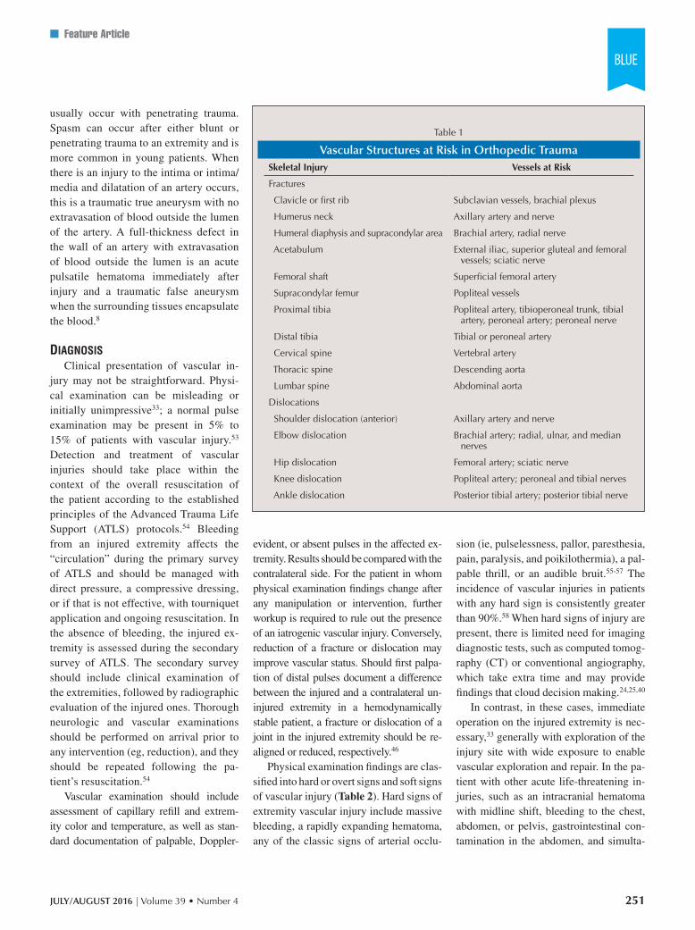
tremity vascular injury is control of bleed-ing, addressed during the circulation step of the primary survey of ATLS.54 Bleeding can be controlled by digital compression, application of a pressure dressing, infla-tion above the systolic blood pressure of a proximally placed blood pressure cuff, or application of a proximal tourniquet.85 In the presence of questionable peripheral pulses associated with gross deformity of the limb from fracture or dislocation, immediate realignment or reduction, re-spectively, should be performed, and the neurovascular status should be reassessed and documented (Figure 1).52 It might be difficult to assess vascular status in trauma patients in shock or hypothermia; such patients should be resuscitated and rewarmed first and then vascular status should be reassessed.8
The presence of vascular injury in or-thopedic trauma does not always warrant
surgical treatment. A single major vessel occlusion distal to the knee or elbow in the absence of severe soft tissue damage or a mangled extremity rarely constitutes a viability risk for the injured extremity. In such cases, observation might be a viable option. Serial imaging surveillance, how-ever, is necessary.75 Nonocclusive arterial injuries such as spasm, intimal flaps, or intramural hematomas detected in angiog-raphy performed for soft signs of vascu-lar injuries seem to heal without surgery in 87% to 95% of cases.63,71 Furthermore, isolated traumatic aneurysms in selected vessels may be treated by therapeutic embolization instead of an open vascu-lar operation.52 In addition, certain vas-cular injuries such as intimal dissections and flaps or small pseudoaneurysms have been shown to be amenable to endovascu-lar treatments with stents or stent grafts.86
opEn Vascular rEpairIn the operating room, an operative
tourniquet can replace the bleeding-con-trol modality used previously. However, if the injury is too proximal or exsangui-nating hemorrhage resumes, a member of the surgical team should be gloved and gowned in a sterile fashion, apply proximal pressure control, and the pa-tient should be prepped and draped with this team member included in the surgical field.52
For all locations of peripheral vascular injury, preparation of the skin and drap-ing should encompass all potential areas of proximal and distal vascular control, the area where a distal fasciotomy would be performed, and one (or the ipsilateral) lower extremity from the groin to the toe-nails for possible retrieval of the greater or lesser saphenous vein from the groin or the ankle.52,75,87 Often, it is helpful to drape the hand or foot of the affected extremity in a sterile plastic bag, so that color changes can be noted (at least in light-skinned patients) and distal pulses can be palpated under sterile conditions after repair is completed.52
Repair of an injured peripheral vessel is performed ideally with the surgeon and assistants wearing magnification loupes and battery-powered headlamps, or the operating microscope. Standard vascular instruments that should be available in-clude appropriate retractors, Metzenbaum scissors, DeBakey forceps, Gerald for-ceps, jeweler’s forceps, fine-tipped needle holders, vessel loops, Fogarty balloon catheters with stopcocks, intraluminal shunts, unfractionated heparin solution, and a contrast agent for a completion an-giography.88
Incisions typically are made longi-tudinally, directly over the target vessel proximal and distal to the injury to ensure adequate exposure for proximal and dis-tal vascular control and repair.75 Incisions should provide comfortable exposure. Af-ter vascular control is achieved, the inci-sions can be extended as needed to expose the zone of vascular injury. When the area
Figure 1: Sagittal computed tomography scan with 3-dimensional reconstruction showing contusion of the left popliteal artery in a 50-year-old woman with an open supracondylar femoral fracture. The pulse returned after reduction of the fracture.
253
Copyright © SLACK inCorporAted
n Feature Article
of injury is in proximity to a joint, a gen-tly curved (lazy “S”) incision to prevent a postoperative scar contracture is recom-mended.55,75,88 Not dissecting far enough proximally and distally from an area of injury is a common error. However, if control of hemorrhage cannot be obtained
or an extensive hematoma is overlying the arterial injury, making it difficult to obtain control close enough to avoid backbleed-ing from collaterals, it is not inappropri-ate to enter the zone of injury directly.55,75 After vascular control is obtained in either classic or direct fashion, vascular occlu-
sion can be maintained by applying a small DeBakey clamp, bulldog clamp, or Silastic (Dow Corning Corporation, Au-burn, Michigan) vessel loops.
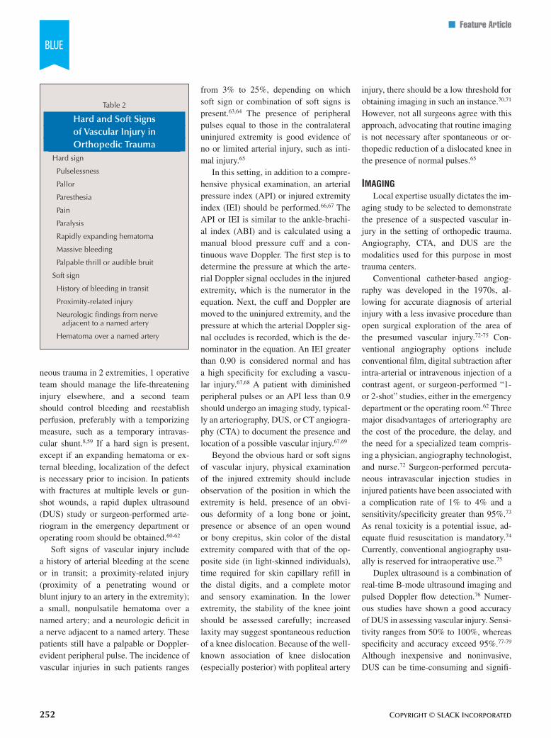
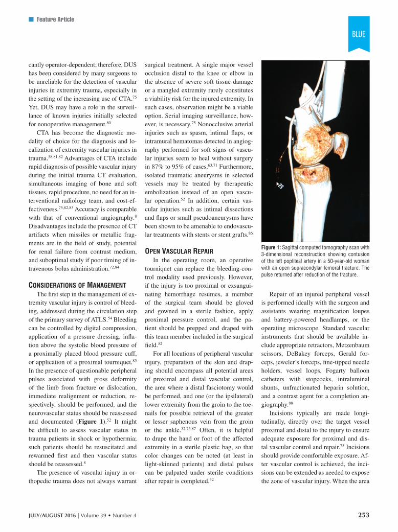
In general, small lacerations can be addressed by lateral angiorrhaphy with 5-0 or 6-0 polypropylene sutures applied transversely (Figure 2). If this leads to significant narrowing, vein patch angio-plasty is a viable alternative. In cases of complete transection of the injured artery, debridement back to healthy intima at both ends is performed, and the feasibility of an end-to-end anastomosis without ten-sion is assessed (Figure 3)88; this normal-ly is performed using 6-0 or 7-0 polypro-pylene sutures applied in a continuous or interrupted technique with 2 stay sutures 180° apart.89
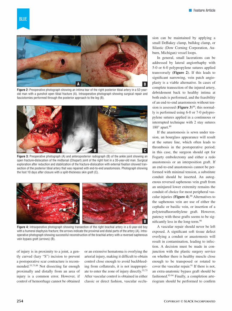
If the anastomosis is sewn under ten-sion, an hourglass appearance will result at the suture line, which often leads to thrombosis in the postoperative period; in this case, the surgeon should opt for Fogarty embolectomy and either a redo anastomosis or an interposition graft. If an end-to-end anastomosis cannot be per-formed with minimal tension, a substitute conduit should be inserted. An autog-enous reversed saphenous vein graft from an uninjured lower extremity remains the conduit of choice for most peripheral vas-cular injuries (Figure 4).88 Alternatives to the saphenous vein are use of either the cephalic or basilic vein, or insertion of a polytetrafluoroethylene graft. However, patency with these grafts seems to be sig-nificantly less in the long-term.90
A vascular repair should never be left exposed. A significant soft tissue defect overlying a conduit or anastomosis will result in contamination, leading to infec-tion. A decision must be made in con-junction with the plastic surgery service on whether there is healthy muscle close enough to be transposed or rotated to cover the vascular repair.91 If there is not, an extra-anatomic bypass graft should be fashioned.92-94 Finally, a completion arte-riogram should be performed to confirm
Figure 2: Preoperative photograph showing an intima tear of the right posterior tibial artery in a 52-year-old man with a gunshot open tibial fracture (A). Intraoperative photograph showing surgical repair and fasciotomies performed through the posterior approach to the leg (B).
BA
Figure 3: Preoperative photograph (A) and anteroposterior radiograph (B) of the ankle joint showing an open fracture-dislocation of the midtarsal (Chopart) joint of the right foot in a 35-year-old man. Surgical exploration after reduction and stabilization of the fracture-dislocation with external fixation showed tran-section of the posterior tibial artery that was repaired with end-to-end anastomosis. Photograph showing the foot 10 days after closure with a split-thickness skin graft (C).
CBA
Figure 4: Intraoperative photograph showing transection of the right brachial artery in a 6-year-old boy with a humeral diaphysis fracture; the arrows indicate the proximal and distal parts of the artery (A). Intra-operative photograph showing successful reconstruction of the brachial artery with a reversed saphenous vein bypass graft (arrows) (B).
BA
254
JULY/AUGUST 2016 | Volume 39 • Number 4
n Feature Article
patency, and identify and address techni-cal issues.88,93,94
comparTmEnT syndromEThe development of compartment syn-
drome portends a poor limb outcome.95,96 The classically described diagnostic signs of the 5 P’s (pain, pallor, paresthesia, pas-sive extension, and pulses intact) repre-sent late signs of compartment syndrome and are frequently absent or obscured in trauma patients because of distracting in-juries and altered mental status. The key clinical findings are pain out of proportion to the associated injury and pain on pas-sive movement of the muscles of the in-volved compartments.95 The incidence of clinically relevant compartment syndrome after extremity vascular injury is unknown because of the widespread use of prophy-lactic fasciotomy in the setting of orthope-dic or vascular surgery.96 Performance of a fasciotomy concomitant with or soon af-ter revascularization of the limb is associ-ated with a fourfold reduction in eventual amputation and other complications.97
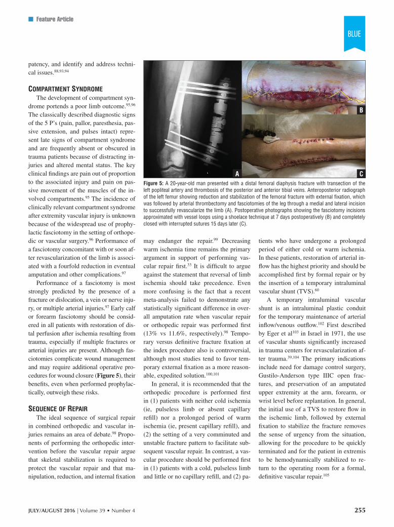
Performance of a fasciotomy is most strongly predicted by the presence of a fracture or dislocation, a vein or nerve inju-ry, or multiple arterial injuries.97 Early calf or forearm fasciotomy should be consid-ered in all patients with restoration of dis-tal perfusion after ischemia resulting from trauma, especially if multiple fractures or arterial injuries are present. Although fas-ciotomies complicate wound management and may require additional operative pro-cedures for wound closure (Figure 5), their benefits, even when performed prophylac-tically, outweigh these risks.
sEquEncE of rEpairThe ideal sequence of surgical repair
in combined orthopedic and vascular in-juries remains an area of debate.98 Propo-nents of performing the orthopedic inter-vention before the vascular repair argue that skeletal stabilization is required to protect the vascular repair and that ma-nipulation, reduction, and internal fixation
may endanger the repair.99 Decreasing warm ischemia time remains the primary argument in support of performing vas-cular repair first.33 It is difficult to argue against the statement that reversal of limb ischemia should take precedence. Even more confusing is the fact that a recent meta-analysis failed to demonstrate any statistically significant difference in over-all amputation rate when vascular repair or orthopedic repair was performed first (13% vs 11.6%, respectively).98 Tempo-rary versus definitive fracture fixation at the index procedure also is controversial, although most studies tend to favor tem-porary external fixation as a more reason-able, expedited solution.100,101
In general, it is recommended that the orthopedic procedure is performed first in (1) patients with neither cold ischemia (ie, pulseless limb or absent capillary refill) nor a prolonged period of warm ischemia (ie, present capillary refill), and (2) the setting of a very comminuted and unstable fracture pattern to facilitate sub-sequent vascular repair. In contrast, a vas-cular procedure should be performed first in (1) patients with a cold, pulseless limb and little or no capillary refill, and (2) pa-
tients who have undergone a prolonged period of either cold or warm ischemia. In these patients, restoration of arterial in-flow has the highest priority and should be accomplished first by formal repair or by the insertion of a temporary intraluminal vascular shunt (TVS).60
A temporary intraluminal vascular shunt is an intraluminal plastic conduit for the temporary maintenance of arterial inflow/venous outflow.102 First described by Eger et al103 in Israel in 1971, the use of vascular shunts significantly increased in trauma centers for revascularization af-ter trauma.39,104 The primary indications include need for damage control surgery, Gustilo-Anderson type IIIC open frac-tures, and preservation of an amputated upper extremity at the arm, forearm, or wrist level before replantation. In general, the initial use of a TVS to restore flow in the ischemic limb, followed by external fixation to stabilize the fracture removes the sense of urgency from the situation, allowing for the procedure to be quickly terminated and for the patient in extremis to be hemodynamically stabilized to re-turn to the operating room for a formal, definitive vascular repair.105
Figure 5: A 20-year-old man presented with a distal femoral diaphysis fracture with transection of the left popliteal artery and thrombosis of the posterior and anterior tibial veins. Anteroposterior radiograph of the left femur showing reduction and stabilization of the femoral fracture with external fixation, which was followed by arterial thrombectomy and fasciotomies of the leg through a medial and lateral incision to successfully revascularize the limb (A). Postoperative photographs showing the fasciotomy incisions approximated with vessel loops using a shoelace technique at 7 days postoperatively (B) and completely closed with interrupted sutures 15 days later (C).
A C
B
255
Copyright © SLACK inCorporAted
n Feature Article
manglEd ExTrEmiTyA mangled extremity results from
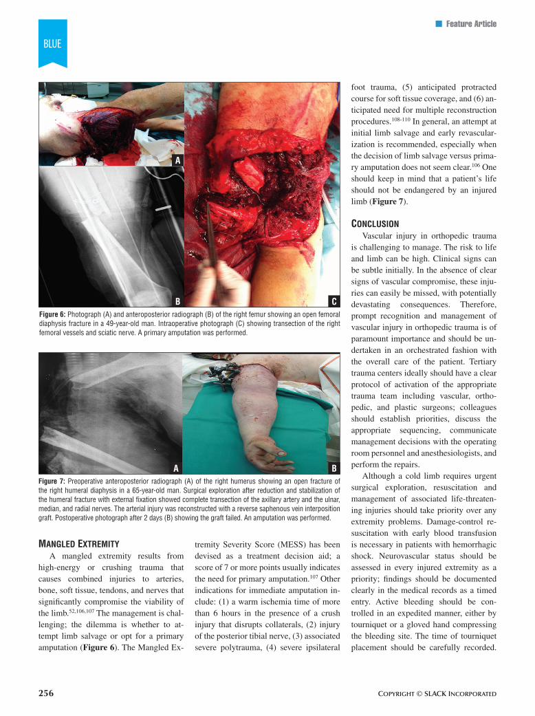
high-energy or crushing trauma that causes combined injuries to arteries, bone, soft tissue, tendons, and nerves that significantly compromise the viability of the limb.52,106,107 The management is chal-lenging; the dilemma is whether to at-tempt limb salvage or opt for a primary amputation (Figure 6). The Mangled Ex-
tremity Severity Score (MESS) has been devised as a treatment decision aid; a score of 7 or more points usually indicates the need for primary amputation.107 Other indications for immediate amputation in-clude: (1) a warm ischemia time of more than 6 hours in the presence of a crush injury that disrupts collaterals, (2) injury of the posterior tibial nerve, (3) associated severe polytrauma, (4) severe ipsilateral
foot trauma, (5) anticipated protracted course for soft tissue coverage, and (6) an-ticipated need for multiple reconstruction procedures.108-110 In general, an attempt at initial limb salvage and early revascular-ization is recommended, especially when the decision of limb salvage versus prima-ry amputation does not seem clear.106 One should keep in mind that a patient’s life should not be endangered by an injured limb (Figure 7).
conclusionVascular injury in orthopedic trauma
is challenging to manage. The risk to life and limb can be high. Clinical signs can be subtle initially. In the absence of clear signs of vascular compromise, these inju-ries can easily be missed, with potentially devastating consequences. Therefore, prompt recognition and management of vascular injury in orthopedic trauma is of paramount importance and should be un-dertaken in an orchestrated fashion with the overall care of the patient. Tertiary trauma centers ideally should have a clear protocol of activation of the appropriate trauma team including vascular, ortho-pedic, and plastic surgeons; colleagues should establish priorities, discuss the appropriate sequencing, communicate management decisions with the operating room personnel and anesthesiologists, and perform the repairs.
Although a cold limb requires urgent surgical exploration, resuscitation and management of associated life-threaten-ing injuries should take priority over any extremity problems. Damage-control re-suscitation with early blood transfusion is necessary in patients with hemorrhagic shock. Neurovascular status should be assessed in every injured extremity as a priority; findings should be documented clearly in the medical records as a timed entry. Active bleeding should be con-trolled in an expedited manner, either by tourniquet or a gloved hand compressing the bleeding site. The time of tourniquet placement should be carefully recorded.
Figure 6: Photograph (A) and anteroposterior radiograph (B) of the right femur showing an open femoral diaphysis fracture in a 49-year-old man. Intraoperative photograph (C) showing transection of the right femoral vessels and sciatic nerve. A primary amputation was performed.
A
CB
Figure 7: Preoperative anteroposterior radiograph (A) of the right humerus showing an open fracture of the right humeral diaphysis in a 65-year-old man. Surgical exploration after reduction and stabilization of the humeral fracture with external fixation showed complete transection of the axillary artery and the ulnar, median, and radial nerves. The arterial injury was reconstructed with a reverse saphenous vein interposition graft. Postoperative photograph after 2 days (B) showing the graft failed. An amputation was performed.
BA
256
JULY/AUGUST 2016 | Volume 39 • Number 4
n Feature Article
Blind clamping or local wound explora-tion in the trauma bay should be discour-aged as potentially detrimental.
A deformed, pulseless extremity should be realigned and a dislocation re-duced; neurovascular status then should be reassessed as reestablishment of flow is not infrequent. Imaging should include DUS, CTA, or on-table angiography; however, imaging should not delay reper-fusion as the injury pattern usually pre-dicts the level of vascular injury in most cases. Reperfusion delay with prolonged warm ischemia (>3-6 hours) leads to ir-reversible tissue damage, with resulting myoglobinuria and acute renal failure that may be life-threatening.
Limb salvage is not always the cor-rect decision; in many cases, primary amputation may be the most reasonable choice. In any case, the patient and rela-tives should be made aware of the possi-ble risks of surgery, potential for multiple procedures, and possibility of immediate or secondary amputation. Preparation for surgery should include (1) administration of broad-spectrum antibiotics, (2) tetanus toxoid, and (3) a bolus of systemic hepa-rin as well as ensuring the injury is iso-lated and bleeding is under control.
The key factors in successful manage-ment are optimal sequence of the repair, adequate exposure and vascular control, debridement of the injured vessel wall to healthy intima, proximal and distal bal-loon catheter thrombectomy, tension-free end-to-end repair or appropriately sized interposition graft, good soft tissue cover-age, stable but expeditious fracture fixa-tion, and adequate fasciotomies. Failure to perform fasciotomies after revascu-larization of an acutely ischemic limb is the most common cause of preventable limb loss. Incisions for fasciotomies or vascular control should preserve perforat-ing vessels, taking into account the future potential need for fashioning flaps for soft tissue coverage. Finally, a bed in the intensive care unit ideally should be re-served for early postoperative monitoring.
rEfErEncEs 1. Rasmussen TE, Woodson J, Rich NM, Mattox
KL. Vascular trauma at a crossroads. J Trau-ma. 2011; 70(5):1291-1293.
2. Carrel A. The surgery of blood vessels. Bull Johns Hopkins Hosp. 1907; 18(190):18-28.
3. Guthrie C. Blood Vessel Surgery and Its Ap-plications. London, United Kingdom: Edward Arnold; 1912.
4. Dente CJ, Feliciano DV. Alexis Carrel (1873-1944): Nobel Laureate, 1912. Arch Surg. 2005; 140(6):609-610.
5. Soubbotitch V. Military experiences of traumat-ic aneurysms. Lancet. 1913; 182(4697):720-721.
6. Makins GH. On Gunshot Injuries to the Blood-Vessels: Founded on Experience Gained in France During the Great War, 1914-1918. Bristol, United Kingdom: J. Wright and Sons; 1919.
7. Noszczyk W, Witkowski M, Weglowski R. The Zamosc period in the work of Romuald Weglowski. Polski Przegl Chir. 1985; 57:440-445.
8. Feliciano DV, Moore FA, Moore EE, et al. Evaluation and management of peripheral vascular injury: Part 1. Western Trauma Asso-ciation/critical decisions in trauma. J Trauma. 2011; 70(6):1551-1556.
9. Debakey ME, Simeone FA. Battle injuries of the arteries in World War II: an analysis of 2,471 cases. Ann Surg. 1946; 123(4):534-579.
10. Rich NM. Vascular trauma historical notes. Perspect Vasc Surg Endovasc Ther. 2011; 23(1):7-12.
11. Jahnke EJ Jr, Seeley SF. Acute vascular in-juries in the Korean War. Ann Surg. 1953; 138(2):158-177.
12. Hughes CW. The primary repair of wounds of major arteries: an analysis of experience in Korea in 1953. Ann Surg. 1955; 141(3):297-303.
13. Rich NM, Rhee P. An historical tour of vas-cular injury management: from its inception to the new millennium. Surg Clin North Am. 2001; 81(6):1199-1215.
14. Rich NM. Vascular trauma in Vietnam. J Car-diovasc Surg (Torino). 1970; 11(5):368-377.
15. Rich NM, Baugh JH, Hughes CW. Acute arte-rial injuries in Vietnam: 1,000 cases. J Trau-ma. 1970; 10(5):359-369.
16. McNamara JH, Brief DK, Beasley W, Wright JK. Vascular injury in Vietnam combat casual-ties: results of treatment at the 24th Evacua-tion Hospital 1 July 1967 to 12 August 1969. Ann Surg. 1973; 178(2):143-147.
17. Iraq Coalition Casualty Count. http://www.icasualties.org. Accessed May 11, 2015.
18. Rotondo MF, Zonies DH. The damage con-trol sequence and underlying logic. Surg Clin North Am. 1997; 77(4):761-777.
19. Perkins JG, Beekley AC. Damage control resuscitation. In: Savitsky E, Eastbridge B, eds. Combat Casualty Care: Lessons Learned From OEF and OIF. Falls Church, VA: Office of the Surgeon General Department of the Army; 2012:121-163.
20. Beekley AC, Sebesta JA, Blackbourne LH, et al, 31st Combat Support Hospital Research Group. Prehospital tourniquet use in Opera-tion Iraqi Freedom: effect on hemorrhage con-trol and outcomes. J Trauma. 2008; 64(suppl 2):S28-S37.
21. Kragh JF Jr, Walters TJ, Baer DG, et al. Sur-vival with emergency tourniquet use to stop bleeding in major limb trauma. Ann Surg. 2009; 249(1):1-7.
22. Owens BD, Kragh JF Jr, Wenke JC, Macaitis J, Wade CE, Holcomb JB. Combat wounds in Operation Iraqi Freedom and Operation En-during Freedom. J Trauma. 2008; 64(2):295-299.
23. Eastridge BJ, Jenkins D, Flaherty S, Schiller H, Holcomb JB. Trauma system development in a theater of war: experiences from Opera-tion Iraqi Freedom and Operation Enduring Freedom. J Trauma. 2006; 61(6):1363-1372.
24. Rasmussen TE, Clouse WD, Jenkins DH, Peck MA, Eliason JL, Smith DL. Echelons of care and the management of wartime vas-cular injury: a report from the 332nd EMDG/Air Force Theater Hospital, Balad Air Base, Iraq. Perspect Vasc Surg Endovasc Ther. 2006; 18(2):91-99.
25. U.S. Army Institute of Surgical Research. Joint Trauma System clinical practice guidelines. http://www.usaisr.amedd.army.mil/cpgs.html. Accessed May 11, 2015.
26. Eastridge BJ, Costanzo G, Jenkins D, et al. Impact of joint theater trauma system initia-tives on battlefield injury outcomes. Am J Surg. 2009; 198(6):852-857.
27. Holcomb JB, McMullin NR, Pearse L, et al. Causes of death in U.S. Special Operations Forces in the global war on terrorism: 2001-2004. Ann Surg. 2007; 245(6):986-991.
28. American College of Surgeons. National Trauma Data Bank. http://www.facs.org/qual-ity programs/trauma/ntdb. Accessed May 13, 2015.
29. American College of Surgeons. National Trauma Data Bank 2014 Annual Report. http://www.facs.org/quality-programs/trauma/ntdb/docpub. Accessed May 13, 2015.
30. Martin MJ, Long WB. Vascular trauma: epi-demiology and natural history. In: Cronenwett JL, Johnston KW, eds. Rutherford’s Vascular Surgery. Vol 2. 8th ed. Philadelphia, PA: Saun-ders; 2014:2422-2437.
31. Haider AH, Hashmi ZG, Gupta S, et al. Benchmarking of trauma care worldwide: the potential value of an International Trau-ma Data Bank (ITDB). World J Surg. 2014; 38(8):1882-1891.
32. Kauvar DS, Wade CE. The epidemiology and
257
Copyright © SLACK inCorporAted
n Feature Article
modern management of traumatic hemor-rhage: US and international perspectives. Crit Care. 2005; 9(suppl 5):S1-S9.
33. Halvorson JJ, Anz A, Langfitt M, et al. Vas-cular injury associated with extremity trauma: initial diagnosis and management. J Am Acad Orthop Surg. 2011; 19(8):495-504.
34. Barmparas G, Inaba K, Talving P, et al. Pediat-ric vs adult vascular trauma: a National Trau-ma Databank review. J Pediatr Surg. 2010; 45(7):1404-1412.
35. White JM, Stannard A, Burkhardt GE, East-ridge BJ, Blackbourne LH, Rasmussen TE. The epidemiology of vascular injury in the wars in Iraq and Afghanistan. Ann Surg. 2011; 253(6):1184-1189.
36. Perkins ZB, De’Ath HD, Aylwin C, Brohi K, Walsh M, Tai NR. Epidemiology and outcome of vascular trauma at a British Major Trauma Centre. Eur J Vasc Endovasc Surg. 2012; 44(2):203-209.
37. Mattox KL, Feliciano DV, Burch J, Beall AC Jr, Jordan GL Jr, De Bakey ME. Five thousand seven hundred sixty cardiovascular injuries in 4459 patients: epidemiologic evolution 1958 to 1987. Ann Surg. 1989; 209(6):698-705.
38. Soreide K, Kruger AJ, Vardal AL, Ellingsen CL, Soreide E, Lossius HM. Epidemiology and contemporary patterns of trauma deaths: changing place, similar pace, older face. World J Surg. 2007; 31(11):2092-2103.
39. Rasmussen TE, Clouse WD, Jenkins DH, Peck MA, Eliason JL, Smith DL. The use of temporary vascular shunts as a damage control adjunct in the management of wartime vascu-lar injury. J Trauma. 2006; 61(1):8-12.
40. Fox CJ, Starnes BW. Vascular surgery on the modern battlefield. Surg Clin North Am. 2007; 87(5):1193-1211.
41. Clouse WD, Rasmussen TE, Peck MA, et al. In-theater management of vascular injury: 2 years of the Balad Vascular Registry. J Am Coll Surg. 2007; 204(4):625-632.
42. Woodward EB, Clouse WD, Eliason JL, et al. Penetrating femoropopliteal injury during modern warfare: experience of the Balad Vas-cular Registry. J Vasc Surg. 2008; 47(6):1255-1264.
43. Frykberg E, Schinco MA. Peripheral vascular injury. In: Feliciano DV, Mattox KL, Moore EE, eds. Trauma. 6th ed. New York, NY: Mc-Graw-Hill; 2006:941-971.
44. Robbs JB, Baker LW. Cardiovascular trauma. Curr Probl Surg. 1984; 21(4):1-87.
45. Cooper C, Rodriguez A, Omert L. Blunt vascular trauma. Curr Probl Surg. 1992; 29(5):281-357.
46. Rozycki GS, Tremblay LN, Feliciano DV, McClelland WB. Blunt vascular trauma in the extremity: diagnosis, management, and out-come. J Trauma. 2003; 55(5):814-824.
47. Oller DW, Rutledge R, Clancy T, et al. Vascu-lar injuries in a rural state: a review of 978 pa-
tients from a state trauma registry. J Trauma. 1992; 32(6):740-745.
48. Kootstra G, Schipper JJ, Boontje AH, Klasen HJ, Binnendijk B. Femoral shaft fracture with injury of the superficial femoral artery in ci-vilian accidents. Surg Gynecol Obstet. 1976; 142(3):399-403.
49. Caudle RJ, Stern PJ. Severe open fractures of the tibia. J Bone Joint Surg Am. 1987; 69(6):801-807.
50. Mullenix PS, Steele SR, Andersen CA, Starnes BW, Salim A, Martin MJ. Limb sal-vage and outcomes among patients with trau-matic popliteal vascular injury: an analysis of the National Trauma Data Bank. J Vasc Surg. 2006; 44(1):94-100.
51. Redmond JM, Levy BA, Dajani KA, Cass JR, Cole PA. Detecting vascular injury in lower-extremity orthopedic trauma: the role of CT angiography. Orthopedics. 2008; 31(8):761-767.
52. Feliciano DV. Evaluation and treatment of vascular injuries. In: Browner BD, Jupiter JB, Levine AM, Trafton PG, Krettek C, eds. Skeletal Trauma. Basic Science, Management, and Reconstruction. 4th ed. Philadelphia, PA: Elsevier Saunders; 2009:323-340.
53. Barnes CJ, Pietrobon R, Higgins LD. Does the pulse examination in patients with trau-matic knee dislocation predict a surgical arte-rial injury? A meta-analysis. J Trauma. 2002; 53(6):1109-1114.
54. American College of Surgeons. ATLS: Ad-vanced Trauma Life Support for Doctors. 8th ed. Chicago, IL: American College of Sur-geons; 2008.
55. Feliciano DV. Management of peripheral arterial injury. Curr Opin Crit Care. 2010; 16(6):602-608.
56. Fox N, Rajani RR, Bokhari F, et al. Evaluation and management of penetrating lower extrem-ity arterial trauma: an Eastern Association for the Surgery of Trauma practice management guideline. J Trauma Acute Care Surg. 2012; 73(5)(suppl 4):S315-S320.
57. Manthey DE, Nicks BA. Penetrating trauma to the extremity. J Emerg Med. 2008; 34(2):187-193.
58. Inaba K, Branco BC, Reddy S, et al. Prospec-tive evaluation of multidetector computed tomography for extremity vascular trauma. J Trauma. 2011; 70(4):808-815.
59. Subramanian A, Vercruysse G, Dente C, Wyrzykowski A, King E, Feliciano DV. A de-cade’s experience with temporary intravascu-lar shunts at a civilian level I trauma center. J Trauma. 2008; 65(2):316-324.
60. Feliciano DV. Evaluation and treatment of vascular injuries. In: Browner BD, Jupiter JB, Levine AM, Trafton PG, Krettek C, eds. Skel-etal Trauma: Basic Science, Management, and Reconstruction. Philadelphia, PA: Elsevier Saunders; 2009:323-340.
61. Callcut RA, Acher CW, Hoch J, Tefera G, Turnipseed W, Mell MW. Impact of intraop-erative arteriography on limb salvage for trau-matic popliteal artery injury. J Trauma. 2009; 67(2):252-257.
62. O’Gorman RB, Feliciano DV. Arteriography performed in the emergency center. Am J Surg. 1986; 152(3):323-325.
63. Frykberg ER, Dennis JW, Bishop K, Laneve L, Alexander RH. The reliability of physical examination in the evaluation of penetrating extremity trauma for vascular injury: results at one year. J Trauma. 1991; 31(4):502-511.
64. Dennis JW, Frykberg ER, Veldenz HC, Huff-man S, Menawat SS. Validation of nonopera-tive management of occult vascular injuries and accuracy of physical examination alone in penetrating extremity trauma: 5- to 10-year follow-up. J Trauma. 1998; 44(2):242-252.
65. Miranda FE, Dennis JW, Veldenz HC, Dovgan PS, Frykberg ER. Confirmation of the safety and accuracy of physical examination in the evaluation of knee dislocation for injury of the popliteal artery: a prospective study. J Trauma. 2002; 52(2):247-251.
66. Johansen K, Lynch K, Paun M, Copass M. Non-invasive vascular tests reliably exclude occult arterial trauma in injured extremities. J Trauma. 1991; 31(4):515-519.
67. Lynch K, Johansen K. Can Doppler pressure measurement replace “exclusion” arteriogra-phy in the diagnosis of occult extremity arte-rial trauma? Ann Surg. 1991; 214(6):737-741.
68. Mills WJ, Barei DP, McNair P. The value of the ankle-brachial index for diagnosing arte-rial injury after knee dislocation: a prospective study. J Trauma. 2004; 56(6):1261-1265.
69. Sadjadi J, Cureton EL, Dozier KC, Kwan RO, Victorino GP. Expedited treatment of lower extremity gunshot wounds. J Am Coll Surg. 2009; 209(6):740-745.
70. Gable DR, Allen JW, Richardson JD. Blunt popliteal artery injury: is physical examination alone enough for evaluation? J Trauma. 1997; 43(3):541-544.
71. Frykberg ER, Vines FS, Alexander RH. The natural history of clinically occult arterial inju-ries: a prospective evaluation. J Trauma. 1989; 29(5):577-583.
72. Miller-Thomas MM, West OC, Cohen AM. Diagnosing traumatic arterial injury in the ex-tremities with CT angiography: pearls and pit-falls. Radiographics. 2005; 25(suppl 1):S133-S142.
73. Itani KM, Burch JM, Spjut-Patrinely V, Rich-ardson R, Martin RR, Mattox KL. Emer-gency center arteriography. J Trauma. 1992; 32(3):302-307.
74. Benko A, Fraser-Hill M, Magner P, et al. Ca-nadian Association of Radiologists: consensus guidelines for the prevention of contrast-in-duced nephropathy. Can Assoc Radiol J. 2007; 58(2):79-87.
258
JULY/AUGUST 2016 | Volume 39 • Number 4
n Feature Article
75. Kauvar DS, Kraiss LW. Vascular trauma: ex-tremity. In: Cronenwett JL, Johnston KW, eds. Rutherford’s Vascular Surgery. Vol 2. 8th ed. Philadelphia, PA: Saunders; 2014:2485-2500.
76. Lynch TG, Hobson RW II. Noninvasive cere-brovascular diagnostic techniques. In: Hobson RW II, Wilson SE, Veith FJ, eds. Vascular Sur-gery: Principles and Practice. 3rd ed. Boca Raton, FL: CRC Press; 2003:123-152.
77. Bynoe RP, Miles WS, Bell RM, et al. Non-invasive diagnosis of vascular trauma by duplex ultrasonography. J Vasc Surg. 1991; 14(3):346-352.
78. Gagne PJ, Cone JB, McFarland D, et al. Prox-imity penetrating extremity trauma: the role of duplex ultrasound in the detection of occult venous injuries. J Trauma. 1995; 39(6):1157-1163.
79. Knudson MM, Lewis FR, Atkinson K, Neu-haus A. The role of duplex ultrasound arterial imaging in patients with penetrating extremity trauma. Arch Surg. 1993; 128(9):1033-1037.
80. Schwartz M, Weaver F, Yellin A, Ralls P. The utility of color flow Doppler examination in penetrating extremity arterial trauma. Am Surg. 1993; 59(6):375-378.
81. Fleiter TR, Mervis S. The role of 3D-CTA in the assessment of peripheral vascular le-sions in trauma patients. Eur J Radiol. 2007; 64(1):92-102.
82. Seamon MJ, Smoger D, Torres DM, et al. A prospective validation of a current practice: the detection of extremity vascular injury with CT angiography. J Trauma. 2009; 67(2):234-243.
83. Rieger M, Mallouhi A, Tauscher T, Lutz M, Jaschke WR. Traumatic arterial injuries of the extremities: initial evaluation with MDCT angiography. AJR Am J Roentgenol. 2006; 186(3):656-664.
84. White PW, Gillespie DL, Feurstein I, et al. Sixty-four slice multidetector computed tomo-graphic angiography in the evaluation of vas-cular trauma. J Trauma. 2010; 68(1):96-102.
85. Welling DR, Burris DG, Hutton JE, Minken SL, Rich NM. A balanced approach to tourni-quet use: lessons learned and relearned. J Am Coll Surg. 2006; 203(1):106-115.
86. Reuben BC, Whitten MG, Sarfati M, Kraiss LW. Increasing use of endovascular therapy in acute arterial injuries: analysis of the Na-tional Trauma Data Bank. J Vasc Surg. 2007; 46(6):1222-1226.
87. Feliciano DV. Vascular injuries. In: Maull KI, Cleveland HC, Strauch GO, Wolferth CC, eds. Advances in Trauma. Vol 2. Chicago, IL: Year Book Medical Publishers; 1987:179-206.
88. Feliciano DV, Moore EE, West MA, et al. Western Trauma Association critical deci-sions in trauma: evaluation and management of peripheral vascular injury: Part II. J Trauma Acute Care Surg. 2013; 75(3):391-397.
89. Ball CG, Feliciano DV. A simple and rapid vascular anastomosis for emergency surgery: a technical case report. World J Emerg Surg. 2009; 4:30.
90. Feliciano DV, Mattox KL, Graham JM, Bi-tondo CG. Five-year experience with PTFE grafts in vascular wounds. J Trauma. 1985; 25(1):71-82.
91. Strinden WD, Dibbell DG Sr, Turnipseed WD, Acher CW, Rao VK, Mixter RC. Coverage of acute vascular injuries of the axilla and groin with transposition muscle flaps: case reports. J Trauma. 1989; 29(4):512-516.
92. Feliciano DV. Heroic procedures in vascular injury management: the role of extra-ana-tomic bypasses. Surg Clin North Am. 2002; 82(1):115-124.
93. Kurtoglu M, Yanar H, Taviloglu K, Sivrikoz E, Plevin R, Aksoy M. Serious lower extrem-ity venous injury management with ligation: prospective overview of 63 patients. Am Surg. 2007; 73(10):1039-1043.
94. Parry NG, Feliciano DV, Burke RM, et al. Management and short-term patency of lower extremity venous injuries with various repairs. Am J Surg. 2003; 186(6):631-635.
95. British Orthopaedic Association. BOAST 10: diagnosis and management of compartment syndrome of the limbs. http://www.boa.ac.uk/wp-content/uploads/2015/01/BOAST-10.pdf. Accessed May 20, 2015.
96. Branco BC, Inaba K, Barmparas G, et al. Inci-dence and predictors for the need for fasciot-omy after extremity trauma: a 10-year review in a mature level I trauma centre. Injury. 2011; 42(10):1157-1163.
97. Farber A, Tan TW, Hamburg NM, et al. Early fasciotomy in patients with extremity vascular injury is associated with decreased risk of ad-verse limb outcomes: a review of the National Trauma Data Bank. Injury. 2012; 43(9):1486-1491.
98. Fowler J, Macintyre N, Rehman S, Gaughan
JP, Leslie S. The importance of surgical se-quence in the treatment of lower extremity injuries with concomitant vascular injury: a meta-analysis. Injury. 2009; 40(1):72-76.
99. Iannacone WM, Taffet R, DeLong WG Jr, Born CT, Dalsey RM, Deutsch LS. Early ex-change intramedullary nailing of distal femo-ral fractures with vascular injury initially sta-bilized with external fixation. J Trauma. 1994; 37(3):446-451.
100. Allen MJ, Nash JR, Ioannidies TT, Bell PR. Major vascular injuries associated with ortho-paedic injuries to the lower limb. Ann R Coll Surg Engl. 1984; 66(2):101-104.
101. Howard PW, Makin GS. Lower limb fractures with associated vascular injury. J Bone Joint Surg Br. 1990; 72(1):116-120.
102. Feliciano DV, Subramanian A. Temporary vascular shunts. Eur J Trauma Emerg Surg. 2013; 39(6):553-560.
103. Eger M, Golcman L, Goldstein A, Hirsch M. The use of a temporary shunt in the manage-ment of arterial vascular injuries. Surg Gyne-col Obstet. 1971; 132(1):67-70.
104. Hancock H, Rasmussen TE, Walker AJ, Rich NM. History of temporary intravascular shunts in the management of vascular injury. J Vasc Surg. 2010; 52(5):1405-1409.
105. Wyrzykowski AD, Feliciano DV. Trauma damage control. In: Feliciano DV, Mattox KL, Moore EE, eds. Trauma. 6th ed. New York, NY: McGraw-Hill; 2008:851-870.
106. Scalea TM, DuBose J, Moore EE, et al. West-ern Trauma Association critical decisions in trauma: management of the mangled extremi-ty. J Trauma Acute Care Surg. 2012; 72(1):86-93.
107. Johansen K, Daines M, Howey T, Helfet D, Hansen ST Jr. Objective criteria accurately predict amputation following lower extremity trauma. J Trauma. 1990; 30(5):568-572.
108. Lange RH. Limb reconstruction versus ampu-tation decision making in massive lower ex-tremity trauma. Clin Orthop Relat Res. 1989; 243:92-99.
109. Hansen ST Jr. The type-IIIC tibial fracture: salvage or amputation. J Bone Joint Surg Am. 1987; 69(6):799-800.
110. Lange RH, Bach AW, Hansen ST Jr, Johan-sen KH. Open tibial fractures with associated vascular injuries: prognosis for limb salvage. J Trauma. 1985; 25(3):203-208.
259