Embed Size (px)
Citation preview
© 2012 Hoover-Plow and Gong, publisher and licensee Dove Medical Press Ltd. This is an Open Access article which permits unrestricted noncommercial use, provided the original work is properly cited.
Vascular Health and Risk Management 2012:8 99–113
Vascular Health and Risk Management
Challenges for heart disease stem cell therapy
Jane Hoover-PlowYanqing GongDepartments of Cardiovascular Medicine and Molecular Cardiology, Joseph J Jacobs Center for Thrombosis and Vascular Biology, Cleveland Clinic Lerner Research Institute, Cleveland, OH, USA
Correspondence: Jane Hoover-Plow Department of Molecular Cardiology, NB50, Cleveland Clinic Lerner Research Institute, 9500 Euclid Avenue, Cleveland, OH 44195, USA Tel +1 216 445 6639 Fax +1 216 445 8204 Email [email protected]
Abstract: Cardiovascular diseases (CVDs) are the leading cause of death worldwide. The
use of stem cells to improve recovery of the injured heart after myocardial infarction (MI)
is an important emerging therapeutic strategy. However, recent reviews of clinical trials of
stem cell therapy for MI and ischemic heart disease recovery report that less than half of
the trials found only small improvements in cardiac function. In clinical trials, bone mar-
row, peripheral blood, or umbilical cord blood cells were used as the source of stem cells
delivered by intracoronary infusion. Some trials administered only a stem cell mobilizing
agent that recruits endogenous sources of stem cells. Important challenges to improve the
effectiveness of stem cell therapy for CVD include: (1) improved identification, recruitment,
and expansion of autologous stem cells; (2) identification of mobilizing and homing agents
that increase recruitment; and (3) development of strategies to improve stem cell survival
and engraftment of both endogenous and exogenous sources of stem cells. This review is an
overview of stem cell therapy for CVD and discusses the challenges these three areas pres-
ent for maximum optimization of the efficacy of stem cell therapy for heart disease, and new
strategies in progress.
Keywords: mobilization, expansion, homing, survival, engraftment
IntroductionThe recovery of function after a myocardial infarction (MI) is dependent on increas-
ing blood flow and regeneration of tissue. Stem cells (SCs) can provide cellular pre-
cursors for cardiomyocyte differentiation, endothelial and supporting cells, as well
as signals for activation of cells and prevention of apoptosis. The results of clinical
trials have been encouraging, however either no change or only small increments
in recovery were found. Recent reviews of completed clinical trials (2002–2010)
for SC therapy report improvements of 10% or less in about half of the studies.1–4
In the review by George,1 13 studies of SC therapy for acute MI were described. In
the eight randomized controlled studies, bone-marrow (BM) cells were administered
by intracoronary injection and left ventricular ejection fraction (LVEF) measured
3–6 months following the MI. In five of the randomized controlled trials, there was
only an average increase of 6% (3%–12%) in cardiac function. Mozid et al2 reported
two additional studies of BM SC therapy for acute MI,5,6 and only one study showed
improvement (5%) of LVEF function. Mozid et al2 also described eight clinical trials
of SC therapy for chronic ischemic heart failure. There was improvement in LVEF
in three of the four studies in patients treated with BM SCs and improvement in two
Dovepress
submit your manuscript | www.dovepress.com
Dovepress 99
R E V I E w
open access to scientific and medical research
Open Access Full Text Article
http://dx.doi.org/10.2147/VHRM.S25665
V
ascu
lar
Hea
lth a
nd R
isk
Man
agem
ent d
ownl
oade
d fr
om h
ttps:
//ww
w.d
ovep
ress
.com
/ by
95.2
16.9
9.24
on
05-A
pr-2
019
For
per
sona
l use
onl
y.
Powered by TCPDF (www.tcpdf.org)
1 / 1

Vascular Health and Risk Management 2012:8
of the four studies in patients transplanted with autologous
skeletal myoblasts. Wen et al4 performed a meta-analysis of
eight randomized controlled trials and concluded that BM
cell therapy provided only moderate (6%–10%) but definite
improvements in LVEF. SC therapy has the potential to pro-
vide gains not only for MI, but also for chronic ischemia and
heart failure. Currently, there are 33 ongoing clinical trials
described on the ClinicalTrials.gov Website7 (see Table 1).
While autologous BM cells are still the major source of SCs
in the ongoing studies, new SC sources are rigorously being
investigated. SC therapy for cardiovascular disease (CVD) is
an intensive area of research, and collective improvements
in the source and number of SCs, and better mobilizing and
homing agents, are needed to increase the effectiveness of
this emerging therapy.
Challenges for SC therapyImproved identification and expansion of autologous SCs and their role in cardiac recoveryIn the 1960s, Till et al,8 while studying the components
responsible for regenerating blood cells, defined two required
properties of SCs: (1) self-renewal – the ability to go through
numerous cycles of cell division while maintaining the undif-
ferentiated state; and (2) potency – the capacity to differentiate
into specialized cell types. SCs are identified by their capacity
to form colonies in culture and by cell surface markers that
are cell specific. The majority of clinical trials of SC therapy
for heart disease have used BM cells, particularly the mono-
nuclear cells (MNCs) (Figure 1). In the ongoing trials listed
Table 1 Ongoing clinical trials of stem-cell therapy for heart diseases
Condition Stem cells Phase Acronym ClinicalTrials.gov NCTID
Congestive heart failure Skeletal myoblasts II/III MARVEL NCT00526253Old MI Skeletal myoblasts II PERCUTANEO NCT00908622Angina, coronary disease Bone marrow II NCT01214499Ischemic heart disease Bone marrow II NCT00690209CAD, AMI Bone marrow I/II REPAIR-ACS NCT00711542MI, ischemia Bone marrow I/II NCT01267331AMI Bone marrow II/III REGEN-AMI NCT00765453CAD Bone marrow II/III NCT00130377Chronic ischemic heart failure Bone marrow II/III REGEN-IHD NCT00747708MI Bone marrow/AC 133 III NCT01167751Congestive heart failure Bone marrow I/II NCT01061580Non-ischemic dilated cardiomyopathy
Bone marrow I/II POSEIDON-DCM NCT01392625
Dilated cardiomyopathy Bone marrow II NOGA-DCM NCT01350310Cardiomyopathy Bone marrow II REGENERATE-DCM NCT01302171Ischemic heart failure Bone marrow/PBC III ESCAPE NCT00841958Left ventricular dysfunction Bone marrow II TIME NCT00684021Left ventricular dysfunction MSC, bone marrow I/II TAC-HFT NCT00768066Ischemia, left ventricular dysfunction
MSC I/II MESAMI NCT01076920
MI Mesenchymal precursors I/II NCT00555828AMI, heart failure MSC III ESTIMATION NCT01394432Chronic ischemic heart disease MSC II MyStromalCell NCT01449032Congestive heart failure MSC I/II NCT00644410Dilated cardiomyopathy CD34+ II NCT00629018AMI CD133+ SELECT-AMI NCT00529932MI CD133+ II/III NCT01187654MI, CAD CD133+ I/II PERFECT NCT00950274CAD CD133+ III NCT01049867MI, heart failure CD133+ II IMPACT-CABG NCT01033617AMI Adipose tissue-derived II/III ADVANCE NCT01216995Heart failure Cardiac progenitor I TICAP NCT01273857Congestive heart failure Cardiac I ALCADIA NCT00981006MI Cardiosphere I CADUCEUS NCT00893360CAD, congestive heart failure Cardiac I SCIPIO NCT00474461
Source: ClinicalTrials.gov website.7 Abbreviations: AMI, acute myocardial infarction; CAD, coronary artery disease; MI, myocardial infarction; MSC, mesenchymal stem cell; PBC, peripheral-blood cell.
submit your manuscript | www.dovepress.com
Dovepress
Dovepress
100
Hoover-Plow and Gong
Vas
cula
r H
ealth
and
Ris
k M
anag
emen
t dow
nloa
ded
from
http
s://w
ww
.dov
epre
ss.c
om/ b
y 95
.216
.99.
24 o
n 05
-Apr
-201
9F
or p
erso
nal u
se o
nly.
Powered by TCPDF (www.tcpdf.org)
1 / 1
Vascular Health and Risk Management 2012:8
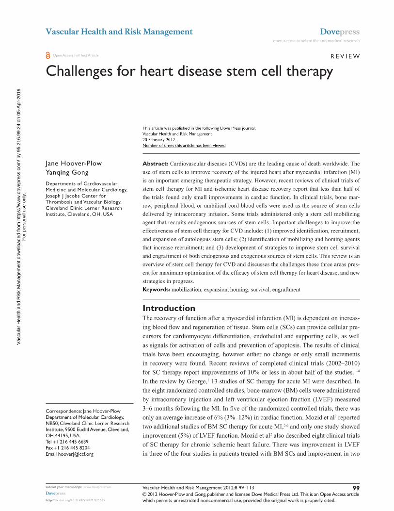
in Table 1, other types of SCs are being tested, including
specific BM, CD34+ or CD133+, and mesenchymal cells.
One study tests adipose tissue-derived SCs, and three trials
are testing cardiac progenitor/stem cells.
Skeletal myoblastsSkeletal myoblasts isolated from muscle biopsies were the
first cells used for the SC therapy for cardiac recovery.9 In
a comparison of rats with chronic MI, treated with human
skeletal myoblasts or BM-derived CD133+ progenitors,
improvements in cardiac function were similar with the
two cell types.10,11 In trials of skeletal myoblast treatment3
in patients with chronic ischemic heart failure, there were
improvements in LVEF in two of four studies (SEISMIC,
TOPCARD-CHD).3 While the initial evaluation in clinical
studies of skeletal myoblast treatment showed there was
improved function, the effect was not sustained, and the cells
were not electrically integrated into the heart.12 Enthusiasm
for this approach has waned. However, second-generation
products are now being developed.9,13 Six trials of skeletal
myoblast therapy have been discontinued, but currently
there are two active trials with skeletal myoblasts (Table 1)
for patients with an old MI (PERCUTANEO) or congestive
heart failure (MARVEL).
Hematopoietic progenitor/stem cells (HPSCs)In clinical trials for MI or ischemic heart disease, BM,
peripheral blood (PB), or umbilical cord blood (UCB) have
been used as the source of SCs.1,3 Autologous BM and PB
have an advantage over UCB cells since UCB cells may be at
risk for immunological rejection. However, the UCB have
a high proliferation potential.12 Autologous BM cells from
aging individuals may have reduced transplant efficiency,
and UCB cells would be advantageous.14,15 A limitation of the
PB is the low yield of SCs. BM is the major source of adult
SCs and the best characterized. The BM cells have long been
used in therapeutic BM replacement for blood diseases.16–18
BM SCs provide the myeloid and lymphoid lineages that
give rise to blood cells.19 The cell surface markers that
identify hematopoietic SCs (HSCs) for humans include:
CD34+, CD59+, Thy1/CD90+, CD38lo/−, c-kit/CD117+,
and lin−. There are differences in mouse HSC markers;
namely, CD34lo/−, Sca-1+, Thy1.1+/lo, and CD38+, but
with c-kit+ and lin− as common markers. The lineage
negative designation includes the absence of 13–14 cell
surface markers found on mature cells. BM has been the
major source of SCs for reported and ongoing clinical trials.
Currently, studies are underway that isolate subsets of the
Stem / Progenitor cells
Endothelial
Stem cells for heart therapy
CardiacHematopoietic Mesenchymal Skeletal
Source
Bone marrow Bone marrow
Peripheral blood Peripheral blood
Umbilical cordblood
Bone marrow
Adipose tissue
Muscle Heart
Epicardium
Infarct border
Figure 1 Types of stem cells in use for heart disease therapy.1–7
Reprinted with permission, Cleveland Clinic Center for Medical Art & Photography © 2011–2012. All Rights Reserved.
submit your manuscript | www.dovepress.com
Dovepress
Dovepress
101
Stem cell therapy challenges
Vas
cula
r H
ealth
and
Ris
k M
anag
emen
t dow
nloa
ded
from
http
s://w
ww
.dov
epre
ss.c
om/ b
y 95
.216
.99.
24 o
n 05
-Apr
-201
9F
or p
erso
nal u
se o
nly.
Powered by TCPDF (www.tcpdf.org)
1 / 1

Vascular Health and Risk Management 2012:8
BM cells such as CD34+, and CD133+ for use in therapy.
Whether these subsets of SCs will have an advantage in heart
disease recovery remains to be seen.
Endothelial SCsStages of lineage development of endothelial SCs and
their sites of origin are less well defined than those for the
hematopoietic lineage.20 The endothelial progenitor cells
(EPCs) found in the PB are thought to originate in the BM
from a subset of SCs or from the myeloid precursors. There
is considerable controversy with regard to the identification
of the EPCs.21 Some investigators have identified the EPCs
as CD34+ cells and/or CD133+ cells,22 while others view
these cells as HPSCs.23,24 Recently,25,26 a consensus definition
of EPC markers was suggested for cross-study comparisons
and with the cell surface markers CD31+, CD34 bright, and
CD45, AC133, CD14, CD14a, CD235a, Live/Dead Violet
negative. Of importance for identification of the EPC is the
ability to become endothelial cells (ECs) in culture. While
CD34+ and/or CD133+ cells in culture may become ECs,
the CD34+ and/or CD133+ cells could be a mixture of sub-
populations. However, the cells identified as CD34+ and/or
CD133+ may be more effective in providing paracrine factors
and stimulating neovascularization than the commonly used
BM MNCs. Tongers et al27 recently described the results of a
clinical trial for patients with refractory angina treated with
intramyocardial autologous CD34+ cells, finding significant
improvements in angina frequency and exercise tolerance.
There is one clinical trial currently underway for treatment
with CD34+ in patients with dilated cardiomyopathy, and five
clinical trials underway for the treatment of MI, CAD, and
heart failure with CD133+ cells. One study, NCT01187654,
will compare the treatment of CD133+ cells and BM MNC
in MI patients. This comparison could be informative as
to whether the CD133+ cells have an advantage over the
more frequently used BM MNC. Bissels et al28 found that
microRNAs were expressed differentially in CD133+,
CD34+, and CD133- cells involved in differentiation, pre-
vention of apoptosis, and cytoskeletal remodeling.
Mesenchymal SCs (MSCs)The MSCs are found in the BM and other tissues. MSCs
are positive for CD44, CD73, CD90 (Thy1), and CD105,
and negative for the hematopoietic markers, CD45, lineage
markers, EC (CD31), and macrophage (CD11b/MAC-1).29
The MSCs have advantages over HSCs.27,30 Compared with
HSCs, MSCs are more abundant, readily proliferate in cul-
ture, and are easily differentiated into different cell types,
such as adipocytes, fibroblasts, osteocytes, and myoblasts.
Further, studies suggest that MSCs may be more potent
for cardiac repair than HPSCs.31 Although the MSCs can
be differentiated into cardiomyocytes, immortalization
was important and could increase the potential of tumor
formation.15 In addition to BM, adipose tissue can also be
used as an abundant source of MSCs.32,33 The MSCs from
UCB, adipose tissue, and BM expressed the same cell surface
markers; however, there are some differences in the percentage
of certain markers and colony heterogeneity. Gaebel et al34
compared treatment of MI in mice with MSCs from UCB,
adipose tissue, and BM. Cells from BM, adipose tissue, and
UCB CD105+ showed improvements in heart functions,
decreased infarct size, and capillary density. UCB CD105
treated mice had reduced collagen deposition compared with
BM and adipose tissue cells, and BM and UCB CD105 cells
additionally had reduced apoptosis when compared with mice
treated with adipose tissue cells. This study suggests that the
function of the MSCs may be dependent on the source. Clini-
cal trials with MSCs35–37 are promising, and currently there are
19 clinical trials underway.7,38 In a recent randomized, double
blind, placebo-controlled study37 with MSC therapy after acute
MI; there was improvement in the global assessment of cardiac
function at 6 months in 45% of the patients.
Cardiac progenitor cells (CPCs)Although it had been believed for a long time that cardiac
myocytes were terminally differentiated, dividing myocytes
found in the heart implied that there are resident or noncardiac
cardiomyocyte progenitor cells.39 There have been intensive
efforts to identify the cardiomyocyte stem and progenitor
cells in the last 10 years.39 Purified cardiomyocytes isolated
from rodent hearts dedifferentiate and divide, expressing SC
markers such as c-kit, Sca-1, Isl1, and Abcg2.40–45 CPCs have
been isolated from human myocardial biopsies.46,47 These
same cells can organize into spheres and re-differentiate into
myocytes and ECs.48 Yamada et al49,50 suggested that CD133+
cells from brown adipose tissue were highly effective in dif-
ferentiation into cardiomyocytes compared with HPSCs, and
that mouse BAT CD133+ cells efficiently induced BM SCs
into cardiomyocytes (CD45- CD31- CD105+) differentiation.
There are four ongoing clinical studies to test autologous
CPCs (Table 1); one study (ALCADIA) will use cardiac-
derived SCs to treat ischemic cardiomyopathy, and two
studies will take advantage of the cardiosphere-derived stem/
progenitor cells (derived from cell outgrowth of autologous
cardiac biopsy) for patients with a recent MI (CADUCEUS)
or heart failure (TICAP). In the SCIPIO trial, patients with
submit your manuscript | www.dovepress.com
Dovepress
Dovepress
102
Hoover-Plow and Gong
Vas
cula
r H
ealth
and
Ris
k M
anag
emen
t dow
nloa
ded
from
http
s://w
ww
.dov
epre
ss.c
om/ b
y 95
.216
.99.
24 o
n 05
-Apr
-201
9F
or p
erso
nal u
se o
nly.
Powered by TCPDF (www.tcpdf.org)
1 / 1

Vascular Health and Risk Management 2012:8
ischemic cardiomyopathy are treated with c-kit+lin− CPCs
derived from the right atrial appendage, and initial results
from 16 patients report that LVEF increased and infarct
size decreased.51
Adipose tissue-derived SCs (ASCs)Cells isolated from adipose tissue can be separated by cen-
trifugation into adipocytes and stromal vascular cells. The
stromal vascular fraction may contain preadipocytes, peri-
cytes and EPCs, adult multipotent MSCs, circulating blood
cells, fibroblasts, ECs, smooth-muscle cells, and immune
cells. This stromal vascular fraction may differentiate into a
number of cell lineages, including the adipocytes, cartilage,
bone skeletal muscle, neuronal cells, ECs, cardiomyocytes,
and smooth-muscle cells.52,53 ASCs are defined as CD44 and
CD105 positive, and Cd11b, CD34, and CD45 negative cells.
Although there is disagreement regarding the capacity of
ASCs to differentiate into ECs, freshly isolated human ASCs
also consist of EPCs (CD11b, CD34, and CD45 positive cells)
and when cultured they have a cobblestone appearance and
take up acetylated low-density lipoprotein. Bai et al54 found
that human freshly isolated adipocytes or cultured adipose
tissue-derived cells underwent cardiomyogenesis through a
fusion-independent pathway. Takahashi et al55 reported that
in rat femoral artery injury, ASCs did not differentiate into
ECs, but were able to inhibit neointimal formation by the
secretion of paracrine factors. There is one ongoing clinical
trial (NCT01216995) testing adipose tissue-derived cells in
patients after an acute MI.
Induced pluripotent stem (iPS) cellsAnother potential source of SCs is iPS cells.56 This source
relies on in vitro de-differentiation of adult cells to
embryonic-like SCs and then reprogramming using specific
culture conditions to induce cardiac lineage cells including
cardiomyocytes, smooth-muscle cells, and ECs. Adult cells
most commonly used for iPS cells are fibroblasts and may
be derived from a variety of tissues such as dermal, liver,
stomach, pancreas, and neural and hematopoietic cells.
Endogenous non-BM SC and iPS cells have been charac-
terized in animal models and some have been identified in
adult humans. Defining these cells and their requirements
for proliferation and mobilization will provide additional
options for enhanced efficacy of SC therapy.
Embryonic SCs (ESCs)The ESCs are the ideal SCs, due to the fact that cultures of
embryonic cells when stimulated can develop into .200
adult cell types.38,57,58 Current efforts focus on establishing
the conditions for directed differentiation of cells by
altering the chemical composition of the culture medium, alter-
ing the culture surface, or inserting genes.58 A major challenge
is the potential of uncontrolled differentiation when injected
directly into an animal, and the potential for tumor formation.
The promise of ESCs is to genetically modify lethal debilitat-
ing chronic disease. There are currently four clinical trials in
progress of human ESCs for spinal cord injury and macular
degeneration, but unfortunately none for cardiac disease.38
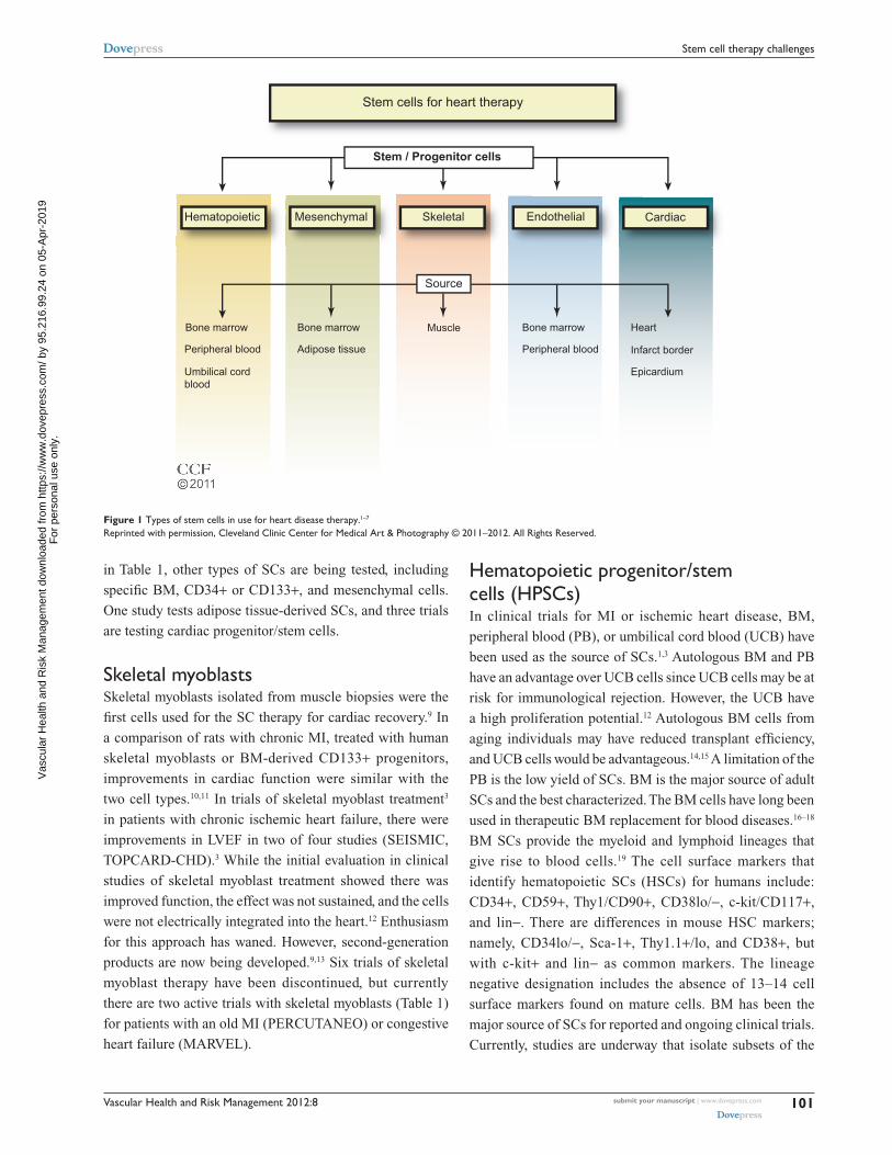
Expansion of SCsA critical step for improved SC therapy is the expansion of
accessible SCs (Figure 2). The homing of cells to injured tis-
sues is very inefficient, and increasing the number of cells that
are available for treatment would be beneficial. Autologous
BM cells, adipose tissue, myocardial, and UCB are cultured
ex vivo to increase the number of cells. Culturing the tissue
also allows selection of specific cells. The ESCs and iPS cells
require additional steps prior to expansion of a preparation.
The iPS cells require de-differentiation as an initial step and
then both iPS cells and ESCs are induced to differentiate prior
to expansion. SCs in culture form colonies, and proliferation
without differentiation requires a specific sequence and tim-
ing of the availability of growth factors and cytokines.59–66
In addition, these cells must maintain their pluripotency. Cells
need to be free of feeder-cells, serum proteins, and microbial
agents. Large-scale expansion with maintenance of pluripo-
tency and transplant safety is required.58,67 Currently, effec-
tive cell culture proliferation is limited,61 and further studies
are needed to understand the requirements for expansion.
New approaches are being investigated including the use
of nanofibers with growth factors, mesenchymal stromal
cells in cultures of HSCs, and genetic manipulation of UCB
HSCs.68–72 To improve SC therapy, improved methods of SC
ex vivo expansion are required.
Identify mobilizing agents with improved effectivenessSC nichesIntensive studies are underway to identify new sources of
stem and progenitor cells for therapy. In addition to BM,
SC niches have been identified (Figure 3) in heart. The SC
niches are defined as a microenvironment with one or more
SC that regulates self-renewal and progeny in vivo.73,74 Self-
renewal occurs in all tissues and in addition to BM, niches
of SCs have been identified in heart, arteries, veins, gonads,
intestine, epidermal tissue, and neural tissue.73,75–77 The
submit your manuscript | www.dovepress.com
Dovepress
Dovepress
103
Stem cell therapy challenges
Vas
cula
r H
ealth
and
Ris
k M
anag
emen
t dow
nloa
ded
from
http
s://w
ww
.dov
epre
ss.c
om/ b
y 95
.216
.99.
24 o
n 05
-Apr
-201
9F
or p
erso
nal u
se o
nly.
Powered by TCPDF (www.tcpdf.org)
1 / 1
Vascular Health and Risk Management 2012:8
non-BM SCs were initially defined by immunofluorescence
in tissue, but given the number of markers needed, this
became untenable, and isolation and identification of SCs
by flow cytometry using multiple markers simultaneously
has made it possible to isolate and investigate the function
of these cells. Recently, lineage mapping has been utilized
to locate niches in animal models by genetically labeling
SC markers and identifying their location in adult tissue.78,79
An example of lineage mapping is the recent study of
Tamura et al78 of neural crest-derived SCs found in the heart
that migrate and differentiate into cardiomyocytes after MI.
The lineage mapping has been utilized for locating SC
niches in a variety of developing organisms.79 The number
of quiescent SCs is small, and better detection methods are
necessary. Further, identifying the regulation and recruitment
of these endogenous SCs in adults is critical.
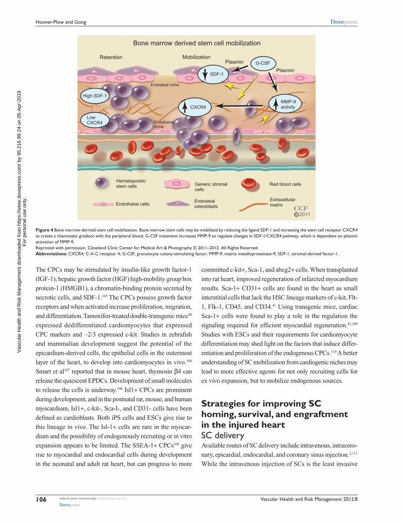
Mobilization of BM SCsIn the BM, SCs reside in an endosteal niche along with stromal
cells, mesenchymal cells, and ECs. The SCs are retained in
the BM with high concentrations of stromal-derived factor
(SDF)-1, the major chemoattractant for SCs. The SDF-1 SC
receptor, CXCR4, is found in low concentrations. Stimula-
tion with cytokines or growth factors may interrupt ligand/
receptor balance. With a decrease in SDF-1 and an increase in
CXCR4 expression, a signaling gradient with the PB allows
the egress of the SCs from the BM (Figure 4). Granulocyte
colony-stimulating factor (G-CSF) is widely used clinically
for SC mobilization and sometimes in conjunction with
other factors57,80 including granulocyte-macrophage colony-
stimulating factor, stem cell factor, fms-like tyrosine kinase
(Flt)-3 ligand, and interleukin-1, -3, -6, -7, -8, -11, and -12
(Figure 3). AMD3100, an inhibitor that blocks SDF-1 bind-
ing to CXCR4; CTCE-0021, a CXCR4 agonist; recombinant
human growth hormone, a pleiotrophic cytokine; parathyroid
hormone; pegfilgrastim, pegylated G-CSF with a prolonged
half-life, and thrombopoietin, a cytokine that regulates mega-
karyocytopoiesis, are also being investigated.80
In addition to cytokines and growth factors, proteases
such as neutrophil elastase, cathespin G, plasmin, and matrix
metalloproteinase (MMP)-9 have been implicated in BM SC
mobilization.81–86 After G-CSF treatment, these proteases
increase in BM as well as in plasma; however, studies83 in
mice deficient in neutrophil elastase or cathespin G suggest
Embryonic
Stem cell therapy
stem cells
Cardiomyocytes
Endothelial cells
Smooth muscle cells
Hematopoietic stem cells Mesenchymal stem cells FibroblastsAdipocytes(adult cells)
Cardiac stem cellsSkeletal myoblasts
stem cell
EXPANSION OF STEM CELLS
DEDIFFERENTIATE(IPS)
DIFFERENTIATE
PROLIFERATE
Figure 2 Expansion of stem cells.Reprinted with permission, Cleveland Clinic Center for Medical Art & Photography © 2011–2012. All Rights Reserved.Notes: Currently, increased numbers of autologous hematopoietic, mesenchymal, cardiac, endothelial, and skeletal stem cells can be generated by expansion in culture with proliferation specific conditions. Adult cells such as fibroblasts or adipocytes may be dedifferentiated in culture to stem cells (iPS cells). MSCs, iPS cells, and ESCs can be induced to differentiate and proliferate in cell culture. Use of differentiated MSCs, iPS cells, and ESCs is in preclinical development.Abbreviations: ESC, embryonic stem cell; iPS, induced pluripotent stem; MSC, mesenchymal stem cell.
submit your manuscript | www.dovepress.com
Dovepress
Dovepress
104
Hoover-Plow and Gong
Vas
cula
r H
ealth
and
Ris
k M
anag
emen
t dow
nloa
ded
from
http
s://w
ww
.dov
epre
ss.c
om/ b
y 95
.216
.99.
24 o
n 05
-Apr
-201
9F
or p
erso
nal u
se o
nly.
Powered by TCPDF (www.tcpdf.org)
1 / 1
Vascular Health and Risk Management 2012:8
these two proteases are not required for HPSC mobilization.
The results of studies81,83,87,88 in MMP-9 deficient mice are
not consistent. While some studies83,88 report MMP-9 is
not required, other studies81,86,87 suggest MMP-9 plays an
important role. These differences may be due to the differ-
ences in genetic background of the mice and to differences
in the dose of the mobilizing agent. In a recent study,86 the
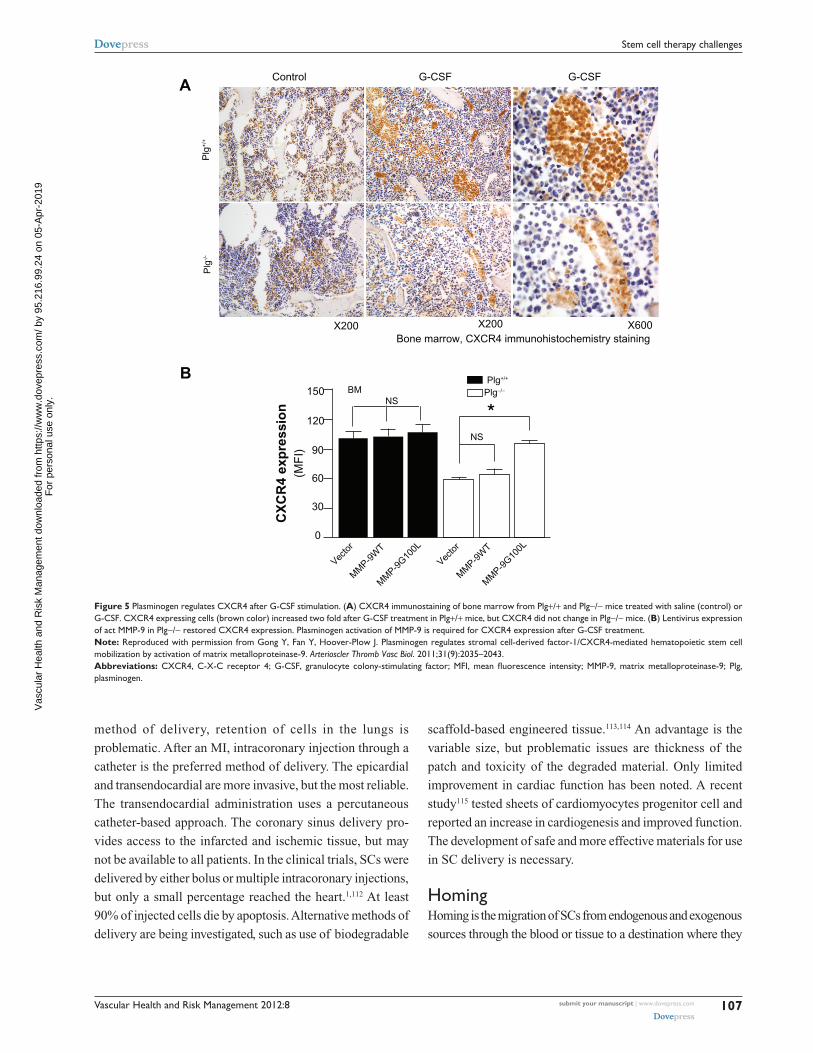
authors of this present paper report that plasmin/MMP-9 is a
major proteolytic pathway required for SC mobilization from
BM (Figure 5). Plasmin activation of MMP-9 regulates the
SDF-1/CXCR4 signaling. In addition, plasmin also promotes
direct degradation of the ECM during SC mobilization.85
G-CSF induced HSC MMP-9 degrades BM SDF-1.83,89,90 The
increase in the number of SC mobilized with G-CSF treat-
ment may not be sufficient for the cardiac remodeling after
MI, and some patients are resistant to G-CSF.91–93 AMD3100,
an inhibitor of CXCR4, is a promising HSC mobilizer under
clinical investigation. Studies report mild and reversible side
effects94–96 and that it works synergistically with G-CSF to
increase CD34+ cells and total white blood count.94,96–98
However, Dai et al recently reported that chronic AMD100
exacerbates cardiac dysfunction after MI in mice.99
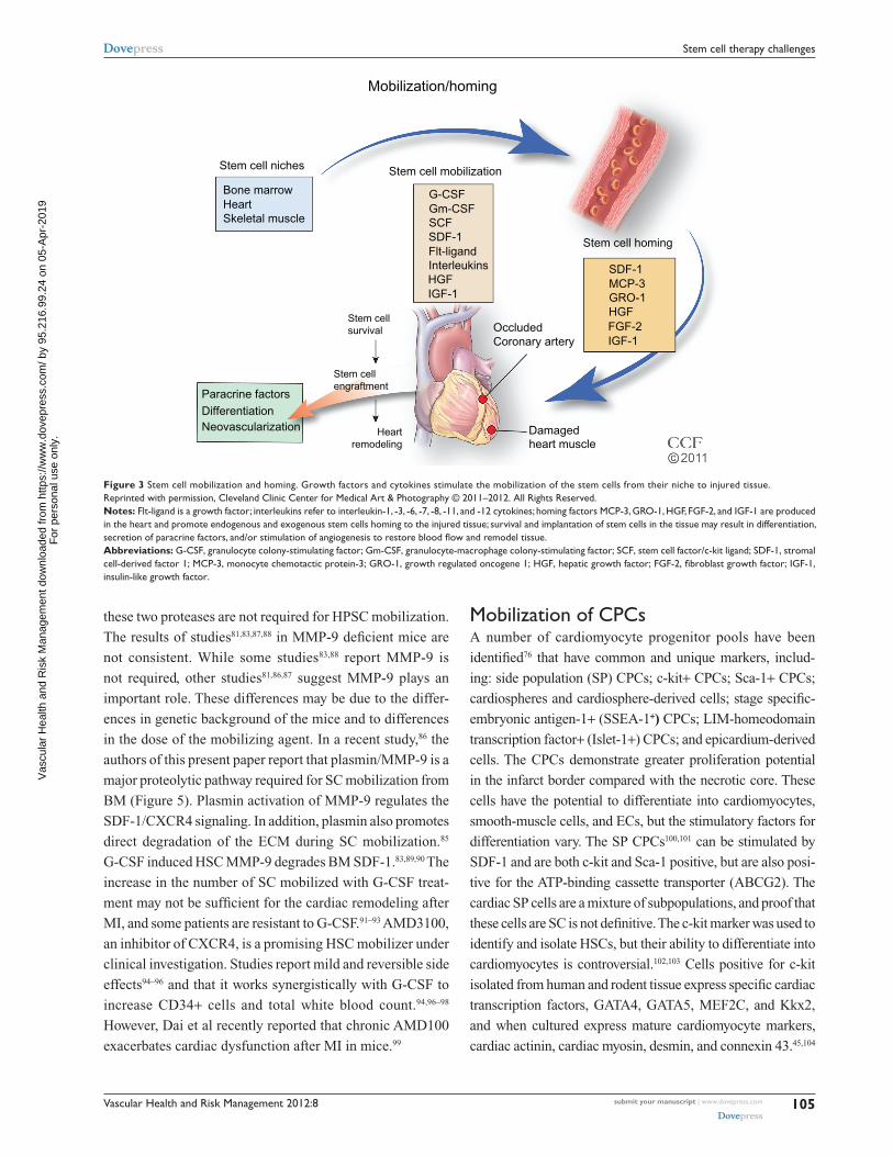
Mobilization of CPCsA number of cardiomyocyte progenitor pools have been
identified76 that have common and unique markers, includ-
ing: side population (SP) CPCs; c-kit+ CPCs; Sca-1+ CPCs;
cardiospheres and cardiosphere-derived cells; stage specific-
embryonic antigen-1+ (SSEA-1+) CPCs; LIM-homeodomain
transcription factor+ (Islet-1+) CPCs; and epicardium-derived
cells. The CPCs demonstrate greater proliferation potential
in the infarct border compared with the necrotic core. These
cells have the potential to differentiate into cardiomyocytes,
smooth-muscle cells, and ECs, but the stimulatory factors for
differentiation vary. The SP CPCs100,101 can be stimulated by
SDF-1 and are both c-kit and Sca-1 positive, but are also posi-
tive for the ATP-binding cassette transporter (ABCG2). The
cardiac SP cells are a mixture of subpopulations, and proof that
these cells are SC is not definitive. The c-kit marker was used to
identify and isolate HSCs, but their ability to differentiate into
cardiomyocytes is controversial.102,103 Cells positive for c-kit
isolated from human and rodent ti ssue express specific cardiac
transcription factors, GATA4, GATA5, MEF2C, and Kkx2,
and when cultured express mature cardiomyocyte markers,
cardiac actinin, cardiac myosin, desmin, and connexin 43.45,104
Bone marrowHeartSkeletal muscle
G-CSFGm-CSFSCFSDF-1Flt-ligandInterleukinsHGFIGF-1
SDF-1MCP-3GRO-1HGFFGF-2IGF-1
Paracrine factorsDifferentiation
Heartremodeling
Damagedheart muscle
Stem cellsurvival Occluded
Coronary artery
Stem cell homing
Stem cell mobilization
Mobilization/homing
Stem cell niches
Stem cellengraftment
Neovascularization
Figure 3 Stem cell mobilization and homing. Growth factors and cytokines stimulate the mobilization of the stem cells from their niche to injured tissue.Reprinted with permission, Cleveland Clinic Center for Medical Art & Photography © 2011–2012. All Rights Reserved.Notes: Flt-ligand is a growth factor; interleukins refer to interleukin-1, -3, -6, -7, -8, -11, and -12 cytokines; homing factors MCP-3, GRO-1, HGF, FGF-2, and IGF-1 are produced in the heart and promote endogenous and exogenous stem cells homing to the injured tissue; survival and implantation of stem cells in the tissue may result in differentiation, secretion of paracrine factors, and/or stimulation of angiogenesis to restore blood flow and remodel tissue.Abbreviations: G-CSF, granulocyte colony-stimulating factor; Gm-CSF, granulocyte-macrophage colony-stimulating factor; SCF, stem cell factor/c-kit ligand; SDF-1, stromal cell-derived factor 1; MCP-3, monocyte chemotactic protein-3; GRO-1, growth regulated oncogene 1; HGF, hepatic growth factor; FGF-2, fibroblast growth factor; IGF-1, insulin-like growth factor.
submit your manuscript | www.dovepress.com
Dovepress
Dovepress
105
Stem cell therapy challenges
Vas
cula
r H
ealth
and
Ris
k M
anag
emen
t dow
nloa
ded
from
http
s://w
ww
.dov
epre
ss.c
om/ b
y 95
.216
.99.
24 o
n 05
-Apr
-201
9F
or p
erso
nal u
se o
nly.
Powered by TCPDF (www.tcpdf.org)
1 / 1
Vascular Health and Risk Management 2012:8
The CPCs may be stimulated by insulin-like growth factor-1
(IGF-1); hepatic growth factor (HGF) high-mobility group box
protein-1 (HMGB1), a chromatin-binding protein secreted by
necrotic cells, and SDF-1.105 The CPCs possess growth factor
receptors and when activated increase proliferation, migration,
and differentiation. Tamoxifen-treated double-transgenic mice48
expressed dedifferentiated cardiomyocytes that expressed
CPC markers and ∼2/3 expressed c-kit. Studies in zebrafish
and mammalian development suggest the potential of the
epicardium-derived cells, the epithelial cells in the outermost
layer of the heart, to develop into cardiomyocytes in vivo.106
Smart et al107 reported that in mouse heart, thymosin β4 can
release the quiescent EPDCs. Development of small molecules
to release the cells is underway.106 Isl1+ CPCs are prominent
during development, and in the postnatal rat, mouse, and human
myocardium, Isl1+, c-kit-, Sca-l-, and CD31- cells have been
defined as cardioblasts. Both iPS cells and ESCs give rise to
this lineage in vivo. The Isl-1+ cells are rare in the myocar-
dium and the possibility of endogenously recruiting or in vitro
expansion appears to be limited. The SSEA-1+ CPCs108 give
rise to myocardial and endocardial cells during development
in the neonatal and adult rat heart, but can progress to more
committed c-kit+, Sca-1, and abcg2+ cells. When transplanted
into rat heart, improved regeneration of infarcted myocardium
results. Sca-1+ CD31+ cells are found in the heart as small
interstitial cells that lack the HSC lineage markers of c-kit, Flt-
1, Flk-1, CD45, and CD34.41 Using transgenic mice, cardiac
Sca-1+ cells were found to play a role in the regulation the
signaling required for efficient myocardial regeneration.42,109
Studies with ESCs and their requirements for cardiomyocyte
differentiation may shed light on the factors that induce differ-
entiation and proliferation of the endogenous CPCs.110 A better
understanding of SC mobilization from cardiogenic niches may
lead to more effective agents for not only recruiting cells for
ex vivo expansion, but to mobilize endogenous sources.
Strategies for improving SC homing, survival, and engraftment in the injured heartSC deliveryAvailable routes of SC delivery include intravenous, intracoro-
nary, epicardial, endocardial, and coronary sinus injection.2,111
While the intravenous injection of SCs is the least invasive
2011
Bone marrow derived stem cell mobilization
Retention Mobilization
LowCXCR4
High SDF-1
SDF-1
CXCR4
G-CSF
MMP-9activity
Hematopoieticstem cells
Endothelial cells
Generic stromalcells
Endostealosteoblasts
Red blood cells
Extracellularmatrix
PlasminPlasmin
Endosteal niche
Endothelialniche
Figure 4 Bone marrow-derived stem cell mobilization. Bone marrow stem cells may be mobilized by reducing the ligand SDF-1 and increasing the stem cell receptor CXCR4 to create a chemotatic gradient with the peripheral blood. G-CSF treatment increases MMP-9 to regulate changes in SDF-1/CXCR4 pathway, which is dependent on plasmin activation of MMP-9.Reprinted with permission, Cleveland Clinic Center for Medical Art & Photography © 2011–2012. All Rights Reserved.Abbreviations: CXCR4, C-X-C receptor 4; G-CSF, granulocyte colony-stimulating factor; MMP-9, matrix metalloproteinase-9; SDF-1, stromal-derived factor-1.
submit your manuscript | www.dovepress.com
Dovepress
Dovepress
106
Hoover-Plow and Gong
Vas
cula
r H
ealth
and
Ris
k M
anag
emen
t dow
nloa
ded
from
http
s://w
ww
.dov
epre
ss.c
om/ b
y 95
.216
.99.
24 o
n 05
-Apr
-201
9F
or p
erso
nal u
se o
nly.
Powered by TCPDF (www.tcpdf.org)
1 / 1
Vascular Health and Risk Management 2012:8
method of delivery, retention of cells in the lungs is
problematic. After an MI, intracoronary injection through a
catheter is the preferred method of delivery. The epicardial
and transendocardial are more invasive, but the most reliable.
The transendocardial administration uses a percutaneous
catheter-based approach. The coronary sinus delivery pro-
vides access to the infarcted and ischemic tissue, but may
not be available to all patients. In the clinical trials, SCs were
delivered by either bolus or multiple intracoronary injections,
but only a small percentage reached the heart.1,112 At least
90% of injected cells die by apoptosis. Alternative methods of
delivery are being investigated, such as use of biodegradable
scaffold-based engineered tissue.113,114 An advantage is the
variable size, but problematic issues are thickness of the
patch and toxicity of the degraded material. Only limited
improvement in cardiac function has been noted. A recent
study115 tested sheets of cardiomyocytes progenitor cell and
reported an increase in cardiogenesis and improved function.
The development of safe and more effective materials for use
in SC delivery is necessary.
HomingHoming is the migration of SCs from endogenous and exogenous
sources through the blood or tissue to a destination where they
150Plg+/+
X200 X200Bone marrow, CXCR4 immunohistochemistry staining
X600
Plg
+/+
Plg−/−
Plg
-/-
NS
(MF
I)C
XC
R4
exp
ress
ion
NS
BM
120
90
60
30
0
Vecto
r
Vecto
r
MM
P-9W
T
MM
P-9W
T
MM
P-9G10
0L
MM
P-9G10
0L
B
AControl G-CSF G-CSF
Figure 5 Plasminogen regulates CXCR4 after G-CSF stimulation. (A) CXCR4 immunostaining of bone marrow from Plg+/+ and Plg−/− mice treated with saline (control) or G-CSF. CXCR4 expressing cells (brown color) increased two fold after G-CSF treatment in Plg+/+ mice, but CXCR4 did not change in Plg−/− mice. (B) Lentivirus expression of act MMP-9 in Plg−/− restored CXCR4 expression. Plasminogen activation of MMP-9 is required for CXCR4 expression after G-CSF treatment.Note: Reproduced with permission from Gong Y, Fan Y, Hoover-Plow J. Plasminogen regulates stromal cell-derived factor-1/CXCR4-mediated hematopoietic stem cell mobilization by activation of matrix metalloproteinase-9. Arterioscler Thromb Vasc Biol. 2011;31(9):2035–2043.Abbreviations: CXCR4, C-X-C receptor 4; G-CSF, granulocyte colony-stimulating factor; MFI, mean fluorescence intensity; MMP-9, matrix metalloproteinase-9; Plg, plasminogen.
submit your manuscript | www.dovepress.com
Dovepress
Dovepress
107
Stem cell therapy challenges
Vas
cula
r H
ealth
and
Ris
k M
anag
emen
t dow
nloa
ded
from
http
s://w
ww
.dov
epre
ss.c
om/ b
y 95
.216
.99.
24 o
n 05
-Apr
-201
9F
or p
erso
nal u
se o
nly.
Powered by TCPDF (www.tcpdf.org)
1 / 1

Vascular Health and Risk Management 2012:8
differentiate and replace or repair injured tissue. After an MI,
expression of several factors has been observed, including tran-
sient increases in cardiac cytokines, SDF-1, MCP-3, GRO-1,
that are chemo-attractants for SCs.116–123 After acute MI, the
expression of these factors leads to SC homing to the infarcted
tissue. However, many of the homing factors are expressed for
only a short period of time after MI. SDF-1, the most studied
homing factor, is expressed by the injured cardiac tissue for
less than 1 week123 and MCP-3 for less than 10 days after MI.124
In preclinical studies, genetic engineering of these factors
into delivered SCs is effective in increasing SC homing.123,125
For example, the delivery of SDF-1 to the myocardium, either
through cell-based gene therapy,123,126 gene transfer,127 or protein-
enhanced128 homing of SCs, results in revascularization and
improvement in cardiac function. Furthermore, overexpress-
ing SDF-1 receptor CXCR4 in SCs leads to greater homing of
SCs and improved left ventricular function when the cells were
delivered within 24 hours of MI.129–131 Studies in animals show
that engineering cells to induce the expression of SC homing
factors or their receptors in myocardial tissue can promote
SC homing from BM to the injured myocardium; however,
these have not to date been tested in humans.132
Survival/engraftmentSurvival and engraftment of SCs is perhaps the most impor-
tant challenge for SC therapy, and the factors necessary for
effective survival and engraftment are not necessarily the
same as those required for homing. After an MI, there is an
enormous loss of cardiomyocytes and supporting cells that
need to be replaced. The environmental signals that may
guide SCs to the cardiomyocyte lineage or to the secretion
of paracrine factors may be absent in the infarcted tissue, and
SCs may provide these signals. Many studies have focused
on strategies to optimize SC migration through injured myo-
cardial tissue. Proteases, adhesion molecules, and integrins
are important in regulating SC migration through injured
myocardial tissue and modulation of the connective tissue
microenvironment to improve SC engraftment.133–136
Several proteases have been identified to have significant
effects on SC mobilization or SC migration and engraftment
in cardiac tissue. SDF-1 and other factors induce the secre-
tion of matrix metalloproteinase MMP-2 and MMP-9.137–139
Of significant interest, proteolytic enzymes, including neu-
trophil elastase, cathepsin G, and MMP-2/9, also negatively
regulate cell migration by cleaving the N-terminal region
of SDF-1 or cleaving CXCR4.90,139–142 Those proteolytic
enzymes are involved in spatial temporal changes in the
locomotion machinery of SCs, thus mediating SC recruit-
ment and engraftment.
Integrins are also key factors for adhesion, rolling and
transmigration of SCs across the endothelium. The HSCs
express several adhesion molecules including multiple inte-
grins. In particular, a dominant role for the α4β1 integrin
very-late antigen [VLA]-4 interaction with vascular cell
adhesion molecule (VCAM)-1 has been suggested by stud-
ies in which exposure to blocking antibodies to VLA-4 or
VCAM-1 significantly reduced the engraftment of transplanted
HSCs.143–145 CD18 expression by the EPCs is necessary for its
interaction with EC surface ICAM-1, and a CD18 neutral-
izing antibody significantly inhibits SC engraftment after
acute MI.146 These studies suggest the potential targets for the
genetic enhancement of SC recruitment and engraftment.
Several other strategies have been proposed: identifying
natural mediators; pre-translational directed differentiation of
SCs to cardiomyocytes; activation of growth factors (FGF-2,
IGF-1a)132 and antiapoptotic factors (p-Akt, SDF-1, BCl-1,
and PDGF); and genetically engineered SCs.125,132 The chal-
lenge to improve survival in SC therapy is to identify effective
ways to increase the number of cells that reach and survive
in the injured heart area.
Assessment of SC therapyThe goals of SC therapy are to: replace lost cardiomyocytes;
increase ECs to improve blood flow; provide paracrine cytok-
ines and growth factors; and improve measurable cardiac
function, including an increase in LVEF; decrease left ventric-
ular end-diastolic diameter; increase myocardial perfusion;
and importantly increase exercise capacity. In clinical trials,
methods to measure cardiac function include echocardiog-
raphy, single photon emission computed tomography, and
magnetic resonance imaging (MRI).1,3,37,147–149 These methods
are well established, but more sensitive methods are neces-
sary to evaluate SC homing and engraftment. Techniques to
evaluate the timing and specific role of narrow populations
of cells, such as MRI150–152 and SC labeling with genetic153,154
and immunofluorescence detectable tags155 are being inves-
tigated in animal models. The lineage/fate mapping110,156–158
has proved to be an informative tool, and further studies in
animal models and ex vivo SC labeling of cells for therapy
will continue to be valuable.
ConclusionSC therapy is an exciting and dynamic area of research with
the potential to improve recovery of CVD, the leading cause of
submit your manuscript | www.dovepress.com
Dovepress
Dovepress
108
Hoover-Plow and Gong
Vas
cula
r H
ealth
and
Ris
k M
anag
emen
t dow
nloa
ded
from
http
s://w
ww
.dov
epre
ss.c
om/ b
y 95
.216
.99.
24 o
n 05
-Apr
-201
9F
or p
erso
nal u
se o
nly.
Powered by TCPDF (www.tcpdf.org)
1 / 1
Vascular Health and Risk Management 2012:8
death. While animal models clearly show benefits of SC therapy
to improve cardiac function after MI and ischemic heart failure,
clinical trials have been disappointing. However, the results
of clinical trials are promising. Better methods are needed
to improve the isolation and identification of SCs, increase
ex vivo expansion of SCs, and increase delivery effectiveness.
A clearer understanding of mobilization and homing of SCs is
needed to identify new and more effective agents. Delineating
the function of specific SCs in remodeling injured tissue
and how resident cardiac SCs may be enhanced is needed to
improve SC engraftment and survival.
AcknowledgmentsThis study was funded by grants from American Heart
Association (AHA0625331B and 09BGIA2050157) and
the National Institutes of Health, National Heart, Lung, and
Blood Institute (R01HL078701). The authors thank Beth
Halasz, CMI, for the artwork for the figures.
DisclosureThe authors report no conflicts of interest in this work.
References 1. George JC. Stem cell therapy in acute myocardial infarction: a review
of clinical trials. Trans Res. 2010;155(1):10–19. 2. Mozid AM, Arnous S, Sammut EC, Mathur A. Stem cell therapy for
heart diseases. Br Med Bull. 2011;98:143–159. 3. Sanz-Ruiz R, Gutierrez Ibanes E, Arranz AV, Fernandez Santos ME,
Fernandez PL, Fernandez-Aviles F. Phases I–III clinical trials using adult stem cells. Stem Cells Int. 2010;2010:579142.
4. Wen Y, Meng L, Xie J, Ouyang J. Direct autologous bone marrow-derived stem cell transplantation for ischemic heart disease: a meta-analysis. Expert Opin Biol Ther. 2011;11(5):559–567.
5. Hirsch A, Nijveldt R, van der Vleuten PA, et al. Intracoronary infusion of mononuclear cells from bone marrow or peripheral blood compared with standard therapy in patients after acute myocardial infarction treated by primary percutaneous coronary intervention: results of the randomized controlled HEBE trial. Eur Heart J. 2011;32(14):1736–1747.
6. Huikuri HV, Kervinen K, Niemela M, et al. Effects of intracoronary injection of mononuclear bone marrow cells on left ventricular function, arrhythmia risk profile, and restenosis after thrombolytic therapy of acute myocardial infarction. Eur Heart J. 2008;29(22):2723–2732.
7. ClinicalTrials.gov [homepage on the Internet]. Available from: http://www.ClinicalTrials.gov. Accessed January 2, 2012.
8. Till JE, McCulloch EA, Siminovitch L. A stochastic model of stem cell proliferation, based on the growth of spleen colony-forming cells. Proc Natl Acad Sci U S A. 1964;51:29–36.
9. Menasche P. Towards the second generation of skeletal myoblasts? Cardiovasc Res. 2008;79(3):355–356.
10. Agbulut O, Vandervelde S, Al Attar N, et al. Comparison of human skeletal myoblasts and bone marrow-derived CD133+ pro-genitors for the repair of infarcted myocardium. J Am Coll Cardiol. 2004;44(2):458–463.
11. Taylor DA, Atkins BZ, Hungspreugs P, et al. Regenerating func-tional myocardium: improved performance after skeletal myoblast transplantation. Nat Med. 1998;4(8):929–933.
12. Gersh BJ, Simari RD, Behfar A, Terzic CM, Terzic A. Cardiac cell repair therapy: a clinical perspective. Mayo Clin Proc. 2009;84(10): 876–892.
13. Haider H, Lei Y, Ashraf M. MyoCell, a cell-based, autologous skeletal myoblast therapy for the treatment of cardiovascular diseases. Curr Opin Mol Ther. 2008;10(6):611–621.
14. Finney MR, Greco NJ, Haynesworth SE, et al. Direct comparison of umbilical cord blood versus bone marrow-derived endothelial precursor cells in mediating neovascularization in response to vascular ischemia. Biol Blood Marrow Transplant. 2006;12(5):585–593.
15. Agarwal U, Ghalayini W, Dong F, et al. Role of cardiac myocyte CXCR4 expression in development and left ventricular remodeling after acute myocardial infarction. Circ Res. 2010;107(5):667–676.
16. Feldman EJ, Gergis U. Management of refractory acute myeloid leuke-mia: re-induction therapy or straight to transplantation? Curr Hematol Malig Rep. 2011. [Epub ahead of print].
17. Stone RM, O’Donnell MR, Sekeres MA. Acute myeloid leukemia. Hematology Am Soc Hematol Educ Program. 2004:98–117.
18. Lodi D, Iannitti T, Palmieri B. Stem cells in clinical practice: applica-tions and warnings. J Exp Clin Cancer Res. 2011;30:9.
19. Sieburg HB, Cho RH, Dykstra B, Uchida N, Eaves CJ, Muller-Sieburg CE. The hematopoietic stem compartment consists of a limited number of discrete stem cell subsets. Blood. 2006;107(6):2311–2316.
20. Alev C, Ii M, Asahara T. Endothelial progenitor cells: a novel tool for the therapy of ischemic diseases. Antioxid Redox Signal. 2011;15(4):949–965.
21. Prater DN, Case J, Ingram DA, Yoder MC. Working hypothesis to rede-fine endothelial progenitor cells. Leukemia. 2007;21(6):1141–1149.
22. Peichev M, Naiyer AJ, Pereira D, et al. Expression of VEGFR-2 and AC133 by circulating human CD34(+) cells identifies a population of functional endothelial precursors. Blood. 2000;95(3):952–958.
23. Timmermans F, Van Hauwermeiren F, De Smedt M, et al. Endothelial outgrowth cells are not derived from CD133+ cells or CD45+ hematopoietic precursors. Arterioscler Thromb Vasc Biol. 2007;27(7):1572–1579.
24. Case J, Mead LE, Bessler WK, et al. Human CD34+AC133+VEGFR-2+ cells are not endothelial progenitor cells but distinct, primitive hematopoietic progenitors. Exp Hematol. 2007;35(7):1109–1118.
25. Mund JA, Case J. The role of circulating endothelial progenitor cells in tumor angiogenesis. Curr Stem Cell Res Ther. 2011;6(2):115–121.
26. Estes ML, Mund JA, Ingram DA, Case J. Identification of endothelial cells and progenitor cell subsets in human peripheral blood. Curr Protoc Cytom. 2010;Chapter 9:Unit 9.33.1–11.
27. Tongers J, Losordo DW, Landmesser U. Stem and progenitor cell-based therapy in ischaemic heart disease: promise, uncertainties, and challenges. Eur Heart J. 2011;32(10):1197–1206.
28. Bissels U, Wild S, Tomiuk S, et al. Combined characterization of microRNA and mRNA profiles delineates early differentiation pathways of CD133+ and CD34+ hematopoietic stem and progenitor cells. Stem Cells. 2011;29(5):847–857.
29. Copland IB. Mesenchymal stromal cells for cardiovascular disease. J Cardiovasc Dis Res. 2011;2(1):3–13.
30. Wagner J, Kean T, Young R, Dennis JE, Caplan AI. Optimizing mesenchymal stem cell-based therapeutics. Curr Opin Biotechnol. 2009;20(5):531–536.
31. Ripa RS, Haack-Sorensen M, Wang Y, et al. Bone marrow derived mesenchymal cell mobilization by granulocyte-colony stimulating factor after acute myocardial infarction: results from the Stem Cells in Myocardial Infarction (STEMMI) trial. Circulation. 2007;116(11 Suppl):I24–I30.
32. Kern S, Eichler H, Stoeve J, Kluter H, Bieback K. Comparative analysis of mesenchymal stem cells from bone marrow, umbilical cord blood, or adipose tissue. Stem Cells. 2006;24(5):1294–1301.
33. Wagner W, Wein F, Seckinger A, et al. Comparative characteristics of mesenchymal stem cells from human bone marrow, adipose tissue, and umbilical cord blood. Exp Hematol. 2005;33(11):1402–1416.
submit your manuscript | www.dovepress.com
Dovepress
Dovepress
109
Stem cell therapy challenges
Vas
cula
r H
ealth
and
Ris
k M
anag
emen
t dow
nloa
ded
from
http
s://w
ww
.dov
epre
ss.c
om/ b
y 95
.216
.99.
24 o
n 05
-Apr
-201
9F
or p
erso
nal u
se o
nly.
Powered by TCPDF (www.tcpdf.org)
1 / 1
Vascular Health and Risk Management 2012:8
34. Gaebel R, Furlani D, Sorg H, et al. Cell origin of human mesenchymal stem cells determines a different healing performance in cardiac regen-eration. PLoS One. 2011;6(2):e15652.
35. Lee JS, Hong JM, Moon GJ, Lee PH, Ahn YH, Bang OY. A long-term follow-up study of intravenous autologous mesenchymal stem cell transplantation in patients with ischemic stroke. Stem Cells. 2010;28(6):1099–1106.
36. Chen SL, Fang WW, Ye F, et al. Effect on left ventricular function of intracoronary transplantation of autologous bone marrow mesenchymal stem cell in patients with acute myocardial infarction. Am J Cardiol. 2004;94(1):92–95.
37. Hare JM, Traverse JH, Henry TD, et al. A randomized, double-blind, placebo-controlled, dose-escalation study of intravenous adult human mesenchymal stem cells (prochymal) after acute myocardial infarction. J Am Coll Cardiol. 2009;54(24):2277–2286.
38. Trounson A, Thakar RG, Lomax G, Gibbons D. Clinical trials for stem cell therapies. BMC Med. 2011;9:52.
39. Carvalho AB, de Carvalho AC. Heart regeneration: past, present and future. World J Cardiol. 2010;2(5):107–111.
40. Martin CM, Meeson AP, Robertson SM, et al. Persistent expression of the ATP-binding cassette transporter, Abcg2, identifies cardiac SP cells in the developing and adult heart. Dev Biol. 2004;265(1):262–275.
41. Oh H, Bradfute SB, Gallardo TD, et al. Cardiac progenitor cells from adult myocardium: homing, differentiation, and fusion after infarction. Proc Natl Acad Sci U S A. 2003;100(21):12313–12318.
42. Matsuura K, Nagai T, Nishigaki N, et al. Adult cardiac Sca-1-positive cells differentiate into beating cardiomyocytes. J Biol Chem. 2004;279(12):11384–11391.
43. Messina E, De Angelis L, Frati G, et al. Isolation and expansion of adult cardiac stem cells from human and murine heart. Circ Res. 2004;95(9):911–921.
44. Laugwitz KL, Moretti A, Lam J, et al. Postnatal isl1+ cardio-blasts enter fully differentiated cardiomyocyte lineages. Nature. 2005;433(7026):647–653.
45. Beltrami AP, Barlucchi L, Torella D, et al. Adult cardiac stem cells are multipotent and support myocardial regeneration. Cell. 2003;114(6): 763–776.
46. Davis DR, Kizana E, Terrovitis J, et al. Isolation and expansion of functionally-competent cardiac progenitor cells directly from heart biopsies. J Mol Cell Cardiol. 2010;49(2):312–321.
47. Davis DR, Ruckdeschel Smith R, Marban E. Human cardiospheres are a source of stem cells with cardiomyogenic potential. Stem Cells. 2010;28(5):903–904.
48. Zhang Y, Li TS, Lee ST, et al. Dedifferentiation and proliferation of mammalian cardiomyocytes. PLoS One. 2010;5(9):e12559.
49. Yamada Y, Yokoyama S, Wang XD, Fukuda N, Takakura N. Cardiac stem cells in brown adipose tissue express CD133 and induce bone marrow nonhematopoietic cells to differentiate into cardiomyocytes. Stem Cells. 2007;25(5):1326–1333.
50. Yamada Y, Wang XD, Yokoyama S, Fukuda N, Takakura N. Cardiac progenitor cells in brown adipose tissue repaired damaged my ocardium. Biochem Biophys Res Commun. 2006;342(2):662–670.
51. Bolli R, Chugh AR, D’Amario D, et al. Cardiac stem cells in patients with ischaemic cardiomyopathy (SCIPIO): initial results of a ran-domised Phase 1 trial. Lancet. 2011;378(9806):1847–1857.
52. Madonna R, Geng YJ, De Caterina R. Adipose tissue-derived stem cells: characterization and potential for cardiovascular repair. Arterioscler Thromb Vasc Biol. 2009;29(11):1723–1729.
53. Madonna R, De Caterina R. Adipose tissue: a new source for cardio-vascular repair. J Cardiovasc Med. 2010;11(2):71–80.
54. Bai X, Yan Y, Song YH, et al. Both cultured and freshly isolated adipose tissue-derived stem cells enhance cardiac function after acute myocardial infarction. Eur Heart J. 2010;31(4):489–501.
55. Takahashi M, Suzuki E, Oba S, et al. Adipose tissue-derived stem cells inhibit neointimal formation in a paracrine fashion in rat femoral artery. Am J Physiol Heart Circ Physiol. 2010;298(2):H415–H423.
56. Martinez-Fernandez A, Nelson TJ, Terzic A. Nuclear reprogramming strategy modulates differentiation potential of induced pluripotent stem cells. J Cardiovasc Transl Res. 2011;4(2):131–137.
57. Thomson JA, Itskovitz-Eldor J, Shapiro SS, et al. Embryonic stem cell lines derived from human blastocysts. Science. 1998;282(5391):1145–1147.
58. Braam SR, Denning C, Mummery CL. Genetic manipulation of human embryonic stem cells in serum and feeder-free media. Methods Mol Biol. 2010;584:413–423.
59. Bhatia M, Bonnet D, Kapp U, Wang JC, Murdoch B, Dick JE. Quantitative analysis reveals expansion of human hematopoietic repopulating cells after short-term ex vivo culture. J Exp Med. 1997;186(4):619–624.
60. Varnum-Finney B, Xu L, Brashem-Stein C, et al. Pluripotent, cytokine-dependent, hematopoietic stem cells are immortalized by constitutive Notch1 signaling. Nat Med. 2000;6(11):1278–1281.
61. Kelly SS, Sola CB, de Lima M, Shpall E. Ex vivo expansion of cord blood. Bone Marrow Transplant. 2009;44(10):673–681.
62. Boitano AE, Wang J, Romeo R, et al. Aryl hydrocarbon receptor antagonists promote the expansion of human hematopoietic stem cells. Science. 2010;329(5997):1345–1348.
63. Peled T, Mandel J, Goudsmid RN, et al. Pre-clinical development of cord blood-derived progenitor cell graft expanded ex vivo with cytokines and the polyamine copper chelator tetraethylenepentamine. Cytotherapy. 2004;6(4):344–355.
64. Zhang CC, Kaba M, Iizuka S, Huynh H, Lodish HF. Angiopoietin-like 5 and IGFBP2 stimulate ex vivo expansion of human cord blood hematopoietic stem cells as assayed by NOD/SCID transplantation. Blood. 2008;111(7):3415–3423.
65. Reya T, Duncan AW, Ailles L, et al. A role for Wnt signalling in self-renewal of haematopoietic stem cells. Nature. 2003;423(6938): 409–414.
66. Murdoch B, Chadwick K, Martin M, et al. Wnt-5A augments repopu-lating capacity and primitive hematopoietic development of human blood stem cells in vivo. Proc Natl Acad Sci U S A. 2003; 100(6): 3422–3427.
67. Perez-Simon JA, Lopez-Villar O, Andreu EJ, et al. Mesenchymal stem cells expanded in vitro with human serum for the treatment of acute and chronic graft-versus-host disease: results of a Phase I/II clinical trial. Haematologica. 2011;96(7):1072–1076.
68. Lu J, Aggarwal R, Pompili VJ, Das H. A novel technology for hematopoietic stem cell expansion using combination of nanofiber and growth factors. Recent Pat Nanotechnol. 2010;4(2):125–135.
69. Walenda T, Bokermann G, Ventura Ferreira MS, et al. Synergistic effects of growth factors and mesenchymal stromal cells for expansion of hematopoietic stem and progenitor cells. Exp Hematol. 2011;39(6): 617–628.
70. Dahlberg A, Delaney C, Bernstein ID. Ex vivo expansion of human hematopoietic stem and progenitor cells. Blood. 2011;117(23): 6083–6090.
71. Delaney C, Ratajczak MZ, Laughlin MJ. Strategies to enhance umbilical cord blood stem cell engraftment in adult patients. Expert Rev Hematol. 2010;3(3):273–283.
72. Madonna R, De Caterina R. Stem cells and growth factor delivery systems for cardiovascular disease. J Biotechnology. 2011;154(4):291–297.
73. Krankel N, Spinetti G, Amadesi S, Madeddu P. Targeting stem cell niches and trafficking for cardiovascular therapy. Pharmacol Ther. 2011;129(1):62–81.
74. Voog J, Jones DL. Stem cells and the niche: a dynamic duo. Cell Stem Cell. 2010;6(2):103–115.
75. Martinez EC, Kofidis T. Adult stem cells for cardiac tissue engineering. J Mol Cell Cardiol. 2011;50(2):312–319.
76. Bollini S, Smart N, Riley PR. Resident cardiac progenitor cells: at the heart of regeneration. J Mol Cell Cardiol. 2011;50(2):296–303.
77. Morrison SJ, Spradling AC. Stem cells and niches: mecha-nisms that promote stem cell maintenance throughout life. Cell. 2008;132(4):598–611.
submit your manuscript | www.dovepress.com
Dovepress
Dovepress
110
Hoover-Plow and Gong
Vas
cula
r H
ealth
and
Ris
k M
anag
emen
t dow
nloa
ded
from
http
s://w
ww
.dov
epre
ss.c
om/ b
y 95
.216
.99.
24 o
n 05
-Apr
-201
9F
or p
erso
nal u
se o
nly.
Powered by TCPDF (www.tcpdf.org)
1 / 1

Vascular Health and Risk Management 2012:8
78. Tamura Y, Matsumura K, Sano M, et al. Neural crest-derived stem cells migrate and differentiate into cardiomyocytes after myocardial infarction. Arterioscler Thromb Vasc Biol. 2011;31(3):582–589.
79. Barker N, Clevers H. Tracking down the stem cells of the intestine: strategies to identify adult stem cells. Gastroenterology. 2007;133(6): 1755–1760.
80. Nervi B, Link DC, DiPersio JF. Cytokines and hematopoietic stem cell mobilization. J Cell Biochem. 2006;99(3):690–705.
81. Heissig B, Hattori K, Dias S, et al. Recruitment of stem and progenitor cells from the bone marrow niche requires MMP-9 mediated release of kit-ligand. Cell. 2002;109(5):625–637.
82. Heissig B, Lund LR, Akiyama H, et al. The plasminogen fibrinolytic pathway is required for hematopoietic regeneration. Cell Stem Cell. 2007;1(6):658–670.
83. Levesque JP, Liu F, Simmons PJ, et al. Characterization of hematopoi-etic progenitor mobilization in protease-deficient mice. Blood. 2004; 104(1):65–72.
84. Pelus LM, Bian H, King AG, Fukuda S. Neutrophil-derived MMP-9 mediates synergistic mobilization of hematopoietic stem and progenitor cells by the combination of G-CSF and the chemokines GRObeta/CXCL2 and GRObetaT/CXCL2delta4. Blood. 2004;103(1): 110–119.
85. Tjwa M, Moura R, Moons L, et al. Fibrinolysis-independent role of plasmin and its activators in the haematopoietic recovery after myeloablation. J Cell Mol Med. 2009;13(11–12):4587–4595.
86. Gong Y, Fan Y, Hoover-Plow J. Plasminogen regulates stromal cell-derived factor-1/CXCR4-mediated hematopoietic stem cell mobilization by activation of matrix metalloproteinase-9. Arterioscler Thromb Vasc Biol. 2011;31(9):2035–2043.
87. Cramer DE, Wagner S, Li B, et al. Mobilization of hematopoi-etic progenitor cells by yeast-derived beta-glucan requires acti-vation of matrix metalloproteinase-9. Stem Cells. 2008;26(5): 1231–1240.
88. Robinson SN, Pisarev VM, Chavez JM, Singh RK, Talmadge JE. Use of matrix metalloproteinase (MMP)-9 knockout mice demonstrates that MMP-9 activity is not absolutely required for G-CSF or Flt-3 ligand-induced hematopoietic progenitor cell mobilization or engraftment. Stem Cells. 2003;21(4):417–427.
89. Jin F, Zhai Q, Qiu L, et al. Degradation of BM SDF-1 by MMP-9: the role in G-CSF-induced hematopoietic stem/progenitor cell mobilization. Bone Marrow Transplant. 2008;42(9):581–588.
90. McQuibban GA, Butler GS, Gong JH, et al. Matrix metalloproteinase activity inactivates the CXC chemokine stromal cell-derived factor-1. J Biol Chem. 2001;276(47):43503–43508.
91. Stiff P, Gingrich R, Luger S, et al. A randomized Phase 2 study of PBPC mobilization by stem cell factor and filgrastim in heavily pr etreated patients with Hodgkin’s disease or non-Hodgkin’s l ymphoma. Bone Marrow Transplant. 2000;26(5):471–481.
92. Holm M. Not all healthy donors mobilize hematopoietic progenitor cells sufficiently after G-CSF administration to allow for subsequent CD34 purification of the leukapheresis product. J Hematother. 1998;7(2):111–113.
93. Anderlini P, Przepiorka D, Seong C, et al. Factors affecting mobilization of CD34+ cells in normal donors treated with filgrastim. Transfusion. 1997;37(5):507–512.
94. Devine SM, Flomenberg N, Vesole DH, et al. Rapid mobilization of CD34+ cells following administration of the CXCR4 antagonist AMD3100 to patients with multiple myeloma and non-Hodgkin’s lymphoma. J Clin Oncol. 2004;22(6):1095–1102.
95. Hendrix CW, Flexner C, MacFarland RT, et al. Pharmacokinetics and safety of AMD-3100, a novel antagonist of the CXCR-4 chemokine recep-tor, in human volunteers. Antimicrob Agents Chemother. 2000;44(6): 1667–1673.
96. Liles WC, Broxmeyer HE, Rodger E, et al. Mobilization of hematopoietic progenitor cells in healthy volunteers by AMD3100, a CXCR4 antagonist. Blood. 2003;102(8):2728–2730.
97. Flomenberg N, Devine SM, Dipersio JF, et al. The use of AMD3100 plus G-CSF for autologous hematopoietic progenitor cell mobilization is superior to G-CSF alone. Blood. 2005;106(5): 1867–1874.
98. Lack NA, Green B, Dale DC, et al. A pharmacokinetic-pharmacodynamic model for the mobilization of CD34+ hematopoi-etic progenitor cells by AMD3100. Clin Pharmacol Ther. 2005;77(5): 427–436.
99. Dai S, Yuan F, Mu J, et al. Chronic AMD3100 antagonism of SDF-1alpha-CXCR4 exacerbates cardiac dysfunction and remodeling after myocardial infarction. J Mol Cell Cardiol. 2010;49(4):587–597.
100. Liang SX, Tan TY, Gaudry L, Chong B. Differentiation and migration of Sca1+/CD31- cardiac side population cells in a murine myocardial ischemic model. Int J Cardiol. 2010;138(1):40–49.
101. Yamahara K, Fukushima S, Coppen SR, et al. Heterogeneic nature of adult cardiac side population cells. Biochem Biophys Res Commun. 2008;371(4):615–620.
102. Scherschel JA, Soonpaa MH, Srour EF, Field LJ, Rubart M. Adult bone marrow-derived cells do not acquire functional attributes of cardiomyocytes when transplanted into peri-infarct myocardium. Mol Ther. 2008;16(6):1129–1137.
103. Orlic D, Kajstura J, Chimenti S, et al. Bone marrow cells regenerate infarcted myocardium. Nature. 2001;410(6829):701–705.
104. Bearzi C, Rota M, Hosoda T, et al. Human cardiac stem cells. Proc Natl Acad Sci U S A. 2007;104(35):14068–14073.
105. Bocchi L, Savi M, Graiani G, et al. Growth factor-induced mobi-lization of cardiac progenitor cells reduces the risk of arrhythmias, in a rat model of chronic myocardial infarction. PLoS One. 2011; 6(3):e17750.
106. Vieira JM, Riley PR. Epicardium-derived cells: a new source of regenerative capacity. Heart. 2011;97(1):15–19.
107. Smart N, Bollini S, Dube KN, et al. De novo cardiomyo-cytes from within the activated adult heart after injury. Nature. 2011;474(7353):640–644.
108. Ott HC, Matthiesen TS, Brechtken J, et al. The adult human heart as a source for stem cells: repair strategies with embryonic-like progenitor cells. Nat Clin Pract Cardiovasc Med. 2007;4 Suppl 1: S27–S39.
109. Tateishi K, Ashihara E, Takehara N, et al. Clonally amplified cardiac stem cells are regulated by Sca-1 signaling for efficient cardiovascular regeneration. J Cell Sci. 2007;120(Pt 10):1791–1800.
110. Chiriac A, Nelson TJ, Faustino RS, Behfar A, Terzic A. Car diogenic induction of pluripotent stem cells streamlined through a con-served SDF-1/VEGF/BMP2 integrated network. PLoS One. 2010;5(4):e9943.
111. Dib N, Khawaja H, Varner S, McCarthy M, Campbell A. Cell therapy for cardiovascular disease: a comparison of methods of delivery. J Cardiovasc Transl Res. 2011;4(2):177–181.
112. Musialek P, Tekieli L, Kostkiewicz M, et al. Randomized transcoro-nary delivery of CD34(+) cells with perfusion versus stop-flow method in patients with recent myocardial infarction: early cardiac retention of (m)Tc-labeled cells activity. J Nucl Cardiol. 2011;18(1):104–116.
113. Guo HD, Cui GH, Wang HJ, Tan YZ. Transplantation of marrow-derived cardiac stem cells carried in designer self-assembling peptide nanofibers improves cardiac function after myocardial infarction. Biochem Biophys Res Commun. 2010;399(1):42–48.
114. Kai D, Prabhakaran MP, Jin G, Ramakrishna S. Guided orienta-tion of cardiomyocytes on electrospun aligned nanofibers for cardiac tissue engineering. J Biomed Mater Res B Appl Biomater. 2011;98B(2):379–386.
115. Zakharova L, Mastroeni D, Mutlu N, et al. Transplantation of cardiac progenitor cell sheet onto infarcted heart promotes cardiogenesis and improves function. Cardiovasc Res. 2010;87(1):40–49.
116. Fukuda S, Bian H, King AG, Pelus LM. The chemokine GRObeta mobilizes early hematopoietic stem cells characterized by enhanced homing and engraftment. Blood. 2007;110(3):860–869.
submit your manuscript | www.dovepress.com
Dovepress
Dovepress
111
Stem cell therapy challenges
Vas
cula
r H
ealth
and
Ris
k M
anag
emen
t dow
nloa
ded
from
http
s://w
ww
.dov
epre
ss.c
om/ b
y 95
.216
.99.
24 o
n 05
-Apr
-201
9F
or p
erso
nal u
se o
nly.
Powered by TCPDF (www.tcpdf.org)
1 / 1
Vascular Health and Risk Management 2012:8
117. Hristov M, Zernecke A, Bidzhekov K, et al. Importance of CXC chemokine receptor 2 in the homing of human peripheral blood endothelial progenitor cells to sites of arterial injury. Circ Res. 2007;100(4):590–597.
118. Penn MS, Khalil MK. Exploitation of stem cell homing for gene delivery. Expert Opin Biol Ther. 2008;8(1):17–30.
119. Lapidot T, Dar A, Kollet O. How do stem cells find their way home? Blood. 2005;106(6):1901–1910.
120. Kurdi M, Booz GW. G-CSF-based stem cell therapy for the heart-unresolved issues part A: paracrine actions, mobilization, and delivery. Congest Heart Fail. 2007;13(4):221–227.
121. Qian H, Tryggvason K, Jacobsen SE, Ekblom M. Contribution of alpha6 integrins to hematopoietic stem and progenitor cell homing to bone marrow and collaboration with alpha4 integrins. Blood. 2006;107(9):3503–3510.
122. Mayorga M, Finan A, Penn M. Pre-transplantation specification of stem cells to cardiac lineage for regeneration of cardiac tissue. Stem Cell Rev. 2009;5(1):51–60.
123. Askari AT, Unzek S, Popovic ZB, et al. Effect of stromal-cell-derived factor 1 on stem-cell homing and tissue regeneration in ischaemic cardiomyopathy. Lancet. 2003;362(9385):697–703.
124. Schenk S, Mal N, Finan A, et al. Monocyte chemotactic protein-3 is a myocardial mesenchymal stem cell homing factor. Stem Cells. 2007;25(1):245–251.
125. Kempf T, Zarbock A, Widera C, et al. GDF-15 is an inhibitor of leukocyte integrin activation required for survival after myocardial infarction in mice. Nat Med. 2011;17(5):581–588.
126. Zhang M, Mal N, Kiedrowski M, et al. SDF-1 expression by mesenchymal stem cells results in trophic support of cardiac myocytes after myocardial infarction. FASEB J. 2007;21(12): 3197–3207.
127. Hiasa K, Egashira K, Kitamoto S, et al. Bone marrow mononuclear cell therapy limits myocardial infarct size through vascular endothelial growth factor. Basic Res Cardiol. 2004;99(3):165–172.
128. Segers VF, Tokunou T, Higgins LJ, MacGillivray C, Gannon J, Lee RT. Local delivery of protease-resistant stromal cell derived factor-1 for stem cell recruitment after myocardial infarction. Circula-tion. 2007;116(15):1683–1692.
129. Kahn J, Byk T, Jansson-Sjostrand L, et al. Overexpression of CXCR4 on human CD34+ progenitors increases their prolifera-tion, migration, and NOD/SCID repopulation. Blood. 2004;103(8): 2942–2949.
130. Cheng Z, Ou L, Zhou X, et al. Targeted migration of mesenchymal stem cells modified with CXCR4 gene to infarcted myocardium improves cardiac performance. Mol Ther. 2008;16(3):571–579.
131. Zhang D, Fan GC, Zhou X, et al. Over-expression of CXCR4 on mesenchymal stem cells augments myoangiogenesis in the infarcted myocardium. J Mol Cell Cardiol. 2008;44(2):281–292.
132. Takehara N, Tsutsumi Y, Tateishi K, et al. Controlled delivery of basic fibroblast growth factor promotes human cardiosphere-derived cell engraftment to enhance cardiac repair for chronic myocardial infarction. J Am Coll Cardiol. 2008;52(23):1858–1865.
133. Ip JE, Wu Y, Huang J, Zhang L, Pratt RE, Dzau VJ. Mesenchymal stem cells use integrin beta1 not CXC chemokine receptor 4 for myocardial migration and engraftment. Mol Biol Cell. 2007;18(8):2873–2882.
134. Borg TK, Markwald R. Periostin: more than just an adhesion molecule. Circ Res. 2007;101(3):230–231.
135. Xiang G, Schuster MD, Seki T, Witkowski P, Eshghi S, Itescu S. Downregulated expression of plasminogen activator inhibitor-1 aug-ments myocardial neovascularization and reduces cardiomyocyte apoptosis after acute myocardial infarction. J Am Coll Cardiol. 2005;46(3):536–541.
136. Shimazaki M, Nakamura K, Kii I, et al. Periostin is essential for cardiac healing after acute myocardial infarction. J Exp Med. 2008;205(2):295–303.
137. Byk T, Kahn J, Kollet O, et al. Cycling G1 CD34+/CD38+ cells potenti-ate the motility and engraftment of quiescent G0 CD34+/CD38-/low severe combined immunodeficiency repopulating cells. Stem Cells. 2005;23(4):561–574.
138. Janowska-Wieczorek A, Marquez LA, Dobrowsky A, Ratajczak MZ, Cabuhat ML. Differential MMP and TIMP production by human marrow and peripheral blood CD34(+) cells in response to chemokines. Exp Hematol. 2000;28(11):1274–1285.
139. Zheng Y, Sun A, Han ZC. Stem cell factor improves SCID-repopulating activity of human umbilical cord blood-derived hematopoietic stem/progenitor cells in xenotransplanted NOD/SCID mouse model. Bone Marrow Transplant. 2005;35(2):137–142.
140. Petit I, Szyper-Kravitz M, Nagler A, et al. G-CSF induces stem cell mobilization by decreasing bone marrow SDF-1 and up-regulating CXCR4. Nature Immunology. 2002;3(7):687–694.
141. Levesque JP, Hendy J, Takamatsu Y, Simmons PJ, Bendall LJ. Disruption of the CXCR4/CXCL12 chemotactic interaction during hematopoietic stem cell mobilization induced by GCSF or cyclophos-phamide. J Clin Invest. 2003;111(2):187–196.
142. Delgado MB, Clark-Lewis I, Loetscher P, et al. Rapid inactivation of stromal cell-derived factor-1 by cathepsin G associated with lymphocytes. Eur J Immunol. 2001;31(3):699–707.
143. Papayannopoulou T, Craddock C, Nakamoto B, Priestley GV, Wolf NS. The VLA4/VCAM-1 adhesion pathway defines contrast-ing mechanisms of lodgement of transplanted murine hemopoietic progenitors between bone marrow and spleen. Proc Natl Acad Sci U S A. 1995;92(21):9647–9651.
144. Papayannopoulou T, Priestley GV, Nakamoto B, Zafiropoulos V, Scott LM, Harlan JM. Synergistic mobilization of hemopoietic pro-genitor cells using concurrent beta1 and beta2 integrin blockade or beta2-deficient mice. Blood. 2001;97(5):1282–1288.
145. Bonig H, Priestley GV, Papayannopoulou T. Hierarchy of molecular-pathway usage in bone marrow homing and its shift by cytokines. Blood. 2006;107(1):79–86.
146. Wu Y, Ip JE, Huang J, et al. Essential role of ICAM-1/CD18 in mediating EPC recruitment, angiogenesis, and repair to the infarcted myocardium. Circ Res. 2006;99(3):315–322.
147. Gu X, Xie Y, Gu J, et al. Repeated intracoronary infusion of peripheral blood stem cells with G-CSF in patients with refractory ischemic heart failure – a pilot study. Circ J. 2011;75(4):955–963.
148. Lasala GP, Silva JA, Kusnick BA, Minguell JJ. Combination stem cell therapy for the treatment of medically refractory coronary ischemia: a Phase I study. Cardiovasc Revasc Med. 2011;12(1): 29–34.
149. Surder D, Schwitter J, Moccetti T, et al. Cell-based therapy for myo-cardial repair in patients with acute myocardial infarction: rationale and study design of the SWiss multicenter Intracoronary Stem cells Study in Acute Myocardial Infarction (SWISS-AMI). Am Heart J. 2010;160(1):58–64.
150. Yao Y, Li Y, Ma G, et al. In vivo magnetic resonance imaging of injected endothelial progenitor cells after myocardial infarction in rats. Mol Imaging Biol. 2011;13(2):303–313.
151. Ebert SN, Taylor DG, Nguyen HL, et al. Noninvasive tracking of car-diac embryonic stem cells in vivo using magnetic resonance imaging techniques. Stem Cells. 2007;25(11):2936–2944.
152. Zhou R, Idiyatullin D, Moeller S, et al. SWIFT detection of SPIO-labeled stem cells grafted in the myocardium. Magn Reson Med. 2010;63(5):1154–1161.
153. Tang J, Wang J, Kong X, et al. Vascular endothelial growth factor promotes cardiac stem cell migration via the PI3 K/Akt pathway. Exp Cell Res. 2009;315(20):3521–3531.
154. Higuchi T, Anton M, Saraste A, et al. Reporter gene PET for moni-toring survival of transplanted endothelial progenitor cells in the rat heart after pretreatment with VEGF and atorvastatin. J Nucl Med. 2009;50(11):1881–1886.
submit your manuscript | www.dovepress.com
Dovepress
Dovepress
112
Hoover-Plow and Gong
Vas
cula
r H
ealth
and
Ris
k M
anag
emen
t dow
nloa
ded
from
http
s://w
ww
.dov
epre
ss.c
om/ b
y 95
.216
.99.
24 o
n 05
-Apr
-201
9F
or p
erso
nal u
se o
nly.
Powered by TCPDF (www.tcpdf.org)
1 / 1
Vascular Health and Risk Management
Publish your work in this journal
Submit your manuscript here: http://www.dovepress.com/vascular-health-and-risk-management-journal
Vascular Health and Risk Management is an international, peer-reviewed journal of therapeutics and risk management, focusing on concise rapid reporting of clinical studies on the processes involved in the maintenance of vascular health; the monitoring, prevention and treatment of vascular disease and its sequelae; and the involvement of
metabolic disorders, particularly diabetes. This journal is indexed on PubMed Central and MedLine. The manuscript management system is completely online and includes a very quick and fair peer-review system, which is all easy to use. Visit http://www.dovepress.com/ testimonials.php to read real quotes from published authors.
Vascular Health and Risk Management 2012:8
155. Adler ED, Bystrup A, Briley-Saebo KC, et al. In vivo detection of embryonic stem cell-derived cardiovascular progenitor cells using Cy3-labeled Gadofluorine M in murine myocardium. JACC Cardiovasc Imaging. 2009;2(9):1114–1122.
156. Hsieh PC, Segers VF, Davis ME, et al. Evidence from a genetic fate-mapping study that stem cells refresh adult mammalian cardiomyocytes after injury. Nat Med. 2007;13(8):970–974.
157. Synnergren J, Akesson K, Dahlenborg K, et al. Molecular signature of cardiomyocyte clusters derived from human embryonic stem cells. Stem Cells. 2008;26(7):1831–1840.
158. Ma Q, Zhou B, Pu WT. Reassessment of Isl1 and Nkx2–5 cardiac fate maps using a Gata4-based reporter of Cre activity. Dev Biol. 2008;323(1):98–104.
submit your manuscript | www.dovepress.com
Dovepress
Dovepress
Dovepress
113
Stem cell therapy challenges
Vas
cula
r H
ealth
and
Ris
k M
anag
emen
t dow
nloa
ded
from
http
s://w
ww
.dov
epre
ss.c
om/ b
y 95
.216
.99.
24 o
n 05
-Apr
-201
9F
or p
erso
nal u
se o
nly.
Powered by TCPDF (www.tcpdf.org)
1 / 1