Embed Size (px)
Citation preview
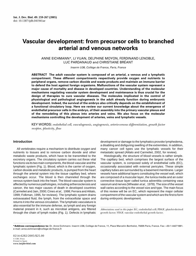
Vascular development: from precursor cells to branched
arterial and venous networks
ANNE EICHMANN*, LI YUAN, DELPHINE MOYON, FERDINAND LENOBLE,LUC PARDANAUD and CHRISTIANE BRÉANT
Inserm U36, Collège de France, Paris, France
ABSTRACT The adult vascular system is composed of an arterial, a venous and a lymphatic
compartment. These different compartments respectively provide oxygen and nutrients to
peripheral organs, remove carbon dioxide and waste products and maintain an immune barrier
to defend the host against foreign organisms. Malfunctions of the vascular system represent a
major cause of mortality and disease in developed countries. Understanding of the molecular
mechanisms regulating vascular system development and maintenance is thus crucial for the
design of therapies to cure vascular diseases. The molecules implicated in the control of
physiological and pathological angiogenesis in the adult already function during embryonic
development. Indeed, the survival of the embryo also critically depends on the establishment of
a functional circulatory loop. Here we review our current knowledge about the emergence of
endothelial precursor cells in the embryo, of their assembly into the primary vascular plexus and
of the remodeling of this plexus into arteries and veins. We also focus on the molecular
mechanisms controlling the development of arteries, veins and lymphatic vessels.
KEY WORDS: endothelial cell, vasculogenesis, angiogenesis, arterio-venous differentiation, growth factorreceptor, plasticity, flow
Int. J. Dev. Biol. 49: 259-267 (2005)doi: 10.1387/ijdb.041941ae
0214-6282/2005/$25.00© UBC PressPrinted in Spainwww.intjdevbiol.com
*Address correspondence to: Dr. Anne Eichmann. Inserm U36, Collège de France, 11, Place Marcelin Berthelot, 75005 Paris, France. Fax: +33-1-44271691.e-mail: [email protected]
Abbreviations used in this paper: EC, endothelial cell; PDGF, platelet-derivedgrowth factor; VEGF, vascular endothelial growth factor.
Introduction
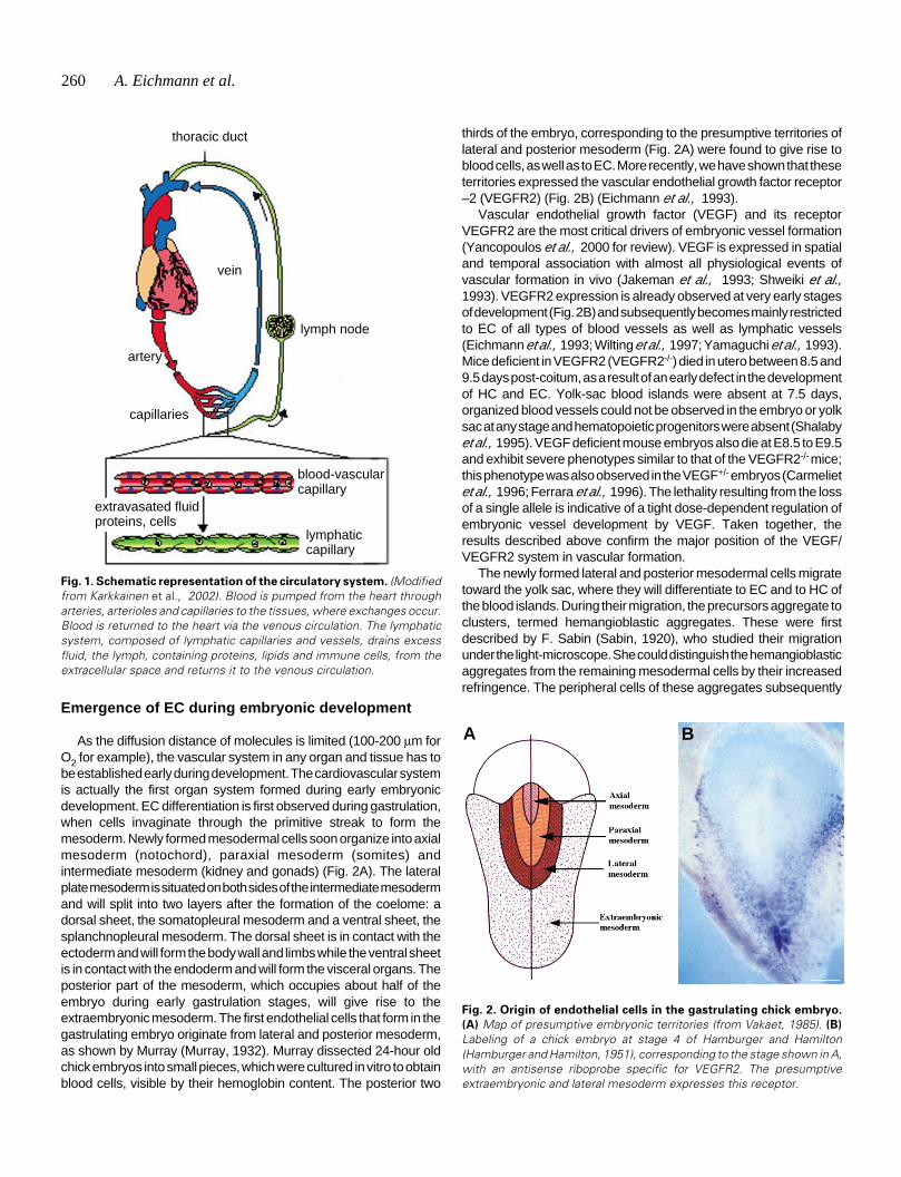
All vertebrates require a mechanism to distribute oxygen andnutrients to tissues and to remove carbon dioxide and othermetabolic waste products, which have to be transmitted to theexcretory organs. The circulatory system carries out these vitalfunctions via its two main components, the blood-vascular and thelymphatic system (Fig. 1). Blood, which is the carrier of oxygen,carbon dioxide and metabolic products, is pumped from the heartthrough the arterial system into the tissue capillary bed, whereexchanges occur. The blood is then channeled through thevenous system back into the heart. The blood-vascular system isaffected by numerous pathologies, including artherosclerosis andcancer, the two major causes of death in developed countries(Carmeliet and Jain, 2000; Cines et al., 1998; Ferrara and Alitalo,1999; Folkman, 1995, for reviews). The lymphatic system drainsextravasated fluid, the lymph, from the extracellular space andreturns it into the venous circulation. The lymphatic vasculature isalso essential for the immune defense, as lymph and any foreignmaterial present in it, such as microbial antigens, are filteredthrough the chain of lymph nodes (Fig. 1). Defects in lymphatic
development or damage to the lymphatics provoke lymphedema,a disabling and disfiguring swelling of the extremities. In addition,many cancer cell types use the lymphatic vessels for theirmetastatic spread (Alitalo and Carmeliet, 2002, for review).
Histologically, the structure of blood vessels is rather simple.The capillary bed, which comprises the largest surface of thevascular system, is composed solely of endothelial cells (EC),occasionally associated with external pericytes. These simplecapillary tubes are surrounded by a basement membrane. Largervessels have additional layers constituting the vessel wall, whichare composed of a muscular layer, the tunica media and an outerconnective tissue layer called tunica adventitia containing vasavasorum and nerves (Wheater et al., 1978). The size of the vesselwall varies according to the vessel size and type. The main focusof this review will be on EC, which represent the major cellularcompartment of the vascular system and which are the first to formduring embryonic development.
260 A. Eichmann et al.
Emergence of EC during embryonic development
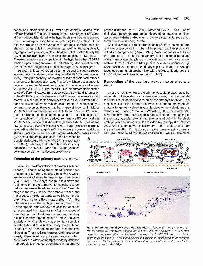
As the diffusion distance of molecules is limited (100-200 µm forO2 for example), the vascular system in any organ and tissue has tobe established early during development. The cardiovascular systemis actually the first organ system formed during early embryonicdevelopment. EC differentiation is first observed during gastrulation,when cells invaginate through the primitive streak to form themesoderm. Newly formed mesodermal cells soon organize into axialmesoderm (notochord), paraxial mesoderm (somites) andintermediate mesoderm (kidney and gonads) (Fig. 2A). The lateralplate mesoderm is situated on both sides of the intermediate mesodermand will split into two layers after the formation of the coelome: adorsal sheet, the somatopleural mesoderm and a ventral sheet, thesplanchnopleural mesoderm. The dorsal sheet is in contact with theectoderm and will form the body wall and limbs while the ventral sheetis in contact with the endoderm and will form the visceral organs. Theposterior part of the mesoderm, which occupies about half of theembryo during early gastrulation stages, will give rise to theextraembryonic mesoderm. The first endothelial cells that form in thegastrulating embryo originate from lateral and posterior mesoderm,as shown by Murray (Murray, 1932). Murray dissected 24-hour oldchick embryos into small pieces, which were cultured in vitro to obtainblood cells, visible by their hemoglobin content. The posterior two
thirds of the embryo, corresponding to the presumptive territories oflateral and posterior mesoderm (Fig. 2A) were found to give rise toblood cells, as well as to EC. More recently, we have shown that theseterritories expressed the vascular endothelial growth factor receptor–2 (VEGFR2) (Fig. 2B) (Eichmann et al., 1993).
Vascular endothelial growth factor (VEGF) and its receptorVEGFR2 are the most critical drivers of embryonic vessel formation(Yancopoulos et al., 2000 for review). VEGF is expressed in spatialand temporal association with almost all physiological events ofvascular formation in vivo (Jakeman et al., 1993; Shweiki et al.,1993). VEGFR2 expression is already observed at very early stagesof development (Fig. 2B) and subsequently becomes mainly restrictedto EC of all types of blood vessels as well as lymphatic vessels(Eichmann et al., 1993; Wilting et al., 1997; Yamaguchi et al., 1993).Mice deficient in VEGFR2 (VEGFR2-/-) died in utero between 8.5 and9.5 days post-coitum, as a result of an early defect in the developmentof HC and EC. Yolk-sac blood islands were absent at 7.5 days,organized blood vessels could not be observed in the embryo or yolksac at any stage and hematopoietic progenitors were absent (Shalabyet al., 1995). VEGF deficient mouse embryos also die at E8.5 to E9.5and exhibit severe phenotypes similar to that of the VEGFR2-/- mice;this phenotype was also observed in the VEGF+/- embryos (Carmelietet al., 1996; Ferrara et al., 1996). The lethality resulting from the lossof a single allele is indicative of a tight dose-dependent regulation ofembryonic vessel development by VEGF. Taken together, theresults described above confirm the major position of the VEGF/VEGFR2 system in vascular formation.
The newly formed lateral and posterior mesodermal cells migratetoward the yolk sac, where they will differentiate to EC and to HC ofthe blood islands. During their migration, the precursors aggregate toclusters, termed hemangioblastic aggregates. These were firstdescribed by F. Sabin (Sabin, 1920), who studied their migrationunder the light-microscope. She could distinguish the hemangioblasticaggregates from the remaining mesodermal cells by their increasedrefringence. The peripheral cells of these aggregates subsequently
Fig. 1. Schematic representation of the circulatory system. (Modifiedfrom Karkkainen et al., 2002). Blood is pumped from the heart througharteries, arterioles and capillaries to the tissues, where exchanges occur.Blood is returned to the heart via the venous circulation. The lymphaticsystem, composed of lymphatic capillaries and vessels, drains excessfluid, the lymph, containing proteins, lipids and immune cells, from theextracellular space and returns it to the venous circulation.
Fig. 2. Origin of endothelial cells in the gastrulating chick embryo.
(A) Map of presumptive embryonic territories (from Vakaet, 1985). (B)
Labeling of a chick embryo at stage 4 of Hamburger and Hamilton(Hamburger and Hamilton, 1951), corresponding to the stage shown in A,with an antisense riboprobe specific for VEGFR2. The presumptiveextraembryonic and lateral mesoderm expresses this receptor.
A B
thoracic duct
vein
lymph node
artery
capillaries
blood-vascularcapillary
lymphaticcapillary
extravasated fluidproteins, cells
Vascular development 261
give rise to smooth muscle cells in the presence ofplatelet-derived growth factor (PDGF)(Yamashita etal., 2000), indicating that rather than being strictlycommitted to only the EC and the HC lineage, thesecells may be pluri-or multipotent progenitors.
Formation of the primary capillary plexus
Following the differentiation of the yolk sac bloodislands, EC surrounding these blood islands soonanastomose to form a capillary meshwork, whichserves as a scaffold for the beginnings of circulation(Fig. 3, 4A). The embryo has thus laid down therudiments of its extraembryonic vascular systembefore the onset of heart beat around the 12-somitestage in the chick. Inside the embryo proper, onemajor vessel, the dorsal aorta, as well as numerouscapillaries have differentiated (Fig. 4A). ECdifferentiation in the embryo proper during thisdevelopmental time window occurs in the absenceof associated hematopoiesis. After the onset ofheartbeat and of blood flow, the yolk sac capillaryplexus is rapidly remodeled into arteries and veinsand a functional circulatory loop essential for survivalis established (Fig. 4B). The newly formed bloodisland HC are channeled through this primitivecirculation. These yolk sac hematopoietic precursorsmostly differentiate into primitive erythrocytes, whichare replaced, as development proceeds, by definitivehematopoietic precursors generated in the embryo
proper (Cumano et al., 2001; Dieterlen-Lievre, 1975). Thesedefinitive precursors are again observed to develop in closeassociation with the endothelium of the dorsal aorta (Jaffredo et al.,1998; Pardanaud et al., 1996).
Collectively, the in situ differentiation of EC from the mesodermand their coalescence into tubes of the primary capillary plexus arecalled vasculogenesis (Risau, 1997). Vasculogenesis results inthe formation of the major embryonic vessels, the dorsal aorta andof the primary vascular plexus in the yolk sac. In the chick embryo,both are formed before the 14ss, prior to the onset of perfusion. Fig.4A shows the structure of the primary capillary plexus at this stagerevealed by immunohistochemistry with the QH1 antibody, specificfor EC in the quail (Pardanaud et al., 1987).
Remodeling of the capillary plexus into arteries andveins
Over the next few hours, the primary vascular plexus has to beremodeled into a system with arteries and veins, to accommodatethe output of the heart and to establish the primary circulation. Thisstep is critical for the embryo’s survival and indeed, many mousemutants for genes involved in vascular development die during this‘remodeling’ phase (Roman and Weinstein, 2000, for review). Wehave recently performed a detailed analysis of the remodeling ofthe primary vascular plexus into arteries and veins in the chickembryo yolk sac, using time-lapse video-microscopy (LeNoble etal., 2004). Fig. 4B shows a chick embryo about 24 hours older thanthe embryo in Fig. 4A, it is obvious that the primary capillary plexushas been remodeled into larger and smaller vessels. The chick
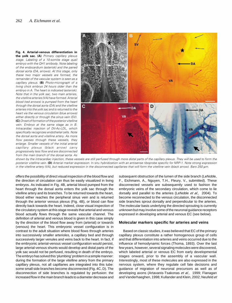
Fig. 3. Differentiation of yolk sac blood islands. (A) Schematic representation: seetext for details. (B) Transverse section through the extraembryonic area of a 15-somitestage embryo, labeled with an antisense riboprobe specific for VEGFR2. Hemangioblasticaggregates are positive. In the blood islands and capillaries, expression of the receptordecreases in the hematopoietic cells (asterisks), but is maintained in the endothelialcells (arrowheads). Bar, 75 µm.
flatten and differentiate to EC, while the centrally located cellsdifferentiate to HC (Fig. 3A). The simultaneous emergence of EC andHC in the blood islands led to the hypothesis that they were derivedfrom a common precursor, the hemangioblast (Sabin, 1920). VEGFR2expression during successive stages of hemangioblast differentiationshows that gastrulating precursors as well as hemangioblasticaggregates are positive, while in the differentiated islands only theEC express this gene and no expression is detected in HC (Fig. 3B).These observations are compatible with the hypothesis that VEGFR2labels a bipotent progenitor and that after lineage diversification; onlyone of the two daughter cells maintains expression of this gene.
To test this idea, we prepared a monoclonal antibody directedagainst the extracellular domain of quail VEGFR2 (Eichmann et al.,1997). Using this antibody, we isolated cells from posterior territoriesof embryos at the gastrulation stage (Fig. 2A), which were subsequentlycultured in semi-solid medium in vitro. In the absence of addedVEGF, the VEGFR2+, but not the VEGFR2- precursors differentiatedto HC of different lineages. In the presence of VEGF, EC differentiationof the VEGFR2+ precursors was induced. These experiments showedthat VEGFR2+ precursors could indeed give rise to EC as well as HC,consistent with the hypothesis that this receptor is expressed by acommon precursor. However, at the single cell level, an individualVEGFR2+ cell would either differentiate to an EC or an HC, but notboth, precluding a direct demonstration of the existence of a‘hemangioblast’. In cultures derived from mouse ES cells, a singleVEGFR2+ cell was found to be able to give rise to both EC as well asHC (Choi et al., 1998; Nishikawa et al., 1998). This cell is currentlyreferred to as the ‘hemangioblast’ in the literature. However, additionalstudies have shown that ES cell-derived VEGFR2+ cells can also
A
B
262 A. Eichmann et al.
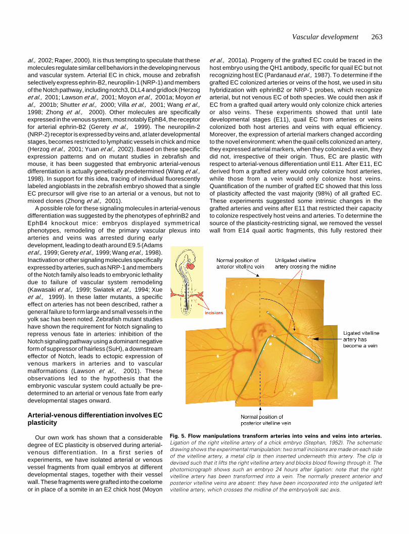
offers the possibility of direct visual inspection of the blood flow andthe direction of circulation can thus be easily visualized in livingembryos. As indicated in Fig. 4B, arterial blood pumped from theheart through the dorsal aorta enters the yolk sac through thevitelline artery and its branches. To be returned towards the heart,blood either reaches the peripheral sinus vein and is returnedthrough the anterior venous plexus (Fig. 4B), or blood can flowdirectly back towards the heart. Indeed, close visual inspection ofthe circulatory system at this stage reveals that arterial and venousblood actually flows through the same vascular channel. Thedefinition of arterial and venous blood is given in this case simplyby the direction of the blood flow away from (arterial) or towards(venous) the heart. This embryonic vessel configuration is incontrast to the adult situation where blood flows through arteriesinto successively smaller arterioles, a capillary bed and throughsuccessively larger venules and veins back to the heart. In fact, ifthe embryonic arterial-venous vessel configuration would persist,large arterial-venous shunts would develop and distal parts of theyolk sac would not be perfused, resulting in death of the embryo.The embryo has solved this ‘plumbing’ problem in a simple manner:during the formation of the large vitelline artery from the primarycapillary plexus, not all capillaries are integrated into this tube:some small side branches become disconnected (Fig. 4C, D). Thedisconnection of side branches is regulated by perfusion: theincreased flow in the main branch leads to a diameter decrease and
subsequent obstruction of the lumen of the side branch (LeNoble,F., Eichmann, A., Nguyen, T.H., Fleury, V., submitted). Thesedisconnected vessels are subsequently used to fashion theembryonic veins of the secondary circulation, which come to liedorsally and parallel to the arteries (LeNoble et al., 2004). Tobecome reconnected to the venous circulation, the disconnectedside branches sprout dorsally and perpendicular to the arteries.The molecular basis underlying the directed sprouting is currentlyunknown but may involve some of the neuronal guidance receptorsexpressed in developing arterial and venous EC (see below).
Molecular markers specific for arteries and veins
Based on classic studies, it was believed that EC of the primarycapillary plexus constitute a rather homogenous group of cellsand that differentiation into arteries and veins occurred due to theinfluence of hemodynamic forces (Thoma, 1893). Over the lastfew years, however, several signaling molecules were discovered,which labeled arterial or venous EC from early developmentalstages onward, prior to the assembly of a vascular wall.Interestingly, most of these molecules are also expressed in thenervous system, where they regulate cell fate decisions andguidance of migration of neuronal precursors as well as ofdeveloping axons (Artavanis-Tsakonas et al., 1999; Flanaganand Vanderhaeghen, 1998; Kullander and Klein, 2002; Neufeld et
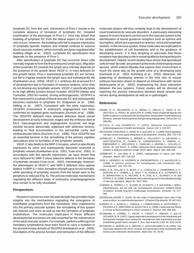
Fig. 4. Arterial-venous differentiation in
the yolk sac. (A) Primary capillary plexusstage. Labeling of a 10-somite stage quailembryo with the QH1 antibody. Note labelingof the endocardium (asterisk) and the paireddorsal aorta (DA, arrows). At this stage, onlythese two major vessels are formed, theremainder of the vascular system is seen as acapillary plexus. (B) Photo-micrograph of aliving chick embryo 24 hours older than theembryo in A. The heart is indicated (asterisk).Note that in the yolk sac, two main arteries,the vitelline arteries (VA) have formed. Arterialblood (red arrows) is pumped from the heartthrough the dorsal aorta (DA) and the vitellinearteries into the yolk sac and is returned to theheart via the venous circulation (blue arrows)either directly or through the sinus vein (SV).(C) Onset of formation of the posterior vitellinevein. Embryo at the same stage as in B.Intracardiac injection of DiI-Ac-LDL, whichspecifically recognizes endothelial cells. Notethe dorsal aorta and vitelline artery. As moreflow passes through these vessels, theyenlarge. Smaller vessels of the initial arterialcapil lary plexus (black arrow) carryprogressively less flow and are disconnectedfrom the main branch of the dorsal aorta. Asshown by the intracardiac injection, these vessels are still perfused through more distal parts of the capillary plexus. They will be used to form theposterior vitelline vein. (D) Arterial marker expression. In situ hybridization with an antisense riboprobe specific for NRP-1. Note strong expressionin the vitelline artery (VA), but reduced expression in the disconnected capillaries that will form the vitelline vein (black arrow). Bars 250 µm.
A
D
B
C
Vascular development 263
al., 2002; Raper, 2000). It is thus tempting to speculate that thesemolecules regulate similar cell behaviors in the developing nervousand vascular system. Arterial EC in chick, mouse and zebrafishselectively express ephrin-B2, neuropilin-1 (NRP-1) and membersof the Notch pathway, including notch3, DLL4 and gridlock (Herzoget al., 2001; Lawson et al., 2001; Moyon et al., 2001a; Moyon etal., 2001b; Shutter et al., 2000; Villa et al., 2001; Wang et al.,1998; Zhong et al., 2000). Other molecules are specificallyexpressed in the venous system, most notably EphB4, the receptorfor arterial ephrin-B2 (Gerety et al., 1999). The neuropilin-2(NRP-2) receptor is expressed by veins and, at later developmentalstages, becomes restricted to lymphatic vessels in chick and mice(Herzog et al., 2001; Yuan et al., 2002). Based on these specificexpression patterns and on mutant studies in zebrafish andmouse, it has been suggested that embryonic arterial-venousdifferentiation is actually genetically predetermined (Wang et al.,1998). In support for this idea, tracing of individual fluorescentlylabeled angioblasts in the zebrafish embryo showed that a singleEC precursor will give rise to an arterial or a venous, but not tomixed clones (Zhong et al., 2001).
A possible role for these signaling molecules in arterial-venousdifferentiation was suggested by the phenotypes of ephrinB2 andEphB4 knockout mice: embryos displayed symmetricalphenotypes, remodeling of the primary vascular plexus into
et al., 2001a). Progeny of the grafted EC could be traced in thehost embryo using the QH1 antibody, specific for quail EC but notrecognizing host EC (Pardanaud et al., 1987). To determine if thegrafted EC colonized arteries or veins of the host, we used in situhybridization with ephrinB2 or NRP-1 probes, which recognizearterial, but not venous EC of both species. We could then ask ifEC from a grafted quail artery would only colonize chick arteriesor also veins. These experiments showed that until latedevelopmental stages (E11), quail EC from arteries or veinscolonized both host arteries and veins with equal efficiency.Moreover, the expression of arterial markers changed accordingto the novel environment: when the quail cells colonized an artery,they expressed arterial markers, when they colonized a vein, theydid not, irrespective of their origin. Thus, EC are plastic withrespect to arterial-venous differentiation until E11. After E11, ECderived from a grafted artery would only colonize host arteries,while those from a vein would only colonize host veins.Quantification of the number of grafted EC showed that this lossof plasticity affected the vast majority (98%) of all grafted EC.These experiments suggested some intrinsic changes in thegrafted arteries and veins after E11 that restricted their capacityto colonize respectively host veins and arteries. To determine thesource of the plasticity-restricting signal, we removed the vesselwall from E14 quail aortic fragments, this fully restored their
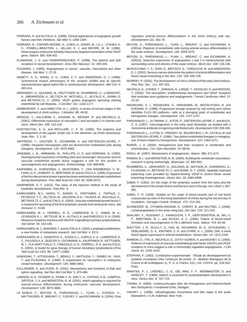
Fig. 5. Flow manipulations transform arteries into veins and veins into arteries.
Ligation of the right vitelline artery of a chick embryo (Stephan, 1952). The schematicdrawing shows the experimental manipulation: two small incisions are made on each sideof the vitelline artery, a metal clip is then inserted underneath this artery. The clip isdevised such that it lifts the right vitelline artery and blocks blood flowing through it. Thephotomicrograph shows such an embryo 24 hours after ligation: note that the rightvitelline artery has been transformed into a vein. The normally present anterior andposterior vitelline veins are absent: they have been incorporated into the unligated leftvitelline artery, which crosses the midline of the embryo/yolk sac axis.
arteries and veins was arrested during earlydevelopment, leading to death around E9.5 (Adamset al., 1999; Gerety et al., 1999; Wang et al., 1998).Inactivation or other signaling molecules specificallyexpressed by arteries, such as NRP-1 and membersof the Notch family also leads to embryonic lethalitydue to failure of vascular system remodeling(Kawasaki et al., 1999; Swiatek et al., 1994; Xueet al., 1999). In these latter mutants, a specificeffect on arteries has not been described, rather ageneral failure to form large and small vessels in theyolk sac has been noted. Zebrafish mutant studieshave shown the requirement for Notch signaling torepress venous fate in arteries: inhibition of theNotch signaling pathway using a dominant negativeform of suppressor of hairless (SuH), a downstreameffector of Notch, leads to ectopic expression ofvenous markers in arteries and to vascularmalformations (Lawson et al., 2001). Theseobservations led to the hypothesis that theembryonic vascular system could actually be pre-determined to an arterial or venous fate from earlydevelopmental stages onward.
Arterial-venous differentiation involves ECplasticity
Our own work has shown that a considerabledegree of EC plasticity is observed during arterial-venous differentiation. In a first series ofexperiments, we have isolated arterial or venousvessel fragments from quail embryos at differentdevelopmental stages, together with their vesselwall. These fragments were grafted into the coelomeor in place of a somite in an E2 chick host (Moyon
264 A. Eichmann et al.
capacity to colonize veins: about 50% of grafted cells were foundin arteries and 50% in veins. The precise nature of the plasticity-restricting signal coming from the vessel wall is yet unknown.
Yolk sac arterial-venous differentiation is flow-regulated
To determine if EC plasticity with respect to arterial-venousdifferentiation also occurred during normal embryonicdevelopment, we examined yolk sacs of embryos at differentstages using a combination of time-lapse video-microscopy onliving embryos and in situ hybridization with arterial and vein-specific markers (LeNoble et al., 2004). We observed that priorto the onset of circulation, arterial markers are expressed in thearterial capillary plexus of the posterior pole of the embryo, theterritory where the vitelline artery will form. As described above,many small capillary branches of this arterial plexus becomedisconnected from the main branch of the vitelline artery during itsformation. Arterial marker expression in these disconnectedbranches is rapidly down-regulated (Fig. 4D). In contrast, arterialmarker expression is maintained in the newly formed vitellineartery (Fig. 4D). The disconnected capillaries that have down-regulated arterial markers now serve to form the vitelline vein.These observations show that the posterior vitelline vein isformed by incorporating previously arterial capillary segments,demonstrating that EC plasticity is required during normal arterial-venous differentiation. Moreover, these experiments suggestedthat hemodynamic forces play a major role during arterial-venousdifferentiation and patterning.
To test this idea directly, we performed several alterations inthe flow pattern of developing chick embryos (LeNoble et al.,2004). We first generated embryos devoid of a circulatory systemby destroying the embryonic heart. In these embryos, the yolk saccontinues to grow for at least 7 days, in spite of the degenerationof the embryo and the lack of perfusion. However, the yolk sacdoes not develop arteries or veins and remains in the configurationof a primary vascular plexus. In situ hybridization with arterialmarkers showed that some regions of this yolk sac expressed thearterial marker ephrinB2, while others did not. Thus, initiation ofarterial marker expression occurs independently of flow.
We next performed ligations of the vitelline artery on one sideof the yolk sac (Fig. 5) (Stephan, 1952). In these embryos, theentire ligated side becomes venularized over a period of 24 hours,as judged first by the direction of blood flow, which becomesreversed in the ligated vessels and second by the expression of
arterial markers, which decreases rapidly following ligation(LeNoble et al., 2004). Thus, manipulation of the flow pattern canmorphologically and genetically transform arteries into veins. Thesame flow manipulation can also transform veins into arteries.Indeed, the vitelline artery on the unligated side significantlyenlarges after ligation, since it receives more flow. Subsequently,this artery crosses over the midline of the embryo-yolk sac axisboth on the anterior and on the posterior pole of the embryo (Fig.5). Time-lapse video-microscopy shows that branches of theanterior vitelline vein are integrated into the vitelline artery as itcrosses the midline. Flow therefore appears as the master-regulator of arterial-venous differentiation in the yolk sac. Ratherthan being pre-determined, the yolk sac vessel EC appear as‘bricks’ that can be used and re-used by the developing vascularsystem to fashion arteries or veins.
Formation of the lymphatic vascular system
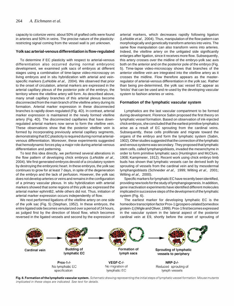
Lymphatics are the last vascular compartment to be formedduring development. Florence Sabin proposed the first theory onlymphatic vessel formation. Based on observation of ink-injectedchick embryos, she concluded that the earliest primitive lymphaticsarise as a result of EC sprouting from the cardinal veins.Subsequently, these cells proliferate and migrate toward theorgans of the embryo and form the lymphatic system (Sabin,1902). Other studies suggested that the connection of the lymphaticand venous systems was secondary. They proposed that lymphaticstem cells, called lymphangioblasts, invaded the mesenchyme inorder to form primitive lymphatic sacs (Huntington and McClure,1908; Kampmeier, 1912). Recent work using chick embryo limbbuds has shown that lymphatic vessels can be derived both bysprouting of vessels from the cardinal vein and by mesodermallymphangioblasts (Schneider et al., 1999; Wilting et al., 2001;Wilting et al., 2000).
Specific markers for lymphatic EC have recently been identified,providing new tools for the study of lymphangiogenesis. In addition,gene inactivation experiments have identified different moleculesimplicated in successive steps of the development of the lymphaticsystem (Fig. 6).
The earliest marker for developing lymphatic EC is thehomeobox transcription factor Prox-1 (prospero-related homeoboxprotein-1) (Wigle and Oliver, 1999). Prox-1 first becomes expressedin the vascular system in the lateral aspect of the posteriorcardinal vein at E9, shortly before the onset of sprouting of
Fig. 6. Formation of the lymphatic vascular system. Schematic drawing representing the initial steps of lymphatic vessel formation. Mouse mutantsimplicated in these steps are indicated. See text for details.
Vascular development 265
lymphatic EC from this vein. Inactivation of Prox-1 results in thecomplete absence of formation of lymphatic EC. Detailedexamination of the phenotype of Prox-1-/- mice has shown thatbudding of lymphatic EC from the lateral aspect of the cardinalvein occurs. However, the budding cells fail to acquire expressionof lymphatic-specific markers and instead continue to expressblood-vascular markers, which normally are down-regulated afterbudding (Wigle et al., 2002). Lymphatic EC specification thusdepends on the presence of Prox-1.
After specification of lymphatic EC has occurred, these cellsnormally migrate to form the first embryonic lymph sacs. Migrationof the lymphatic EC towards the lymph sacs critically depends onthe presence of the growth factor VEGF-C: in mice deficient forthis growth factor, Prox-1 expressing lymphatic EC are formed,but fail to migrate towards the lymph sacs and subsequently die(Karkkainen et al., 2004). VEGF-C-/- embryos die at around E17of development due to formation of massive oedema, since theydo not develop any lymphatic vessels. VEGF-C specifically bindsto its high affinity tyrosine kinase receptor VEGFR3 (Alitalo andCarmeliet, 2002 for review). During embryogenesis, VEGFR3 isfirst expressed in a subset of blood vascular EC and subsequentlybecomes restricted to lymphatic EC (Kaipainen et al., 1995;Wilting et al., 1997). Consistent with the early expression,VEGFR3 involvement in embryonic angiogenesis has beenconfirmed by targeted gene inactivation (Dumont et al., 1998).The VEGFR3 deficient mice showed defective blood vesseldevelopment at early embryonic stages and the embryos died atE9.5. Vasculogenesis and angiogenesis occurred, but largevessels became abnormally organized with defective lumens,leading to fluid accumulation in the pericardial cavity andcardiovascular failure (Dumont et al., 1998). Thus VEGFR3 hasan essential function in the remodeling of the primary capillaryvasculature prior to formation of the lymphatic vessels.
VEGF-C also binds to the NRP-2 receptor, which is specificallyexpressed by veins and subsequently becomes restricted tolymphatic vessels (Karkkainen et al., 2001; Yuan et al., 2002). Inaccordance with this specific expression, we have shown thatmice deficient for NRP-2 show selective defects in the formationof lymphatic vessels (Yuan et al., 2002). Interestingly, however,the phenotypes of VEGF-C and NRP-2 deficient mice appeardistinct. In NRP-2-/- mice, formation of lymph sacs occurs normally,while sprouting of lymphatic vessels from the lymph sacs to theperiphery is reduced (Fig. 6). The precise molecular mechanismsregulating the different steps of embryonic lymphangiogenesisthus remain to be fully elucidated.
Perspectives
Research carried out over the past decade has provided majorinsights into the mechanisms regulating the emergence ofendothelial progenitors from the mesoderm, their coalescenceinto the primary vascular system, the remodeling of this systeminto arteries and veins as well as into the formation of lymphaticendothelium. The molecules implicated in these differentdevelopmental processes are also essential for the maintenanceof the adult vascular system. For example, patients suffering fromhereditary lymphedema have been found to carry a mutation inthe tyrosine kinase domain of VEGFR3 (Karkkainen et al., 2000).Elucidation of the precise function and interaction of the different
molecular players will thus certainly lead to the development ofnovel treatments for vascular disorders. A particularly interestingaspect of recent research carried out on the vascular system is theidentification of neural guidance receptors such as ephrins andneuropilins, which are expressed on arteries, veins and lymphaticvessels. In the nervous system, these molecules are implicated inthe establishment of cell boundaries and in the guidance ofdeveloping axons. It is thus tempting to speculate that thesereceptors may also play a role in vessel guidance during embryonicdevelopment. Indeed, recent studies have shown that specializedcells termed ‘tip cells’ are present at the ends of developing vesselsprouts, which extend filipodia that explore their environment inmuch the same way as the growth cone of a developing axon(Gerhardt et al., 2003; Ruhrberg et al., 2002). Moreover, thepatterning of developing arteries in the limb skin of mouseembryos has been shown to depend on interactions with nerves(Mukouyama et al., 2002), emphasizing the close interactionbetween the two systems. Future studies will be directed atexploring the precise interactions between blood vessels andnerves during development as well as in pathologies.
References
ADAMS, R. H., WILKINSON, G. A., WEISS, C., DIELLA, F., GALE, N. W.,DEUTSCH, U., RISAU, W. and KLEIN, R. (1999). Roles of ephrinB ligands andEphB receptors in cardiovascular development: demarcation of arterial/venousdomains, vascular morphogenesis and sprouting angiogenesis. Genes Dev 13:295-306.
ALITALO, K. and CARMELIET, P. (2002). Molecular mechanisms oflymphangiogenesis in health and disease. Cancer Cell 1: 219-227.
ARTAVANIS-TSAKONAS, S., RAND, M. D. and LAKE, R. J. (1999). Notch signaling:cell fate control and signal integration in development. Science 284: 770-776.
CARMELIET, P., FERREIRA, V., BREIER, G., POLLEFEYT, S., KIECKENS, L.,GERTSENSTEIN, M., FAHRIG, M., VANDENHOECK, A., HARPAL, K.,EBERHARDT, C., DECLERCQ, C., PAWLING, J., MOONS, L., COLLEN, D.,RISAU, W. and NAGY, A. (1996). Abnormal blood vessel development andlethality in embryos lacking a single VEGF allele. Nature 380: 435-439.
CARMELIET, P. and JAIN, R. K. (2000). Angiogenesis in cancer and otherdiseases. Nature 407: 249-257.
CHOI, K., KENNEDY, M., KAZAROV, A., PAPADIMITRIOU, J. C. and KELLER, G.(1998). A common precursor for hematopoietic and endothelial cells.Development 125: 725-732.
CINES, D. B., POLLAK, E. S., BUCK, C. A., LOSCALZO, J., ZIMMERMAN, G. A.,MCEVER, R. P., POBER, J. S., WICK, T. M., KONKLE, B. A., SCHWARTZ, B.S., BARNATHAN, E. S., MCCRAE, K. R., HUG, B. A., SCHMIDT, A. M. andSTERN, D. M. (1998). Endothelial cells in physiology and in the pathophysiologyof vascular disorders. Blood 91: 3527-3561.
CUMANO, A., FERRAZ, J. C., KLAINE, M., DI SANTO, J. P. and GODIN, I. (2001).Intraembryonic, but not yolk sac hematopoietic precursors, isolated beforecirculation, provide long-term multilineage reconstitution. Immunity 15: 477-485.
DIETERLEN-LIEVRE, F. (1975). On the origin of haemopoietic stem cells in theavian embryo: an experimental approach. J Embryol Exp Morphol 33: 607-619.
DUMONT, D. J., JUSSILA, L., TAIPALE, J., LYMBOUSSAKI, A., MUSTONEN, T.,PAJUSOLA, K., BREITMAN, M. and ALITALO, K. (1998). Cardiovascular failurein mouse embryos deficient in VEGF receptor-3. Science 282: 946-949.
EICHMANN, A., CORBEL, C., NATAF, V., VAIGOT, P., BREANT, C. and LEDOUARIN, N. M. (1997). Ligand-dependent development of the endothelial andhemopoietic lineages from embryonic mesodermal cells expressing vascularendothelial growth factor receptor 2. Proc Natl Acad Sci USA 94: 5141-5146.
EICHMANN, A., MARCELLE, C., BREANT, C. and LE DOUARIN, N. M. (1993). Twomolecules related to the VEGF receptor are expressed in early endothelial cellsduring avian embryonic development. Mech Dev 42: 33-48.
266 A. Eichmann et al.
FERRARA, N. and ALITALO, K. (1999). Clinical applications of angiogenic growthfactors and their inhibitors. Nat Med 5: 1359-1364.
FERRARA, N., CARVER-MOORE, K., CHEN, H., DOWD, M., LU, L., O’SHEA, K.S., POWELL-BRAXTON, L., HILLAN, K. J. and MOORE, M. W. (1996).Heterozygous embryonic lethality induced by targeted inactivation of the VEGFgene. Nature 380: 439-442.
FLANAGAN, J. G. and VANDERHAEGHEN, P. (1998). The ephrins and Ephreceptors in neural development. Annu Rev Neurosci 21: 309-345.
FOLKMAN, J. (1995). Angiogenesis in cancer, vascular, rheumatoid and otherdisease. Nat Med 1: 27-31.
GERETY, S. S., WANG, H. U., CHEN, Z. F. and ANDERSON, D. J. (1999).Symmetrical mutant phenotypes of the receptor EphB4 and its specifictransmembrane ligand ephrin-B2 in cardiovascular development. Mol Cell 4:403-414.
GERHARDT, H., GOLDING, M., FRUTTIGER, M., RUHRBERG, C., LUNDKVIST,A., ABRAMSSON, A., JELTSCH, M., MITCHELL, C., ALITALO, K., SHIMA, D.and BETSHOLTZ, C. (2003). VEGF guides angiogenic sprouting utilizingendothelial tip cell filopodia. J Cell Biol 161: 1163-1177.
HAMBURGER, V. and HAMILTON, H. L. (1951). A series of normal stages in thedevelopment of chick embryo. J Morphol 88: 49-92.
HERZOG, Y., KALCHEIM, C., KAHANE, N., RESHEF, R. and NEUFELD, G.(2001). Differential expression of neuropilin-1 and neuropilin-2 in arteries andveins. Mech Dev 109: 115-119.
HUNTINGTON, S. G. and MCCLURE, C. F. W. (1908). The anatomy anddevelopment of the jugular lymph sac in the domestic cat (Felis domestica).Anat. Rec. 2: 1-19.
JAFFREDO, T., GAUTIER, R., EICHMANN, A. and DIETERLEN-LIEVRE, F.(1998). Intraaortic hemopoietic cells are derived from endothelial cells duringontogeny. Development 125: 4575-4583.
JAKEMAN, L. B., ARMANINI, M., PHILLIPS, H. S. and FERRARA, N. (1993).Developmental expression of binding sites and messenger ribonucleic acid forvascular endothelial growth factor suggests a role for this protein invasculogenesis and angiogenesis. Endocrinology 133: 848-859.
KAIPAINEN, A., KORHONEN, J., MUSTONEN, T., VAN HINSBERGH, V. W.,FANG, G. H., DUMONT, D., BREITMAN, M. and ALITALO, K. (1995). Expressionof the fms-like tyrosine kinase 4 gene becomes restricted to lymphatic endotheliumduring development. Proc Natl Acad Sci USA 92: 3566-3570.
KAMPMEIER, O. F. (1912). The value of the injection method in the study oflymphatic development. Anat Rec 6.
KARKKAINEN, M.J., HAIKO, P., SAINIO, K., PARTANEN, J., TAIPALE, J.,PETROVA, T.V., JELTSCH, M., JACKSON, D.G., TALIKKA, M., RAUVALA, H.,BETSHOLTZ, C. and ALITALO, K. (2004). Vascular endothelial growth factor Cis required for sprouting of the first lymphatic vessels from embryonic veins. NatImmunol. 5: 74-80.
KARKKAINEN, M. J., FERRELL, R. E., LAWRENCE, E. C., KIMAK, M. A.,LEVINSON, K. L., MCTIGUE, M. A., ALITALO, K. and FINEGOLD, D. N. (2000).Missense mutations interfere with VEGFR-3 signalling in primary lymphoedema.Nat Genet 25: 153-159.
KARKKAINEN, M. J., MAKINEN, T. and ALITALO, K. (2002) Lymphatic endothelium:a new frontier of metastasis research. Nat Cell Biol 4: E2-5.
KARKKAINEN, M. J., SAARISTO, A., JUSSILA, L., KARILA, K. A., LAWRENCE, E.C., PAJUSOLA, K., BUELER, H., EICHMANN, A., KAUPPINEN, R., KETTUNEN,M. I., YLA-HERTTUALA, S., FINEGOLD, D. N., FERRELL, R. E. and ALITALO,K. (2001). A model for gene therapy of human hereditary lymphedema.nProcNatl Acad Sci USA 98: 12677-12682.
KAWASAKI, T., KITSUKAWA, T., BEKKU, Y., MATSUDA, Y., SANBO, M., YAGI,T. and FUJISAWA, H. (1999). A requirement for neuropilin-1 in embryonicvessel formation. Development 126: 4895-4902.
KULLANDER, K. and KLEIN, R. (2002). Mechanisms and functions of Eph andephrin signalling. Nat Rev Mol Cell Biol 3: 475-486.
LAWSON, N. D., SCHEER, N., PHAM, V. N., KIM, C. H., CHITNIS, A. B., CAMPOS-ORTEGA, J. A. and WEINSTEIN, B. M. (2001). Notch signaling is required forarterial-venous differentiation during embryonic vascular development.Development 128: 3675-3683.
LE NOBLE, F., MOYON, D., PARDANAUD, L., YUAN, L., DJONOV, V.,MATTHIJSEN, R., BREANT, C., FLEURY, V. and EICHMANN, A. (2004). Flow
regulates arterial-venous differentiation in the chick embryo yolk sac.Development 131: 361-75.
MOYON, D., PARDANAUD, L., YUAN, L., BREANT, C. and EICHMANN, A.(2001a). Plasticity of endothelial cells during arterial-venous differentiation inthe avian embryo. Development 128: 3359-3370.
MOYON, D., PARDANAUD, L., YUAN, L., BREANT, C. and EICHMANN, A.(2001b). Selective expression of angiopoietin 1 and 2 in mesenchymal cellssurrounding veins and arteries of the avian embryo. Mech Dev 106: 133-136.
MUKOUYAMA, Y. S., SHIN, D., BRITSCH, S., TANIGUCHI, M. and ANDERSON,D. J. (2002). Sensory nerves determine the pattern of arterial differentiation andblood vessel branching in the skin. Cell 109: 693-705.
MURRAY, P. (1932). The development «in vitro» of blood of the early chick embryo.Proc Roy. Soc. 111: 497-521.
NEUFELD, G., COHEN, T., SHRAGA, N., LANGE, T., KESSLER, O. and HERZOG,Y. (2002). The neuropilins: multifunctional semaphorin and VEGF receptorsthat modulate axon guidance and angiogenesis. Trends Cardiovasc Med 12:13-19.
NISHIKAWA, S. I., NISHIKAWA, S., HIRASHIMA, M., MATSUYOSHI, N. andKODAMA, H. (1998). Progressive lineage analysis by cell sorting and cultureidentifies FLK1+VE-cadherin+ cells at a diverging point of endothelial andhemopoietic lineages. Development 125: 1747-1757.
PARDANAUD, L., ALTMANN, C., KITOS, P., DIETERLEN-LIEVRE, F. and BUCK,C. A. (1987). Vasculogenesis in the early quail blastodisc as studied with amonoclonal antibody recognizing endothelial cells. Development 100: 339-349.
PARDANAUD, L., LUTON, D., PRIGENT, M., BOURCHEIX, L. M., CATALA, M. andDIETERLEN-LIEVRE, F. (1996). Two distinct endothelial lineages in ontogeny,one of them related to hemopoiesis. Development 122: 1363-1371.
RAPER, J. A. (2000). Semaphorins and their receptors in vertebrates andinvertebrates. Curr Opin Neurobiol 10: 88-94.
RISAU, W. (1997). Mechanisms of angiogenesis. Nature 386: 671-674.
ROMAN, B. L. and WEINSTEIN, B. M. (2000). Building the vertebrate vasculature:research is going swimmingly. Bioessays 22: 882-893.
RUHRBERG, C., GERHARDT, H., GOLDING, M., WATSON, R., IOANNIDOU, S.,FUJISAWA, H., BETSHOLTZ, C. and SHIMA, D. T. (2002). Spatially restrictedpatterning cues provided by heparin-binding VEGF-A control blood vesselbranching morphogenesis. Genes Dev 16: 2684-2698.
SABIN, F. (1902). On the origin of the lymphatic system from the veins and thedecelopment of the lymph hearts and thoracic duct in the pig. Am J Anat 1: 367-391.
SABIN, F. R. (1920). Studies on the origin of blood-vessels and of red bloodcorpuscules as seen in the living blastoderm of chicks during the second day ofincubation. Carnegie Contrib. Embryol. 272: 214-262.
SCHNEIDER, M., OTHMAN-HASSAN, K., CHRIST, B. and WILTING, J. (1999).Lymphangioblasts in the avian wing bud. Dev Dyn 216: 311-319.
SHALABY, F., ROSSANT, J., YAMAGUCHI, T. P., GERTSENSTEIN, M., WU, X.F., BREITMAN, M. L. and SCHUH, A. C. (1995). Failure of blood-islandformation and vasculogenesis in Flk-1-deficient mice. Nature 376: 62-66.
SHUTTER, J. R., SCULLY, S., FAN, W., RICHARDS, W. G., KITAJEWSKI, J.,DEBLANDRE, G. A., KINTNER, C. R. and STARK, K. L. (2000). Dll4, a novelNotch ligand expressed in arterial endothelium. Genes Dev 14: 1313-1318.
SHWEIKI, D., ITIN, A., NEUFELD, G., GITAY-GOREN, H. and KESHET, E. (1993).Patterns of expression of vascular endothelial growth factor (VEGF) and VEGFreceptors in mice suggest a role in hormonally regulated angiogenesis. J ClinInvest 91: 2235-2243.
STEPHAN, F. (1952). Contribution expérimentale l’Étude du développement dusystème circulatoire chez l’embryon de poulet. In «Bulletin Biologique de laFrance et de la Belgique» (L. P. U. d. France, Ed.), Vol. LXXXVI, pp. 218-310,Paris.
SWIATEK, P. J., LINDSELL, C. E., DEL AMO, F. F., WEINMASTER, G. andGRIDLEY, T. (1994). Notch1 is essential for postimplantation development inmice. Genes Dev 8: 707-719.
THOMA, R. (1893). «Untersuchungen über die Histogenese und Histomechanikdes Gefssytems.» Ferdinand Enke, Stuttgart.
VAKAET, L. (1985). «Morphogenetic movements and fate maps in the avianblastoderm.» G.M. Edelman, New York.
Vascular development 267
VILLA, N., WALKER, L., LINDSELL, C. E., GASSON, J., IRUELA-ARISPE, M. L.and WEINMASTER, G. (2001). Vascular expression of Notch pathway receptorsand ligands is restricted to arterial vessels. Mech Dev 108: 161-164.
WANG, H. U., CHEN, Z. F. and ANDERSON, D. J. (1998). Molecular distinction andangiogenic interaction between embryonic arteries and veins revealed byephrin-B2 and its receptor Eph-B4. Cell 93: 741-753.
WHEATER, P. R., BURKITT, H. G. and DANIELS, V. G. (1978). «FunctionalHistology: a Text and Colour Atlas.» Churchill Livestone,
WIGLE, J. T., HARVEY, N., DETMAR, M., LAGUTINA, I., GROSVELD, G., GUNN,M. D., JACKSON, D. G. and OLIVER, G. (2002). An essential role for Prox1 inthe induction of the lymphatic endothelial cell phenotype. EMBO J 21: 1505-1513.
WIGLE, J. T. and OLIVER, G. (1999). Prox1 function is required for the developmentof the murine lymphatic system. Cell 98: 769-778.
WILTING, J., EICHMANN, A. and CHRIST, B. (1997). Expression of the avianVEGF receptor homologues Quek1 and Quek2 in blood-vascular and lymphaticendothelial and non-endothelial cells during quail embryonic development. CellTissue Res 288: 207-223.
WILTING, J., PAPOUTSI, M., OTHMAN-HASSAN, K., RODRIGUEZ-NIEDENFUHR,M., PROLS, F., TOMAREV, S. I. and EICHMANN, A. (2001). Development ofthe avian lymphatic system. Microsc Res Tech 55: 81-91.
WILTING, J., PAPOUTSI, M., SCHNEIDER, M. and CHRIST, B. (2000). The lymphatic
endothelium of the avian wing is of somitic origin. Dev Dyn 217: 271-278.
XUE, Y., GAO, X., LINDSELL, C. E., NORTON, C. R., CHANG, B., HICKS, C.,GENDRON-MAGUIRE, M., RAND, E. B., WEINMASTER, G. and GRIDLEY, T.(1999). Embryonic lethality and vascular defects in mice lacking the Notchligand Jagged1. Hum Mol Genet 8: 723-730.
YAMAGUCHI, T. P., DUMONT, D. J., CONLON, R. A., BREITMAN, M. L. andROSSANT, J. (1993). flk-1, an flt-related receptor tyrosine kinase is an earlymarker for endothelial cell precursors. Development 118: 489-498.
YAMASHITA, J., ITOH, H., HIRASHIMA, M., OGAWA, M., NISHIKAWA, S.,YURUGI, T., NAITO, M. and NAKAO, K. (2000). Flk1-positive cells derived fromembryonic stem cells serve as vascular progenitors. Nature 408: 92-96.
YANCOPOULOS, G. D., DAVIS, S., GALE, N. W., RUDGE, J. S., WIEGAND, S. J.and HOLASH, J. (2000). Vascular-specific growth factors and blood vesselformation. Nature 407: 242-248.
YUAN, L., MOYON, D., PARDANAUD, L., BREANT, C., KARKKAINEN, M. J.,ALITALO, K. and EICHMANN, A. (2002). Abnormal lymphatic vessel developmentin neuropilin 2 mutant mice. Development 129: 4797-4806.
ZHONG, T. P., CHILDS, S., LEU, J. P. and FISHMAN, M. C. (2001). Gridlocksignalling pathway fashions the first embryonic artery. Nature 414: 216-220.
ZHONG, T. P., ROSENBERG, M., MOHIDEEN, M. A., WEINSTEIN, B. andFISHMAN, M. C. (2000). gridlock, an HLH gene required for assembly of theaorta in zebrafish. Science 287: 1820-1824.