Embed Size (px)
Citation preview
Dr Milind PatilDr Dipen Patel
2nd year ResidentsSurgical ‘F’ unit
Introduction The venous drainage system of the lower
extremity consists of three sets of veins:Deep veins,Superficial veinsPerforating veins. All veins contain delicate one-way valves that
normally open to allow blood to flow toward the heart and prevent blood from flowing in a retrograde fashion after the valves close .
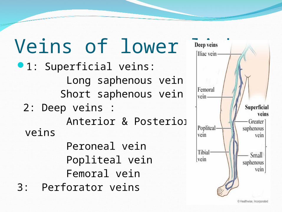
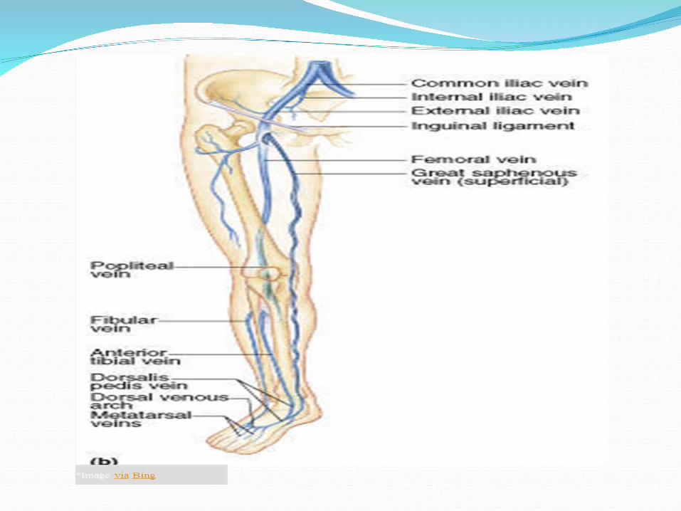
Veins of lower limb 1: Superficial veins: Long saphenous vein Short saphenous vein 2: Deep veins : Anterior & Posterior Tibial veins Peroneal vein Popliteal vein Femoral vein 3: Perforator veins
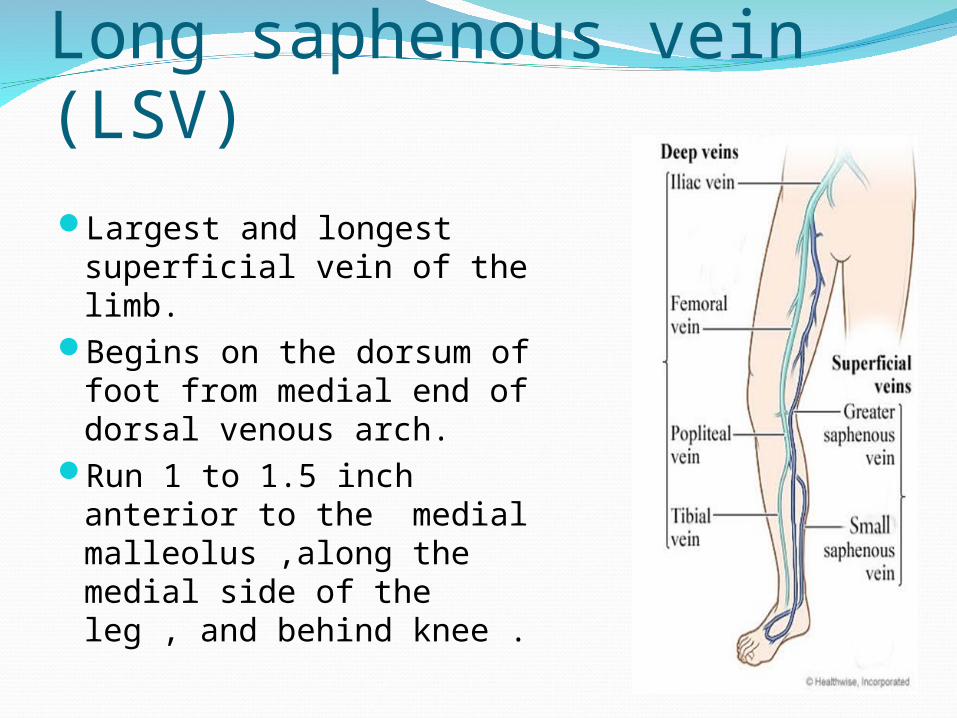
Long saphenous vein (LSV)Largest and longest
superficial vein of the limb.
Begins on the dorsum of foot from medial end of dorsal venous arch.
Run 1 to 1.5 inch anterior to the medial malleolus ,along the medial side of the leg , and behind knee .
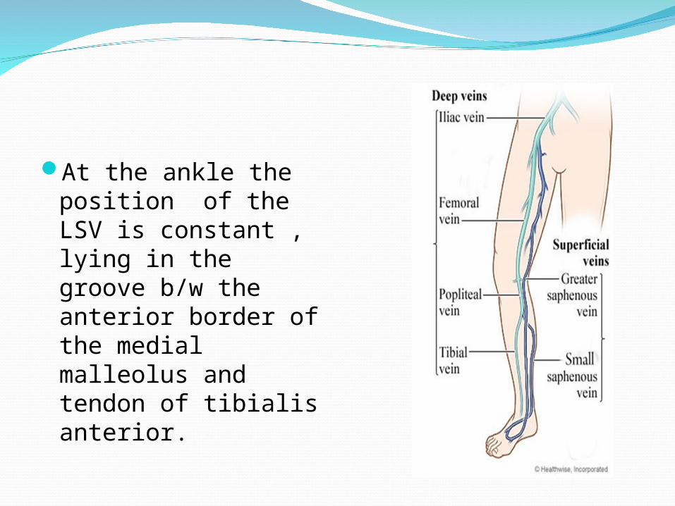
At the ankle the position of the LSV is constant , lying in the groove b/w the anterior border of the medial malleolus and tendon of tibialis anterior.
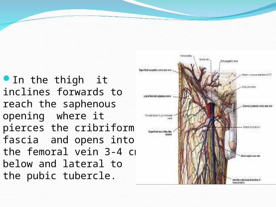
In the thigh it inclines forwards to reach the saphenous opening where it pierces the cribriform fascia and opens into the femoral vein 3-4 cm below and lateral to the pubic tubercle.
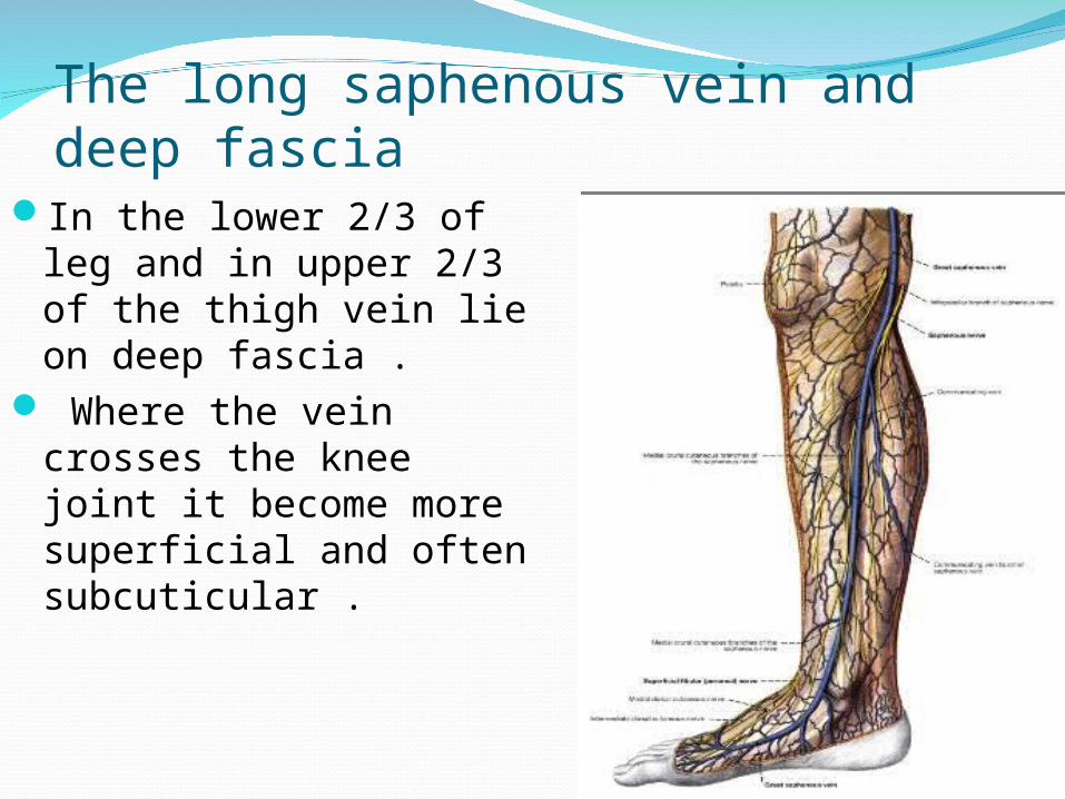
The long saphenous vein and deep fascia In the lower 2/3 of leg
and in upper 2/3 of the thigh vein lie on deep fascia .
Where the vein crosses the knee joint it become more superficial and often subcuticular .
The structures accompanying the LSV In the leg saphenous nerve lies in close
relation with the LSV.The nerve is very closely applied to the vein
in lower 2/3 of leg and often injured in exploring or stripping the saphenous vein .
In the thigh medial femoral cutaneous nerve run in close relation with vein .
Throughout its length the LSV is accompanied by lymphatic trunks draining the dorsum of foot and anterior and medial aspects of the legs and thigh .
This lymphatic drain in superficial inguinal lymph nodes.
Tributaries of LSV and communication Just below knee LSV receive posterior arch
vein (Leonardo's vein) which collect the blood from post-medial aspect of calf .
Anterior veins of leg(stocking vein) ascend across the shin and join either LSV or posterior arch vein .
There is a free anastomosis b/w tributaries of short saphenous vein and venous arch connecting medial ankle perforating vein and this medial ankle perforating veins are connected with LSV in lower third of leg .
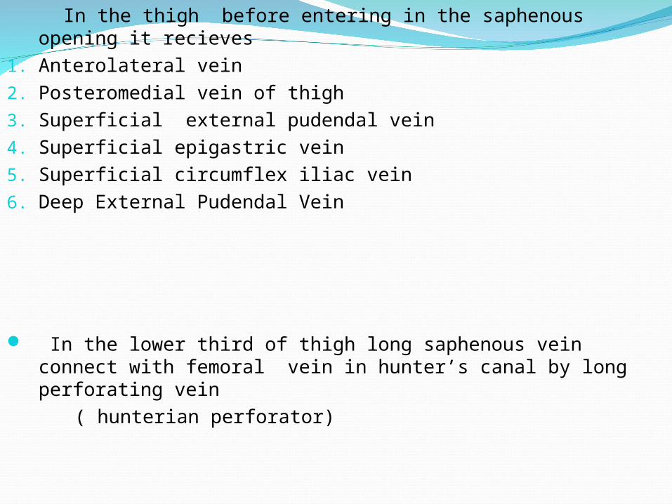
In the thigh before entering in the saphenous opening it recieves
1. Anterolateral vein
2. Posteromedial vein of thigh 3. Superficial external pudendal vein
4. Superficial epigastric vein
5. Superficial circumflex iliac vein
6. Deep External Pudendal Vein
In the lower third of thigh long saphenous vein connect with femoral vein in hunter’s canal by long perforating vein
( hunterian perforator)
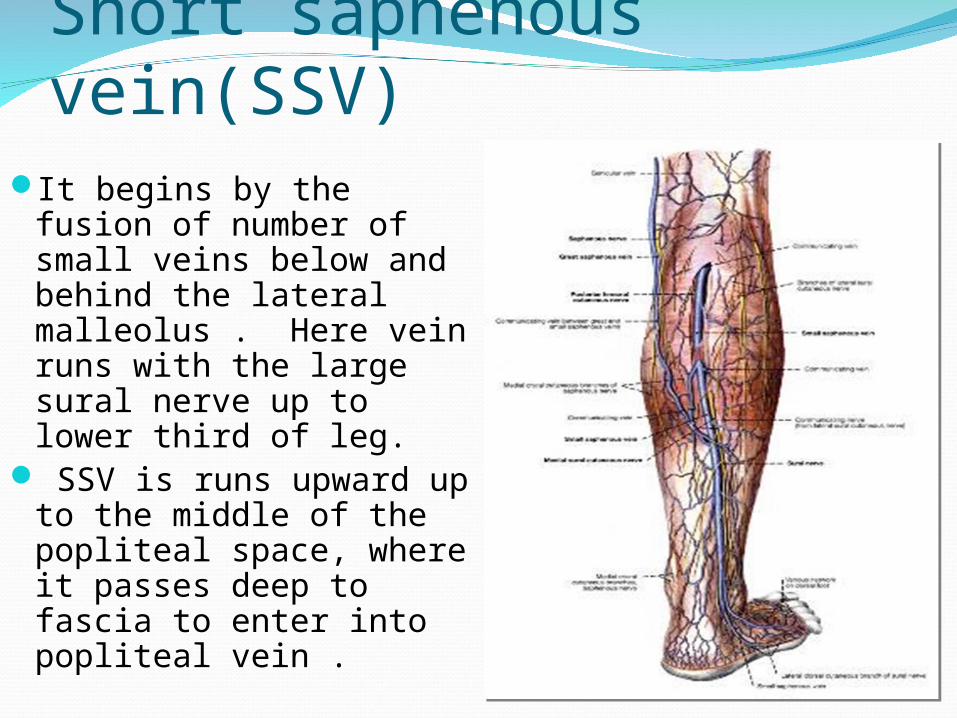
Short saphenous vein(SSV)It begins by the fusion
of number of small veins below and behind the lateral malleolus . Here vein runs with the large sural nerve up to lower third of leg.
SSV is runs upward up to the middle of the popliteal space, where it passes deep to fascia to enter into popliteal vein .
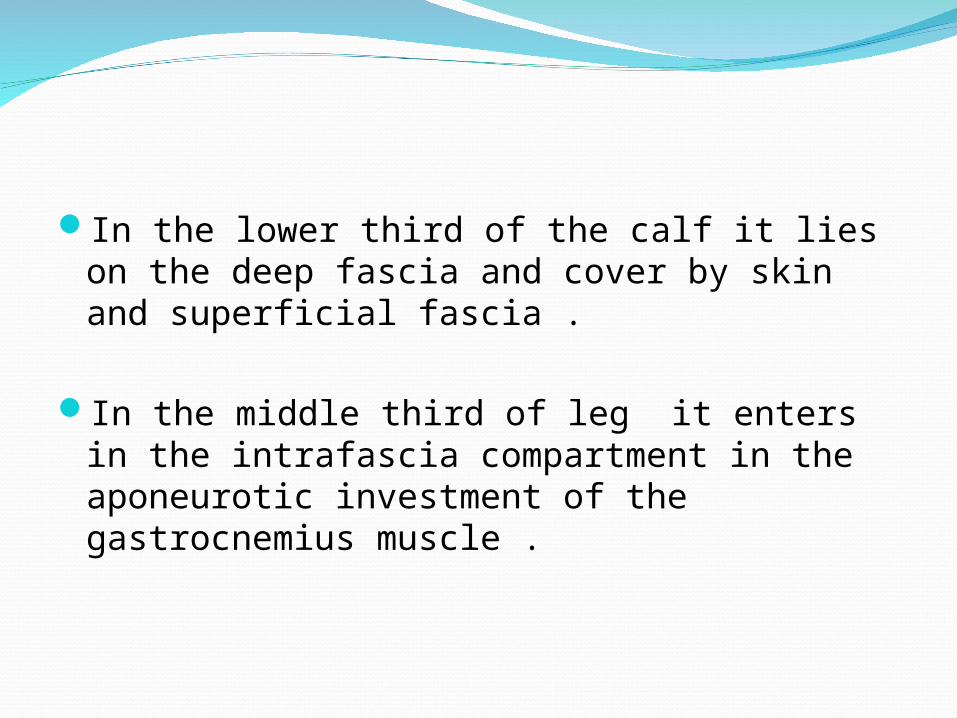
In the lower third of the calf it lies on the deep fascia and cover by skin and superficial fascia .
In the middle third of leg it enters in the intrafascia compartment in the aponeurotic investment of the gastrocnemius muscle .
Upper third of leg it penetrates the deep fascia and enter popliteal space and lie b/w head of two gastrocnemius muscle which lies 1.25cm below the transvers skin crease behind knee .
Here SSV join popliteal vein .
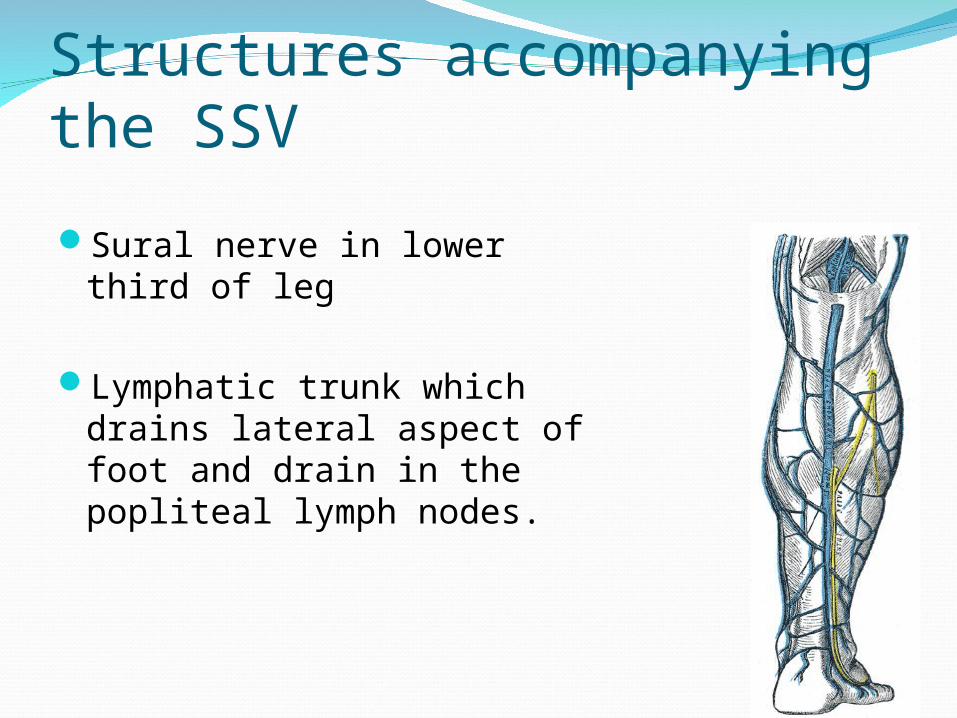
Structures accompanying the SSV
Sural nerve in lower third of leg
Lymphatic trunk which drains lateral aspect of foot and drain in the popliteal lymph nodes.
Where the vein passes through fascia Posterior cuteneous nerve emerges out from deep to superficial.
In the upper part of vein it communicates with LSV via the posteromedial vein of Leg.
SSV may run above the popliteal space and end in deep veins in lower thigh or may end in LSV in upper thigh.

Deep veins This veins lie in deep fascial plane and are
supported by powerful muscles of leg.These are 1: Anterior and posterior Tibial veins 2: Peroneal vein 3: Popliteal vein 4: Femoral vein These veins accompany with Arteries.
Perforating veins These are communicating veins b/w
superficial and deep veins .
Two type: 1 Indirect veins 2 Direct veins
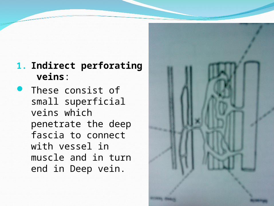
1. Indirect perforating veins:
These consist of small superficial veins which penetrate the deep fascia to connect with vessel in muscle and in turn end in Deep vein.
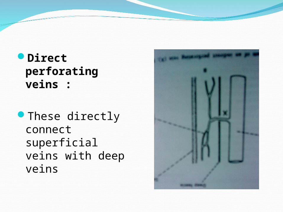
Direct perforating veins :
These directly connect superficial veins with deep veins
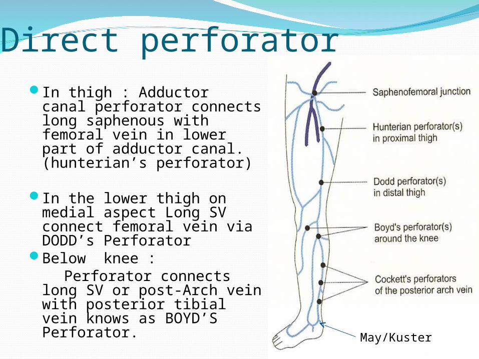
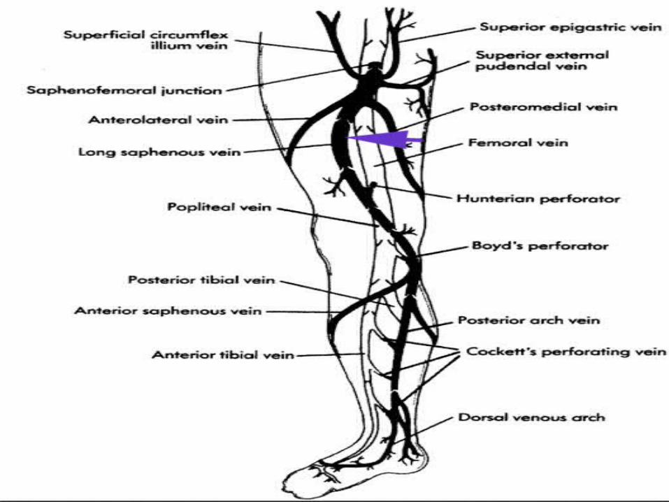
Direct perforator In thigh : Adductor canal
perforator connects long saphenous with femoral vein in lower part of adductor canal. (hunterian’s perforator)
In the lower thigh on medial aspect Long SV connect femoral vein via DODD’s Perforator
Below knee : Perforator connects long
SV or post-Arch vein with posterior tibial vein knows as BOYD’S Perforator. May/Kuster
In leg : 1.Lateral perforator is presented at the
junction of mid & lower third of leg .It connect SSV with peroneal vein.
2. Medially there are three perforator which
connect posterior arch vein with posterior tibial vein , know as COCKETT’S Perforator
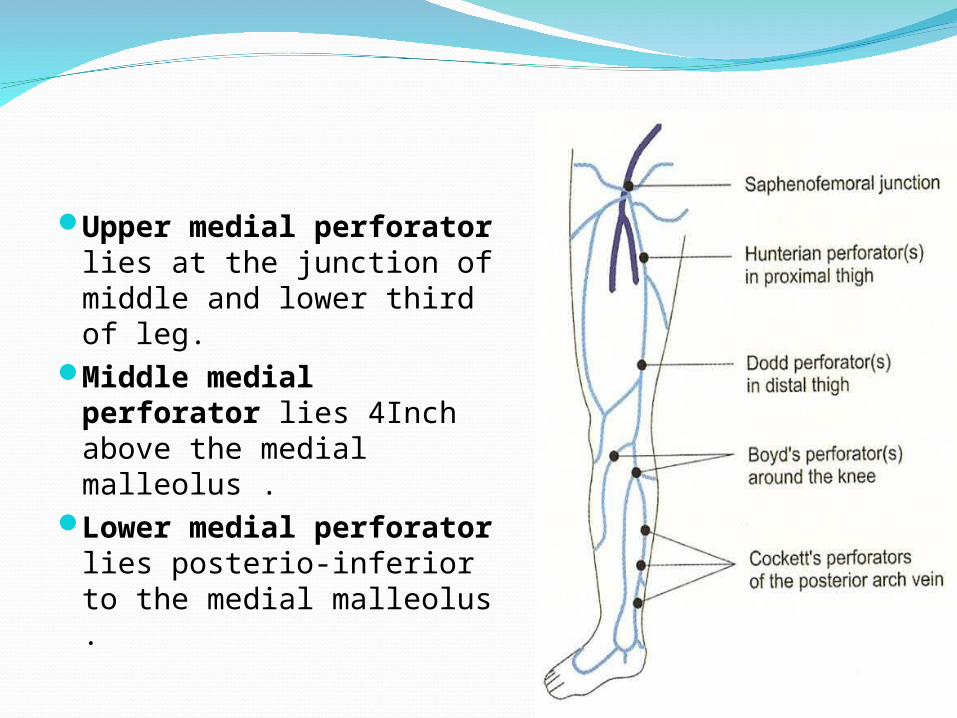
Upper medial perforator lies at the junction of middle and lower third of leg.
Middle medial perforator lies 4Inch above the medial malleolus .
Lower medial perforator lies posterio-inferior to the medial malleolus .
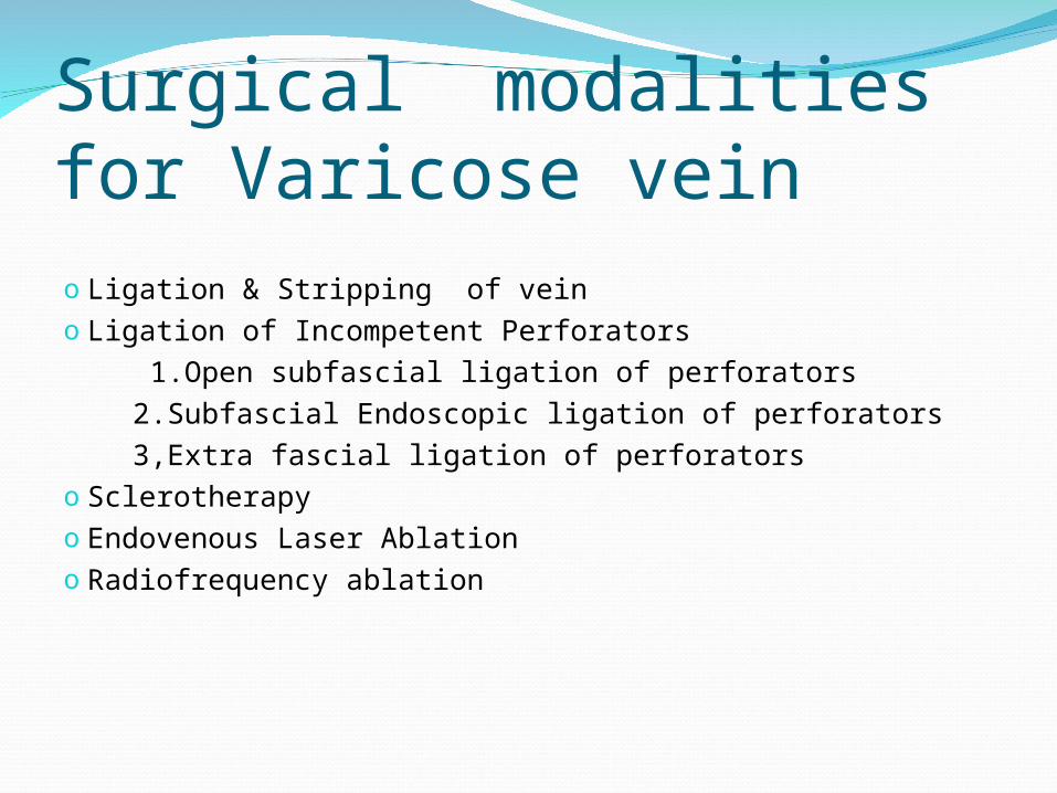
Surgical modalities for Varicose veino Ligation & Stripping of vein o Ligation of Incompetent Perforators 1.Open subfascial ligation of perforators 2.Subfascial Endoscopic ligation of perforators 3,Extra fascial ligation of perforatorso Sclerotherapyo Endovenous Laser Ablationo Radiofrequency ablation
SurgeryLigation and stripping of varicose vein :Indication :
LSV /SSV incompetency .Perforating vein incompetency.
ContraindicationsDVTPregnancyThrombophlebitisPeripheral vascular disease
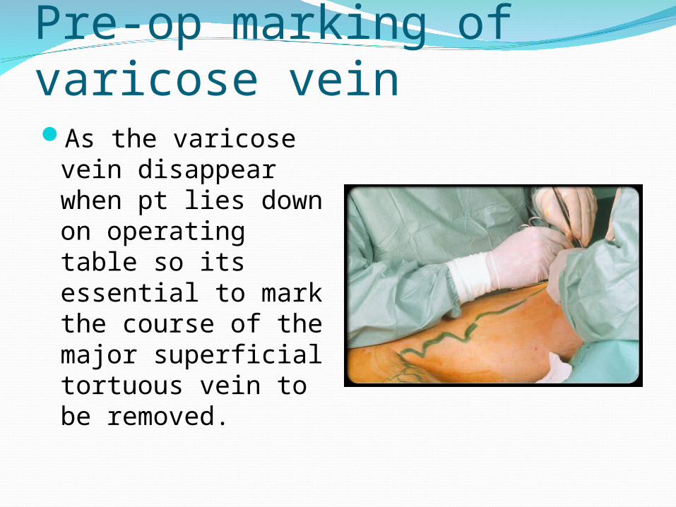
Pre-op marking of varicose vein As the varicose
vein disappear when pt lies down on operating table so its essential to mark the course of the major superficial tortuous vein to be removed.
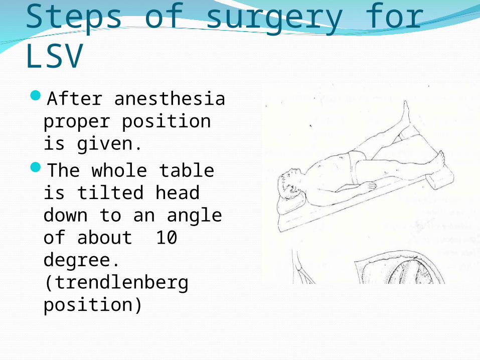
Steps of surgery for LSV After anesthesia
proper position is given.
The whole table is tilted head down to an angle of about 10 degree. (trendlenberg position)
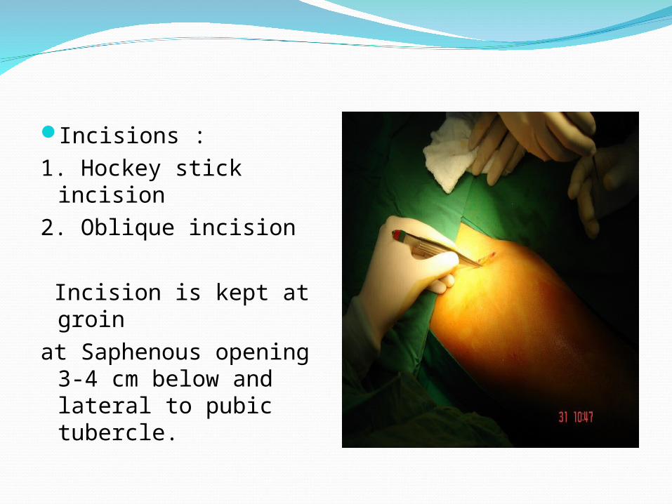
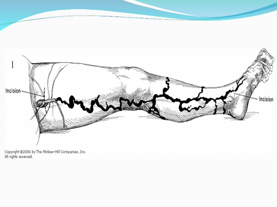
Incisions :1. Hockey stick incision2. Oblique incision Incision is kept at
groin at Saphenous opening
3-4 cm below and lateral to pubic tubercle.
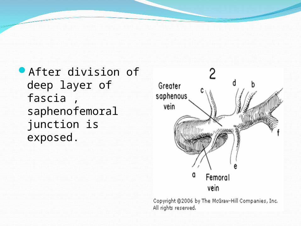
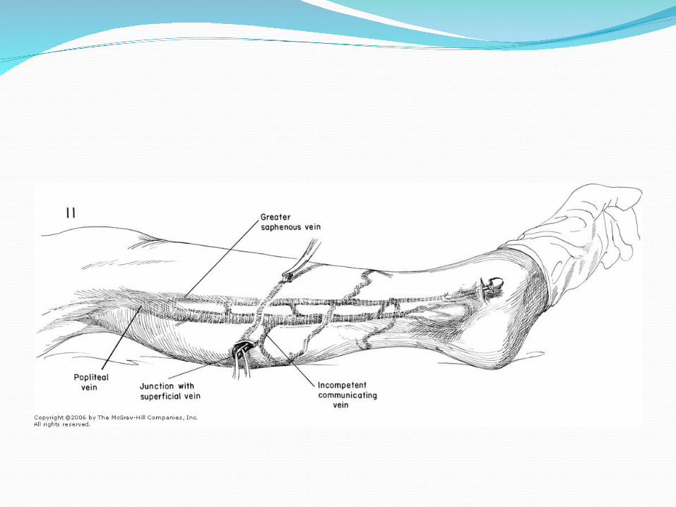
After division of deep layer of fascia , saphenofemoral junction is exposed.
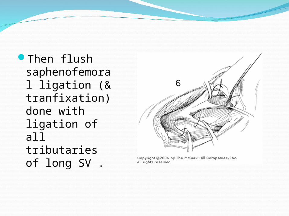
Then flush saphenofemoral ligation (& tranfixation) done with ligation of all tributaries of long SV .
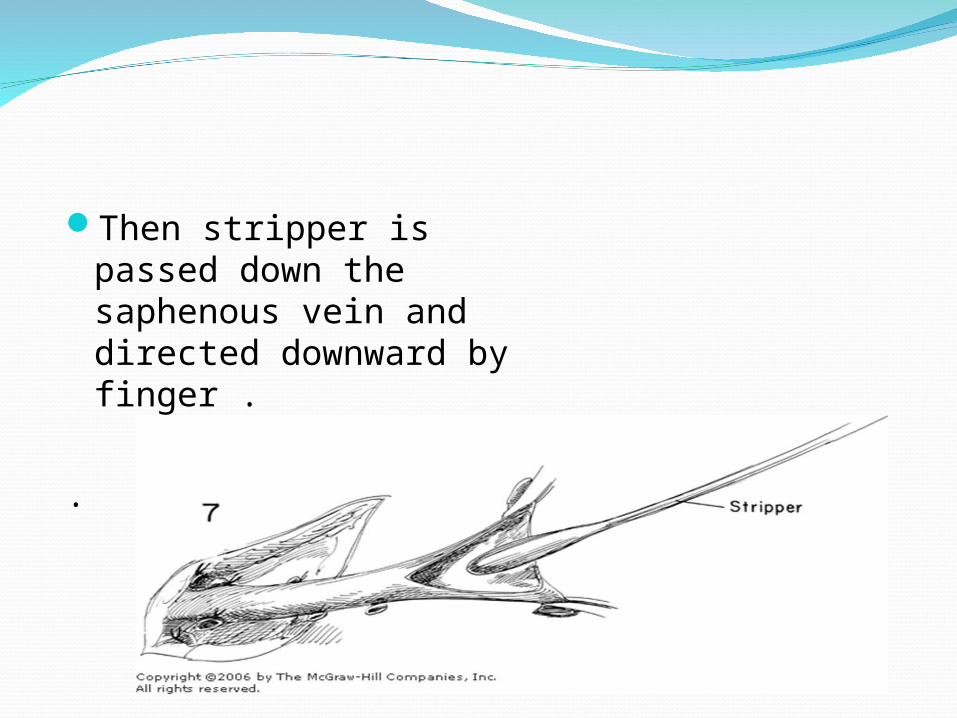
Then stripper is passed down the saphenous vein and directed downward by finger .
.
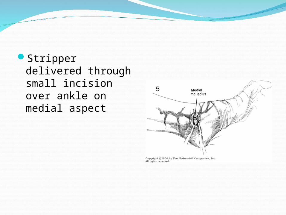
Stripper delivered through small incision over ankle on medial aspect
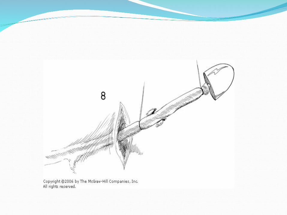
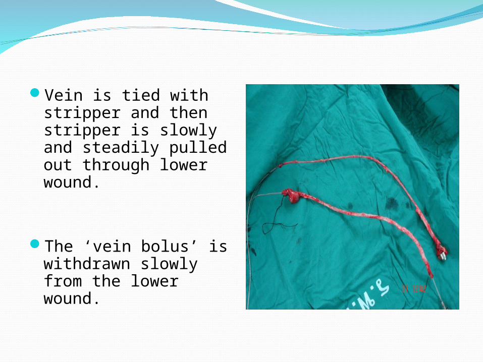
Vein is tied with stripper and then stripper is slowly and steadily pulled out through lower wound.
The ‘vein bolus’ is withdrawn slowly from the lower wound.
The residual veins are then ‘wormed out ‘ using multiple stab avulsions using vein hooks ,from the preoperative marked sites.
Post operatively limb elevation and compression stockings are given .
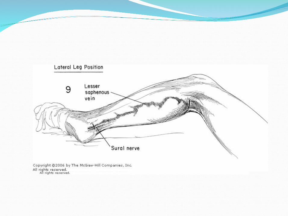
STEPS OF SURGERY FOR SSVAfter anesthesia proper position is given.The patient must be face down and the knee
is flexed a little, by placing sandbag under the ankle .
Some prefer lateral leg position.The foot of the table is tilted up a little, so
that legs are above the heart.
Incision is kept atleast 5 cm long, transversely across the popliteal fossa, in one of the transverse line of skin about the level with knee joint.
The incision is deepened until the deep fascia and short saphenous vein lies deep to this.
The fascia is divided transversely in the line of incision.
The short saphenous vein is then seen or sought for betweeen the two heads of gastrocnemius.
As soon as the SSV is identified, it is lifted up in a pair of artery forceps and the knee is flexed still further.
Then flush saphenopopliteal ligation (& transfixation) done with ligation of all the side branches of SSV, right upto its junction with the popliteal vein.
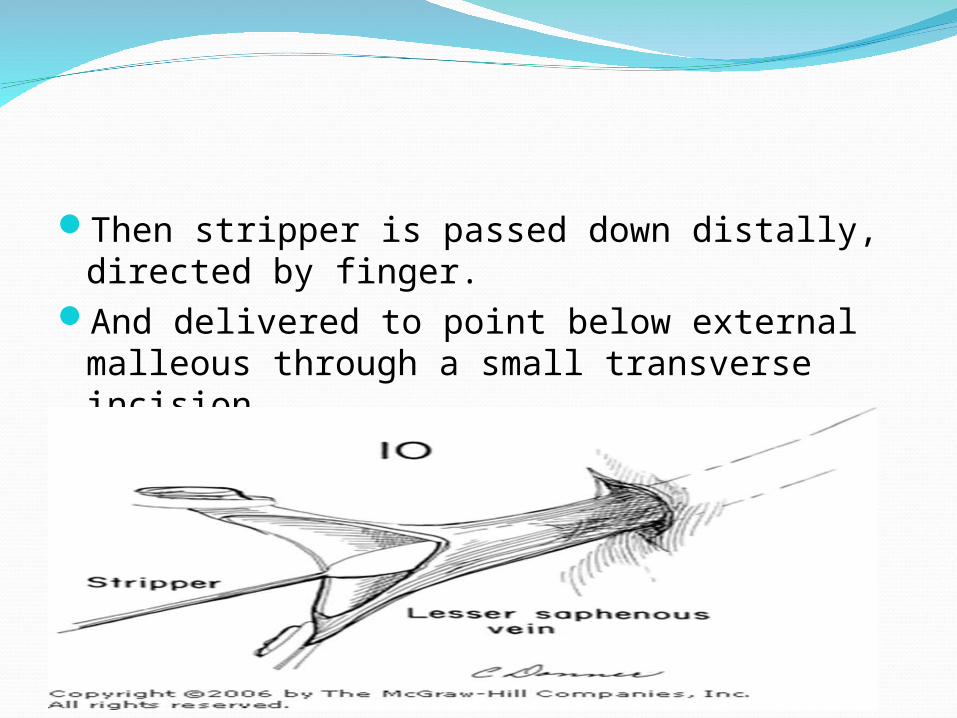
Then stripper is passed down distally, directed by finger.
And delivered to point below external malleous through a small transverse incision.
INTRA- OPERATIVE COMPLICATIONS OF THE SURGERYBLEEDING FROM A TORN SAPHENA VARIX INJURY TO COMMON FEMORAL VEIN INJURY TOCOMMON FEMORAL ARTERYINJURY TO SAPHANEOUS NERVEINJURY TO SURAL NERVE
IMMEDIATE POST-OP CAREThree factors to be kept in mind in the first
week :
1 Maintenance of firm elastic pressure over whole limb.
2 Regular movement and exercise of the legs3 Elevation of the foot of the bed 6 to 9
inches so that the legs are just above the heart level when the patient is in bed.
POSITION :
The foot of the bed is raised 6 to 9 inches
Patient is not allowed more than 2 pillows.
BANDAGING :
The original firm crepe bandage put on at the operation should remain untouched for seven days
GETTING UP :Started 24 hrs after the operation.When the foot is placed on the ground for the
first time, extra firm webbing elastic bandage are placed over knee and ankle.
At 7 days the stitches are removed.A firm webbing elastic bandage from ankle to
knee is worn through-out the day for a whole fortnight.
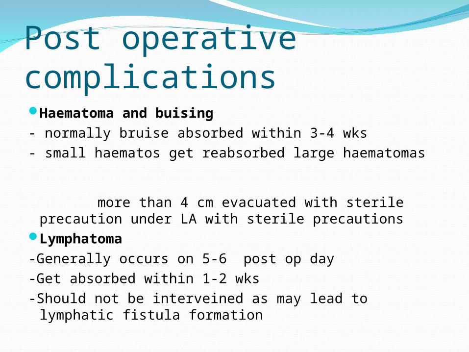
Post operative complicationsHaematoma and buising- normally bruise absorbed within 3-4 wks- small haematos get reabsorbed large haematomas
more than 4 cm evacuated with sterile precaution under LA with sterile precautions
Lymphatoma-Generally occurs on 5-6 post op day-Get absorbed within 1-2 wks -Should not be interveined as may lead to lymphatic
fistula formation
Wound sepsisPost operative saphenous neuritisLymphoedema of legInduration of stripper tractDVT and embolism
Extra fascial ligation of perforators(Cocketts procedure)Not commonly employedAim is to clear all the extrafascial veinsMore traumatic due to adherence of
subcutaneous fat and connective tissue to the fascia
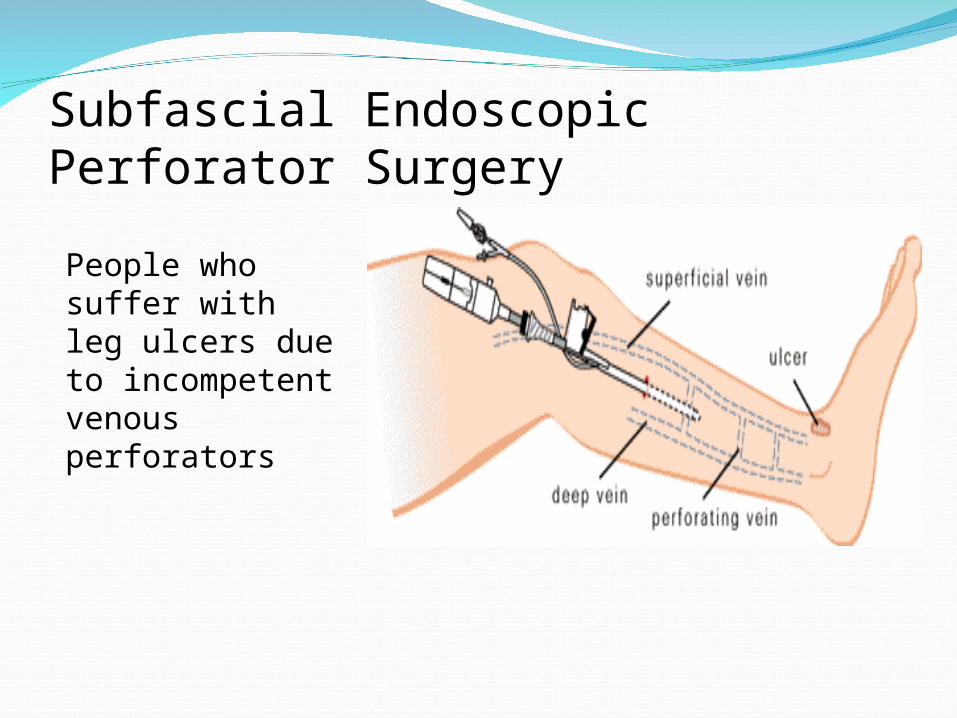
Subfascial Endoscopic Perforator Surgery
People who suffer with leg ulcers due to incompetent venous perforators
Indication :Incompetent perforating veins in calf with no
superficial venous reflux or no evidence of DVT on Doppler .
Patient with LSV / SSV varicosity with ulcer
Procedure Using spinal or general anesthesia a ¾ inch
incision is made on the inside of the calf. An instrument is inserted deep to the fascia
of the leg and a large balloon is inflated with water to create a working space.
The balloon is then emptied and the space is insufflated with air.
The camera is inserted and the perforator veins can be seen in the space passing from superficial to deep layers.
Another small incision is made in the calf for passage of another instrument.
The perforator veins are carefully dissected, Clips are applied and the veins are divided
if necessary. All trocars are then removed and the wounds
are closed. The patient is generally sent home the same
day of surgery with elastic stocking.
Obliteration of venous lumen - Methods
1. Foam Sclerotherapy2. Laser3. Radiofrequency Ablation
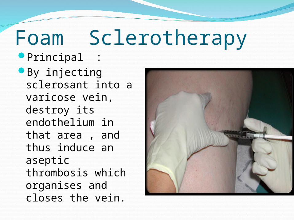
Foam SclerotherapyPrincipal : By injecting
sclerosant into a varicose vein, destroy its endothelium in that area , and thus induce an aseptic thrombosis which organises and closes the vein.
Indication :
Residual vein after surgery
Large venous telangiectases.
Isolated small dilated veins
Contraindication :
PregnancyPelvic tumor Sup thromboplebitis
at the time of procedure
DVTPrevious h/o reaction
to sclerosant
SOLUTIONS :
SODIUM TETRADECYL SULPHATESOD.MORRHUATEHYPERTONIC SALINE SOL.POLYDOCANOL,SOTRADECOLETHANOLAMINE OLEATEGLUCOSE COMBINATIONS
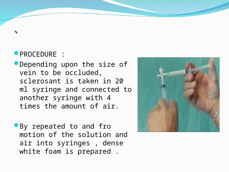
`PROCEDURE :Depending upon the size of
vein to be occluded, sclerosant is taken in 20 ml syringe and connected to another syringe with 4 times the amount of air.
By repeated to and fro motion of the solution and air into syringes , dense white foam is prepared .
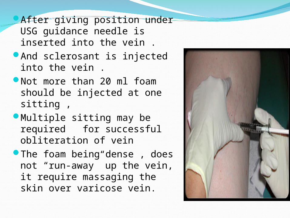
After giving position under USG guidance needle is inserted into the vein .
And sclerosant is injected into the vein .
Not more than 20 ml foam should be injected at one sitting ,
Multiple sitting may be required for successful obliteration of vein
The foam being dense , does not “run-away” up the vein, it require massaging the skin over varicose vein.
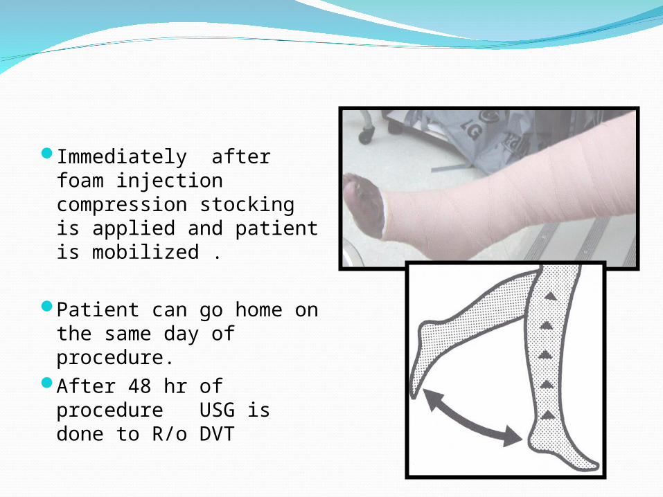
Immediately after foam injection compression stocking is applied and patient is mobilized .
Patient can go home on the same day of procedure.
After 48 hr of procedure USG is done to R/o DVT
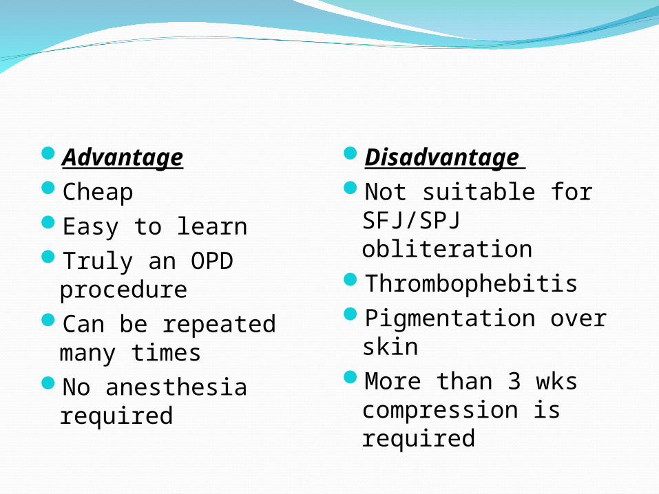
AdvantageCheapEasy to learn Truly an OPD
procedure Can be repeated
many timesNo anesthesia
required
Disadvantage Not suitable for
SFJ/SPJ obliterationThrombophebitisPigmentation over
skin More than 3 wks
compression is required
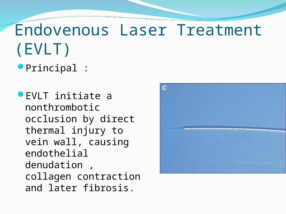
Endovenous Laser Treatment (EVLT)Principal :
EVLT initiate a nonthrombotic occlusion by direct thermal injury to vein wall, causing endothelial denudation , collagen contraction and later fibrosis.
Indication :Long saphenous vein
varicosityShort saphenous
vein varicosity
Contraindication :Superficial vein
thrombophlebitisDVT
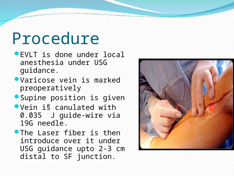
Procedure EVLT is done under local
anesthesia under USG guidance.
Varicose vein is marked preoperatively
Supine position is givenVein is canulated with
0.035” J guide-wire via 19G needle.
The Laser fiber is then introduce over it under USG guidance upto 2-3 cm distal to SF junction.
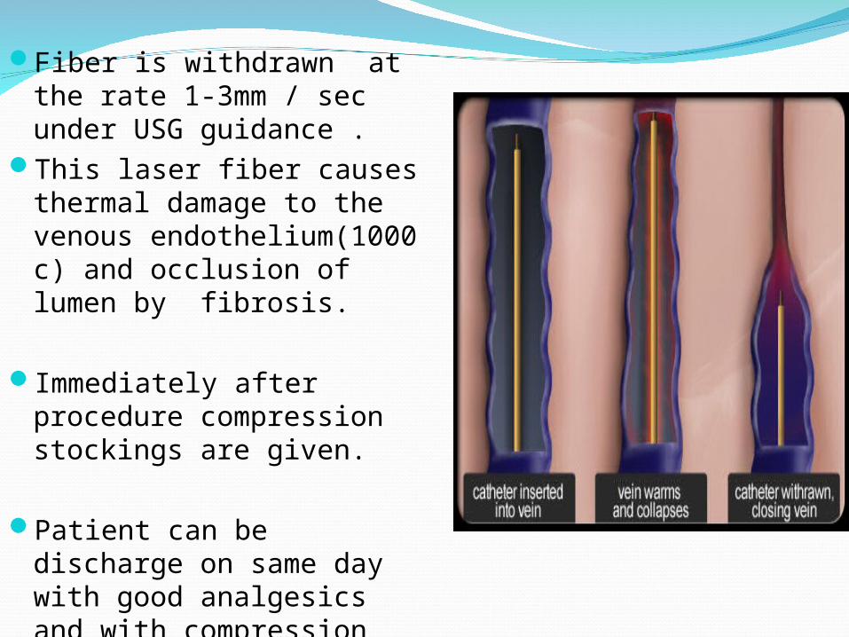
Fiber is withdrawn at the rate 1-3mm / sec under USG guidance .
This laser fiber causes thermal damage to the venous endothelium(1000 c) and occlusion of lumen by fibrosis.
Immediately after procedure compression stockings are given.
Patient can be discharge on same day with good analgesics and with compression stockings.
ADVANTAGEMinimal invasive
procedure No post op scarDone with local
anesthesiaMinimal post-op
pain Recurrence rate ( at
2 year f/u only 3%
DISADVANTAGECostly procedureHigh technical skills
reqColor Doppler and
Radiologist is req Skin burnsThrombophebitis Paresthesia
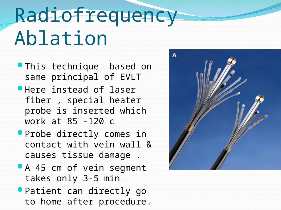
Radiofrequency AblationThis technique based on
same principal of EVLT Here instead of laser fiber ,
special heater probe is inserted which work at 85 -120 c
Probe directly comes in contact with vein wall & causes tissue damage .
A 45 cm of vein segment takes only 3-5 min
Patient can directly go to home after procedure.
TRIVEXAlternative to avulsion phlebectomy for
superficial vein excision.
In this technique with the help of transcutaneous light, veins are seen and extracted with the help of suction dissector.
Thank You