Embed Size (px)
Citation preview
Loyola University ChicagoLoyola eCommons
Master's Theses Theses and Dissertations
1934
Variations of the Middle Meningeal Artery in theMiddle Cranial FossaClemens F. DerezinskiLoyola University Chicago
This Thesis is brought to you for free and open access by the Theses and Dissertations at Loyola eCommons. It has been accepted for inclusion inMaster's Theses by an authorized administrator of Loyola eCommons. For more information, please contact [email protected].
This work is licensed under a Creative Commons Attribution-Noncommercial-No Derivative Works 3.0 License.Copyright © 1934 Clemens F. Derezinski
Recommended CitationDerezinski, Clemens F., "Variations of the Middle Meningeal Artery in the Middle Cranial Fossa" (1934). Master's Theses. Paper 14.http://ecommons.luc.edu/luc_theses/14
VARBTIDNS OF THE RIDDLE MENINGEAL &RTERY
IN THE MIDDI.Ji CRANrAL FOSSA.
A THESIS
SUBMITTED TO THE FACULTY
a£ ·'Ule
G!WlUATE SCHOOL
ot
I.D'm!A. UNI.VERS.I'rr
in
CANDIDACY' FOR THX
DE.GBEE OF BSTER OF SCIENCE
-Cl.eJaDs r ... ~' ».s.:M.
:l
ln'troduct.i.on
Iixc:rea:seti advances in the surgery of the head include
o,per-ations upon the Cfa.s:serfan gang~ion and l:f'ga:tions a£ the
middi:e mertinge·a:I artery· whell' it lm:s been suo cfec·ted to trauma.
s·ince this artery is intimate I%· relat.ed to both opera:tions •
it :Col~ows tha.t exact tmowledge of its course and variations
i.s nee::ess:sry to the el:infcian for suc·cess:ru~ results.. Kana'v:'e":t
and: Da..vis(r-z2J: :Dave painted out the close rela:t..i.Gns of this
vesse~ to the semilunar- ganglion; in connection with :rra:et.ures
at the temporal. r-egi.on of the skull• Rowan (t"22) stressed the
importance o,f the presence: a.f a eamd. for the anterior branch
of the arte:rlf.. B~lett. ("02) empha:si.z-ed the varia'bill.ty Ott
the 12ssel enc:oom.tere:d in surgery oC the mi.ddl.e cranial". :fossa:.
Ya:tsuta. ("951 di.scttssed the surg:lea.l aD&torq of the artery· ..
Thes-e tmd ul Octher tmthO'rs who have studied the Yariati.ans oC
the artery hava done sa with their own particular phase a:f the
problem: in mind.. NO:- one. b.ofteTer• has presented as ful:.1. and
complete a pi.cture o--f the important variatio-ns as pos.s.ible ..
The pu;rpGSe of this res.eerch is • therefo-re,. to, determine
the co\ll"'"se. and -variations of the Biddle menigea:l arte.ry as
exactl:y as po·ssible • and to interpret the resuJ..ts in the l.ight
ot ellr.dcal. and anatomi.cal s.igm.:fi:canee ..
Gra:te.fu:t appreci.at.io.n is due Dr ... s .. B..Cl:umdler. Asaociat.e
Pro:ress.or of .Ana.t.Otq' at Looyok trni.veruty lredica:l. Scho.Gl..• :Cor-
auggas.Ung the pro;bl.eDr a:ad fer the helpful. advice and critiei

2
rendered throughout the course of thi.s research.. Thanks are
a·Iso due to Dr.O.F .Kampmeier of illinois Medical Schoo.l. and
Dr..B..J .Anson Oif Northwestern Medical,. who, have made their
respec-tive bone collections av:a:ilable for study.. Had it not
D:een :for their crooperation. the IItlmber of sku~ls wou~d have
been too small to he conclusive.
Literatura
The descr-iptions o-f the course of the middle menigeal
artery as gLven in the standard text-books of anatomy are
essentially the same.. According t.o Cunningham "s Anatomy( '31) •
"'It is the largest branch of the internal maxillary artery.
It passes through the :foramen spinosum and enters the middle
cranial fo.ssa:. In this :fossa it passes forwards, for a short
distance.- in a groove on the great wing of the sphenoid, and
divides into an anterior and posterior terminal branch.. The
artery lies in the out,er layer of the dura: mater.. The
anterior termi.nal branch pa.ssea upwards along the great wing
of the sphenoid to the sphenoidd angle of the parietal bone •
where it is sometimes enclosed in a bo.n~ canal. The posterior
terminal branch passes backwards to the squamous part of the
temporal hone, where it sends branches upwards to the vertex
and backwards to the occ:ipu.t.. The smaller branches of the
terminal arteries anastomose with each ather and with those
of the oppGsite side, and with. the anterior and posterior
meningeal arteries.• The descriptions in Gray's Anatomy ('30),
3
Morris,.. Ana tomy('32) • and Davis' Applied Anatomy ( t26) are
quite similar to that given in Cunningham's text-book .. Marris'
Anatomy adds that the canal for the anterior branch. attains
a size ranging from six to twelve millimeters, and Davis' text
points out that this canal is located in the region of the
pterion. These discussions give the impression that the
course of the artery is essentially the same in all cases, and
that no variations occur, with the possible exception of its
relation to a. canal.
Ka:navel and Dav-is ('22). in an examination of one hundred
skulls, found six distinct variations of the middle meningeal
artery, all of which were confined to the posterior branch.
They were as follows: (1) in 41 per cent. of the cases, the
posterior branch was given off midway between the foramen
spinosum and the region of the pterion; (2) in 36 per cent.
of the instances, it was given off at a higher level., near
the region of the pterion, and (3) in 8 per cent. of the cases,
at a lower level, very soon after the artery emerged from the
foramen spinosum; (4.) two posterior branches arose at separate
intervals in 8 per c.ent .. and (5) simultaneously, in 5 per cent.
of the cases; (6) no posteri.or branch was found in two
instances.
Bartlett (lQ6) in his observations of one hundred half
skulls, found no instances of double branching of the posterior
ramus. However, he descri ood two other variations not here-to-
4
fore discussed: (I.) in which the anterior branch was absent
and (2) in which the main trunk. in addition to its anterior
and posterior branches,_ gave off another ranrt1s which coursed
f'"orwards and media.lly along the side of the sella turci.ca. He
The regards this additional branch as an anomalous condition.
frequency of occurence of these variations was not mentioned.
Bartlett. also discussed the variations in the relations
of the foramen spinosum, by means of which the middle
meningeal artery gains entrance into the middle cranial fossa.
He found the foremen spinosum absent in one instance. In this
case the artery entered the middle cranial cavity through the
:toramen ovale, accompanying the mandibular nerva.. In all
other cases the foramen spinosum was present. The distance
between the foramen spillosum and the foramen ovale was less
than one millimeter in four cases, from one to two millimeters
in eleven eases, from two to three millimeters in thirty six
cases~ from three to five millimeters in thirty four cases.
and from five to· ten millimeters in twelve instances. In only
one instance did the. distance exceed t.en. millimeters. Whether
these variations occured unilaterally or bilaterally was not
mentioned. Kanavel and Davis record the fact that the foramer1
ap.inosum was found to be continous with the foramen ovale in
four per cent. of their cases.
Yatsuta (~95). in the resuLts ob.tained from an inspection
of seventy five skulls, reported that the fora.men spinosum wa.s
a;.
present in every case. He no.ted that the posterior branch
exhibited an inconstant course. r:t arose at a ~evel varying
from 1.5 em. to 3.1 em. superior to the foramen spinosum.
He. added that the anterior branch was contained in a bony
canal in almost. every case, and that the canal was situa.ted
posterior to the sphenoparietal sinus.
Rowan C '22} $ in a detai.led account of the frequency of
the presence of a canal for the anterior bran~ of the middle
meningeal ar~ery~ noted that it was present in 110 of the 195
lateral halves of crania examined (56...4 per cent.). The canal
occured bilaterally in 31 per cent. of U1e crania, and was
absent on both sides of the cranium in 25 per cent. of the
cases. The bon~ canal was found on the ri~t side only in
27.1 per cent of the instances, and on the left in 30 per cent.
of the specimens~
The literature just cited shows that the standard text
books of anatomy assume that no variations occur in the course
of the middle, meningeal artery, with the possible exception
of the presence of a canal. The clinical literature, on the
other hand, describes eight different varia tiona. A~l these
variations were not described by the same author, and the
number of crania examined was relatively small ..
Methods and Ma-terials
Observations have been carried out on 430 whole and 49
half crania o.btained fro.m the anatomical laboratories of
Ei
Loyoila~Worthwestern and illinois Medical. Schools. Of these,
324 whole and 39 half crania were bare of all soft tissues;
as a result, the course of the middle meningeal artery was
deduced from the presence of the groove which the vessel made
upon the floor and side's of' the cranium. The remaining 106
whole and 1.0 hal.f crania were obtained direetl.y from cadavers
with the meninges still adherent to the bony parts.. Here, the
course of the artery could be directly traced as it traversed
upwards. thr~gh the dura mater, toward the region of the
calvarium. There were, in all, 907 lateral halves of crania
examined. However, not every cranium, because of trauma. or
other circumstances which affected its physical condition,
presented a complete picture of the co.urse of the artery. For
this. rea.sona there is a diff'erence in the number of crania
examined for each type of varia.tion.
The scope of this research embraces the variations of the
middle meningeal artery f'ound in the middle cranial foasa,
commencing at the entrance of th~ vessel into the middle
cranial cavity. and terminating when the artery crosses at
the level of the ·lesser wing of the sphenoid. For this reason
the ~iations of the middle meningeal artery have been
classified according to .•. its entrance into, its course
within, and its exit from, the middle·cranial fossa.
Results
Entrance into the middle cranial fossa.
7:"
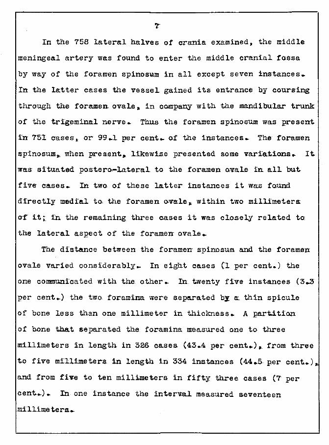
In the 758 lateral halves of crania examined, the middle
meningeed artery was found to enter the middle cranial fossa
by way of the fora~en spinosum in all except seven instances.
In the latter cases the vessel gained its entrance by coursing
through the foramen ovale • in company with the mandibular trunk
of the trigeminal nerve.. Thus the foramen s.pinosum was present.
fn 7'5:1 cases,. or 99 .. 1 per cent .. of the instances .. The foramen
spinosum., when pres.ent._ likewise presented some variations. It.
was: situa.ted postero-lateral to the foramen ovale in all but.
five cases.. In two of these latter instances it was fo.und
direcUy medial to the foramen a.val.e.~ within two millimeters.
ot it;. in the remaining three cases it was cl.o.sely related to:
the lateral aspect of the foramen o:vale ..
The distance between the foramen spinosum and the foramen
oval.e varied considerably.. In eight cases (l per cent.) the
one communi:cated with the other.. In twenty five instances (3..3
per cent .. ) the two foramina were separated by a thin spicule
of bone less than one millimeter in thickness.. A part.itLon
of bone that separated the foramina measured one to three
m:i.lli:meters in length in 326 cases (43 .. 4 per cent .. ) • from three
to five millimeters in length in 334 instances (44.5. per cent.) •
and from five to ten millimeters in fifty three cases (7 per
cent .. ). In one instance the interval measured seventeen
m-illime tera ..
a
Course within the m:iddl.e crania!. fossa:
The variations of the middl.e meningeal artery as it
courses through the middle cranial fossa are divided into thaae
of its anterior and pos~rior branches ..
a: .. Variations of the anterior branch..
This branch was present in all but five o! the 903 lateral.
halves of crania examined.. In nine cases (1 per cent .. ) the
middle meningeal artery._ immediately after its exit from the
foramen spino.sum. bifurcated into two anterior branches. The
lateral branch coursed in the manner typi.cal of the anterior
ra!IllXS .- while the medial. ramus proeeeded forwards along the
side of the s.ella. turcica. either to supply the meninges in
that region or el.se to bend la.terally to. anastomose with branchEs
from the anterior meningeal. artery.. The medial anterior
branch was intimately related to the mandibular and maxillary
nerves as it coursed along the lateraT aspects of the foramen
o~l.e and foramen rotundum.. In 99 instances (1.0 .. 9 per cent .. )
the anterior branch (or the main trunk, if the posterio.r branch
aros.e at a high. l.eve 1) gave off a medial. branch midway in its
course. This ramus proceeded medially towards the lesser
wing of the sphenoid bone.. In a few ins-tances it supplied the.
meninges in that region,. and in all other cases it was seen
to. enter the. orbital cavity: either through the superio.r orbital
fissure or by wa;x of a separate foramen, to anasto.mo.se with
the. ophthalmi.c artery •.
9
Two anterior branches were found to be bilaterally
present in one cranium:*' and a medial terminal branch occured
on both sides in forty eight crania ..
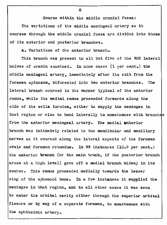
a. Variations of the posterior branch!
Eight distinct variations were noti.ced in the 894 lateral
halves of crania observed.. The posteri.or branch was a.bsent
in six cases (...6 per cent .. Y.. It was found as a. single branch
in ?22 cases [80 .. 7 per cent.Y,. and when in this state it was
given off at one of three levels:. in the ma:jarit:Y of the cases
(295 instances, or 33 per cent.). it was situated midway
b-e1.we en the foramen spinasum· and the region of the pte.rion;
les.s frequently (258 instances .. or 28'.8 per cent..)"' it.. arose
very s.oon after the artery pro-per emerged from the foramen
spinosum.. In the remaining 169 instances (18 .. 9 per cent. •. ) i.t
originated at a. high level. at or near the region of the
pt-erion.
Two posterior branches were given o,ff simultaneously in
thirteen cases (L.A. per cent .. ).. Two po-aterior branches arose
at separate intervals in forty eight cases (16..5 per cent .. ) ..
These branches._ like those of the single posterior ramus, were
given off at various levels.
A. bony canal containing the posterior branch of tha
middle meningeal artery was found in seven instances(..8per cent.
In thirty two cases (4 ... 5 per c~ent.) the posterior branch
bifurcated very near its origin ..
lO
Irs to the bilatera~.is:m or these variations,. fifty two of'
the 423 whale crania inspected exhibited a bilateral presence
of the single posterior branch ar-ising at a level midway
between the f'oramen spinosum and the region of the pterion
('12 .. 3 per een~.. The same branch originated a.t a lower l.evel.
near the foramen spinosum. on both sides in a like number of
specimens.. In thirty three crania ('r .a per cent .. ) i.t was
bilaterally situated high up near the regi:on of the pterion.
Two po,sterior branches arose at separated inter-vals on each
side in forty eight whole specimens ...
It is to be noted that two variations may be. found on the
same lateral hall o-f a specimen, 1.e ... a. variatiOn of the
po:ster:tor branch. occuring together ..
Exit f'rom the middle cranial. :rossa.
The po·sterior branch presented no. significant variations
for the reason that it usua.lly breaks into a. large network
of smaller branches as it reaches the calvarium.. The anterior
branch.., on the o;ther hand, continued for a longer period of
time before subdividing. .Kbove the level of the lesser wing
o,f the sphenoid bone • which bounds the upper limit of the
middle crania~ fossa • this branch coursed upward to.ward the
region of the calvarium as a single artery in 842 out of the
870 cases examined ("96 .. 7 per cent .. ).. In the remaining twenty
eight cases (3..3 per cent ... )., it divided into two ascending
branches ..
ll
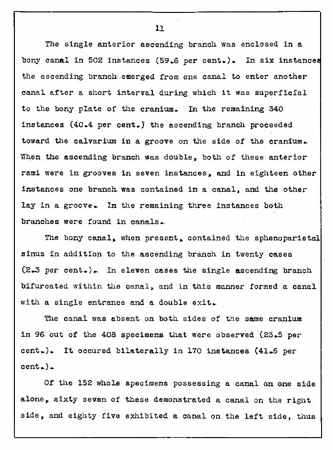
The single anterior asc-ending branch was enclosed in a
bony cana.l in 502 instances (59 .. 6 per cent.). In six instancet:
the ascending branch emerged from one canal to enter another
canal after a short interval during which it was superficial
to the bony plate of the cranium. In the remaining 340
instances (40.4 per cent.) the ascending branch pro.ceeded
toward the cal.varium in a groove on the side o.f the cranium.
Vrhen the ascending branch was double, both of these anterior
rami were in grooves in seven instances. and in eighteen other
instances one branch.was contained in a canal, and the other
lay in a groove. In the remaining three instances both
branches were found in canals ..
The bony canal, when present,. contained the sphenoparietal
sinus in addition to the ascending branch in twenty cases
(~..3 per cent.).. In eleven cases the single ascending branch
b:.ifurcated within the canal, and in this manner formed a canal
with a single entrance and a double exit ..
The canal was absent on both sides of the same cranium
in 9& out of the 408 specimens that were observed (23 .. 5 per
cent.). It occured bilaterally in lTO instances (41.6 per
cent .. ) ..
Of the 152 whole specimens possessing a canal on one side
alone,. sixty se"'l"en of these demonstrated a canal on the right
side, and eighty five exhibited a canal on the left side, thus
.· often showing that this bony tunnel tends to be preseLc, mo~e on the
left side than the right side.
An inspection of 52.2 canals revealed considerable differ-
ences in their lengths • which varied from one to thirty five
millimeters .. The l.ength of the canals, with their number of
percentage and instances of occurence, are as fallows:
Length N~ .. of instances Percentage
1 to 5 mm 1.23 23..5
5 to 1.0 mm 118 2Z..6
10 to 1.5 mm ~3-l.. 25.1
15 to 20 mm 95 ~7.7
20 to 25 mm 32 6.1.
25 t.o so mm: 19- 5.6
30 to 35 mm 5 .5
It- should be nnted that the length of the canal measures
from one to fifteen millimeters in the greatestt number of
cases .•
The relations of tbe entrance of' the canal for the
anterior branch of the middle meningeal artery were found to
be at variance with those described by the standard text-books
of anatomy. Cunningham's ,41\atcmry ( '31) and D.aTis Applied
Anatomy ( '2.6) state that this e·n.trance o;f the canal lies in
at. the pterion. Of the 41.9 lateral halves of crania contain
ing a canal only s.ixty of these canals demonstrated a definite
relationship to the pterion, t.hat is, they were topographical!;
situa.ted withi'n a:. :Cive millimeter of it.. The canal was
related posterior to the pterion. in the region of' the Sy-~vi
point. in ~65. or 39'.S per cent. of the remaining 559 cases.
The distance between it and this landmark ranged from five
to ten millimeters in ninety cases, from ten to fifteen
millimeters in fifty six other instances and from fifteen to
twenty millimeters. in seventeen additional cases.. The
distance measured twenty four mi.llime.te.rs in t.w:o inatances ..
The. entrance of the bony canal was situated inferior to
the pterion in 132 casws (31.5 per cent.J. In seventy five
o.f these cases it was located :rrom five to ten millimeters
in:Cerior to the pterion. and in thirty aight other in&t.aucaa .•
f'rom ten to f'if'teen mi.llimeters inferior to it, while in the
remaining sevent.een cases it was from fifteen to twenty
millimeters inferior to that landmark.. In two instances the
distance measured t.wenty three millimeters.
In two cases the caEal was located superior to the
pterion at a distanc:e a£ thirteen mi~limeters... and was :round
to be anterior to it in two addi tiona!. cases at a.. di.s~ce.
of twelve a:nd fifteen millimeters reapectivecy ..
The cana~ was bath posterior and s.uperi.or to this
to-pographical l.andmark w.ithin a_ fifteen millimeter radius
in forty nine lateral ha:tvea o.f crania (ll .. ? per cent)
anterior and inferilir w:ithilr a ten. millimeter range in s.ix
t.een o;ther ease a (3.8 per c.e.nt .. ).
~4
ObviousLy, the pterion is not a constant ~andmark :Cor the
location of the canal for the anterior branch of the midd~e
meningea~ artery. Inasmuch as the entrance of" this canal. is
:round to be posterior to it in more than one third of the
cases, and inferior to it in almost as many instances, it
would seenr that there is no dependable landmark for the canal.
the pt.erion least of a~l.
Summary and conclusions.
By way of' summary, the observations made on 907 lateral.
halves of crania have brought out the fo~lowing facts:.
In. 99: ... I. per cent of the cases the midd~ meningea.~ artery
entered the middle craniaL fossa by way of the fora.men
spinosum. When the f'ora.men was absent, the artery gained.
entrance into the midd~e crania~ cavity thro.ugh the foramen
ovale, accompanying the mandibu~r trunk, of the trigemina~
nerve. When the f"oramen spinosum was present, it infrequent~y
communicated with the foramen ovale, or was separated from it
by a thin spicule of bone.. In the greater IIUmber of instances
it was situated posterolatera~ to the foramen ovale at a
distance varying from one to seventeen millimeters. In the
majority of the cases the distance measured one to. f"ive
millimeters.
The anterior branch. of the middle meningeal artery
usually was. found to be a sing~e branch. In one per cent. of
the cases it was given off either by the main trunk or the
l.5
anterior branch. This additional bran~ coursed forwards and
medially. and in the majority of the c:ases it anastomosed with
the ophthalmic artery within the orbi ta.l cavity ..
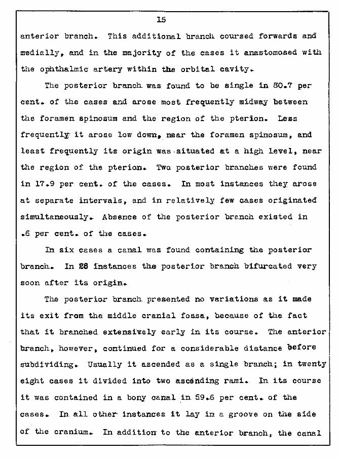
The posterior branch was found to he single in 80.7 per
cent •. of the cases and arose most frequently midway between
the foramen spinosum and the region of the pterion. Less
frequently it arose low down• near the foramen spinosum, and
least frequently its origin was .situated at a high Level, near
the region of the pterion. Twa. posterior branches were found
in 17.9 per c:ent. of the cases. In most instances they arose
a.t separate intervals,. and in relati.vely few ca.ses originated
simultaneously.. Absence of the posterior branch existed in
.5 per c:ent. of the eases.
In six cases a canal was found containing the posterior
branch. In 28 instances the posterior branch bifurcated very
soon a:Cter its origin.
The posterio.r hranch presented no variations as it made
its exit from the middle cranial fossa, because of the fact
tha.t it branched extensively early in its course.. The anterior
branch, however, continued for a considerable dis.tance \lefore
subdividing. Usually it ascended as a single branchi in twenty
eight cases it divided into two ascending rami. In its course
it was contained in a bony canal .in 59 .. 6 per cent .. o.f the
cases.. In all other instances it lay in a groove on the side
of the cranium. In addition to the anterior branch,. the canal
:t6
also contained the sphenopari.eta.l sinus in 2.3 per cent. of
the cas-es. The length o-r the canal varied from one to thirty
five millimeters:;· in most of the cases observed it measured
from five to fifteen millimeters in length. It was present
more often on the left side than the right side. In many
cases (41..0 per aent .. ) the c:ana.l was bilat.eral and in almast
one half a.s many instances it was bilaterally abs.ent ..
The reJ.ations of the entrance of the bony canal were
found to be quite variable. In o.nly 14 .• 3 per cent. of the
cases was it related directly to tJh.e pterion. In the greatest
number of cases(39..5 per cent .. ) it was situated posterior to
it, at a distance varying from five to twenty millimeters. In
those instances it was intimately related to the Sylvian pa.int.
Less frequently, yet in sufficiently large number (31..5 per
cent .. },. it was located in:Cerior to the pterion, also a-t a
distance varying :from five to twenty millimeters. In very few
instances it was found either anterior or superior to that
landmark. There is thererore no constant landmark for the
entrance of the canal.
Literature, cited
Bartlett, w.. 1.902- Contribution to the surgical anatomy of the
Gwmingham
Davis
Gray
mi-ddle cranial fos.sa. Annals of Surgery, voT.36 •
PP• 580-702
193l Text-book of Anato.my • &th ed., p.906 ..
Ox:f"ard university Press. London.
l926 Ten-book of .Applied Anatomy • 6th ed., pp .. :r
l7. u·ppincott Gompany. Philadelphia.
1.930 Text-book of Ana.tomy, 22nd ed., pp.564-565.
Lea and Fehiger. Philadelphia ..
Kanavel, !;.B .. ,, and Davis, L...E. .. Surgi:cal Anatomy of the
1\ro.rris
Rowan, J..E.
trigeminal nerve... Surgery • Gynecology • and
Obstetrics, vol .• 34, pp.357-366 ..
l932 Human Anatomy, Sth ed., pp.623-624 ..
Bla.kiston 's Son and Company. Phi.l.a.delphia.
l922 Anterior branch of the middle meningeal
artery:. its anatomical turmel and surgical
importance.. Illinois Medical Journal, vol.41,
pp.205-209.
* Yatsuta*' K .. Z .. 1.905 Surgical anatomy of the middle menigeal
artery. Vra.chebnaia Gazeta, vol.l2, pp.4.97-501.
*The article was kindly translated from the
Rus.sian by Dr.J .M..Essenberg, Associate Pnufessor
of Anatomy at Loyola Uni:versity Medical School.
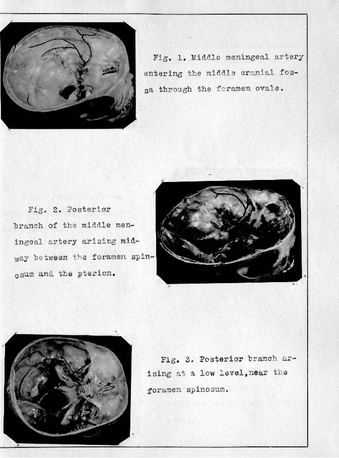
Fig. 2. Posterior
Fig . 1 . Mi ddle meningeal artery
entering the middle cranial f os
sa through the f oramen ovale.
branch of the middle men
ingeal artery arising mid
waY be tween the foramen spin-
0sum and the pterion .
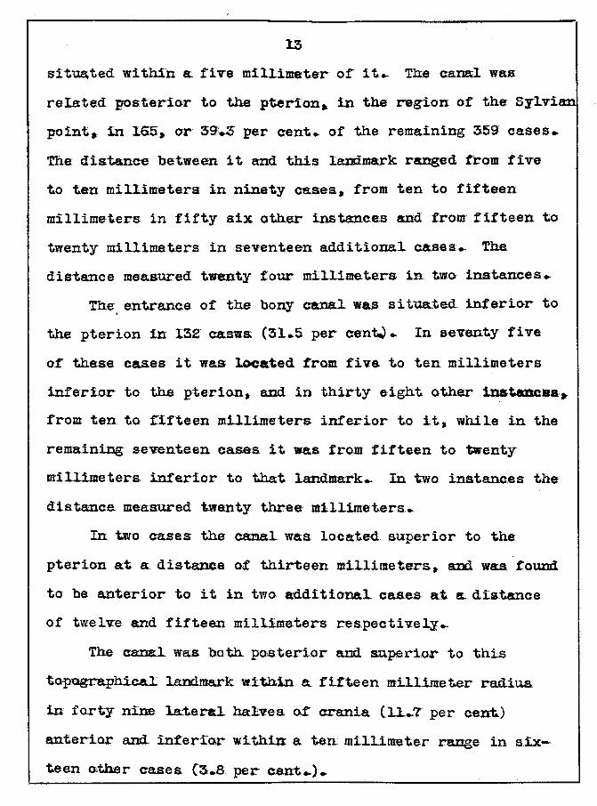
Fig. 3. Posterior branch ar
ising at a low level, near t h e
toramen spinosum.
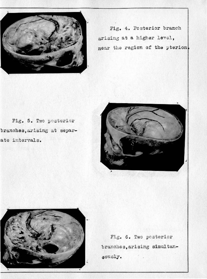
Fig. 5. Two poster i or
branches, arising a t separ
ate i n t ervals.
Fig . 4. Pos terior branch
a.r · sing at a higher le el ,
near t he region of the pterion
Fig. 6. Two pos t er ior
br anches,arising simultan
eously.
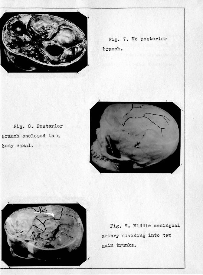
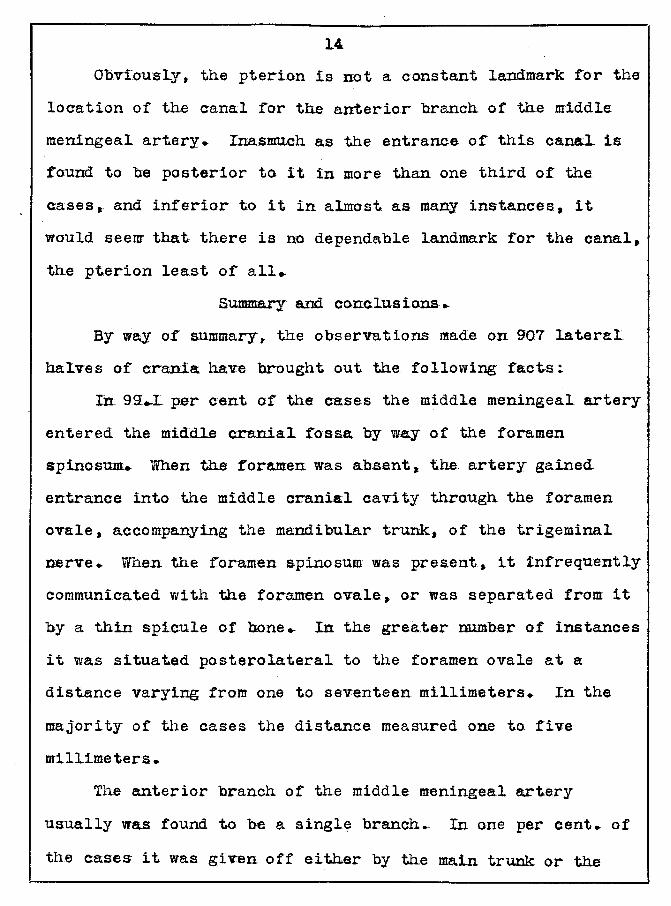
Fig. 8. Posterior
branch enclosed in a
bony canal.
Fig. 7 . No pos terior
branch .
Fig. 9. Middle meningeal
artery dividing i n to two
main trunks.
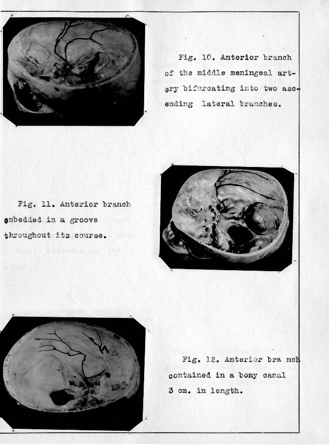
Fig. 11 . Anterior branch
omb edded in a groove
throughout its course.
Fig. 10. Anterior branch
of t he middle meningeal art
ary bifurcating i n to two asc
ending lateral branches.
Fig. 12 . Anterior bra
contained in a bony canal
3 em. in length.
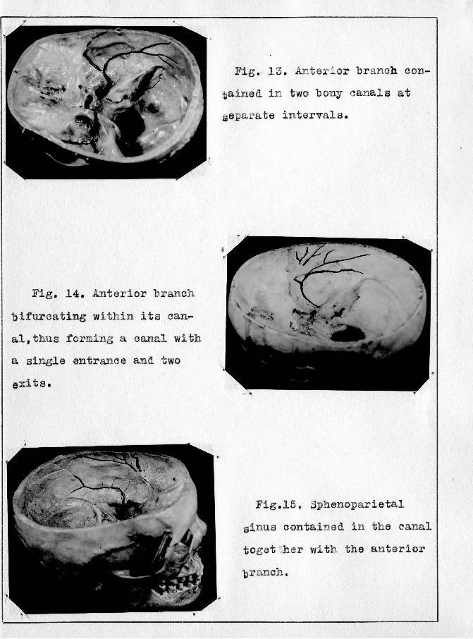
Fig. 14. Anterior branch
bifurcating within its can
al,thus :forming a canal with
a single entrance and two
exits.
Fig. 13. Anteri or branch con
tained i n two bony canal s at
separ ate intervals .
Fig.l5. Sphenoparietal
sinus contained in the canal
toget ~~-her with the an terior
cranch.