Embed Size (px)
Citation preview

Copyright 0 1986 by the Genetics Society of America
VARIATION AND GENOMIC LOCALIZATION OF GENES ENCODING DROSOPHILA MELANOGASTER MALE
ACCESSORY GLAND PROTEINS SEPARATED BY SODIUM
ELECTROPHORESIS DODECYL SULFATE-POLYACRYLAMIDE GEL
MICHAEL WHALEN AND THOMAS G. WILSON’
Department of Zoology, University of Vermont, Burlington, Vermont 05405
Manuscript received December 3 1, 1985 Revised copy accepted May 4, 1986
ABSTRACT
Accessory gland proteins from Drosophila melanogaster males have been sepa- rated by sodium dodecyl sulfate-polyacrylamide gel electrophoresis into nine major bands. When individual males from 175 strains were examined, consid- erable polymorphism for nearly one-half of the major protein bands was seen, including null alleles for three bands. Variation was observed not only among long-established laboratory strains but also among stocks recently derived from natural populations. There was little difference in the amount of variation be- tween P and M strains, indicating that P element mutagenesis is not a factor producing the variation. Codominant expression of variants for each of five bands was found in heterozygotes, suggesting structural gene variation and not posttranslational modification variation. Stocks carrying electrophoretic variants of four of the major proteins were used to map the presumed structural genes for these proteins; the loci were found to be dispersed on the second chromo- some. Since males homozygous for variant proteins were fertile, the polymor- phism seems to have little immediate effect on successful sperm transfer. We propose that a high degree of polymorphism can be tolerated because these proteins play a nutritive rather than enzymatic role in Drosophila reproduction.
PERM transfer in many animals is accompanied by accessory gland secre- S tions. Components of accessory gland secretions have been investigated in a number of insects (FUCHS and HISS 1970; PEFEROEN and DE LOOF 1984; BARKER and DAVEY 1982; BLACK, LANDERS and HAPP 1982), including Dro- sophila melanoguster (CHEN and BUHLER 1970; STUMM-ZOLLINGER and CHEN 1985). The protein fraction can be readily separated by sodium dodecyl sulfate- polyacrylamide gel electrophoresis (NaDodS0,-PAGE), and from eight to ten major bands have been found after Coomassie blue staining (STUMM-ZOLLIN- GER and CHEN 1985). The functions of these proteins are unknown, although functions for two other ejaculate proteins have been discovered, including
’ To whom reprint requests should be sent.
Genetics 114: 77-92 September, 1986.
78 M. WHALEN AND T. G. WILSON
esterase-6 (MANE, TOMPKINS and RICHMOND 1983) and a low molecular weight peptide that stimulates female oviposition (LEAHY and LOWE 1967).
A better understanding of these proteins would also help to evaluate the significance of accessory gland secretion serving as a barrier leading to or maintaining sympatric speciation. For example, it has been shown that the oviposition-enhancement peptide in D. melanogaster ejaculate (LEAHY and LOWE 1967) cannot be furnished by injection of accessory gland contents from other species into virgin D. melanogaster females (BAUMANN and CHEN 1973; FUYAMA 1983; CHEN, STUMM-ZOLLINGER and CALDELARI 1985). Even when heterospecific mating does occur between males of D. suzukii and females of D. pulchrella, a close relative, hybrid progeny are rare unless inseminated females are injected with an accessary gland extract of D. pulchrella (FUYAMA 1983). These results indicate that at least one component, presumably a pro- tein, of male accessory gland secretion is necessary for fertile heterospecific mating between Drosophila species. Since the electrophoretic patterns of ac- cessory gland proteins are species-specific (CHEN 1976; CHEN, STUMM-ZOLLIN- GER and CALDELARI 1985), perhaps additional proteins are important in re- productive isolation of Drosophila species.
Our first objective in a study of secretory proteins in D. melanogaster has been a localization of the genes controlling the major proteins that are seen on our NaDodS04-PAGE gels after silver-staining. After mapping these genes, we can determine whether they are tightly clustered, as are several chorion protein genes (SPRADLING et al. 1980) and histone genes (LIFTON et al. 1977), or dispersed, as are the larval serum protein genes (ROBERTS and EVANS- ROBERTS 1979). Since mapping these genes by recombination requires a stock carrying an electrophoretic variant of the protein for which the gene is being mapped, various D. melanogaster strains that had been collected worldwide were examined for variant proteins. Since considerable heterospecific diversity of accessory gland proteins has been found (CHEN 1976; CHEN, STUMM-ZOLLIN- GER and CALDELARI 1985), it was likely that occasional electrophoretic variants might also occur among D. melanogaster populations. In fact, we found several major proteins to be invariant and two to be represented by rare allelic var- iants. However, we did not expect the large amont of variability that was found for nearly one-half of the other major proteins. This was surprising for several reasons: (1 ) since NaDodSOe-PAGE is usually considered to be a gel sieve for protein size but not charge (WEBER and OSBORN 1969), gene deletions or insertions were implicated as a cause of the genetic variability; (2) other studies of abundant proteins in D. melanogaster and humans separated by size in the second dimension of two-dimensional electrophoretic gels showed depressed variability of these proteins (LEIGH BROWN and LANGLEY 1979; SMITH, RACINE and LANGLEY 1980); and (3) males carrying these variants in homozygous condition were fertile, indicating toleration of considerable size variation in these proteins.
The purpose of this paper is to demonstrate not only this large amount of genetic variability among D. melanogaster strains but also that the variability is

ACCESSORY GLAND PROTEIN VARIATION 79
genetically controlled. The implications of this variability in relation to a pos- sible function of these proteins will be discussed.
MATERIALS AND METHODS
D. melunogaster stocks: Two collections of wild-type strains were examined in this work. The first consisted of 100 stock constructions, each of which had a second or third chromosome derived from males caught at one of four locations in the United States in 1977 and inserted into an isogenic laboratory strain. Thus, each stock was homozygous for either a second or third chromosome from a natural population fly inserted into an isogenic background genome (LAURIE-AHLBERG et al. 1980). By using these stocks we could assess the genetic variation of 50 second and 50 third chromo- somes without the problem of variability contributed by the remaining chromosomes. Genetic variability of enzyme activity in D. melanogaster has been traced to unlinked modifiers in several studies (LAURIE-AHLBERG et al. 1980; TEPPER et al. 1984); there- fore, the possibility that electrophoretic variation could result from unlinked modifier genes cannot be disregarded.
The second collection consisted of 49 P strains and 23 M strains obtained from M. KIDWELL and categorized in her laboratory. Each strain descended from a single fertile female collected worldwide. P strains are characterized by the presence of P elements, middle repetitive DNA sequences that can move about in the genome under certain conditions; M strains lack P elements (RUBIN, KIDWELL and BINCHAM 1982; ENCELS 1983). P elements are characteristic of strains caught within the past 30 years and are usually absent from long-established laboratory strains (KIDWELL 1983). All flies were raised on a standard yeast-cornmeal-molasses-agar diet at 25 O .
Strains carrying electrophoretic variants of the major accessory gland proteins and used in this study are described in Table 1. The multiply marked second chromosome all was used for mapping studies, and its composition is described in Table 1. The reference strain for electrophoretic mobility of the gland proteins was an inbred stock of Oregon-RC.
Electrophoresis: Males were selected at 2- 10 days after eclosion for electrophoresis. The electrophoretic pattern of accessory gland proteins has been found to remain qualitatively unaltered after the first day of adult life (CHEN 1976); therefore, age differences were reflected only by differences in protein accumulation. The accessory glands and, for ease of transfer, the ejaculatory duct were dissected into Drosophila Ringer’s solution (EPHRUSSI and BEADLE 1936). Each pair of glands plus duct was transferred to a 10 pl drop of Laemmli NaDodS04-PAGE buffer (LAEMMLI 1970), which effectively lyses the glands and solubilizes the proteins. After removal of gland debris several minutes later, the sample drop was then loaded onto a 0.75 mm 5.7596, 8% or 10% acrylamide analytical gel (LAEMMLI 1970) and was electrophoresed for 2- 4 hr at 17” until the bromphenol blue dye front reached the end of the gel. The gel was fixed in methanol-acetic acid, postfixed in 6.25% glutaraldehyde and then silver- stained (MERRIL, SWITZER and VAN KEUREN 1979) or stained with Coomassie blue. We have found the glutaraldehyde fixation step necessary for reproducible and quantitative visualization of the silver-stained protein bands. Usually, the gel was destained and then restained to improve resolution of several of the bands. Acrylamide and other gel component chemicals were obtained from Biorad Corporation. Each strain was exam- ined at least twice to verify the observed gel pattern.
RESULTS
Comparison of gel stains: We needed a sensitive staining method capable of detecting accessory gland proteins from single males in order to assess genetic variability as well as simplify mapping of the genes controlling these proteins. Silver-staining the gel provided more than adequate sensitivity, but

80 M. WHALEN AND T. G. WILSON
TABLE 1
Characteristics of D. melanogaster strains used in this study
Strain Phenotype or genotype Useful characteristic Reference
Oregon-RC
W83-2
RI-02
Swedish-C +/TM3
+ISM5
+ICY0 alllall
Wild type
Wild type
Wild type-second-chromosomal substi-
Wild type Heterozygote has blunt bristles and
nicked wings due to dominant mu- tations Stubble and Serrate; homozy- gous T M 3 is lethal
Heterozygote has upturned wings due to dominant mutation Curly; homo- zygous SM5 is lethal
A balancer chromosome similar to SM5 Second chromosome (all) marked with
recessive mutations aristaless (al), dumpy (dp), black (b), purple @r), curved (c), plexus ( p x ) and speck (sp)
tution line
Reference strain for elec- trophoretic mobility
Variant alleles for both bands C and grey-1
Variant allele for band K
Variant allele for band B No recombination between
T M 3 and homologous chromosome
N o recombination between SM5 and homologous chromosome
Used to map genes for bands B, grey-I, C and K
b
~~~ ~~ ~
LINDSLEY and GRELL (1 968).
LAURIE-AHLBERG et al. (1 980). ’ Collection of M. KIDWELL.
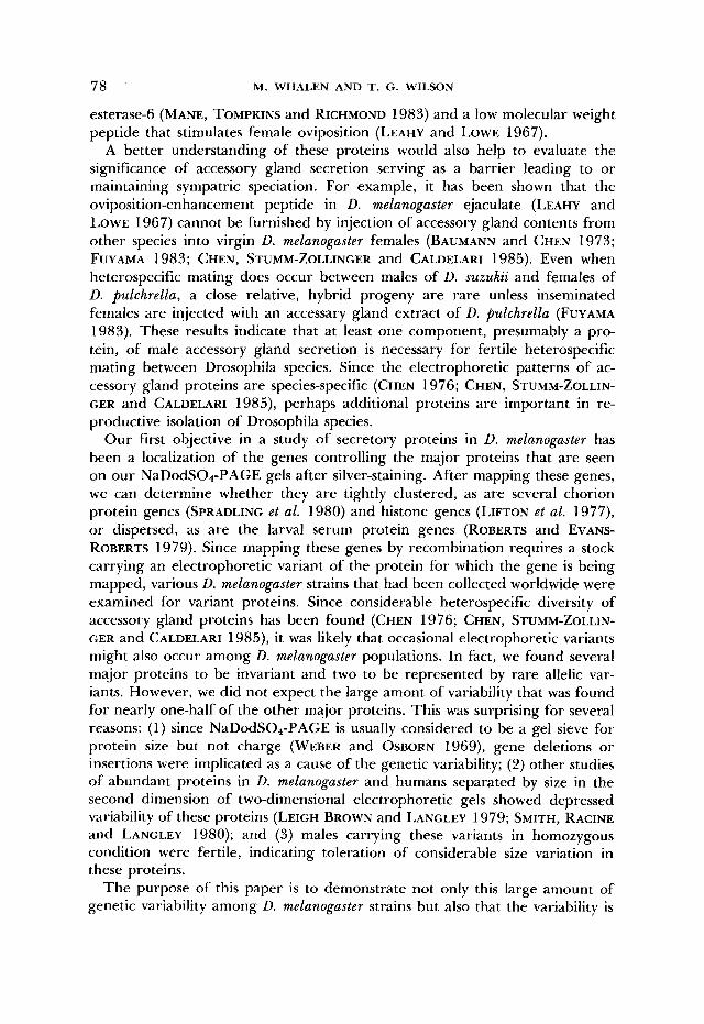
it was necessary to determine if the same protein bands that stained heavily with silver reagent would also stain heavily with Coomassie blue. If so, these bands could justifiably be termed major proteins. Therefore, samples from a male accessory gland homogenate from each of several wild-type strains were electrophoresed in duplicate under nearly identical conditions, and one gel was stained with silver reagent, the other with Commassie blue. As shown in Figure 1, staining intensity was comparable for the major bands on both gels. Silver- staining was more intense for both high and low molecular weight regions of the gel, however, and several bands stained heavily with silver but not with Coomassie blue (heavy arrows). Other bands (light arrows) designate erratic proteins that were not reproducibly observed and were not further studied.
Identification of major proteins: The sensitivity of silver-staining allowed easy visualization of the major protein bands from a single male (Figure 2). These brown-staining bands are identified by the letters A-K. Three additional major bands stain greenish-grey in color. One migrates slower than band A, another migrates between bands F and G, and the third migrates between A and B. Since this last band stained reproducibly (although poorly in Figure 1 gels), it was studied in these fly strains and is termed band grey-1 in Figure 2.
Tissue specificity: For ease of dissection and tissue transfer, each accessory gland pair with attached ejaculatory duct was typically dissected into Ringer’s solution and subsequently lysed in NaDodSOa-PAGE buffer. T o determine which proteins were specific to these tissues as well as to identify proteins in
ACCESSORY GLAND PROTEIN VARIATION 81
- E”
II
3 1 2 3 FIGURE l.--Comparison of D. mefanogasfer male accessory gland proteins separated by Na-
DodSO4-PAGE and stained with silver reagent (left gel) or Coomassie blue (right gel). Each lane contains the homogenate of one pair (left gel) or three pairs (right gel) of glands from males o f Urbana-S strain (lane 1). Oregon-RC strain (lane 2) or Swedish-C strain (lane 3). Direction of migration is top (cathode) to bottom (anode).
common with testes, various tissues from the reproductive apparatus were examined. As shown in Figure 3. nearly all of the high molecular weight proteins were found to be specific to the accessory glands. Bands F, H and I were limited to the ejaculatory duct tissue; lanes containing only accessory gland homogenate lacked these bands (not shown). Therefore, these bands appear to be either specific to ejaculatory duct tissue or were modification products from precursor protein from the ejaculatory duct or testes. Bands D, E and G appeared as minor bands in ejaculatory duct tissue (Figure 3, lane 2); possibly, these bands were derived from the accessory gland and were secreted into the ejaculatory duct. There seemed to be no similarity between major proteins specific to testes and those specific to accessory glands or ejaculatory duct. Since the presence of bands F, H and I provided convenient marker

82 M. WHALEN AND T. C. WILSON
---e-- w
FIGURE ‘L.-Silver-stained NaI)cdSO,-PAGE of male accessory gland extracts. Fach lane r e p resents an individual male honiozvgous for a second chroniosonle from a natural population (described in WHALEN 1986). except lane 6, which is a standard laboratory strain (Oregon-RC) used for reference. Gel calibration proteins consisted of six polypeptide ranging from 29 to 205 kD (Sigma Chemical kit SDSZOO). Estimated molecular masses (kD) are A, 165-170; grey-I, 145- 163; R. 1.50-140; C. 125-128; D, 104; E. 89; F, 68; G, 60; H, 50; 1. 49: J, 45; K. 43.
proteins for comparative purposes, we continued to include the ejaculatory duct in the accessory gland dissection and lysate, but we ruled out these pro- teins as accessory glandderived proteins.

ACCFSSORY GLAND PROTEIN VARIATION
- 83
Y FIGURE S.-Silver-stained NaDodSO,-PhGI.: of homogenates from accessory glands plus at-
tached ejaculatory duct (lane I ) , ejaculatory duct (lane 2) and testes (lane 3). Each lane contains one fly equivalent from a homogenate o f five males, a procedure done to minimize individual variation of quantity of protein present.
Second chromosome variability: Fach of the 50 homozygous secondchro- mosomal stocks was examined, and a representative gel is shown in Figure 2 for 1 1 of the strains. On this particular gel, four variants (lanes 3, 4, 7, 10) for band A are apparent, six (lanes 1-4, 7, 9) for band grey-1 (including a null), six (lanes 7-12) for band B, four (lanes 1, 2, 4, 7) for band C, two (lanes 7, 8) for J and three (lanes 3, 4, 7) for K (including a null). Bands B, I and K appear as single bands on 210% acrylamide gels, but can be resolved into four closely spaced bands (B) or doublets (I and K) with the use of a lower (<8%) percentage of acrylamide gel. These subbands migrate as a family of bands, and a particular variant results in a migration shift of the entire family (Figure 2). We assume that each family of bands represents either posttranslational modification of a single gene product or perhaps an event such as differential R N A splicing of a single transcript (MAEDA et al. 1985). but we cannot rule o u t the possibility of breakdown of a single polypeptide during sample prepa-

84 M. WHALEN AND T. G. WILSON
TABLE 2
NaDodSOd-PAGE variants for six major proteins from D. melunogaster male accessory glands
No. of variants
Second chromo- Third chromo- Band soma1 strains soma1 strains P strains M strains
A 4 1 3 3 Grey- 1 6 1 6 5 B 11 1 2 7 2 7 C 4 1 5 3
2 1 2 1 K 3 1 2 2 J
A minimum estimate is given for band B in the P and M strains since heterozygosity precluded a positive allele identification in several instances. T h e Drosophila strains are more fully described elsewhere (WHALEN 1986).
ration, The band patterns of all strains carrying a variant protein were care- fully compared to determine if each variant band was expressed independently of the remaining bands. In no other instance was a variant band associated with a position change of one or more other bands. Therefore, it appears that, with the exception of members of the B-, I- and K-band families, the remaining polymorphic bands represent single gene products.
An analysis of the 50 second-chromosomal stocks revealed a surprising num- ber of different electrophoretic variants for bands A, grey-1, B and C (Table 2). If these variants were separated by size during the electrophoresis, then they represent protein size variations of as much as 20 kilodaltons (kD) (band grey-1). Null alleles for bands grey-1 and K were found. Accessory gland bands D, E and G and ejaculatory duct bands F, H and I were monomorphic, sug- gesting either no genetic variation for size was present or that genes on the second chromosome do not control electrophoretic variability of these proteins.
Third chromosomal variability: When the 50 homozygous third chromo- somal stocks were examined, no variants were found for any major band (Table 2). Thus, genes affecting the mobility of these bands were not located on the third chromosome. This invariability had two implications: (1) extrachromo- somal modifiers on the third chromosome could be disregarded as additional sources of variation, and (2) the monomorphic bands are either truly mono- morphic or else variants of these bands are controlled by genes on either the X-chromosome or fourth chromosome. P and M strains: Additional strains were examined to verify the polymor-
phism observed among the previous strains, as well as to search for X-chro- mosomal and fourth chromosomal variation. Since these strains were simply cultures derived from single fertilized females, variability from all chromo- somes was possible. Also, these strains had been classified into P and M strains (M. KIDWELL), and a comparison of the variability between these strain types could also indicate the impact of mobile genetic elements on band polymor- phism, since movement of P elements can result in elevated mutation rates
ACCF-WRY GLAND PROTEIN VARIATION 85
- 1 2 3 4
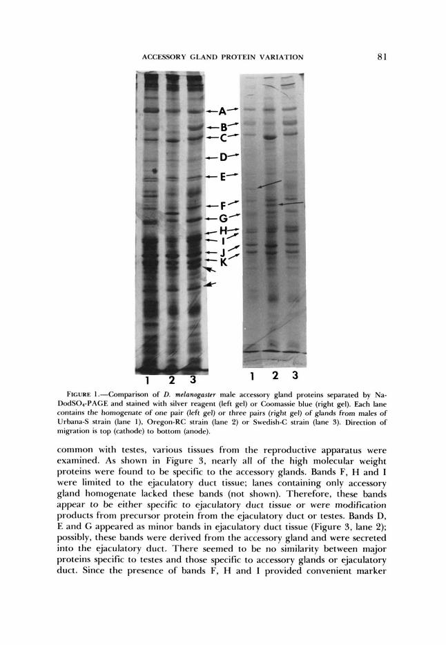
FIGURE 4.-A portion o f a silver-stained SaDo<lS( ),-PAGE showing codominant expression of band C variants. l ane I , heterozygous oll/SAI5 male having the Fast allele (C3) derived from the S M 5 chromosome; lane 4. stock W83-2: lanes 2 and 3, Mf83-2/SM5 males.
(KIDWELL. KIDWELL and SVED 1977). including deletions and insertions (SIM- MONS and LIM 1980; GREEN 1982; LEVIS, O'HARE and RURIN 1984). T h e results showed variability for bands A, grey-I, B, C and K, including the presence of several new alleles for B and C as well as a null allele for C not present in the previously examined stocks. Variability was found for these bands in wild-type stocks maintained in the laboratory for the last 50 years, as well as in stocks derived from wild-caught flies within the past several years. There seemed to be little difference between the band variability of the P and M strains (Table 2). T h e monomorphic bands remained so in these stocks, indicating that neither the X-chromosome nor the fourth chromosome is a source of variation for these proteins; thus, they appear to be truly mono- morphic on NaDodSO.,-PAGE.
Codominance of expression: If the genetic variation is due to mutation within the structural gene coding for each of these proteins and not to varia- tion of a posttranslational event acting on a monomorphic gene product, one would expect the alleles to be expressed codominantly in heterozygotes. This prediction proved true for heterozygotes constructed from flies carrying var- iants for the following proteins: band A, grey-I, B, C and K. Band J could not be evaluated due to poor resolution of variant bands in heterozygotes. Figure 4 shows codominant expression of two alleles controlling band C. These results had two implications: ( 1) posttranslational modification variation could be discounted as the cause of the observed polymorphism in these bands, since codominant expression would not be expected with this possibility; and (2) the X-chromosome could be eliminated as a source of polymorphism, since males carry only one X-chromosome. T h e simplest interpretation is that the variabil- ity of these proteins results from structural gene lesions.
Localization of genes: Examination of chromosome substitution stocks re- vealed polymorphism for bands A, grey-I, B, C, J and K in males only from the second chromosome stocks, and examination of 75 additional laboratory and wild-caught strains showed no additional major proteins to be polymorphic
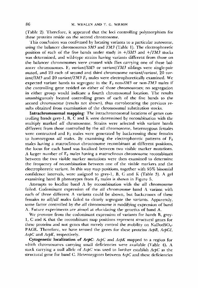
86 M. WHALEN AND T. G. WILSON
(Table 2). Therefore, it appeared that the loci controlling polymorphism for these proteins reside on the second chromosome.
This conclusion was confirmed by locating variants to a particular autosome, using the balancer chromosomes SM5 and TM3 (Table 1). The electrophoretic position of each of the five bands under study in +/SA45 and +/TM3 stocks was determined, and wild-type strains having variants different from those on the balancer chromosomes were crossed with flies carrying one of these bal- ancer chromosomes. F1 variant/SM5 or variant/TM3 siblings were single-pair mated, and 20 each of second and third chromosome variant/variant, 20 var- iant/SMS and 20 variant/TM3 FP males were electrophoretically examined. We expected variant bands to segregate in the F2 non-SMS or non-TM? males if the controlling gene resided on either of those chromosomes; no segregation in either group would indicate a fourth chromosomal location. The results unambiguously located controlling genes of each of the five bands to the second chromosome (results not shown), thus corroborating the previous re- sults obtained from examination of the chromosomal substitution stocks.
Intrachromosomal mapping: The intrachromosomal locations of genes con- trolling bands grey-I, B, C and K were determined by recombination with the multiply marked all chromosome. Strains were selected with variant bands different from those controlled by the all chromosome, heterozygous females were contructed and F2 males were generated by backcrossing these females to homozygous all males. By examining the electrophoretic patterns of F2 males having a matroclinous chromosome recombinant at different positions, the locus for each band was localized between two visible marker mutations. A larger number of F2 males having a matroclinous chromosome recombinant between the two visible marker mutations were then examined to determine the frequency of recombination between one of the visible markers and the electrophoretic variant. In this way map positions, together with 95% binomial confidence intervals, were assigned to grey-1, B, C and K (Table 3). A gel examining band B phenotypes from F2 males is shown in Figure 5 .
Attempts to localize band A by recombination with the all chromosome failed. Codominant expression of the all chromosome band A variant with each of three different A variants could be shown, but backcrosses of these females to all/all males failed to clearly segregate the variants. Apparently, some factor controlled by the all chromosome is modifying expression of band A. Future experiments are aimed at elucidating the genetics of band A.
We presume from the codominant expression of variants for bands B, grey- 1, C and K that the recombinant map positions represent structural genes for these proteins and not genes that merely control the mobility on NaDodS04- PAGE. Therefore, we have termed the genes for these proteins AcpB, AcpGI, AcpC and AcpK, respectively.
Cytogenetic localization of AcpC: AcPC and AcpK mapped to a region for which chromosomes carrying small deficiencies were available (Table 4). A stock carrying a null allele of AcpC was used to further establish AcpC as the structural gene for band C . Heterozygotes between AcpC and these deficiencies

ACCFSORY GLAND PROTEIN VARIATION 87 TABLE 3
Recombinational map positions on chromosome 9 of D. melanogastcr for four major accessory gland proteins
Protein No. of Calculated map 95% confidence
Recombinant class recombinants location interval
Grey- I a1 dp S + a1 dp F +
3 13.5 12.9- 14.1 207
42.8 41.2-44.4 U + F b pr c px sp 41 216 + S b pr c px sp
C
K
a1 dp b S + a1 dp b F +
+ F fir c fix sfi + s pr c px sp
149 51
5 75
53.0 52.6-53.4
54.1 53.8-54.4 . . . F and S refer to fast and slow variants for each protein for which the gene is being mapped.
+ indicates all remaining chromosomal markers a re wild-type alleles. T h e variants carried by the all chromosomes are Grey-l , F; U, S; C. F; K. F. T h e map positions of marker mutations used in the calculations are: dp. 13.0; b. 48.5; and pr. 54.5 (LINDSLEY and GRELL 1968).
B
1 5 10 15 FIGURE 5.--Silver-stained NaDodSO,-PAGE of accessory gland proteins from individual + F/S
b pr c px +/a1 dp S b pr c px sp males, identifying the band U allele in each recombinant second chromosome. Recombinant chromosomes carrying the F alleles (from a Swedish4 chromosome) appear as heterozygotes (e.g., lane 3). whereas those carrying the S allele (from the all chromosome) appear as homozygotes (e.g.. lane 1) with the S allele from the all chromosome.
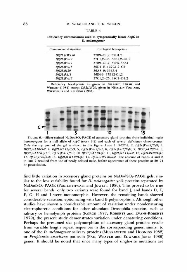
were examined (Figure 6). It is clear that AcpC is uncovered by the region 36D1-36E3,4 and that AcpC is the structural gene for band C.
DISCUSSION
Numerous studies have assessed genetic variability by examining the electro- phoretic mobility of proteins. T h e vast majority of these separations have been under nondenaturing conditions, during which the net charge of a protein determines its mobility. Isozymes a re usually readily separated under these conditions, and polymorphism of many enzymes has been found to be consid- erable, especially among invertebrates (AYALA 1984). Recently, the variability of abundant, presumably nonenzymatic, proteins has been assessed on two- dimentional gels and found to be relatively low, for example, in whole D. melunoguster (LEIGH BROWN and LANGLEY 1979) and human kidney samples (SMITH, RACINE and LANGLEY 1980). Since two-dimensional gels separate pro- teins on the basis of size as well as charge, we expected from these studies to
88 M. WHALEN AND T. G. WILSON
TABLE 4
Deficiency chromosomes used to cytogentically locate AcpC in D. melanogmter
Chromosome dmignarion Cytological hrmkpoincs ~~
Dfi2L)TWI30 37B9-C1.2: 37D1.2 Dfi2L)VA I2 37C1.2-C5: 38R1.2-C1.2 Df(2L)VA17 37B9-CI ,2; 37F5-38A I Df(2L)VA 18 SGDI-EI; 37CI , 2 4 3 Df(2L)H20 36A8-9; 36E3,4 f)f(2L)hh18 36E4-6; 37B12-C1.2 Dfl2L)VA13 37C1.2-C5; 38C1 -DI .2
Deficiency breakpoints as given in GILBERT. HIRSH and WRICHI' ( 1 984) except Dfi2L)H20. given in N ~ ~ L E I N - V O L H A R D . WIFSCHAUS and KLUI)ING ( 1 984).
c I
c D - - - Iu
C- 1 5 10 15
FIGURE 6.--Silver-stained NaDodSO,-PAGE of accessory gland proteins from individual males heterozygous for a null allele of ArpC (stock S 2 ) and each of several deficiency chromosomes. Only the top part of the gel is shown in this figure. Lane I . S-2/S-2; 2, Dfl2L)VA18/CyO: 3, Dfl2L)VA 18/S-2; 4. D!2L)VA12/CyO: 5 , Dfl2L)VA12/S-2; 6. Dfl2L)hh18/CyO: 7. Dfl2L)hh18/S-P; 8. Dfl2L)VA I7/CyO; 9. Dfl2L)VA I7/S-2: IO. Dfl2L)VA 13/CyO; 1 I , Dfl2L)VA 13/S-2; 12, Dfl2L)H2O/CyO: 13, Dfl2L)H2O/S-2; 14. Dfl2L))Tw130/CyO; 15, Dfl2L)7W13O/S-2. The absence of bands A and B in lane 2 resulted from use of newly eclosed male, before appearance of these proteins at 20-24 hr posteclosion.
find little variation in accessory gland proteins on NaDodS04-PAGE gels, sim- ilar to the low variability found for D. melanogaster yolk proteins separated by NaDodS04-PAGE (POSTLETHWAIT and JOWETT 1980). This proved to be true for several bands: only two variants were found for band J, and bands D, E, F, G , H and I were monomorphic. However, the remaining bands showed considerable variation, epitomizing with band B polymorphism. Although other studies have shown a considerable amount of variation under nondenaturing electrophoretic conditions for other abundant Drosophila proteins, such as salivary or hemolymph proteins (KORCE 1977; ROBERTS and EVANS-ROBERTS 1979). the present study demonstrates variation under denaturing conditions. Perhaps the presumed size polymorphism of accessory gland proteins results from variable length repeat sequences in the corresponding genes, similar to one of the D. melanogaster salivary proteins (MUSKAVITCH and HOCNFS~ 1982) or Periplaneta americana oothecin (PAu, WEAVER and EDWARDS-JONES 1986) genes. I t should be noted that since many types of single-site mutations are

ACCESSORY GLAND PROTEIN VARIATION 89
not detected by NaDodSO,-PAGE, our method underestimates the variability in these proteins.
The dogma of NaDodS0,-PAGE dictates that the variation seen in these proteins is due to size differences, presumably resulting from deletions or insertions in the structural genes. However, since certain types of single-site mutations involving noncharged amino acid substitutions have been shown to result in altered mobility of the lesioned protein on NaDodS04-PAGE (DE JONG, ZWEERS and COHEN 1978; NOEL, NIKAIDO and AMES 1979), it is possible that the variation results from point mutations of this type and not from deletions. Further work will be necessary to distinguish between these inter- pretations of the observed variation.
In this work we have utilized electrophoretic variants for four of the major accessory gland proteins to map the loci controlling their mobility on Na- DodS04-PAGE. Since each of the variant proteins for bands A, grey-1, B, C and K are expressed codominantly in heterozygotes, it appears likely that the variation seen is due to variation in the structural genes encoding these pro- teins and not to posttranslational events. This evidence is strongest for band C, for which deficiency chromosomes were identified that uncovered the gene for this protein and deleted the protein entirely in deJficiency/nulZ males.
It is clear that the genes controlling the variability map to the second chro- mosome. This is evident not only from examination of the second- and third- chromosomal substitution lines but also by recombinational mapping in the present study. Since there is no obvious reason to maintain evolutionarily the accessory gland protein genes on the second chromosome, this localization suggests that these genes might be in the process of dispersing from a more tightly clustered gene family and becoming completely dispersed throughout the genome. Until more is known of the homology and function of these proteins, however, little can be concluded from the genomic locations.
The second- and third-chromosomal substitution stocks were constructed by crossing males from natural populations with females having chromosomes derived from long-established laboratory stocks (LAURIE-AHLBERG et al. 1980). Concerns have been raised by several investigators (VOELKER et al. 1980; TEP- PER et al. 1984) that genetic variability in these and similarly constructed stocks, instead of reflecting naturally occurring variation from the paternally derived chromosomes, might result from P-element mutagenesis induced dur- ing stock construction. This P-element mutation possibility can be discounted in the present study, however. Variability in accessory gland proteins as re- flected by migration changes on NaDodS04-PAGE gels was seen only among the second chromosomal substitution lines. If P-element mutagenesis occurring during the stock constructions contributed to the observed band variability, then band variation would have been equally as likely in the third-chromosome substitution lines, and this result was not seen. A final implication of the lack of variation of the third-chromosome lines is that the stocks were accurately constructed and have not undergone breakdown since their construction in 1978.
The high variability observed for some of these proteins may also offer clues

90 M. WHALEN AND T. G. WILSON
as to their function. Elevated polymorphism has been interpreted as more permissible for functionally less important proteins (KIMURA and OHTA 1974). If this idea is extended to the present work, then the polymorphic accessory gland proteins may be nonenzymatic proteins, the structure of which can tol- erate considerable size or amino acid variation without loss of function. We suggest that the function of these proteins is nutritive, serving as a source of amino acids for the female or her oocytes. In indirect support of this idea, we wish to offer three findings: (1) A nutritive role for male accessory gland products has been demonstrated in other insects (FRIEDEL and GILLOTT 1977; BOGGS and GILBERT 1979; SCHAL and BELL 1982). (2) Following insemination, male accessory gland proteins have been shown capable of being rapidly trans- ported into the female hemolymph in grasshoppers (FRIEDEL and GILLOTT 1977) and D. melanogaster (MANE, TOMPKINS and RICHMOND 1983), indicating that the ejaculated proteins can leave the seminal recepticle, pass into the female’s hemolymph and be utilized there or incorporated, either intact or broken, down into the oocyte. (3) The extreme species divergence, even for sibling species, of Drosophila male accessory gland proteins as seen on Na- DodS04-PAGE (CHEN 1976) argues against specific enzymatic functions of these proteins as necessary for successful sperm transfer. To this diversity must now be added D. melanogaster intraspecific diversity, including null alleles at three loci, that apparently has little immediate effect on male fertility. If, however, the sole function of many of these proteins is a source of amino acids for the female or for oogenesis, it is not surprising that considerable polymor- phism can be tolerated.
We wish to thank C. LAURIE-AHLBERG, M. KIDWELL and T. WRIGHT for supplying stocks; J. HERBERS and W. KILPATRICK for comments on the manuscript. Supported in part by an institu- tional award from the University of Vermont and by a grant from the National Institutes of Health. This work was a portion of an M.S. thesis submitted by M.W. to the Cell Biology Program at the University of Vermont.
LITERATURE CITED
AYALA, F. J., 1984
BARKER, J. F. and K. G. DAVEY, 1982
BAUMANN, H. and P. S. CHEN, 1973
Molecular polymorphism: how much is there and why is there so much? Dev.
Intraglandular synthesis of protein in the transport acces-
Geschlechtsspezifische, ninhydrinpositive Substanzen in Ad-
Cytodifferentiation in the accessory glands of Tenebvio molitor VIII. Crossed immunoelectrophoretic analysis of terminal differentiation in the postecdysial tubular accessory glands. Dev. Biol. 9 4 106-1 15.
Male contribution to egg production in butterflies: evidence for transfer of nutrients at mating. Science 206: 83-84.
Species specific protein patterns in Drosophila paragonial glands. Experientia 32: 549-551.
Paragonial substance and other ninhydrin-positive components
Genet. 4 379-391.
sory reproductive gland in the male of Rhodnius prolixus. Insect Biochem. 12: 157-159.
ultmannchen von Drosophila funebris. Rev. Suisse Zool. 8 0 685-690.
BLACK, P. N., M. H. LANDERS and G. M. HAPP, 1982
BOGGS, C. L. and L. E. GILBERT, 1979
CHEN, P. S., 1976
CHEN, P. S. and R. BUHLER, 1970 in male and female adults of Drosophila melanogaster. J. Insect Physiol. 1 6 615-627.

ACCESSORY GLAND PROTEIN VARIATION 91
CHEN. P. S.. E. STUMM-ZOLLINGER and M. CALDELARI, 1985 Protein metabolism of Drosofihila male accessory glands-11. Species-specificity of secretion proteins. Insect Biochem. 1 5 385- 390.
DE JONG, W. W., A. ZWEERS and L. H. COHEN, 1978 Influence of single amino acid substitutions on electrophoretic mobility of sodium dodecyl sulfate-protein complexes. Biochem. Biophys. Res. Commun. 82: 532-539.
ENGELS, W. R., 1983 The P family of transposable elements in Drosophila. Annu. Rev. Genet.
EPHRUSSI, B. and G. W. BEADLE, 1936 A technique of transplantation for Drosophila. Am. Nat.
FRIEDEL, T. and C. GILLOTT, 1977 Contributions of male-produced proteins to vitellogenesis in
FUCHS, M. S. and E. A. HIS, 1970 The partial purification and separation of the protein com-
FUYAMA, Y., 1983 Species-specificity of paragonial substances as an isolating mechanism in Dro-
GILBERT, D. G., J. HIRXH and T. R. F. WRIGHT, 1984 Molecular mapping of a gene cluster
GREEN, M. M., 1982 Genetic instability in Drosophila melanogaster: deletion induction by insertion
KIDWELL, M. G., 1983 Evolution of hybrid dysgenesis determinants in Drosophila melanogaster.
KIDWELL, M. G., J. F. KIDWELL and J. A. SVED, 1977 Hybrid dysgenesis in Drosophila melano- gaster: a syndrome of aberrant traits including mutation, sterility, and male recombination. Genetics 86: 813-833.
KIMURA, M. and T. OHTA, 1974 On some principles governing molecular evolution. Proc. Natl.
KORGE, G., 1977 Larval saliva in Drosophila melanogaster: production, composition, and relation- ship to chromosome puffs. Dev. Biol. 5 8 339-355.
LAEMMLI, U. K., 1970 Cleavage of structural proteins during the assembly of the head of bac- teriophage T4. Nature 227: 680-685.
LAURIE-AHLBERG, C. C., G. MARONI, G. C. BEWLEY, J. C. LUCCHESI and B. S. WEIR, 1980 Quantitative genetic variation of enzyme activities in natural populations of Drosophila melanogaster. Proc. Natl. Acad. Sci. USA 77: 1073-1077.
Purification of the male factor increasing egg deposition in D. melanogaster. Life Sci. 6 151-156.
Reevaluation of genic heterozygosity in a natural population of Drosophila melanogaster by two-dimensional electrophoresis. Proc. Natl. Acad. Sci. USA 76: 2381-2384.
Effects of transposable element insertions on RNA encoded by the white gene of Drosophila. Cell 38: 471-481.
The organization of the histone genes in Drosophila melanogaster: functional and evolutionary implication. Cold Spring Harbor Symp. Quant. Biol. 42: 1047-1051.
Genetic variations of Drosophila melanogaster. Carnegie Inst. Wash. Publ. 627.
Differential RNA splicing and post-
17: 315-344.
7 0 218-225.
Melanoplus sanguinipes. J. Insect Physiol. 23: 145-1 5 1 .
ponents of matrone from Aedes aegypti. J. Insect Physiol. 16: 913-939.
sophila. Experientia 3 9 190-192.
flanking the Drosophila dopa decarboxylase gene. Genetics 106: 679-694.
sequences. Proc. Natl. Acad. Sci. USA 7 9 5367-5369.
Proc. Natl. Acad. Sci. USA 8 0 1655-1659.
Acad. Sci. USA 71: 2848-2852.
LEAHY, M. G. and M. L. LOWE, 1967
LEIGH BROWN, A. J. and C. H. LANGLEY, 1979
LEVIS, R., K. O'HARE and G. M. RUBIN, 1984
LIFTON, R. P., M. L. GOLDBERG, R. W. KARP and D. S. HOGNESS, 1977
LINDSLEY, D. L. and E. H. GRELL, 1968
MAEDA, N., H. S. KIM, E. A. AZEN and 0. SMITHIES, 1985
92 M. WHALEN AND T. G. WILSON
translational cleavages in the human salivary proline-rich protein gene system. J. Biol. Chem. 260: 11123-11130.
Male esterase 6 catalyzes the synthesis of a sex pheromone in Drosophila melanogaster females. Science 222: 419-421.
Trace polypeptides in cellular extracts and human body fluids detected by two-dimensional electrophoresis and a highly sensitive silver stain. Proc. Natl. Acad. Sci. USA 76: 4335-4339.
MANE, S. D., L. TOMPKINS and R. C. RICHMOND, 1983
MERRIL, C. R., R. C. SWITZER and M. L. VAN KEUREN, 1979
MUSKAVITCH, M. A. T. and D. S. HOGNESS, 1982 An expandable gene that encodes a Drosophila glue protein is not expressed in variants lacking remote upstream sequences. Cell 29: 1041- 1051.
NOEL, D., K. NIKAIDO and G. F. L. AMES, 1979 A single amino acid substitution in a histidine- transport protein drastically alters its mobility in sodium dodecyl sulfate-polyacrylamide gel electrophoresis. Biochemistry 18: 4 159-41 65.
Mutations affecting the pattern of the larval cuticle in Drosophila melanagaster 1. Zygotic loci on the second chromosome. Wilhelm Roux Arch. Entwicklungsmech. Org. 193: 267-283.
Regulation of cockroach oothecin synthesis by juvenile hormone. Arch. Insect Biochem. Physiol. (Suppl. 1): 59-73.
lntraglandular and extraglandular synthesis of proteins secreted by the accessory reproductive glands of the Colorado potato beetle, Leptinotarsa decemlineata. Insect Biochem. 1 4 407-416.
Genetic analysis of the hormonally regulated yolk
The genetic and cytogenetic localization of the
NUSSLEIN-VOLHARD, C., E. WIESCHAUS and H. KLUDING, 1984
PAU, R. N., R. J. WEAVER and K. EDWARDS-JONES, 1986
PEFEROEN, M. and A. DE LOOF, 1984
POSTLETHWAIT, J. H. and T . JOWETT, 1980
ROBERTS, D. B. and S. EVANS-ROBERTS, 1979
polypeptide genes in D. melanogaster. Cell 20: 671-678.
three structural genes coding for the major protein of Drosophila serum. Genetics 93: 663- 679.
RUBIN, G. M., M. G. KIDWELL and P. M. BINGHAM, 1982 The molecular basis of P-M hybrid
SCHAL, C. and W. J. Bell, 1982 Ecological correlates of paternal investment of urates in a tropical
SIMMONS, M. T. and J. K. LIM, 1980 Site specificity of mutations arising in dysgenic hybrids of
SMITH, S. C., R. R. RACINE and C. H. LANGLEY, 1980 Lack of genic variation in the abundant
SPRADLING, A. C., M. E. DIGAN, A. P. MAHOWALD, M. SCOTT and E. A. Craig, 1980 Two
STUMM-ZOLLINGER, E. and P. S. CHEN, 1985
dysgenesis: the nature of induced mutations. Cell 29: 987-994.
cockroach. Science 218: 170-173.
Drosophila melanogaster. Proc. Natl. Acad. Sci. USA 77: 6042-6046.
proteins of human kidney. Genetics 96: 967-974.
clusters of genes for major chorion proteins of Drosophila melanogaster. Cell 1 9 905-914. Protein metabolism of Drosophila melanagaster male
accessory glands-I. Insect Biochem. 15: 375-383. Studies of esterase 6 in
Drosophila melunogaster XIV. Variation of esterase 6 levels controlled by unlinked genes in natural populations. Genet. Res. 43: 181-190.
VOELKER, R. A., C. H. LANGLEY, A. J. LEIGH-BROWN, S. OHNISHI, B. DICKSON, E. MONTGOMERY and S. C. SMITH, 1980 Enzyme null alleles in natural populations of Drosophila melanogaster: frequencies in a North Carolina population. Proc. Natl. Acad. Sci. USA 77: 1091-1095.
The reliability of molecular weight determinations by dodecyl sulfate-polyacrylamide gel electrophoresis. J. Biol. Chem. 244: 4406-44 12.
Genetic analysis of the accessory gland proteins of Drosophila melanogaster. M. S. Thesis, University of Vermont, Burlington.
Communicating editor: V. G. FINNERTY
TEPPER, C. S., A. L. TERRY, J. E. HOLMES and R. C. RICHMOND, 1984
WEBER, K. and M. OSBORN, 1969
WHALEN, M., 1986