Embed Size (px)
Citation preview
Variable motif utilization in homeotic selector(Hox)–cofactor complex formation controls specificityKatherine M. Lellia, Barbara Norob,1, and Richard S. Mannb,2
Departments of aGenetics and Development and bBiochemistry and Molecular Biophysics, Columbia University Medical Center, New York, NY 10032
Edited* by Eric H. Davidson, California Institute of Technology, Pasadena, CA, and approved November 18, 2011 (received for review August 29, 2011)
Homeotic selector (Hox) proteins often bind DNA cooperativelywith cofactors such as Extradenticle (Exd) and Homothorax (Hth)to achieve functional specificity in vivo. Previous studies identifiedthe Hox YPWM motif as an important Exd interaction motif. Usinga comparative approach, we characterize the contribution of thisand additional conserved sequence motifs to the regulation ofspecific target genes for three Drosophila Hox proteins. We findthat Sex combs reduced (Scr) uses a simple interaction mechanism,where a single tryptophan-containing motif is necessary for Exd-dependent DNA-binding and in vivo functions. Abdominal-A (AbdA)is more complex, using multiple conserved motifs in a context-dependent manner. Lastly, Ultrabithorax (Ubx) is the most flexible,in that it uses multiple conserved motifs that function in parallel toregulate target genes in vivo. We propose that using differentbinding mechanisms with the same cofactor may be one strategyto achieve functional specificity in vivo.
Understanding the molecular processes by which gene ex-pression is regulated remains at the core of many biological
questions. The predominant model of eukaryotic gene regulationemphasizes the role of site-specific transcription factors in targetgene selection. The initial binding of these transcription factorsanchors the rest of the transcriptional regulatory complex, orenhanceosome, to the target site. Recruitment of additionalproteins is often required to determine the regulatory sign,whether a gene is activated or repressed, and if this state will bemaintained. In some cases, the DNA sequence can provideconsiderable insight into which other proteins are recruited (1).However, enhanceosome formation also requires protein-pro-tein interactions: Mutational analyses of transcription factorsdemonstrate that sequences outside of the DNA-binding domaincan influence regulatory activity, in part, by influencing the as-sembly of DNA-bound protein complexes (2, 3).The homeotic selector (Hox) proteins provide a powerful
system in which to study the role of protein-protein interactionsin enhanceosome formation and transcription factor function.Best known for their role in anterior-posterior patterning, Hoxproteins contain a highly conserved DNA-binding domain,termed the homeodomain (4). Although most homeodomainsbind similar AT-rich sequences in vitro (5), each Hox proteindisplays a high level of functional specificity in vivo (6). Theseobservations imply that residues outside of the DNA-bindingdomain influence specificity in vivo. One way Hox proteinsachieve higher specificity is through cooperative interactions withDNA-binding cofactors, such as Extradenticle [Exd in Drosoph-ila, Pre-B-cell leukemia homeobox (Pbx) in vertebrates] andHomothorax [Hth in Drosophila, Myeloid ecotropic viral in-tegration site (Meis) in vertebrates] (7, 8). Exd and Hth, bothmembers of the three-amino acid loop extension (TALE) familyof homeodomain proteins, are obligate dimer partners for bothnuclear translocation and transcriptional activity in vivo (9, 10).Previous genetic analyses highlight the importance of exd and hthfor Hox function during embryogenesis (10–12). In additionto Exd and Hth, the abdominal Hox proteins Ultrabithorax(Ubx) and AbdominalA (AbdA) have the ability to bind co-operatively with another homeodomain protein, Engrailed (En)(1). As with Exd-Hth-Hox interaction, En–Hox–DNA complexformation has been shown to be critical for Hox-mediated generegulation in vivo (1).
Biochemical and X-ray crystallography studies identified thehighly conserved Hox motif called YPWM as one mode by whichHox proteins interact with Exd-Hth. However, despite beingevolutionarily conserved and present in most Hox proteins, theimportance of the YPWM motif appears to vary (2). For ex-ample, although vertebrate Hoxa1 and Deformed (Dfd) requirethe YPWMmotif for Pbx/Exd-dependent functions (13, 14), Ubxand AbdA do not require YPWM for some Exd-dependentfunctions (15–19). In the case of Ubx and AbdA, a shared six-amino acid motif C-terminal to the homeodomain, termedUbdA, has also been suggested to contribute to interactions withExd (17, 18, 20). Interestingly, in addition to UbdA, both Ubxand AbdA have other evolutionarily conserved residues in the Cterminus that may also be important for mediating Hox functionsin vivo (16, 21–23). These C-terminal sequences, which are dis-tinct in Ubx and AbdA, could modify UbdA-dependent inter-actions so that its function may not be equivalent in bothproteins. Further, the presence of multiple Exd interactionmotifs poses the question of what determines which mode ofinteraction is most relevant for a given in vivo function. In thecase of Ubx, both the protein context and the target site havebeen suggested to influence how individual motifs are used (18).Currently, it is generally unknown how different modes of
cofactor interaction influence Hox specificity. In the case of Sexcombs reduced (Scr), structural studies demonstrated that theYPWM-Exd interaction helps position amino acids of the Hoxlinker region, which separates YPWM from the homeodomain,to make critical contacts in the minor groove of a specific DNA-binding site (24). These observations raise the possibility thatother modes of Exd-Hox interaction could also alter the way inwhich these protein complexes recognize and bind to their targetDNA sequences. In addition, alternate modes of protein–DNAcomplex formation may have an impact on the recruitment ofcoactivators and corepressors, as has been suggested for theglucocorticoid receptor (25).In the current study, we use a comparative approach to
characterize conserved sequence motifs for three Hox proteinsand analyze their requirement for different in vivo functions. Wedemonstrate that the YPWM motif is critical for Scr to carry outExd-dependent functions in vivo. In contrast to the single Exdinteraction motif of Scr, AbdA and Ubx are more complex. Inaddition to the previously described YPWM and UbdA motifs,both AbdA and Ubx have other conserved motifs that contributeto cooperative complex formation with Exd-Hth in vitro. How-ever, the in vivo requirements for conserved motifs vary accord-ing to the Hox protein. AbdA uses motifs in a context-dependentmanner, whereas Ubx is more flexible, with some motifs appar-ently acting in a redundant manner for certain readouts. Our
Author contributions: K.M.L., B.N., and R.S.M. designed research; K.M.L. and B.N. per-formed research; K.M.L. and B.N. contributed new reagents/analytic tools; K.M.L. and B.N.analyzed data; and K.M.L. and R.S.M. wrote the paper.
The authors declare no conflict of interest.
*This Direct Submission article had a prearranged editor.1Present address: Department of Neuroscience, Columbia University, New York,NY 10032.
2To whom correspondence should be addressed. E-mail: [email protected].
This article contains supporting information online at www.pnas.org/lookup/suppl/doi:10.1073/pnas.1114118109/-/DCSupplemental.
21122–21127 | PNAS | December 27, 2011 | vol. 108 | no. 52 www.pnas.org/cgi/doi/10.1073/pnas.1114118109
results suggest that having multiple sequence motifs adds com-plexity to the assembly and function of Hox complexes in vivo.
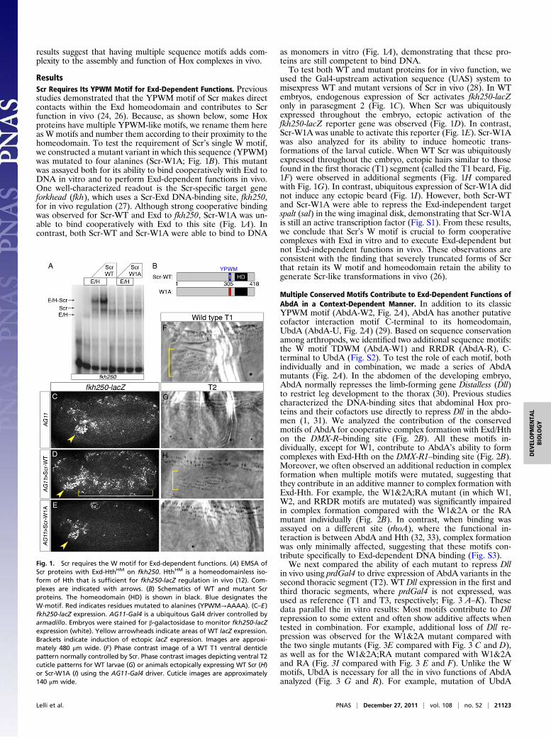
ResultsScr Requires Its YPWM Motif for Exd-Dependent Functions. Previousstudies demonstrated that the YPWM motif of Scr makes directcontacts within the Exd homeodomain and contributes to Scrfunction in vivo (24, 26). Because, as shown below, some Hoxproteins have multiple YPWM-like motifs, we rename them hereas W motifs and number them according to their proximity to thehomeodomain. To test the requirement of Scr’s single W motif,we constructed a mutant variant in which this sequence (YPWM)was mutated to four alanines (Scr-W1A; Fig. 1B). This mutantwas assayed both for its ability to bind cooperatively with Exd toDNA in vitro and to perform Exd-dependent functions in vivo.One well-characterized readout is the Scr-specific target geneforkhead (fkh), which uses a Scr-Exd DNA-binding site, fkh250,for in vivo regulation (27). Although strong cooperative bindingwas observed for Scr-WT and Exd to fkh250, Scr-W1A was un-able to bind cooperatively with Exd to this site (Fig. 1A). Incontrast, both Scr-WT and Scr-W1A were able to bind to DNA
as monomers in vitro (Fig. 1A), demonstrating that these pro-teins are still competent to bind DNA.To test both WT and mutant proteins for in vivo function, we
used the Gal4-upstream activation sequence (UAS) system tomisexpress WT and mutant versions of Scr in vivo (28). In WTembryos, endogenous expression of Scr activates fkh250-lacZonly in parasegment 2 (Fig. 1C). When Scr was ubiquitouslyexpressed throughout the embryo, ectopic activation of thefkh250-lacZ reporter gene was observed (Fig. 1D). In contrast,Scr-W1A was unable to activate this reporter (Fig. 1E). Scr-W1Awas also analyzed for its ability to induce homeotic trans-formations of the larval cuticle. When WT Scr was ubiquitouslyexpressed throughout the embryo, ectopic hairs similar to thosefound in the first thoracic (T1) segment (called the T1 beard, Fig.1F) were observed in additional segments (Fig. 1H comparedwith Fig. 1G). In contrast, ubiquitous expression of Scr-W1A didnot induce any ectopic beard (Fig. 1I). However, both Scr-WTand Scr-W1A were able to repress the Exd-independent targetspalt (sal) in the wing imaginal disk, demonstrating that Scr-W1Ais still an active transcription factor (Fig. S1). From these results,we conclude that Scr’s W motif is crucial to form cooperativecomplexes with Exd in vitro and to execute Exd-dependent butnot Exd-independent functions in vivo. These observations areconsistent with the finding that severely truncated forms of Scrthat retain its W motif and homeodomain retain the ability togenerate Scr-like transformations in vivo (26).
Multiple Conserved Motifs Contribute to Exd-Dependent Functions ofAbdA in a Context-Dependent Manner. In addition to its classicYPWM motif (AbdA-W2, Fig. 2A), AbdA has another putativecofactor interaction motif C-terminal to its homeodomain,UbdA (AbdA-U, Fig. 2A) (29). Based on sequence conservationamong arthropods, we identified two additional sequence motifs:the W motif TDWM (AbdA-W1) and RRDR (AbdA-R), C-terminal to UbdA (Fig. S2). To test the role of each motif, bothindividually and in combination, we made a series of AbdAmutants (Fig. 2A). In the abdomen of the developing embryo,AbdA normally represses the limb-forming gene Distalless (Dll)to restrict leg development to the thorax (30). Previous studiescharacterized the DNA-binding sites that abdominal Hox pro-teins and their cofactors use directly to repress Dll in the abdo-men (1, 31). We analyzed the contribution of the conservedmotifs of AbdA for cooperative complex formation with Exd/Hthon the DMX-R–binding site (Fig. 2B). All these motifs in-dividually, except for W1, contribute to AbdA’s ability to formcomplexes with Exd-Hth on the DMX-R1–binding site (Fig. 2B).Moreover, we often observed an additional reduction in complexformation when multiple motifs were mutated, suggesting thatthey contribute in an additive manner to complex formation withExd-Hth. For example, the W1&2A;RA mutant (in which W1,W2, and RRDR motifs are mutated) was significantly impairedin complex formation compared with the W1&2A or the RAmutant individually (Fig. 2B). In contrast, when binding wasassayed on a different site (rhoA), where the functional in-teraction is between AbdA and Hth (32, 33), complex formationwas only minimally affected, suggesting that these motifs con-tribute specifically to Exd-dependent DNA binding (Fig. S3).We next compared the ability of each mutant to repress Dll
in vivo using prdGal4 to drive expression of AbdA variants in thesecond thoracic segment (T2). WT Dll expression in the first andthird thoracic segments, where prdGal4 is not expressed, wasused as reference (T1 and T3, respectively; Fig. 3 A–K). Thesedata parallel the in vitro results: Most motifs contribute to Dllrepression to some extent and often show additive affects whentested in combination. For example, additional loss of Dll re-pression was observed for the W1&2A mutant compared withthe two single mutants (Fig. 3E compared with Fig. 3 C and D),as well as for the W1&2A;RA mutant compared with W1&2Aand RA (Fig. 3I compared with Fig. 3 E and F). Unlike the Wmotifs, UbdA is necessary for all the in vivo functions of AbdAanalyzed (Fig. 3 G and R). For example, mutation of UbdA
Fig. 1. Scr requires the W motif for Exd-dependent functions. (A) EMSA ofScr proteins with Exd-HthHM on fkh250. HthHM is a homeodomainless iso-form of Hth that is sufficient for fkh250-lacZ regulation in vivo (12). Com-plexes are indicated with arrows. (B) Schematics of WT and mutant Scrproteins. The homeodomain (HD) is shown in black. Blue designates theW-motif. Red indicates residues mutated to alanines (YPWM→AAAA). (C–E)fkh250-lacZ expression. AG11-Gal4 is a ubiquitous Gal4 driver controlled byarmadillo. Embryos were stained for β-galactosidase to monitor fkh250-lacZexpression (white). Yellow arrowheads indicate areas of WT lacZ expression.Brackets indicate induction of ectopic lacZ expression. Images are approxi-mately 480 μm wide. (F) Phase contrast image of a WT T1 ventral denticlepattern normally controlled by Scr. Phase contrast images depicting ventral T2cuticle patterns for WT larvae (G) or animals ectopically expressing WT Scr (H)or Scr-W1A (I) using the AG11-Gal4 driver. Cuticle images are approximately140 μm wide.
Lelli et al. PNAS | December 27, 2011 | vol. 108 | no. 52 | 21123
DEV
ELOPM
ENTA
LBIOLO
GY
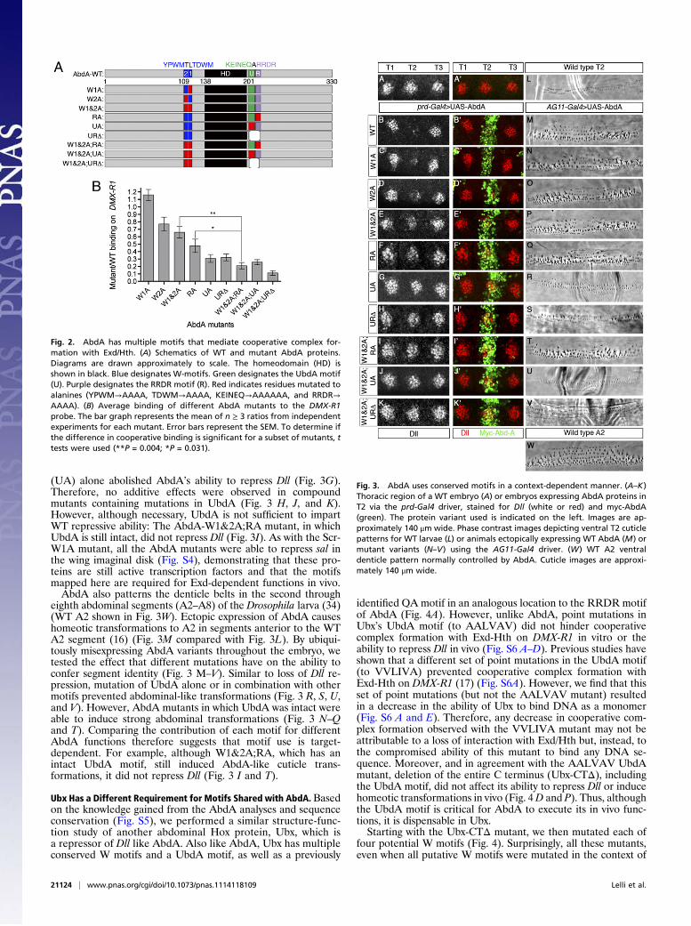
(UA) alone abolished AbdA’s ability to repress Dll (Fig. 3G).Therefore, no additive effects were observed in compoundmutants containing mutations in UbdA (Fig. 3 H, J, and K).However, although necessary, UbdA is not sufficient to impartWT repressive ability: The AbdA-W1&2A;RA mutant, in whichUbdA is still intact, did not repress Dll (Fig. 3I). As with the Scr-W1A mutant, all the AbdA mutants were able to repress sal inthe wing imaginal disk (Fig. S4), demonstrating that these pro-teins are still active transcription factors and that the motifsmapped here are required for Exd-dependent functions in vivo.AbdA also patterns the denticle belts in the second through
eighth abdominal segments (A2–A8) of the Drosophila larva (34)(WT A2 shown in Fig. 3W). Ectopic expression of AbdA causeshomeotic transformations to A2 in segments anterior to the WTA2 segment (16) (Fig. 3M compared with Fig. 3L). By ubiqui-tously misexpressing AbdA variants throughout the embryo, wetested the effect that different mutations have on the ability toconfer segment identity (Fig. 3 M–V). Similar to loss of Dll re-pression, mutation of UbdA alone or in combination with othermotifs prevented abdominal-like transformations (Fig. 3 R, S, U,and V). However, AbdA mutants in which UbdA was intact wereable to induce strong abdominal transformations (Fig. 3 N–Qand T). Comparing the contribution of each motif for differentAbdA functions therefore suggests that motif use is target-dependent. For example, although W1&2A;RA, which has anintact UbdA motif, still induced AbdA-like cuticle trans-formations, it did not repress Dll (Fig. 3 I and T).
Ubx Has a Different Requirement for Motifs Shared with AbdA. Basedon the knowledge gained from the AbdA analyses and sequenceconservation (Fig. S5), we performed a similar structure-func-tion study of another abdominal Hox protein, Ubx, which isa repressor of Dll like AbdA. Also like AbdA, Ubx has multipleconserved W motifs and a UbdA motif, as well as a previously
identified QA motif in an analogous location to the RRDR motifof AbdA (Fig. 4A). However, unlike AbdA, point mutations inUbx’s UbdA motif (to AALVAV) did not hinder cooperativecomplex formation with Exd-Hth on DMX-R1 in vitro or theability to repress Dll in vivo (Fig. S6 A–D). Previous studies haveshown that a different set of point mutations in the UbdA motif(to VVLIVA) prevented cooperative complex formation withExd-Hth on DMX-R1 (17) (Fig. S6A). However, we find that thisset of point mutations (but not the AALVAV mutant) resultedin a decrease in the ability of Ubx to bind DNA as a monomer(Fig. S6 A and E). Therefore, any decrease in cooperative com-plex formation observed with the VVLIVA mutant may not beattributable to a loss of interaction with Exd/Hth but, instead, tothe compromised ability of this mutant to bind any DNA se-quence. Moreover, and in agreement with the AALVAV UbdAmutant, deletion of the entire C terminus (Ubx-CTΔ), includingthe UbdA motif, did not affect its ability to repress Dll or inducehomeotic transformations in vivo (Fig. 4D and P). Thus, althoughthe UbdA motif is critical for AbdA to execute its in vivo func-tions, it is dispensable in Ubx.Starting with the Ubx-CTΔ mutant, we then mutated each of
four potential W motifs (Fig. 4). Surprisingly, all these mutants,even when all putative W motifs were mutated in the context of
Fig. 2. AbdA has multiple motifs that mediate cooperative complex for-mation with Exd/Hth. (A) Schematics of WT and mutant AbdA proteins.Diagrams are drawn approximately to scale. The homeodomain (HD) isshown in black. Blue designates W-motifs. Green designates the UbdA motif(U). Purple designates the RRDR motif (R). Red indicates residues mutated toalanines (YPWM→AAAA, TDWM→AAAA, KEINEQ→AAAAAA, and RRDR→AAAA). (B) Average binding of different AbdA mutants to the DMX-R1probe. The bar graph represents the mean of n ≥ 3 ratios from independentexperiments for each mutant. Error bars represent the SEM. To determine ifthe difference in cooperative binding is significant for a subset of mutants, ttests were used (**P = 0.004; *P = 0.031).
Fig. 3. AbdA uses conserved motifs in a context-dependent manner. (A–K)Thoracic region of a WT embryo (A) or embryos expressing AbdA proteins inT2 via the prd-Gal4 driver, stained for Dll (white or red) and myc-AbdA(green). The protein variant used is indicated on the left. Images are ap-proximately 140 μm wide. Phase contrast images depicting ventral T2 cuticlepatterns for WT larvae (L) or animals ectopically expressing WT AbdA (M) ormutant variants (N–V) using the AG11-Gal4 driver. (W) WT A2 ventraldenticle pattern normally controlled by AbdA. Cuticle images are approxi-mately 140 μm wide.
21124 | www.pnas.org/cgi/doi/10.1073/pnas.1114118109 Lelli et al.
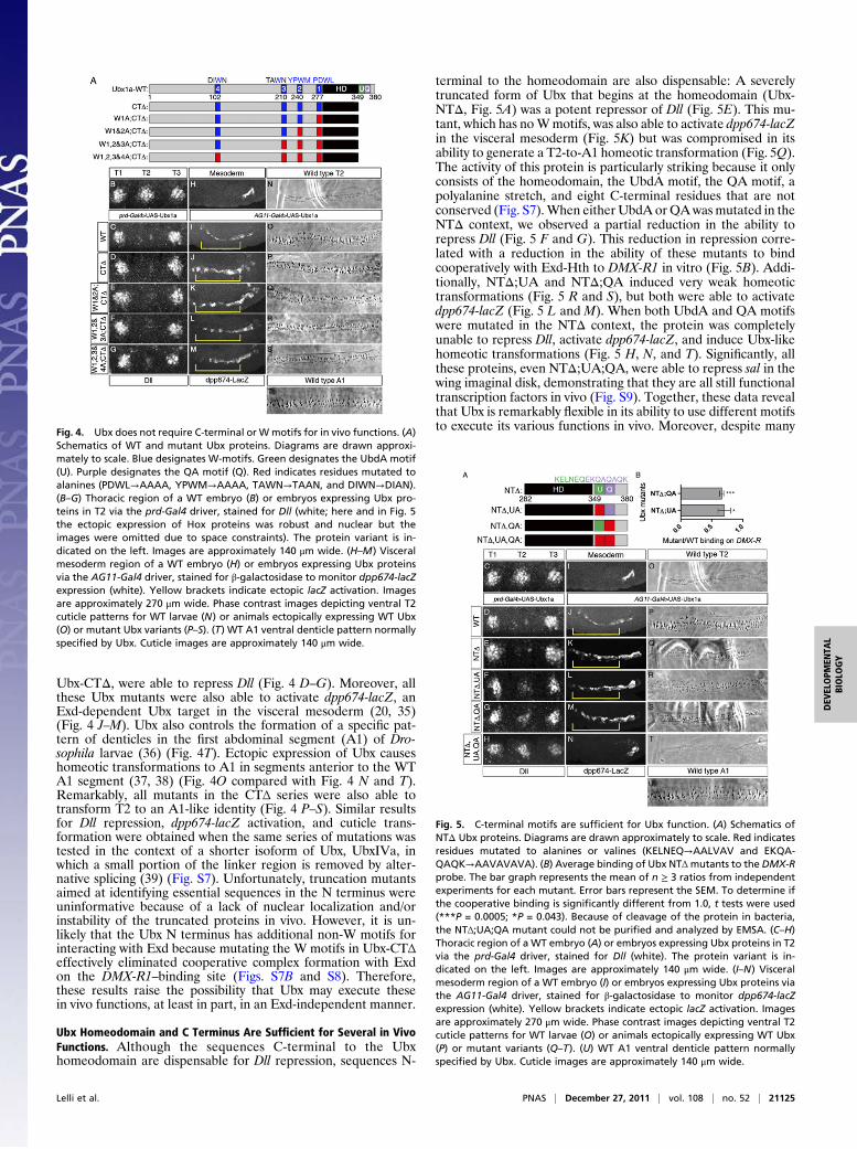
Ubx-CTΔ, were able to repress Dll (Fig. 4 D–G). Moreover, allthese Ubx mutants were also able to activate dpp674-lacZ, anExd-dependent Ubx target in the visceral mesoderm (20, 35)(Fig. 4 J–M). Ubx also controls the formation of a specific pat-tern of denticles in the first abdominal segment (A1) of Dro-sophila larvae (36) (Fig. 4T). Ectopic expression of Ubx causeshomeotic transformations to A1 in segments anterior to the WTA1 segment (37, 38) (Fig. 4O compared with Fig. 4 N and T).Remarkably, all mutants in the CTΔ series were also able totransform T2 to an A1-like identity (Fig. 4 P–S). Similar resultsfor Dll repression, dpp674-lacZ activation, and cuticle trans-formation were obtained when the same series of mutations wastested in the context of a shorter isoform of Ubx, UbxIVa, inwhich a small portion of the linker region is removed by alter-native splicing (39) (Fig. S7). Unfortunately, truncation mutantsaimed at identifying essential sequences in the N terminus wereuninformative because of a lack of nuclear localization and/orinstability of the truncated proteins in vivo. However, it is un-likely that the Ubx N terminus has additional non-W motifs forinteracting with Exd because mutating the W motifs in Ubx-CTΔeffectively eliminated cooperative complex formation with Exdon the DMX-R1–binding site (Figs. S7B and S8). Therefore,these results raise the possibility that Ubx may execute thesein vivo functions, at least in part, in an Exd-independent manner.
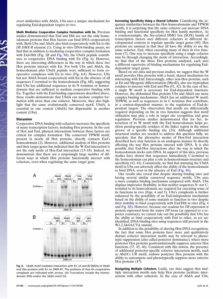
Ubx Homeodomain and C Terminus Are Sufficient for Several in VivoFunctions. Although the sequences C-terminal to the Ubxhomeodomain are dispensable for Dll repression, sequences N-
terminal to the homeodomain are also dispensable: A severelytruncated form of Ubx that begins at the homeodomain (Ubx-NTΔ, Fig. 5A) was a potent repressor of Dll (Fig. 5E). This mu-tant, which has noWmotifs, was also able to activate dpp674-lacZin the visceral mesoderm (Fig. 5K) but was compromised in itsability to generate a T2-to-A1 homeotic transformation (Fig. 5Q).The activity of this protein is particularly striking because it onlyconsists of the homeodomain, the UbdA motif, the QA motif, apolyalanine stretch, and eight C-terminal residues that are notconserved (Fig. S7).When either UbdA orQAwasmutated in theNTΔ context, we observed a partial reduction in the ability torepress Dll (Fig. 5 F and G). This reduction in repression corre-lated with a reduction in the ability of these mutants to bindcooperatively with Exd-Hth to DMX-R1 in vitro (Fig. 5B). Addi-tionally, NTΔ;UA and NTΔ;QA induced very weak homeotictransformations (Fig. 5 R and S), but both were able to activatedpp674-lacZ (Fig. 5 L and M). When both UbdA and QA motifswere mutated in the NTΔ context, the protein was completelyunable to repress Dll, activate dpp674-lacZ, and induce Ubx-likehomeotic transformations (Fig. 5 H, N, and T). Significantly, allthese proteins, even NTΔ;UA;QA, were able to repress sal in thewing imaginal disk, demonstrating that they are all still functionaltranscription factors in vivo (Fig. S9). Together, these data revealthat Ubx is remarkably flexible in its ability to use different motifsto execute its various functions in vivo. Moreover, despite manyFig. 4. Ubx does not require C-terminal or Wmotifs for in vivo functions. (A)
Schematics of WT and mutant Ubx proteins. Diagrams are drawn approxi-mately to scale. Blue designates W-motifs. Green designates the UbdA motif(U). Purple designates the QA motif (Q). Red indicates residues mutated toalanines (PDWL→AAAA, YPWM→AAAA, TAWN→TAAN, and DIWN→DIAN).(B–G) Thoracic region of a WT embryo (B) or embryos expressing Ubx pro-teins in T2 via the prd-Gal4 driver, stained for Dll (white; here and in Fig. 5the ectopic expression of Hox proteins was robust and nuclear but theimages were omitted due to space constraints). The protein variant is in-dicated on the left. Images are approximately 140 μm wide. (H–M) Visceralmesoderm region of a WT embryo (H) or embryos expressing Ubx proteinsvia the AG11-Gal4 driver, stained for β-galactosidase to monitor dpp674-lacZexpression (white). Yellow brackets indicate ectopic lacZ activation. Imagesare approximately 270 μm wide. Phase contrast images depicting ventral T2cuticle patterns for WT larvae (N) or animals ectopically expressing WT Ubx(O) or mutant Ubx variants (P–S). (T) WT A1 ventral denticle pattern normallyspecified by Ubx. Cuticle images are approximately 140 μm wide.
Fig. 5. C-terminal motifs are sufficient for Ubx function. (A) Schematics ofNTΔ Ubx proteins. Diagrams are drawn approximately to scale. Red indicatesresidues mutated to alanines or valines (KELNEQ→AALVAV and EKQA-QAQK→AAVAVAVA). (B) Average binding of Ubx NTΔmutants to the DMX-Rprobe. The bar graph represents the mean of n ≥ 3 ratios from independentexperiments for each mutant. Error bars represent the SEM. To determine ifthe cooperative binding is significantly different from 1.0, t tests were used(***P = 0.0005; *P = 0.043). Because of cleavage of the protein in bacteria,the NTΔ;UA;QA mutant could not be purified and analyzed by EMSA. (C–H)Thoracic region of a WT embryo (A) or embryos expressing Ubx proteins in T2via the prd-Gal4 driver, stained for Dll (white). The protein variant is in-dicated on the left. Images are approximately 140 μm wide. (I–N) Visceralmesoderm region of a WT embryo (I) or embryos expressing Ubx proteins viathe AG11-Gal4 driver, stained for β-galactosidase to monitor dpp674-lacZexpression (white). Yellow brackets indicate ectopic lacZ activation. Imagesare approximately 270 μm wide. Phase contrast images depicting ventral T2cuticle patterns for WT larvae (O) or animals ectopically expressing WT Ubx(P) or mutant variants (Q–T). (U) WT A1 ventral denticle pattern normallyspecified by Ubx. Cuticle images are approximately 140 μm wide.
Lelli et al. PNAS | December 27, 2011 | vol. 108 | no. 52 | 21125
DEV
ELOPM
ENTA
LBIOLO
GY
overt similarities with AbdA, Ubx uses a unique mechanism forregulating Exd-dependent targets in vivo.
UbdA Mediates Cooperative Complex Formation with En. Previousstudies demonstrated that Exd and Hth are not the only home-odomain proteins that Hox factors can bind DNA cooperativelywith: Both Ubx and AbdA also bind cooperatively with En to theDll DMX-R element (1). Using in vitro DNA-binding assays, wefind that in addition to mediating cooperative complex formationwith Exd-Hth (Fig. 2B and Fig. S8), the UbdA peptide contrib-utes to cooperative DNA binding with En (Fig. 6). However,there are interesting differences in the way in which these twoHox proteins interact with En. For both Ubx and AbdA, thehomeodomain plus UbdA peptide were sufficient to form co-operative complexes with En in vitro (Fig. 6A). However, Ubxbut not AbdA bound cooperatively with En in the absence of allsequences C-terminal to the homeodomain (Fig. 6B), suggestingthat Ubx has additional sequences in its N terminus or homeo-domain that are sufficient to mediate cooperative binding withEn. Together with the Exd-binding experiments described above,these results demonstrate that UbdA can mediate complex for-mation with more than one cofactor. Moreover, they also high-light that the same evolutionarily conserved motif, UbdA, isessential in one context (AbdA) but dispensable in anothercontext (Ubx).
DiscussionCooperative DNA binding with cofactors increases the specificityof many transcription factors, including Hox proteins. In the caseof Hox and Exd, physical interactions between these factors arecritical for complex formation: The conserved YPWM motif,present in nearly all Hox proteins, directly contacts Exd’shomeodomain (2). However, additional analysis of Hox proteinsand their target genes has indicated that the W-Exd interaction isnot the only mode of Hox-Exd interaction (13–18). Indeed, wedemonstrate that there are a surprisingly large number of dif-ferent ways in which Hox proteins functionally interact withcofactors, even when regulating the same target gene.
Increasing Specificity Using a Shared Cofactor. Considering the se-quence similarities between the Hox homeodomains and YPWMmotifs, it is surprising that the same cofactor can increase DNAbinding and functional specificity for Hox family members. Asa counterexample, the Sry-related HMG box (SOX) family oftranscription factors uses different cofactors in different de-velopmental contexts to regulate specific target genes (40). Hoxproteins are unusual in that they all have the ability to use thesame cofactor, Exd, when executing many of their in vivo func-tions (7). One way to increase specificity using a single cofactormay be through changing the mode of interaction. Accordingly,we find that of the three Hox proteins analyzed, each usesa different repertoire of binding mechanisms for regulating Exd-dependent target genes.Based on our data and previous studies, we suggest that the W
motif provides Hox proteins with a basal, shared mechanism forinteracting with Exd. Interestingly, other non-Hox proteins, suchas En and Myogenic differentiation (MyoD), also use tryptophanresidues to interact with Exd and Pbx (41, 42). In the case of Scr,a single W motif is necessary for Exd-dependent functions.However, the abdominal Hox proteins Ubx and AbdA use morecomplex binding mechanisms. AbdA has an additional W motif,TDWM, as well as sequences in its C terminus that contribute,in a context-dependent manner, to the regulation of Exd-de-pendent targets. The observation that motifs are differentiallyrequired depending on the in vivo function suggests that motifutilization may play a role in target site recognition and generegulation. Previous studies demonstrated that for Scr, in-teraction of its W motif with the Exd homeodomain helps po-sition paralog-specific residues of the linker region in the minorgroove of a specific binding site (24). Although additionalstructural studies are needed to address this question fully, wespeculate that the alternative modes of Hox-Exd interactiondescribed here may function in an analogous manner by directlyaffecting the way Hox proteins interact with DNA. It is alsopossible that Exd-Hox interactions alter the way in which thehomeodomain docks onto DNA. In support of this idea, previousstudies have shown that sequences immediately C-terminal tothe homeodomain can play a role in homeodomain structure andspecificity (43, 44). Consistently, we find that mutating the UbdAmotif in Ubx can adversely affect the ability of the homeodomainto bind DNA, even in the absence of Exd (Fig. S6).Our results also reveal that despite sharing binding sites and
having several similar conserved sequence motifs, Ubx usesa more complex binding mechanism compared with AbdA. Ubxdisplays impressive flexibility, in that neither sequences N- nor C-terminal to its homeodomain are required for executing some ofits functions in vivo (Figs. 4 and 5). Ubx’s complexity is furtherenhanced by the possibility of an Exd-independent mechanismbased on the ability of some mutants to function in vivo despitetheir inability to bind cooperatively with Exd/Hth in vitro (Fig. 4and Fig. S8). However, because our readout for Dll expression isprotein expressed from the native Dll locus (as opposed to a re-porter construct), we cannot rule out the possibility that Ubx hasthe ability to bind cooperatively with Exd to other, as yet un-identified, DNA-binding sites using sequences still present in theW1,2,3&4A;CTΔ mutant.In addition to the possibility of altering Hox-DNA recognition,
the fact that some Hox proteins have more and qualitativelydistinct cofactor interaction motifs may be relevant to pheno-typic suppression (also called posterior dominance), where moreposterior Hox proteins posttranslationally suppress anterior Hoxfunctions (37, 45, 46). Consistent with this notion, the presenceof additional posterior-specific cofactor interaction motifs, suchas AbdA’s UR motif, endows posterior Hox proteins with theability to outcompete and phenotypically suppress more anteriorHox proteins (47).
Navigating Multiple Cofactors. Lastly, our data suggest that mul-tiple interaction motifs may help Hox proteins facilitate inter-actions with other cofactors. In the case of AbdA and Ubx,
Fig. 6. UbdA motif mediates interaction with En. (A and B) EMSAs of AbdAand Ubx proteins with En on DMX-R1. The positions of Hox–En cooperativecomplexes are indicated with arrows. (A) Truncations include the homeo-domain (HD) and/or the UbdA motif (U).
21126 | www.pnas.org/cgi/doi/10.1073/pnas.1114118109 Lelli et al.
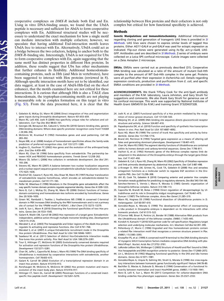
cooperative complexes on DMX-R include both Exd and En.Using in vitro DNA-binding assays, we found that the UbdApeptide is necessary and sufficient for AbdA to form cooperativecomplexes with En. Additional structural studies will be nec-essary to understand the exact mechanism for how a single motifcan mediate interaction with multiple cofactors; however, wespeculate that having additional Exd-interaction motifs leavesUbdA free to interact with En. Alternatively, UbdA could act asa bridge between the two cofactors, helping to anchor both to theDMX-R–binding site. Interestingly, UbdA is not required for Ubxto form cooperative complexes with En, again suggesting that thesame motif has distinct properties in different Hox proteins. Inaddition, these results suggest that Ubx has other mechanismsthat further enhance its flexibility. Additional homeodomain-containing proteins, such as Hth (and Meis in vertebrates), havebeen suggested to interact with Hox proteins (reviewed in 8).Although specific interaction motifs have yet to be identified, ourdata suggest, at least in the case of AbdA-Hth-Exd on the rhoAenhancer, that the motifs examined here are not critical for theseinteractions. It is curious that although Hth is also a TALE classhomeodomain, the tryptophan-containing motifs are not playinga measurable role in complex formation on this target in vitro(Fig. S3). From the data presented here, it is clear that the
relationship between Hox proteins and their cofactors is not onlycomplex but critical for how functional specificity is achieved.
MethodsGenetic Manipulations and Immunohistochemistry. Additional informationregarding cloning and generation of transgenic UAS lines is provided in SIMethods. UAS lines were chosen to express similar levels of tagged Hoxproteins. Either AG11-GAL4 or prd-GAL4 was used for ectopic expression asindicated. Flip-out clones were generated using hs-flp; act<y<Gal4, UAS-GFP. Antibodies used are described in SI Methods. Z-series of embryos werecollected on a Leica SP5 confocal microscope. Cuticle images were collectedon a Zeiss Axioplan 2 microscope.
EMSAs. EMSAs were carried out as previously described (31). CooperativeDNA binding was calculated as a ratio of the amount of mutant Exd–Hthcomplex to the amount of WT Exd–Hth complex in the same gel. Proteinswere all purified after their expression in Escherichia coli. Details regardingexpression constructs, production and purification from E. coli, and specificEMSA conditions are provided in SI Methods.
ACKNOWLEDGMENTS. We thank Tiffany Cook for the anti-Spalt antibodyand members of the R.M. laboratory, Laura Johnston, and Gary Struhl forsuggestions and comments. We also thank Gary Struhl for generous use ofhis confocal microscope. This work was supported by National Institutes ofHealth Grant GM54510 (to R.M.) and training Grant 5T32DK07328.
1. Gebelein B, McKay DJ, Mann RS (2004) Direct integration of Hox and segmentationgene inputs during Drosophila development. Nature 431:653–659.
2. Mann RS, Lelli KM, Joshi R (2009) Hox specificity unique roles for cofactors and col-laborators. Curr Top Dev Biol 88:63–101.
3. Georges AB, Benayoun BA, Caburet S, Veitia RA (2010) Generic binding sites, genericDNA-binding domains: Where does specific promoter recognition come from? FASEBJ 24:346–356.
4. McGinnis W, Krumlauf R (1992) Homeobox genes and axial patterning. Cell 68:283–302.
5. Noyes MB, et al. (2008) Analysis of homeodomain specificities allows the family-wideprediction of preferred recognition sites. Cell 133:1277–1289.
6. Hughes CL, Kaufman TC (2002) Hox genes and the evolution of the arthropod bodyplan. Evol Dev 4:459–499.
7. Mann RS, Chan SK (1996) Extra specificity from extradenticle: The partnership be-tween HOX and PBX/EXD homeodomain proteins. Trends Genet 12:258–262.
8. Moens CB, Selleri L (2006) Hox cofactors in vertebrate development. Dev Biol 291:193–206.
9. Stevens KE, Mann RS (2007) A balance between two nuclear localization sequencesand a nuclear export sequence governs extradenticle subcellular localization. Genetics175:1625–1636.
10. Rieckhof GE, Casares F, Ryoo HD, Abu-Shaar M, Mann RS (1997) Nuclear translocationof extradenticle requires homothorax, which encodes an extradenticle-related ho-meodomain protein. Cell 91:171–183.
11. Peifer M, Wieschaus E (1990) Mutations in the Drosophila gene extradenticle affect theway specific homeo domain proteins regulate segmental identity. Genes Dev 4:1209–1223.
12. Noro B, Culi J, McKay DJ, Zhang W, Mann RS (2006) Distinct functions of homeo-domain-containing and homeodomain-less isoforms encoded by homothorax. GenesDev 20:1636–1650.
13. Green NC, Rambaldi I, Teakles J, Featherstone MS (1998) A conserved C-terminaldomain in PBX increases DNA binding by the PBX homeodomain and is not a primarysite of contact for the YPWM motif of HOXA1. J Biol Chem 273:13273–13279.
14. Joshi R, Sun L, Mann R (2010) Dissecting the functional specificities of two Hox pro-teins. Genes Dev 24:1533–1545.
15. Galant R, Walsh CM, Carroll SB (2002) Hox repression of a target gene: Extradenticle-independent, additive action through multiple monomer binding sites. Development129:3115–3126.
16. Merabet S, et al. (2003) The hexapeptide and linker regions of the AbdA Hox proteinregulate its activating and repressive functions. Dev Cell 4:761–768.
17. Merabet S, et al. (2007) A unique Extradenticle recruitment mode in the DrosophilaHox protein Ultrabithorax. Proc Natl Acad Sci USA 104:16946–16951.
18. Saadaoui M, et al. (2011) Selection of distinct Hox-Extradenticle interaction modesfine-tunes Hox protein activity. Proc Natl Acad Sci USA 108:2276–2281.
19. Tour E, Hittinger CT, McGinnis W (2005) Evolutionarily conserved domains requiredfor activation and repression functions of the Drosophila Hox protein Ultrabithorax.Development 132:5271–5281.
20. Chan SK, Jaffe L, Capovilla M, Botas J, Mann RS (1994) The DNA binding specificity ofUltrabithorax is modulated by cooperative interactions with extradenticle, anotherhomeoprotein. Cell 78:603–615.
21. Galant R, Carroll SB (2002) Evolution of a transcriptional repression domain in aninsect Hox protein. Nature 415:910–913.
22. Ronshaugen M, McGinnis N, McGinnis W (2002) Hox protein mutation and macro-evolution of the insect body plan. Nature 415:914–917.
23. Hittinger CT, Stern DL, Carroll SB (2005) Pleiotropic functions of a conserved insect-specific Hox peptide motif. Development 132:5261–5270.
24. Joshi R, et al. (2007) Functional specificity of a Hox protein mediated by the recog-
nition of minor groove structure. Cell 131:530–543.25. Meijsing SH, et al. (2009) DNA binding site sequence directs glucocorticoid receptor
structure and activity. Science 324:407–410.26. Papadopoulos DK, et al. (2010) Function and specificity of synthetic Hox transcription
factors in vivo. Proc Natl Acad Sci USA 107:4087–4092.27. Ryoo HD, Mann RS (1999) The control of trunk Hox specificity and activity by Extra-
denticle. Genes Dev 13:1704–1716.28. Brand AH, Perrimon N (1993) Targeted gene expression as a means of altering cell
fates and generating dominant phenotypes. Development 118:401–415.29. Chan SK, Mann RS (1993) The segment identity functions of Ultrabithorax are contained
within its homeo domain and carboxy-terminal sequences. Genes Dev 7:796–811.30. Vachon G, et al. (1992) Homeotic genes of the Bithorax complex repress limb de-
velopment in the abdomen of the Drosophila embryo through the target gene Distal-
less. Cell 71:437–450.31. Gebelein B, Culi J, Ryoo HD, ZhangW, Mann RS (2002) Specificity of Distalless repression
and limb primordia development by abdominal Hox proteins. Dev Cell 3:487–498.32. Li-Kroeger D, Witt LM, Grimes HL, Cook TA, Gebelein B (2008) Hox and senseless
antagonism functions as a molecular switch to regulate EGF secretion in the Dro-
sophila PNS. Dev Cell 15:298–308.33. Uhl JD, Cook TA, Gebelein B (2010) Comparing anterior and posterior Hox complex
formation reveals guidelines for predicting cis-regulatory elements. Dev Biol 343:154–166.34. Sánchez-Herrero E, Vernós I, Marco R, Morata G (1985) Genetic organization of
Drosophila bithorax complex. Nature 313:108–113.35. Capovilla M, Brandt M, Botas J (1994) Direct regulation of decapentaplegic by Ul-
trabithorax and its role in Drosophila midgut morphogenesis. Cell 76:461–475.36. Duncan I (1987) The bithorax complex. Annu Rev Genet 21:285–319.37. Mann RS, Hogness DS (1990) Functional dissection of Ultrabithorax proteins in D.
melanogaster. Cell 60:597–610.38. González-Reyes A, Morata G (1990) The developmental effect of overexpressing
a Ubx product in Drosophila embryos is dependent on its interactions with other
homeotic products. Cell 61:515–522.39. O’Connor MB, Binari R, Perkins LA, Bender W (1988) Alternative RNA products from
the Ultrabithorax domain of the bithorax complex. EMBO J 7:435–445.40. Kondoh H, Kamachi Y (2010) SOX-partner code for cell specification: Regulatory target
selection and underlying molecular mechanisms. Int J Biochem Cell Biol 42:391–399.41. Peltenburg LT, Murre C (1996) Engrailed and Hox homeodomain proteins contain
a related Pbx interaction motif that recognizes a common structure present in Pbx.EMBO J 15:3385–3393.
42. Knoepfler PS, et al. (1999) A conserved motif N-terminal to the DNA-binding domainsof myogenic bHLH transcription factors mediates cooperative DNA binding with pbx-
Meis1/Prep1. Nucleic Acids Res 27:3752–3761.43. LaRonde-LeBlanc NA, Wolberger C (2003) Structure of HoxA9 and Pbx1 bound to DNA:
Hox hexapeptide and DNA recognition anterior to posterior. Genes Dev 17:2060–2072.44. Lin L, McGinnis W (1992) Mapping functional specificity in the Dfd and Ubx homeo
domains. Genes Dev 6:1071–1081.45. González-Reyes A, Urquia N, Gehring WJ, Struhl G, Morata G (1990) Are cross-regula-
tory interactions between homoeotic genes functionally significant? Nature 344:78–80.46. Bachiller D, Macías A, Duboule D, Morata G (1994) Conservation of a functional hi-
erarchy between mammalian and insect Hox/HOM genes. EMBO J 13:1930–1941.47. Noro B, Lelli K, Sun L, Mann RS (2011) Competition for cofactor-dependent DNA
binding underlies Hox phenotypic suppression. Genes Dev 25:2327–2332.
Lelli et al. PNAS | December 27, 2011 | vol. 108 | no. 52 | 21127
DEV
ELOPM
ENTA
LBIOLO
GY