Embed Size (px)
Citation preview
Variability of the pullout strength of cancellous bone screws withcement augmentation
P. Procter a,b, P. Bennani b, C.J. Brown b,⁎, J. Arnoldi c, D.P. Pioletti d, S. Larsson e
a Stryker Osteosynthesis, Selzach, Switzerlandb School of Engineering and Design, Brunel University, Uxbridge, UKc Clinic of Plastic and Hand Surgery, University Hospital Bern, Bern, Switzerlandd Laboratory of Biomechanical Orthopedics, EPFL, Lausanne, Switzerlande Department of Orthopaedics, Uppsala University Hospital, Uppsala, Sweden
a b s t r a c ta r t i c l e i n f o
Article history:Received 4 July 2014Accepted 2 March 2015
Keywords:Cancellous boneBone screwsPullout testingHuman cadaverFinite element modelling
Background:Orthopaedic surgeons often face clinical situations where improved screw holding power in cancel-lous bone is needed. Injectable calcium phosphate cements are one option to enhance fixation.Methods: Paired screw pullout tests were undertaken in which human cadaver bone was augmented with calci-um phosphate cement. A finite element model was used to investigate sensitivity to screw positional placement.Findings: Statistical analysis of the data concluded that the pullout strength was generally increased by cementaugmentation in the in vitro human cadaver tests. However, when comparing the individual paired samplestherewere surprising results with lower strength than anticipated after augmentation, in apparent contradictionto the generally expected conclusion. Investigation using the finite element model showed that these strengthreductions could be accounted for by small screwpositional changes. A change of 0.5mmmight result in predict-ed pullout force changes of up to 28%.Interpretation: Small changes in screw position might lead to significant changes in pullout strength sufficient toexplain the lower than expected individual pullout values in augmented cancellous bone. Consequently whilstthe addition of cement at a position of low strength would increase the pullout strength at that point, it mightnot reach the pullout strength of the un-augmented paired test site. However, the overall effect of cement aug-mentation produces a significant improvement at whatever point in the bone the screw is placed. The use ofpolymeric bone-substitute materials for tests may not reveal the natural variation encountered in tests usingreal bone structures.
© 2015 Elsevier Ltd. All rights reserved.
1. Introduction
Many orthopaedic surgeons report difficulties of fixing bone screws(e.g. Andersson et al., 2000 or Strømsøe, 2004), especially into poorquality cancellous bone of low apparent density—bone with low bonevolume to total volume (BV/TV) ratios. Bone quality and quantityin patients presenting with fragility fractures are often variable (e.g.Berlemann and Schwarzenbach, 1997; Chen et al., 2009; Thiele et al.,2007), and control of screw fixation in a surgical situation is challenging.
The number of clinically reported screw failures is high especially inpatientswith poor bone quality. Hip fracture screw failure rates vary be-tween 3% and 5% (Jesudason and Jeyem, 2006; Kim et al., 2001) whilefor proximal humeral fractures rates can vary between 15% and 40%(Singer et al., 1998; Cantlon and Egol, 2013). It has been suggested(Procter, 2013) that the total number of screw failures due to loosening
and/or migration worldwide is at least one million annually, and whilesome failures will not have significant clinical consequences, somemay need immediate and costly surgical revision. To overcome someof these difficulties, the use of cement for screw augmentation is oftenconsidered. The clinical evidence for cement augmentation is presentedin a recentmeta-analysis by Namdari et al. (2013). They concluded that“Augmentation of intertrochanteric femur fractures with polymethylmethacrylate or calcium–phosphate may provide benefits in terms ofradiographic parameters and complication rates”.
Screw pullout strength is often predicted using pullout tests frombone models such as Sawbones™ (Ramaswamy et al., 2010; Flahiffet al., 1995; Yánez et al., 2010; Augat et al., 2002; Brown et al., 2000;Schoenfeld et al., 2008; Patel et al., 2010), and tests reported by Asniset al. (1996) suggest that good agreement is reached in porous foamsof various densities. However Chapman et al. (1996) found the strengthrange for the tests in a homogenous Sawbones™ material and thosedata available for humanbonediffer considerably. Tests of screwpulloutwith cement augmentation are therefore even more difficult, as bone-substitute materials with appropriate mechanical strength and stiffness
Clinical Biomechanics 30 (2015) 500–506
⁎ Corresponding author at: School of Engineering and Design, Brunel University,Kingston Lane, Uxbridge UB8 3PH, UK.
E-mail address: [email protected] (C.J. Brown).
http://dx.doi.org/10.1016/j.clinbiomech.2015.03.0030268-0033/© 2015 Elsevier Ltd. All rights reserved.
Contents lists available at ScienceDirect
Clinical Biomechanics
j ourna l homepage: www.e lsev ie r .com/ locate /c l inb iomech
properties are often porous but with closed pores (as recommended byASTM F543-07e1, 2007), meaning that cement distribution is limitedand unrealistic, and in consequence the increase in measured pulloutstrengths can be unrealistically low. There are grades of Sawbones™with interconnected porosity. However the cement distribution is unre-alistically large and measured pullout forces will be overstated. The useof real bone may therefore be preferred (Andreassen et al., 2004;Collinge et al., 2007; Eriksson et al., 2002; Hoshikawa et al., 2003;Larsson and Procter, 2011; Leung et al., 2006; Mader et al., 2003;McKoy and An, 2000; Renner et al., 2004; Verlaan et al., 2006), but thefundamental structure can lead to significant variability (Bayraktaret al., 2004; Keaveny et al., 1994; Rincon Kohli, 2003), particularlywhen it involves poor quality osteopenic or osteoporotic bone. Repeat-ability of results can be difficult. Seebeck et al. (2005) have shown thatthis variability exists in pullout of screws from human bone. The evalu-ation of fixation devices through testing in poor quality bone, eitherhuman or animal, therefore often requires a large number of specimensto achieve significance.
Orthopaedic practitioners have used injectable cement in a pre-drilled screw hole, back-filled with cement prior to screw insertion toincrease pullout strength and improve “stability”. It is generallyregarded as beneficial in its usewith both cancellous and cortical screws(Gefen, 2002; Gausepohl et al., 2001). Particularly, Larsson et al. (2012)have shown that bone augmentation using calcium phosphate cementsin a lapine in vivo model gives significantly improved pullout strength.As a result it might be expected that augmentation with cement shouldalways improve fixation, and consequently improve surgical outcome.
As additional evidence of augmented screw performance throughthe use of a particular calcium phosphate cement (Hydroset®) inhuman bone, a series of human cadaver tests was carried out usingten paired femurs, correcting for local density as determined throughCT scans. However, in the analysis of the results of pullout force normal-ised for density from these tests on human bone it became evident thata small number of anomalous results were present. Because of theunexpected results a further series of ten human cadaver tests, wasundertaken. These again showed similar anomalous results.
This pullout strength data from tests using bone screws inserted intocalcium phosphate cement in human femurs is presented below. Ourhypothesis is that these variations could occur as a result of smallchanges of screw insertion position, and finite element models providedata on the consequent variability of pull out strength. The discussiondemonstrates how this might explain the unexpected test dataobtained.
2. Methods
Two methods are presented. First, the methodology for the humancadaver study is detailed. Secondly, a brief outline of a simplified finiteelement model used to demonstrate relative values of screw pullout ispresented; fuller descriptions and validation methods are givenelsewhere (Brown et al., 2013).
2.1. Human cadaver study
The distal parts of eleven human femurs (numbers 1 to 11)were dis-sected from cadavers that were previously fixed in a solution of 91% al-cohol, 2% formaldehyde and 7% phenol, and were kept in a refrigeratorat approximately 4 °C. No precise identification of the bones was avail-able but they were thought to be mostly frommiddle-aged donors whodid not suffer from diseases such as osteoporosis or arthritis.
Two cylinders of 25 mm diameter and about 20 mm length wereextracted from the condyle of each femur. A hollow mill placed on apower drill running at 400 r.p.m. was used to cut out the cylinders.The specimens were then separated from the bone with a manualsaw. The specimens were taken on the anterior–lateral and on theanterior–medial side of the condyles and then put in hermetically
closed tubes (standard laboratory Falcon tubes), identified by the sam-ple references (ID) and then again stored in the refrigerator at approxi-mately 4 °C.
The accuracy of the bone mineral density measurements (BMD)made using a CT scanner (Densiscan1000, QCT, Scanco, Brüttisellen,Switzerland) was checked with a reference “phantom”. The specimenswere placed in the scanner within the Falcon tube. A special fixturemade of polystyrene foam and a metal point was used to align the sur-face of the sample (cortical side)with the scan starting plan and to alignthe axis of the sample with the axis of the scanner. Seven slices, startingfrom the bone surface in the direction of the bone core, were scannedeach 2 mm. The mean density was then calculated in circles of about6 mm diameter on the slices where the metal point was no longer visi-ble. This procedure ensured a density measurement of the exact bonevolume in which the screw was to be placed. The Falcon tubes contain-ing the samples were then put back to the refrigerator at 4 °C.
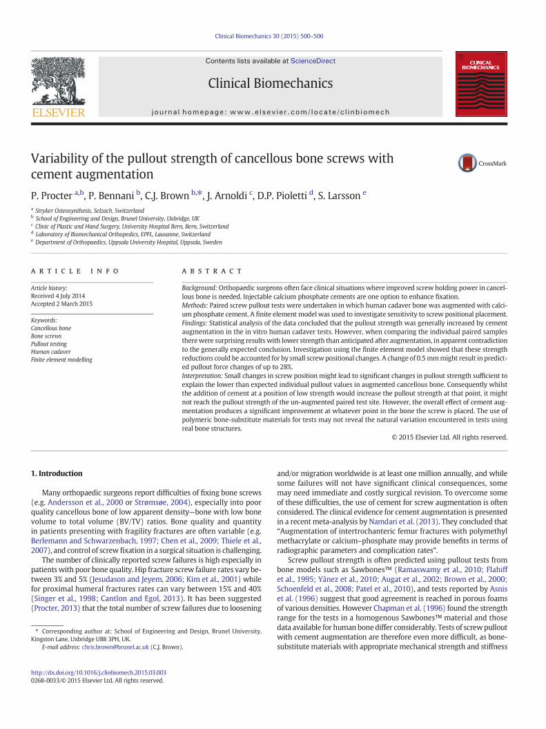
To place the screws and cement, the Falcon tube containing the spec-imens was plunged for about 1 h in a water container heated to 37 °C(Fig. 1). The samples, which had reached the body temperatureafter that time, were then placed in a purpose-made fixture box(Fig. 1) and a hole of 2.5 mm diameter was drilled 10 mm deep at thesame location and along the axis of the bone cylinder that had beenscanned. The hole was then tapped 10 mm deep with the correctmanufacturer-recommended tap for 4.0 mm screws. Finally, the speci-mens were placed back in the Falcon tubes and plunged in the 37 °Cbath.
The calcium phosphate cement (Hydroset®, Stryker) packs werestored for about 1 h in the room where the bone augmentation proce-dure took place. The room temperature was set at 19 °C. The bone/screw samples, kept in the Falcon tubes, were taken out from the37 °C warm bath just before augmentation. The Hydroset liquid wasmixed with the powder for 45 s. At 2 min, the mixture was injected inthe pre-drilled holes and at 3 min the screws (standard orthopaedicscrews 4 mm diameter × 35 mm long) were inserted 10 mm deep.After that the specimens were again placed in the special fixture boxand back into the Falcon tubes immediately after screwing, and thenplunged in the 37 °C bath for 4 h. The specimen selection process foraugmented or non-augmented was randomised. Where the bone sam-ple of the pair was not augmented, the same procedure for screw place-ment and temperature control was followed—with the omission ofcement injection. One randomly selected sample (numbered 2) was
Fig. 1. Falcon tube and specimen.
501P. Procter et al. / Clinical Biomechanics 30 (2015) 500–506
used to trial the protocol for both augmented and non-augmentedscrews.
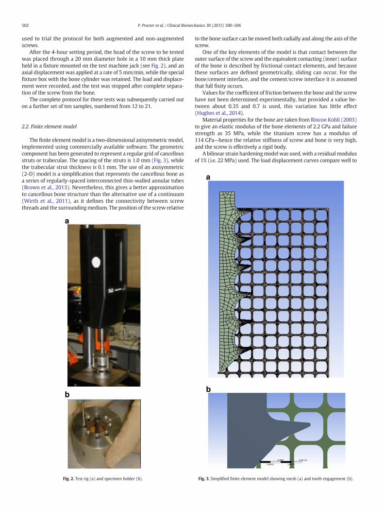
After the 4-hour setting period, the head of the screw to be testedwas placed through a 20 mm diameter hole in a 10 mm thick plateheld in a fixture mounted on the test machine jack (see Fig. 2), and anaxial displacement was applied at a rate of 5 mm/min, while the specialfixture box with the bone cylinder was retained. The load and displace-ment were recorded, and the test was stopped after complete separa-tion of the screw from the bone.
The complete protocol for these tests was subsequently carried outon a further set of ten samples, numbered from 12 to 21.
2.2. Finite element model
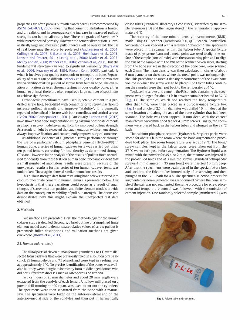
The finite elementmodel is a two-dimensional axisymmetricmodel,implemented using commercially available software. The geometriccomponent has been generated to represent a regular grid of cancellousstruts or trabeculae. The spacing of the struts is 1.0 mm (Fig. 3), whilethe trabecular strut thickness is 0.1 mm. The use of an axisymmetric(2-D) model is a simplification that represents the cancellous bone asa series of regularly-spaced interconnected thin-walled annular tubes(Brown et al., 2013). Nevertheless, this gives a better approximationto cancellous bone structure than the alternative use of a continuum(Wirth et al., 2011), as it defines the connectivity between screwthreads and the surroundingmedium. The position of the screw relative
to the bone surface can bemoved both radially and along the axis of thescrew.
One of the key elements of the model is that contact between theouter surface of the screw and the equivalent contacting (inner) surfaceof the bone is described by frictional contact elements, and becausethese surfaces are defined geometrically, sliding can occur. For thebone/cement interface, and the cement/screw interface it is assumedthat full fixity occurs.
Values for the coefficient of friction between the bone and the screwhave not been determined experimentally, but provided a value be-tween about 0.35 and 0.7 is used, this variation has little effect(Hughes et al., 2014).
Material properties for the bone are taken from Rincon Kohli (2003)to give an elastic modulus of the bone elements of 2.2 GPa and failurestrength as 35 MPa, while the titanium screw has a modulus of114 GPa—hence the relative stiffness of screw and bone is very high,and the screw is effectively a rigid body.
A bilinear strain hardeningmodel was used, with a residualmodulusof 1% (i.e. 22 MPa) used. The load displacement curves compare well to
Fig. 3. Simplified finite element model showing mesh (a) and tooth engagement (b).Fig. 2. Test rig (a) and specimen holder (b).
502 P. Procter et al. / Clinical Biomechanics 30 (2015) 500–506
those given by Andrews EWGibson (2001) who use a constant value ofresidual modulus of 25 MPa for similar reasons of numerical stability. Asimilar failure criterion has also been used, where the slope of the load–displacement characteristic reduces to 95% of the elastic value.
The bone is restrained at the largest radius (10 mm), and pullout isachieved by the incremental displacement of the screw along itsaxis—the results presented being the reaction forces.
3. Results
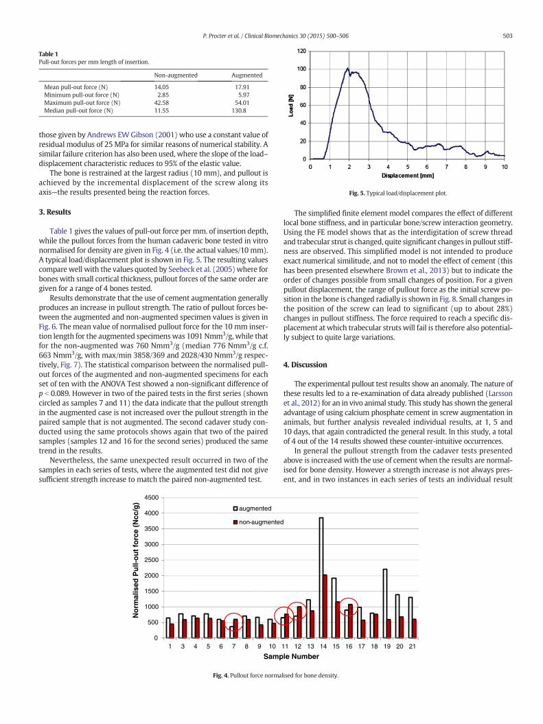
Table 1 gives the values of pull-out force per mm. of insertion depth,while the pullout forces from the human cadaveric bone tested in vitronormalised for density are given in Fig. 4 (i.e. the actual values/10 mm).A typical load/displacement plot is shown in Fig. 5. The resulting valuescomparewell with the values quoted by Seebeck et al. (2005)where forbones with small cortical thickness, pullout forces of the same order aregiven for a range of 4 bones tested.
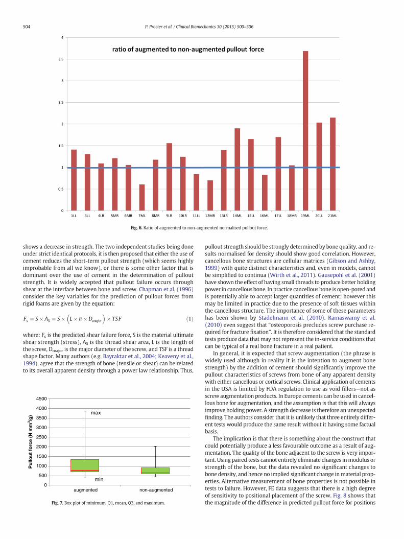
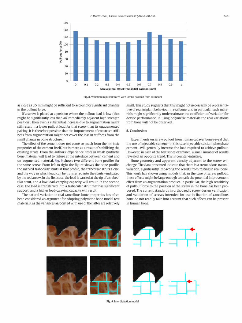
Results demonstrate that the use of cement augmentation generallyproduces an increase in pullout strength. The ratio of pullout forces be-tween the augmented and non-augmented specimen values is given inFig. 6. The mean value of normalised pullout force for the 10 mm inser-tion length for the augmented specimenswas 1091 Nmm3/g, while thatfor the non-augmented was 760 Nmm3/g (median 776 Nmm3/g c.f.663 Nmm3/g, with max/min 3858/369 and 2028/430 Nmm3/g respec-tively, Fig. 7). The statistical comparison between the normalised pull-out forces of the augmented and non-augmented specimens for eachset of ten with the ANOVA Test showed a non-significant difference ofp b 0.089. However in two of the paired tests in the first series (showncircled as samples 7 and 11) the data indicate that the pullout strengthin the augmented case is not increased over the pullout strength in thepaired sample that is not augmented. The second cadaver study con-ducted using the same protocols shows again that two of the pairedsamples (samples 12 and 16 for the second series) produced the sametrend in the results.
Nevertheless, the same unexpected result occurred in two of thesamples in each series of tests, where the augmented test did not givesufficient strength increase to match the paired non-augmented test.
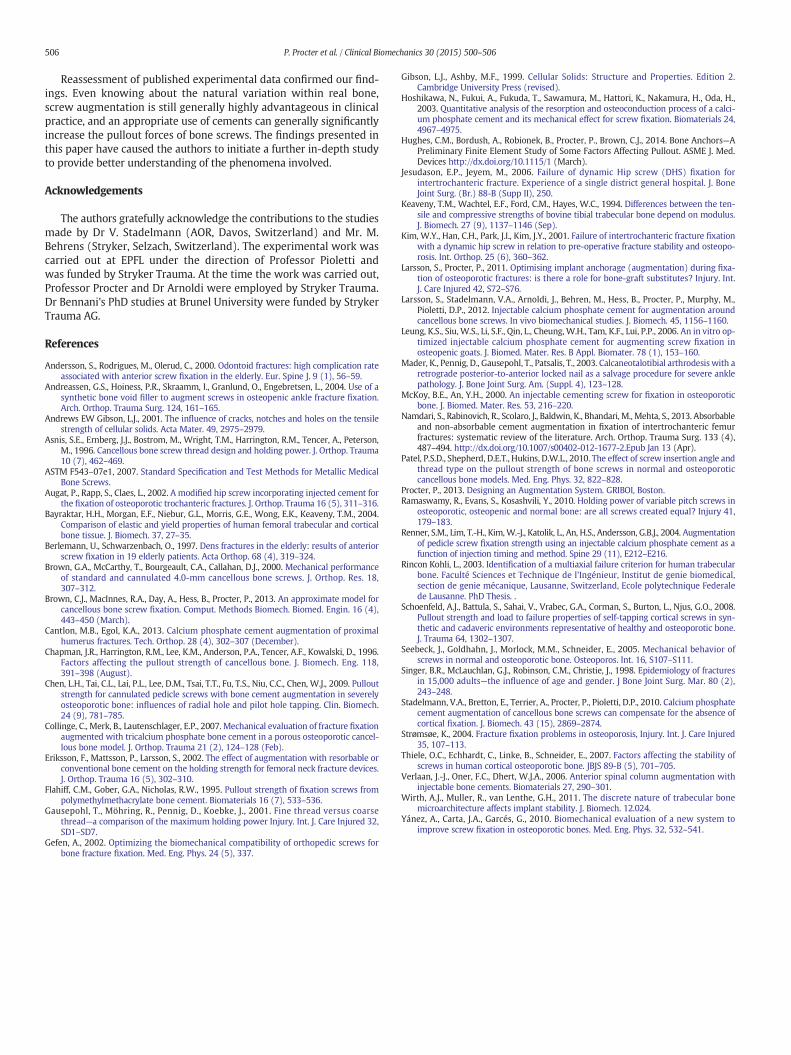
The simplified finite element model compares the effect of differentlocal bone stiffness, and in particular bone/screw interaction geometry.Using the FE model shows that as the interdigitation of screw threadand trabecular strut is changed, quite significant changes in pullout stiff-ness are observed. This simplified model is not intended to produceexact numerical similitude, and not to model the effect of cement (thishas been presented elsewhere Brown et al., 2013) but to indicate theorder of changes possible from small changes of position. For a givenpullout displacement, the range of pullout force as the initial screw po-sition in the bone is changed radially is shown in Fig. 8. Small changes inthe position of the screw can lead to significant (up to about 28%)changes in pullout stiffness. The force required to reach a specific dis-placement at which trabecular struts will fail is therefore also potential-ly subject to quite large variations.
4. Discussion
The experimental pullout test results show an anomaly. The nature ofthese results led to a re-examination of data already published (Larssonet al., 2012) for an in vivo animal study. This study has shown the generaladvantage of using calcium phosphate cement in screw augmentation inanimals, but further analysis revealed individual results, at 1, 5 and10 days, that again contradicted the general result. In this study, a totalof 4 out of the 14 results showed these counter-intuitive occurrences.
In general the pullout strength from the cadaver tests presentedabove is increased with the use of cement when the results are normal-ised for bone density. However a strength increase is not always pres-ent, and in two instances in each series of tests an individual result
0
500
1000
1500
2000
2500
3000
3500
4000
4500
1 3 4 5 6 7 8 9 10 11 12 13 14 15 16 17 18 19 20 21
Sample Number
augmented
non-augmented
Nor
mal
ised
Pul
l-out
forc
e (N
cc/g
)
Fig. 4. Pullout force normalised for bone density.
Table 1Pull-out forces per mm length of insertion.
Non-augmented Augmented
Mean pull-out force (N) 14.05 17.91Minimum pull-out force (N) 2.85 5.97Maximum pull-out force (N) 42.58 54.01Median pull-out force (N) 11.55 130.8
Fig. 5. Typical load/displacement plot.
503P. Procter et al. / Clinical Biomechanics 30 (2015) 500–506
shows a decrease in strength. The two independent studies being doneunder strict identical protocols, it is then proposed that either the use ofcement reduces the short-term pullout strength (which seems highlyimprobable from all we know), or there is some other factor that isdominant over the use of cement in the determination of pulloutstrength. It is widely accepted that pullout failure occurs throughshear at the interface between bone and screw. Chapman et al. (1996)consider the key variables for the prediction of pullout forces fromrigid foams are given by the equation:
Fs ¼ S" AS ¼ S" L" π " Dmajor
! "" TSF ð1Þ
where: Fs is the predicted shear failure force, S is the material ultimateshear strength (stress), AS is the thread shear area, L is the length ofthe screw, Dmajor is the major diameter of the screw, and TSF is a threadshape factor. Many authors (e.g. Bayraktar et al., 2004; Keaveny et al.,1994), agree that the strength of bone (tensile or shear) can be relatedto its overall apparent density through a power law relationship. Thus,
pullout strength should be strongly determined by bone quality, and re-sults normalised for density should show good correlation. However,cancellous bone structures are cellular matrices (Gibson and Ashby,1999) with quite distinct characteristics and, even in models, cannotbe simplified to continua (Wirth et al., 2011). Gausepohl et al. (2001)have shown the effect of having small threads to produce better holdingpower in cancellous bone. In practice cancellous bone is open-pored andis potentially able to accept larger quantities of cement; however thismay be limited in practice due to the presence of soft tissues withinthe cancellous structure. The importance of some of these parametershas been shown by Stadelmann et al. (2010). Ramaswamy et al.(2010) even suggest that “osteoporosis precludes screw purchase re-quired for fracture fixation”. It is therefore considered that the standardtests produce data that may not represent the in-service conditions thatcan be typical of a real bone fracture in a real patient.
In general, it is expected that screw augmentation (the phrase iswidely used although in reality it is the intention to augment bonestrength) by the addition of cement should significantly improve thepullout characteristics of screws from bone of any apparent densitywith either cancellous or cortical screws. Clinical application of cementsin the USA is limited by FDA regulation to use as void fillers—not asscrew augmentation products. In Europe cements can be used in cancel-lous bone for augmentation, and the assumption is that this will alwaysimprove holding power. A strength decrease is therefore an unexpectedfinding. The authors consider that it is unlikely that three entirely differ-ent tests would produce the same result without it having some factualbasis.
The implication is that there is something about the construct thatcould potentially produce a less favourable outcome as a result of aug-mentation. The quality of the bone adjacent to the screw is very impor-tant. Using paired tests cannot entirely eliminate changes inmodulus orstrength of the bone, but the data revealed no significant changes tobone density, and hence no implied significant change inmaterial prop-erties. Alternative measurement of bone properties is not possible intests to failure. However, FE data suggests that there is a high degreeof sensitivity to positional placement of the screw. Fig. 8 shows thatthe magnitude of the difference in predicted pullout force for positions
0
500
1000
1500
2000
2500
3000
3500
4000
4500
augmented non-augmented
Pul
lout
forc
e (N
mm
3 /g) max
min
Fig. 7. Box plot of minimum, Q1, mean, Q3, and maximum.
Fig. 6. Ratio of augmented to non-augmented normalised pullout force.
504 P. Procter et al. / Clinical Biomechanics 30 (2015) 500–506
as close as 0.5 mmmight be sufficient to account for significant changesin the pullout force.
If a screw is placed at a position where the pullout load is low (thatmight be significantly less than an immediately adjacent high strengthposition), then even a substantial increase due to augmentation mightstill result in a lower pullout load for that screw than its unaugmentedpairing. It is therefore possible that the improvement of construct stiff-ness from augmentation might not cover the loss in stiffness from thesmall change in bone structure.
The effect of the cement does not come so much from the intrinsicproperties of the cement itself, but is more as a result of stabilising theexisting struts. From the authors' experience, tests in weak syntheticbone material will lead to failure at the interface between cement andun-augmented material. Fig. 9 shows two different bone profiles forthe same screw. From left to right the figure shows the bone profile,the marked trabecular struts at that profile, the trabecular struts alone,and the way in which load can be transferred into the struts—indicatedby the red arrow. In thefirst case, the load is carried at the tip of a trabec-ular strut, and a low load-carrying capacity will result. In the secondcase, the load is transferred into a trabecular strut that has significantsupport, and a higher load-carrying capacity will result.
The natural variation in real cancellous bone properties has oftenbeen considered an argument for adopting polymeric bone model testmaterials, as the variances associatedwith use of the latter are relatively
small. This study suggests that this might not necessarily be representa-tive of real implant behaviour in real bone, and in particular such mate-rials might significantly underestimate the coefficient of variation fordevice performance. In using polymeric materials the real variationsfrom bone will not be observed.
5. Conclusion
Experiments on screw pullout from human cadaver bone reveal thatthe use of injectable cement—in this case injectable calcium phosphatecement—will generally increase the load required to achieve pullout.However, in each of the test series examined, a small number of resultsrevealed an opposite trend. This is counter-intuitive.
Bone geometry and apparent density adjacent to the screw willchange. The data presented indicate that there is a tremendous naturalvariation, significantly impacting the results from testing in real bone.This work has shown using models that, in the case of screw pullout,these effects might be large enough tomask the potential improvementeffect from an augmentation product. In particular, the high sensitivityof pullout force to the position of the screw in the bone has been pro-posed. The current standards in orthopaedic screw design verificationand validation of screws intended for use in fixation of cancellousbone do not readily take into account that such effects can be presentin human bone.
Fig. 9. Interdigitation model.
Fig. 8. Variation in pullout force with lateral position from FE model.
505P. Procter et al. / Clinical Biomechanics 30 (2015) 500–506
Reassessment of published experimental data confirmed our find-ings. Even knowing about the natural variation within real bone,screw augmentation is still generally highly advantageous in clinicalpractice, and an appropriate use of cements can generally significantlyincrease the pullout forces of bone screws. The findings presented inthis paper have caused the authors to initiate a further in-depth studyto provide better understanding of the phenomena involved.
Acknowledgements
The authors gratefully acknowledge the contributions to the studiesmade by Dr V. Stadelmann (AOR, Davos, Switzerland) and Mr. M.Behrens (Stryker, Selzach, Switzerland). The experimental work wascarried out at EPFL under the direction of Professor Pioletti andwas funded by Stryker Trauma. At the time the work was carried out,Professor Procter and Dr Arnoldi were employed by Stryker Trauma.Dr Bennani's PhD studies at Brunel University were funded by StrykerTrauma AG.
References
Andersson, S., Rodrigues, M., Olerud, C., 2000. Odontoid fractures: high complication rateassociated with anterior screw fixation in the elderly. Eur. Spine J. 9 (1), 56–59.
Andreassen, G.S., Hoiness, P.R., Skraamm, I., Granlund, O., Engebretsen, L., 2004. Use of asynthetic bone void filler to augment screws in osteopenic ankle fracture fixation.Arch. Orthop. Trauma Surg. 124, 161–165.
Andrews EW Gibson, L.J., 2001. The influence of cracks, notches and holes on the tensilestrength of cellular solids. Acta Mater. 49, 2975–2979.
Asnis, S.E., Ernberg, J.J., Bostrom, M., Wright, T.M., Harrington, R.M., Tencer, A., Peterson,M., 1996. Cancellous bone screw thread design and holding power. J. Orthop. Trauma10 (7), 462–469.
ASTM F543–07e1, 2007. Standard Specification and Test Methods for Metallic MedicalBone Screws.
Augat, P., Rapp, S., Claes, L., 2002. A modified hip screw incorporating injected cement forthe fixation of osteoporotic trochanteric fractures. J. Orthop. Trauma 16 (5), 311–316.
Bayraktar, H.H., Morgan, E.F., Niebur, G.L., Morris, G.E., Wong, E.K., Keaveny, T.M., 2004.Comparison of elastic and yield properties of human femoral trabecular and corticalbone tissue. J. Biomech. 37, 27–35.
Berlemann, U., Schwarzenbach, O., 1997. Dens fractures in the elderly: results of anteriorscrew fixation in 19 elderly patients. Acta Orthop. 68 (4), 319–324.
Brown, G.A., McCarthy, T., Bourgeault, C.A., Callahan, D.J., 2000. Mechanical performanceof standard and cannulated 4.0-mm cancellous bone screws. J. Orthop. Res. 18,307–312.
Brown, C.J., MacInnes, R.A., Day, A., Hess, B., Procter, P., 2013. An approximate model forcancellous bone screw fixation. Comput. Methods Biomech. Biomed. Engin. 16 (4),443–450 (March).
Cantlon, M.B., Egol, K.A., 2013. Calcium phosphate cement augmentation of proximalhumerus fractures. Tech. Orthop. 28 (4), 302–307 (December).
Chapman, J.R., Harrington, R.M., Lee, K.M., Anderson, P.A., Tencer, A.F., Kowalski, D., 1996.Factors affecting the pullout strength of cancellous bone. J. Biomech. Eng. 118,391–398 (August).
Chen, L.H., Tai, C.L., Lai, P.L., Lee, D.M., Tsai, T.T., Fu, T.S., Niu, C.C., Chen, W.J., 2009. Pulloutstrength for cannulated pedicle screws with bone cement augmentation in severelyosteoporotic bone: influences of radial hole and pilot hole tapping. Clin. Biomech.24 (9), 781–785.
Collinge, C., Merk, B., Lautenschlager, E.P., 2007. Mechanical evaluation of fracture fixationaugmented with tricalcium phosphate bone cement in a porous osteoporotic cancel-lous bone model. J. Orthop. Trauma 21 (2), 124–128 (Feb).
Eriksson, F., Mattsson, P., Larsson, S., 2002. The effect of augmentation with resorbable orconventional bone cement on the holding strength for femoral neck fracture devices.J. Orthop. Trauma 16 (5), 302–310.
Flahiff, C.M., Gober, G.A., Nicholas, R.W., 1995. Pullout strength of fixation screws frompolymethylmethacrylate bone cement. Biomaterials 16 (7), 533–536.
Gausepohl, T., Möhring, R., Pennig, D., Koebke, J., 2001. Fine thread versus coarsethread—a comparison of the maximum holding power Injury. Int. J. Care Injured 32,SD1–SD7.
Gefen, A., 2002. Optimizing the biomechanical compatibility of orthopedic screws forbone fracture fixation. Med. Eng. Phys. 24 (5), 337.
Gibson, L.J., Ashby, M.F., 1999. Cellular Solids: Structure and Properties. Edition 2.Cambridge University Press (revised).
Hoshikawa, N., Fukui, A., Fukuda, T., Sawamura, M., Hattori, K., Nakamura, H., Oda, H.,2003. Quantitative analysis of the resorption and osteoconduction process of a calci-um phosphate cement and its mechanical effect for screw fixation. Biomaterials 24,4967–4975.
Hughes, C.M., Bordush, A., Robionek, B., Procter, P., Brown, C.J., 2014. Bone Anchors—APreliminary Finite Element Study of Some Factors Affecting Pullout. ASME J. Med.Devices http://dx.doi.org/10.1115/1 (March).
Jesudason, E.P., Jeyem, M., 2006. Failure of dynamic Hip screw (DHS) fixation forintertrochanteric fracture. Experience of a single district general hospital. J. BoneJoint Surg. (Br.) 88-B (Supp II), 250.
Keaveny, T.M., Wachtel, E.F., Ford, C.M., Hayes, W.C., 1994. Differences between the ten-sile and compressive strengths of bovine tibial trabecular bone depend on modulus.J. Biomech. 27 (9), 1137–1146 (Sep).
Kim, W.Y., Han, C.H., Park, J.I., Kim, J.Y., 2001. Failure of intertrochanteric fracture fixationwith a dynamic hip screw in relation to pre-operative fracture stability and osteopo-rosis. Int. Orthop. 25 (6), 360–362.
Larsson, S., Procter, P., 2011. Optimising implant anchorage (augmentation) during fixa-tion of osteoporotic fractures: is there a role for bone-graft substitutes? Injury. Int.J. Care Injured 42, S72–S76.
Larsson, S., Stadelmann, V.A., Arnoldi, J., Behren, M., Hess, B., Procter, P., Murphy, M.,Pioletti, D.P., 2012. Injectable calcium phosphate cement for augmentation aroundcancellous bone screws. In vivo biomechanical studies. J. Biomech. 45, 1156–1160.
Leung, K.S., Siu, W.S., Li, S.F., Qin, L., Cheung,W.H., Tam, K.F., Lui, P.P., 2006. An in vitro op-timized injectable calcium phosphate cement for augmenting screw fixation inosteopenic goats. J. Biomed. Mater. Res. B Appl. Biomater. 78 (1), 153–160.
Mader, K., Pennig, D., Gausepohl, T., Patsalis, T., 2003. Calcaneotalotibial arthrodesis with aretrograde posterior-to-anterior locked nail as a salvage procedure for severe anklepathology. J. Bone Joint Surg. Am. (Suppl. 4), 123–128.
McKoy, B.E., An, Y.H., 2000. An injectable cementing screw for fixation in osteoporoticbone. J. Biomed. Mater. Res. 53, 216–220.
Namdari, S., Rabinovich, R., Scolaro, J., Baldwin, K., Bhandari, M., Mehta, S., 2013. Absorbableand non-absorbable cement augmentation in fixation of intertrochanteric femurfractures: systematic review of the literature. Arch. Orthop. Trauma Surg. 133 (4),487–494. http://dx.doi.org/10.1007/s00402-012-1677-2.Epub Jan 13 (Apr).
Patel, P.S.D., Shepherd, D.E.T., Hukins, D.W.L., 2010. The effect of screw insertion angle andthread type on the pullout strength of bone screws in normal and osteoporoticcancellous bone models. Med. Eng. Phys. 32, 822–828.
Procter, P., 2013. Designing an Augmentation System. GRIBOI, Boston.Ramaswamy, R., Evans, S., Kosashvili, Y., 2010. Holding power of variable pitch screws in
osteoporotic, osteopenic and normal bone: are all screws created equal? Injury 41,179–183.
Renner, S.M., Lim, T.-H., Kim,W.-J., Katolik, L., An, H.S., Andersson, G.B.J., 2004. Augmentationof pedicle screw fixation strength using an injectable calcium phosphate cement as afunction of injection timing and method. Spine 29 (11), E212–E216.
Rincon Kohli, L., 2003. Identification of a multiaxial failure criterion for human trabecularbone. Faculté Sciences et Technique de l’Ingénieur, Institut de genie biomedical,section de genie mécanique, Lausanne, Switzerland, Ecole polytechnique Federalede Lausanne. PhD Thesis. .
Schoenfeld, A.J., Battula, S., Sahai, V., Vrabec, G.A., Corman, S., Burton, L., Njus, G.O., 2008.Pullout strength and load to failure properties of self-tapping cortical screws in syn-thetic and cadaveric environments representative of healthy and osteoporotic bone.J. Trauma 64, 1302–1307.
Seebeck, J., Goldhahn, J., Morlock, M.M., Schneider, E., 2005. Mechanical behavior ofscrews in normal and osteoporotic bone. Osteoporos. Int. 16, S107–S111.
Singer, B.R., McLauchlan, G.J., Robinson, C.M., Christie, J., 1998. Epidemiology of fracturesin 15,000 adults—the influence of age and gender. J Bone Joint Surg. Mar. 80 (2),243–248.
Stadelmann, V.A., Bretton, E., Terrier, A., Procter, P., Pioletti, D.P., 2010. Calcium phosphatecement augmentation of cancellous bone screws can compensate for the absence ofcortical fixation. J. Biomech. 43 (15), 2869–2874.
Strømsøe, K., 2004. Fracture fixation problems in osteoporosis, Injury. Int. J. Care Injured35, 107–113.
Thiele, O.C., Echhardt, C., Linke, B., Schneider, E., 2007. Factors affecting the stability ofscrews in human cortical osteoporotic bone. JBJS 89-B (5), 701–705.
Verlaan, J.-J., Oner, F.C., Dhert, W.J.A., 2006. Anterior spinal column augmentation withinjectable bone cements. Biomaterials 27, 290–301.
Wirth, A.J., Muller, R., van Lenthe, G.H., 2011. The discrete nature of trabecular bonemicroarchitecture affects implant stability. J. Biomech. 12.024.
Yánez, A., Carta, J.A., Garcés, G., 2010. Biomechanical evaluation of a new system toimprove screw fixation in osteoporotic bones. Med. Eng. Phys. 32, 532–541.
506 P. Procter et al. / Clinical Biomechanics 30 (2015) 500–506


![EffectsofCondylarElasticPropertiesto ...downloads.hindawi.com/journals/bmri/2009/509848.pdf · cancellous bone [24], cortical bone [21], dentate mandible [18], enamel and dentin [25],](https://img.pdfslide.us/doc/110x75/5f3e17f75ef2332a7542381d/effectsofcondylarelasticpropertiesto-cancellous-bone-24-cortical-bone-21.jpg)