Embed Size (px)
Citation preview
Vapor Phase Exchange of Self-Assembled Monolayers forEngineering of Biofunctional SurfacesL. Kankate,*,†,‡ A. Aguf,† H. Großmann,§ M. Schnietz,† R. Tampe,§ A. Turchanin,∥ and A. Golzhauser†
†Department of Physics, Physics of Supramolecular Systems and Surfaces, University of Bielefeld, 33615 Bielefeld, Germany‡Fritz-Haber-Institut der Max-Planck-Gesellschaft, Faradayweg 4-6, 14195 Berlin, Germany§Institute of Biochemistry, Biocenter, Goethe-University, 60438 Frankfurt, Germany∥Institute of Physical Chemistry, Friedrich Schiller University Jena, Lessingstr. 10, 07743 Jena, Germany
ABSTRACT: We show that 4′-nitro-1,1′-biphenyl-4-thiol self-assembled monolayers (NBPT SAMs) on gold can be exchangedwith 11-(mercaptoundecyl)triethylene glycol (C11EG3OH) SAMs viavapor deposition (VD). The pristine and the exchanged SAMsobtained by VD as well as solution method (SM) were characterizedby X-ray photoelectron spectroscopy (XPS) and polarizationmodulation infrared reflection−absorption spectroscopy (PM-IRRAS). Using surface plasmon resonance (SPR), it is shown thatC11EG3OH SAMs on gold obtained by vapor deposition exchange(VDEx) have the same protein resistivity as SAMs obtained by thedirect self-assembly process. As expected, the cross-linked NBPTSAM are found to be resistive to both exchange processes, VDEx andsolution method exchange (SMEx). In this way, VDEx opens up anelegant and new approach of patterning SAM surfaces in situ atvacuum conditions without using any solvents. By combining electron irradiation-induced chemical lithography of NBPT SAMswith VDEx, biofunctional patterned substrates were engineered and used for immobilization of protein arrays.
■ INTRODUCTION
The exchange of self-assembled monolayers (SAMs) insolvents, i.e., replacement of one kind of SAM formingmolecules on a surface with another one dissolved in a solvent,is employed in both fundamental studies1−4 and nano-technology applications.5−10 The poly(ethylene glycol)(PEG)-terminated SAMs possess biorepulsive propertiespreventing the unspecific adsorption of proteins andcells.11−13 On the other hand, the phenyl rings in 4′-nitro-1,1′-biphenyl-4-thiol (NBPT) SAMs are cross-linked, and theterminal nitro groups are converted into amino groups viaelectron irradiation providing anchoring places for biofunction-alization.5 As the cross-linked, amino-terminated areas areresistive to the exchange reactions, it is possible, employingelectron beam lithography and subsequent chemical function-alization, to create biofunctional patterns in NBPT SAMs andto exchange the non-cross-linked areas with PEG-terminatedSAMs forming a protein resistive background in this way.5 Inanother approach an irradiation promoted exchange of SAMshas been conducted for protein immobilization on surfacesselectively.8−10 Typically for the preparation of SAMs andsolely for the exchange reactions the solution-based methods(SM) are used.1−10 However, the vapor (or gas phase)deposition (VD) of SAM is more advantageous.14−20 By thistechnique the SAM preparation is conducted in a definedenvironment providing high purity and reproducibility, which isadvantages from the technological point of view as well as the
possibility for the characterization of monolayers by vacuum-based analysis techniques like, e.g., electron spectroscopy. Todemonstrate the potential of this method for both fundamentalstudies and nanotechnology, we have employed the VDtechnique for growth of various aromatic and aliphatic SAMson gold and copper substrates.16−18 Here we report, to the bestof our knowledge for the first time, on a dry exchange of SAMsemploying vapor deposition in vacuum (VDEx) to pattern thesurfaces. We demonstrate that NBPT SAMs on gold can becompletely exchanged with 11-(mercaptoundecyl)triethyleneglycol (C11EG3OH) SAMs. The process is characterized by insitu X-ray photoelectron spectroscopy (XPS) and by ex situpolarization modulation infrared reflection−absorption spec-troscopy (PM-IRRAS). Using surface plasmon resonance(SPR) measurements, we show that C11EG3OH SAMsprepared by VDEx have the same protein resistive propertiesas SAMs prepared by the direct self-assembly in solvents. Weemploy electron beam projection lithography to generate thecross-linked areas in NBPT SAMs and to conduct the areaselective exchange of non-cross-linked SAMs with C11EG3OH.In this way we generate micropatterns for immobilization of theproteins arrays showing the potential of this new technique fornano-biotechnology applications.
Received: November 28, 2016Revised: February 27, 2017Published: March 24, 2017
Article
pubs.acs.org/Langmuir
© XXXX American Chemical Society A DOI: 10.1021/acs.langmuir.6b04207Langmuir XXXX, XXX, XXX−XXX
■ EXPERIMENTAL SECTIONSubstrates. For SAM preparation different types of gold substrates
were used depending on the applied characterization techniques.Thermally evaporated gold layers with a thickness of 30 or 100 nm onsilicon (100) wafers with a 5 nm thick titanium adhesive layer (G.Albert PVD-Coatings) were employed for XPS or PM-IRRAS,respectively. For SPR measurements the borosilicate glass substrates12 × 12 mm2 with a thickness of 0.3 mm (D263, PEO GmbH) werecleaned in a SC1 (standard cleaning solution 1:1 NH3 (25%) + 1H2O2 (30%) + 5 Milli-Q water) for 20 min, extensively rinsed withMilli-Q water, and dried with nitrogen. A 2−5 nm adhesion layer ofchrome and a 40−50 nm layer of gold were then thermally evaporatedin a Bal-Tec MED 020 vacuum coating system. The SAMs preparedon the different substrates showed a similar quality as characterized byXPS.Molecular Precursors for SAM Preparation. 4′-Nitro-1,1′-
biphenyl-4-thiol (NBPT, Taros, 99%) and 11-(mercaptoundecyl)-triethylene glycol (C11EG3OH, Sigma-Aldrich, 95%) were used. Atroom temperature, NBPT is solid and C11EG3OH is in liquid state.X-ray Photoelectron Spectroscopy (XPS). The XPS data were
recorded using a monochromatic Al X-ray source (1486.7 eV) and ahemispherical electron energy analyzer (Omicron, Sphera). Toprovide a precise energy calibration, the XPS binding energies werereferenced to the Au 4f7/2 peak at 84.0 eV. The XPS data weremeasured at a photoelectron emission angle, θ, of 8° with anacceptance angle of 14° of the energy analyzer and an energyresolution of 0.9 eV. For the analysis of the XP spectra, a Shirleybackground subtraction and symmetric Voigt functions were used forfitting.21 The separation between S 2p1/2 and Sp3/2 was fixed to 1.2 eV.To determine the film thickness, the Au 4f7/2 signal intensity wasassumed to be exponentially attenuated by the SAM overlayer ofthickness d, according to d = λ cos θ ln(I0(Au 4f7/2)/I(Au 4f7/2)),where I0(Au 4f7/2) and I(Au 4f7/2) signals are the Au 4f7/2 intensitiesfrom the bare and SAM covered Au surfaces, respectively. Thephotoelectron attenuation length, λ, was taken to be 36 Å.5,22 Thecalculation of element ratios was conducted in frames of the statisticalmodel.21
Polarization Modulation Infrared Reflection−AbsorptionSpectroscopy (PM-IRRAS). The infrared spectra were recordedusing a nitrogen-purged Bruker Vertex70 spectrometer with a PMA50polarization modulator unit. The spectra were taken at an incidentangle of 80° with a resolution of 4 cm−1. Depending on the region ofinterest the photoelastic modulator was operated at 3000 or 1200cm−1. A typical measurement time was about 10 min. As referencesamples, hexadecanethiol (Sigma-Aldrich, 95%) SAMs incubated atroom temperature on gold substrates in 1 mM ethanol solution for 24h were measured. For these samples asymmetric C−H stretchingmodes of the alkyl chains νa were typically observed at 2919 cm−1.Surface Plasmon Resonance (SPR). Measurements were
performed in a Biacore T100 (GE Healthcare) at a flow rate of 10μL/min. Substrates were mounted on the sample holder using double-sided tape. Bovine serum albumin (Roth) and myoglobin (Sigma)were dissolved in HBS (HEPES buffered saline: 20 mM HEPES, 150mM NaCl, prepared in Milli-Q, pH 7.5) at 1 and 5 mg/mL. Eachmeasurement was composed of the following steps: (i) 30 minequilibration with running buffer (HBS), (ii) injection of a 1 mg/mLprotein solution for 10 min followed by a dissociation phase of 5 min,(iii) injection of a 5 mg/mL protein solution for 10 min followed by adissociation phase of 5 min, and (iv) injection of a NaCl standardsolution (0.4 M in HBS). Steps i and iv were performed in all four flowchannels, while in steps ii and iii one flow channel was dedicated toone protein solution. To quantify the level of nonspecific proteinadhesion the SPR signal differences were calculated at defined timepoints before (t1 = 250 s) and after the injection (t2 = 1150 s) of theproteins. Because of possible thickness variations of the Au layer oneach sample, all values were normalized with the SPR signal shiftgenerated by the NaCl standard solution.Laser Scanning Microscopy (LSM). Laser scanning microscopy
was performed using a Plan-NeoFluor 63× oil immersion objective
(NA 1.4), an inverted microscope setup (Axiovert 200M, Carl Zeiss,Jena), and an Ar laser at 488 nm.
E-Beam Irradiation. Electron irradiation of VD and SM NBPTSAM through a stencil mask (from Quantifoil Jena) was carried outusing a flood gun (Specs FG20) in high vacuum (pressure <5 × 10⁻⁷
mbar). The energy of electrons was 100 eV. The used dose was 50mC/cm2. Some of the irradiation experiments were performed in theultrahigh vacuum chamber maintained at a pressure of 1 × 10⁻¹⁰ mbar,where the energy of irradiating electrons was 50 eV.
SAM Preparation by Vapor Deposition (VD). The experimentswere carried out in a UHV system (Multiprobe, Omicron) equippedwith a preparation chamber and an analysis chamber enabling in situXPS characterization of the samples. The base pressure of the analysischamber and the preparation chamber were 1 × 10⁻¹⁰ and 1 × 10⁻⁹
mbar, respectively. The organic molecules were evaporated from aKnudsen type evaporator (TCE-BSC, Kentax) containing three quartzcrucibles for evaporation of different compounds. Prior to depositionsubstrates were cleaned by argon ion sputtering (1 keV). The Knudsencell (evaporation) temperature T and the pressure during depositionwere 353 K and ∼2 × 10⁻⁸ mbar for both molecules. The time of thedeposition was 3 h (dose 30 langmuirs) for C11EG3OH and 2.5 h(dose 20 langmuirs) for NBPT (1 langmuir = 10−6 Torr·s). Thefurther details of NBPT and C11EG3OH VD SAM preparation arepresented in refs 16 and 17, respectively. To estimate the depositionpressure of C11EG3OH, the pressure values obtained by a vacuumgauge calibrated to pure nitrogen were corrected by the sensitivityfactor of 8.0 for decanethiol.19 The sensitivity factor of nitrobenzene,7.2,26 was used to obtain the pressure of NBPT.
SAM Exchange by Vapor Deposition (VDEx). The dry exchangeexperiments were performed as follows. First, NBPT was vapordeposited on gold substrate at room temperature. Then, the freshlyprepared NBPT SAM at RT was in situ, i.e., without any air exposure,exposed to the molecular beam of C11EG3OH (Knudsen celltemperature T = 353 K). To provide reproducible molecular fluxesin different experiments a fixed amount of C11EG3OH (0.3 mL) wasloaded into the evaporator, and the distance to the substrate was keptconstant (7 cm). To achieve the complete exchange, the dose of ∼50langmuirs was applied. The exposure time required to achieve the finalexchange was 5 h. After VDEx the samples were analyzed by XPS.
SAM Preparation by Solution Method (SM). C11EG3OH SAM.Gold (Au/Si or Au/glass) substrates were cleaned by giving them anultrasonic bath in ethanol and by UVO plasma cleaning. Subsequently,they were rinsed by ethanol and acetonitrile. The cleaned substrateswere incubated into the freshly prepared acetonitrile (HPLC grade)based 0.5 mM solution of C11EG3OH (Sigma-Aldrich, 95%) fordifferent times. Then the samples were given an ultrasonic bath in usedsolvents for 3 min and rinsed with the same solvent to remove thephysisorbed molecules. Thereafter, the samples were dried withnitrogen gas and transferred into the UHV apparatus for character-ization.
NBPT SAM. For preparation of the NBPT SAM by the solutionmethod, the above-mentioned substrate cleaning and sample washingsteps were repeated using only ethanol. The cleaned substrate wasincubated in dry dimethylformamide (DMF) based 10 mM NBPTsolution for 3 days. Subsequently, the sample was rinsed with DMF.Then the sample was given an ultrasonic bath in ethanol for 3 min andrinsed with ethanol and blown with nitrogen gas.
SAM Exchange by Solution Method (SMEx). The solution-basedNBPT SAM on gold was incubated in a freshly prepared acetonitrilebased 0.5 mM solution of C11EG3OH for different incubation times.Afterward, the above-mentioned cleaning procedure for C11EG3OHSAM was repeated.
TrisNTA Functionalization of Electron-Irradiated NBPT SAMin Solution. This step of the sample preparation is required toconfirm the formation of biocompatible patterns using the VDExprocedure. The chemicals are usually stored at −20 °C undernitrogen. To avoid residual gases condensation, the chemicals werewarmed to room temperature before opening the container. 3-(Maleimido)propionic acid N-hydroxysuccinimide ester (Sigma-Aldrich) was dissolved in 20 μL of dry dimethylformamide (DMF,
Langmuir Article
DOI: 10.1021/acs.langmuir.6b04207Langmuir XXXX, XXX, XXX−XXX
B
Merck) by dipping a glass pipet into the dry powder, transferring it tothe reaction tube containing the solvent, and mixing gently. Then, 5μL of this solution was applied onto the e-beam-irradiated samplesurface, which was then covered with a clean covering slide. After 20min samples were rinsed three times with dry DMF, cleaned in anultrasonic bath for 5 min in dry DMF, and dried with nitrogen.Chelator coupling was performed by adding 10 μL of 10 mM tris-N-nitrilotriacetic acid thiol (trisNTA thiol)23−25 in a HBS buffer (pH7.5) and covered that with the covering slide again. The chelator wasallowed to react with the surfaces for 2 h. Finally, samples werethoroughly rinsed with deionized water and dried with nitrogen. ThetrisNTA thiol was synthesized in our group.25
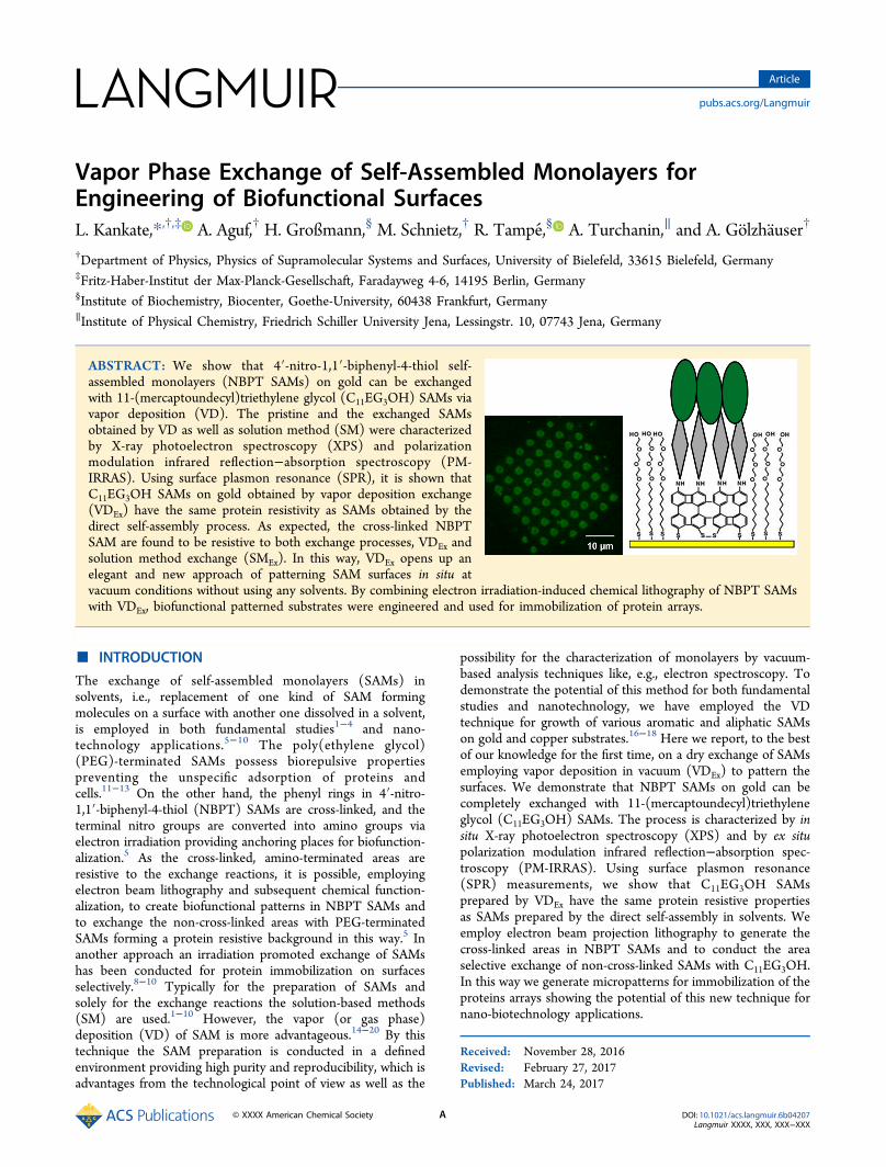
■ RESULTS AND DISCUSSIONExchanged SAMs and SAMs on Plain Gold. Figure 1
shows the XP spectra of C11EG3OH and NBPT SAMs. For
each an S 2p doublet is positioned at 162 and 163.2 eVrespectively for S 2p3/2 and S 2p1/2 which are assigned tothiolate species confirming the formation of sulfur−gold bonds.Neither physisorbed nor oxidized sulfur species were detected.For the NBPT SAM obtained by VD (Figure 1a), the C 1sspectrum consists of a main peak at 284.2 eV originated fromaromatic rings. A peak centered at 285.2 eV is assigned to C−Sand C−N bonding, and those at 287.5 and 291.6 eV are due tothe aromatic shake-up process.The peak at 532.5 eV originates from oxygen of terminal
nitro groups. This is consistent with the previously reportedsolution SAM.5,16 Note that in contrast to the solution basedNBPT SAM, the SAM prepared by VD undergoes a partialreduction due to released thiol hydrogen.16 Thus, we observetwo peaks in the N 1s spectrum: at 405.6 eV for NO2 and at399.3 eV due to NH2. The XPS data for the chemicalcomposition are gathered in Table 1.
Figure 1. XP spectra of vapor deposited (VD) NBPT SAM on 30 nmAu/Si (a); vapor (VD) and solution (SM)deposited C11EG3OH SAMson Au (c, e). Spectra depicted by (b) and (d) were obtained fromNBPT SAMs after their exposure to C11EG3OH molecules in vaporand solution showing clearly the NBPT SAM can be exchanged withC11EG3OH by both methods, VDEx and SMEx. VD NBPT SAM wasused for VDEx. Solution-based NBPT SAM was used for SMEx. Note incontrast to solution NBPT SAM on Au vapor deposited NBPT SAMare partially reduced showing two peaks in N 1s region, nitro andamino. Preparation conditions for C11EG3OH SAM: the times ofexposure were 5 h for VDEx (dose 50 langmuirs) and 3 h for VD (dose30 langmuirs). The time of incubation was 24 h for both SM andSMEx. Growth conditions for NBPT SAM: The time of exposure was2.5 h (dose 20 langmuirs). Surface temperature was RT for each.
Table 1. XPS Data Fitting Parameters and Effective Film Thickness for C11EG3OH Exchange SAMs and VD NBPT SAMs onAua
SAM d (Å) S 2p C 1s O 1s N 1s IC1s(i):IC1s(ii) O:N C:S C1s(ii):O1s
VDEx & SMEx C11EG3OH 19 162.0 (1.0) (i) 284.8 (1.2) 533.1 (1.4) NA 1.1 NA 16 1.7163.2 (1.0) (ii) 285.8 (1.4)
VD NBPT 11 162.0 (1.0) 284.2 (1.2) 532.5 (1.8) 405.6 (1.5) NA 1.8 11 NA163.2 (1.0) 285.2 (1.4) 399.3 (1.3)
287.5 (2.8)291.6 (3.7)
aThe fwhm values are depicted in parentheses. Peak positions of all C11EG3OH SAMs are identical. IC1s(i)/IC1s(ii) is alkane carbon to ethercarbon intensity ratio. The last three columns provide the various chemical compositions, which are reasonably in agreement with theoretical values.
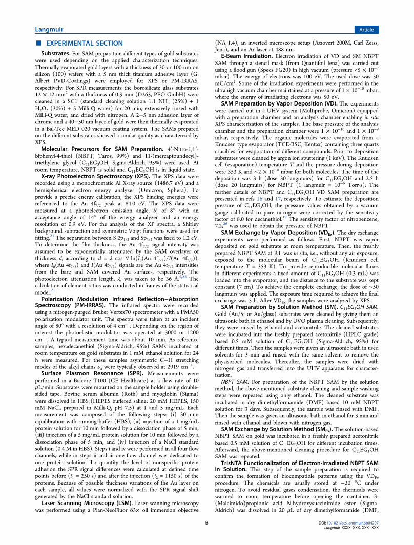
Figure 2. PM-IRRAS various stretching vibration spectra of SM basedNBPT SAM on Au (a), C11EG3OH SAM on Au by VD (c), and SM(e); C11EG3OH SAMs obtained by both exchange methods, VDEx (b)and SMEx (d). Alky and ether ν(C−H) modes are emphasized.Growth conditions: see Figure 1.
Langmuir Article
DOI: 10.1021/acs.langmuir.6b04207Langmuir XXXX, XXX, XXX−XXX
C
The calculation of element ratios was conducted in frames ofthe statistical model.21 The C:S and O:N ratios (for nitrogroups) are 11 and 1.8, respectively, which are in goodagreement with expected values. For C11EG3OH SAMs onplain gold surfaces obtained by VD and SM (Figure 1c,e), apeak at 533.1 eV is expected from oxygen of terminal ethergroups. The C 1s spectrum consists of two components,positioned at 284.8 and 286.8 eV accounting for carbon in thealkyl chain and in the ether part, respectively.17
The spectra in Figure 1b are obtained from VD based NBPTSAM which was exposed to the C11EG3OH molecular beam, inUHV for 5 h (exposure ∼50 langmuirs), in situ i.e. as-preparedNBPT SAM, without exposure to air. The solution basedNBPT SAM on Au was immersed in 0.5 mM acetonitrilesolution of C11EG3OH SAM for 24 h and characterized withXPS (Figure 1d). The nitrogen signal in both samplesdisappeared. The shapes of the C 1s and O 1s spectra clearlyshow that the NBPT SAM exchanged with C11EG3OH SAM byboth deposition methods. The spectral features and peakpositions of exchanged SAMs are identical to the SAMsprepared on clean gold surfaces. The maximum thicknesses ofC11EG3OH SAMs on plain Au surfaces were measured to be 19Å for VD and 18 Å for SM. For thickness calculationphotoelectron attenuation length, λ, was taken to be 36 Å.5,22
The thickness of the SAM obtained by both exchange methods(VDEx and SMEx) was 19 Å. By assuming the all-trans alkanechain with a tilt angle of 30° and helical configuration of ethergroups the calculated thickness of the monolayer should be 23Å.17,27 As revealed by PM-IRRAS measurements (as discussedlatter), the configuration of terminal ether groups of all the
SAMs is mainly amorphous like, and therefore the measuredthicknesses are slightly lower than the calculated value. The C:Sratio was calculated to be 16, which is in good agreement withthe theoretical value of 17. The ether carbon to oxygen ratio
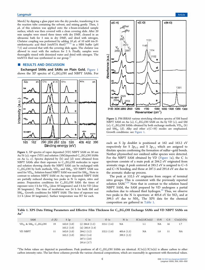
Figure 3. SPR sensogram shows two injections of BSA (1 g/mL and 5mg/mL) applied to different C11EG3OH SAM covered and bare Ausurfaces. Raw data were normalized to the Au surface using a 0.4 MNaCl standard solution. SAMs were prepared on 50 nm Au/glass, withgrowth conditions mentioned in Figure 1.
Table 2. Normalized Values of Adsorbed Protein in pg/mm2 after an Injection of a 1 mg/mL Protein Solution for 10 min forC11EG3OH SAM on Au/Glass, Obtained by Various Methodsa
protein plain Au SM VD VDEx
BSA 2000.72 19.74 (0.99%) 32.62 (1.63%) 44.88 (2.24%)myoglobin 1991.41 49.88 (2.51%) 27.89 (1.40%) 37.33 (1.87%)
aValues in parentheses represent percentages of nonspecific protein adsorption based on the signal measured on the plain Au surface (i.e., withoutSAM).
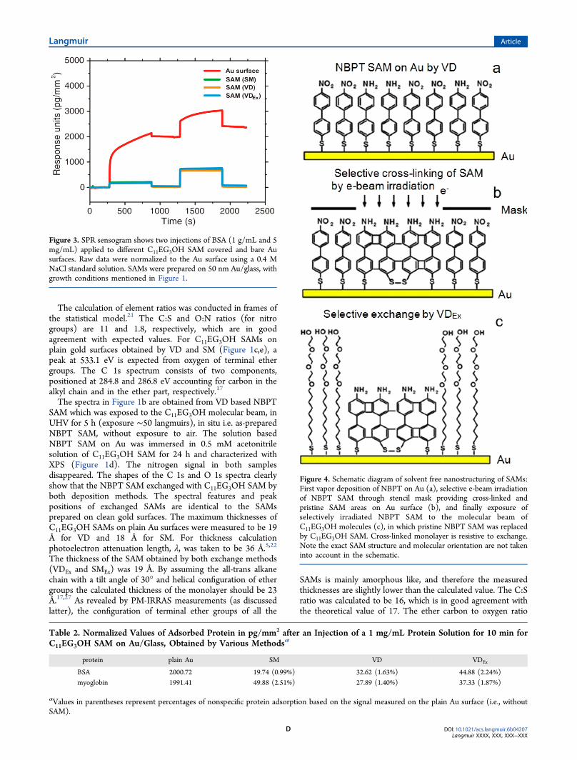
Figure 4. Schematic diagram of solvent free nanostructuring of SAMs:First vapor deposition of NBPT on Au (a), selective e-beam irradiationof NBPT SAM through stencil mask providing cross-linked andpristine SAM areas on Au surface (b), and finally exposure ofselectively irradiated NBPT SAM to the molecular beam ofC11EG3OH molecules (c), in which pristine NBPT SAM was replacedby C11EG3OH SAM. Cross-linked monolayer is resistive to exchange.Note the exact SAM structure and molecular orientation are not takeninto account in the schematic.
Langmuir Article
DOI: 10.1021/acs.langmuir.6b04207Langmuir XXXX, XXX, XXX−XXX
D
was measured to be about 1.6, i.e., close to the theoretical valueof 1.5. The intensity ratio of alkyl to ether carbon is ∼1.1, whichis much lower than the expected value of 1.8. Obviously, this isdue to the attenuation of the carbon signal originated from thealkyl chain, suggesting the existence of a standing phase of themolecular backbone in the SAM.Furthermore, the cross-linked ABPT (amino-terminated
BPT) SAM was obtained by exposing the VD NBPT SAM toelectrons with the energy of 50 eV and dose of 50 mC/cm2
under UHV.5 The cross-linked SAM was then exposed to theC11EG3OH molecular beam under conditions identical to thosementioned above of the final exchange by VD. As expected, thecross-linked SAM was not exchanged as confirmed by XPS.A reverse exchange of SAMs, i.e., exchanging C11EG3OH
SAM with NBPT SAM was not observed in the case of bothexchange methods under conditions similar to those mentionedabove, or even with increasing the molecular flux by of theorder of 1 or the solution concentration up to 1 mM andincreasing the incubation time up to 90 h.
Polarization Modulation Infrared Reflection−Absorp-tion Spectroscopy (PM-IRRAS) Characterization. Figure 2depicts PM-IRRAS spectra for solution NBPT and C11EG3OH(both exchange and usual) SAMs on 100 nm Au/Si. The peakassignment is based on refs 27−30. The IR results are inagreement with XPS data, confirming that NBPT SAM isexchanged with C11EG3OH SAM by VDEx and SMEx. ForNBPT SAM, C−H aromatic stretching vibrations are observedat 2964, 2929, and 2856 cm−1 (Figure 2a). Note that thepositions of these peaks in SAMs are largely shifted towardhigher wavelengths compared to C−H stretching of phenylmolecules in liquid (for instance, these bands for nitrobenzenein liquid state are observed at 3111, 3093, and 3031 cm−1).30
However, the corresponding C−H stretching vibration ofbiphenyl thiols (without functional groups) SAMs on gold donot exhibit such a large shift.29 The cause of this shift in peakposition is not yet clearly understood; probably this might beconnected to the order of the monolayer. The spectra of NBPTsamples exposed to C11EG3OH molecules in the solution andvapor phase (Figure 2b,d) clearly show that the C−H aromaticstretching vibrations vanish and new bands appear at 2921 and2851 cm−1, which are attributed to asymmetric (νa) andsymmetric (νs) C−H stretching modes of alkyl chains,respectively. The C−H symmetric stretching (νs) of highlyordered alkanethiol SAMs on gold is expected at 2917 cm−1
(and that of neat liquid is at 2925 cm−1), suggesting theC11EG3OH SAM having more gauche defects than alkanethiolsSAMs on gold surfaces.27 The bands at 2950 and 2870 cm−1 areexpected from asymmetric (νa) and symmetric (νs) C−Hstretching modes of ether groups.27,28 The peak positions andshapes of both exchange SAMs and SAMs obtained on plain Ausurfaces are identical.The main peak of C−O−C stretching of VD SAMs appears
reproducibly at about 1131 cm−1; however, that for solutionSAMs shows a large variation from sample to sample, 1125−1140 cm−1 (in Figure 2, C−O−C peak positions are 1127 and1140 cm−1 respectively for SMEx and SM samples). Thisvariation in peak positions is probably due to the adsorption ofsome water molecules on the ether chain during the formationof SAM in ambient.31 This causes the change in the structure ofSAMs.17 These peak positions are expected from theamorphous structure of terminal ether groups, which seemsto be important for protein resistivity characteristics ofSAMs.27,28 The main peaks are accompanied by weak shoulders
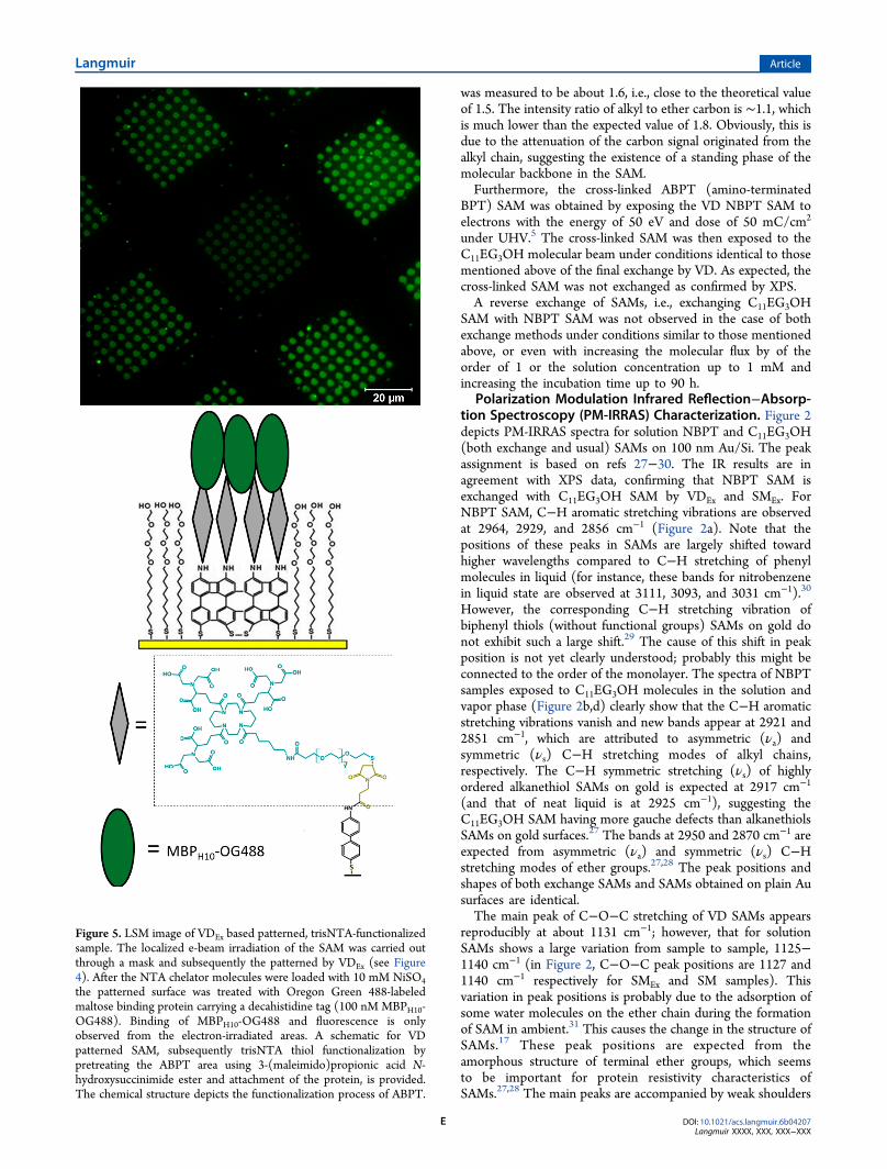
Figure 5. LSM image of VDEx based patterned, trisNTA-functionalizedsample. The localized e-beam irradiation of the SAM was carried outthrough a mask and subsequently the patterned by VDEx (see Figure4). After the NTA chelator molecules were loaded with 10 mM NiSO4the patterned surface was treated with Oregon Green 488-labeledmaltose binding protein carrying a decahistidine tag (100 nM MBPH10-OG488). Binding of MBPH10-OG488 and fluorescence is onlyobserved from the electron-irradiated areas. A schematic for VDpatterned SAM, subsequently trisNTA thiol functionalization bypretreating the ABPT area using 3-(maleimido)propionic acid N-hydroxysuccinimide ester and attachment of the protein, is provided.The chemical structure depicts the functionalization process of ABPT.
Langmuir Article
DOI: 10.1021/acs.langmuir.6b04207Langmuir XXXX, XXX, XXX−XXX
E
at 1102, 1114, and 1145 cm−1 that originate from the helicaland all-trans configurations,27,28 respectively. The peak at 1351cm−1 stems from asymmetric O−N−O stretching mode, whichdoes not completely disappear in both types of exchangesamples thus the samples still comprise some amount of NBPT(∼5%) molecules. The removal of the rest amount of NBPT isdifficult even heating the sample to 80 °C. Note that theunbound NBPT molecules can also be desorbed at thistemperature. This is not surprising since it is not possible to getclose-packed monolayers from C11EG3OH
17 molecules, so arelatively large number of defects on surfaces are expected,which can be occupied by NBPT as thiolate and NBPT thiolateshould survive up to at 100 °C.16 Nevertheless, theincorporation of a small number of NBPT molecules doesnot affect the protein resistivity characteristics of the SAM asdiscussed below.Protein Resistivity Test by SPR. To study the protein
resistance of C11EG3OH SAMs obtained by exchange in vapordeposition (VDEx) and comparing them to SAMs usuallyprepared on plain gold surfaces (obtained by VD as well asSM), surface plasmon resonance (SPR) experiments wereperformed. Two different protein solutions (containing bovineserum albumin (BSA) and myoglobin) with a concentration of1 and 5 mg/mL were injected onto each surface. To check thequality of the SAMs in terms of the protein resistance a bareAu/glass substrate was included as negative control. Aquantification of the level of the nonspecific protein adsorptionwas done by comparing the SPR signals before and after theinjection of the proteins.Figure 3 shows the results for BSA. Upon injection of the 1
mg/mL protein onto the bare Au surface a strong shift of theSPR response is observed. Two components of this shift can bedistinguished: The first, immediate one is due to the higherrefractive index of the protein solution compared to therefractive index of the running buffer; the second one, which isslower and which shows a characteristic curvature, is caused bythe adsorption of proteins onto the Au surface. After theinjection is stopped and the running buffer is flown over thesurface again, the SPR signal rapidly decreases, which, again, isbased on the refractive index change of the solutions. It remainsthen at a level of about 2000 RU (response units) higher thanbefore the injection, indicating an increase of the surface massof about 2 ng/mm2 of the adsorbed protein. A subsequentinjection of a solution containing 5 times as much proteinfurther increases the signal to 2400 RU. The mass increase afterthe second injection is much lower, which suggests that most ofthe binding sites were already occupied during first proteininjection and the surface coverage is nearly saturated.The above experiment was repeated with C11EG3OH SAMs
prepared by SM, VD, and VDEx on gold/glass. The mostobvious difference of the SAMs compared to the bare goldsurface is a rectangular signal progression without clear bindingkinetics during the first injection. Only the refractive index shiftupon injection of the protein solution was observed. When theinjection was stopped, the signal returned nearly to its initialvalue, stabilizing at around 30 RU. Thus, only a very smallamount of BSA remained on surfaces. This was observed forboth proteins that were applied, indicating the biocompatiblecharacteristics of the tested C11EG3OH SAMs. To quantifythese properties further, we calculated the signal differences(ΔRU) before and after the first injection for each surface andfor each protein. Since all substrates were self-made a variationin gold thickness could not be excluded. Therefore, the RU
values for the measured samples had to be normalized. To thisend the signal shift of a 0.4 M NaCl solution was measured onall surfaces and used as a reference for the signals measuredduring protein injection. The results are included in Table 2.Compared to the bare Au surface the percentage of nonspecificadsorption of the tested proteins was roughly between ∼1%and 2% on all SAMs. This demonstrates the equivalent qualityof SAMs with respect to protein resistance, no matter if theSAM was prepared by SM, VD, or VDEx.
Patterning the Surfaces for Protein Chips Application.As mentioned before, the cross-linked monolayer is resistive tothe exchange whereas the pristine monolayer can be replacedby C11EG3OH SAM. It is possible to obtain the nanoscaledpatterns using VD in the following way (see schematic inFigure 4). First we conducted the electron irradiation of NBPTSAM obtained by VD (and also SM NBPT SAM) through astencil mask to generate amino-terminated cross-linked SAMareas and pristine SAM areas on surfaces according to theprocedure reported previously for the solution NBPT SAM5
(see Figure 4b). The energy of irradiating electrons was 100 eV,and the dose used was of 50 mC/cm2. Thereafter, NBPTmolecules were exchanged with C11EG3OH by VD. Thisprovides us with a contamination-free biocompatible nano-structured pattern on the surface obtained by using vapordeposition, without using any solvent. This structure consists ofthe area of amino-terminated cross-linked biphenyl thiol(ABPT) SAM and C11EG3OH SAM (see Figure 4c). For acontrol this VDEx nanostructuring process was also conductedon the solution based NBPT SAM.In order to confirm the pattern formation by VD exchange
strategy, which is interesting for application as protein chips,the ABPT areas of both structured samples were functionalizedwith trisNTA chelators as described in the ExperimentalSection, and specific protein binding was checked by loadingthe chelators with Ni2+ ions and adding MBPH10-OG488. Inorder to activate the chelators for binding of His-taggedproteins, the structure surfaces were treated with 10 mMNiSO4, followed by adding 1 M imidazole to removenonspecifically adsorbed Ni2+ ions. After that Oregon Green488-labeled maltose binding protein carrying a decahistidine tag(MBPH10-OG488) with a concentration of 100 nM was added.The procedure for the synthesis of MBPH10 is reported in thepast.32 The incubation time of each step was 5 min and wasfollowed by a thorough rinsing with HBS-T (20 mM HEPES,150 mM NaCl, 0.05% Tween P20, prepared in Milli-Q, pH7.5). It should be noted that the NTA adsorbent when chargedwith Ni2+ ions has a high affinity for the proteins carryinghistidine tag. This concept was used in chromatography for thespecific binding of the proteins,33,34 and it was further adoptedfor the NTA-functionalized SAMs.24,25 The specific binding ofthe proteins this way was confirmed for trisNTA-functionalizedirradiated NBPT SAMs.5,35 Detailed information for thestructure of Ni2+ ions embedded in NTA matrix and itsspecific interaction with the His-tagged protein can be found inref 24. In ref 35, MBPH10 was injected on Ni2+ ions loadedtrisNTA thiol-functionalized ABPT SAMs, and subsequently itwas washed with imidazole and ethylenediaminetetraacetic acid(EDTA) to detach Ni2+ ions from the surfaces. It was possibleto remove the protein completely after washing the sample asconfirmed by SPR32 and by atomic force microscopy.5 Thus,the adsorption of the protein is specific to Ni2+ ions inbuilt intriNTA. The protein chip can be regenerated and reused formultiple times.5,35
Langmuir Article
DOI: 10.1021/acs.langmuir.6b04207Langmuir XXXX, XXX, XXX−XXX
F
For imaging each sample was mounted face down in a self-made sample holder. Figure 5 shows the corresponding laserscanning microscopy (LSM) image. On the sample thefluorescence patterns closely resemble the mask that was usedfor the e-beam lithography. Therefore, the fluorescence onlyappears in areas where the surface was irradiated by theelectron while the outside areas remain dark. This result provestwo important things. First, since binding of MBPH10-OG488only occurs in irradiated areas, the trisNTA molecules can onlybe found there; i.e., the coupling reactions are restricted solelyto the cross-linked ABPT regions. Second, the areas aroundpatterns were dark because the molecules from the pristineSAM were replaced by protein resistive C11EG3OH SAM. TheVDEx-based nanostuctured pattern obtained using eithersolution-based NBPT or VD NBPT on Au gave the sameresult. A schematic in Figure 5 exhibits VD patterned SAMused for immobilization of MBPH10-OG488 on trisNTA thiol-functionalized ABPT area. The chemical structure depicts thetrisNTA functionalization of ABPT. The chemical reaction forthe same was reported previously.35
■ CONCLUSIONS
We have found a new way of exchanging NBPT SAM withC11EG3OH SAM on Au surfaces, i.e., exchange by vapordeposition (VDEx) in ultrahigh vacuum. The quality, the filmthickness, and protein resistivity of VDEx SAM are very similarto SAMs usually obtained on plain gold surfaces. The cross-linked SAMs are resistive to vapor (similar to solution)exchange. This opens up a new way of generating nanostructureunder ultrahigh vacuum conditions, without using solvents. Asan example of combining electron induced chemical lithog-raphy and VDEx, the nanoscaled pattern surfaces containingcross-linked ABPT and C11EG3OH SAM areas were generatedusing vapor deposition. The pattern surfaces were further usedfor protein chip application. The reported results open broadavenues for implementation of the vapor deposition techniquesin engineering of biofunctional surfaces and interfaces.
■ AUTHOR INFORMATION
Corresponding Author*E-mail [email protected].
ORCIDL. Kankate: 0000-0002-6624-8012R. Tampe: 0000-0002-0403-2160NotesThe authors declare no competing financial interest.
■ REFERENCES(1) Chidsey, C. E. D.; Bertozzi, C. R.; Putvinski, T. M.; Mujsce, A. M.Coadsorption of Ferrocene-Terminated and Unsubstituted Alkane-thiols on Gold: Electroactive Self-Assembled Monolayers. J. Am. Chem.Soc. 1990, 112, 4301−4306.(2) Schlenoff, J. B.; Li, M.; Ly, H. Stability and Self-Exchange inAlkanethiol Monolayers. J. Am. Chem. Soc. 1995, 117, 12528−12536.(3) Saavedra, H. M.; Barbu, C. M.; Dameron, A. A.; Mullen, T. J.;Crespi, V. H.; Weiss, P. S. 1-Adamantanethiolate MonolayerDisplacement Kinetics Follow a Universal Form. J. Am. Chem. Soc.2007, 129, 10741−10746.(4) Hohman, J. N.; Thomas, J. C.; Zhao, Y.; Auluck, H.; Kim, M.;Vijselaar, W.; Kommeren, S.; Terfort, A.; Weiss, P. S. ExchangeReactions between Alkanethiolates and Alkaneselenols on Au{111}. J.Am. Chem. Soc. 2014, 136, 8110−8121.
(5) Turchanin, A.; Tinazli, A.; El-Desawy, M.; Großmann, H.;Schnietz, M.; Solak, H. H.; Tampe, R.; Golzhauser, A. Molecular Self-Assembly, Chemical Lithography, and Biochemical Tweezers: A Pathfor the Fabrication of Functional Nanometer-Scale Protein Arrays.Adv. Mater. 2008, 20, 471−477.(6) Khan, M. N.; Zharnikov, M. Irradiation Promoted ExchangeReaction with Disulfide Substituents. J. Phys. Chem. C 2013, 117,14534−14543.(7) Khan, M. N.; Zharnikov, M. Fabrication of ss DNA/Oligo-(ethylene glycol) Monolayers by Promoted Exchange Reaction withThiol and Disulfide Substituents. J. Phys. Chem. C 2014, 118, 3093−3101.(8) Yan, R.; Le Pleux, L.; Mayor, M.; Zharnikov, M. PromotedExchange Reaction between Alkanethiolate Self-Assembled Mono-layers and an Azide-Bearing Substituent. J. Phys. Chem. C 2016, 120,25967−25976.(9) Jeyachandran, Y. L.; Zharnikov, M. Comprehensive Analysis ofthe Effect of Electron Irradiation on Oligo(ethylene glycol)Terminated Self-Assembled Monolayers Applicable for Specific andNonspecific Patterning of Proteins. J. Phys. Chem. C 2012, 116,14950−14959.(10) Jeyachandran, Y. L.; Meyerbroker, N.; Terfort, A.; Zharnikov,M. Maskless Ultraviolet Projection Lithography with a BiorepellingMonomolecular Resist. J. Phys. Chem. C 2015, 119, 494−501.(11) Pale-Grosdemange, C.; Simon, E. S.; Prime, K. L.; Whitesides,G. M. Formation of Self-Assembled Monolayers by Chemisorption ofDerivatives of Aligo(ethylene glycol) of Structure HS(CH2)11(OCH2CH2)mOH on Gold. J. Am. Chem. Soc. 1991, 113, 12−20.(12) Herrwerth, S.; Eck, W.; Reinhardt, S.; Grunze, M. Factors thatDetermine the Protein Resistance of Oligo ether Self-AssembledMonolayers − Internal Hydrophilicity, Terminal Hydrophilicity, andLateral Packing Density. J. Am. Chem. Soc. 2003, 125, 9359−9366.(13) Singhvi, R.; Kumar, A.; Lopez, G. P.; Stephanopoulos, G. N.;Wang, D. C.; Whitesides, G. M.; Ingber, D. E. Engineering Cell Shapeand Function. Science 1994, 264, 696−698.(14) Poirier, G. E.; Pylant, E. D. The Self-Assembly Mechanism ofAlkanethiols on Au(111). Science 1996, 272, 1145−1148.(15) Schreiber, F. Structure and Growth of Self-AssemblingMonolayers. Prog. Surf. Sci. 2000, 65, 151−256.(16) Kankate, L.; Turchanin, A.; Golzhauser, A. On the Release ofHydrogen from the S-H Groups in the Formation of Self-AssembledMonolayers of Thiols. Langmuir 2009, 25, 10435−10438.(17) Kankate, L.; Großmann, H.; Werner, U.; Tampe, R.; Turchanin,A.; Golzhauser, A. Protein Resistant Oligo(ethylene glycol) Termi-nated Self-Assembled Monolayers of Thiols on Gold by VaporDeposition in Vacuum. Biointerphases 2010, 5, 30−36.(18) Matei, D. G.; Weber, N.-E.; Kurasch, S.; Wundrack, S.;Woszczyna, M.; Grothe, M.; Weimann, T.; Ahlers, F.; Stosch, R.;Kaiser, U.; Turchanin, A. Functional Single-Layer Graphene Sheetsfrom Aromatic Monolayers. Adv. Mater. 2013, 25, 4146−4151.(19) Eberhardt, A.; Fenter, P.; Eisenberger, P. Growth Kinetics inSelf-Assembling Monolayers: A Unique Adsorption Mechanism. Surf.Sci. 1998, 397, L285−L290.(20) Poirier, G. E. Mechanism of Formation of Au Vacancy Islands inAlkanethiol Monolayers on Au(111). Langmuir 1997, 13, 2019−2026.(21) Briggs, D.; Grant, J. T. Surface Analyses by Auger and X-RayPhotoelectron Spectroscopy; Surface Spectra Limited: Chichester, UK,2003.(22) Laibinis, P. E.; Bain, C. D.; Whitesides, G. M. Attenuation ofPhotoelectrons in Monolayers of n-Alkanethiols Adsorbed on Copper,Silver, and Gold. J. Phys. Chem. 1991, 95, 7017−7021.(23) Baldauf, C.; Schulze, K.; Lueders, P.; Bordignon, E.; Tampe, R.In-situ spin labeling of his-tagged proteins: distance measurementsunder in-cell conditions. Chem. - Eur. J. 2013, 19, 13714−13719.(24) Lata, S.; Reichel, A.; Brock, R.; Tampe, R.; Piehler, J. HighAffinity Adoption for Switchable Recognition of Histidine-TaggedProteins. J. Am. Chem. Soc. 2005, 127, 10205−10215.(25) Tinazli, A.; Tang, J.; Valiokas, R.; Picuric, S.; Lata, S.; Piehler, J.;Liedberg, B.; Tampe, R. High Affinity Chelator Thiols for Switchable
Langmuir Article
DOI: 10.1021/acs.langmuir.6b04207Langmuir XXXX, XXX, XXX−XXX
G
and Oriented Immobilization of Histidine-Tagged Proteins: A GenericPlatform for Protein Chip Technology. Chem. - Eur. J. 2005, 11,5249−5259.(26) Summers, R. L. NASA technical note, 1969.(27) Harder, H.; Grunze, M.; Dahint, R.; Whitesides, G. M.; Laibinis,P. E. Molecular Conformation in Oligo(ethylene glycol)-TerminatedSelf-Assembled Monolayers on Gold and Silver Surfaces DeterminesTheir Ability To Resist Protein Adsorption. J. Phys. Chem. B 1998,102, 426−436.(28) Valiokas, R.; Svedhem, S.; Ostblom, M.; Svensson, S. C.;Liedberg, T. B. Influence of Specific Intermolecular Interactions on theSelf-Assembly and Phase Behavior of Oligo(Ethylene Glycol)Terminated Alkanethiolates on Gold. J. Phys. Chem. B 2001, 105,5459−5469.(29) Geyer, W.; Stadler, V.; Eck, W.; Zharnikov, M.; Golzhauser, A.;Grunze, M. Electron-Induced CrossLinking of Aromatic Self-Assembled Monolayers: Negative Resists for Nanolithography. Appl.Phys. Lett. 1999, 75, 2401−2403.(30) Laposa, J. D. Vibrational Spectra of Nitrobenzene-d5.Spectrochim. Acta, Part A 1979, 35, 65−71.(31) Skoda, M. W.; Jacobs, R. M.; Willis, J.; Schreiber, F. Hydrationof Oligo(ethylene glycol) Self-Assembled Monolayers Studied UsingPolarization Modulation Infrared Spectroscopy. Langmuir 2007, 23,970−974.(32) Lata, S.; Piehler, J. Stable and Functional Immobilization ofHistidine-Tagged Proteins via Multivalent Chelator Head groups on aMolecular Polyethylene glycol) Brush. Anal. Chem. 2005, 77, 1096−1105.(33) Porath, J.; Carlsson, J.; Olsson, I.; Belfrage, G. Metal ChelateAffinity Chromatography, A New Approach to Protein Fractionation.Nature 1975, 258, 598−599.(34) Hochuli, E.; Doebeli, H.; Schacher, A. New Metal ChelateAdsorbent Selective for Proteins and Peptides Containing Neighbour-ing Histidine residues. J. Chromatogr. 1987, 411, 177−184.(35) Grossmann, H. Funktionalisierung mikro- und nanostruktur-ierter Oberflachen zur spezifischen Proteinimmobilisierung. Ph.D.Thesis, Goethe-University, Frankfurt am Main, Germany, 2014.
Langmuir Article
DOI: 10.1021/acs.langmuir.6b04207Langmuir XXXX, XXX, XXX−XXX
H