Embed Size (px)
Citation preview
VanillinsÐa novel family of DNA-PK inhibitorsStephen Durant* and Peter Karran
Mammalian DNA Repair, Cancer Research UK, London Research Institute, Clare Hall Laboratories, Blanche Lane,South Mimms, Potters Bar, Herts, EN6 3LD, UK
Received as resubmission July 15, 2003; Accepted August 4, 2003
ABSTRACT
Non-homologous DNA end-joining (NHEJ) is amajor pathway of double strand break (DSB) repairin human cells. Here we show that vanillin (3-meth-oxy-4-hydroxybenzaldehyde)Ða naturally occurringfood component and an acknowledged antimuta-gen, anticlastogen and anticarcinogenÐis an inhibi-tor of NHEJ. Vanillin blocked DNA end-joining byhuman cell extracts by directly inhibiting the activityof DNA-PK, a crucial NHEJ component. Inhibitionwas selective and vanillin had no detectable effecton other steps of the NHEJ process, on an unrelatedprotein kinase or on DNA mismatch repair by cellextracts. Subtoxic concentrations of vanillin did notaffect the ATM/ATR-dependent phosphorylation ofChk2 or the S-phase checkpoint response after ion-ising radiation. They signi®cantly potentiated thecytotoxicity of cisplatin, but did not affect sensitivityto UVC. A limited screen of structurally related com-pounds identi®ed two substituted vanillin deriva-tives that were 100- and 50-fold more potent thanvanillin as DNA-PK inhibitors. These compoundsalso sensitised cells to cisplatin. The inhibition ofNHEJ is consistent with the antimutagenic andother biological properties of vanillin, possibly alter-ing the balance between DSB repair by NHEJ andhomologous recombination.
INTRODUCTION
DNA double strand breaks (DSBs) are a major threat to cellsurvival and genomic integrity. They can arise followingexposure to exogenous DNA damaging agents as well asthrough endogenous cellular events such as recombination orreplication stalling. Unrepaired DSBs are potentially lethaland their misrepair may lead to chromosome rearrangements,telomere loss and mutation, all of which are common eventsduring the development of cancer (1,2).
Human cells use at least two distinct pathways to rejoinDSBs. Homologous recombination (HR) utilises homologoussequences from an undamaged chromatid to effect repair. HRrequires several members of the hRAD51 family of proteins,together with the products of hRAD52, hRAD54, Brca1,Brca2, XRCC2 and XRCC3 genes (3). It seems likely that thecomplex of RAD50/NBS1/MRE11 proteins is also involved at
an early stage of rejoining by HR possibly by tethering thebroken DNA ends in close proximity and promoting theirresection to provide the substrate for HR (4). Mutationalinactivation of the HR pathway has signi®cant effects on thesensitivity of cells to DNA damaging agents as well as thedevelopment of cancer (5).
Non-homologous end-joining (NHEJ) is involved in V(D)Jrecombination to generate antibody diversity (6). It alsoappears to be the predominant pathway of DSB repair in post-embryonic human cells (7). NHEJ directly ligates severedDNA ends with no apparent requirement for extensivesequence homology. The rejoining process may result in thedeletion of short stretches of nucleotides and is thereforepotentially mutagenic. NHEJ requires DNA-dependentprotein kinase (DNA-PK), which comprises the catalyticsubunit DNA-PKcs and the DNA end-binding heterodimerKu70/Ku80 (8). The Ku complex also binds to inositolhexakisphosphate (IP6) (9) as well as to the DNA ligase IV/XRCC4 heterodimer. It is thought that broken ends arerecognised by the Ku heterodimer, which then recruits DNA-PKcs, thereby activating its kinase activity. Several proteincomponents of the NHEJ pathway as well as several non-participants are phosphorylated by this kinase (10). The Ku70/Ku80/DNA-PKcs complex also protects the DNA termini fromnuclease attack. After modi®cation to provide suitable 3¢OH/5¢P termini, end-joining is carried out by XRCC4/DNA ligaseIV (2). More recently, the Artemis gene product has also beenimplicated in NHEJ (11). Since the Artemis protein can cleaveDNA hairpins generated by the RAG proteins, its involvementmost likely re¯ects a speci®c role in V(D)J recombination aswell as in 5¢±3¢ overhang processing in NHEJ. Mutations thatinactivate the components of NHEJ confer immune disordersand cellular sensitivity to DSB-inducing agents. They are notgenerally associated with an increased cancer incidence. It hasbeen suggested that cancer only develops when key signallingproteins, such as ATM (ataxia telagiectasia mutated) and p53,are also mutated (12,13).
Wortmannin is a sterol fungal metabolite of complexstructure (Fig. 1). It strongly inhibits the activity of membersof the family of phosphatidylinositol-3 kinase-related kinases(PIKK). This large family includes DNA-PK, the DNAdamage signalling proteins ATM, the related ATR and thenutrient-dependent cell growth regulator mTOR (target ofrapamycin protein, also known as FRAP, RAFT1 or RAPT)(14±16). Wortmannin inhibits NHEJ catalysed by cell extracts(17) and sensitises cells to agents that produce DSBs (18,19).
Several natural plant-derived compounds, including vanil-lin, cinnamaldehyde, coumarin, umbelliferone, anisaldehyde
*To whom correspondence should be addressed. Tel: +44 207 2693882; Fax: +44 207 2693801; Email: [email protected]
Nucleic Acids Research, 2003, Vol. 31, No. 19 5501±5512DOI: 10.1093/nar/gkg753
Nucleic Acids Research, Vol. 31 No. 19 ã Oxford University Press 2003; all rights reserved
Dow
nloaded from https://academ
ic.oup.com/nar/article/31/19/5501/1092754 by U
niversity of Bologna user on 30 January 2022

and tannic acid, have moderate antimutagenic properties.They also sensitise cells to the lethal effects of DNA-damaging agents (20). Vanillin (4-hydroxy-3-methoxybenz-aldehyde, Fig. 1) has consistently proven to be the mosteffective antimutagen amongst the group (21±23). It occursnaturally in the pods of Vanilla planifolia, Vanilla tahitensisand Vanilla pompona and is also synthesised on a large scalein the food industry for use as a ¯avouring agent. Vanillinitself exhibits little cytotoxicity, mutagenicity or clastogeni-city in model systemsÐincluding cultured mammalian cells(24,25). It does, however, potentiate the cytotoxicity of someDNA damaging agents including hydrogen peroxide, mito-mycin C (MMC), N-methyl-N-nitrosoguanidine and 6-thio-guanine (25,26) and suppresses UV- and X-ray-inducedchromosome aberrations (27). Vanillin is also an attestedanticarcinogen in rats. It decreases the number of smallintestinal tumours induced by several carcinogens in amultiple organ bioassay (28) and reduces the number ofpreneoplastic glutathione S-transferase p isoenzyme-positivefoci induced by treatment with 2-amino-3-methylimid-azo[4,5]quinoline in a hepatocarcinoma model (29). Themechanisms underlying these various biological effectsremain unde®ned.
Here we report that vanillin inhibits DNA repair by NHEJand is a selective inhibitor of DNA-PK activity. UnrelatedDNA repair reactions and other steps in the NHEJ processwere unaffected by vanillin. Sub-toxic concentrations ofvanillin produced a dose-dependent sensitisation to cisplatinin a model human tumour cell line. A limited screen of relatedcompounds identi®ed vanillin derivatives that were morepotent DNA-PK inhibitors and also sensitised cells to killingby cisplatin. Thus, vanillin is a representative of a family ofcompounds that might have bene®cial antimutagenic effectsas well as the ability to potentiate the effectiveness ofanticancer drugs.
MATERIALS AND METHODS
Cell culture and toxicity assays
A2780-SC1 human ovarian carcinoma cells were grown inDMEM supplemented with 10% fetal calf serum (FCS) inhumidi®ed incubators containing 10% CO2. Vanillin (Sigma)was dissolved in H2O by heating to 70°C for 10 min. Forcisplatin survival tests, cells were grown continuously with orwithout vanillin at 100 or 300 mM. Cells were inoculated ontoplates, allowed to adhere in DMEM and treated with cisplatin
for 1 h. Cisplatin was removed by replacing with fresh DMEMand cells incubated for 7±10 days. For ionising radiation (IR)survival assays, cells were resuspended in PBS, irradiated andresuspended in fresh DMEM followed by inoculation ontoplates. After 7±10 days incubation, surviving colonies werestained with Giemsa and counted.
Human GM00558 lymphoma cells, ATLD-D5037(MRE11-de®cient) and TK6 and WTK1 B-lymphoblastoidcell lines were grown in RPMI supplemented with 10% FCS in5% CO2. Cell growth assays were performed by treating cellsfor 1 h with cisplatin or exposing cells to IR in PBSA at 5 3105 cells/ml. Cells were harvested and resuspended in freshRPMI and 10% FCS at 5 3 105/ml and inoculated into 24-wellplates. Cells were maintained in exponential growth byappropriate dilution and counted daily.
Whole cell extract preparation
Whole cell extracts were made from exponentially growingGM00558 lymphoblasts. Brie¯y, cells were pelleted andwashed by resuspension in 53 pellet volume of hypotonicbuffer containing 10 mM Tris±HCl pH 8.0, 1 mM EDTA and5 mM dithiothreitol (DTT). They were then resuspended in thesame buffer (23 pellet volume) and incubated for 20 min onice followed by dounce homogenisation in the presence ofprotease inhibitors (2.1 mg/ml aprotinin, 1 mg/ml pepstatin,chymostatin, leupeptin and 0.17 mg/ml AEBSF). Thehomogenate was then left on ice for 20 min prior to theaddition of 1/4 vol of 53 high salt buffer (83.5 mM Tris±HClpH 7.5, 1.65 M KCl, 3.3 mM EDTA and 1 mM DTT).Homogenate was then separated by centrifugation at37 000 r.p.m. for 3 h (using SW41 rotor). The resultingsupernatant was dialysed overnight against 20 mM Tris±HClpH 8.0, 0.1 M potassium acetate, 20% glycerol, 0.5 mMEDTA and 1 mM DTT. Extract was ®nally snap frozen asliquid N2 beads and stored at ±80°C.
DNA end-joining assay
End-joining was measured using the plasmid-based in vitroDNA end-joining assay (17). In 10 ml reactions, 10 mg (1±3 ml)of whole cell extract with or without vanillin was mixed with10 ng of pDEA7Z plasmid linearised with EcoRI restrictionenzyme and labelled with [g-32P]ATP (10 mCi/ml,Amersham). Reactions were incubated for 2 h at 37°C in50 mM Tris±HCl pH 8.0, 60 mM potassium acetate, 0.5 mMMg acetate, 1 mM ATP, 1 mM DTT and 0.1 mg/ml BSA.After incubation, 2 ml 53 deproteinisation mix (10 mg/mlproteinase K, 2.5% SDS, 50 mM EDTA and 100 mM Tris±HCl pH 7.5) was added and samples loaded onto a 0.6%agarose gel. Electrophoresis was carried out at 80 V for 2 hand the gel was dried and exposed to Bio-Rad ®lm overnight.Quantitation was carried using a Storm 840 PhosphorImagerand IQ software.
Mismatch repair assay
Mismatch repair was assayed in 25 ml reactions containing:30 mM HEPES±KOH pH 8.0, 7 mM MgCl2, 0.5 mM DTT,0.1 mM each dNTP (Pharmacia Biotech, Uppsala, Sweden),4 mM ATP, 40 mM phosphocreatine, 1 mg of creatinephosphokinase (rabbit type I), 40 ng (141 fmol) of substrateand HeLa cell extract. After 20 min incubation at 37°C, thereaction was terminated by the addition of 10 mM EDTA.
Figure 1. Structures of vanillin and wortmannin.
5502 Nucleic Acids Research, 2003, Vol. 31, No. 19
Dow
nloaded from https://academ
ic.oup.com/nar/article/31/19/5501/1092754 by U
niversity of Bologna user on 30 January 2022

Following proteinase K digestion and extraction with phenol±chloroform, DNA was digested with 10 U MluI for 1 h.Digested DNA was then separated on a 0.8% agarose gelcontaining ethidium bromide and the gel was scanned by aCCD camera in the Gel Doc 1000 system (Bio-RadLaboratories, Hercules, CA).
DNA-dependent protein kinase activity
Protein kinase activation was assayed using the SignaTECTâDNA-Dependent Protein Kinase Assay System (Promega).Following the manufacturer's protocol, reactions (25 ml)contained puri®ed DNA-PK or whole cell extract, DNA-PKactivation buffer, reaction buffer, a DNA-PK biotinylatedp53-derived peptide substrate and 0.5 mCi [g-32P]ATP(10 mCi/ml). Appropriate concentrations of vanillin or itsderivatives were included throughout the incubation. Sampleswere incubated at 30°C for 5 min. Termination buffer was thenadded and 10 ml of each reaction mixture was spotted ontoSAM2â capture membrane. After washing with 2 M NaClmembranes were dried and incorporated 32P-phosphorylatedsubstrate measured by scintillation counting.
Ligase adenylation
Adenylation was determined by the method of Tomkinsonet al. (30). [a-32P]ATP incorporation during the adenylation ofpuri®ed Ligase I was measured by incubating appropriateconcentrations of Ligase I and 1 mCi [a-32P]ATP (10 mCi/ml,Amersham) with or without vanillin for 10 min at roomtemperature in 60 mM Tris pH 8.0, 10 mM MgCl2, 5 mM DTTand 50 mg/ml BSA. Ten micrograms of GM00558 extract wasused as a source of other ligases for adenylation. Reactionswere stopped by adding SDS and heating to 95°C for 5 min.Products were analysed by 8% SDS±polyacrylamide gelelectrophoresis. The gel was ®xed in 10% acetic acid, driedand exposed to ®lm.
Ku binding
The assay used to measure the binding of Ku to IP6 wasdeveloped by Dr L. Hanakahi (personal communication).Brie¯y, using Bio-Rad BioSpin 30 columns, Ku bound to3HTdR-labelled IP6 was eluted and measured by scintillationcounting.
Radioresistant DNA synthesis (RDS)
Exponentially growing cells were grown in DMEM supple-mented with FCS. Irradiated cells (1 ml) were incubated for 3 hat 37°C in the presence of 1 mCi 3HTdR (1 mCi/mlAmersham). Aliquots were placed onto separate 1.5 31.5 cm 3 MM ®lter papers. 50 ml 10% SDS and 1 mMEDTA was then added to each ®lter paper. All ®lters were thenwashed twice in 10% cold TCA, twice in ethanol and dried.DNA synthesis was measured by incorporation of 3HTdR byscintillation counting.
Phosphorylation of Chk2
Phosphorylation of Chk2 was analysed by western blot usingphospho-Chk2 (Thr68) antibody (Cell Signaling NEB, Herts,UK). GM00558 cells treated with and without vanillin wereused to make extracts 2 h post-IR exposure. Electrophoresis ofprotein extracts was then carried out using 10% SDS±PAGE.Protein blots were incubated with a 10003 dilution of primary
phospho-Chk2 antibody followed by incubation with a30003 dilution of secondary antibody (Bio-Rad). Blots werevisualised by ECL solution and exposed to ®lm.
Protein kinase C (PKC) activity
PKC was assayed using the SignaTECTâ Protein kinase CAssay System (Promega). Reactions (25 ml) contained PKC,PKC activation and co-activation buffers and a biotinylatedPKC peptide substrate with or without appropriate concentra-tions of vanillin or derivatives. The reaction was started byaddition of 0.5 mCi [g-32P]ATP (10 mCi/ml) and incubationwas at 30°C for 5 min. After adding termination buffer, 10 mlof each reaction mixture was spotted onto SAM2â capturemembrane. Each membrane was washed with 2 M NaCl anddried. Radioactivity was determined by scintillation counting.50 mM of the known PKC inhibitor, Myristoylated peptide(Myr-RFARKGALRQKNV; Promega), was used as a positivecontrol for inhibition.
Immuno¯uorescence studies
A2780-SC1 cells were grown on cover slips in DMEMsupplemented with 10% fetal bovine serum for 36 h. Vanillinwas added at a ®nal concentration of 300 mM. Cisplatin (5 mM)was added for 1 h. When wortmannin was used, it was addedfor the last 30 min of cisplatin treatment. Cells were thenwashed with PBS and incubated for a further 24 h in thepresence or absence of vanillin. Cells were then washed threetimes in PBS, treated with 0.5% NaCl pH 7.0, ®xed in 4%paraformaldehyde for 30 min and treated with 0.5% TritonX-100 for 10 min. Cover slips were incubated for 2 h at roomtemperature in primary antibody (rabbit polyclonal a-Rad51-ICRF-FBE2 and mouse monoclonal a-RPA ICRF 70A fromR. Wood) followed by 1 h at room temperature with thesecondary antibodies (Alexa 488-conjugated goat anti-rabbitand Alexa 546-conjugated goat anti-mouse, MolecularProbes). Nuclei were visualised using To-Pro DNA stain.Images were captured using a Zeiss Laser ScanningMicroscope LSM 510 equipped with photomultiplier.
Vanillin-derivative screen
A total of 21 candidate compounds were selected from acohort of vanillin-like structures identi®ed in the MaybridgeChemicals database. These candidate compounds wereselected on the basis of (i) benzaldehyde-like structure and(ii) a similar molecular weight. Compounds (100 mM) weretested for inhibition in the SignaTect DNA-PK assay.
RESULTS
Inhibition of DNA end-joining by vanillin
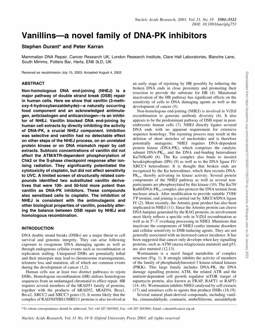
Extracts of GM00558 lymphoma cells were used to assayDNA end-joining of a linearised plasmid. After a 2-hincubation with extract, the predominant product was lineardimer DNA molecules with some trimer and tetramer forms(Fig. 2a). Inclusion of vanillin at a range of concentrationsbetween 100 mM and 1 mM resulted in a progressive decreasein the yield of ligated products. At 100 mM vanillin, no trimerswere detectable and a concentration of 1 mM effectivelyabolished the formation of dimer molecules. The extent ofinhibition by 100 mM vanillin was comparable to that
Nucleic Acids Research, 2003, Vol. 31, No. 19 5503
Dow
nloaded from https://academ
ic.oup.com/nar/article/31/19/5501/1092754 by U
niversity of Bologna user on 30 January 2022
produced by 0.25 mM wortmannin. By computing thecombined yield of dimers and higher order multimers fromsix independent assays, the IC50 value for inhibition ofrejoining by vanillin was estimated to be 300 mM (right panel).
The same concentrations of vanillin did not detectablyaffect mismatch repair. hMutSa- and hMutLa-dependentcorrection of a T:C mispair by HeLa cell extracts wasunaffected by vanillin concentrations up to 1 mM (Fig. 2b).Similarly, inclusion of wortmannin (0.25 and 2.5 mM) waswithout detectable effect (data not shown). We conclude thatthe effect of vanillin on NHEJ by cell extracts does not re¯ecta general inhibition of in vitro DNA repair.
Inhibition of enzyme reaction by vanillin
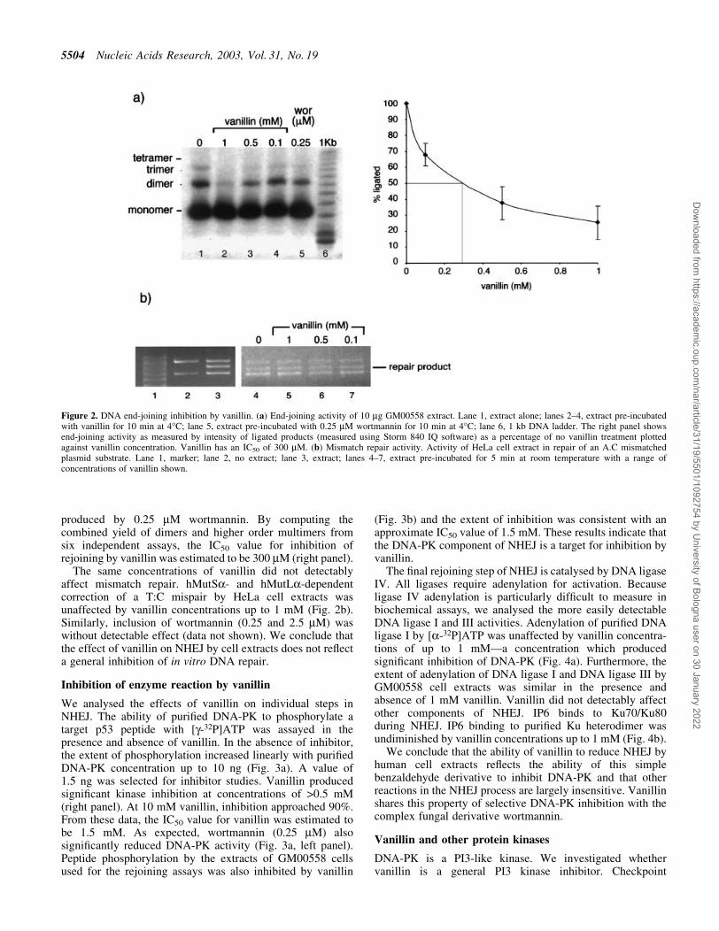
We analysed the effects of vanillin on individual steps inNHEJ. The ability of puri®ed DNA-PK to phosphorylate atarget p53 peptide with [g-32P]ATP was assayed in thepresence and absence of vanillin. In the absence of inhibitor,the extent of phosphorylation increased linearly with puri®edDNA-PK concentration up to 10 ng (Fig. 3a). A value of1.5 ng was selected for inhibitor studies. Vanillin producedsigni®cant kinase inhibition at concentrations of >0.5 mM(right panel). At 10 mM vanillin, inhibition approached 90%.From these data, the IC50 value for vanillin was estimated tobe 1.5 mM. As expected, wortmannin (0.25 mM) alsosigni®cantly reduced DNA-PK activity (Fig. 3a, left panel).Peptide phosphorylation by the extracts of GM00558 cellsused for the rejoining assays was also inhibited by vanillin
(Fig. 3b) and the extent of inhibition was consistent with anapproximate IC50 value of 1.5 mM. These results indicate thatthe DNA-PK component of NHEJ is a target for inhibition byvanillin.
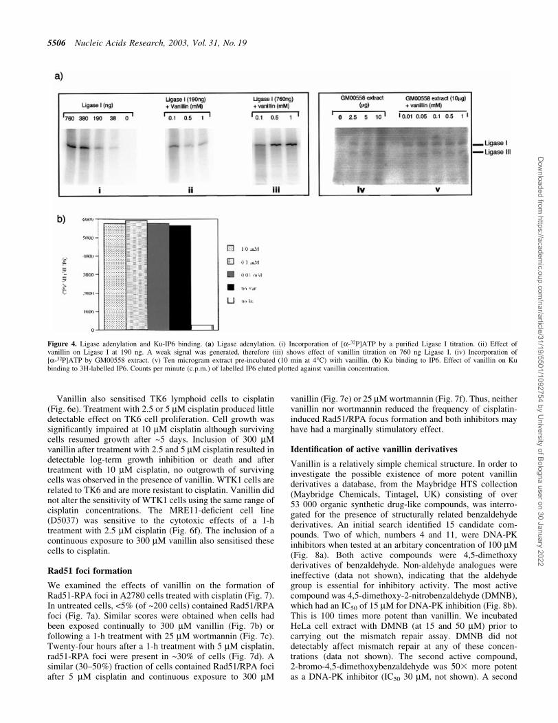
The ®nal rejoining step of NHEJ is catalysed by DNA ligaseIV. All ligases require adenylation for activation. Becauseligase IV adenylation is particularly dif®cult to measure inbiochemical assays, we analysed the more easily detectableDNA ligase I and III activities. Adenylation of puri®ed DNAligase I by [a-32P]ATP was unaffected by vanillin concentra-tions of up to 1 mMÐa concentration which producedsigni®cant inhibition of DNA-PK (Fig. 4a). Furthermore, theextent of adenylation of DNA ligase I and DNA ligase III byGM00558 cell extracts was similar in the presence andabsence of 1 mM vanillin. Vanillin did not detectably affectother components of NHEJ. IP6 binds to Ku70/Ku80during NHEJ. IP6 binding to puri®ed Ku heterodimer wasundiminished by vanillin concentrations up to 1 mM (Fig. 4b).
We conclude that the ability of vanillin to reduce NHEJ byhuman cell extracts re¯ects the ability of this simplebenzaldehyde derivative to inhibit DNA-PK and that otherreactions in the NHEJ process are largely insensitive. Vanillinshares this property of selective DNA-PK inhibition with thecomplex fungal derivative wortmannin.
Vanillin and other protein kinases
DNA-PK is a PI3-like kinase. We investigated whethervanillin is a general PI3 kinase inhibitor. Checkpoint
Figure 2. DNA end-joining inhibition by vanillin. (a) End-joining activity of 10 mg GM00558 extract. Lane 1, extract alone; lanes 2±4, extract pre-incubatedwith vanillin for 10 min at 4°C; lane 5, extract pre-incubated with 0.25 mM wortmannin for 10 min at 4°C; lane 6, 1 kb DNA ladder. The right panel showsend-joining activity as measured by intensity of ligated products (measured using Storm 840 IQ software) as a percentage of no vanillin treatment plottedagainst vanillin concentration. Vanillin has an IC50 of 300 mM. (b) Mismatch repair activity. Activity of HeLa cell extract in repair of an A.C mismatchedplasmid substrate. Lane 1, marker; lane 2, no extract; lane 3, extract; lanes 4±7, extract pre-incubated for 5 min at room temperature with a range ofconcentrations of vanillin shown.
5504 Nucleic Acids Research, 2003, Vol. 31, No. 19
Dow
nloaded from https://academ
ic.oup.com/nar/article/31/19/5501/1092754 by U
niversity of Bologna user on 30 January 2022
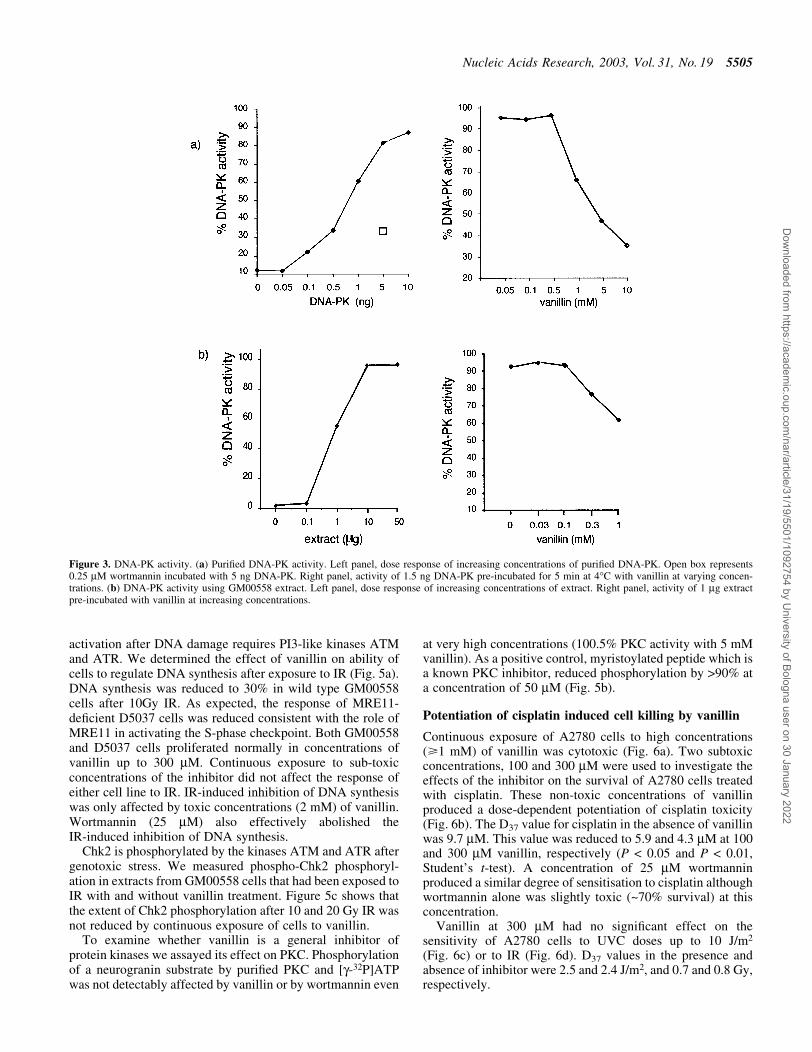
activation after DNA damage requires PI3-like kinases ATMand ATR. We determined the effect of vanillin on ability ofcells to regulate DNA synthesis after exposure to IR (Fig. 5a).DNA synthesis was reduced to 30% in wild type GM00558cells after 10Gy IR. As expected, the response of MRE11-de®cient D5037 cells was reduced consistent with the role ofMRE11 in activating the S-phase checkpoint. Both GM00558and D5037 cells proliferated normally in concentrations ofvanillin up to 300 mM. Continuous exposure to sub-toxicconcentrations of the inhibitor did not affect the response ofeither cell line to IR. IR-induced inhibition of DNA synthesiswas only affected by toxic concentrations (2 mM) of vanillin.Wortmannin (25 mM) also effectively abolished theIR-induced inhibition of DNA synthesis.
Chk2 is phosphorylated by the kinases ATM and ATR aftergenotoxic stress. We measured phospho-Chk2 phosphoryl-ation in extracts from GM00558 cells that had been exposed toIR with and without vanillin treatment. Figure 5c shows thatthe extent of Chk2 phosphorylation after 10 and 20 Gy IR wasnot reduced by continuous exposure of cells to vanillin.
To examine whether vanillin is a general inhibitor ofprotein kinases we assayed its effect on PKC. Phosphorylationof a neurogranin substrate by puri®ed PKC and [g-32P]ATPwas not detectably affected by vanillin or by wortmannin even
at very high concentrations (100.5% PKC activity with 5 mMvanillin). As a positive control, myristoylated peptide which isa known PKC inhibitor, reduced phosphorylation by >90% ata concentration of 50 mM (Fig. 5b).
Potentiation of cisplatin induced cell killing by vanillin
Continuous exposure of A2780 cells to high concentrations(>1 mM) of vanillin was cytotoxic (Fig. 6a). Two subtoxicconcentrations, 100 and 300 mM were used to investigate theeffects of the inhibitor on the survival of A2780 cells treatedwith cisplatin. These non-toxic concentrations of vanillinproduced a dose-dependent potentiation of cisplatin toxicity(Fig. 6b). The D37 value for cisplatin in the absence of vanillinwas 9.7 mM. This value was reduced to 5.9 and 4.3 mM at 100and 300 mM vanillin, respectively (P < 0.05 and P < 0.01,Student's t-test). A concentration of 25 mM wortmanninproduced a similar degree of sensitisation to cisplatin althoughwortmannin alone was slightly toxic (~70% survival) at thisconcentration.
Vanillin at 300 mM had no signi®cant effect on thesensitivity of A2780 cells to UVC doses up to 10 J/m2
(Fig. 6c) or to IR (Fig. 6d). D37 values in the presence andabsence of inhibitor were 2.5 and 2.4 J/m2, and 0.7 and 0.8 Gy,respectively.
Figure 3. DNA-PK activity. (a) Puri®ed DNA-PK activity. Left panel, dose response of increasing concentrations of puri®ed DNA-PK. Open box represents0.25 mM wortmannin incubated with 5 ng DNA-PK. Right panel, activity of 1.5 ng DNA-PK pre-incubated for 5 min at 4°C with vanillin at varying concen-trations. (b) DNA-PK activity using GM00558 extract. Left panel, dose response of increasing concentrations of extract. Right panel, activity of 1 mg extractpre-incubated with vanillin at increasing concentrations.
Nucleic Acids Research, 2003, Vol. 31, No. 19 5505
Dow
nloaded from https://academ
ic.oup.com/nar/article/31/19/5501/1092754 by U
niversity of Bologna user on 30 January 2022
Vanillin also sensitised TK6 lymphoid cells to cisplatin(Fig. 6e). Treatment with 2.5 or 5 mM cisplatin produced littledetectable effect on TK6 cell proliferation. Cell growth wassigni®cantly impaired at 10 mM cisplatin although survivingcells resumed growth after ~5 days. Inclusion of 300 mMvanillin after treatment with 2.5 and 5 mM cisplatin resulted indetectable log-term growth inhibition or death and aftertreatment with 10 mM cisplatin, no outgrowth of survivingcells was observed in the presence of vanillin. WTK1 cells arerelated to TK6 and are more resistant to cisplatin. Vanillin didnot alter the sensitivity of WTK1 cells using the same range ofcisplatin concentrations. The MRE11-de®cient cell line(D5037) was sensitive to the cytotoxic effects of a 1-htreatment with 2.5 mM cisplatin (Fig. 6f). The inclusion of acontinuous exposure to 300 mM vanillin also sensitised thesecells to cisplatin.
Rad51 foci formation
We examined the effects of vanillin on the formation ofRad51-RPA foci in A2780 cells treated with cisplatin (Fig. 7).In untreated cells, <5% (of ~200 cells) contained Rad51/RPAfoci (Fig. 7a). Similar scores were obtained when cells hadbeen exposed continually to 300 mM vanillin (Fig. 7b) orfollowing a 1-h treatment with 25 mM wortmannin (Fig. 7c).Twenty-four hours after a 1-h treatment with 5 mM cisplatin,rad51-RPA foci were present in ~30% of cells (Fig. 7d). Asimilar (30±50%) fraction of cells contained Rad51/RPA fociafter 5 mM cisplatin and continuous exposure to 300 mM
vanillin (Fig. 7e) or 25 mM wortmannin (Fig. 7f). Thus, neithervanillin nor wortmannin reduced the frequency of cisplatin-induced Rad51/RPA focus formation and both inhibitors mayhave had a marginally stimulatory effect.
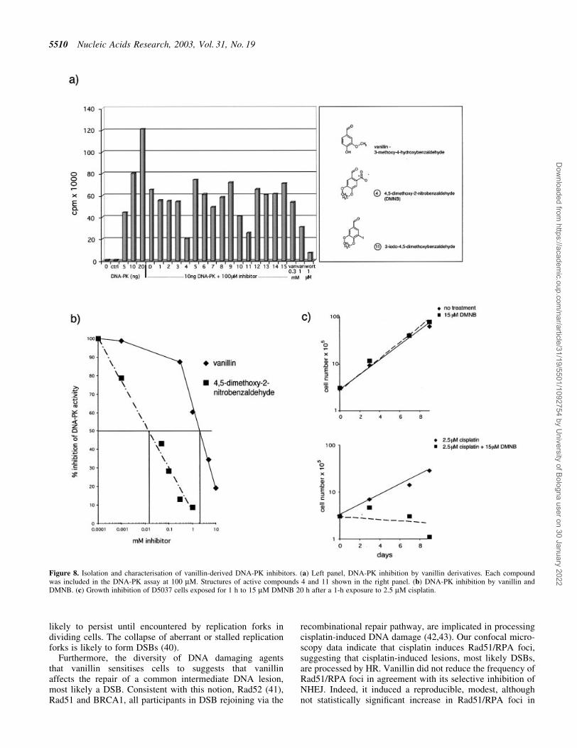
Identi®cation of active vanillin derivatives
Vanillin is a relatively simple chemical structure. In order toinvestigate the possible existence of more potent vanillinderivatives a database, from the Maybridge HTS collection(Maybridge Chemicals, Tintagel, UK) consisting of over53 000 organic synthetic drug-like compounds, was interro-gated for the presence of structurally related benzaldehydederivatives. An initial search identi®ed 15 candidate com-pounds. Two of which, numbers 4 and 11, were DNA-PKinhibitors when tested at an arbitary concentration of 100 mM(Fig. 8a). Both active compounds were 4,5-dimethoxyderivatives of benzaldehyde. Non-aldehyde analogues wereineffective (data not shown), indicating that the aldehydegroup is essential for inhibitory activity. The most activecompound was 4,5-dimethoxy-2-nitrobenzaldehyde (DMNB),which had an IC50 of 15 mM for DNA-PK inhibition (Fig. 8b).This is 100 times more potent than vanillin. We incubatedHeLa cell extract with DMNB (at 15 and 50 mM) prior tocarrying out the mismatch repair assay. DMNB did notdetectably affect mismatch repair at any of these concen-trations (data not shown). The second active compound,2-bromo-4,5-dimethoxybenzaldehyde was 503 more potentas a DNA-PK inhibitor (IC50 30 mM, not shown). A second
Figure 4. Ligase adenylation and Ku-IP6 binding. (a) Ligase adenylation. (i) Incorporation of [a-32P]ATP by a puri®ed Ligase I titration. (ii) Effect ofvanillin on Ligase I at 190 ng. A weak signal was generated, therefore (iii) shows effect of vanillin titration on 760 ng Ligase I. (iv) Incorporation of[a-32P]ATP by GM00558 extract. (v) Ten microgram extract pre-incubated (10 min at 4°C) with vanillin. (b) Ku binding to IP6. Effect of vanillin on Kubinding to 3H-labelled IP6. Counts per minute (c.p.m.) of labelled IP6 eluted plotted against vanillin concentration.
5506 Nucleic Acids Research, 2003, Vol. 31, No. 19
Dow
nloaded from https://academ
ic.oup.com/nar/article/31/19/5501/1092754 by U
niversity of Bologna user on 30 January 2022

screen using 4,5-dimethoxybenzaldehyde as the searchstructure identi®ed a further six derivatives. None of thesedemonstrated DNA-PK inhibitory activity (data not shown).
The effect of DMNB on cisplatin-treated D5037 cells wasexamined. Continuous exposure of DMNB (at its IC50 forDNA-PK of 15 mM) was lethal to the cells (data not shown).Therefore, cells were not treated with DMNB until 20 hafter cisplatin treatment (approximately one cell doublingtime). DMNB was added for 1 h only. This treatment did not
affect cell growth but signi®cantly sensitised the cells tocisplatin. None treated with cisplatin and DMNB was viable(Fig. 8c).
DISCUSSION
The genoprotective properties of vanillin have been acknow-ledged for a number of years. Its antimutagenic, anticlasto-genic and anticancer effects are all well documented in cellculture and animal models. Vanillin also enhances the toxicityof a range of structurally diverse DNA damaging agents in avariety of cell lines. The mechanistic basis for these biologicaleffects has remained unde®ned. It has been postulated thatvanillin exerts its antimutagenic activity in damaged cells bypromoting recombination and rejoining of DNA at homo-logous sites (25). It has also been proposed that vanillin mayinhibit a DNA repair process, such that enhancement oflethality decreases mutant yield by killing cells with poten-tially mutagenic lesions (23). Our demonstration that vanillinselectively blocks DNA repair by the relatively error-proneNHEJ pathway by inhibiting DNA-PK provides a plausibleexplanation for its antimutagenic and genotoxic enhancingeffects. Reducing DSB misrepair by the potentially mutagenicNHEJ pathway and imposing a greater reliance on HRrecombination might explain the antimutagenic effect innormal cells. The impairment of NHEJ pathway of DSB repairis consistent with a potentiation of the cytotoxicity ofgenotoxic agents.
It is presently unclear whether vanillin is a generalphosphatidylinositol kinase-related kinase inhibitor likewortmannin. Wortmannin is known to inhibit, albeit lesseffectively, the PI3KKs ATM and ATR, components inex-tricably linked to checkpoint activation and DSB repair (31).However, the observation that radiation-sensitivity of XRCC4defective cells is not further enhanced by wortmannin suggestsinhibition of DNA-PK is likely to be its most signi®cantbiological property (32). Our observations on radiation-resistant DNA synthesis suggest that the checkpoint proteinATM is not affected by non-toxic concentrations of vanillinand only affected at toxic concentrations of the inhibitor.However, the concentration of wortmannin (25 mM) generallyused to sensitise cells to IR did abrogate the S phasecheckpoint in our wild-type cells. It restored post-radiationlevels of replication comparable to those of MRE11 cells. Theextent to which inhibition of the intra-S checkpoint contributesto the sensitising effect of wortmannin is currently unclear.IR-induced phosphorylation of the down-stream effectorChk2, which is dependent on functional ATM/ATR afterDNA damage, was not inhibited by vanillin at its IC50 valuefor DNA-PK inhibition. This provides direct evidence thatvanillin does not signi®cantly impair ATM/ATR-dependentkinase activity.
Vanillin, like wortmannin, reacts preferentially with proteinlysine residues. Wortmannin binds irreversibly to lysine inthe phosphotransferase domain of the phosphatidylinositolkinase-related kinases. (16,33). Lysine 3751 of DNA-PKcs, islikely to be the site of nucleophilic attack by wortmannin (34).Vanillin also binds preferentially to lysine, although in thiscase via Schiff Base formation (35). It is likely therefore thatvanillin can also modify the key active site lysine of DNA-PK.Non-aldehyde analogues did not inhibit DNA-PK, suggesting
Figure 5. Kinase activity. (a) Effect of DNA-PK inhibitors on PKC activity.Using 10 ng of PKC vanillin, DMNB, wortmannin (wort) and myristoylatedpeptide inhibitor was added at increasing concentrations. (b) Phosphoryl-ation of Chk2. Extracts made from GM00558 cells treated with 0, 10 and20 Gy IR and incubated with 300 mM vanillin or 15 mM DMNB were usedto measure Phospho-Chk2 by western blot. (c) Radio-resistant DNA synthe-sis. GM00558 and D5037 (MRE11-de®cient) cells incubated with 3H TdRfor 3 h. Vanillin was added as a continuous exposure while wortmannin wasadded during the ®rst hour of incubation. Curves show levels of DNAsynthesis with and without 300 mM vanillin. Single points indicate levels ofDNA synthesis after exposure to 10 Gy IR at higher concentrations ofvanillin and wortmannin.
Nucleic Acids Research, 2003, Vol. 31, No. 19 5507
Dow
nloaded from https://academ
ic.oup.com/nar/article/31/19/5501/1092754 by U
niversity of Bologna user on 30 January 2022
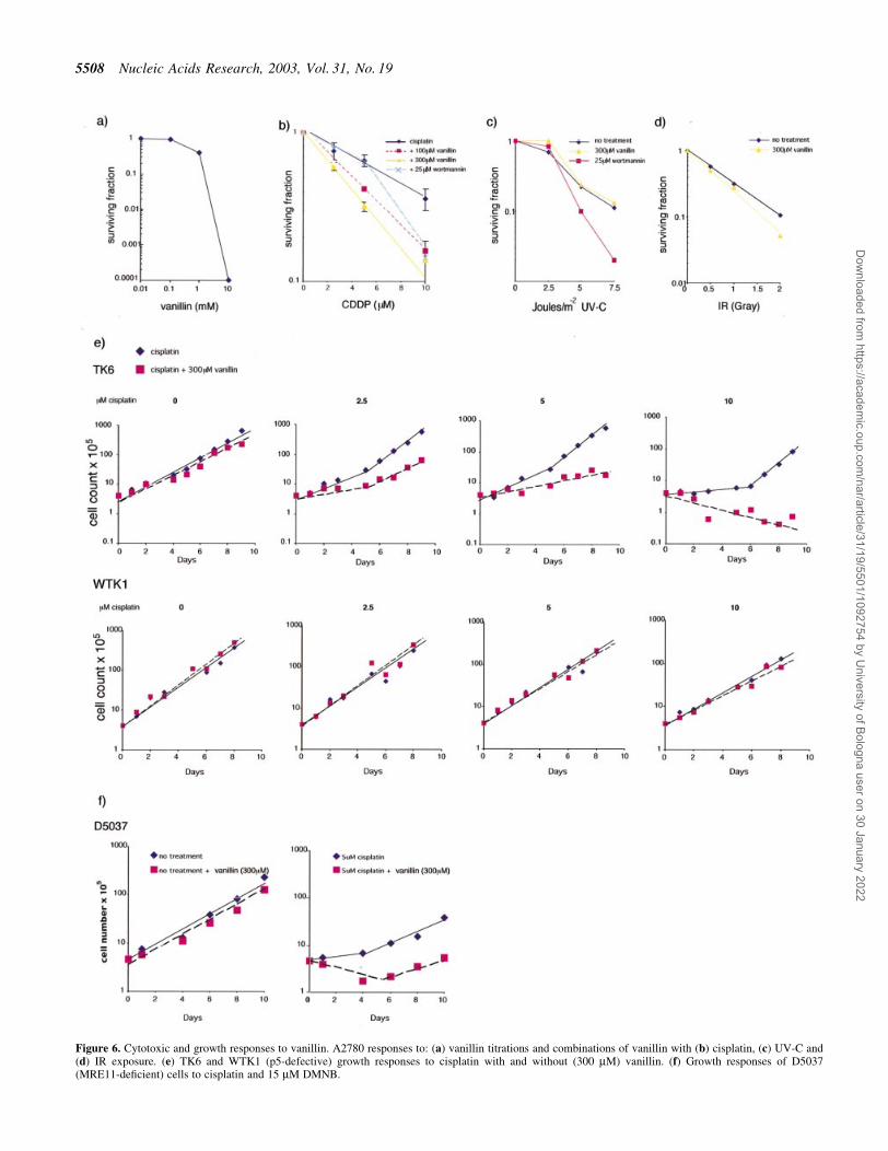
Figure 6. Cytotoxic and growth responses to vanillin. A2780 responses to: (a) vanillin titrations and combinations of vanillin with (b) cisplatin, (c) UV-C and(d) IR exposure. (e) TK6 and WTK1 (p5-defective) growth responses to cisplatin with and without (300 mM) vanillin. (f) Growth responses of D5037(MRE11-de®cient) cells to cisplatin and 15 mM DMNB.
5508 Nucleic Acids Research, 2003, Vol. 31, No. 19
Dow
nloaded from https://academ
ic.oup.com/nar/article/31/19/5501/1092754 by U
niversity of Bologna user on 30 January 2022

that the aldehyde group plays a critical role in inhibition. Inagreement with this idea, acroleinÐa simple a,b unsaturatedaliphatic aldehydeÐalso inhibits DNA-PK albeit with arelatively high IC50 (S.D., unpublished).
Wortmannin is more potent than vanillin as an inhibitor ofend-joining and of DNA-PK. The IC50 for vanillin is ~5000-fold higher. The biological effects of the inhibitors do notre¯ect this difference, however. The wortmannin concentra-tion typically used to sensitise cells to IR (25 mM) is 100 timeshigher than its biochemical IC50 (18,36). In contrast, vanillinproduced a detectable sensitisation at or considerably belowits biochemical IC50 for end-joining and DNA-PK inhibition.It seems likely that these differences re¯ect the relative sizesof the vanillin and wortmannin molecules. Vanillin is morelikely to enter cells freely whereas the more complexwortmannin probably requires active transport. In addition,although we did not ®nd evidence of inhibition of other DNArepair reactions, we cannot exclude the possibility that vanillin
may affect cell sensitivity through interference with othercomponents of the cellular responses to DNA damage.
Previous cytotoxicity studies have shown that vanillinsensitised rodent cells to several DNA damaging treatments.We observed that non-toxic concentrations of vanillin pro-duced a signi®cant sensitisation to cisplatin in human A2780cells. However, we did not observe any signi®cant effects ofvanillin on IR or UV sensitivity. We did see evidence of avanillin-induced increase in sensitisation to IR at higher dosesbut the reason for a considerably weaker effect compared tothat seen for cisplatin is presently unclear. It may re¯ect thedifferences between IR and cisplatin in the way they produceDSBs. Cisplatin produces predominantly DNA intrastrandcrosslinks. The majority of these (>90%) are between adjacentpurines (1,2 adducts) (37) and are relatively refractory toremoval by nucleotide excision repair (NER) (38). Theremainder are 1,3 crosslinks that are ef®ciently excised byNER (39). Cisplatin therefore introduces many lesions that are
Figure 7. Confocal microscopy. RPA/Rad51 foci formation in the nuclei of A2780 cells: (a) untreated, (b) treated with 300 mM vanillin, (c) exposed for 1 hto 25 mM wortmannin followed by 20 h incubation, (d) exposed to 5 mM cisplatin for 1 h followed by 20 h incubation, (e) exposed to cisplatin while incontinuous exposure to vanillin (300 mM) and (f) exposed to cisplatin followed by a 1 h exposure to wortmannin 20 h later. RPA (red), Rad51 (green) andmerge (yellow).
Nucleic Acids Research, 2003, Vol. 31, No. 19 5509
Dow
nloaded from https://academ
ic.oup.com/nar/article/31/19/5501/1092754 by U
niversity of Bologna user on 30 January 2022
likely to persist until encountered by replication forks individing cells. The collapse of aberrant or stalled replicationforks is likely to form DSBs (40).
Furthermore, the diversity of DNA damaging agentsthat vanillin sensitises cells to suggests that vanillinaffects the repair of a common intermediate DNA lesion,most likely a DSB. Consistent with this notion, Rad52 (41),Rad51 and BRCA1, all participants in DSB rejoining via the
recombinational repair pathway, are implicated in processingcisplatin-induced DNA damage (42,43). Our confocal micro-scopy data indicate that cisplatin induces Rad51/RPA foci,suggesting that cisplatin-induced lesions, most likely DSBs,are processed by HR. Vanillin did not reduce the frequency ofRad51/RPA foci in agreement with its selective inhibition ofNHEJ. Indeed, it induced a reproducible, modest, althoughnot statistically signi®cant increase in Rad51/RPA foci in
Figure 8. Isolation and characterisation of vanillin-derived DNA-PK inhibitors. (a) Left panel, DNA-PK inhibition by vanillin derivatives. Each compoundwas included in the DNA-PK assay at 100 mM. Structures of active compounds 4 and 11 shown in the right panel. (b) DNA-PK inhibition by vanillin andDMNB. (c) Growth inhibition of D5037 cells exposed for 1 h to 15 mM DMNB 20 h after a 1-h exposure to 2.5 mM cisplatin.
5510 Nucleic Acids Research, 2003, Vol. 31, No. 19
Dow
nloaded from https://academ
ic.oup.com/nar/article/31/19/5501/1092754 by U
niversity of Bologna user on 30 January 2022
cisplatin-treated cells. This is consistent with an increasedDSB processing by HR being a consequence of inhibition ofthe alternative NHEJ pathway.
Vanillin sensitised A2780, TK6 and GM00558 cells tocisplatin. In contrast, neither the TK6 derivative, WTK1 norRaji Burkitt's lymphoma cells (data not shown) were affectedby vanillin. Both WTK1 and Raji are more resistant tocisplatin than GM00558, most likely because of a defectivep53. The possibility that the sensitising effects of NHEJinhibition are con®ned to cells expressing WT p53 proteinwould be an interesting area for future investigation.
The simplicity of the vanillin molecule makes it anattractive candidate for modi®cation in search of more activeformulations. Our limited analysis of benzaldehyde deriva-tives identi®ed two additional methoxybenzaldehydes thatwere signi®cantly better than vanillin as inhibitors of DNA-PK. One was a 2-nitro compound, the other contained a 3-iodogroup. In the case of DMNB, the difference in IC50 was100-fold. It seems likely that the increased activity of DMNBis possibly due, at least in part, to the electron-withdrawingproperties of the 2-nitro group that would increase aldehydereactivity towards protein amino groups. The iodo groupdoes not share this property, however, and it is possible thatthe hydrophobicity of both these groups is a signi®cantin¯uenceÐpossibly by facilitating the reaction in relativelyhydrophobic areas of the protein.
DMNB, like vanillin did not affect PKC activity and did notinhibit Chk2 phosphorylation after IR, indicating that DMNBretains the relative speci®city after modi®cation. However,since vanillin and its more active derivatives are simplebenzaldehydes it is unlikely that their effects are completelyspeci®c for DNA-PK. Indeed, some benzaldehyde derivativescan protect cells from cisplatin-mediated cell inactivation(44). This is apparently due to the ability of benzaldehydes tobind reversibly to outer surface membrane proteins (45),therefore interfering with cellular transport and reducing drugaccumulation. However, substituted benzaldehydes withelectron-donating groups at the 2-position have the greatestprotective effect and no protection was seen using benzalde-hydes without these electron-donating groups. Therefore theinhibitory action on DNA-PK speci®cally by vanillin and themore potent Schiff base-strengthening electrophilic groupson dimethoxybenzaldehydes might explain the cisplatin-sensitising effect of this particular group of compounds.
In conclusion, we have shown that speci®c members ofthe vanillin family are simple and relatively speci®c lowmolecular weight inhibitors of DNA-PK. This family of novelvanillin-based molecules should be useful tools in assessingthe biochemical mechanism of DNA-PK and the relativecontribution of NHEJ and other pathways to DSB repair. Inview of the already established biological effects of vanillin,the possible long-term bene®cial effects of these compoundsin preventing genomic instability and cancer might warrantfurther investigation.
ACKNOWLEDGEMENTS
We would like to thank Pauline Branch and Peter Macphersonfor assistance with tissue culture and the mismatch repairassay, respectively. We are also grateful to Les Hanakahi forkindly donating puri®ed DNA-PK samples and assistance with
the Ku-binding assay, to Peter Robbins and Debbie Barnes forkindly supplying puri®ed Ligase I samples and to Professor A.Taylor at the University of Birmingham for supplying theMRE11-de®cient D5037 lymphoblastoid cell line. Our thanksalso go to Todd Duncan for assistance on compound structuresearches from the Maybridge Chemicals database.
REFERENCES
1. Haber,J.E. (2000) Partners and pathways repairing a double-strand break.Trends Genet., 16, 259±264.
2. Khanna,K.K. and Jackson,S.P. (2001) DNA double-strand breaks:signalling, repair and the cancer connection. Nature Genet., 27, 247±254.
3. Karran,P. (2000) DNA double strand break repair in mammalian cells.Curr. Opin. Genet. Dev., 10, 144±150.
4. de Jager,M., van Noort,J., van Gent,D.C., Dekker,C., Kanaar,R. andWyman,C. (2001) Human Rad50/Mre11 is a ¯exible complex that cantether DNA ends. Mol. Cell, 8, 1129±1135.
5. Godthelp,B.C., Wiegant,W.W., van Duijn-Goedhart,A., Scharer,O.D.,van Buul,P.P., Kanaar,R. and Zdzienicka,M.Z. (2002) MammalianRad51C contributes to DNA cross-link resistance, sister chromatidcohesion and genomic stability. Nucleic Acids Res., 30, 2172±2182.
6. Fugmann,S.D., Lee,A.I., Shockett,P.E., Villey,I.J. and Schatz,D.G.(2000) The RAG proteins and V(D)J recombination: complexes, endsand transposition. Annu. Rev. Immunol., 18, 495±527.
7. van Gent,D.C., Hoeijmakers,J.H.J. and Kanaar,R. (2001) Chromosomalstability and the DNA double strand break connection. Nature Rev.Genet., 2, 196±206.
8. Jackson,S.P. (2002) Sensing and repairing DNA double-strand breaks.Carcinogenesis, 23, 687±696.
9. Hanakahi,L.A., Bartlet-Jones,M., Chappell,C., Pappin,D. and West,S.(2000) Binding of inositol phosphate to DNA-PK and stimulation ofdouble-strand break repair. Cell, 102, 721±729.
10. Karmaker,P., Piotrowski,J., Brosh,R.M., Sommers,J.A., LeesMillers,S.P., Cheng,W.-H., Snowden,C.M., Ramsden,D.A. andBohr,V.A. (2002) Werner protein is a target of DNA-dependent proteinkinase in vivo and in vitro and its catalytic activities are regulated byphosphorylation. J. Biol. Chem., 277, 18291±18302.
11. Moshous,D., Callebaut,I., de Chasseval,R., Corneo,B.,Cavazzano-Calvo,M., Le Deist,F., Tezcan,I., Sanal,O., Bertrand,S.Y.,Phillipe,N., Fischer,A. and de Villartey,J.P. (2001) Artemis a novel DNAdouble-strand break repair/V(D)J recombination protein, is mutated inhuman severe combined immune de®ciency. Cell, 105, 177±186.
12. Luo,C.-M., Tang,W., Mekeel,K.L., DeFrank,J.S., Anne,R. andPowell,S.N. (1996) High frequency and error-prone DNA recombinationin Ataxia Telangiecstasia cell lines. J. Biol. Chem., 271, 4497±4503.
13. O'Driscoll,M., Cerosaletti,K.M., Girard,P.M., Dai,Y., Stumm,M.,Kysela,B., Hirsch,B., Gennery,A., Palmer,S.E., Seidel,J. et al. (2001)DNA ligase IV mutations identi®ed in patients exhibiting developmentaldelay and immunode®ciency. Mol. Cell, 8, 1175±1185.
14. Zakian,V.A. (1995) ATM-related genes: what do they tell us about aboutfunctions of the human gene? Cell, 82, 685±687.
15. Hunter,T. (1995) When is a lipid kinase not a lipid kinase? When it is aprotein kinase. Cell, 83, 1±4.
16. Sarkaria,J.N., Tibbetts,R.S., Busby,E.C., Kennedy,A.P., Hill,D.E. andAbraham,R.T. (1998) Inhibition of phosphoinositide 3-kinase relatedkinases by the radiosensitizing agent wortmannin. Cancer Res., 58,4375±4382.
17. Baumann,P. and West,S.C. (1998) DNA end-joining catalyzed by humancell-free extracts. Proc. Natl Acad. Sci. USA, 95, 14066±14070.
18. O'Gorman,D.M., McKenna,S.L., McGahon,A.J., Knox,K.A. andCotter,T.G. (2000) Sensitisation of HL60 human leukaemia cells tocytotoxic drug-induced apoptosis by inhibition of PI3-kinase survivalsignals. Leukemia, 4, 602±611.
19. Chernikova,S.B., Lindquist,K.L. and Elkind,M.M. (2001) Cell cycle-dependent effects of wortmannin on radiation survival and mutation.Radiat. Res., 155, 826±831.
20. Ohta ,T. (1993) Modi®cation of genotoxicity by naturally occurring¯avorings and their derivatives. Crit. Rev. Toxicol., 23, 127±146.
21. Watanabe,K., Ohta,T., Watanabe,M., Kato,T. and Shirasu,Y. (1990)Inhibition of induction of adaptive response by o-vanillin in Escherichiacoli B. Mutat. Res., 243, 273±280.
Nucleic Acids Research, 2003, Vol. 31, No. 19 5511
Dow
nloaded from https://academ
ic.oup.com/nar/article/31/19/5501/1092754 by U
niversity of Bologna user on 30 January 2022
22. Keshava,C., Keshava,N., Ong,T. and Nath,J. (1998) Protective effect ofvanillin on radiation-induced micronuclei and chromosomal aberrationsin V79 cells. Mutat. Res., 397, 149±159.
23. Gustafson,D.L., Franz,H.R., Ueno,A.M., Smith,C.R., Doolittle,D.J. andWaldren,C.C. (2000) Vanillin (3-methoxy-4-hydroxybenzaldehyde)inhibits mutation induced by hydrogen peroxide, N-methyl-N-nitrosoguanidine and mitomycin C but not 137Cs g-radiation at the CD59locus in human±hamster hybrid AL cells. Mutagenesis, 15, 207±213.
24. Jansson,T. and Zech,L. (1987) Effects of vanillin on sister-chromatidexchanges and chromosome aberrations in human lymphocytes. Mutat.Res., 190, 221±224.
25. Tamai,K., Tezuka,H. and Kuroda,Y. (1992) Direct modi®cations byvanillin in cytotoxicity and genetic changes induced by EMS and H2O2 incultured chinese hamster cells. Mutat. Res., 268, 231±237.
26. Imanishi,H., Sasaki,Y.F., Matsumoto,K., Watanabe,M., Ohta ,T.,Shirasu,Y. and Tutikawa,K. (1990) Suppression of 6-TG-resistantmutations in V79 cells and recessive spot formations in mice by vanillin.Mutat. Res. 243, 151±158.
27. Sasaki,Y.F., Imanishi,H., Watanabe,M., Ohta,T. and Shirasu,Y. (1990)Suppressing effects of antimutagenic ¯avorings on chromosomeaberrations induced by UV-light or X-rays in cultured Chinese hamstercells. Mutat. Res., 229, 1±10.
28. Akagi,K., Hirose,M., Hoshiya,T., Mizoguchi,Y., Ito,N. and Shrai,T.(1995) Modulating effects of elagic acid, vanillin and quercetin in a ratmedium term multi-organ carcinogenesis model, Cancer Lett., 94,113±121.
29. Tsuda,H., Uehara,N., Iwahori,Y., Asamoto,M., Iigo,M., Nagao,M.,Matsumoto,K., Ito,M. and Hirono,I. (1994) Chemopreventive effects ofbeta-carotene, alpha-tocopherol and ®ve naturally occurring antioxidantson initiation of hepatocarcinogenesis by 2-amino-3-methylimidazo[4,5-f]quinoline in the rat. Jpn J. Cancer Res., 85, 1214±1219.
30. Tomkinson,A.E., Lasko,D.D., Daly,G. and Lindahl,T. (1990)Mammalian DNA ligases. Catalytic domain and size of DNA ligase I.J. Biol. Chem., 265, 12611±12617.
31. Boulton,S., Kyle,S., Yalcintepe,L. and Durkacz,B.W. (1996)Wortmannin is a potent inhibitor of DNA double strand break but notsingle strand break repair in Chinese hamster ovary cells.Carcinogenesis, 17, 2285±2290.
32. DelacoÃte,F., Han,M., Stamato,T.D., Jasin,M. and Lopez,B.S. (2002) Anxrcc4 defect or Wortmannin stimulates homologous recombinationspeci®cally induced by double-strand breaks in mammalian cells.Nucleic Acids Res., 30, 3454±3463.
33. Wymann,M.P., Bulgarelli-Leva,G., Zvelebil,M.J., Pirola,L.,Vanhaesebroeck,B., Water®eld,M.D. and Panayotou,G. (1996)Wortmannin inactivates phosphoinositide 3-kinase by covalent
modi®cation of Lys-802, a residue involved in the phosphate transferreaction. Mol. Cell. Biol., 16, 1722±1733.
34. Izzard,R.A., Jackson,S.P. and Smith,G.C.M. (1999) Competitive andnoncompetitive inhibition of the DNA-dependent protein kinase. CancerRes., 59, 2581±2586.
35. Chobpattana,W., Jeon,I.J. and Smith,J.S. (2000) Kinetics of interaction ofvanillin with amino acids and peptides in model systems. J. Agric. FoodChem., 48, 3885±3889.
36. Price,B.D. and Youmell,M.B. (1996) The phosphatidylinositol 3-kinaseinhibitor wortmannin sensitizes murine ®broblasts and human tumor cellsto radiation and blocks induction of p53 following DNA damage. CancerRes., 56, 246±250.
37. Fichtinger-Schepman,A.M., van der veer,J.L., den Hartog,J.H.,Lohman,P.H. and Reedijk,J. (1985) Adducts of the anticancer drugcis-diamminedichloroplatinum(II) with DNA: formation, identi®cationand quanti®cation. Biochemistry, 24, 707±713.
38. Szymkowski,D.E., Yarema,K., Essigman,J.M., Lippard,S.J. andWood,R.D. (1992) An intrastrand d(GpG) platinum cross-link in duplexM13 DNA is refractory to repair by human cell extracts. Proc. Natl Acad.Sci. USA, 89, 10772±10776.
39. Moggs,J.G., Yarema,K.J., Essigmann,J.M. and Wood,R.D. (1996)Analysis of incision sites produced by human cell extracts and puri®edproteins during nucleotide excision repair of a 1,3-intrastrand d(GpTpG)-cisplatin adduct. J. Biol. Chem., 271, 7177±7186.
40. Morishita,T., Tsutsui,Y., Iwasaki,H. and Shinagawa,H. (2002) TheSchizosaccharomyces pombe rad60 gene is essential for repairing double-strand DNA breaks spontaneously occurring during replication andinduced by DNA-damaging agents. Mol. Cell. Biol., 22, 3537±3548.
41. Durant,S.T., Morris,M.M., Illand,M., McKay,H.J., McCormick,C.,Hirst,G.L., Borts,R.H. and Brown,R. (1999). Dependence on RAD52 andRAD1 for anticancer drug resistance mediated by inactivation ofmismatch repair genes. Curr. Biol., 9, 51±54.
42. Britten,R.A., Kuny,S. and Perdue,S. (1999) Modi®cation of non-conservative double-strand break (DSB) rejoining activity after theinduction of cisplatin resistance in human tumour cells. Br. J. Cancer, 79,843±849.
43. Venkitaraman,A.R. (2001) Functions of BRCA1 and BRCA2 in thebiological response to DNA damage. J. Cell Sci., 114, 3591±3598.
44. Dornish,J.M. and Pettersen,E.O. (1989) Modulation of cis-diammineplatinum(II)-induced cytotoxicity by benzaldehyde derivatives.Cancer Lett., 46, 63±68.
45. Miyakawa,T., Zundel,J.-L. and Sakaguchi,K. (1979) Selective inhibitoryeffect of benzaldehyde on the growth of simian virus 40-transformedcells. Biochem. Biophys. Res. Commun., 87, 1024±1030.
5512 Nucleic Acids Research, 2003, Vol. 31, No. 19
Dow
nloaded from https://academ
ic.oup.com/nar/article/31/19/5501/1092754 by U
niversity of Bologna user on 30 January 2022


![Nucleic Acid Extraction echniques T€¦ · nucleic acid without ampli cation inhibitors or contaminants such as protein, car-bohydrate, and other nucleic acids [ 8 ] . There are](https://img.pdfslide.us/doc/110x75/601f05c49bc97203e65e2d57/nucleic-acid-extraction-echniques-t-nucleic-acid-without-ampli-cation-inhibitors.jpg)


![Index [] · Index Bold page numbers = tabulated PK data a 852 A 418 abacavir 438 abanoquil 425 abecarnil 299 ... alcoholism 194, 204 alcuronium 300 aldehyde dehydrogenase inhibitors](https://img.pdfslide.us/doc/110x75/5e4e2954b23a5a57321683a8/index-index-bold-page-numbers-tabulated-pk-data-a-852-a-418-abacavir-438-abanoquil.jpg)


![Provincial Constituency Reference Map - District Peshawar · T uc l fa j n between ALHASAN [] ... PK - 9 PK - 5 PK - 11 PK - 4 PK - 3 PK - 2 PK - 1 Legend Districts Boundary Provincial](https://img.pdfslide.us/doc/110x75/5c01b81309d3f22b088d1121/provincial-constituency-reference-map-district-t-uc-l-fa-j-n-between-alhasan.jpg)