Embed Size (px)
Citation preview
8/8/2019 Van Herick En
http://slidepdf.com/reader/full/van-herick-en 1/4
Cataract
Glaucoma
Retina
Refractive
Table 1
Grading according to Van Herick
Cataract I Glaucoma I Retina I Refractive
Grade Relation between corneal slit image SC Interpretation
and anterior chamber depth CA
4 1 : 1 or higher Angle closure very unlikely; Chamber angle approx. 35°… 45°
3 1 : ½ Angle closure unlikely; Chamber angle approx. 20°… 35°
2 1 : ¼ Angle closure possible; Chamber angle approx. 20°
1 1 : < ¼ Angle closure likely; Chamber angle approx. 10°
0 closed Angle closure; Chamber angle approx. 0°
Van Herick’s Method for the Estimation of theChamber Angle
Performed on the slit lamp without any additional aids, the
Van Herick test allows quick assessment of the lateral
chamber angle.
A narrow slit of light is projected onto the peripheral cornea
at an angle of 60° as near as possible to the limbus. This
results in a slit image on the surface of the cornea (SC,
Fig. 1), the width of which is used as reference for the
assessment of the conditions in the chamber angle. The width
of the chamber angle (CA, Fig. 1) can be described by the
distance between the corneal slit image (SC, Fig. 1) and the
slit image on the iris (SI, Fig. 1).
Grading
If the distance between the posterior surface of the cornea
and the iris (CA) has at least the same width as the slit (SC)
projected onto the cornea, the chamber angle is widely open.
Angle closure is very unlikely. This state corresponds to Grade 4
of the grading according to Van Herick (Fig. 2). If CA is half
of SC, Grade 3 according to Van Herick (Fig. 3). The chamber
angle is open and an angle closure is unlikely.
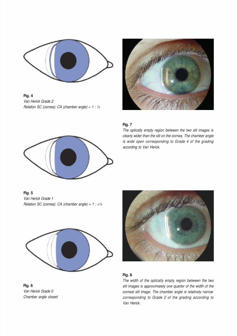
Angle closure is possible, if CA is a quarter of SC. Van Herick
classifies this state as Grade 2 (Fig. 4). In this case, the
affected eye should be measured with a gonioscope. With
Grade 1 (Fig. 5), the w idth of CA is smaller t han a quarter of
SC. Looking through a gonioscope, a dangerously narrow
chamber angle is visible; angle closure is likely. If between
the corneal slit image and the slit image on the iris no space
is visible (Fig. 6), the chamber angle is closed and an angle
closure already existing. This grading is summarized in Table 1.
8/8/2019 Van Herick En
http://slidepdf.com/reader/full/van-herick-en 2/4
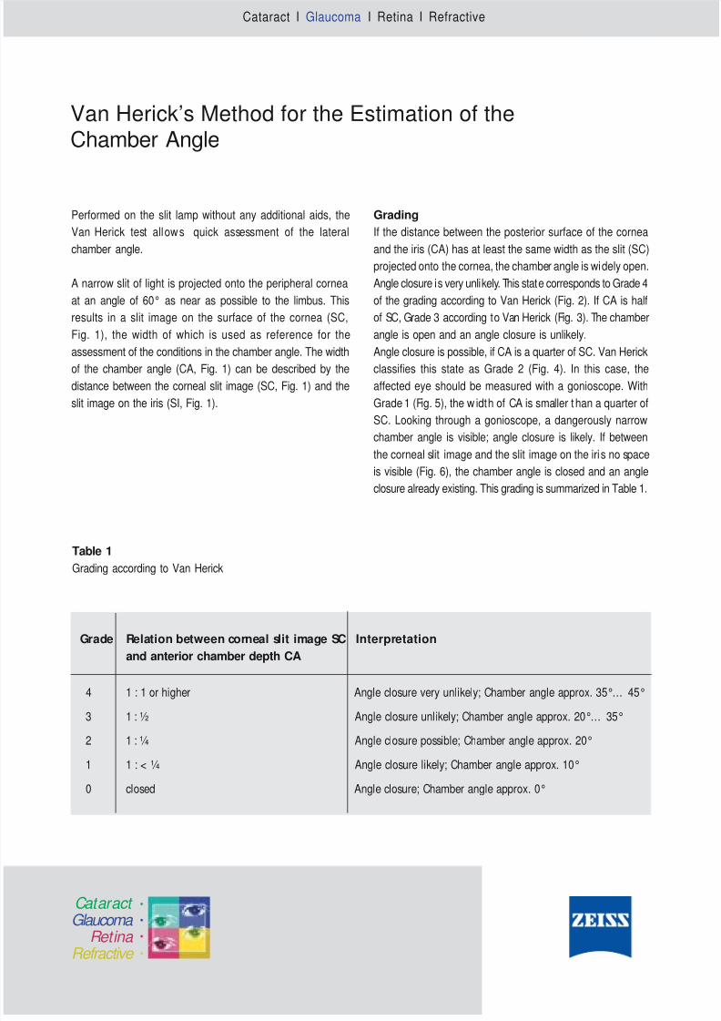
Fig. 1
Schematic diagram of the slit image in the Van Herick
method
Fig. 2
Van Herick Grade 4 Relation SC (cornea): CA (chamber angle) = 1 : 1 or higher
Fig. 3
Van Herick Grade 3 Relation SC (cornea): CA (chamber angle) = 1 : ½
Relevance and limits of the method
Using this method the chamber angle can be assessed
temporally and nasally. In practice, it is advisable to form the
average of two estimations. From this, one can infer anaccurate assessment of the total chamber angle. However,
the results obtained in this way cannot replace gonioscopy.
The method rather serves for the quick assessment of the
risk of an angle closure without stressing the patient.
This method is of part icular significance before the diagnostic
application of a mydriatic. If a narrow chamber angle exists,
the administration of a mydriatic may cause an angle
closure.Previous assessment of the chamber angle by the
Van Herick method can help to estimate and minimize the
risk of a provoked angle closure.
Conclusion
The Van Herick method for the estimation of the chamber
angle used in a slit lamp examination does not require
appreciable t ime. Moreover, the performance of this test does
not represent additional stress to the patient. Thus, the
Van Herick test is a method that is very well suitable for the
quick and easy assessment of the chamber angle.
Caption SC - Slit on cornea
CA - Chamber angle
SI - Slit on iris
8/8/2019 Van Herick En
http://slidepdf.com/reader/full/van-herick-en 3/4
Fig. 7
The optically empty region between the two slit images is
clearly wider than the slit on the cornea. The chamber angle
is wide open corresponding to Grade 4 of the grading
according to Van Herick.
Fig. 4
Van Herick Grade 2
Relation SC (cornea): CA (chamber angle) = 1 : ¼
Fig. 5
Van Herick Grade 1
Relation SC (cornea): CA (chamber angle) = 1 : <¼
Fig. 6
Van Herick Grade 0
Chamber angle closed
Fig. 8
The width of the optically empty region between the two
slit images is approximately one quarter of the width of the
corneal slit image. The chamber angle is relatively narrow
corresponding to Grade 2 of the grading according to
Van Herick.
8/8/2019 Van Herick En
http://slidepdf.com/reader/full/van-herick-en 4/4
Carl Zeiss Meditec AG Phone: +49 (0) 36 41/ 220-333Goeschwitzer Str. 51-52 Fax: +49 (0) 36 41/ 220-28207740 Jena [email protected] www.meditec.zeiss.com
Publication-No.000000
-1276-
334
References
1. Bonomi L: Usefulness of the Van Herick test. Glaucoma
World Newsletter 1997; No. 3
2. Gross G: Differentialdiagnose der
Winkelblockglaukome. Der Augenspiegel 1999; 9:
13-26
3. Martonyi CL et al: Clinical Slit Lamp Biomicroscopy
and Photo Slit Lamp Biomicrography. 2nd edition; 1985
4. Van Herick W, Shaffer RN, Schwartz A: Estimation of
width of angle of anterior chamber. Incidence and
significance of the narrow angle. American Journal of
Ophthalmology 1969; 68: 626-629
Authors
Robert Wilke, qualified physician; [email protected]
Burkhard Wagner, product manager; [email protected]