Embed Size (px)
Citation preview
Valvular Heart Disease and Infective Endocarditis
LECTURE IN INTERNAL MEDICINE FOR V COURSE STUDENTS
M. Yabluchansky, L. Bogun, L. Martymianova, O. Bychkova, N. Lysenko, M. Brynza V.N. Karazin National University Medical School’ Internal Medicine Dept.
Valvular Heart Disease
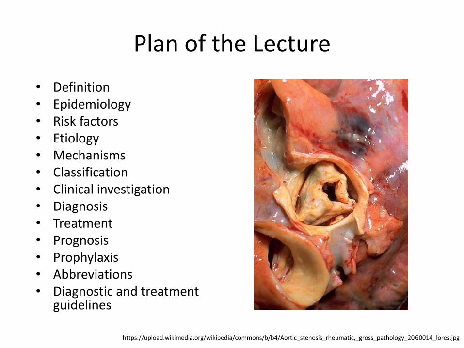
Plan of the Lecture
• Definition • Epidemiology • Risk factors • Etiology • Mechanisms • Classification • Clinical investigation • Diagnosis • Treatment • Prognosis • Prophylaxis • Abbreviations • Diagnostic and treatment
guidelines
https://upload.wikimedia.org/wikipedia/commons/b/b4/Aortic_stenosis_rheumatic,_gross_pathology_20G0014_lores.jpg
Definition Valvular heart disease (VHD) is any disease process involving one or more of the four valves of the
heart (the aortic and mitral valves on the left and
the pulmonary and tricuspid valves on the right).
There are two types of heart valve disease:
• Valvular stenosis, that occurs when a heart valve doesn't fully open due to stiff or fused leaflets.
• Valvular insufficiency (regurgitation, incompetence, "leaky valve“), that occurs when a valve does not close tightly
http://www.webmd.com/heart-disease/guide/heart-valve-disease https://en.wikipedia.org/wiki/Valvular_heart_disease
US MLE STEP 2 TEST
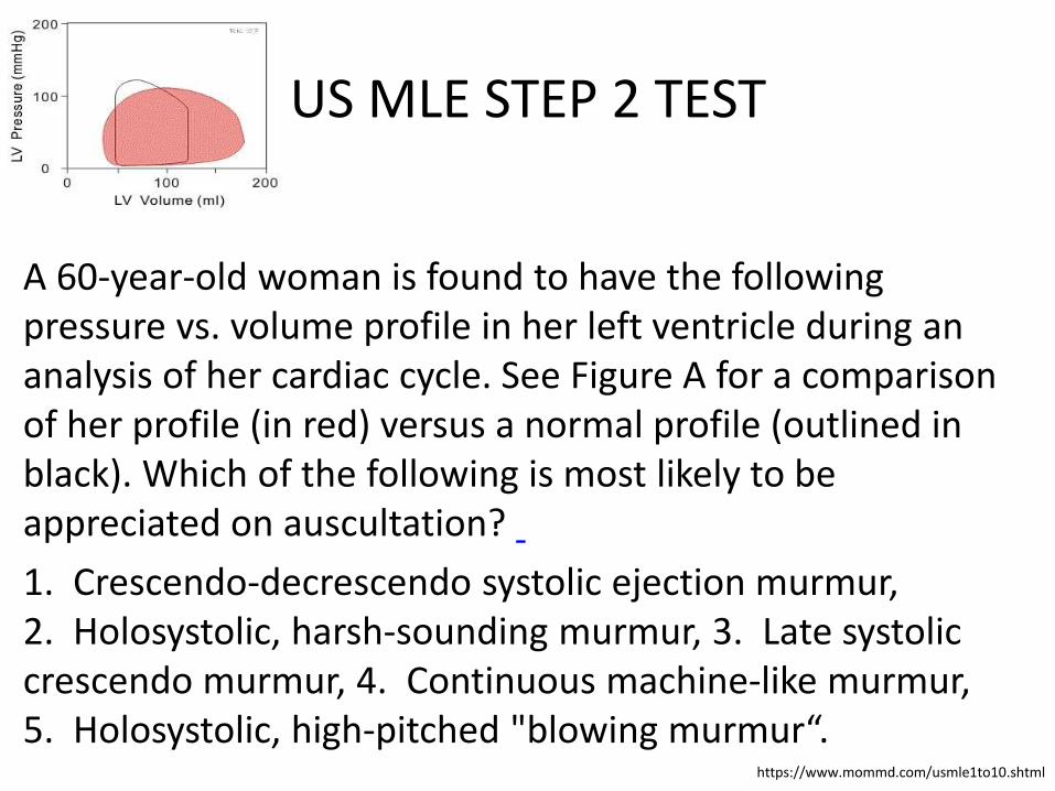
A 60-year-old woman is found to have the following pressure vs. volume profile in her left ventricle during an analysis of her cardiac cycle. See Figure A for a comparison of her profile (in red) versus a normal profile (outlined in black). Which of the following is most likely to be appreciated on auscultation?
1. Crescendo-decrescendo systolic ejection murmur, 2. Holosystolic, harsh-sounding murmur, 3. Late systolic crescendo murmur, 4. Continuous machine-like murmur, 5. Holosystolic, high-pitched "blowing murmur“.
https://www.mommd.com/usmle1to10.shtml
US MLE STEP 2 EXPLANATION
The correct answer is 5. The diagram shown is consistent with mitral regurgitation (mitral insufficiency). Mitral regurgitation presents with a holosystolic, high-pitched "blowing murmur." Incorrect answers: 1: Crescendo-decrescendo systolic ejection murmurs are associated with aortic stenosis (AS), 2: Holosystolic, harsh-sounding murmurs are associated with ventricular septal defects, 3: Late systolic crescendo murmurs are associated with mitral valve prolapse, 4: Continuous machine-like murmurs are associated with patent ductus arteriosus.
https://www.mommd.com/usmle1to10.shtml
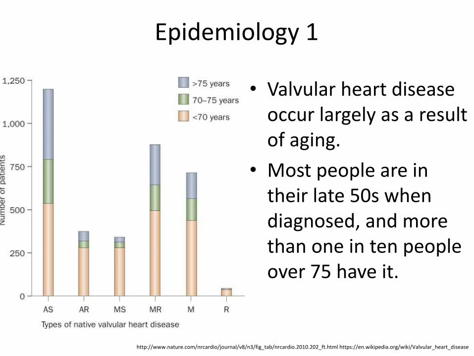
Epidemiology 1
http://www.nature.com/nrcardio/journal/v8/n3/fig_tab/nrcardio.2010.202_ft.html https://en.wikipedia.org/wiki/Valvular_heart_disease
• Valvular heart disease occur largely as a result of aging.
• Most people are in their late 50s when diagnosed, and more than one in ten people over 75 have it.
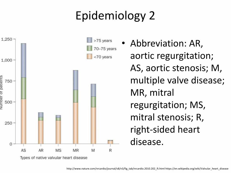
Epidemiology 2
http://www.nature.com/nrcardio/journal/v8/n3/fig_tab/nrcardio.2010.202_ft.html https://en.wikipedia.org/wiki/Valvular_heart_disease
• Abbreviation: AR, aortic regurgitation; AS, aortic stenosis; M, multiple valve disease; MR, mitral regurgitation; MS, mitral stenosis; R, right-sided heart disease.
Risk Factors and Etiology 1 • Aortic stenosis: calcification of aortic valve,
rheumatic fever.
• Aortic regurgitation: 1) acute: infective endocarditis, trauma; 2) chronic: 2a) primary valvular: rheumatic fever, bicuspid aortic valve, Marfan's syndrome, Ehlers–Danlos syndrome, ankylosing spondylitis, systemic lupus erythematosus; 2b) disease of the aortic root: syphilitic aortitis, osteogenesis imperfecta, aortic dissection, Behçet's disease, reactive arthritis, systemic hypertension.
https://en.wikipedia.org/wiki/Valvular_heart_disease
Mechanisms 1
• Aortic stenosis: obstruction through the aortic ostium causes increased pressure in the LV and impaired flow through the aorta.
• Aortic regurgitation: insufficiency of the aortic valve causes backflow of blood into the LV.
• Mitral stenosis: progressive obstruction of the mitral ostium causes increased pressure in the left atrium and the pulmonary circulation; congestion may cause thromboembolism, and atrial hypertension may cause atrial fibrillation.
https://en.wikipedia.org/wiki/Valvular_heart_disease
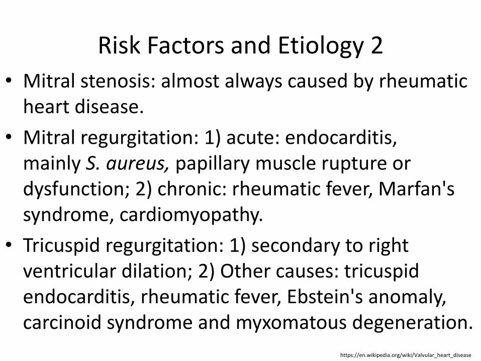
Risk Factors and Etiology 2
• Mitral stenosis: almost always caused by rheumatic heart disease.
• Mitral regurgitation: 1) acute: endocarditis, mainly S. aureus, papillary muscle rupture or dysfunction; 2) chronic: rheumatic fever, Marfan's syndrome, cardiomyopathy.
• Tricuspid regurgitation: 1) secondary to right ventricular dilation; 2) Other causes: tricuspid endocarditis, rheumatic fever, Ebstein's anomaly, carcinoid syndrome and myxomatous degeneration.
https://en.wikipedia.org/wiki/Valvular_heart_disease
Mechanisms 2
• Mitral regurgitation: insufficiency of the mitral valve causes backflow of blood into the left atrium during systole.
• Tricuspid regurgitation: insufficiency of the tricuspid valve causes backflow of blood into the right atrium during systole.
https://en.wikipedia.org/wiki/Valvular_heart_disease
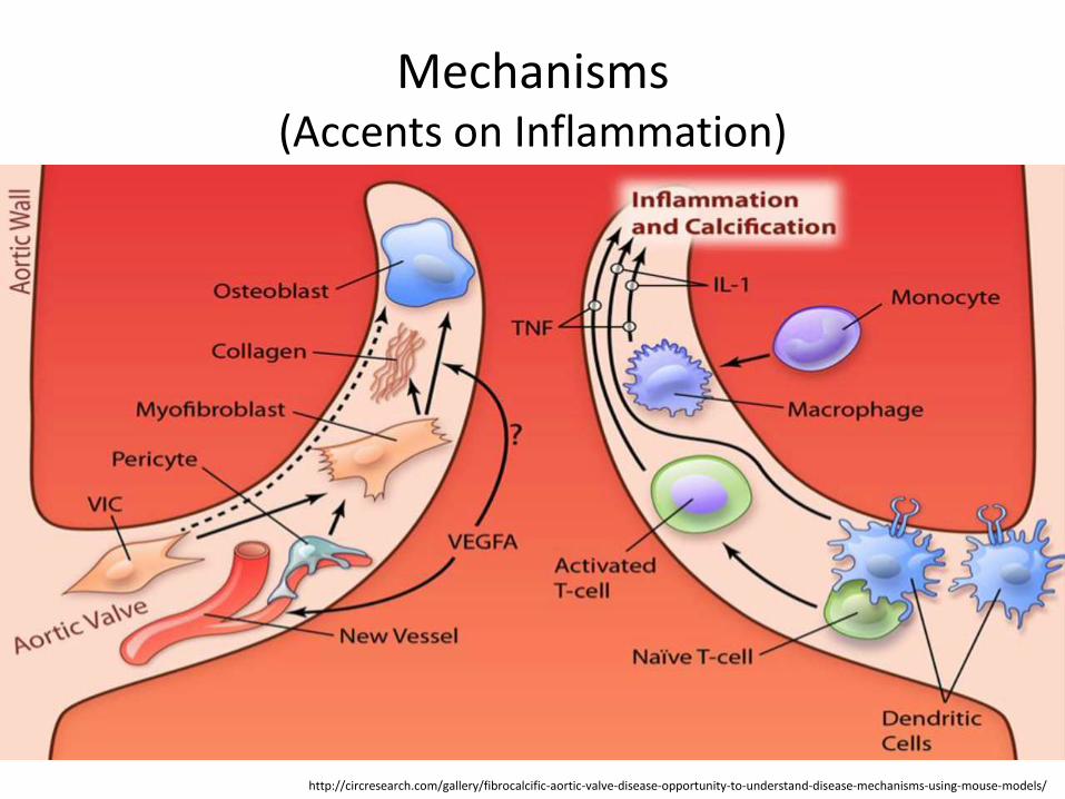
Mechanisms (Accents on Inflammation)
http://circresearch.com/gallery/fibrocalcific-aortic-valve-disease-opportunity-to-understand-disease-mechanisms-using-mouse-models/
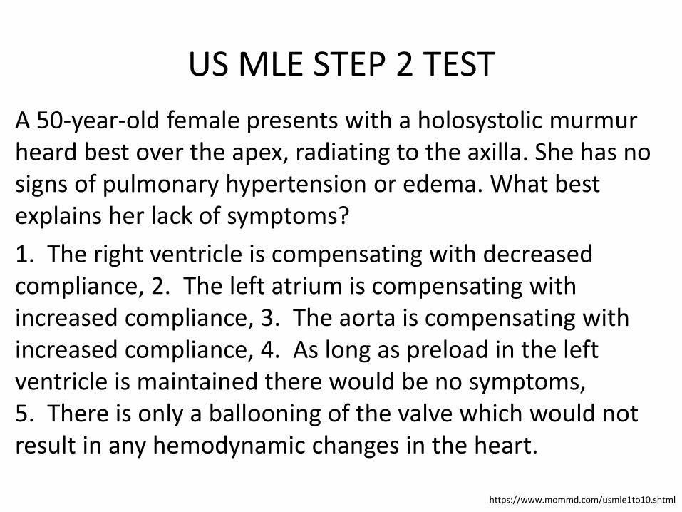
US MLE STEP 2 TEST
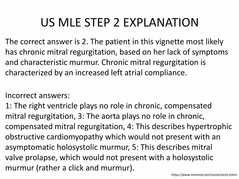
A 50-year-old female presents with a holosystolic murmur heard best over the apex, radiating to the axilla. She has no signs of pulmonary hypertension or edema. What best explains her lack of symptoms?
1. The right ventricle is compensating with decreased compliance, 2. The left atrium is compensating with increased compliance, 3. The aorta is compensating with increased compliance, 4. As long as preload in the left ventricle is maintained there would be no symptoms, 5. There is only a ballooning of the valve which would not result in any hemodynamic changes in the heart.
https://www.mommd.com/usmle1to10.shtml
US MLE STEP 2 EXPLANATION
The correct answer is 2. The patient in this vignette most likely has chronic mitral regurgitation, based on her lack of symptoms and characteristic murmur. Chronic mitral regurgitation is characterized by an increased left atrial compliance.
Incorrect answers: 1: The right ventricle plays no role in chronic, compensated mitral regurgitation, 3: The aorta plays no role in chronic, compensated mitral regurgitation, 4: This describes hypertrophic obstructive cardiomyopathy which would not present with an asymptomatic holosystolic murmur, 5: This describes mitral valve prolapse, which would not present with a holosystolic murmur (rather a click and murmur).
https://www.mommd.com/usmle1to10.shtml
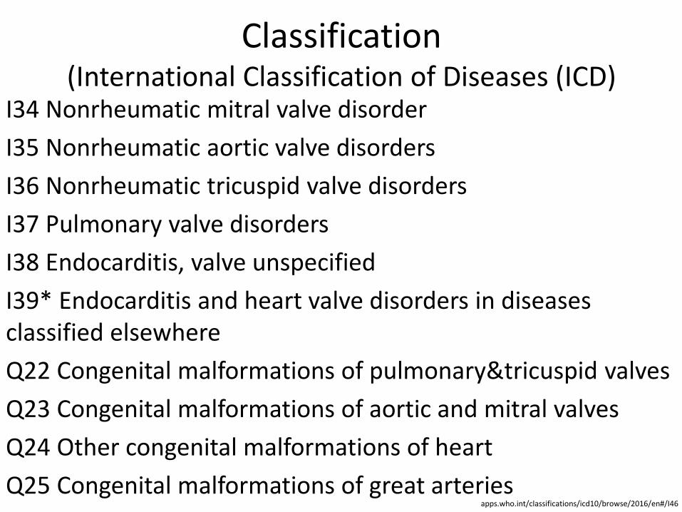
Classification (International Classification of Diseases (ICD)
apps.who.int/classifications/icd10/browse/2016/en#/I46
I34 Nonrheumatic mitral valve disorder
I35 Nonrheumatic aortic valve disorders
I36 Nonrheumatic tricuspid valve disorders
I37 Pulmonary valve disorders
I38 Endocarditis, valve unspecified
I39* Endocarditis and heart valve disorders in diseases classified elsewhere
Q22 Congenital malformations of pulmonary&tricuspid valves
Q23 Congenital malformations of aortic and mitral valves
Q24 Other congenital malformations of heart
Q25 Congenital malformations of great arteries
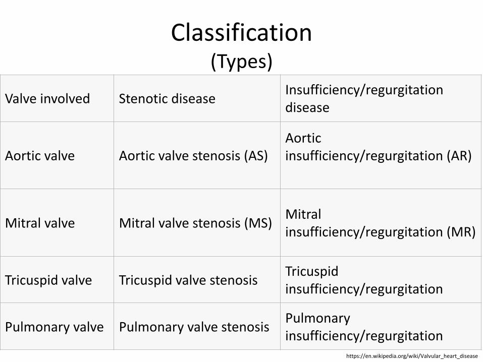
Classification (Types)
https://en.wikipedia.org/wiki/Valvular_heart_disease
Valve involved Stenotic disease Insufficiency/regurgitation disease
Aortic valve Aortic valve stenosis (AS) Aortic insufficiency/regurgitation (AR)
Mitral valve Mitral valve stenosis (MS) Mitral insufficiency/regurgitation (MR)
Tricuspid valve Tricuspid valve stenosis Tricuspid insufficiency/regurgitation
Pulmonary valve Pulmonary valve stenosis Pulmonary insufficiency/regurgitation
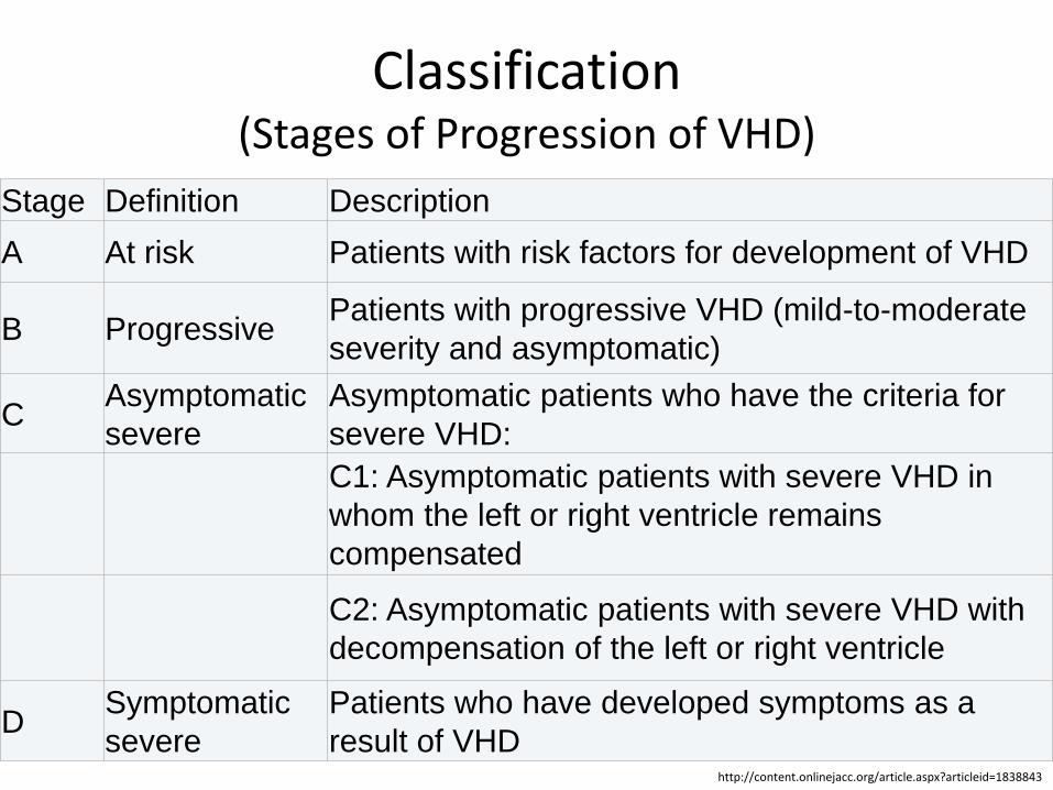
Classification (Stages of Progression of VHD)
http://content.onlinejacc.org/article.aspx?articleid=1838843
Stage Definition Description
A At risk Patients with risk factors for development of VHD
B Progressive Patients with progressive VHD (mild-to-moderate
severity and asymptomatic)
C Asymptomatic
severe
Asymptomatic patients who have the criteria for
severe VHD:
C1: Asymptomatic patients with severe VHD in
whom the left or right ventricle remains
compensated
C2: Asymptomatic patients with severe VHD with
decompensation of the left or right ventricle
D Symptomatic
severe
Patients who have developed symptoms as a
result of VHD
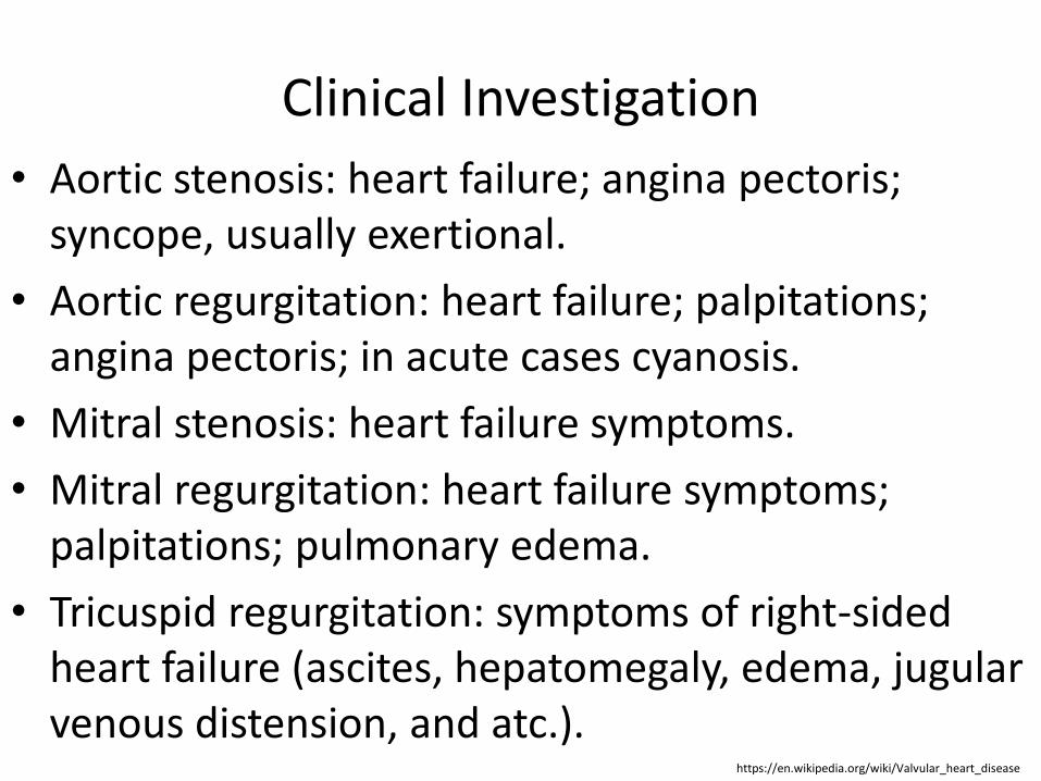
Clinical Investigation
• Aortic stenosis: heart failure; angina pectoris; syncope, usually exertional.
• Aortic regurgitation: heart failure; palpitations; angina pectoris; in acute cases cyanosis.
• Mitral stenosis: heart failure symptoms.
• Mitral regurgitation: heart failure symptoms; palpitations; pulmonary edema.
• Tricuspid regurgitation: symptoms of right-sided heart failure (ascites, hepatomegaly, edema, jugular venous distension, and atc.).
https://en.wikipedia.org/wiki/Valvular_heart_disease
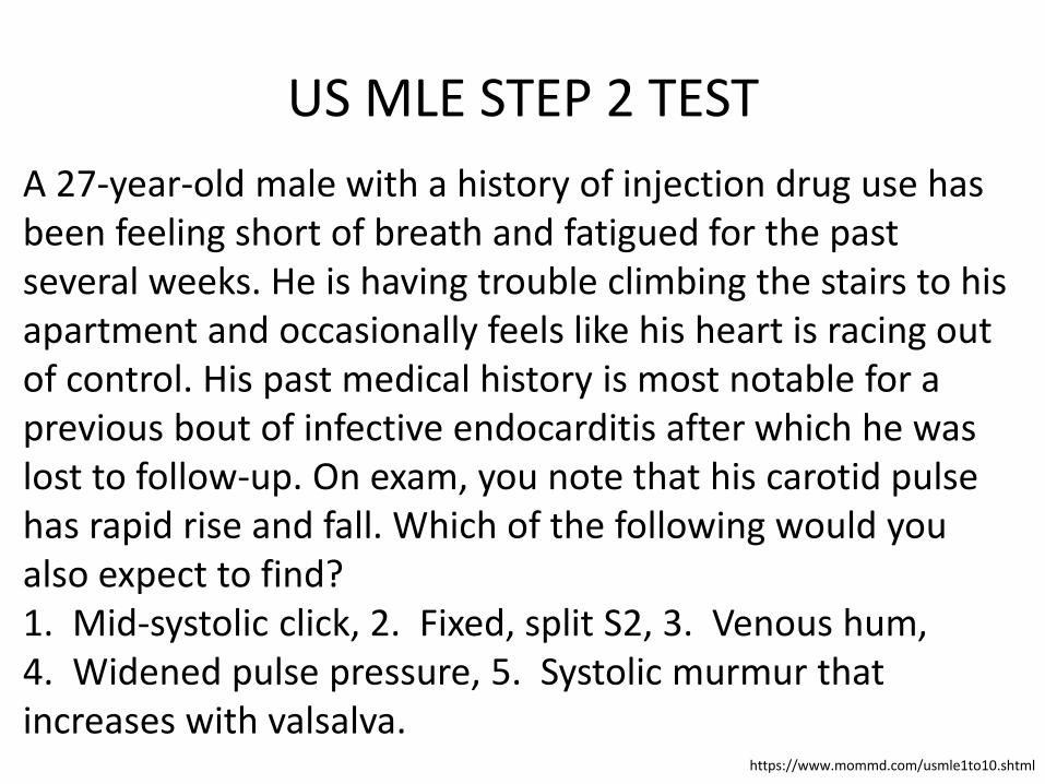
US MLE STEP 2 TEST
A 27-year-old male with a history of injection drug use has been feeling short of breath and fatigued for the past several weeks. He is having trouble climbing the stairs to his apartment and occasionally feels like his heart is racing out of control. His past medical history is most notable for a previous bout of infective endocarditis after which he was lost to follow-up. On exam, you note that his carotid pulse has rapid rise and fall. Which of the following would you also expect to find? 1. Mid-systolic click, 2. Fixed, split S2, 3. Venous hum, 4. Widened pulse pressure, 5. Systolic murmur that increases with valsalva.
https://www.mommd.com/usmle1to10.shtml
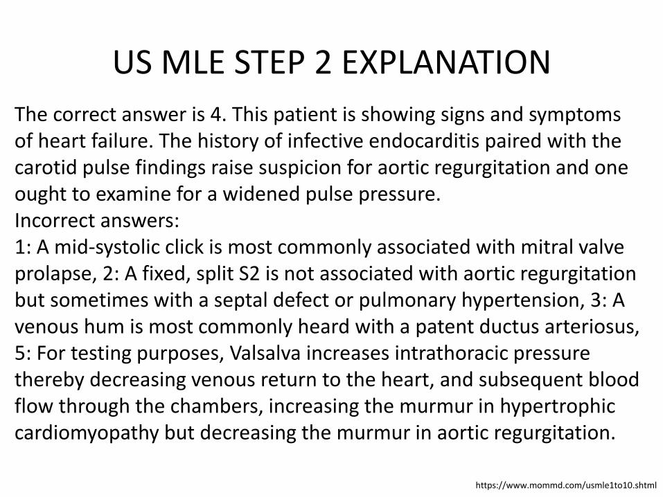
US MLE STEP 2 EXPLANATION
The correct answer is 4. This patient is showing signs and symptoms of heart failure. The history of infective endocarditis paired with the carotid pulse findings raise suspicion for aortic regurgitation and one ought to examine for a widened pulse pressure. Incorrect answers: 1: A mid-systolic click is most commonly associated with mitral valve prolapse, 2: A fixed, split S2 is not associated with aortic regurgitation but sometimes with a septal defect or pulmonary hypertension, 3: A venous hum is most commonly heard with a patent ductus arteriosus, 5: For testing purposes, Valsalva increases intrathoracic pressure thereby decreasing venous return to the heart, and subsequent blood flow through the chambers, increasing the murmur in hypertrophic cardiomyopathy but decreasing the murmur in aortic regurgitation.
https://www.mommd.com/usmle1to10.shtml
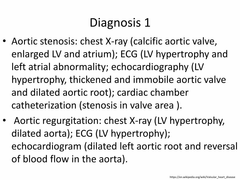
Diagnosis 1
• Aortic stenosis: chest X-ray (calcific aortic valve, enlarged LV and atrium); ECG (LV hypertrophy and left atrial abnormality; echocardiography (LV hypertrophy, thickened and immobile aortic valve and dilated aortic root); cardiac chamber catheterization (stenosis in valve area ).
• Aortic regurgitation: chest X-ray (LV hypertrophy, dilated aorta); ECG (LV hypertrophy); echocardiogram (dilated left aortic root and reversal of blood flow in the aorta).
https://en.wikipedia.org/wiki/Valvular_heart_disease
Diagnosis 2
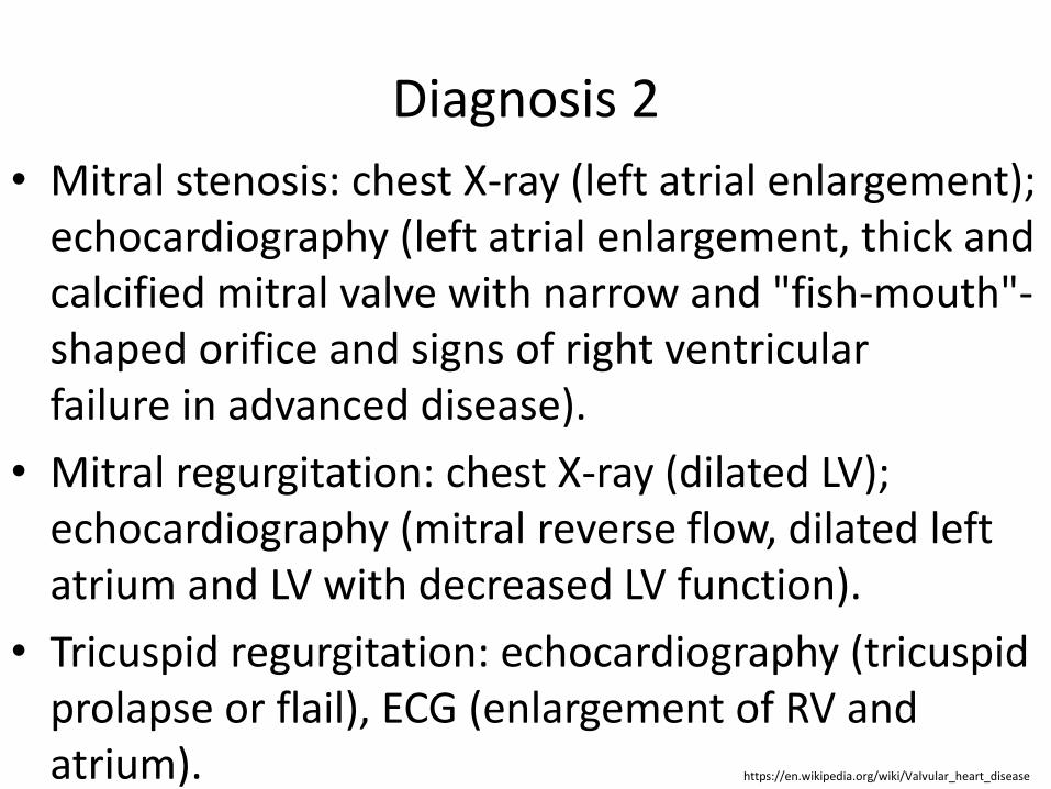
• Mitral stenosis: chest X-ray (left atrial enlargement); echocardiography (left atrial enlargement, thick and calcified mitral valve with narrow and "fish-mouth"-shaped orifice and signs of right ventricular failure in advanced disease).
• Mitral regurgitation: chest X-ray (dilated LV); echocardiography (mitral reverse flow, dilated left atrium and LV with decreased LV function).
• Tricuspid regurgitation: echocardiography (tricuspid prolapse or flail), ECG (enlargement of RV and atrium).
https://en.wikipedia.org/wiki/Valvular_heart_disease
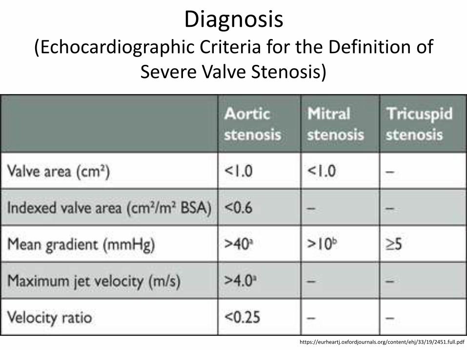
Diagnosis (Echocardiographic Criteria for the Definition of
Severe Valve Stenosis)
https://eurheartj.oxfordjournals.org/content/ehj/33/19/2451.full.pdf
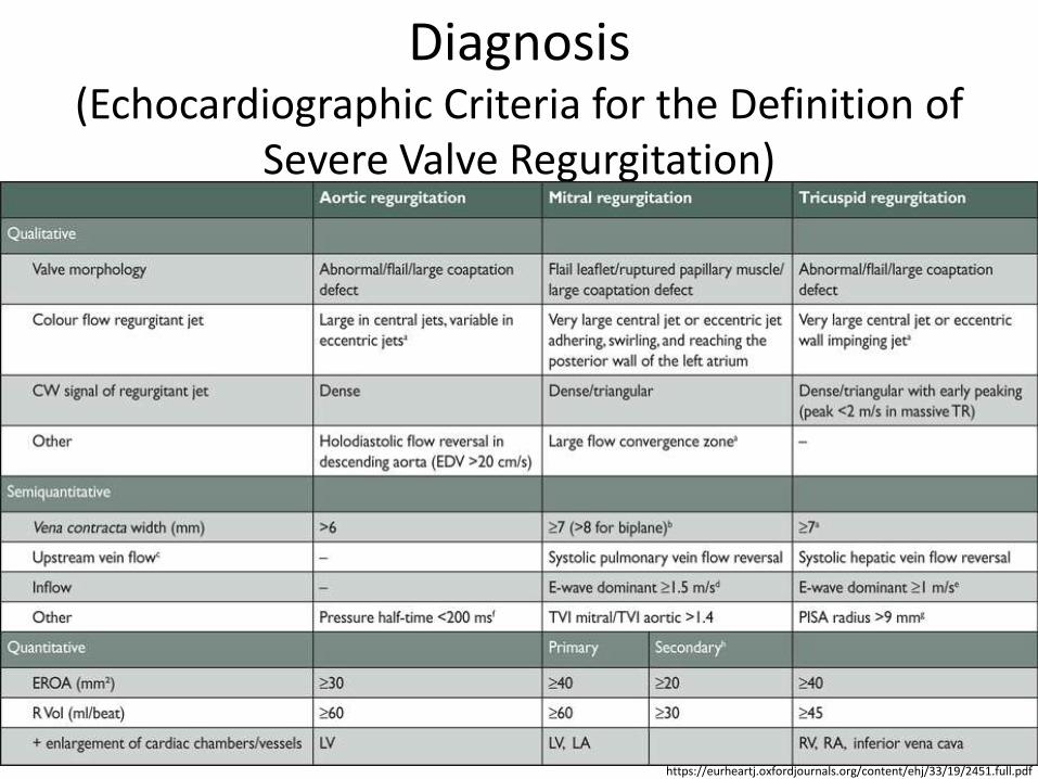
Diagnosis (Echocardiographic Criteria for the Definition of
Severe Valve Regurgitation)
https://eurheartj.oxfordjournals.org/content/ehj/33/19/2451.full.pdf
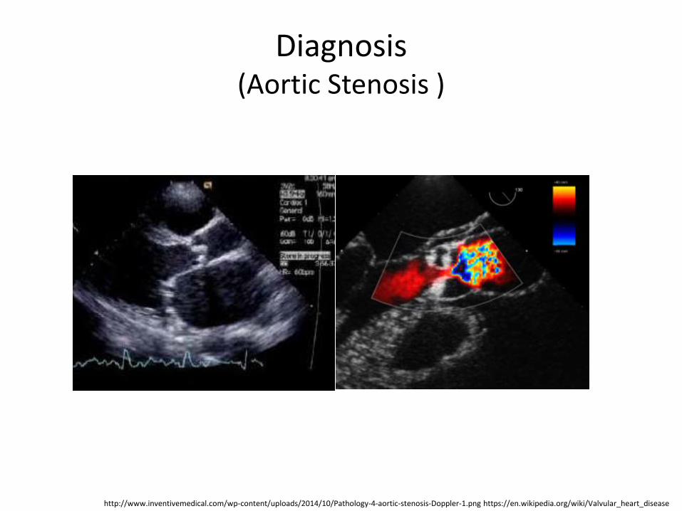
Diagnosis (Aortic Stenosis )
http://www.inventivemedical.com/wp-content/uploads/2014/10/Pathology-4-aortic-stenosis-Doppler-1.png https://en.wikipedia.org/wiki/Valvular_heart_disease
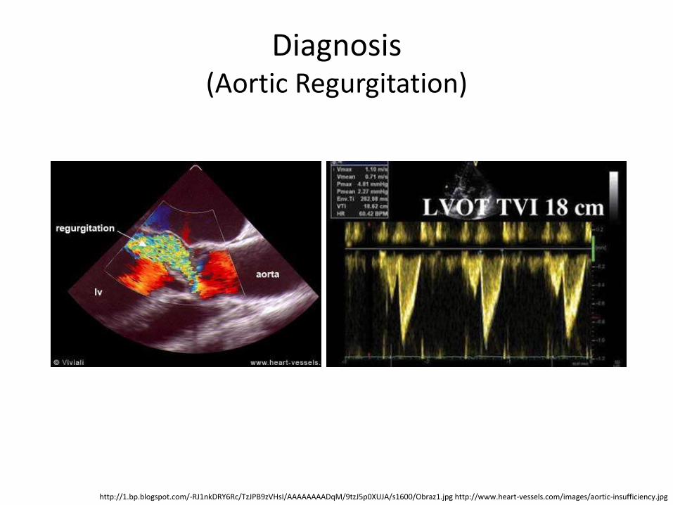
Diagnosis (Aortic Regurgitation)
http://1.bp.blogspot.com/-RJ1nkDRY6Rc/TzJPB9zVHsI/AAAAAAAADqM/9tzJ5p0XUJA/s1600/Obraz1.jpg http://www.heart-vessels.com/images/aortic-insufficiency.jpg
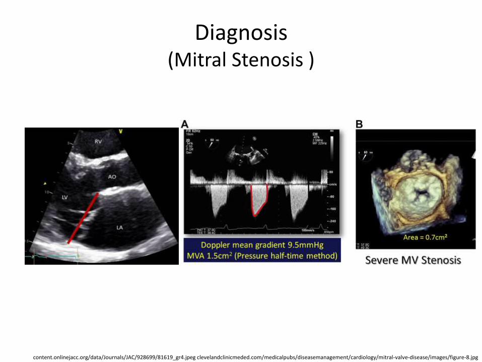
Diagnosis (Mitral Stenosis )
content.onlinejacc.org/data/Journals/JAC/928699/81619_gr4.jpeg clevelandclinicmeded.com/medicalpubs/diseasemanagement/cardiology/mitral-valve-disease/images/figure-8.jpg
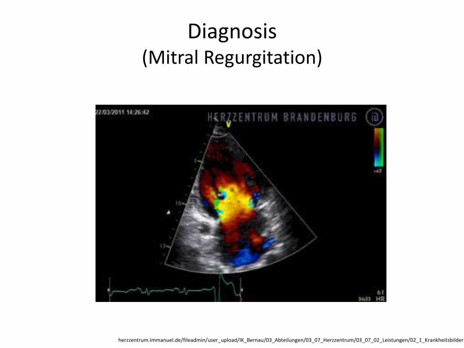
Diagnosis (Mitral Regurgitation)
herzzentrum.immanuel.de/fileadmin/user_upload/IK_Bernau/03_Abteilungen/03_07_Herzzentrum/03_07_02_Leistungen/02_1_Krankheitsbilder
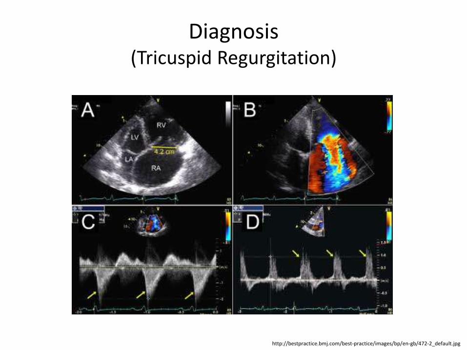
Diagnosis (Tricuspid Regurgitation)
http://bestpractice.bmj.com/best-practice/images/bp/en-gb/472-2_default.jpg
US MLE STEP 2 TEST
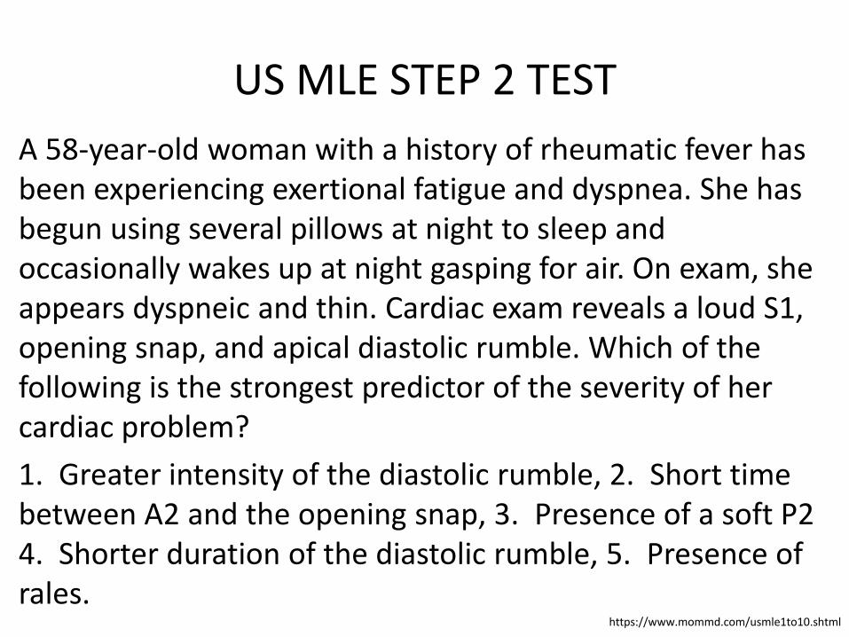
A 58-year-old woman with a history of rheumatic fever has been experiencing exertional fatigue and dyspnea. She has begun using several pillows at night to sleep and occasionally wakes up at night gasping for air. On exam, she appears dyspneic and thin. Cardiac exam reveals a loud S1, opening snap, and apical diastolic rumble. Which of the following is the strongest predictor of the severity of her cardiac problem?
1. Greater intensity of the diastolic rumble, 2. Short time between A2 and the opening snap, 3. Presence of a soft P2 4. Shorter duration of the diastolic rumble, 5. Presence of rales.
https://www.mommd.com/usmle1to10.shtml
US MLE STEP 2 EXPLANATION
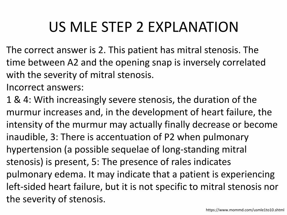
The correct answer is 2. This patient has mitral stenosis. The time between A2 and the opening snap is inversely correlated with the severity of mitral stenosis. Incorrect answers: 1 & 4: With increasingly severe stenosis, the duration of the murmur increases and, in the development of heart failure, the intensity of the murmur may actually finally decrease or become inaudible, 3: There is accentuation of P2 when pulmonary hypertension (a possible sequelae of long-standing mitral stenosis) is present, 5: The presence of rales indicates pulmonary edema. It may indicate that a patient is experiencing left-sided heart failure, but it is not specific to mitral stenosis nor the severity of stenosis.
https://www.mommd.com/usmle1to10.shtml
Treatment (Aortic Stenosis) 1
• No treatment in asymptomatic patients.
• If symptomatic, treated with aortic valve replacement surgery.
• Medical therapy and percutaneous balloon valvuloplasty have relatively poor effect: - any angina is treated with short-acting nitrovasodilators, beta-blockers and/or calcium blockers;
https://en.wikipedia.org/wiki/Valvular_heart_disease
Treatment (Aortic Stenosis) 2
• If Medical therapy and percutaneous balloon valvuloplasty have relatively poor effect: - any hypertension is treated aggressively, but caution must be taken in administering beta-blockers; - Any heart failure is treated with diuretics, nitrovasodilators and, if not contraindicated, cautious inpatient administration of ACE inhibitors.
https://en.wikipedia.org/wiki/Valvular_heart_disease
Treatment (Aortic Regurgitation)
• If stable and asymptomatic - conservative treatment such as low sodium diet, diuretics, ACE inhibitors/angiotensin II receptor antagonists, calcium blockers and avoiding very strenuous activity.
• Aortic valve replacement in symptomatic patients or progressive LV dilation or systolic ventricular diameter >55 mm, immediately if acute.
• Endocarditis prophylaxis is indicated before dental, gastrointestinal or genitourinary procedures.
https://en.wikipedia.org/wiki/Valvular_heart_disease
Treatment (Mitral Stenosis) 1
• No therapy is required for asymptomatic patients.
• Diuretics for any pulmonary congestion or edema.
• If stenosis is severe, surgery is recommended.
• Any atrial fibrillation is treated accordingly (rate or rrhythm control, and chronic anticoagulant administration).
• Prophylaxis of infective endocarditis .
https://en.wikipedia.org/wiki/Valvular_heart_disease
Treatment (Mitral Stenosis) 2
• Surgically, by mitral valvuloplasty with a percutaneously inserted balloon, unless significant mitral regurgitation or too much calcification (indicated in ostium area < 1-1.2 cm2).
• Other options include valvulotomy or mitral valve replacement by open surgery.
https://en.wikipedia.org/wiki/Valvular_heart_disease
Treatment (Mitral Regurgitation) 1
• Medically:
• afterload reduction with vasodilators;
• any hypertension is treated aggressively, e.g. by diuretics and low sodium diet;
• antiarrhythmics;
• chronic anticoagulation in concomitant mitral valve prolapse or atrial fibrillation.
https://en.wikipedia.org/wiki/Valvular_heart_disease
Treatment (Mitral Regurgitation) 2
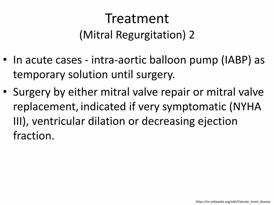
• In acute cases - intra-aortic balloon pump (IABP) as temporary solution until surgery.
• Surgery by either mitral valve repair or mitral valve replacement, indicated if very symptomatic (NYHA III), ventricular dilation or decreasing ejection fraction.
https://en.wikipedia.org/wiki/Valvular_heart_disease
US MLE STEP 2 TEST
A 68-year-old woman is admitted to the hospital following an acute change in mental status. She has a mechanical heart valve. Her medications are carvedilol, lisinopril, atorvastatin, and warfarin. Coagulation studies show an INR of 3.1, which is within the target range set by her cardiologist. For which of the following comorbid medical conditions would this medical regimen and INR be most appropriate?
1. Mechanical aortic valve with hyperlipidemia, 2. Mechanical aortic valve with atrial fibrillation, 3. Mechanical aortic valve with diabetes mellitus, 4. Mechanical aortic valve with hypertension, 5. Mechanical aortic valve with history of systemic embolization.
https://www.mommd.com/usmle1to10.shtml
US MLE STEP 2 EXPLANATION
The correct answer is 2. The patient described in the question stem has a mechanical heart valve and an INR of 3.1. INR of 2.5-3.5 is recommended for patients with a bileaflet mechanical aortic valve and atrial fibrillation.
Incorrect answers: 1: INR of 2-3 is indicated for a bileaflet mechanical aortic valve, 3: The presence of diabetes is not the most important factor determining the appropriate level of anticoagulation for this patient. Answer 2 is a better answer, 4: The presence of hypertension is not the most important factor determining the appropriate level of anticoagulation for this patient. Answer 2 is a better answer, 5: INR of 2.5-3.5 + ASA is indicated for a mechanical valve with a history of systemic embolization. The patient is not taking aspirin.
https://www.mommd.com/usmle1to10.shtml
Treatment (Tricuspid Regurgitation)
• Treatment of underlying cause.
• Surgery:
– tricuspid valvular repair;
–Valvuloplasty;
– valve replacement(rarely performed).
https://en.wikipedia.org/wiki/Valvular_heart_disease
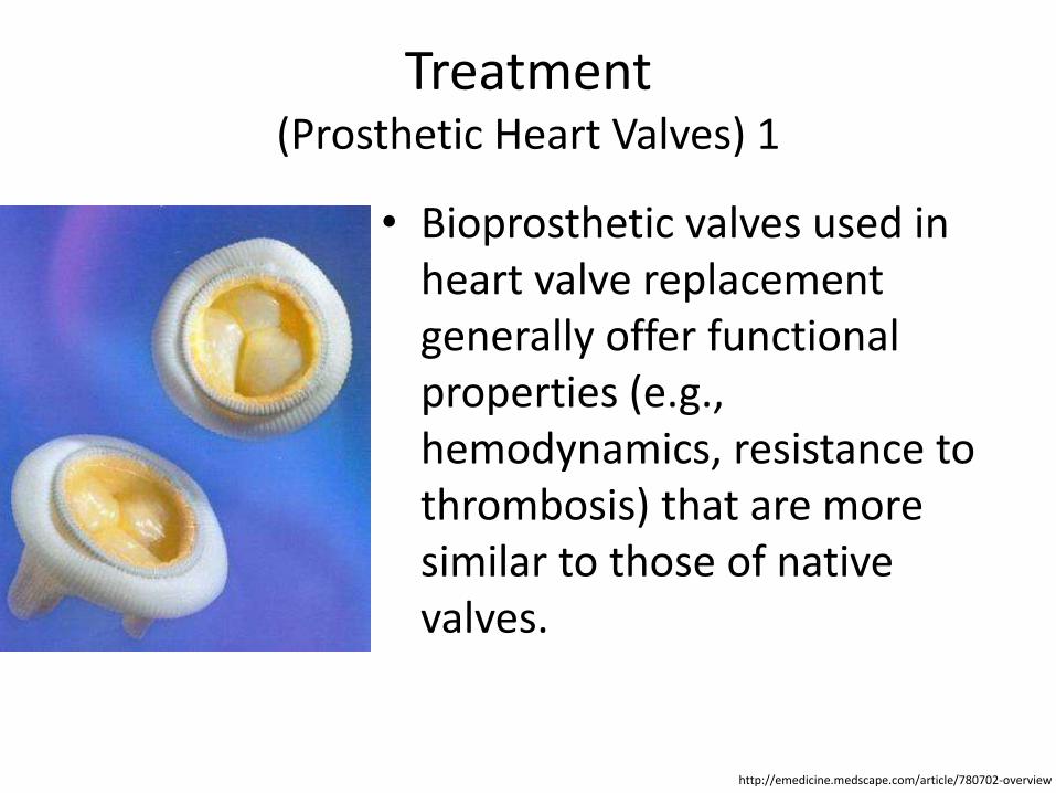
Treatment (Prosthetic Heart Valves) 1
• Bioprosthetic valves used in heart valve replacement generally offer functional properties (e.g., hemodynamics, resistance to thrombosis) that are more similar to those of native valves.
http://emedicine.medscape.com/article/780702-overview
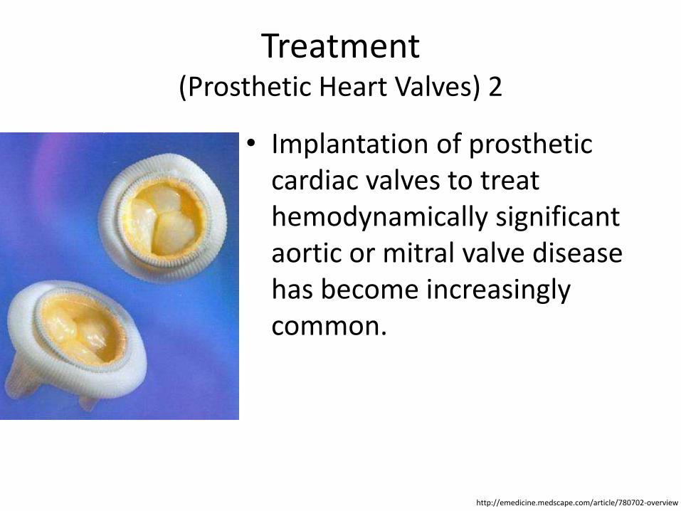
Treatment (Prosthetic Heart Valves) 2
• Implantation of prosthetic cardiac valves to treat hemodynamically significant aortic or mitral valve disease has become increasingly common.
http://emedicine.medscape.com/article/780702-overview
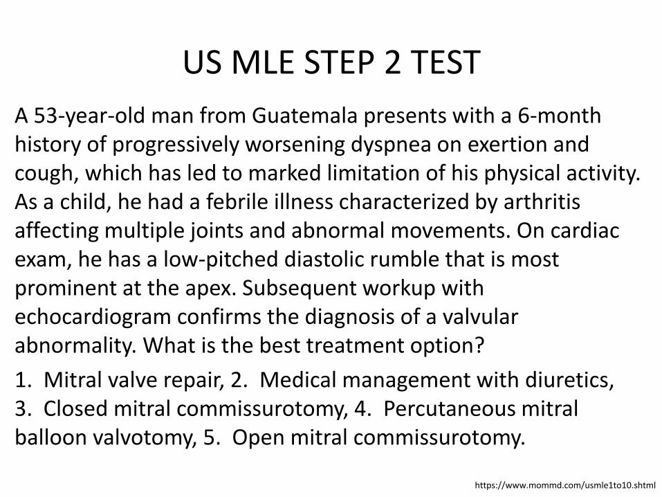
US MLE STEP 2 TEST
A 53-year-old man from Guatemala presents with a 6-month history of progressively worsening dyspnea on exertion and cough, which has led to marked limitation of his physical activity. As a child, he had a febrile illness characterized by arthritis affecting multiple joints and abnormal movements. On cardiac exam, he has a low-pitched diastolic rumble that is most prominent at the apex. Subsequent workup with echocardiogram confirms the diagnosis of a valvular abnormality. What is the best treatment option?
1. Mitral valve repair, 2. Medical management with diuretics, 3. Closed mitral commissurotomy, 4. Percutaneous mitral balloon valvotomy, 5. Open mitral commissurotomy.
https://www.mommd.com/usmle1to10.shtml
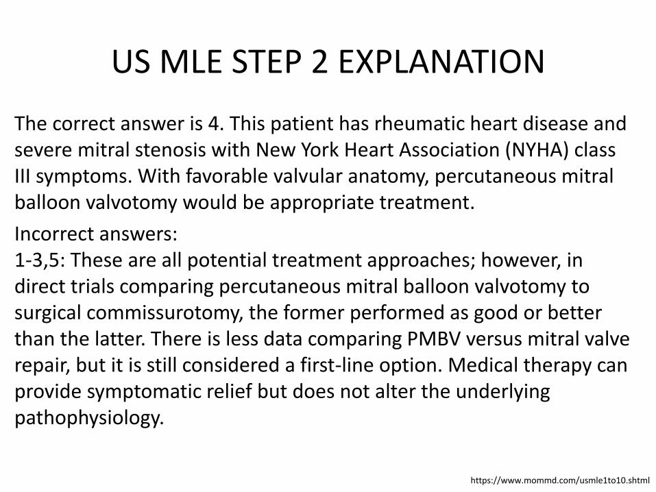
US MLE STEP 2 EXPLANATION
The correct answer is 4. This patient has rheumatic heart disease and severe mitral stenosis with New York Heart Association (NYHA) class III symptoms. With favorable valvular anatomy, percutaneous mitral balloon valvotomy would be appropriate treatment.
Incorrect answers: 1-3,5: These are all potential treatment approaches; however, in direct trials comparing percutaneous mitral balloon valvotomy to surgical commissurotomy, the former performed as good or better than the latter. There is less data comparing PMBV versus mitral valve repair, but it is still considered a first-line option. Medical therapy can provide symptomatic relief but does not alter the underlying pathophysiology.
https://www.mommd.com/usmle1to10.shtml
Treatment (Prosthetic Heart Valves Malfunction) 1
• Acute prosthetic valve failure: sudden onset of dyspnea, syncope, or precordial pain.
• Acute aortic valve failure: sudden death; survivors have acute severe dyspnea, sometimes accompanied by precordial pain, or syncope.
• Subacute valvular failure: symptoms of gradually worsening congestive heart failure; they also may present with unstable angina or, at times, may be entirely asymptomatic.
http://emedicine.medscape.com/article/780702-overview
Treatment (Prosthetic Heart Valves Malfunction) 2
• Embolic complications: symptoms related to the site of embolization (e.g., stroke, myocardial infarction, sudden death, or symptoms of visceral or peripheral embolization).
• Anticoagulant-related hemorrhage: symptoms related to the site of hemorrhage.
http://emedicine.medscape.com/article/780702-overview
Prognosis
• The prognosis for patients with valvular heart disease has improved over the past 15 years.
• A better understanding of the proper timing of surgery is one of the key reasons.
• In general, surgery for stenotic valvular disease can be delayed until symptoms appear.
• In regurgitant valvular heart disease, prognostically important left ventricular dysfunction may develop in the absence of symptoms, and thus valve surgery for asymptomatic patients is entirely appropriate.
http://www.nejm.org/doi/full/10.1056/NEJM199707033370107
Prophylaxis • Prompt treatment of strep infections can prevent
rheumatic fever, which damages the heart valves.
• An exercise, a heart-healthy diet, and medicines that lower cholesterol might prevent aortic stenosis (thickening and stiffening of the aortic valve).
• Heart-healthy eating, physical activity, other heart-healthy lifestyle changes, and medicines aimed at preventing a heart attack, high blood pressure, or heart failure also may help prevent heart valve disease.
https://www.nhlbi.nih.gov/health/health-topics/topics/hvd/prevention
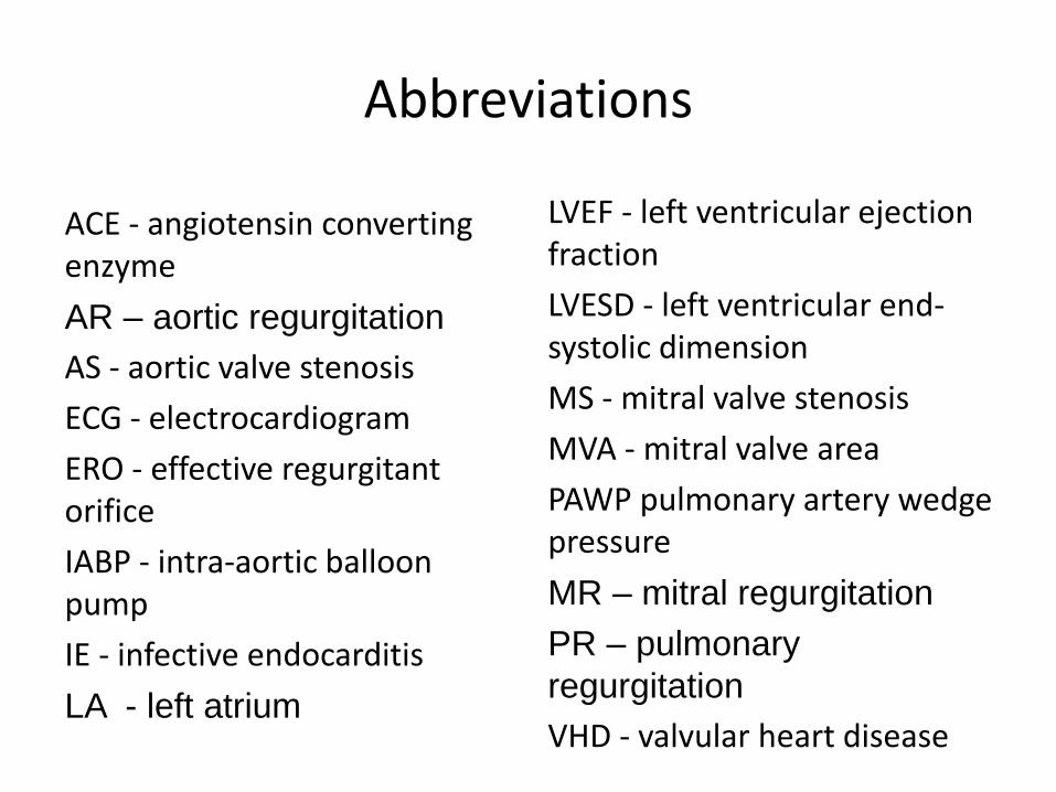
Abbreviations
ACE - angiotensin converting enzyme
AR – aortic regurgitation
AS - aortic valve stenosis
ECG - electrocardiogram
ERO - effective regurgitant orifice
IABP - intra-aortic balloon pump
IE - infective endocarditis
LA - left atrium
LVEF - left ventricular ejection fraction
LVESD - left ventricular end-systolic dimension
MS - mitral valve stenosis
MVA - mitral valve area
PAWP pulmonary artery wedge pressure
MR – mitral regurgitation
PR – pulmonary
regurgitation
VHD - valvular heart disease
Diagnostic and treatment guidelines
2014 AHA/ACC Guideline for the Management of Patients With Valvular Heart Disease
Guidelines on the management of valvular heart disease (version 2012)
Prosthetic Heart Valves
Infective Endocarditis
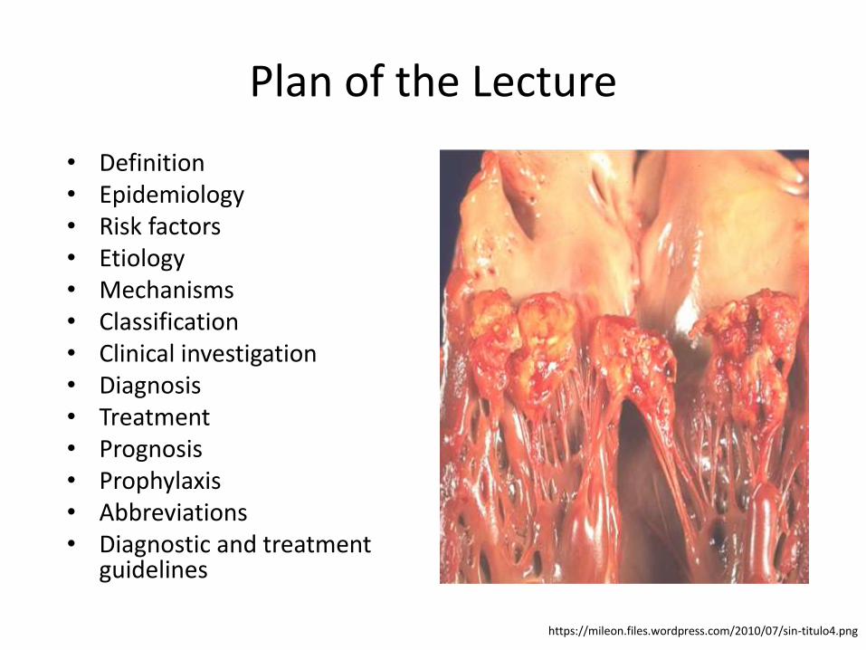
Plan of the Lecture
• Definition • Epidemiology • Risk factors • Etiology • Mechanisms • Classification • Clinical investigation • Diagnosis • Treatment • Prognosis • Prophylaxis • Abbreviations • Diagnostic and treatment
guidelines
https://mileon.files.wordpress.com/2010/07/sin-titulo4.png
Definition
Infective endocarditis (IE) is a potentially lethal disease caused largely by bacteria that enter the bloodstream and settle in the heart, which may include one or more heart valves, the mural endocardium, or a septal defect or a blood vessel, with intracardiac effects that include severe valvular insufficiency and intractable congestive heart failure and myocardial abscesses.
emedicine.medscape.com/article/216650-overview circ.ahajournals.org/content/early/2015/09/15/CIR.0000000000000296
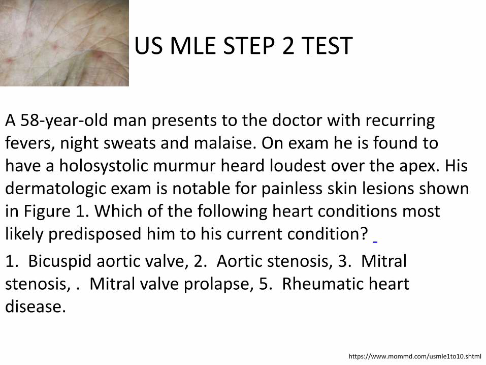
US MLE STEP 2 TEST
A 58-year-old man presents to the doctor with recurring fevers, night sweats and malaise. On exam he is found to have a holosystolic murmur heard loudest over the apex. His dermatologic exam is notable for painless skin lesions shown in Figure 1. Which of the following heart conditions most likely predisposed him to his current condition?
1. Bicuspid aortic valve, 2. Aortic stenosis, 3. Mitral stenosis, . Mitral valve prolapse, 5. Rheumatic heart disease.
https://www.mommd.com/usmle1to10.shtml
US MLE STEP 2 EXPLANATION
The correct answer is 4. The clinical presentation is consistent with native valve bacterial endocarditis (NVBE). The most common cardiac abnormality predisposing to NVBE in 15- to 60-year olds is mitral valve prolapse (MVP). Incorrect answers: 1, 2, 3: These cardiac conditions do not directly increase the risk of IE, 5: Although rheumatic heart disease is associated with IE, mitral valve prolapse is the most common cardiac condition that predisposes patients to NVBE.
https://www.mommd.com/usmle1to10.shtml
Epidemiology 1
http://circ.ahajournals.org/content/early/2015/09/15/CIR.0000000000000296
• IE is an uncommon infectious disease with an annual incidence ranging from 3 to 7 per 100000 person-years in the most contemporary population surveys.
• Although relatively rare, IE continues to be characterized by increased morbidity and mortality and is now the third or fourth most common life-threatening infection syndrome, after sepsis, pneumonia, and intra-abdominal abscess.
Epidemiology 2
http://circ.ahajournals.org/content/early/2015/09/15/CIR.0000000000000296
• Characteristics of IE patients have shifted toward an increased mean patient age, a higher proportion of prosthetic valves and other cardiac devices, and a decreasing proportion of rheumatic heart disease.
• The proportion of IE patients undergoing surgery has increased over time to reach ≈50%.
Risk Factors • Artificial heart valves.
• Intracardiac devices.
• Unrepaired cyanotic congenital heart defects.
• History of infective endocarditis.
• Chronic rheumatic heart disease.
• Age-related degenerative valvular lesions.
• Hemodialysis.
• Coexisting conditions, especially ones that suppress immunity (diabetes mellitus, alcohol abuse, HIV/AIDS, and intravenous drug).
https://en.wikipedia.org/wiki/Infective_endocarditis
Etiology
• Many microorganisms can cause infective endocarditis.
• These are generally isolated by blood culture, where the patient's blood is removed, and any growth is noted and identified.
• The term bacterial endocarditis (BE) commonly is used, reflecting the fact that most cases of IE are due to bacteria; however, infective endocarditis (IE) has become the preferred term.
https://en.wikipedia.org/wiki/Infective_endocarditis
Etiology (Bacterial) 1
• Staphylococcus aureus followed by Streptococci of the viridans group and coagulase negative Staphylococci are the three most common organisms responsible for infective endocarditis.
• Other Streptococci and Enterococci are also a frequent cause of infective endocarditis.
• Enterococcus can enter the bloodstream as a consequence of abnormalities in the gastrointestinal or genitourinary tracts.
https://en.wikipedia.org/wiki/Myocarditis#Causes
Etiology (Bacterial) 2
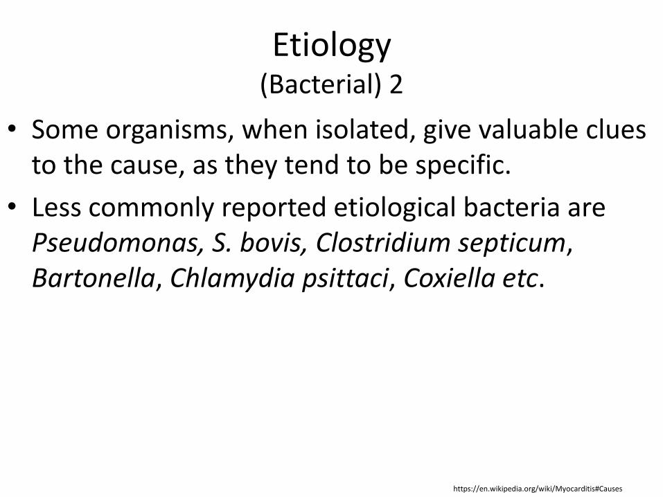
• Some organisms, when isolated, give valuable clues to the cause, as they tend to be specific.
• Less commonly reported etiological bacteria are Pseudomonas, S. bovis, Clostridium septicum, Bartonella, Chlamydia psittaci, Coxiella etc.
https://en.wikipedia.org/wiki/Myocarditis#Causes
Etiology (Fungal)
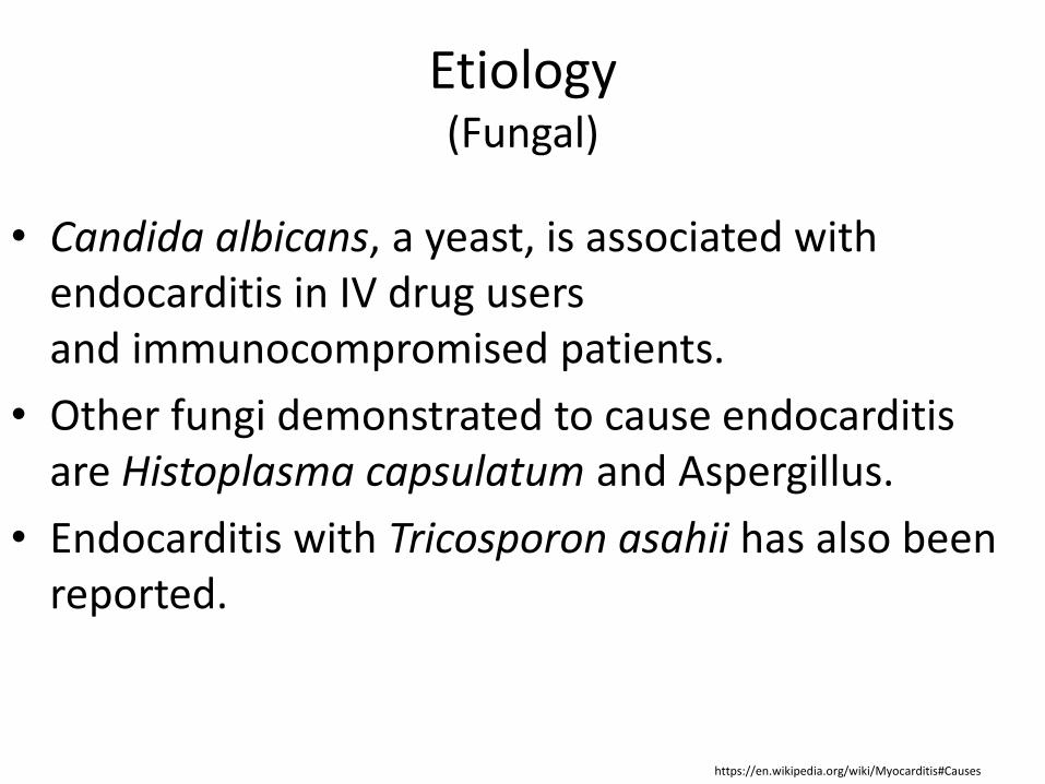
• Candida albicans, a yeast, is associated with endocarditis in IV drug users and immunocompromised patients.
• Other fungi demonstrated to cause endocarditis are Histoplasma capsulatum and Aspergillus.
• Endocarditis with Tricosporon asahii has also been reported.
https://en.wikipedia.org/wiki/Myocarditis#Causes
US MLE STEP 2 TEST
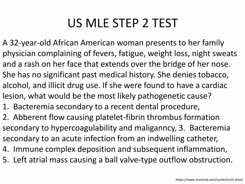
A 32-year-old African American woman presents to her family physician complaining of fevers, fatigue, weight loss, night sweats and a rash on her face that extends over the bridge of her nose. She has no significant past medical history. She denies tobacco, alcohol, and illicit drug use. If she were found to have a cardiac lesion, what would be the most likely pathogenetic cause? 1. Bacteremia secondary to a recent dental procedure, 2. Abberent flow causing platelet-fibrin thrombus formation secondary to hypercoagulability and maliganncy, 3. Bacteremia secondary to an acute infection from an indwelling catheter, 4. Immune complex deposition and subsequent inflammation, 5. Left atrial mass causing a ball valve-type outflow obstruction.
https://www.mommd.com/usmle1to10.shtml
US MLE STEP 2 EXPLANATION
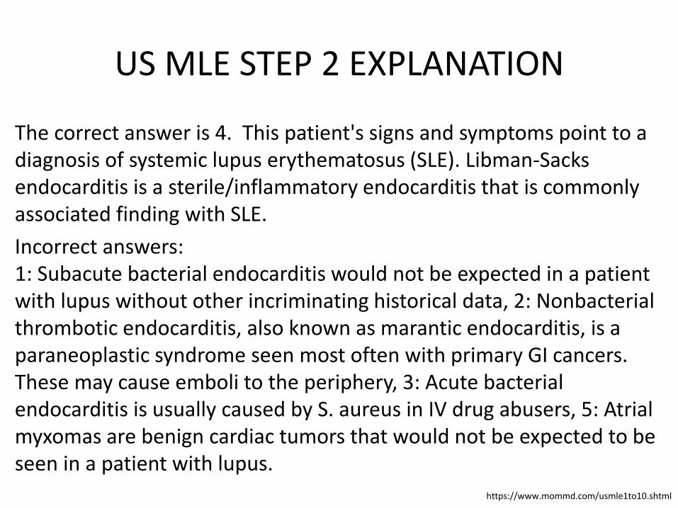
The correct answer is 4. This patient's signs and symptoms point to a diagnosis of systemic lupus erythematosus (SLE). Libman-Sacks endocarditis is a sterile/inflammatory endocarditis that is commonly associated finding with SLE.
Incorrect answers: 1: Subacute bacterial endocarditis would not be expected in a patient with lupus without other incriminating historical data, 2: Nonbacterial thrombotic endocarditis, also known as marantic endocarditis, is a paraneoplastic syndrome seen most often with primary GI cancers. These may cause emboli to the periphery, 3: Acute bacterial endocarditis is usually caused by S. aureus in IV drug abusers, 5: Atrial myxomas are benign cardiac tumors that would not be expected to be seen in a patient with lupus.
https://www.mommd.com/usmle1to10.shtml
Mechanisms 1
http://emedicine.medscape.com/article/216650-overview#a3 https://en.wikipedia.org/wiki/Myocarditis#Causes
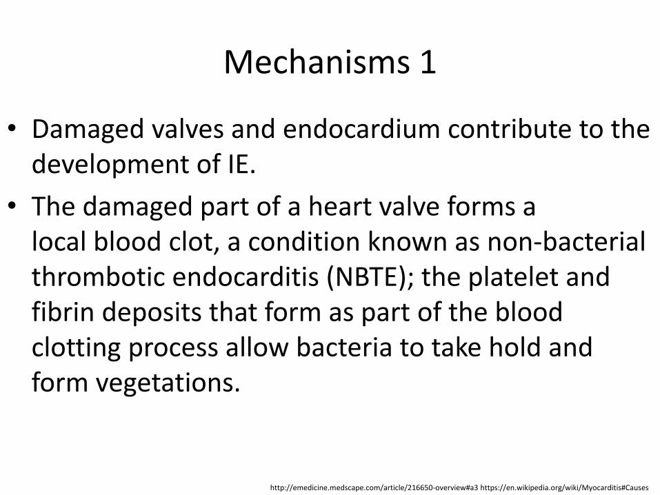
• Damaged valves and endocardium contribute to the development of IE.
• The damaged part of a heart valve forms a local blood clot, a condition known as non-bacterial thrombotic endocarditis (NBTE); the platelet and fibrin deposits that form as part of the blood clotting process allow bacteria to take hold and form vegetations.
Mechanisms 2
http://emedicine.medscape.com/article/216650-overview#a3 https://en.wikipedia.org/wiki/Myocarditis#Causes
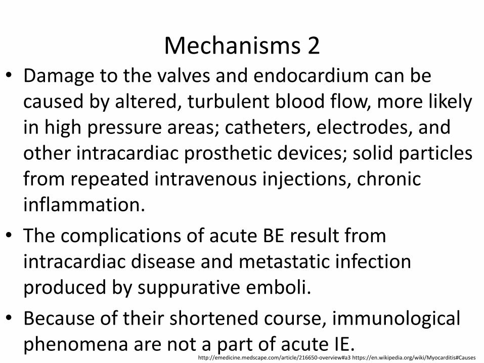
• Damage to the valves and endocardium can be caused by altered, turbulent blood flow, more likely in high pressure areas; catheters, electrodes, and other intracardiac prosthetic devices; solid particles from repeated intravenous injections, chronic inflammation.
• The complications of acute BE result from intracardiac disease and metastatic infection produced by suppurative emboli.
• Because of their shortened course, immunological phenomena are not a part of acute IE.
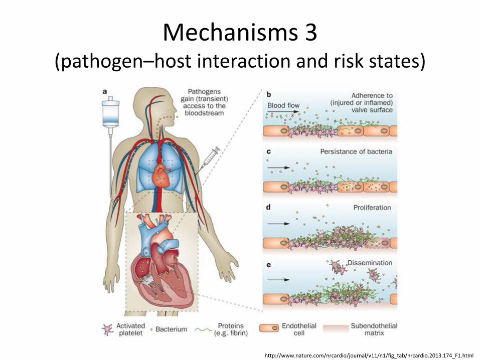
Mechanisms 3 (pathogen–host interaction and risk states)
http://www.nature.com/nrcardio/journal/v11/n1/fig_tab/nrcardio.2013.174_F1.html
US MLE STEP 2 TEST
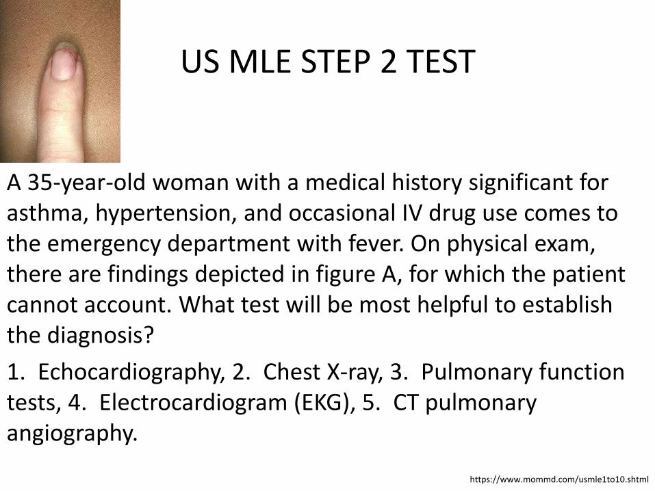
A 35-year-old woman with a medical history significant for asthma, hypertension, and occasional IV drug use comes to the emergency department with fever. On physical exam, there are findings depicted in figure A, for which the patient cannot account. What test will be most helpful to establish the diagnosis?
1. Echocardiography, 2. Chest X-ray, 3. Pulmonary function tests, 4. Electrocardiogram (EKG), 5. CT pulmonary angiography.
https://www.mommd.com/usmle1to10.shtml
US MLE STEP 2 EXPLANATION The correct answer is 1. Fever, subungual splinter hemorrhages (in image A), and history of IV drug abuse raise concern for infective endocarditis. An echocardiogram will likely confirm the diagnosis by demonstrating a valvular vegetation.
Incorrect answers: 2. Chest X-Ray can occasionally reveal important diagnostic clues (e.g. septic pulmonary emboli) but would not show a valve vegetation, which indicates active endocarditis, 3. Pulmonary function tests would help diagnose asthma, but not infective endocarditis, 4. EKGs rarely reveal diagnostic findings of infective endocarditis, but may help determine the presence of emboli to the coronary circulation if there are ischemic changes, 5. CT pulmonary angiography would help diagnose pulmonary embolus (PE), but not infective endocarditis. Sometimes PE is a complication of infective endocarditis, but unlikely in the patient in this scenario.
https://www.mommd.com/usmle1to10.shtml
Classification (International Classification of Diseases (ICD))
apps.who.int/classifications/icd10/browse/2016/en#/I46
133 Acute and subacute endocarditis
I33.0 Acute and subacute infective endocarditis
Endocarditis (acute)(subacute):bacterial
infective NOS, lenta, malignant, septic, ulcerative.
I33.9 Acute endocarditis, unspecified.
Classification (the Rate of Progression and Severity of Disease) 1
http://www.merckmanuals.com/professional/cardiovascular-disorders/endocarditis/infective-endocarditis https://en.wikipedia.org/wiki/Infective_endocarditis
• Subacute bacterial endocarditis (SBE) is often due to streptococci of low virulence (mainly viridans streptococci) and mild to moderate illness which progresses slowly over weeks and months and has low propensity to hematogenously seed extracardiac sites.
Classification (the Rate of Progression and Severity of Disease) 2
http://www.merckmanuals.com/professional/cardiovascular-disorders/endocarditis/infective-endocarditis https://en.wikipedia.org/wiki/Infective_endocarditis
• Acute bacterial endocarditis (ABE) is a fulminant illness over days to weeks, and is more likely due to Staphylococcus aureus which has much greater virulence, or disease-producing capacity and frequently causes metastatic infection.
• Prosthetic valvular endocarditis (PVE) develops in 2 to 3% of patients within 1 yr. after valve replacement and in 0.5%/yr. thereafter.
Classification (the Rate of Progression and Severity of Disease) 3
http://www.merckmanuals.com/professional/cardiovascular-disorders/endocarditis/infective-endocarditis https://en.wikipedia.org/wiki/Infective_endocarditis
• The terms short incubation (meaning less than about six weeks), and long incubation (greater than about six weeks) are preferred.
Clinical Investigation (Signs and Symptoms) 1
• Fever (97% of patients), malaise and endurance fatigue (90% of patients).
• A new or changing heart murmur, weight loss, and coughing (35% of patients).
• Vascular phenomena: septic embolism (causing stroke or gangrene of fingers), Janeway lesions (painless hemorrhagic cutaneous lesions on the palms and soles), intracranial hemorrhage, conjunctival and splinter hemorrhages, kidney and splenic infarcts.
https://en.wikipedia.org/wiki/Infective_endocarditis
Clinical Investigation (Signs and Symptoms) 2
• Immunologic phenomena: glomerulonephritis , Osler's nodes (painful subcutaneous lesions in the distal fingers), Roth's spots on the retina, positive serum rheumatoid factor)
• Other signs: night sweats, rigors, anemia, etc.
https://en.wikipedia.org/wiki/Infective_endocarditis
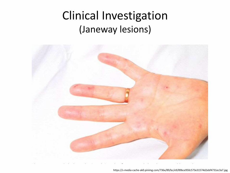
Clinical Investigation (Janeway lesions)
https://s-media-cache-ak0.pinimg.com/736x/89/bc/e9/89bce950c573e31574d2ebf4731ec3a7.jpg
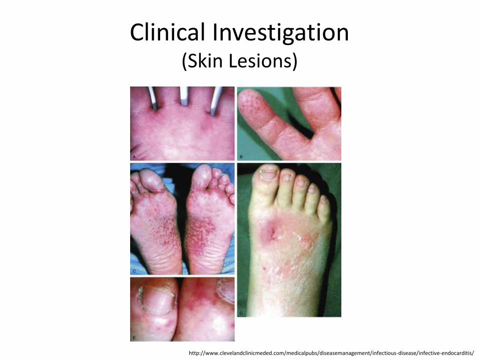
Clinical Investigation (Skin Lesions)
http://www.clevelandclinicmeded.com/medicalpubs/diseasemanagement/infectious-disease/infective-endocarditis/
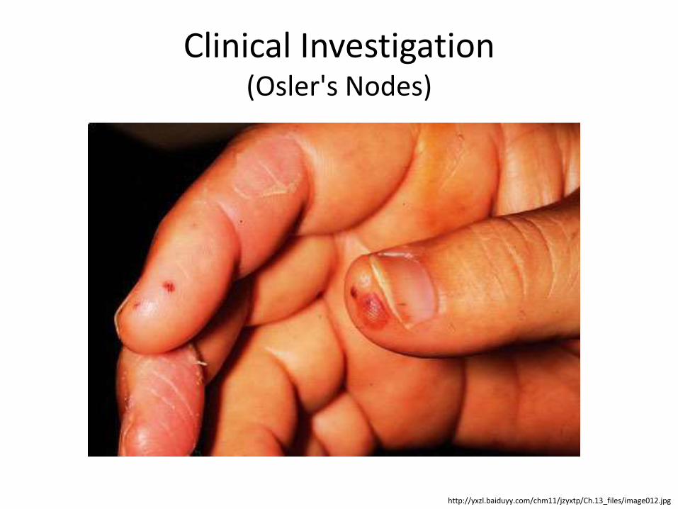
Clinical Investigation (Osler's Nodes)
http://yxzl.baiduyy.com/chm11/jzyxtp/Ch.13_files/image012.jpg
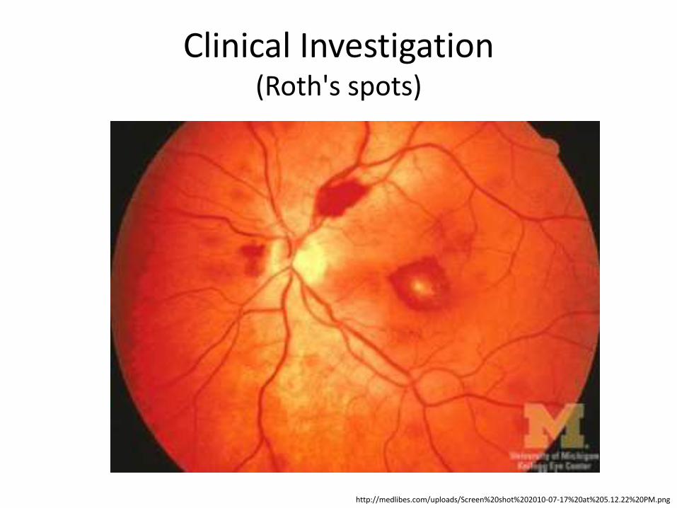
Clinical Investigation (Roth's spots)
http://medlibes.com/uploads/Screen%20shot%202010-07-17%20at%205.12.22%20PM.png
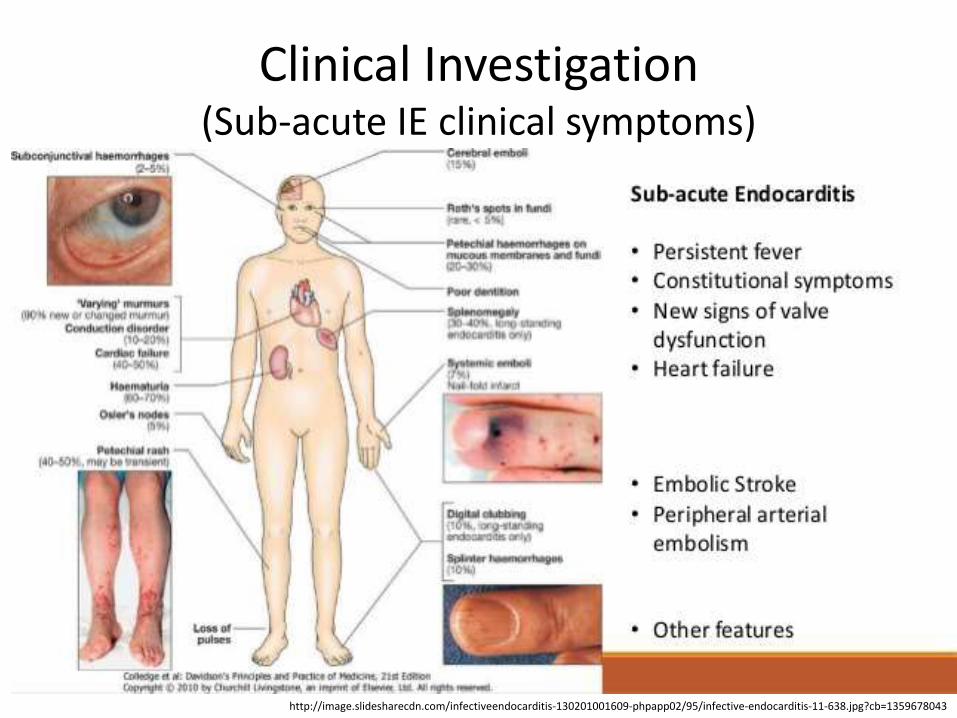
Clinical Investigation (Sub-acute IE clinical symptoms)
http://image.slidesharecdn.com/infectiveendocarditis-130201001609-phpapp02/95/infective-endocarditis-11-638.jpg?cb=1359678043
US MLE STEP 2 TEST
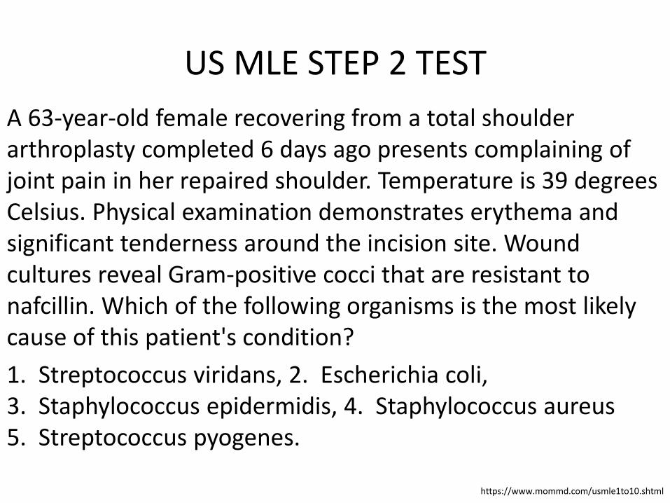
A 63-year-old female recovering from a total shoulder arthroplasty completed 6 days ago presents complaining of joint pain in her repaired shoulder. Temperature is 39 degrees Celsius. Physical examination demonstrates erythema and significant tenderness around the incision site. Wound cultures reveal Gram-positive cocci that are resistant to nafcillin. Which of the following organisms is the most likely cause of this patient's condition?
1. Streptococcus viridans, 2. Escherichia coli, 3. Staphylococcus epidermidis, 4. Staphylococcus aureus 5. Streptococcus pyogenes.
https://www.mommd.com/usmle1to10.shtml
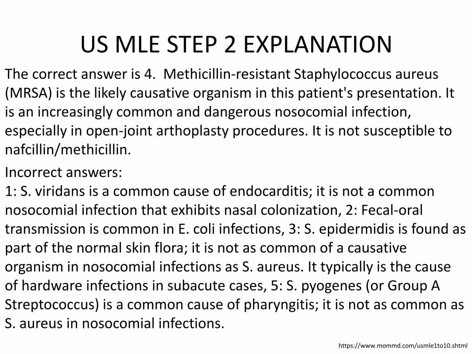
US MLE STEP 2 EXPLANATION The correct answer is 4. Methicillin-resistant Staphylococcus aureus (MRSA) is the likely causative organism in this patient's presentation. It is an increasingly common and dangerous nosocomial infection, especially in open-joint arthoplasty procedures. It is not susceptible to nafcillin/methicillin.
Incorrect answers: 1: S. viridans is a common cause of endocarditis; it is not a common nosocomial infection that exhibits nasal colonization, 2: Fecal-oral transmission is common in E. coli infections, 3: S. epidermidis is found as part of the normal skin flora; it is not as common of a causative organism in nosocomial infections as S. aureus. It typically is the cause of hardware infections in subacute cases, 5: S. pyogenes (or Group A Streptococcus) is a common cause of pharyngitis; it is not as common as S. aureus in nosocomial infections.
https://www.mommd.com/usmle1to10.shtml
Diagnosis 1
http://circ.ahajournals.org/content/early/2015/09/15/CIR.0000000000000296
• The diagnosis of IE is straightforward in the minority of patients who present with a consistent history and classic oslerian manifestations: sustained bacteremia or fungemia, evidence of active valvulitis, peripheral emboli, and immunological vascular phenomena.
• In most patients, however, the “textbook” history and physical examination findings may be few or absent.
Diagnosis 2
http://circ.ahajournals.org/content/early/2015/09/15/CIR.0000000000000296
• Acute IE may evolve too quickly for the development of immunological vascular phenomena, which are more characteristic of the later stages of the more insidious subacute form of untreated IE.
• The variability in clinical presentation of IE and the importance of early accurate diagnosis require a diagnostic strategy that is both sensitive for disease detection and specific for its exclusion across all forms of the disease.
Diagnosis 3
http://circ.ahajournals.org/content/early/2015/09/15/CIR.0000000000000296
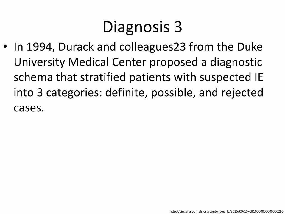
• In 1994, Durack and colleagues23 from the Duke University Medical Center proposed a diagnostic schema that stratified patients with suspected IE into 3 categories: definite, possible, and rejected cases.
Diagnosis (the Modified Duke Criteria: Definite, Possible,
and Rejected Cases) 1
http://circ.ahajournals.org/content/early/2015/09/15/CIR.0000000000000296
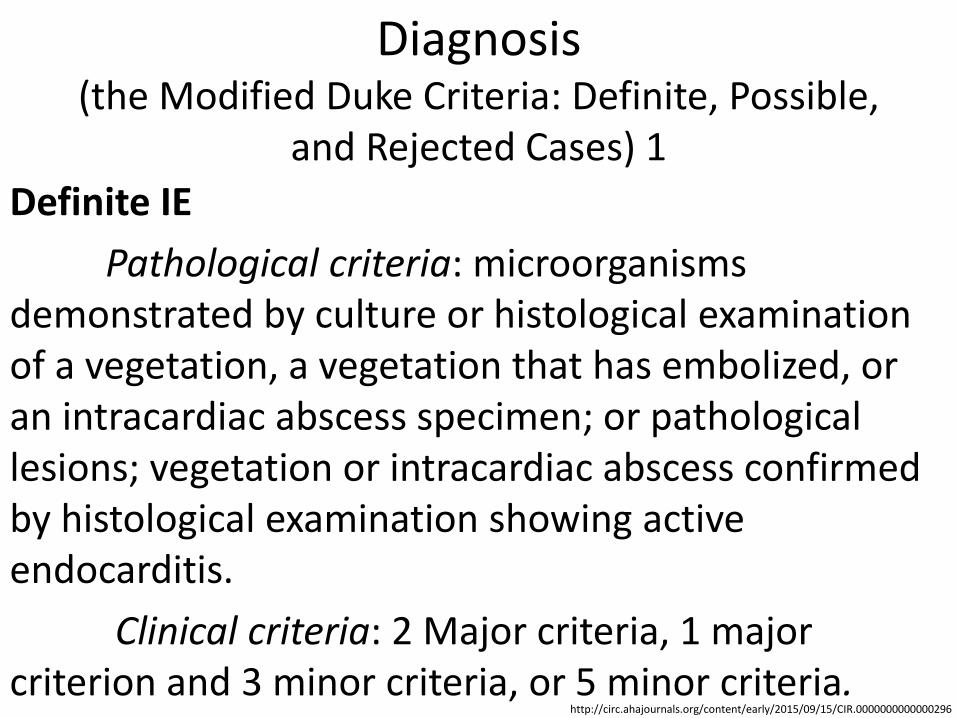
Definite IE
Pathological criteria: microorganisms demonstrated by culture or histological examination of a vegetation, a vegetation that has embolized, or an intracardiac abscess specimen; or pathological lesions; vegetation or intracardiac abscess confirmed by histological examination showing active endocarditis.
Clinical criteria: 2 Major criteria, 1 major criterion and 3 minor criteria, or 5 minor criteria.
Diagnosis (the Modified Duke Criteria: Definite, Possible,
and Rejected Cases) 2
http://circ.ahajournals.org/content/early/2015/09/15/CIR.0000000000000296
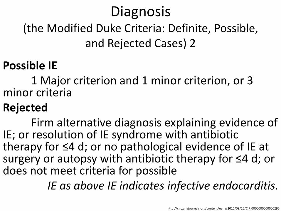
Possible IE 1 Major criterion and 1 minor criterion, or 3 minor criteria Rejected Firm alternative diagnosis explaining evidence of IE; or resolution of IE syndrome with antibiotic therapy for ≤4 d; or no pathological evidence of IE at surgery or autopsy with antibiotic therapy for ≤4 d; or does not meet criteria for possible
IE as above IE indicates infective endocarditis.
Diagnosis (the Modified Duke Criteria: Major Criteria)
http://circ.ahajournals.org/content/early/2015/09/15/CIR.0000000000000296
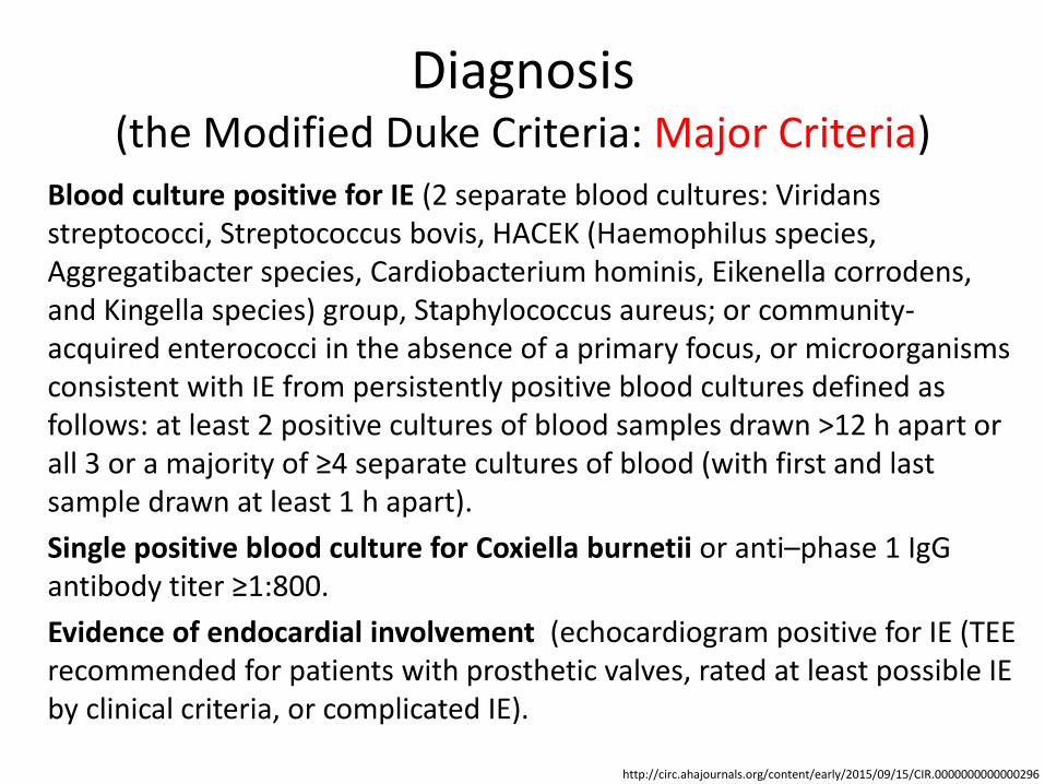
Blood culture positive for IE (2 separate blood cultures: Viridans streptococci, Streptococcus bovis, HACEK (Haemophilus species, Aggregatibacter species, Cardiobacterium hominis, Eikenella corrodens, and Kingella species) group, Staphylococcus aureus; or community-acquired enterococci in the absence of a primary focus, or microorganisms consistent with IE from persistently positive blood cultures defined as follows: at least 2 positive cultures of blood samples drawn >12 h apart or all 3 or a majority of ≥4 separate cultures of blood (with first and last sample drawn at least 1 h apart).
Single positive blood culture for Coxiella burnetii or anti–phase 1 IgG antibody titer ≥1:800.
Evidence of endocardial involvement (echocardiogram positive for IE (TEE recommended for patients with prosthetic valves, rated at least possible IE by clinical criteria, or complicated IE).
Diagnosis (the Modified Duke Criteria: Minor Criteria)
http://circ.ahajournals.org/content/early/2015/09/15/CIR.0000000000000296
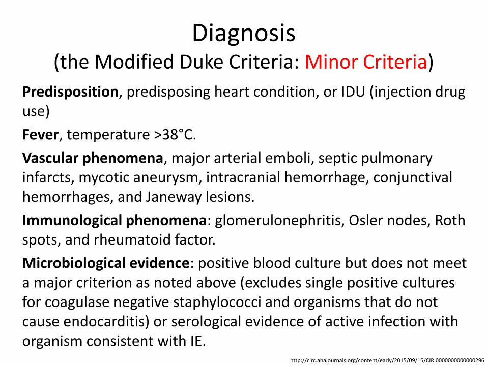
Predisposition, predisposing heart condition, or IDU (injection drug use)
Fever, temperature >38°C.
Vascular phenomena, major arterial emboli, septic pulmonary infarcts, mycotic aneurysm, intracranial hemorrhage, conjunctival hemorrhages, and Janeway lesions.
Immunological phenomena: glomerulonephritis, Osler nodes, Roth spots, and rheumatoid factor.
Microbiological evidence: positive blood culture but does not meet a major criterion as noted above (excludes single positive cultures for coagulase negative staphylococci and organisms that do not cause endocarditis) or serological evidence of active infection with organism consistent with IE.
Diagnosis (Laboratory Investigations and Biomarkers) 1
http://eurheartj.oxfordjournals.org/content/early/2015/08/28/eurheartj.ehv319
• Sepsis severity may be indicated by the demonstration of a number of laboratory investigations, including the degree of leucocytosis/leucopoenia, the number of immature white cell forms, concentrations of C-reactive protein (CRP) and procalcitonin, erythrocyte sedimentation rate (ESR) and markers of end-organ dysfunction (lactataemia, elevated bilirubin, thrombocytopenia and changes in serum creatinine concentration); however, none are diagnostic for IE.
Diagnosis (Laboratory Investigations and Biomarkers) 2
http://eurheartj.oxfordjournals.org/content/early/2015/08/28/eurheartj.ehv319
• Further, certain laboratory investigations are used in surgical scoring systems relevant to risk stratification in patients with IE, including bilirubin, creatinine and platelet count and creatinine clearance.
• The pattern of increase in inflammatory mediators or immune complexes may support, but not prove, the diagnosis of IE.
Diagnosis (Imaging Techniques) 1
http://eurheartj.oxfordjournals.org/content/early/2015/08/28/eurheartj.ehv319
• Imaging plays a key role in both the diagnosis and management of IE.
• Echocardiography is useful for the prognostic assessment of patients with IE, for its follow-up under therapy and during and after surgery.
• Echocardiography is particularly useful for initial assessment of the embolic risk and in decision making in IE.
Diagnosis (Imaging Techniques) 2
http://eurheartj.oxfordjournals.org/content/early/2015/08/28/eurheartj.ehv319
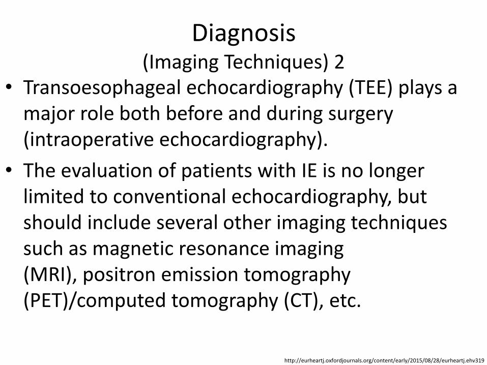
• Transoesophageal echocardiography (TEE) plays a major role both before and during surgery (intraoperative echocardiography).
• The evaluation of patients with IE is no longer limited to conventional echocardiography, but should include several other imaging techniques such as magnetic resonance imaging (MRI), positron emission tomography (PET)/computed tomography (CT), etc.
US MLE STEP 2 TEST
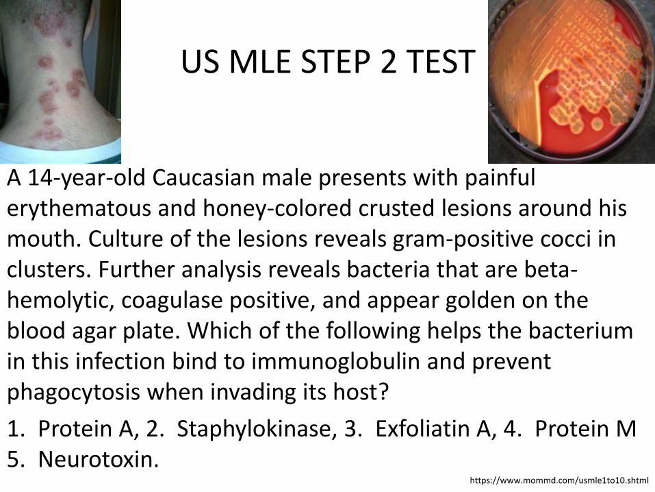
A 14-year-old Caucasian male presents with painful erythematous and honey-colored crusted lesions around his mouth. Culture of the lesions reveals gram-positive cocci in clusters. Further analysis reveals bacteria that are beta-hemolytic, coagulase positive, and appear golden on the blood agar plate. Which of the following helps the bacterium in this infection bind to immunoglobulin and prevent phagocytosis when invading its host?
1. Protein A, 2. Staphylokinase, 3. Exfoliatin A, 4. Protein M 5. Neurotoxin.
https://www.mommd.com/usmle1to10.shtml
US MLE STEP 2 EXPLANATION The correct answer is 1. The patient described above has a Staphylococcus aureus skin infection (impetigo) as evidenced by the description of the lesion and causative bacteria. Protein A is present on the surface of S. aureus and is able to bind to the Fc region of immunoglobulin molecules. This region of the immunoglobulin is normally bound by phagocytic cells in order to absorb opsonized bacteria; protein A disrupts this action and prevents phagocytosis. Incorrect answers: 2: Staphylokinase, like streptokinase, leads to the breakdown of fibrin clots, 3: Exfoliatin A leads to the breakdown of desmosomes and can lead to scalded skin syndrome, 4: Protein M is a surface protein on Streptococcus pyogenes that helps the bacterium avoid phagocytosis, 5: Neurotoxin would be consistent with a Clostridium botulinum infection (botulinum toxin).
https://www.mommd.com/usmle1to10.shtml
Diagnosis (Echocardiography as the Reference Method)
http://eurheartj.oxfordjournals.org/content/35/10/624
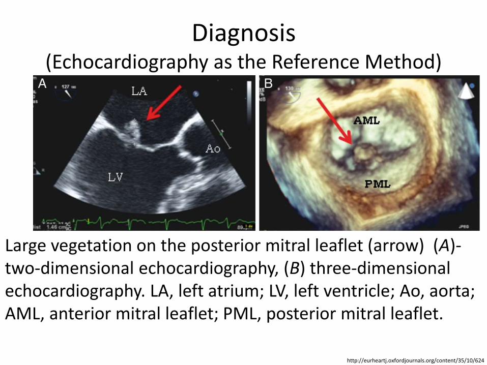
Large vegetation on the posterior mitral leaflet (arrow) (A)- two-dimensional echocardiography, (B) three-dimensional echocardiography. LA, left atrium; LV, left ventricle; Ao, aorta; AML, anterior mitral leaflet; PML, posterior mitral leaflet.
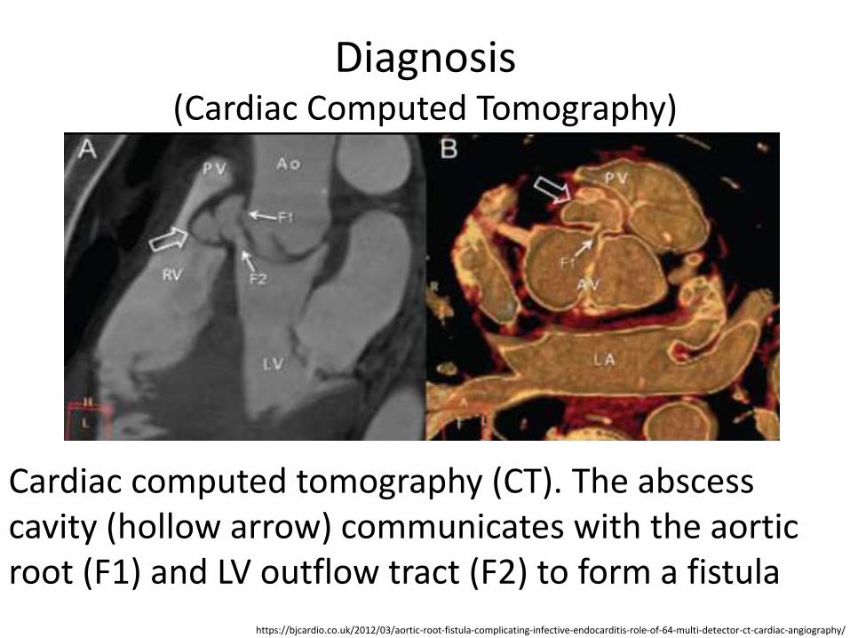
Diagnosis (Cardiac Computed Tomography)
https://bjcardio.co.uk/2012/03/aortic-root-fistula-complicating-infective-endocarditis-role-of-64-multi-detector-ct-cardiac-angiography/
Cardiac computed tomography (CT). The abscess cavity (hollow arrow) communicates with the aortic root (F1) and LV outflow tract (F2) to form a fistula
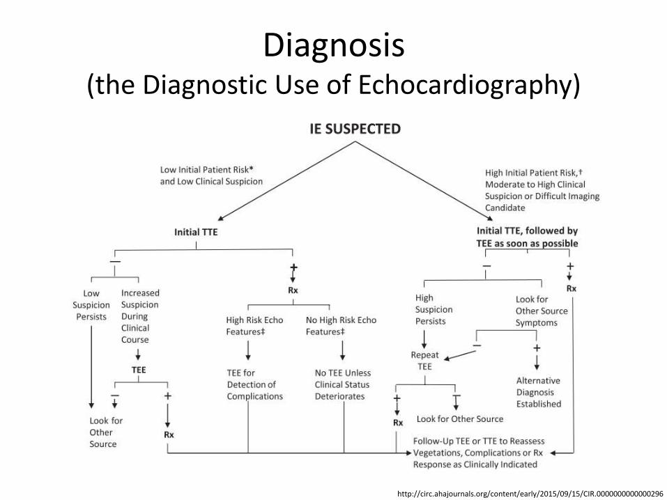
Diagnosis (the Diagnostic Use of Echocardiography)
http://circ.ahajournals.org/content/early/2015/09/15/CIR.0000000000000296
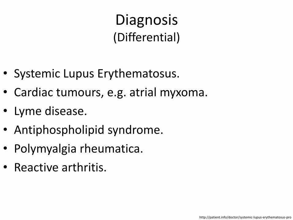
Diagnosis (Differential)
http://patient.info/doctor/systemic-lupus-erythematosus-pro
• Systemic Lupus Erythematosus.
• Cardiac tumours, e.g. atrial myxoma.
• Lyme disease.
• Antiphospholipid syndrome.
• Polymyalgia rheumatica.
• Reactive arthritis.
Treatment (Antimicrobial Therapy: Principles) 1
• The primary goal of antibiotic treatment is to eradicate infection, including sterilizing vegetations, although the unique characteristics of infected vegetations (focal infection with high bacterial density, slow rate of bacterial growth within biofilms, and low microorganism metabolic activity, impaired immunity ) can pose a variety of challenges.
http://circ.ahajournals.org/content/early/2015/09/15/CIR.0000000000000296
Treatment (Antimicrobial Therapy: Principles) 2
• Antibiotics may fail to eradicate infection as a result of increased binding of the drug to serum proteins, perturbations of antibiotic penetration into the vegetation, and unique antibiotic pharmacokinetic/pharmacodynamic (PK/PD) features.
• Therefore, prolonged, parenteral, bactericidal therapy is required for attempted infection cure.
http://circ.ahajournals.org/content/early/2015/09/15/CIR.0000000000000296
Treatment (Antimicrobial Therapy: Inoculum Effect) 1
• The effect of high bacterial densities on antimicrobial activity is called the inoculum effect in which certain groups of antimicrobials commonly used to treat IE such as β-lactams and glycopeptides (and, to a lesser extent, lipopeptides such as daptomycin) are less active against highly dense bacterial populations.
http://circ.ahajournals.org/content/early/2015/09/15/CIR.0000000000000296
Treatment (Antimicrobial Therapy: Inoculum Effect) 2
• The effective minimum inhibitory concentration (MIC) at the site of infection with bacterial densities of 108 to 1011 colony-forming units per 1 g tissue can be much higher than anticipated by in vitro susceptibility tests that use a standard inoculum (105.5 colony-forming units per milliliter).
http://circ.ahajournals.org/content/early/2015/09/15/CIR.0000000000000296
Treatment (Antimicrobial Therapy: Bactericidal Drugs) 1
• Data from clinical investigations support the need for bactericidal antibiotics to sterilize vegetations in IE with high bacterial densities.
• For enterococci, bactericidal activity can be achieved by the combination of certain β-lactam antibiotics (e.g., penicillin, ampicillin, and piperacillin) with an aminoglycoside.
http://circ.ahajournals.org/content/early/2015/09/15/CIR.0000000000000296
Treatment (Antimicrobial Therapy: Bactericidal Drugs) 2
• The bactericidal effect achieved by a combination of antibacterial drugs that alone only inhibit bacterial growth is called synergy.
• The rate of bactericidal activity against some other organisms can also be enhanced by a combination of a β-lactam antibiotic plus an aminoglycoside.
http://circ.ahajournals.org/content/early/2015/09/15/CIR.0000000000000296
Treatment (Antimicrobial Therapy: Duration) 1
• The duration of therapy in IE must be sufficient to ensure complete eradication of microorganisms within vegetations.
• When the bactericidal activity is known to be more rapid or the likely vegetation bacterial burden is lower, then the clinician may prescribe a shorter duration of antimicrobial therapy in unique instances.
http://circ.ahajournals.org/content/early/2015/09/15/CIR.0000000000000296
Treatment (Antimicrobial Therapy: Duration) 2
• Combination therapy with penicillin or ceftriaxone and an aminoglycoside for 2 weeks is highly effective in viridans group streptococci (VGS) IE in very select patients with uncomplicated infection.
• Both β-lactam therapy alone and combination therapy with nafcillin and an aminoglycoside for only 2 weeks have been effective in patients with uncomplicated right-sided IE caused by S aureus; monotherapy with a β-lactam would be selected for use in cases of uncomplicated IE.
http://circ.ahajournals.org/content/early/2015/09/15/CIR.0000000000000296
US MLE STEP 2 TEST
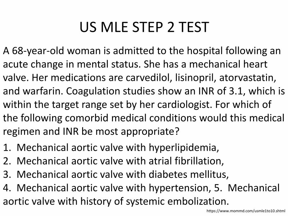
A 68-year-old woman is admitted to the hospital following an acute change in mental status. She has a mechanical heart valve. Her medications are carvedilol, lisinopril, atorvastatin, and warfarin. Coagulation studies show an INR of 3.1, which is within the target range set by her cardiologist. For which of the following comorbid medical conditions would this medical regimen and INR be most appropriate?
1. Mechanical aortic valve with hyperlipidemia, 2. Mechanical aortic valve with atrial fibrillation, 3. Mechanical aortic valve with diabetes mellitus, 4. Mechanical aortic valve with hypertension, 5. Mechanical aortic valve with history of systemic embolization.
https://www.mommd.com/usmle1to10.shtml
US MLE STEP 2 EXPLANATION
The correct answer is 2. The patient described in the question stem has a mechanical heart valve and an INR of 3.1. INR of 2.5-3.5 is recommended for patients with a bileaflet mechanical aortic valve and atrial fibrillation. Incorrect answers: 1: INR of 2-3 is indicated for a bileaflet mechanical aortic valve, 3: The presence of diabetes is not the most important factor determining the appropriate level of anticoagulation for this patient. Answer 2 is a better answer, 4: The presence of hypertension is not the most important factor determining the appropriate level of anticoagulation for this patient. Answer 2 is a better answer, 5: INR of 2.5-3.5 + ASA is indicated for a mechanical valve with a history of systemic embolization. The patient is not taking aspirin.
https://www.mommd.com/usmle1to10.shtml
Treatment (Fungi)
• Fungal IE is rare but can develop in a wide range of patients.
• A 2-phase treatment of fungal IE has evolved. The initial or induction phase consists of control of infection. Treatment includes a combination of a parenteral antifungal agent, usually an amphotericin B–containing product, and valve surgery.
• Antifungal therapy usually is given for >6 weeks.
http://circ.ahajournals.org/content/early/2015/09/15/CIR.0000000000000296
Treatment (Long-Term Follow-Up) 1
• Months to years after completion of medical therapy for IE, patients should have ongoing observation for and education about recurrent infection and delayed onset of worsening valve dysfunction.
• Daily dental hygiene should be stressed, with serial evaluations by a dentist who is familiar with this patient population.
http://circ.ahajournals.org/content/early/2015/09/15/CIR.0000000000000296
Treatment (Long-Term Follow-Up) 2
• Patients should be questioned about symptoms of heart failure, and a thorough physical examination should be done.
• Additional evaluation with echocardiography is indicated in selected patients with positive findings from history and physical examination.
• Patients should be instructed to seek immediate medical evaluation for persistent fever.
http://circ.ahajournals.org/content/early/2015/09/15/CIR.0000000000000296
Treatment (The ‘Endocarditis Team’) 1
• IE is not a single disease, but rather may present with very different aspects depending on the first organ involved, the underlying cardiac disease (if any), the microorganism involved, the presence or absence of complications and the patient's characteristics, so no single practitioner will be able to manage and treat it.
http://eurheartj.oxfordjournals.org/content/early/2015/08/28/eurheartj.ehv319
Treatment (The ‘Endocarditis Team’) 2
• Very high level of expertise is needed from practitioners from several specialties, including cardiologists, cardiac surgeons, internal diseases specialists, microbiologists, neurologists, neurosurgeons, experts in coronary heart disease and others.
• About half of the patients with IE undergo surgery during the hospital course.
http://eurheartj.oxfordjournals.org/content/early/2015/08/28/eurheartj.ehv319
Prognosis 1 • Prognosis largely depends on whether or not
complications develop.
• If left untreated, IE is generally fatal.
• Early detection and appropriate treatment of this uncommon disease can be lifesaving.
• The overall mortality rate has remained stable at 14.5%.
http://emedicine.medscape.com/article/216650-overview#a6
Prognosis 2 • Increased mortality rates are associated with
increased age, infection involving the aortic valve, development of congestive heart failure, central nervous system (CNS) complications, and underlying disease such as diabetes mellitus
• Acute endocarditis due to S aureus is associated with a high mortality rate (30-40%), except when it is associated with IV drug use.
http://emedicine.medscape.com/article/216650-overview#a6
Prophylaxis 1
• There is no clinical evidence that it reduces the incidence of IE and there are negative effects (e.g. allergy and increased bacterial resistance) of taking antibiotics that may outweigh the benefits.
• Antibiotics were historically commonly recommended to prevent IE in those with heart problems undergoing dental procedures (known as dental antibiotic prophylaxis) and in now days they are less commonly recommended for this procedure.
http://www.merckmanuals.com/professional/cardiovascular-disorders/endocarditis/infective-endocarditis https://www.nice.org.uk/guidance/published?type=cg
Prophylaxis 2
• Preventive dental examination and therapy before surgery to repair heart valves or congenital heart lesions is recommended.
http://www.merckmanuals.com/professional/cardiovascular-disorders/endocarditis/infective-endocarditis https://www.nice.org.uk/guidance/published?type=cg
Key Points 1 • Because the normal heart is relatively resistant to
infection, endocarditis occurs when there is a predisposing abnormality of the endocardium.
• Predisposing abnormalities include congenital heart defects, rheumatic valvular disease, bicuspid or calcific aortic valves, mitral valve prolapse, hypertrophic cardiomyopathy, prior endocarditis, and presence of a prosthetic valve.
• Local cardiac consequences include myocardial abscess, conduction system abnormalities, and sudden, severe valvular regurgitation.
http://www.merckmanuals.com/professional/cardiovascular-disorders/endocarditis/infective-endocarditis
Key Points 2 • Systemic consequences include immune
phenomena and septic emboli, which may affect any organ put particularly the lungs, kidneys, spleen, central nervous system (CNS), skin, retina.
• Diagnose using blood cultures and Duke criteria.
• Treat with a prolonged course of antimicrobial therapy; surgery may be needed for mechanical complications or resistant organisms.
• Give antimicrobial prophylaxis for patients at high risk of an adverse outcome from infective endocarditis.
http://www.merckmanuals.com/professional/cardiovascular-disorders/endocarditis/infective-endocarditis
Abbreviations ABE - acute bacterial endocarditis
ACE - angiotensin converting enzyme
BE - bacterial endocarditis
CMR - cardiac magnetic resonance
CNS – central nervous system
CRP - C-reactive protein
CT - computed tomography
ECG - electrocardiogram
ESR - erythrocyte sedimentation rate
IDU - injection drug use
IE - infective endocarditis
HIV - human immunodeficiency virus
MIC - minimum inhibitory concentration
MRI - magnetic resonance imaging
NBTE - non-bacterial thrombotic endocarditis
LVADs - left ventricular assistive devices
PK/PD - pharmacokinetic/pharmacodynamic
SBE - subacute bacterial endocarditis
TEE - transesophageal echocardiography
TTE - transthoracic echocardiography
PET - positron emission tomography
Diagnostic and treatment guidelines
2015 ESC Guidelines for the management of infective endocarditis
Infective Endocarditis in Adults: Diagnosis, Antimicrobial Therapy, and Management of Complications
Prophylaxis against infective endocarditis: antimicrobial prophylaxis against infective endocarditis in adults and children undergoing interventional procedures
Infective Endocarditis
Infective Endocarditis
Cardiac imaging in infectious endocarditis