Embed Size (px)
Citation preview
SHOULDER
Value of additional acromioclavicular cerclage for horizontalstability in complete acromioclavicular separation:a biomechanical study
Tim Saier • Arne J. Venjakob • Philipp Minzlaff •
Peter Fohr • Filip Lindell • Andreas B. Imhoff •
Stephan Vogt • Sepp Braun
Received: 27 August 2013 / Accepted: 3 February 2014
� Springer-Verlag Berlin Heidelberg 2014
Abstract
Purpose To evaluate whether isolated anatomical cora-
coclavicular (CC) ligament reconstruction with two suture-
button devices provides equal horizontal acromioclavicular
joint (ACJ) stability compared to additional ACJ suture
tape cerclage.
Methods A servohydraulic testing machine was used to
assess horizontal ACJ translation in 12 fresh-frozen human
shoulders during 5,000 cycles of dynamic anteroposterior
directed loading (70 N). Horizontal ACJ stability was
assessed for native specimen (n = 6) and compared to
specimen with dissected AC ligaments but intact CC lig-
aments (n = 6). After complete AC/CC dissection, an
anatomical CC reconstruction was performed with two
suture-button devices (n = 6) and compared to the addi-
tional ACJ suture tape cerclage (n = 6).
Results Native specimen showed an mean horizontal
amplitude of 10.8 mm [standard deviation (SD) 3.29].
After 5,000 cycles of horizontal loading (70 N), mean
amplitude increased by 1.5 mm (SD 0.75, p = 0.005).
Specimen with dissected AC ligaments started at an mean
amplitude of 14.1 mm (SD 4.11), which was increased by
0.9 mm (SD 0.56, n.s.) after loading. Initially, amplitude of
specimen with anatomical CC reconstruction was 13.2 mm
(SD 2.75), which increased by 2.9 mm (SD 1.45,
p = 0.001) after loading. The specimen with additional AC
cerclage initially showed an amplitude of 10.6 mm (SD
2.35). After loading, translation was increased by 3.0 mm
(SD 0.97, p = 0.001). There was no failure of any surgical
reconstruction in the tests.
Conclusion The results of this study suggest that only
combined AC and CC reconstruction can adequately re-
establish physiological horizontal ACJ stability. Therefore,
it is likely that a combined surgical procedure with double
suture-button devices and AC suture tape cerclage can
adequately re-establish horizontal AC joint stability in case
of an acute injury (Ctype Rockwood IV and may allow
superior clinical outcomes for patients, especially if early
functional rehabilitation is intended).
Keywords Acromioclavicular joint � Acute separation �Rockwood classification � Coracoclavicular ligaments �Acromioclavicular ligaments � Horizontal instability
Introduction
The balance of mobility and joint stability is a significant
challenge for surgical treatment of acromioclavicular
joint (ACJ) separations. The individual anatomy of the
ACJ with its wide variety in 3D intra-articular angle,
Sepp Braun and Stephan Vogt have contributed equally for senior
authorship.
T. Saier � A. J. Venjakob � P. Minzlaff �A. B. Imhoff (&) � S. Vogt � S. Braun
Department for Orthopaedic Sports Medicine, Klinikum rechts
der Isar, Technische Universitat Munchen, Ismaningerstr. 22,
81675 Munich, Germany
e-mail: [email protected]
T. Saier
Berufsgenossenschaftliche Unfallklinik Murnau,
Prof.-Kuntscher-Str. 8, 82418 Murnau, Germany
P. Fohr � F. Lindell
Department of Biomechanics, Klinikum rechts der Isar,
Technische Universitat Munchen, Ismaningerstr. 22,
81675 Munich, Germany
123
Knee Surg Sports Traumatol Arthrosc
DOI 10.1007/s00167-014-2895-7
range of motion, strength, and joint congruity compli-
cates the effort to anatomically restore the ACJ after
complete traumatic separation [13]. At present, there is a
wide consensus on nonoperative treatment for acute
Rockwood types I and II [23] and surgical treatment of
Rockwood types IV–VI injuries [14, 26]. It still remains
controversial whether Rockwood type III lesions require
surgical reconstruction or not [16, 18]. There is no clear
evidence for either treatment to result in superior out-
comes [1].
Especially, the distinct functional anatomy of the two
coracoclavicular (CC) ligaments (conoid and trapezoid
ligament) has led the focus to anatomically reconstruct
these structures for improved function and stability [5, 10,
15, 20]. Techniques to arthroscopically reconstruct the
conoid and trapezoid ligament by using suture-button
devices have been described [18]. This approach is thought
to limit intraoperative morbidity and—in contrast to open
procedures—allows diagnosis and treatment of concomi-
tant intra-articular pathologies (e.g. SLAP lesions) [25]. As
this approach alone does not directly address the disrupted
AC ligament complex, horizontal instability due to insuf-
ficient healing could result. While there is broad consensus
that vertical reposition and stability are attributed to the CC
ligaments [29], horizontal stability is mainly contributed to
the ACJ complex [8]. With the isolated reconstruction of
the CC ligaments, a very protective post-operative protocol
may be needed to achieve sustained long-term horizontal
ACJ stability. Since it has been shown clinically that hor-
izontal instability of the ACJ may result in severe chronic
pain and functional shoulder impairment [22], there is a
raising focus on the relevance of specific techniques
thought to improve horizontal stability (e.g. ACJ cerclage)
[24].
To our knowledge, there is no data on the effect of
dynamic loading on horizontal ACJ stability after ana-
tomical reconstruction of the CC ligaments versus an
additional stabilization of the ACJ using a modified figure
of eight suture cerclage. Thus, the purpose of this study
was to biomechanically investigate horizontal stability of
these surgical techniques during extensive bidirectional
anteroposterior dynamic loading. This new set-up simu-
lates a more realistic approach to study cyclic physiological
horizontal loading (e.g. repetitive range-of-motion during
rehabilitation) of the ACJ compared to conventional load-
to-failure experiments.
It was hypothesized that isolated anatomical CC liga-
ment reconstruction provides equal horizontal ACJ stabil-
ity if compared to native shoulders with intact AC and CC
ligaments. Secondarily the authors hypothesized that an
additional ACJ cerclage provides no additional horizontal
ACJ stability compared to the isolated CC ligaments
reconstruction.
To our knowledge, this is the first study that uses—
extensive—bidirectional anterior–posterior dynamic load-
ing in the horizontal plane of the AC joint.
Materials and methods
Specimen preparation
A sample size of twelve fresh-frozen cadaveric human
shoulders (9 right/3 left shoulders, mean age
59 ± 13 years) was used for the study. Each specimen was
thawed for 12 h at room temperature for consecutive test-
ing. The specimens were dissected carefully off all soft
tissue including the rotator cuff, and the glenohumeral joint
was exarticulated. The AC ligaments, the CC ligaments,
and the coracoacromial (CA) ligament were left intact.
Any preexisting lesion of the above-mentioned liga-
mentous restraints (AC, CC, CA ligaments) and severe
degenerative changes to the AC joint were ruled out by
visual inspection. The exposed biomaterials were kept
moist with sterile saline solution throughout the time of
testing to prevent tissue dehydration. The scapula and
clavicle of each specimen were potted in a custom mould
with quick-setting polyurethane cement (RenCast� FC 53
Isocyanate/FC 53 Polyol, Huntsman, Belgium).
Before moulding, the scapula was adjusted within the
horizontal and frontal plane to allow for maximal pivoting
of the clavicle with tensioned CC ligaments in the hori-
zontal plane. Secondarily, moulding of minimum 2/3 of the
clavicle was carried out. For this, the moulded scapula was
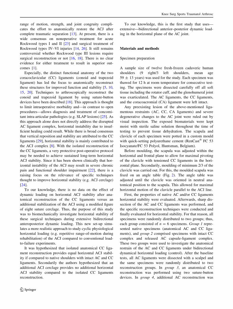
fixed on an angle table (Fig. 2). The angle table was
adjusted until the clavicle was oriented in neutral ana-
tomical position to the scapula. This allowed for maximal
horizontal motion of the clavicle parallel to the ACJ line.
First, the properties of native AC and/or CC ligaments
horizontal stability were evaluated. Afterwards, sharp dis-
section of the AC and CC ligaments was performed, and
the specific reconstruction techniques were conducted and
finally evaluated for horizontal stability. For that reason, all
specimens were randomly distributed to two groups; thus,
each group consisted of n = 6 specimens. Group 1 repre-
sented native specimens (anatomical AC and CC liga-
ments), and group 2 comprised specimens with intact CC
complex and released AC capsule-ligament complex.
These two groups were used to investigate the anatomical
restrain of the AC and CC ligaments under bidirectional
dynamical horizontal loading (control). After the baseline
tests, all AC ligaments were dissected with a scalpel and
the same specimens were randomly distributed to two
reconstruction groups. In group 3, an anatomical CC
reconstruction was performed using two suture-button
devices. In group 4, additional AC reconstruction was
Knee Surg Sports Traumatol Arthrosc
123
performed using a suture tape cerclage in a modified figure
of eight configuration.
Reconstruction techniques
Group 3
The ACJ was anatomically reduced, and reconstruction
was performed in neutral testing position with the speci-
men mounted into the servohydraulic testing machine.
As described by Walz et al. [27], an anatomical CC
ligament reconstruction with two suture-button devices
(TightRope, Arthrex, Naples, FL, USA) was performed
using a standard AC drill guide (Arthrex, Naples, FL, USA).
A 2.4-mm guide wire was drilled through the centre of the
clavicle, about 45 mm medial from the lateral clavicular
edge, perforating the base of the coracoid posteriorly, about
5 mm lateral to the medial border (conoidal position). A
second drill guide wire (trapezoidal position) was placed
about 25 mm medial to the lateral clavicular edge. In the
coracoid, the wire was positioned 10 mm anterior to the
conoidal wire and 5 mm medial to the lateral border of the
coracoid. Both guide wires were over-reamed with a 4.0-
mm cannulated drill. Subsequently, the suture-button
devices were pulled in with shuttle wires. After flipping the
inferior buttons, the free limbs of the sutures were tightened
over the two clavicular buttons and tied to complete the
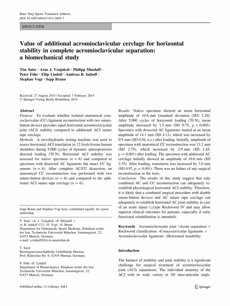
reposition (Fig. 1b). According to Rios et al. [17], this
technique and positioning of the suture-button devices may
re-establish the anatomical properties of the CC ligaments.
Group 4
The anatomical CC ligament reconstruction was performed
as described above in the specimens of group 3. In addition,
the ACJ were stabilized in a modified figure of eight con-
figuration suture tape cerclage (FiberTape, Arthrex, Naples,
FL, USA; Fig. 1c). Two vertical 2.4-mm holes were drilled
into the acromion, located in line with the anterior and
posterior margin of the ACJ, and a 2.4-mm hole was drilled
in anterior–posterior direction through the lateral clavicle,
about 1.5 cm medial to the medial ACJ line. The suture tape
was shuttled through the acromial tunnels using a passing
wire as an ‘‘U’’ under the acromion. The longer posterior
free limb of the tape was crossed towards anterior over the
ACJ and passed through the clavicular tunnel to posterior.
Now, the anterior acromial branch was crossed over the
ACJ and passed through the clavicular tunnel from posterior
towards anterior. Finally, the cerclage was stabilized with a
frame running parallel to the anterior and posterior margin
of the ACJ. After the longer end was shuttled through the
two acromial tunnels in the same way as the first ‘‘U’’, the
reconstruction was completed by knotting the tape anterior
to the clavicle (Fig. 1c).
Biomechanical testing
To evaluate the main outcome measure of total maximum
mean amplitude of horizontal translation [mm], each
specimen was mounted in a servohydraulic testing machine
(Zwick/Roell HC 10, ±10 kN maximum force, Zwick
GmbH & Co KG, Ulm, Germany) with the potted scapula
fixed to an angle table which was attached to the base plate
of the testing system. The potted clavicle was connected to
an adjustable suspension device with the force sensor
(Modell 1010-AF, ±2 kN maximum load, Huppert Mess-
technik GmbH, Herrenberg, Germany) and the servohy-
draulic cylinder of the device (Figs. 1, 2).
Position measurement of the testing machine (Zwick/
Roell HC10) was carried out using a LVDT sensor (RDP
Fig. 1 Specimen (right side) mounted into the servohydraulic testing
system. a Native specimen (group 1) with anatomical Ligg. conoid-
eum (a) and trapezoideum (b). b After CC-/AC-Ligg.-dissection and
CC reconstruction (group 3) with two anatomically implanted suture-
button systems (c TightRope, Arthrex, Naples, FL, USA). c Specimen
in axial view after anatomical CC-Ligg. reconstruction (e TightRope,
Arthrex, Naples, FL, USA) and suture tape AC-cerclage (d FiberTape,
Arthrex, Naples, FL, USA) (group 4)
Knee Surg Sports Traumatol Arthrosc
123
192028, RDP Electronics Ltd, Wolverhampton, United
Kingdom) with a measurement accuracy of 0.20 lm. In
general, the testing machine presents an accuracy class of
±0.5 %, which represents for the present study an absolute
resolution between 2.5 and 100 lm.
The software of the servohydraulic testing system
(testXpert Research Dynamic Sequencer, Zwick GmbH &
Co KG, Ulm, Germany) was configured to start from the
predefined neutral position (max. ?4 N load) after pre-
conditioning the specimen with 50 cycles of an anterior and
posterior-directed force with 70 N at a frequency of 1 Hz.
Subsequently, the software was set to run 5,000 cycles of
anterior- and posterior-directed loads with 70 N at 1 Hz,
resulting in 10,000 stresses and strains. To outrule bias of
clavicular and coracoidal bending and resistance to flexural
strength, the applied force of 70 N was chosen according to
the study by Costic et al. [4] and Lee et al. [10]. For each
test, the combined anterior and posterior translation [total
maximum mean amplitude (mm)] after preconditioning
(50 cycles) was used as reference value to determine any
increase in the amplitude after 5,000 cycles [total maxi-
mum mean amplitude; total maximum mean change
(mm)]. Secondarily, maximum mean anterior and posterior
translation (mm) was recorded. To facilitate data collec-
tion, the maximal singular cyclic translation within a block
of 50 cycles was recorded.
In summary, this set-up represents a novel protocol for
dynamic bidirectional horizontal ACJ stability evaluation.
IRB approval was obtained by the institutional ethics
committee (Faculty of Medicine, Technische Universitaet
Muenchen, ID number: 5431/12).
Statistical analysis
Statistical analysis was done using SPSS software package
(Version 20, SPSS Inc., Chicago, IL, USA). All data are
reported as mean ± standard deviation (SD). The level of
significance was set to 0.05.
Two-sided level of significance of 5 % was used.
ANOVA was used for global and Tukey’s test for pairwise
comparison.
Results
During the entire testing, there was no failure of any
reconstruction (e.g. knot suture-button device, cerclage
failure, or fracture).
Group 1: native specimens
After 50 cycles of preconditioning, maximum mean ampli-
tude reached 10.8 mm (SD 3.29) and increased to 12.3 mm
(SD 3.31) after 5,000 cycles (see Fig. 3). Thus, under
repetitive anteroposterior directed loading, mean amplitude
increased significantly by 1.5 mm (SD 0.75, p = 0.005).
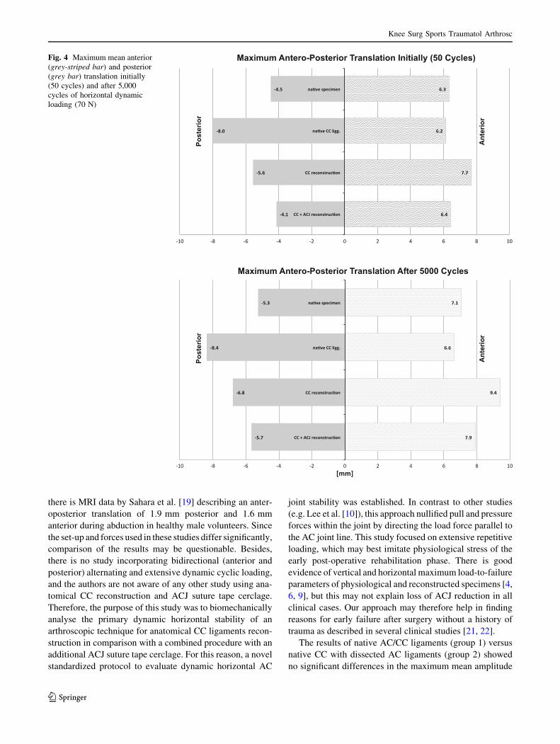
Maximal difference of mean posterior and anterior
translation increased significantly by 0.9 mm (SD 0.72,
p = 0.029), respectively, and by 0.6 mm (SD 0.27,
p = 0.002; see Fig. 4).
Group 2: native specimens with dissected AC ligaments
After preconditioning maximum mean amplitude was
14.1 mm (SD 4.11), following extensive dynamic loading,
it increased to 15.0 mm (SD 4.34) (see Fig. 3). Thus, this
group showed an increase of 0.9 mm (SD 0.56, n.s.).
Maximal difference between mean posterior and ante-
rior translation increased significantly by 0.4 mm (SD 0.2,
p = 0.007), respectively, and by 0.5 mm (SD 0.47,
p = 0.05; see Fig. 4).
Fig. 2 Test set-up of the specimen mounted into the servohydraulic
testing machine. a servohydraulic cylinder, b force sensor, c moulded
specimen with adjustable clavicle suspension device
Knee Surg Sports Traumatol Arthrosc
123
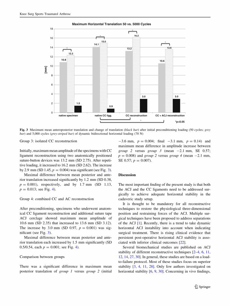
Group 3: isolated CC reconstruction
Initially, maximum mean amplitude of the specimens with CC
ligament reconstruction using two anatomically positioned
suture-button devices was 13.2 mm (SD 2.75). After repeti-
tive loading, it increased to 16.2 mm (SD 2.62). The increase
by 2.9 mm (SD 1.45, p = 0.004) was significant (see Fig. 3).
Maximal difference between mean posterior and ante-
rior translation increased significantly by 1.2 mm (SD 0.38,
p = 0.001), respectively, and by 1.7 mm (SD 1.13,
p = 0.013; see Fig. 4).
Group 4: combined CC and AC reconstruction
After preconditioning, specimens who underwent anatom-
ical CC ligament reconstruction and additional suture tape
ACJ cerclage showed maximum mean amplitude of
10.6 mm (SD 2.35) that increased to 13.6 mm (SD 3.12).
The increase by 3.0 mm (SD 0.97, p = 0.001) was sig-
nificant (see Fig. 3).
Maximal difference between mean posterior and ante-
rior translation each increased by 1.5 mm significantly (SD
0.5/0.54, each p = 0.001; see Fig. 4).
Comparison between groups
There was a significant difference in maximum mean
posterior translation of group 1 versus group 2 (initial
-3.6 mm, p = 0.004; final -3.1 mm, p = 0.14) and
maximum mean difference in amplitude increase between
group 2 versus group 3 (mean -2.1 mm, SE 0.57;
p = 0.008) and group 2 versus group 4 (mean -2.1 mm,
SE 0.57; p = 0.007).
Discussion
The most important finding of the present study is that both
the ACJ and the CC ligaments need to be addressed sur-
gically to achieve adequate horizontal stability in the
cadaveric study setup.
It is thought to be mandatory for all reconstructive
techniques to restore the physiological three-dimensional
position and restraining forces of the ACJ. Multiple sur-
gical techniques have been proposed to address separations
of the ACJ [1]. Recently, there is a trend to take dynamic
horizontal ACJ instability into account when indicating
surgical treatment. There is rising clinical evidence that
persistent post-operative horizontal ACJ stability is asso-
ciated with inferior clinical outcomes [22].
Several biomechanical studies are published on ACJ
stability of different reconstructive techniques [2–4, 6, 11,
12, 14, 27, 30]. In general, these studies are based on a load-
to-failure protocol. Most of these studies focus on superior
stability [3, 4, 11, 28]. Only few authors investigated on
horizontal stability [6, 9, 30]. Concerning in vivo findings,
10.8
14.1
13.2
10.6
12.3
15.0
16.2
13.6
1.5 0.9
3.0 3.0
0
2
4
6
8
10
12
14
16
18
native specimen
[mm
]
x
Maximum Horizontal Translation 50 vs. 5000 Cycles
*
*
*
*
*
* *p<0.05
native CC ligg. CC reconstruction CC + ACJ reconstruction
Fig. 3 Maximum mean anteroposterior translation and change of translation (black bar) after initial preconditioning loading (50 cycles; grey
bar) and 5,000 cycles (grey-striped bar) of dynamic bidirectional horizontal loading (70 N)
Knee Surg Sports Traumatol Arthrosc
123
there is MRI data by Sahara et al. [19] describing an anter-
oposterior translation of 1.9 mm posterior and 1.6 mm
anterior during abduction in healthy male volunteers. Since
the set-up and forces used in these studies differ significantly,
comparison of the results may be questionable. Besides,
there is no study incorporating bidirectional (anterior and
posterior) alternating and extensive dynamic cyclic loading,
and the authors are not aware of any other study using ana-
tomical CC reconstruction and ACJ suture tape cerclage.
Therefore, the purpose of this study was to biomechanically
analyse the primary dynamic horizontal stability of an
arthroscopic technique for anatomical CC ligaments recon-
struction in comparison with a combined procedure with an
additional ACJ suture tape cerclage. For this reason, a novel
standardized protocol to evaluate dynamic horizontal AC
joint stability was established. In contrast to other studies
(e.g. Lee et al. [10]), this approach nullified pull and pressure
forces within the joint by directing the load force parallel to
the AC joint line. This study focused on extensive repetitive
loading, which may best imitate physiological stress of the
early post-operative rehabilitation phase. There is good
evidence of vertical and horizontal maximum load-to-failure
parameters of physiological and reconstructed specimens [4,
6, 9], but this may not explain loss of ACJ reduction in all
clinical cases. Our approach may therefore help in finding
reasons for early failure after surgery without a history of
trauma as described in several clinical studies [21, 22].
The results of native AC/CC ligaments (group 1) versus
native CC with dissected AC ligaments (group 2) showed
no significant differences in the maximum mean amplitude
Fig. 4 Maximum mean anterior
(grey-striped bar) and posterior
(grey bar) translation initially
(50 cycles) and after 5,000
cycles of horizontal dynamic
loading (70 N)
Knee Surg Sports Traumatol Arthrosc
123
of horizontal translation before and after extensive
dynamic cyclic loading. This finding may be explained by
the fact that the CC ligaments are stronger than the ACJ
complex. Since translation after preconditioning of the
native specimens was smaller, the authors hypothesize that
the majority of horizontal resistance was build up by the
ACJ complex, leaving the CC ligaments not totally loaded.
Thus, the overall bearing structure in native specimens is
weaker and allows for a greater elongation during exten-
sive dynamic loading. These findings are in accordance
with preliminary anatomical studies published by Debski
et al. [5] and Fukuda et al. [7] showing that the AC liga-
ments primarily stabilize the ACJ in the anterior–posterior
plane. In line with Fukuda et al. using a similarly directed
load, our data suggest by a non-significant trend that the
AC ligaments play a particular role in posterior stabiliza-
tion of the lateral clavicle. Thus, after dissection of the ACJ
ligaments, translation must increase in order to resist the
force of 70 N.
Likewise, comparing the two intervention groups, the
isolated CC reconstruction (group 3) showed an increased
horizontal translation after preconditioning versus the
additional ACJ cerclage (group 4). The authors conclude
that the ACJ cerclage must be considered the major time
zero resistance to anteroposterior translation.
After extensive loading of the groups, there was a sig-
nificant increase in anterior–posterior translation of the
lateral clavicle in all groups. As for group 1 versus 2, as
well as for group 3 versus 4, it must be assumed that
especially the ACJ stability was weakened until there was a
status, when the increased force absorption by the CC
restraints limited the elongation of the ACJ structures. The
higher amplitude increase within the intervention groups
may be due to the fact that the reconstruction techniques
cannot reach up to biology, e.g. due to slipping within the
drill holes and/or a smaller insertion zone compared to the
widespread anchorage of the native ligaments. This finding
is contrary to the study by Rios et al. [17] that stated re-
establishment of the anatomical properties of the CC lig-
aments after anatomical CC reconstruction. However, none
of the AC or CC reconstructions failed by suture breakage
or fractures of the bones during testing with extensive
cyclic loading. But, since the anterior and posterior
excursions of the groups were alike, the intervention
groups are considered to resemble anatomical characteris-
tics of horizontal range of motion.
Most importantly, combined CC and ACJ stabilization
(group 4) led to significantly increased horizontal ACJ
stability compared to isolated CC ligaments reconstruction
(group 3). Thus, the authors conclude that only the com-
bined reconstructive approach (group 4) may successfully
restrain repetitive anterior–posterior load. The tendency for
dynamic wear, as observed in the isolated CC ligament
reconstruction group (group 3), may explain early non-
traumatic failures, as has been described before [22].
A limitation of this study is that there is no means to
evaluate biological factors such as tissue healing and
scarring of disrupted ligaments in a biomechanical cadaver
study.
Transferring the experimental results to clinical practice,
this study supports a combined AC- and CC reconstruction
in ACJ separations CRockwood IV, especially if early
functional stability is intended. Therefore, the authors
promote to surgically address both the CC ligaments and
the ACJ in complete ACJ separation.
Conclusion
The specimens undergoing combined surgical reconstruc-
tion of the CC ligaments with two suture-button devices
and AC joint stabilization with suture tape cerclage showed
physiological horizontal AC joint stability during initial
cycles.
Over time, dynamic cyclic loading weakened horizontal
AC joint stability in all the tested groups significantly. The
specimens that underwent combined CC and AC ligaments
reconstruction terminated cyclic loading superior to the
group with dissected AC ligaments, but reconstructed CC
ligaments and were only minimally inferior to native
conditions.
Transferring the results to clinical practice, this study is
in support of a combined AC- and CC reconstruction in
ACJ separations CRockwood IV, especially if early func-
tional stability is intended. Thus, the authors promote a
combined CC and ACJ surgical treatment in complete ACJ
separation.
References
1. Beitzel K, Cote MP, Apostolakos J, Solovyova O, Judson CH,
Ziegler CG, Edgar CM, Imhoff AB, Arciero RA, Mazzocca AD
(2013) Current concepts in the treatment of acromioclavicular
joint dislocations. Arthroscopy 29(2):387–397
2. Beitzel K, Obopilwe E, Chowaniec DM, Nowak MD, Hanypsiak
BT, Guerra JJ, Arciero RA, Mazzocca AD (2012) Biomechanical
properties of repairs for dislocated AC joints using suture button
systems with integrated tendon augmentation. Knee Surg Sports
Traumatol Arthrosc 20(10):1931–1938
3. Clevenger T, Vance RE, Bachus KN, Burks RT, Tashjian RZ
(2011) Biomechanical comparison of acromioclavicular joint
reconstructions using coracoclavicular tendon grafts with and
without coracoacromial ligament transfer. Arthroscopy 27(1):
24–30
4. Costic RS, Labriola JE, Rodosky MW, Debski RE (2004)
Biomechanical rationale for development of anatomical recon-
structions of coracoclavicular ligaments after complete acromi-
oclavicular joint dislocations. Am J Sports Med 32(8):1929–1936
Knee Surg Sports Traumatol Arthrosc
123
5. Debski RE, Parsons IM, Woo SL, Fu FH (2001) Effect of cap-
sular injury on acromioclavicular joint mechanics. J Bone Joint
Surg Am 83(9):1344–1351
6. Deshmukh AV, Wilson DR, Zilberfarb JL, Perlmutter GS (2004)
Stability of acromioclavicular joint reconstruction: biomechani-
cal testing of various surgical techniques in a cadaveric model.
Am J Sports Med 32(6):1492–1498
7. Fukuda K, Craig EV, An KN, Cofield RH, Chao EY (1986)
Biomechanical study of the ligamentous system of the acromio-
clavicular joint. J Bone Joint Surg Am 68(3):434–440
8. Klimkiewicz JJ, Williams GR, Sher JS, Karduna A, Des Jardins J,
Iannotti JP (1999) The acromioclavicular capsule as a restraint to
posterior translation of the clavicle: a biomechanical analysis.
J Shoulder Elb Surg 8(2):119–124
9. Ladermann A, Gueorguiev B, Stimec B, Fasel J, Rothstock S,
Hoffmeyer P (2013) Acromioclavicular joint reconstruction: a
comparative biomechanical study of three techniques. J Shoulder
Elb Surg 22(2):171–178
10. Lee KW, Debski RE, Chen CH, Woo SL, Fu FH (1997) Func-
tional evaluation of the ligaments at the acromioclavicular joint
during anteroposterior and superoinferior translation. Am J Sports
Med 25(6):858–862
11. Lee SJ, Keefer EP, McHugh MP, Kremenic IJ, Orishimo KF,
Ben-Avi S, Nicholas SJ (2008) Cyclical loading of coracocla-
vicular ligament reconstructions: a comparative biomechanical
study. Am J Sports Med 36(10):1990–1997
12. Martetschlager F, Buchholz A, Sandmann G, Siebenlist S, Dobele S,
Hapfelmeier A, Stockle U, Millett PJ, Elser F, Lenich A (2013)
Acromioclavicular and coracoclavicular PDS augmentation for
complete AC joint dislocation showed insufficient properties in a
cadaver model. Knee Surg Sports Traumatol Arthrosc 21(2):438–444
13. Mazzocca AD, Arciero RA, Bicos J (2007) Evaluation and
treatment of acromioclavicular joint injuries. Am J Sports Med
35(2):316–329
14. Mazzocca AD, Santangelo SA, Johnson ST, Rios CG, Dumonski
ML, Arciero RA (2006) A biomechanical evaluation of an ana-
tomical coracoclavicular ligament reconstruction. Am J Sports
Med 34(2):236–246
15. Mazzocca AD, Spang JT, Rodriguez RR, Rios CG, Shea KP,
Romeo AA, Arciero RA (2008) Biomechanical and radiographic
analysis of partial coracoclavicular ligament injuries. Am J Sports
Med 36(7):1397–1402
16. Murena L, Canton G, Vulcano E, Cherubino P (2013) Scapular
dyskinesis and SICK scapula syndrome following surgical treat-
ment of type III acute acromioclavicular dislocations. Knee Surg
Sports Traumatol Arthrosc 21(5):1146–1150
17. Rios CG, Arciero RA, Mazzocca AD (2007) Anatomy of the
clavicle and coracoid process for reconstruction of the coraco-
clavicular ligaments. Am J Sports Med 35(5):811–817
18. Rios CG, Mazzocca AD (2008) Acromioclavicular joint problems
in athletes and new methods of management. Clin Sports Med
27(4):763–788
19. Sahara W, Sugamoto K, Murai M, Tanaka H, Yoshikawa H
(2006) 3D kinematic analysis of the acromioclavicular joint
during arm abduction using vertically open MRI. J Orthop Res
24(9):1823–1831
20. Salzmann GM, Paul J, Sandmann GH, Imhoff AB, Schottle PB
(2008) The coracoidal insertion of the coracoclavicular liga-
ments: an anatomic study. Am J Sports Med 36(12):2392–2397
21. Salzmann GM, Walz L, Buchmann S, Glabgly P, Venjakob A,
Imhoff AB (2010) Arthroscopically assisted 2-bundle anatomical
reduction of acute acromioclavicular joint separations. Am J
Sports Med 38(6):1179–1187
22. Scheibel M, Droschel S, Gerhardt C, Kraus N (2011) Arthro-
scopically assisted stabilization of acute high-grade acromiocla-
vicular joint separations. Am J Sports Med 39(7):1507–1516
23. Simovitch R, Sanders B, Ozbaydar M, Lavery K, Warner JJ
(2009) Acromioclavicular joint injuries: diagnosis and manage-
ment. J Am Acad Orthop Surg 17(4):207–219
24. Sobhy MH (2012) Midterm results of combined acromioclavic-
ular and coracoclavicular reconstruction using nylon tape.
Arthroscopy 28(8):1050–1057
25. Tischer T, Salzmann GM, El-Azab H, Vogt S, Imhoff AB (2009)
Incidence of associated injuries with acute acromioclavicular
joint dislocations types III through V. Am J Sports Med
37(1):136–139
26. Venjakob AJ, Salzmann GM, Gabel F, Buchmann S, Walz L,
Spang JT, Vogt S, Imhoff AB (2013) Arthroscopically assisted
2-bundle anatomic reduction of acute acromioclavicular joint
separations: 58-month findings. Am J Sports Med 41(3):615–621
27. Walz L, Salzmann GM, Fabbro T, Eichhorn S, Imhoff AB (2008)
The anatomic reconstruction of acromioclavicular joint disloca-
tions using 2 TightRope devices: a biomechanical study. Am J
Sports Med 36(12):2398–2406
28. Wellmann M, Habermeyer P (2010) Update on shoulder surgery
2010: current treatment strategies for traumatic lesions of the
shoulder. Der Unfallchirurg 113(6):481–490
29. Yoo YS, Tsai AG, Ranawat AS, Bansal M, Fu FH, Rodosky MW,
Smolinski P (2010) A biomechanical analysis of the native cor-
acoclavicular ligaments and their influence on a new recon-
struction using a coracoid tunnel and free tendon graft.
Arthroscopy 26(9):1153–1161
30. Zooker CC, Parks BG, White KL, Hinton RY (2010) TightRope
versus fiber mesh tape augmentation of acromioclavicular joint
reconstruction: a biomechanical study. Am J Sports Med
38(6):1204–1208
Knee Surg Sports Traumatol Arthrosc
123


![Preconception Laparoscopic Cervical Cerclage: The ... · 1965 [5], trans-abdominal cervical cerclage prior to pregnancy has emerged as a safe and effective intervention for improving](https://img.pdfslide.us/doc/110x75/5f8851cdb723447a244bb50e/preconception-laparoscopic-cervical-cerclage-the-1965-5-trans-abdominal.jpg)
![Cervicalstitch(cerclage)forpreventingpretermbirthin ... › download › pdf › 131165057.pdf · [Intervention Review] Cervical stitch (cerclage) for preventing preterm birth in](https://img.pdfslide.us/doc/110x75/5f03a1417e708231d409ff53/cervicalstitchcerclageforpreventingpretermbirthin-a-download-a-pdf-a.jpg)