Embed Size (px)
Citation preview

© 2014. Published by The Company of Biologists Ltd | Disease Models & Mechanisms (2014) 7, 107-117 doi:10.1242/dmm.013219
107
ABSTRACTFetal valproate syndrome (FVS) is caused by in utero exposure to thedrug sodium valproate. Valproate is used worldwide for the treatmentof epilepsy, as a mood stabiliser and for its pain-relieving properties.In addition to birth defects, FVS is associated with an increased riskof autism spectrum disorder (ASD), which is characterised byabnormal behaviours. Valproate perturbs multiple biochemicalpathways and alters gene expression through its inhibition of histonedeacetylases. Which, if any, of these mechanisms is relevant to thegenesis of its behavioural side effects is unclear. Neuroanatomicalchanges associated with FVS have been reported and, among these,altered serotonergic neuronal differentiation is a consistent finding.Altered serotonin homeostasis is also associated with autism. Herewe have used a chemical-genetics approach to investigate theunderlying molecular defect in a zebrafish FVS model. Valproatecauses the selective failure of zebrafish central serotonin expression.It does so by downregulating the proneural gene ascl1b, an orthologof mammalian Ascl1, which is a known determinant of serotonergicidentity in the mammalian brainstem. ascl1b is sufficient to rescueserotonin expression in valproate-treated embryos. Chemical andgenetic blockade of the histone deacetylase Hdac1 downregulatesascl1b, consistent with the Hdac1-mediated silencing of ascl1bexpression by valproate. Moreover, tonic Notch signalling is crucial forascl1b repression by valproate. Concomitant blockade of Notchsignalling restores ascl1b expression and serotonin expression in bothvalproate-exposed and hdac1 mutant embryos. Together, these dataprovide a molecular explanation for serotonergic defects in FVS andhighlight an epigenetic mechanism for genome-environmentinteraction in disease.
KEY WORDS: Serotonin, Fetal valproate syndrome, Zebrafish,Notch, Proneural gene, Hdac1
RESEARCH ARTICLE
1Division of Developmental Biology, MRC National Institute for Medical Research,The Ridgeway, Mill Hill, London, NW7 1AA, UK. 2National Hospital for Neurologyand Neurosurgery, Queen Square, London, WC1N 3BG, UK. 3Division ofDevelopmental Neurobiology, MRC National Institute for Medical Research, TheRidgeway, Mill Hill, London, NW7 1AA, UK. 4London Research Institute, CancerResearch UK, Lincoln’s Inn Fields Laboratories, 44 Lincoln’s Inn Fields, London,WC2A 3LY, UK. *Present address: Wellcome Trust Sanger Institute, Hinxton, Cambridge, CB101SA, UK.
‡Authors for correspondence ([email protected]; [email protected])
This is an Open Access article distributed under the terms of the Creative CommonsAttribution License (http://creativecommons.org/licenses/by/3.0), which permits unrestricteduse, distribution and reproduction in any medium provided that the original work is properlyattributed.
Received 6 June 2013; Accepted 17 October 2013
INTRODUCTIONValproate (VPA) is a fatty acid derivative widely prescribed for itsanticonvulsant, mood-stabilising and pain-relieving properties, butit has teratogenic and neuropsychiatric side effects upon in uteroexposure, collectively termed fetal valproate syndrome (FVS). Theunderlying molecular cause of FVS is unknown, but candidatemechanisms are the dysregulation of transcription factors importantfor brain development, disruption of signal transduction pathways,inositol depletion and direct inhibition of epigenetic regulators suchas the histone deacetylases (HDACs) (Chen et al., 1997; Detich etal., 2003; Einat et al., 2003; Marchion et al., 2005; Milutinovic etal., 2007; Phiel et al., 2001; Williams et al., 2002).
Fetal VPA exposure is associated with a 3- to 46-fold increasedrisk of autism spectrum disorder (ASD) (Bromley et al., 2013;Christensen et al., 2013; Dufour-Rainfray et al., 2011; Rasalam etal., 2005). Animal models of FVS display autism-like behaviours(Dufour-Rainfray et al., 2010; Kim et al., 2011; Yochum et al., 2008)and neuroanatomical abnormalities that are also reported in ASD(Ingram et al., 2000; Rodier et al., 1996). In these models, activityof the neurotransmitter serotonin (5HT) is altered, which has beenimplicated in the regulation of numerous behaviours, includingsocial interaction (Ansorge et al., 2004; Patterson, 2006). Alteredhippocampal and blood 5HT levels have been reported in animalmodels of FVS, which correlate with impaired 5HT neuronaldifferentiation (Dufour-Rainfray et al., 2010; Kuwagata et al., 2009;Miyazaki et al., 2005; Narita et al., 2002; Oyabu et al., 2013) andautism-like behaviours (Lin et al., 2013; Tsujino et al., 2007; Wanget al., 2013). Interestingly, in one of these rat models, treatment witha 5HT1A receptor agonist improved the abnormal behaviours,implying a deficit of 5HT signalling (Wang et al., 2013). Bycontrast, in the other study, VPA increased brain 5HT levels (Tsujinoet al., 2007).
Significantly, 5HT is also implicated in autism pathogenesis. InASD, 30% of subjects have elevated 5HT blood levels (Mulder etal., 2004; Schain and Freedman, 1961), central 5HT homeostasis isaltered (Chugani et al., 1999; Chugani et al., 1997) and anassociation with stereotyped behaviour has been reported (Kolevzonet al., 2010; Sacco et al., 2010). Selective serotonin reuptakeinhibitors (SSRIs) improve some manifestations of autism, includingstereotypical behaviours (Hollander et al., 2003; McDougle et al.,2000), whereas depletion of the 5HT precursor tryptophanexacerbates these symptoms (Bauman et al., 2006). Genetic orpharmacological perturbation of the 5HT system is associated withautism-like behaviours in humans and in rodents (Bauman et al.,2006; Cook et al., 1997; Kane et al., 2012; Klauck et al., 1997; Nabiet al., 2004; Nakatani et al., 2009; Sutcliffe et al., 2005; Veenstra-VanderWeele et al., 2012). In particular, an allelic polymorphism ofthe serotonin transporter gene (SERT), which is a determinant of
Valproic acid silencing of ascl1b/Ascl1 results in the failure ofserotonergic differentiation in a zebrafish model of fetal valproatesyndromeJohn Jacob1,2,‡, Vanessa Ribes1, Steven Moore1, Sean C. Constable3, Noriaki Sasai1, Sebastian S. Gerety3,*,Darren J. Martin4, Chris P. Sergeant4, David G. Wilkinson3 and James Briscoe1,‡
Dis
ease
Mod
els
& M
echa
nism
s

108
5HT activity, is associated with ASD (Devlin et al., 2005).Furthermore, a mouse model of one of the human gain-of-functionSERT genetic variants displays ASD-like behaviours andhyperserotonaemia (Veenstra-VanderWeele et al., 2012). Therefore,increases and decreases in central 5HT activity seem to producecommon behavioural phenotypes, which is consistent with the viewthat autism can result from positive and negative changes inneurotransmitter signalling (Zoghbi and Bear, 2012).
5HT neurons in the hindbrain are derived from progenitorsexposed to the signalling molecule sonic hedgehog (Shh) (Jessell,2000). Serotonergic progenitor identity is characterised byexpression of the transcription factors Nkx2.2, Foxa2 and Ascl1, allof which are required for 5HT neuronal differentiation (Briscoe etal., 1999; Jacob et al., 2007; Pattyn et al., 2004). Newly born 5HTneurons express post-mitotic determinants, including thetranscription factor Pet1 (Hendricks et al., 2003) and subsequentlythe 5HT biosynthetic enzyme Tph2 (Zhang et al., 2004).
We investigated the molecular pathophysiology underlyingserotonergic deficits in a zebrafish FVS model because this couldprovide mechanistic insight into the genesis of core autism
behaviours. Importantly, hindbrain development and neuronalsubtype diversity and serotonergic differentiation show strongconservation (Lillesaar, 2011). Furthermore, zebrafish exposed toVPA display morphological defects similar to those described inFVS, suggesting the validity of our system for modelling features ofhuman FVS (Gurvich et al., 2005; Herrmann, 1993). We show thatVPA specifically blocks hindbrain 5HT expression in zebrafish.Acting via Hdac1, VPA silences the zebrafish ortholog ofmammalian Ascl1, ascl1b, by unmasking tonic Notch repression.Moreover, Ascl1b is sufficient to rescue 5HT expression in VPA-treated embryos.
RESULTSVPA impairs central 5HT neuronal differentiationTo assess the effect of VPA on brainstem development, we exposedzebrafish gastrulae at 50% epiboly to 0.625 mM VPA until 27 hourspost-fertilisation (hpf), at which time the drug was removed and theembryos were allowed to develop until 48 hpf. Treatment fromgastrulation was based on the heightened risk of teratogenicity inhuman infants exposed to VPA during the first trimester ofpregnancy (Ornoy, 2009). Immunostaining for a range of hindbrainneuronal subtypes, specifically motor neurons, 5HT neurons,GABA-ergic neurons and Mauthner neurons, revealed a failure of5HT neuronal differentiation marked by absence of 5HT expression(Fig. 1A). Additionally, Mauthner neurons, which express aneurofilament-associated antigen that is detected by the 3A10monoclonal antibody, were also absent (Hatta, 1992; Schier et al.,1996) (Fig. 1A). Isl1-positive motor neurons and GABA-ergicneurons were present. The spatial distribution of motor neurons inVPA-treated embryos appeared subtly altered, suggesting migratorydefects, but we did not pursue these changes further (Fig. 1A).Instead we focused on the striking serotonergic phenotype.
We speculated that exposure to VPA from gastrulation couldaffect common steps in the differentiation of multiple neuronallineages. Therefore, we treated embryos with 0.625 mM VPA from24-48 hpf and used appropriate markers to detect brainstem neuronalsubtypes (Fig. 1B,C). Somatic motor, GABA-ergic and Mauthnerneurons were still present (Fig. 1C), but there was a specific deficitof 5HT neuronal differentiation, marked by the absence of 5HT andtph2 expression (Fig. 1B). Moreover, there was an absence or severereduction of pet1 expression, which indicates that VPA acts at a stepproximal to or at the early stages of 5HT neuronal differentiation(Fig. 1B). Lower doses than this did not consistently lead to the lossof 5HT expression (see supplementary material Fig. S1).
Having established that brainstem 5HT expression wasspecifically lacking in embryos treated with VPA from 24 hpf, weaddressed whether there was a delay in the differentiation of 5HTneurons. VPA was removed at 48 hours and embryos were analysedafter a further 24 hours of incubation (Fig. 1D). There was limitedrecovery of 5HT expression at 72 hpf [mean number of 5HTneurons in controls=26.7±1.7 (s.d.), n=3; mean number of 5HTneurons in VPA-treated condition=1.8±1.3, n=4], which suggeststhat VPA does not merely retard the differentiation of 5HT neurons.We conclude that VPA specifically blocks the differentiation of 5HTneurons in embryos exposed to the drug between 24 and 48 hpf.This period coincides with the onset of differentiation of 5HTneurons between 25 and 30 hpf (Lillesaar et al., 2007). Because thepost-mitotic differentiation of 5HT neurons is well underway from48 hpf onwards (Lillesaar et al., 2007; McLean and Fetcho, 2004),we asked whether VPA could abolish 5HT expression after 48 hpf.VPA exposure between 48 hpf and 72 hpf had no effect on 5HTexpression (Fig. 1D) (mean number of 5HT neurons=25.8±2.5,
RESEARCH ARTICLE Disease Models & Mechanisms (2014) doi:10.1242/dmm.013219
TRANSLATIONAL IMPACT
Clinical issueThe drug valproate is used worldwide as an anticonvulsant agent, as amood stabiliser and for its pain-relieving properties. Valproate isteratogenic (interferes with early development) and fetal exposurecauses fetal valproate syndrome (FVS), which is characterised by aspectrum of morphological, cognitive and behavioural deficits. Recentpopulation-based epidemiological studies have highlighted thesignificantly increased risk of autism spectrum disorders (ASDs) inchildren exposed to valproate in utero. The in vivo mechanism ofvalproate action that is pertinent to its neuropsychiatric side effects is notclear. Multiple in vitro mechanisms have been described, includinginhibition of histone deacetylases. Studies on animal models of FVShave identified biochemical and cellular perturbations of the centralserotonergic system. Altered serotonin homeostasis is also a feature ofidiopathic autism; therefore, uncovering the pathogenesis of serotonindeficits in FVS could reveal the molecular underpinnings of corebehavioural abnormalities in autism.
Results Zebrafish are highly suited to pharmacological and genetics approachesthat can be combined to provide novel insights into disease mechanisms.In this article, the authors describe a zebrafish model of FVS thatdisplays a failure of serotonergic differentiation in the brainstem inresponse to valproate treatment. They show that a critical proneuralgene, ascl1b, is silenced by valproate through a mechanism thatdepends on inhibition of the histone deacetylase Hdac1. Theirexperiments further show that valproate unmasks tonic repression of theascl1b gene by the Notch pathway. If the Notch pathway is blocked,valproate is no longer able to silence ascl1b. Importantly, restoration ofAscl1b in the presence of valproate rescues the expression of serotoninin the zebrafish brainstem.
Implications and future directions This study shows that valproate represses expression of ascl1b, leadingto defects in the serotonergic system in zebrafish. The failure inserotonergic differentiation in this new model is reminiscent of defectsreported previously for mice exposed to valproate, suggesting that themechanism unveiled herein is likely to provide a common molecularexplanation for serotonergic abnormalities in this disorder. Indeed, theconservation of the differentiation pathways of serotonergic neurons inzebrafish and in humans suggests that these findings will be relevant tounderstanding the complex pathophysiology of FVS. More broadly, theauthors highlight an epigenetic mechanism at work in an iatrogenic formof ASD that could also be relevant to idiopathic, common forms ofautism.
Dis
ease
Mod
els
& M
echa
nism
s
n=4). These data suggest that VPA acts on serotonergic progenitors,rather than on post-mitotic neurons.
To test whether specific neuronal subtypes are vulnerable to VPAapplication from 24 hpf because they only differentiate at or after 24hpf, we extended the range of neuronal subtypes assayed. CerebellarPurkinje neurons begin to differentiate from 3 dpf and are markedby expression of Parv7 (Bae et al., 2009). Wild-type embryos wereincubated in VPA for a prolonged period, from 24 hpf to 4.5 dpf (seesupplementary material Fig. S2). Immunostaining against Parv7showed the persistence of this cell type in VPA-treated embryos(supplementary material Fig. S2). The differential sensitivity of 5HTand Purkinje neurons suggests that vulnerability to the effects ofVPA is not linked directly to the timing of neuronal differentiation.
Inhibition of Hdac1 by VPA accounts for the failure of 5HTneuronal differentiationNext, we sought to identify the molecular pathway targeted by VPAin blocking 5HT neuronal differentiation. Previous studies haveshown both in vivo and in vitro that VPA is an inhibitor of HDACsat therapeutic concentrations (Göttlicher et al., 2001; Gurvich et al.,2005; Kook et al., 2003; Phiel et al., 2001; Tremolizzo et al., 2002;Yildirim et al., 2003). To test the involvement of Hdac1, we used azebrafish hdac1 mutant line, hdac1s436, that was previously isolatedin a forward genetic screen (Noël et al., 2008). Immunostaining of
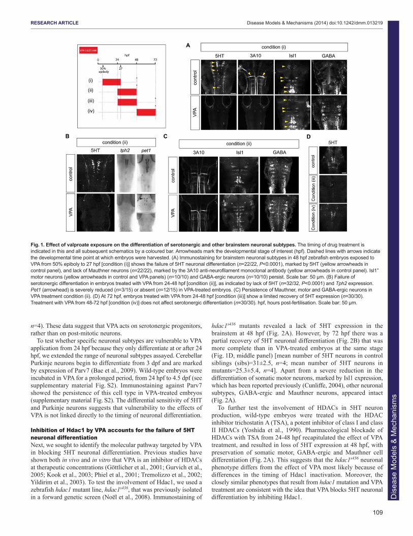
hdac1s436 mutants revealed a lack of 5HT expression in thebrainstem at 48 hpf (Fig. 2A). However, by 72 hpf there was apartial recovery of 5HT neuronal differentiation (Fig. 2B) that wasmore complete than in VPA-treated embryos at the same stage(Fig. 1D, middle panel) [mean number of 5HT neurons in controlsiblings (sibs)=31±2.5, n=4; mean number of 5HT neurons inmutants=25.3±5.4, n=4]. Apart from a severe reduction in thedifferentiation of somatic motor neurons, marked by Isl1 expression,which has been reported previously (Cunliffe, 2004), other neuronalsubtypes, GABA-ergic and Mauthner neurons, appeared intact(Fig. 2A).
To further test the involvement of HDACs in 5HT neuronproduction, wild-type embryos were treated with the HDACinhibitor trichostatin A (TSA), a potent inhibitor of class I and classII HDACs (Yoshida et al., 1990). Pharmacological blockade ofHDACs with TSA from 24-48 hpf recapitulated the effect of VPAtreatment, and resulted in loss of 5HT expression at 48 hpf, withpreservation of somatic motor, GABA-ergic and Mauthner celldifferentiation (Fig. 2A). This suggests that the hdac1s436 neuronalphenotype differs from the effect of VPA most likely because ofdifferences in the timing of Hdac1 inactivation. Moreover, theclosely similar phenotypes that result from hdac1 mutation and VPAtreatment are consistent with the idea that VPA blocks 5HT neuronaldifferentiation by inhibiting Hdac1.
109
RESEARCH ARTICLE Disease Models & Mechanisms (2014) doi:10.1242/dmm.013219
Fig. 1. Effect of valproate exposure on the differentiation of serotonergic and other brainstem neuronal subtypes. The timing of drug treatment isindicated in this and all subsequent schematics by a coloured bar. Arrowheads mark the developmental stage of interest (hpf). Dashed lines with arrows indicatethe developmental time point at which embryos were harvested. (A) Immunostaining for brainstem neuronal subtypes in 48 hpf zebrafish embryos exposed toVPA from 50% epiboly to 27 hpf [condition (i)] shows the failure of 5HT neuronal differentiation (n=22/22, P<0.0001), marked by 5HT (yellow arrowheads incontrol panel), and lack of Mauthner neurons (n=22/22), marked by the 3A10 anti-neurofilament monoclonal antibody (yellow arrowheads in control panel). Isl1+
motor neurons (yellow arrowheads in control and VPA panels) (n=10/10) and GABA-ergic neurons (n=10/10) persist. Scale bar: 50 μm. (B) Failure ofserotonergic differentiation in embryos treated with VPA from 24-48 hpf [condition (ii)], as indicated by lack of 5HT (n=32/32, P<0.0001) and Tph2 expression.Pet1 (arrowhead) is severely reduced (n=3/15) or absent (n=12/15) in VPA-treated embryos. (C) Persistence of Mauthner, motor and GABA-ergic neurons inVPA treatment condition (ii). (D) At 72 hpf, embryos treated with VPA from 24-48 hpf [condition (iii)] show a limited recovery of 5HT expression (n=30/30).Treatment with VPA from 48-72 hpf [condition (iv)] does not affect serotonergic differentiation (n=30/30). hpf, hours post-fertilisation. Scale bar: 50 μm.
Dis
ease
Mod
els
& M
echa
nism
s
110
VPA downregulates expression of the proneural gene ascl1bin serotonergic progenitors through a mechanismconsistent with Hdac1 inhibitionNext we examined the expression of transcription factors that specifyserotonergic progenitors. We reasoned that, because 5HT neurons areabsent, any disruption of serotonergic fate determinants might occurbefore the first 5HT neurons are born. We therefore exposed 24 hpfembryos to VPA and analysed gene expression by in situ hybridisation8 hours later. There was a striking reduction of ascl1b expression by32 hpf that was not apparent at 28 hpf, just 4 hours earlier (Fig. 2C).Colocalisation of ascl1b and nkx2.2 in the hindbrain by fluorescentdouble in situ hybridisation confirmed the expression of ascl1b inserotonergic progenitors (Fig. 2D). Expression of the related geneascl1a and other fate determinants, nkx2.2 and foxa2, and thesignalling molecule shh were unchanged, which implies that the lossof ascl1b expression is not due to depletion of progenitors (Fig. 2E).
Further evidence implicating Hdac1 as the mediator of the effectsof VPA on 5HT neurons is that, in hdac1s436 mutants, expression ofascl1b is also severely reduced, a finding that has been reportedpreviously in a different hdac1-null mutant line (Fig. 2C) (Cunliffe,2004).
In contrast to the downregulation of ascl1b, another bHLHproneural gene, ptf1a, which is required for the generation ofcerebellar Purkinje neurons (Hori et al., 2008), was not decreasedby VPA. Ptf1a expression in the cerebellar primordium is firstdetected at 48 hpf (Kani et al., 2010). We monitored ptf1aexpression by in situ hybridisation for ptf1a transcripts in wild-typezebrafish embryos and by an eGFP reporter in a transgenic ptf1a-egfp line at 52 hpf in embryos continuously exposed to VPA from24 hpf (see supplementary material Fig. S2) (Pisharath et al., 2007).The persistent ptf1a expression suggests that proneural genes aredifferentially sensitive to VPA and this is consistent with thecontinued differentiation of Purkinje neurons after VPA exposure(see supplementary material Fig. S2).
Notch signalling represses ascl1b in the presence of VPAVPA has previously been reported to upregulate Notch signalling ina variety of systems (Greenblatt et al., 2007; Stockhausen et al.,2005). In zebrafish embryos this effect seems likely to be mediatedby Hdac1 (Cunliffe, 2004). One possibility, therefore, was thatHdac1 was inhibiting the Notch pathway and thus decreasing theNotch-mediated inhibition of ascl1b via Her genes, which are the
RESEARCH ARTICLE Disease Models & Mechanisms (2014) doi:10.1242/dmm.013219
Fig. 2. Failure of serotonergic differentiation in VPA-treated embryos is mimicked by blockade of Hdac1 activity and is associated withdownregulation of ascl1b. (A) hdac1 mutation or pharmacological blockade mimics the effect of VPA on the differentiation of brainstem neuronal subtypes. At48 hpf, hdac1s436 mutants lack 5HT neurons (n=15/15, P<0.0001) and show a severe depletion of Isl1+ motor neurons. Treatment with TSA from 24 to 48 hpf[condition (vii)] selectively affects 5HT expression (n=30/30, P<0.0001), but other neuronal subtypes are retained. Scale bar: 50 μm. (B) Partial recovery of 5HTneuronal differentiation in hdac1s436 mutants at 72 hpf (n=14/14). Scale bar: 50 μm. (C) Short duration (8 hour) exposure to VPA from 24 hpf [condition (vi)]leads to the downregulation of ascl1b expression (n=15/15), and similar loss of ascl1b expression is observed in hdac1s436 mutants (n=10/10). Rhombomeres(r) marked by arrowheads. Scale bar: 50 μm. (D) Double in situ hybridisation showing ascl1b expression in serotonergic progenitors, marked by expression ofnkx2.2. Co-labelled cells are indicated by white arrowheads. Scale bar: 20 μm. (E) Short-duration VPA treatment [condition (viii)] does not affect the expressionof the paralogous gene ascl1a, or other progenitor-expressed determinants of serotonergic fate, nkx2.2, foxa2 or shh (arrowheads).
Dis
ease
Mod
els
& M
echa
nism
s

major effectors of Notch signalling in zebrafish (Cunliffe, 2004;Louvi and Artavanis-Tsakonas, 2006). A second possiblemechanism is through direct upregulation of ascl1b by Hdac1(Cunliffe, 2004; Harrison et al., 2011; Wang et al., 2009). Finally,Hdac1 repression of specific members of the Her family, inparticular her6, which in turn represses ascl1b, is a third possibility(Cunliffe, 2004; Harrison et al., 2011).
her4 is a direct target of Notch signalling that is widely used as areadout of Notch activity (Takke and Campos-Ortega, 1999; Takkeet al., 1999; Yeo et al., 2007). The downregulation of ascl1b inembryos following an 8-hour treatment with VPA from 24 hpf(Fig. 2C) was not accompanied by obvious elevation of her4expression (Fig. 3A,B). Quantitative reverse-transcriptase PCRconfirmed that there was a modest decrease in her4 expressionrelative to untreated control embryos (Fig. 3A). her6 is also a targetof the Notch pathway and to date is the only Her gene reported toundergo negative regulation by Hdac1 (Cunliffe, 2004). In situhybridisation for her6 and a panel of additional Her family membersin wild-type embryos similarly treated with VPA also showed nodifference in expression compared with controls (data not shown).
To investigate the involvement of the Notch pathway in ascl1brepression by VPA, we inhibited Notch signalling over the sameperiod and determined whether VPA could still repress ascl1b.Embryos at 22 hpf were treated with a small-molecule inhibitor ofNotch signalling, the γ-secretase inhibitor DAPT, which is knownto have potent activity in vivo in zebrafish (Geling et al., 2002;Imbimbo, 2008). DAPT treatment alone from 22-32 hpf effectivelydownregulated her4 expression, whereas ascl1b expression wasmaintained (Fig. 3A,C). Strikingly, following the addition of VPA at24 hpf, her4 remained repressed and ascl1b expression persisted
(Fig. 3A,C). Therefore, the repression of ascl1b by short-termexposure to VPA is prevented by concomitant blockade of Notchsignalling, which implies that a basal level of Notch signallingrepresses ascl1b when Hdac1 activity is blocked.
To confirm that the repression of Ascl1b by VPA is not due toincreased Notch signalling secondary to blockade of Hdac1function, we analysed her4 expression in hdac1s436 mutants at 32 hpf(Fig. 3B). There was a marked reduction of her4 expression inmutant hindbrains, indicative of reduced Notch signalling (Fig. 3B).Therefore, VPA inhibition of Hdac1 does not repress ascl1b throughthe upregulation of Notch signalling, or via Hdac1-mediatedrepression of her4 or her6. Instead, Hdac1 acts independently ofNotch activity to regulate ascl1b. Moreover, a parallel, tonic levelof Notch signalling was sufficient to repress ascl1b transcriptionwhen Hdac1 was inhibited by VPA.
Hdac1 and Notch have opposing effects on ascl1bTo confirm the parallel roles for Hdac1 and Notch signalling, wetested whether DAPT could restore ascl1b expression in hdac1s436
mutants. hdac1s436 mutants and sibs were treated with DAPT from22 hpf (Fig. 4A). At 32 hpf, sibs treated with DAPT showed strongexpression of ascl1b, which was virtually absent by 48 hpfpresumably because of depletion of the progenitor pool caused bythe blockade of Notch signalling. Remarkably, DAPT restoredascl1b expression in hdac1s436 mutants, and expression wasmaintained at least up to 48 hpf (compare Fig. 2C and Fig. 4A). Theother progenitor markers, nkx2.2, foxa2 and shh, were not altered byDAPT in either sibs or hdac1s436 embryos at 48 hpf (Fig. 4B). Thesedata confirm that Hdac1 and Notch exert positive and negativeregulatory effects, respectively, on ascl1b. Given the finding that
111
RESEARCH ARTICLE Disease Models & Mechanisms (2014) doi:10.1242/dmm.013219
Fig. 3. VPA treatment exposes cryptic transcriptional repression ofascl1b by basal levels of Notch signalling. (A) Quantitative reverse-transcriptase PCR showing that VPA mildly reduces her4 expression,whereas DAPT strongly reduces her4 expression to between 12% and23% of control levels (*P=0.017, **P=0.001). (B) Short duration (8 hour)treatment with VPA [condition (viii)] does not upregulate her4 expressionat 32 hpf (n=12/12). Downregulation of her4 expression in hdac1s436
mutant hindbrain at 32 hpf (n=5/5). Scale bar: 50 μm. (C) Concomitantblockade of Notch signalling using the γ-secretase inhibitor DAPTprevents the downregulation of ascl1b by VPA. Loss of her4 expressionin DAPT-treated embryos [conditions (ix) and (x)] (upper panels)(n=35/35). Persistence of ascl1b expression in embryos treated withVPA and DAPT [condition (x)] (n=23/23).
Dis
ease
Mod
els
& M
echa
nism
s

112
DAPT could prevent the repression of ascl1b by VPA, theexpression of ascl1b was then analysed in a Notch-signallingmutant, mindbomb (mib). These mutants lack an E3 ubiquitin ligase,which is crucial for Notch signalling (Itoh et al., 2003; Schier et al.,1996). Consistent with the data from chemical inhibition of Notchsignalling, the expression of ascl1b was restored in 32 hpf mibmutants treated with VPA from 50% epiboly to 27 hpf (Fig. 4C).
Failure of 5HT neuronal differentiation in VPA-treatedembryos is due to repression of ascl1bFinally, we asked whether replacement of ascl1b was sufficient torescue 5HT neuronal differentiation in VPA-treated embryos. To testthis, a plasmid encoding a Myc-tagged full-length Ascl1b (ascl1b-myc) under the control of UAS was injected into a stable transgeniczebrafish line, ubi:ERT2-GAL4 (Gerety et al., 2013), which allowsthe regulation of ascl1b-myc by 4-hydroxytamoxifen (4-OHT). Incontrol embryos treated with VPA alone, Ascl1b-Myc could not bedetected by immunostaining for Myc antigen (Fig. 5A) and 5HT-expressing neurons were absent. Addition of 4-OHT resulted instrong and widespread expression of Ascl1b-Myc and the rescue of5HT neuronal differentiation in 16% of embryos (Fig. 5A,P=0.0058).
If repression of ascl1b is the reason for the failure of 5HTneuronal differentiation upon VPA exposure, then restoration ofascl1b expression in hdac1s436 and mib mutants should be sufficientto rescue 5HT expression. Indeed, immunostaining of DAPT-treatedhdac1s436 embryos at 48 hpf revealed the rescue of 5HT expressionin the raphe (Fig. 5B). We also exposed mib mutants to VPA fromgastrulation (50% epiboly) to 27 hpf, and then incubated theembryos in normal medium until 48 hpf, at which time they werefixed and immunostained for 5HT. We had already established thatan identical VPA regime abolishes 5HT expression on a wild-type
background (Fig. 1A). Untreated mib mutants lacked 5HT neuronsbut, remarkably, 5HT neurons were generated in mib mutantembryos exposed to VPA (Fig. 5C). These findings confirmed thatthe loss of 5HT expression in VPA-treated embryos was not due toneuronal death, but instead was due to a block in theirdifferentiation.
DISCUSSIONUsing a zebrafish model of FVS, we have identified a molecularmechanism to explain the loss of 5HT neurons, which have beenimplicated in the neuropsychiatric manifestations of FVS. Twodifferent VPA treatment regimes caused the failure of serotonergicdifferentiation, and the late-dosing regime (24-48 hpf) seemed tohave greater selectivity for the serotonergic system than the early-dosing schedule (50% epiboly to 27 hpf). There is wide variation inthe teratogenic dose of VPA in human and animal studies(Christensen et al., 2013; Ornoy, 2009), which makes directcomparisons of dosing regimes between different model organismsand with humans exposed to VPA in utero difficult to interpret.Nevertheless, the altered serotonergic differentiation in this zebrafishmodel is reminiscent of similar deficits that are found in rodentmodels, which correlate with autism-like behavioural abnormalities(Dufour-Rainfray et al., 2010; Dufour-Rainfray et al., 2011).
Recovery of ascl1b expression in VPA-treated hdac1s436 or mibmutant embryos is associated with rescue of 5HT neuronaldifferentiation. However, direct replacement of Ascl1b is sufficientto rescue 5HT expression in only 16% of embryos. Suboptimaltiming or level of the ectopically expressed Ascl1b expression mightexplain the lower efficiency. Alternatively, non-physiological,persistent high-level expression of Ascl1b in post-mitotic neuronsmight account for the low rate of rescue (Cai et al., 2000).Nevertheless, taken together these findings indicate that the failure
RESEARCH ARTICLE Disease Models & Mechanisms (2014) doi:10.1242/dmm.013219
Fig. 4. Ascl1b transcriptional regulation by opposingHdac1 and Notch activities. (A) Blockade of Notchsignalling by DAPT in hdac1s436 mutants [condition (xi)]results in the recovery of ascl1b expression at 32 hpf and48 hpf (arrowheads) (n=15/15 for both developmentalstages combined). Arrowheads indicate the expressiondomain of ascl1b. (B) DAPT treatment of hdac1s436
mutants [condition (xi)] does not affect the expression ofnkx2.2 (n=7/7), foxa2 (n=8/8) and shh (n=6/6)(arrowheads) at 48 hpf. Arrowheads indicate theexpression domain of the respective genes in the ventralmidline of the embryo. Scale bar: 50 μm. (C) VPAtreatment of embryos on the Notch signalling mutantmindbomb (mib) background [condition (i)] is associatedwith recovery of ascl1b expression (n=11/11). (D) Modelof transcriptional regulation of ascl1b by Hdac1 and theNotch pathway. Solid arrows indicate direct positiveregulation. Lines with an orthogonal bar representinhibition. The inhibitory arrow with a question mark aboveit, from Hdac1 to Her, takes account of the possibility thatanother Her gene (or another transcription factor ofunknown identity) that represses ascl1b is in turnrepressed by Hdac1.
Dis
ease
Mod
els
& M
echa
nism
s

of 5HT neuronal differentiation in our model of FVS is mostprobably due to the loss of ascl1b expression. We cannot, however,exclude the possibility that VPA disrupts the expression of other,unknown, genes that are crucial for 5HT neuronal differentiation.We anticipate that the finding of downregulated expression of ascl1bin our zebrafish model will prompt closer analysis of rodent FVSmodels for changes in the expression of the mammalian orthologAscl1 in serotonergic progenitors. Ascl1 is known to be required forneuronal-subtype specification, which is separable from its better-known proneural function (Goridis and Brunet, 1999). In mammals,a requirement for Ascl1 in serotonergic differentiation has previouslybeen demonstrated, including for the expression of 5HT itself, andthis subtype-specification function cannot be substituted by otherproneural genes (Jacob et al., 2009; Parras et al., 2002; Pattyn et al.,2004). Additionally, Ascl1 has been shown to be essential for theacquisition of noradrenergic traits in all mammalian central andperipheral neurons (Blaugrund et al., 1996; Guillemot et al., 1993;Hirsch et al., 1998). At least in part, Ascl1 regulates neurotransmitterphenotype in noradrenergic neurons in collaboration with Phox2genes (Pattyn et al., 2000). However, Phox2 genes are not expressedin the mammalian serotonergic lineage. Instead, Ascl1 regulation ofthe zinc-finger transcription-factor-encoding gene Insm1 in post-mitotic serotonergic precursors is a crucial component of themammalian serotonergic transcriptional programme (Jacob et al.,2009).
Using the HDAC inhibitor TSA and an hdac1 mutant line, wehave provided evidence that the effect of VPA on serotonergicdifferentiation is mediated through Hdac1 inhibition. These findingsare consistent with previous reports that VPA inhibits HDACs invivo (Gurvich et al., 2005). Moreover, Hdac1 is one of the keymolecular factors that regulate the transcription of ascl1b and thesedata are consistent with the previously identified positive regulatoryrole of Hdac1 (Cunliffe, 2004; Harrison et al., 2011). The regulationof ascl1b might be direct given that Hdac1 binds to the ascl1bpromoter (Harrison et al., 2011). It has been proposed that Hdac1promotes activation of ascl1b directly by participating in cycles ofdeacetylation and histone-acetyltransferase-dependent acetylation oftranscription-unit-associated histones, and/or by maintaining theascl1b promoter in a transcriptionally poised state (Wang et al.,2009). Nevertheless, we do not rule out the possibility that Hdac1functions indirectly by inhibiting the expression of a transcriptionalrepressor of ascl1b (Fig. 4D).
A second transcriptional regulator of ascl1b that is unmasked byVPA is repressive regulation by the Notch pathway. It was shownpreviously that VPA upregulates Notch signalling and this isassociated with increased expression of Notch effector genes(Greenblatt et al., 2007; Stockhausen et al., 2005). Therefore, weexpected that repression of ascl1b by VPA might be mediated byenhanced Notch signalling. However, short-duration VPA treatmentis sufficient to downregulate ascl1b without concomitantly
113
RESEARCH ARTICLE Disease Models & Mechanisms (2014) doi:10.1242/dmm.013219
Fig. 5. Ascl1b is sufficient to rescue serotonergic differentiation in VPA-treated embryos. (A) Mis-expression of Myc-tagged Ascl1b in VPA-treatedtransgenic embryos rescues 5HT neuronal differentiation. A stable transgenic line expressing ERT2-GAL4 under the control of the ubiquitin (ubi) promoter wasinjected at the one-cell stage with plasmid DNA encoding Ascl1b-Myc under the control of UAS. Embryos were treated with VPA with or without 4-hydroxytamoxifen (4-OHT) [condition (xii)]. Myc immunostaining in shown in green. The upper and lower left panels show low-power views (scale bar: 500 μm)of Myc-immunostained zebrafish embryos treated with VPA in the absence (upper panel) or presence (lower panel) of 4-OHT. Addition of 4-OHT leads towidespread expression of Ascl1b-Myc (bottom left panel). Panels to the right show high-power views (scale bar: 50 μm) of Myc (green) and 5HT (red)immunostaining in the hindbrain of transgenic embryos. Embryos treated with VPA alone fail to express 5HT (n=51/51). Addition of 4-OHT and induction ofAscl1b-Myc expression rescues 5HT expression in 8/51 VPA-treated embryos (P=0.0058). Upper right and bottom right panels are high-power (scale bar: 20μm) views of the boxed areas in the panels immediately to the left. The bottom, middle panel inset shows a higher-power view of the 5HT-expressing cells inthe boxed area. The bottom right panel shows merged channels representing Ascl1b-Myc (green) and 5HT (red) immunostaining. (B) In hdac1s436 mutantstreated with DAPT [condition (xi)], there is a rescue of 5HT neuronal differentiation at 48 hpf (n=22/22, P<0.0001). Scale bar: 50 μm. (C) On the mib mutantbackground there is a recovery of 5HT neurons at 48 hpf in VPA-treated embryos [condition (i)] (n=12/12, P=0.0002). Inset shows a high-power image of theboxed region.
Dis
ease
Mod
els
& M
echa
nism
s
114
enhancing Notch signalling. Consistent with this, the expression ofa panel of Her genes, including her4, a target of Notch signalling,and her6, are not appreciably altered under these conditions.Nevertheless, our findings using chemical and genetic blockade ofNotch function demonstrate that parallel Notch signallingparticipates in the VPA-mediated downregulation of ascl1b(Fig. 4D). Importantly, only a basal level of Notch signalling isrequired for VPA downregulation of ascl1b. Together, our findingslead to a revised view of ascl1b regulation based on a derepressionmodel, because removal of the basal inhibitory Notch input whenHdac1 function is blocked by VPA is sufficient for ascl1btranscription.
The regulatory relationship between Hdac1 and the Notchpathway in the control of ascl1b expression reported in a previousstudy (Cunliffe, 2004) provided an opportunity to evaluate furtherwhether VPA blockade of serotonergic differentiation occurs viaHdac1 inhibition. In hdac1 mutants, we found that her4 expressionis reduced (Fig. 3B). Our result differs from the latter study thatproposed increased Notch signalling in hdac1 mutants based onenhanced her6 expression (Cunliffe, 2004). A possible explanationfor the difference is that her6 expression in hdac1 mutants isdevelopmental-stage-dependent. The loss of ascl1b expression inhdac1s436 mutants and in VPA-treated embryos (Fig. 2C) occurs inthe absence of enhanced Notch pathway activity during the periodof treatment. This is consistent with Hdac1 inhibition as the salientin vivo mechanism of VPA. Moreover, these data are also consistentwith direct regulation of ascl1b by Hdac1.
Because VPA has been reported to have a variety of modes ofaction, identifying which of these mechanisms is relevant in vivofor the behavioural manifestations of FVS is challenging. This isan important question, however, because identification of themolecular mechanism has potential therapeutic significance. Theinvolvement of Hdac1 in the serotonergic deficit in FVS stronglysuggests that an epigenetic mode of action could mediate at leastsome of the behavioural manifestations of FVS. This is not anisolated finding, because epigenetic contributions to other ASDshave been noted. Methyl-CpG-binding protein 2 (MECP2), whichis implicated as the cause of the ASD-associated disease Rettsyndrome contains a transcriptional repression domain, whichphysically interacts with the transcriptional co-repressor Sin3A(Amir et al., 1999). In turn, Sin3A recruits HDAC1 and HDAC2,which are believed to mediate transcriptional repression byMECP2 (Jones et al., 1998; Nan et al., 1998). More broadly,epigenetic modifications seem to regulate autism susceptibility; forexample, autism is associated with duplications of 15q11-13,which is an imprinted region of the genome where DNAmethylation status is associated with Prader-Willi syndrome andAngelman syndrome (Dykens et al., 2011). In conclusion,epigenetic regulation of gene expression seems to be an emergingpoint of convergence for environmental agents and hereditaryfactors in ASD pathogenesis.
MATERIALS AND METHODSZebrafish strains and husbandryZebrafish embryos were staged according to hpf and morphological criteria(Kimmel et al., 1995). mib mutant embryos were obtained by incrosses ofheterozygote mib zebrafish. They were identified by the irregular appearanceof the hindbrain, markedly reduced hindbrain ventricle and loss of earlysomite boundaries (Stickney et al., 2000; van Eeden et al., 1996). hdac1s436
mutants were generated by incrosses of heterozygote hdac1s436 zebrafish(Noël et al., 2008). Mutants were identified by their smaller CNS, narrowanterior rhombomeres, reduced midbrain ventricle, cleft eyes, retinalhypopigmentation, curled shape and, at 48 hpf, additionally by absent
pectoral fins (Cunliffe, 2004). ubi:ERT2-GAL4 transgenic zebrafish weregenerated as described (Gerety et al., 2013).
Transgenic constructs and transient transgenic zebrafishDNA encoding Ascl1b tagged with six copies of Myc at the C-terminus wasmade by gene synthesis (Genwiz). ascl1b-myc was subcloned into a 5× UASplasmid containing a miniTOL2 backbone to facilitate genomic integrationin zebrafish (Balciunas et al., 2006). Transient transgenic embryos weregenerated by co-injecting 10-20 pg of UAS:ascl1b-myc plasmid DNA with25 pg of tol2 transposase mRNA into one-cell-stage embryos obtained byintercrossing UBI:ERT2-GAL4 transgenic zebrafish.
Reverse transcriptase PCR (RT-PCR) and primer sequencesBetween 30 and 40 embryos at 32 hpf were used for each condition andexperiments were performed in triplicate. At the end of the period of drugtreatment embryos were placed in Trizol, homogenised, chloroform wasadded, the sample was centrifuged at 4°C, glycogen and isopropanol wereadded, the sample was then centrifuged again at 4°C, 75% ethanol wasadded, centrifugation at 4°C was repeated and the pellet was re-suspendedin water. RNA purification was performed using the RNeasy Micro Kit(Qiagen) according to the manufacturer’s instructions. cDNA synthesis wasperformed using the Superscript First-Strand Synthesis System for RT-PCR(Invitrogen), according to the manufacturer’s instructions. Quantitative RT-PCR was performed using the 7900HT Fast Real-Time PCR System(Applied Biosystems).
The following primers were used for RT-PCR: her4 5′-AGCAGCAGCCCGACTCCAGA-3′, 5′-GCTGACGGCCTCCTGCACAC-3′; beta-actin2 5′-CGAGCTGTCTTCCCATCCA-3′, 5′-TCACCAACGT -AGCTGTCTTTCTG-3′. Data was analysed using the Student’s t-test.
Immunohistochemistry and in situ hybridisationFor immunofluorescence and in situ hybridisation, embryos were fixed in4% paraformaldehyde (PFA) either for 2-3 hours at room temperature orovernight at 4°C. For immunofluorescence, embryos were rinsed three timesin 0.1% Tween in phosphate buffered saline (PBST), incubated for 20minutes in 0.01 mg/ml proteinase K (Roche) in PBST/0.2% BSA/2% heat-inactivated goat serum solution (PBT), post-fixed in PFA, washed threetimes in PBT, blocked in PBT containing 1% DMSO, 0.5% Triton-X and1% BSA (PBDT), and then incubated for 24 hours at 4°C in primaryantibody in PBST. The following day, embryos were washed four times inPBT, blocked again in PBDT and then incubated in secondary antibodyovernight in PBDT. Further washes in PBT were performed the next day,and embryos were then transferred to 70% glycerol and mounted undercoverslips for viewing. Immunofluorescence staining was visualised byconfocal microscopy (Leica TCS SP2). For single and double in situhybridisation, embryos were processed as described (Lauter et al., 2011).Fluorescent signals were developed with Fast Blue and Fast Red.Differences between embryos were analysed using Fisher’s exact test.
The following antibodies were used: rabbit anti-5HT (1/500, Millipore),rabbit anti-GABA (1/500, Sigma), 3A10 mouse anti-neurofilament (1/25,DSHB), mouse anti-Parvalbumin7 (1/1000, gift from Dr Masahiko Hibi,Nagoya University, Japan), 4D5 mouse anti-Isl1 (1/45, DSHB), mouse anti-myc (1/500, Santa Cruz). The following riboprobes were used: foxa2(Strähle et al., 1993), nkx2.2 (Barth and Wilson, 1995), shh (Krauss et al.,1993), her4 (Takke et al., 1999), her6, tph2 (also called tphR) (Teraoka etal., 2004), ascl1a, ascl1b (Allende and Weinberg, 1994), ptf1a (Zecchin etal., 2004). pet1 (IMAGE ID: 7000463) was subcloned from p-Express1 intopBluescript, then linearised with EcoRI (Roche) and transcribed using T7Polymerase (Promega).
DrugsEmbryos were dechorionated manually prior to drug treatment. Sodiumvalproate (Sigma) was dissolved in water as a 2 M stock solution and wasused at a final concentration of 0.625 mM. This dose is comparable to dosesfound to inhibit HDACs in vivo in other studies (Gurvich et al., 2005).Lower doses did not consistently abolish 5HT expression in the hindbrain(see supplementary material Fig. S1). DAPT (Calbiochem) was dissolved in
RESEARCH ARTICLE Disease Models & Mechanisms (2014) doi:10.1242/dmm.013219
Dis
ease
Mod
els
& M
echa
nism
s
DMSO as a 46 mM stock solution and used at 200 μM final concentration,which is similar to doses previously reported to block Notch signalling inzebrafish (Geling et al., 2002). A 5 mM TSA solution in DMSO (Sigma)was diluted to a final concentration of 2 μM in fish water prior to use, whichis comparable to doses used in vivo to inhibit HDACs (Gurvich et al., 2005).4-OHT (Sigma) was dissolved in ethanol at 12.5 mg/ml. Dilutions of 4-OHTwere made to 1.5 μM in standard fish water just prior to use. In all drugconditions, controls were treated with an equivalent amount of vehiclediluted in fish water. Stock solutions of all these drugs were stored at −20°C.
AcknowledgementsWe thank Johanna Fischer and Dr Elke Ober for providing reagents and hdac1s436
mutants, and Dr Alex Gould and Dr Vincent Cunliffe for advice. Mindbomb mutantswere kindly provided by Dr Cristian Soza-Ried and Dr David Ish-Horowicz (CancerResearch UK). Anti-Parv7 antibody was a gift from Dr Masahiko Hibi. ZebrafishHer riboprobes were a gift from Dr Yi-Chuan Cheng. The zebrafish pet1 plasmidwas a gift from Dr Laure Bally-Cuif. We thank Rawan Alsubaie and Milla Tomovafor technical assistance.
Competing interestsThe authors declare no competing financial interests.
Author contributionsJ.J., V.R. and J.B. conceived experiments and J.J., V.R. and S.M. performedexperiments. J.J. and J.B. wrote the paper. S.C.C., N.S., S.S.G. and D.G.W.contributed expertise and/or reagents. D.J.M. and C.P.S. provided mib mutants.
FundingThis work was funded by an Autism Speaks Grant (#1299) to Alex Gould and J.B.and by the MRC (U117560541).
Supplementary materialSupplementary material available online athttp://dmm.biologists.org/lookup/suppl/doi:10.1242/dmm.013219/-/DC1
ReferencesAllende, M. L. and Weinberg, E. S. (1994). The expression pattern of two zebrafish
achaete-scute homolog (ash) genes is altered in the embryonic brain of the cyclopsmutant. Dev. Biol. 166, 509-530.
Amir, R. E., Van den Veyver, I. B., Wan, M., Tran, C. Q., Francke, U. and Zoghbi, H.Y. (1999). Rett syndrome is caused by mutations in X-linked MECP2, encodingmethyl-CpG-binding protein 2. Nat. Genet. 23, 185-188.
Ansorge, M. S., Zhou, M., Lira, A., Hen, R. and Gingrich, J. A. (2004). Early-lifeblockade of the 5-HT transporter alters emotional behavior in adult mice. Science306, 879-881.
Bae, Y. K., Kani, S., Shimizu, T., Tanabe, K., Nojima, H., Kimura, Y., Higashijima, S.and Hibi, M. (2009). Anatomy of zebrafish cerebellum and screen for mutationsaffecting its development. Dev. Biol. 330, 406-426.
Balciunas, D., Wangensteen, K. J., Wilber, A., Bell, J., Geurts, A., Sivasubbu, S.,Wang, X., Hackett, P. B., Largaespada, D. A., McIvor, R. S. et al. (2006).Harnessing a high cargo-capacity transposon for genetic applications in vertebrates.PLoS Genet. 2, e169.
Barth, K. A. and Wilson, S. W. (1995). Expression of zebrafish nk2.2 is influenced bysonic hedgehog/vertebrate hedgehog-1 and demarcates a zone of neuronaldifferentiation in the embryonic forebrain. Development 121, 1755-1768.
Bauman, M. L., Anderson, G., Perry, E. and Ray, M. (2006). Neuroanatomical andneurochemical studies of the autistic brain: current thought and future direction. InUnderstanding Autism (ed. S. O. Moldin and J. L. R. Rubenstein), pp. 303-322. BocaRaton, FL: CRC Press; Taylor & Francis Group.
Blaugrund, E., Pham, T. D., Tennyson, V. M., Lo, L., Sommer, L., Anderson, D. J.and Gershon, M. D. (1996). Distinct subpopulations of enteric neuronal progenitorsdefined by time of development, sympathoadrenal lineage markers and Mash-1-dependence. Development 122, 309-320.
Briscoe, J., Sussel, L., Serup, P., Hartigan-O’Connor, D., Jessell, T. M.,Rubenstein, J. L. and Ericson, J. (1999). Homeobox gene Nkx2.2 andspecification of neuronal identity by graded Sonic hedgehog signalling. Nature 398,622-627.
Bromley, R. L., Mawer, G. E., Briggs, M., Cheyne, C., Clayton-Smith, J., García-Fiñana, M., Kneen, R., Lucas, S. B., Shallcross, R., Baker, G. A.; Liverpool andManchester Neurodevelopment Group (2013). The prevalence ofneurodevelopmental disorders in children prenatally exposed to antiepileptic drugs.J. Neurol. Neurosurg. Psychiatry 84, 637-643.
Cai, L., Morrow, E. M. and Cepko, C. L. (2000). Misexpression of basic helix-loop-helix genes in the murine cerebral cortex affects cell fate choices and neuronalsurvival. Development 127, 3021-3030.
Chen, G., Yuan, P., Hawver, D. B., Potter, W. Z. and Manji, H. K. (1997). Increase inAP-1 transcription factor DNA binding activity by valproic acid.Neuropsychopharmacology 16, 238-245.
Christensen, J., Grønborg, T. K., Sørensen, M. J., Schendel, D., Parner, E. T.,Pedersen, L. H. and Vestergaard, M. (2013). Prenatal valproate exposure and riskof autism spectrum disorders and childhood autism. JAMA 309, 1696-1703.
Chugani, D. C., Muzik, O., Behen, M., Rothermel, R., Janisse, J. J., Lee, J. andChugani, H. T. (1999). Developmental changes in brain serotonin synthesis capacityin autistic and nonautistic children. Ann. Neurol. 45, 287-295.
Chugani, D. C., Muzik, O., Rothermel, R., Behen, M., Chakraborty, P., Mangner, T.,da Silva, E. A. and Chugani, H. T. (1997). Altered serotonin synthesis in thedentatothalamocortical pathway in autistic boys. Ann. Neurol. 42, 666-669.
Cook, E. H., Jr, Courchesne, R., Lord, C., Cox, N. J., Yan, S., Lincoln, A., Haas, R.,Courchesne, E. and Leventhal, B. L. (1997). Evidence of linkage between theserotonin transporter and autistic disorder. Mol. Psychiatry 2, 247-250.
Cunliffe, V. T. (2004). Histone deacetylase 1 is required to repress Notch target geneexpression during zebrafish neurogenesis and to maintain the production ofmotoneurones in response to hedgehog signalling. Development 131, 2983-2995.
Detich, N., Bovenzi, V. and Szyf, M. (2003). Valproate induces replication-independent active DNA demethylation. J. Biol. Chem. 278, 27586-27592.
Devlin, B., Cook, E. H., Jr, Coon, H., Dawson, G., Grigorenko, E. L., McMahon, W.,Minshew, N., Pauls, D., Smith, M., Spence, M. A. et al.; CPEA Genetics Network(2005). Autism and the serotonin transporter: the long and short of it. Mol. Psychiatry10, 1110-1116.
Dufour-Rainfray, D., Vourc’h, P., Le Guisquet, A. M., Garreau, L., Ternant, D.,Bodard, S., Jaumain, E., Gulhan, Z., Belzung, C., Andres, C. R. et al. (2010).Behavior and serotonergic disorders in rats exposed prenatally to valproate: a modelfor autism. Neurosci. Lett. 470, 55-59.
Dufour-Rainfray, D., Vourc’h, P., Tourlet, S., Guilloteau, D., Chalon, S. and Andres,C. R. (2011). Fetal exposure to teratogens: evidence of genes involved in autism.Neurosci. Biobehav. Rev. 35, 1254-1265.
Dykens, E. M., Lee, E. and Roof, E. (2011). Prader-Willi syndrome and autismspectrum disorders: an evolving story. J. Neurodevelopmental Disorders 3, 225-237.
Einat, H., Yuan, P., Gould, T. D., Li, J., Du, J., Zhang, L., Manji, H. K. and Chen, G.(2003). The role of the extracellular signal-regulated kinase signaling pathway inmood modulation. J. Neurosci. 23, 7311-7316.
Geling, A., Steiner, H., Willem, M., Bally-Cuif, L. and Haass, C. (2002). A gamma-secretase inhibitor blocks Notch signaling in vivo and causes a severe neurogenicphenotype in zebrafish. EMBO Rep. 3, 688-694.
Gerety, S. S., Breau, M. A., Sasai, N., Xu, Q., Briscoe, J. and Wilkinson, D. G.(2013). An inducible transgene expression system for zebrafish and chick.Development 140, 2235-2243.
Goridis, C. and Brunet, J. F. (1999). Transcriptional control of neurotransmitterphenotype. Curr. Opin. Neurobiol. 9, 47-53.
Göttlicher, M., Minucci, S., Zhu, P., Krämer, O. H., Schimpf, A., Giavara, S.,Sleeman, J. P., Lo Coco, F., Nervi, C., Pelicci, P. G. et al. (2001). Valproic aciddefines a novel class of HDAC inhibitors inducing differentiation of transformed cells.EMBO J. 20, 6969-6978.
Greenblatt, D. Y., Vaccaro, A. M., Jaskula-Sztul, R., Ning, L., Haymart, M.,Kunnimalaiyaan, M. and Chen, H. (2007). Valproic acid activates notch-1 signalingand regulates the neuroendocrine phenotype in carcinoid cancer cells. Oncologist12, 942-951.
Guillemot, F., Lo, L. C., Johnson, J. E., Auerbach, A., Anderson, D. J. and Joyner,A. L. (1993). Mammalian achaete-scute homolog 1 is required for the earlydevelopment of olfactory and autonomic neurons. Cell 75, 463-476.
Gurvich, N., Berman, M. G., Wittner, B. S., Gentleman, R. C., Klein, P. S. andGreen, J. B. (2005). Association of valproate-induced teratogenesis with histonedeacetylase inhibition in vivo. FASEB J. 19, 1166-1168.
Harrison, M. R., Georgiou, A. S., Spaink, H. P. and Cunliffe, V. T. (2011). Theepigenetic regulator Histone Deacetylase 1 promotes transcription of a coreneurogenic programme in zebrafish embryos. BMC Genomics 12, 24.
Hatta, K. (1992). Role of the floor plate in axonal patterning in the zebrafish CNS.Neuron 9, 629-642.
Hendricks, T. J., Fyodorov, D. V., Wegman, L. J., Lelutiu, N. B., Pehek, E. A.,Yamamoto, B., Silver, J., Weeber, E. J., Sweatt, J. D. and Deneris, E. S. (2003).Pet-1 ETS gene plays a critical role in 5-HT neuron development and is required fornormal anxiety-like and aggressive behavior. Neuron 37, 233-247.
Herrmann, K. (1993). Effects of the anticonvulsant drug valproic acid and relatedsubstances on the early development of the zebrafish (Brachydanio rerio). Toxicol. InVitro 7, 41-54.
Hirsch, M. R., Tiveron, M. C., Guillemot, F., Brunet, J. F. and Goridis, C. (1998).Control of noradrenergic differentiation and Phox2a expression by MASH1 in thecentral and peripheral nervous system. Development 125, 599-608.
Hollander, E., Phillips, A. T. and Yeh, C. C. (2003). Targeted treatments for symptomdomains in child and adolescent autism. Lancet 362, 732-734.
Hori, K., Cholewa-Waclaw, J., Nakada, Y., Glasgow, S. M., Masui, T., Henke, R. M.,Wildner, H., Martarelli, B., Beres, T. M., Epstein, J. A. et al. (2008). A nonclassicalbHLH Rbpj transcription factor complex is required for specification of GABAergicneurons independent of Notch signaling. Genes Dev. 22, 166-178.
Imbimbo, B. P. (2008). Therapeutic potential of gamma-secretase inhibitors andmodulators. Curr. Top. Med. Chem. 8, 54-61.
Ingram, J. L., Peckham, S. M., Tisdale, B. and Rodier, P. M. (2000). Prenatalexposure of rats to valproic acid reproduces the cerebellar anomalies associatedwith autism. Neurotoxicol. Teratol. 22, 319-324.
Itoh, M., Kim, C. H., Palardy, G., Oda, T., Jiang, Y. J., Maust, D., Yeo, S. Y., Lorick,K., Wright, G. J., Ariza-McNaughton, L. et al. (2003). Mind bomb is a ubiquitinligase that is essential for efficient activation of Notch signaling by Delta. Dev. Cell 4,67-82.
115
RESEARCH ARTICLE Disease Models & Mechanisms (2014) doi:10.1242/dmm.013219
Dis
ease
Mod
els
& M
echa
nism
s
116
Jacob, J., Ferri, A. L., Milton, C., Prin, F., Pla, P., Lin, W., Gavalas, A., Ang, S. L.and Briscoe, J. (2007). Transcriptional repression coordinates the temporal switchfrom motor to serotonergic neurogenesis. Nat. Neurosci. 10, 1433-1439.
Jacob, J., Storm, R., Castro, D. S., Milton, C., Pla, P., Guillemot, F., Birchmeier, C.and Briscoe, J. (2009). Insm1 (IA-1) is an essential component of the regulatorynetwork that specifies monoaminergic neuronal phenotypes in the vertebratehindbrain. Development 136, 2477-2485.
Jessell, T. M. (2000). Neuronal specification in the spinal cord: inductive signals andtranscriptional codes. Nat. Rev. Genet. 1, 20-29.
Jones, P. L., Veenstra, G. J., Wade, P. A., Vermaak, D., Kass, S. U., Landsberger,N., Strouboulis, J. and Wolffe, A. P. (1998). Methylated DNA and MeCP2 recruithistone deacetylase to repress transcription. Nat. Genet. 19, 187-191.
Kane, M. J., Angoa-Peréz, M., Briggs, D. I., Sykes, C. E., Francescutti, D. M.,Rosenberg, D. R. and Kuhn, D. M. (2012). Mice genetically depleted of brainserotonin display social impairments, communication deficits and repetitivebehaviors: possible relevance to autism. PLoS ONE 7, e48975.
Kani, S., Bae, Y. K., Shimizu, T., Tanabe, K., Satou, C., Parsons, M. J., Scott, E.,Higashijima, S. and Hibi, M. (2010). Proneural gene-linked neurogenesis inzebrafish cerebellum. Dev. Biol. 343, 1-17.
Kim, K. C., Kim, P., Go, H. S., Choi, C. S., Yang, S. I., Cheong, J. H., Shin, C. Y. andKo, K. H. (2011). The critical period of valproate exposure to induce autisticsymptoms in Sprague-Dawley rats. Toxicol. Lett. 201, 137-142.
Kimmel, C. B., Ballard, W. W., Kimmel, S. R., Ullmann, B. and Schilling, T. F.(1995). Stages of embryonic development of the zebrafish. Dev. Dyn. 203, 253-310.
Klauck, S. M., Poustka, F., Benner, A., Lesch, K. P. and Poustka, A. (1997).Serotonin transporter (5-HTT) gene variants associated with autism? Hum. Mol.Genet. 6, 2233-2238.
Kolevzon, A., Newcorn, J. H., Kryzak, L., Chaplin, W., Watner, D., Hollander, E.,Smith, C. J., Cook, E. H., Jr and Silverman, J. M. (2010). Relationship betweenwhole blood serotonin and repetitive behaviors in autism. Psychiatry Res. 175, 274-276.
Kook, H., Lepore, J. J., Gitler, A. D., Lu, M. M., Wing-Man Yung, W., Mackay, J.,Zhou, R., Ferrari, V., Gruber, P. and Epstein, J. A. (2003). Cardiac hypertrophyand histone deacetylase-dependent transcriptional repression mediated by theatypical homeodomain protein Hop. J. Clin. Invest. 112, 863-871.
Krauss, S., Concordet, J. P. and Ingham, P. W. (1993). A functionally conservedhomolog of the Drosophila segment polarity gene hh is expressed in tissues withpolarizing activity in zebrafish embryos. Cell 75, 1431-1444.
Kuwagata, M., Ogawa, T., Shioda, S. and Nagata, T. (2009). Observation of fetalbrain in a rat valproate-induced autism model: a developmental neurotoxicity study.Dev. Neurosci. 27, 399-405.
Lauter, G., Söll, I. and Hauptmann, G. (2011). Two-color fluorescent in situhybridization in the embryonic zebrafish brain using differential detection systems.BMC Dev. Biol. 11, 43.
Lillesaar, C. (2011). The serotonergic system in fish. J. Chem. Neuroanat. 41, 294-308.
Lillesaar, C., Tannhauser, B., Stigloher, C., Kremmer, E. and Bally-Cuif, L. (2007).The serotonergic phenotype is acquired by converging genetic mechanisms withinthe zebrafish central nervous system. Dev. Dyn. 236, 1072-1084.
Lin, H. C., Gean, P. W., Wang, C. C., Chan, Y. H. and Chen, P. S. (2013). Theamygdala excitatory/inhibitory balance in a valproate-induced rat autism model.PLoS ONE 8, e55248.
Louvi, A. and Artavanis-Tsakonas, S. (2006). Notch signalling in vertebrate neuraldevelopment. Nat. Rev. Neurosci. 7, 93-102.
Marchion, D. C., Bicaku, E., Daud, A. I., Sullivan, D. M. and Munster, P. N. (2005).Valproic acid alters chromatin structure by regulation of chromatin modulationproteins. Cancer Res. 65, 3815-3822.
McDougle, C. J., Kresch, L. E. and Posey, D. J. (2000). Repetitive thoughts andbehavior in pervasive developmental disorders: treatment with serotonin reuptakeinhibitors. J. Autism Dev. Disord. 30, 427-435.
McLean, D. L. and Fetcho, J. R. (2004). Ontogeny and innervation patterns ofdopaminergic, noradrenergic, and serotonergic neurons in larval zebrafish. J. Comp.Neurol. 480, 38-56.
Milutinovic, S., D’Alessio, A. C., Detich, N. and Szyf, M. (2007). Valproate induceswidespread epigenetic reprogramming which involves demethylation of specificgenes. Carcinogenesis 28, 560-571.
Miyazaki, K., Narita, N. and Narita, M. (2005). Maternal administration of thalidomideor valproic acid causes abnormal serotonergic neurons in the offspring: implicationfor pathogenesis of autism. Int. J. Dev. Neurosci. 23, 287-297.
Mulder, E. J., Anderson, G. M., Kema, I. P., de Bildt, A., van Lang, N. D., den Boer,J. A. and Minderaa, R. B. (2004). Platelet serotonin levels in pervasivedevelopmental disorders and mental retardation: diagnostic group differences,within-group distribution, and behavioral correlates. J. Am. Acad. Child Adolesc.Psychiatry 43, 491-499.
Nabi, R., Serajee, F. J., Chugani, D. C., Zhong, H. and Huq, A. H. (2004).Association of tryptophan 2,3 dioxygenase gene polymorphism with autism. Am. J.Med. Genet. B. Neuropsychiatr. Genet. 125B, 63-68.
Nakatani, J., Tamada, K., Hatanaka, F., Ise, S., Ohta, H., Inoue, K., Tomonaga, S.,Watanabe, Y., Chung, Y. J., Banerjee, R. et al. (2009). Abnormal behavior in achromosome-engineered mouse model for human 15q11-13 duplication seen inautism. Cell 137, 1235-1246.
Nan, X., Ng, H. H., Johnson, C. A., Laherty, C. D., Turner, B. M., Eisenman, R. N.and Bird, A. (1998). Transcriptional repression by the methyl-CpG-binding proteinMeCP2 involves a histone deacetylase complex. Nature 393, 386-389.
Narita, N., Kato, M., Tazoe, M., Miyazaki, K., Narita, M. and Okado, N. (2002).Increased monoamine concentration in the brain and blood of fetal thalidomide- andvalproic acid-exposed rat: putative animal models for autism. Pediatr. Res. 52, 576-579.
Noël, E. S., Casal-Sueiro, A., Busch-Nentwich, E., Verkade, H., Dong, P. D.,Stemple, D. L. and Ober, E. A. (2008). Organ-specific requirements for Hdac1 inliver and pancreas formation. Dev. Biol. 322, 237-250.
Ornoy, A. (2009). Valproic acid in pregnancy: how much are we endangering theembryo and fetus? Reprod. Toxicol. 28, 1-10.
Oyabu, A., Narita, M. and Tashiro, Y. (2013). The effects of prenatal exposure tovalproic acid on the initial development of serotonergic neurons. Dev. Neurosci. 31,202-208.
Parras, C. M., Schuurmans, C., Scardigli, R., Kim, J., Anderson, D. J. andGuillemot, F. (2002). Divergent functions of the proneural genes Mash1 and Ngn2 inthe specification of neuronal subtype identity. Genes Dev. 16, 324-338.
Patterson, P. H. (2006). Modeling features of autism in animals. In UnderstandingAutism (ed. S. O. Moldin and J. L. R. Rubenstein), pp. 277-302. Boca Raton, FL:Taylor & Francis Group.
Pattyn, A., Goridis, C. and Brunet, J. F. (2000). Specification of the centralnoradrenergic phenotype by the homeobox gene Phox2b. Mol. Cell. Neurosci. 15,235-243.
Pattyn, A., Simplicio, N., van Doorninck, J. H., Goridis, C., Guillemot, F. andBrunet, J. F. (2004). Ascl1/Mash1 is required for the development of centralserotonergic neurons. Nat. Neurosci. 7, 589-595.
Phiel, C. J., Zhang, F., Huang, E. Y., Guenther, M. G., Lazar, M. A. and Klein, P. S.(2001). Histone deacetylase is a direct target of valproic acid, a potentanticonvulsant, mood stabilizer, and teratogen. J. Biol. Chem. 276, 36734-36741.
Pisharath, H., Rhee, J. M., Swanson, M. A., Leach, S. D. and Parsons, M. J. (2007).Targeted ablation of beta cells in the embryonic zebrafish pancreas using E. colinitroreductase. Mech. Dev. 124, 218-229.
Rasalam, A. D., Hailey, H., Williams, J. H., Moore, S. J., Turnpenny, P. D., Lloyd, D.J. and Dean, J. C. (2005). Characteristics of fetal anticonvulsant syndromeassociated autistic disorder. Dev. Med. Child Neurol. 47, 551-555.
Rodier, P. M., Ingram, J. L., Tisdale, B., Nelson, S. and Romano, J. (1996).Embryological origin for autism: developmental anomalies of the cranial nerve motornuclei. J. Comp. Neurol. 370, 247-261.
Sacco, R., Curatolo, P., Manzi, B., Militerni, R., Bravaccio, C., Frolli, A., Lenti, C.,Saccani, M., Elia, M., Reichelt, K. L. et al. (2010). Principal pathogeneticcomponents and biological endophenotypes in autism spectrum disorders. AutismRes. 3, 237-252.
Schain, R. J. and Freedman, D. X. (1961). Studies on 5-hydroxyindole metabolism inautistic and other mentally retarded children. J. Pediatr. 58, 315-320.
Schier, A. F., Neuhauss, S. C., Harvey, M., Malicki, J., Solnica-Krezel, L., Stainier,D. Y., Zwartkruis, F., Abdelilah, S., Stemple, D. L., Rangini, Z. et al. (1996).Mutations affecting the development of the embryonic zebrafish brain. Development123, 165-178.
Stickney, H. L., Barresi, M. J. and Devoto, S. H. (2000). Somite development inzebrafish. Dev. Dyn. 219, 287-303.
Stockhausen, M. T., Sjölund, J., Manetopoulos, C. and Axelson, H. (2005). Effectsof the histone deacetylase inhibitor valproic acid on Notch signalling in humanneuroblastoma cells. Br. J. Cancer 92, 751-759.
Strähle, U., Blader, P., Henrique, D. and Ingham, P. W. (1993). Axial, a zebrafishgene expressed along the developing body axis, shows altered expression incyclops mutant embryos. Genes Dev. 7, 1436-1446.
Sutcliffe, J. S., Delahanty, R. J., Prasad, H. C., McCauley, J. L., Han, Q., Jiang, L.,Li, C., Folstein, S. E. and Blakely, R. D. (2005). Allelic heterogeneity at theserotonin transporter locus (SLC6A4) confers susceptibility to autism and rigid-compulsive behaviors. Am. J. Hum. Genet. 77, 265-279.
Takke, C. and Campos-Ortega, J. A. (1999). her1, a zebrafish pair-rule like gene,acts downstream of notch signalling to control somite development. Development126, 3005-3014.
Takke, C., Dornseifer, P., v Weizsäcker, E. and Campos-Ortega, J. A. (1999). her4,a zebrafish homologue of the Drosophila neurogenic gene E(spl), is a target ofNOTCH signalling. Development 126, 1811-1821.
Teraoka, H., Russell, C., Regan, J., Chandrasekhar, A., Concha, M. L., Yokoyama,R., Higashi, K., Take-Uchi, M., Dong, W., Hiraga, T. et al. (2004). Hedgehog andFgf signaling pathways regulate the development of tphR-expressing serotonergicraphe neurons in zebrafish embryos. J. Neurobiol. 60, 275-288.
Tremolizzo, L., Carboni, G., Ruzicka, W. B., Mitchell, C. P., Sugaya, I., Tueting, P.,Sharma, R., Grayson, D. R., Costa, E. and Guidotti, A. (2002). An epigeneticmouse model for molecular and behavioral neuropathologies related toschizophrenia vulnerability. Proc. Natl. Acad. Sci. USA 99, 17095-17100.
Tsujino, N., Nakatani, Y., Seki, Y., Nakasato, A., Nakamura, M., Sugawara, M. andArita, H. (2007). Abnormality of circadian rhythm accompanied by an increase infrontal cortex serotonin in animal model of autism. Neurosci. Res. 57, 289-295.
van Eeden, F. J., Granato, M., Schach, U., Brand, M., Furutani-Seiki, M., Haffter, P.,Hammerschmidt, M., Heisenberg, C. P., Jiang, Y. J., Kane, D. A. et al. (1996).Mutations affecting somite formation and patterning in the zebrafish, Danio rerio.Development 123, 153-164.
Veenstra-VanderWeele, J., Muller, C. L., Iwamoto, H., Sauer, J. E., Owens, W. A.,Shah, C. R., Cohen, J., Mannangatti, P., Jessen, T., Thompson, B. J. et al.(2012). Autism gene variant causes hyperserotonemia, serotonin receptorhypersensitivity, social impairment and repetitive behavior. Proc. Natl. Acad. Sci.USA 109, 5469-5474.
RESEARCH ARTICLE Disease Models & Mechanisms (2014) doi:10.1242/dmm.013219
Dis
ease
Mod
els
& M
echa
nism
s
Wang, C. C., Lin, H. C., Chan, Y. H., Gean, P. W., Yang, Y. K. and Chen, P. S. (2013).5-HT1A-receptor agonist modified amygdala activity and amygdala-associated socialbehavior in a valproate-induced rat autism model. Int. J. Neuropsychopharmacol. 16,2027-2039.
Wang, Z., Zang, C., Cui, K., Schones, D. E., Barski, A., Peng, W. and Zhao, K.(2009). Genome-wide mapping of HATs and HDACs reveals distinct functions inactive and inactive genes. Cell 138, 1019-1031.
Williams, R. S., Cheng, L., Mudge, A. W. and Harwood, A. J. (2002). A commonmechanism of action for three mood-stabilizing drugs. Nature 417, 292-295.
Yeo, S. Y., Kim, M., Kim, H. S., Huh, T. L. and Chitnis, A. B. (2007). Fluorescentprotein expression driven by her4 regulatory elements reveals the spatiotemporalpattern of Notch signaling in the nervous system of zebrafish embryos. Dev. Biol.301, 555-567.
Yildirim, E., Zhang, Z., Uz, T., Chen, C. Q., Manev, R. and Manev, H. (2003).Valproate administration to mice increases histone acetylation and 5-lipoxygenasecontent in the hippocampus. Neurosci. Lett. 345, 141-143.
Yochum, C. L., Dowling, P., Reuhl, K. R., Wagner, G. C. and Ming, X. (2008). VPA-induced apoptosis and behavioral deficits in neonatal mice. Brain Res. 1203, 126-132.
Yoshida, M., Kijima, M., Akita, M. and Beppu, T. (1990). Potent and specific inhibitionof mammalian histone deacetylase both in vivo and in vitro by trichostatin A. J. Biol.Chem. 265, 17174-17179.
Zecchin, E., Mavropoulos, A., Devos, N., Filippi, A., Tiso, N., Meyer, D., Peers, B.,Bortolussi, M. and Argenton, F. (2004). Evolutionary conserved role of ptf1a in thespecification of exocrine pancreatic fates. Dev. Biol. 268, 174-184.
Zhang, X., Beaulieu, J. M., Sotnikova, T. D., Gainetdinov, R. R. and Caron, M. G.(2004). Tryptophan hydroxylase-2 controls brain serotonin synthesis. Science 305,217.
Zoghbi, H. Y. and Bear, M. F. (2012). Synaptic dysfunction in neurodevelopmentaldisorders associated with autism and intellectual disabilities. Cold Spring Harb.Perspect. Biol. 4, a009886.
117
RESEARCH ARTICLE Disease Models & Mechanisms (2014) doi:10.1242/dmm.013219
Dis
ease
Mod
els
& M
echa
nism
s


![Depakene (valproic acid) Solution Depakene (valproic acid ... · Depakene (valproic acid) Solution Depakene (valproic acid) Capsule, Liquid Filled [Abbott Laboratories] BOX WARNING](https://img.pdfslide.us/doc/110x75/5e1de40c443159751c398549/depakene-valproic-acid-solution-depakene-valproic-acid-depakene-valproic.jpg)