Embed Size (px)
Citation preview
ARTICLEPEDIATRICS Volume 138 , number 1 , July 2016 :e 20153303
Validity of Newborn Clinical Assessment to Determine Gestational Age in BangladeshAnne CC Lee, MD, MPH, a, b Luke C. Mullany, PhD, b Karima Ladhani, ScM, a, c Jamal Uddin, MBBS, d Dipak Mitra, PhD, b Parvez Ahmed, MBBS, e Parul Christian, DrPH, b Alain Labrique, PhD, b Sushil K. DasGupta, e R. Peter Lokken, MD, MPH, f Mohammed Quaiyum, MBBS, e Abdullah H Baqui, DrPH, b for the Projahnmo Study Group
abstractBACKGROUND: Gestational age (GA) is frequently unknown or inaccurate in pregnancies in low-
income countries. Early identification of preterm infants may help link them to potentially
life-saving interventions.
METHODS: We conducted a validation study in a community-based birth cohort in rural
Bangladesh. GA was determined by pregnancy ultrasound (<20 weeks). Community health
workers conducted home visits (<72 hours) to assess physical/neuromuscular signs
and measure anthropometrics. The distribution, agreement, and diagnostic accuracy
of different clinical methods of GA assessment were determined compared with early
ultrasound dating.
RESULTS: In the live-born cohort (n = 1066), the mean ultrasound GA was 39.1 weeks (SD
2.0) and prevalence of preterm birth (<37 weeks) was 11.4%. Among assessed newborns
(n = 710), the mean ultrasound GA was 39.3 weeks (SD 1.6) (8.3% preterm) and by Ballard
scoring the mean GA was 38.9 weeks (SD 1.7) (12.9% preterm). The average bias of the
Ballard was –0.4 weeks; however, 95% limits of agreement were wide (–4.7 to 4.0 weeks)
and the accuracy for identifying preterm infants was low (sensitivity 16%, specificity
87%). Simplified methods for GA assessment had poor diagnostic accuracy for identifying
preterm births (community health worker prematurity scorecard [sensitivity/specificity:
70%/27%]; Capurro [5%/96%]; Eregie [75%/58%]; Bhagwat [18%/87%], foot length <75
mm [64%/35%]; birth weight <2500 g [54%/82%]). Neonatal anthropometrics had poor to
fair performance for classifying preterm infants (areas under the receiver operating curve
0.52–0.80).
CONCLUSIONS: Newborn clinical assessment of GA is challenging at the community level in
low-resource settings. Anthropometrics are also inaccurate surrogate markers for GA in
settings with high rates of fetal growth restriction.
aDepartment of Pediatric Newborn Medicine, Brigham and Women’s Hospital, Boston, Massachusetts; bDepartment of International Health, Johns Hopkins Bloomberg School of Public Health, Baltimore, Maryland; cDepartment of Global Health & Population, Harvard T. H. Chan School of Public Health, Boston, Massachusetts; dChild Health Research Foundation, Shishu Hospital, Dhaka, Bangladesh; eInternational Centre for Diarrheal
Disease Research, Dhaka, Bangladesh; and fDepartment of Radiology, University of Illinois Hospital and Health
Sciences System, Chicago, Illinois
Dr Lee conceptualized and designed the study, obtained funding, implemented the study,
performed data analysis, and drafted the initial manuscript; Dr Mullany helped conceptualize
and design the study, obtain funding, performed data analysis, and reviewed and revised the
manuscript; Ms Ladhani performed data analysis, and reviewed and revised the manuscript;
Drs Uddin, Ahmed, and Mitra helped design the data collection instruments, coordinate and
supervise data collection, train physician and community health workers, and reviewed and
NIH
To cite: Lee AC, Mullany LC, Ladhani K, et al. Validity of Newborn Clinical
Assessment to Determine Gestational Age in Bangladesh. Pediatrics.
2016;138(1):e20153303
WHAT’S KNOWN ON THIS SUBJECT: Most preterm infants
are born in and die in low-income countries where
gestational age (GA) is unknown or inaccurate. Postnatal
clinical assessments are sometimes used to estimate the
maturity or GA of infants, primarily in high-income settings.
WHAT THIS STUDY ADDS: Compared to ultrasound
dating, clinical newborn assessments of GA performed
by community health workers were inaccurate, with wide
margins of error (±4 weeks) and poor diagnostic accuracy.
Anthropometrics were inaccurate predictors of GA in a
setting where fetal growth restriction is common.
by guest on August 8, 2018www.aappublications.org/newsDownloaded from
LEE et al
Preterm birth (<37 weeks’ gestation)
is the leading cause of mortality in
children <51 and results in 1 million
neonatal deaths annually.2 Almost
all (99%) occur in low- and middle-
income countries (LMICs), 1 where
preterm infants carry a seven-fold
increased mortality risk compared
with their full-term counterparts.3 Of
the 15 million annual preterm births
globally, 10 million occur in homes
or first-level facilities in LMICs.4 In
these settings, preterm infants are
commonly unrecognized and/or fail
to seek medical care.
Accurate and feasible methods of
determining gestational age (GA)
are urgently needed in LMICs to
facilitate the early recognition and
referral of premature infants, and
the delivery of potentially life-saving
interventions. Pregnancy dating is
frequently uncertain in low-resource
settings due to late presentation for
antenatal care, challenges of last
menstrual period (LMP) recall, and
unavailability of ultrasonography.
In high-income countries, postnatal
clinical assessment of infant
physical and neurologic maturity
was commonly used to estimate
GA before ultrasound was widely
available.5, 6 The Dubowitz and
Ballard scores may predict GA ±
14 days of LMP dating.6 However,
these methods are complex, require
neurologic examination, and
computation, and, thus, may not be
feasible for frontline health workers
in LMICs.4, 5 Additionally, neurologic
examinations may be influenced
by other morbidities, such as birth
asphyxia, infection, or congenital
anomalies.
Simplified methods to identify
premature infants that rely on
fewer characteristics, 7 external
signs only, 8, 9 or individual physical
anthropometrics10–12 have been
described and developed for
lower resource settings. The
Eregie, Capurro, and Parkin scores
(Supplemental Table 5) have been
reported to estimate GA in high
correlation with the Dubowitz
score.7, 13 Foot length has also
been explored as a potential single
screening measure for prematurity
and low birth weight.10, 11
In South Asia, another challenge is
the high prevalence of fetal growth
restriction, which may influence
the validity of the postnatal clinical
maturity assessment. Bhagwat et al14
described a simplified algorithm for
GA determination (Supplemental
Table 5) that correlated well with
LMP-based GA in 2 hospital-based
studies in India.14, 15 Narayanan et al16
developed a 6-sign examination,
including ophthalmic assessment
of the anterior vascular capsule
of the lens.17 When performed by
physicians in a tertiary-level hospital
in New Delhi, this assessment dated
95% of newborns within 11 days of
LMP dates.16
The current evidence base regarding
GA assessment in LMICs is limited
by several factors. Clinical newborn
assessments have been traditionally
used by medical professionals
(physicians, midwives, or nurses),
and have not been evaluated when
performed by nonmedically trained
frontline health workers, who are
often the first and only newborn
contact in LMICs, or in community
settings in which 40 million infants
are born annually.18 Perhaps the
greatest limitation is that few studies
have validated GA methods against
a gold standard of early ultrasound
in LMICs.5 The aim of our study was
to validate a simple prematurity
scorecard as well as standard clinical
assessments of GA performed by
frontline community health workers
(CHWs) in rural Bangladesh, as
compared with early pregnancy
ultrasonography.
METHODS
Study Site
This study was conducted by the
Projahnmo study group19 in its
Bangladesh field site located in
Sylhet district (Kanaighat and
Zakiganj subdistricts: 670 km2).
The Projahnmo study group is a
collaboration of the Ministry of
Health and Family Welfare of the
Government of Bangladesh, the
International Centre for Diarrheal
Disease Research-Bangladesh,
Shimantik nongovernmental
organization, Child Health Research
Foundation, Brigham and Women’s
Hospital/Harvard Medical School,
and the Johns Hopkins Bloomberg
School of Public Health. The
population has an annual birth cohort
of 15 000, with high baseline rates
of home birth (∼90%) and neonatal
mortality (36.8/1000 live births).20
The study area is served by CHWs:
women residents of the community
with at least 10th grade education,
as well as 6 weeks of specialized
training on basic maternal and
newborn care. The CHWs for this
study had on average 5 years of
newborn care experience.
Pregnancy Surveillance, Eligibility, and Enrollment
This study was nested within
a cluster randomized trial
(clinicaltrials.gov: NCT01572532)
funded by the Eunice Kennedy Shriver
National Institute of Child Health
and Human Development evaluating
the impact of a community-based
screening and treatment program
for maternal genitourinary tract
infections on the rate of preterm
birth.21 During monthly pregnancy
surveillance visits, if a period
was missed, a home pregnancy
test was performed, and mothers
identified at <20 weeks gestation
were enrolled after obtaining verbal
consent. Exclusion criteria included
intrauterine fetal demise, severe
congenital anomalies, or withdrawal
of consent. The study was approved
by the Ethical Review Committees of
the International Centre for Diarrheal
Disease Research-Bangladesh, the
Johns Hopkins Bloomberg School of
2 by guest on August 8, 2018www.aappublications.org/newsDownloaded from
PEDIATRICS Volume 138 , number 1 , July 2016
Public Health, and Partners Health
Care Institutional Review Boards.
Ultrasonography
A study ultrasonographer (medical
physician with ultrasound
certification) was trained and
standardized in early pregnancy
biometry for pregnancy dating, and
scans were performed in the field
clinic by using a portable Nanomax
Sonosite ultrasound machine
(Fuji Sonosite, Inc, Bothell, WA). For
fetuses <14 weeks by LMP, crown
rump length was measured, and
for those 14 to 19 weeks, biparietal
diameter (BPD) and femoral length
were also measured. Three measures
of each biometric parameter were
obtained. An external radiologist (PL)
reviewed a random 10% of images
for a quality control assessment,
based on a predetermined checklist
(Supplemental Figure 5). GA was
estimated as per Hadlock et al, by
using median crown rump length to
date pregnancies <14 weeks22 and
BPD for pregnancies ≥14 weeks.23
Neonatal Assessment
A literature review was conducted
to identify existing postnatal
clinical assessments and a range of
potential individual neuromuscular
and physical clinical signs to be
included (Supplemental Table 5).
Signs were performed individually
during the assessment, then
combined in the analytic stage into
the different scoring systems. The
neonatal assessment included 6
neuromuscular signs, followed by 12
physical signs and 7 anthropometrics.
Signs from the Ballard, Eregie, Parkin,
Capurro, and Bhagwat scores were
included with minor modifications
(Supplemental Table 6; Supplemental
Fig 6).6–9, 13, 14, 24 For the Eregie, we
also tested the score by using local
standards for head circumference
and mid-upper arm circumference
(MUAC). The assessment required 30
to 45 minutes to perform.
We also designed a simple CHW
scorecard to screen for prematurity
(Supplemental Fig 7). The criteria
selected were most strongly
correlated with GA based on previous
literature, feasible for nonmedically
trained providers, and culturally
acceptable. The scoring system
included 5 physical characteristics
categorized into 3 GA categories
(red zone: <34 weeks, yellow zone:
34–36 weeks, and green zone: term
≥37 weeks). The number in each
color zone was totaled, with the
highest number corresponding to the
assigned GA category.
Birth weight, infant length, foot
length, breast bud diameter, head
circumference, MUAC, and chest
circumference were measured
thrice. The following devices were
used: KL-218 digital weighing
scale (precision 10g; Dongguan
Manufacturing, Hong Kong, China),
JiVitA infant length board (JiVitA,
Gaibandha, Bangladesh), 25, 26 and
JiVitA measuring tape (JiVtA).26 Foot
length was measured from base of
the heel to tip of the hallux with a
clear plastic metric ruler (locally
purchased, Sylhet, Bangladesh)
using methods described by
Marchant et al.12
A total of 24 CHWs were trained
and standardized in the newborn
assessment (detailed in the text
of the Supplemental Information).
Refresher training was conducted
after 6 months.
A home visit was conducted by
the CHW as soon as possible after
delivery notification. Newborns
visited >72 hours were excluded
from the analysis. The assessment
was not performed if the family
refused or if the infant had signs
of very severe illness. For quality
control, a study physician conducted
independent examinations on a
random 10% of newborns, and
also directly observed 5% of CHW
assessments.
Data Analysis
Stata 12.0 (StataCorp, College Station,
TX) was used for analyses. Preterm
birth was defined as <37 weeks of
gestation by early ultrasound dating.
Small for gestational age (SGA) was
defined as <10% birth weight for GA
by using the INTERGROWTH-21st
birth weight standard.27 For analysis
of individual signs, the correlation
of scores with GA was determined
by the Spearman rank correlation
coefficient. The percentage of
preterm births was determined
for each category and the Pearson
χ2 statistic was used to determine
the significance of the difference in
proportions.
We assessed the agreement of gold
standard ultrasound dating with
postnatal GA determination by using
Bland-Altman analysis to determine
the mean bias (difference) and 95%
limits of agreement (LOA). The
Stata batplot command was used,
allowing for assessment of trends
and the adjustment of LOA by a
regression model of the difference
and averages of measures. The
trend significance was tested with
the Pearson correlation coefficient.
Linear regression was performed
to determine the trend line of
mean difference. Lin’s concordance
analysis28 was also performed to
assess the correlation of GA methods.
For neonatal anthropometrics,
receiver operating curves (ROCs)
were generated and area under
the curve (AUC) calculated
for the diagnostic accuracy of
anthropometrics to identify preterm
births. The best anthropometric
cutoff for a measure was chosen
as that with the highest average
sensitivity and specificity. For
all methods, we calculated the
sensitivity, specificity, positive
predictive value (PPV), and negative
predictive value (NPV) for the
identification of preterm infants.
3 by guest on August 8, 2018www.aappublications.org/newsDownloaded from

LEE et al
RESULTS
The pregnancy cohort was enrolled
from May 2012 to December 2013
(Fig 1). A total of 1380 mothers
consented, of whom 1162 were
enrolled and 1066 infants were
born alive. Among livebirths, mean
GA was 39.1 weeks (SD 2.0) and
preterm prevalence was 11.4%, with
early-moderate preterm birth (<34
weeks) prevalence of 2.6%. A total of
710 newborns were assessed at <72
hours of life (651 term, 59 preterm)
by a CHW. Losses to follow-up were
higher in the preterm group (n = 62),
particularly as these infants were
more likely to have died (n = 8), been
excluded for illness (n = 14), or born
in the hospital and thus visited at >72
hours (n = 34) or lost to follow-up
(n = 6). CHWs performed on average
3 to 4 newborn assessments per
month, with a total of 35 assessments
per CHW over the study period.
Among assessed infants, a histogram
of the GA distribution is shown in Fig
2. Mean ultrasound-based GA was
39.3 weeks (SD 1.6, range 29.6–44.0),
with 59 births (8.3%) <37 weeks
and 7 (1.0%) <34 weeks. The mean
birth weight was 2787 g (SD 416)
(among term infants: 2820 g, SD 400;
preterm infants: 2435 g, SD 423). The
prevalence of SGA in the population
was 32.4% using the INTERGROWTH-
21st standard.27 The average z-score
for birth weight was –1.03 (SD 1.02),
length –0.29 (SD 1.54), and head
circumference –0.23 (SD 1.37).
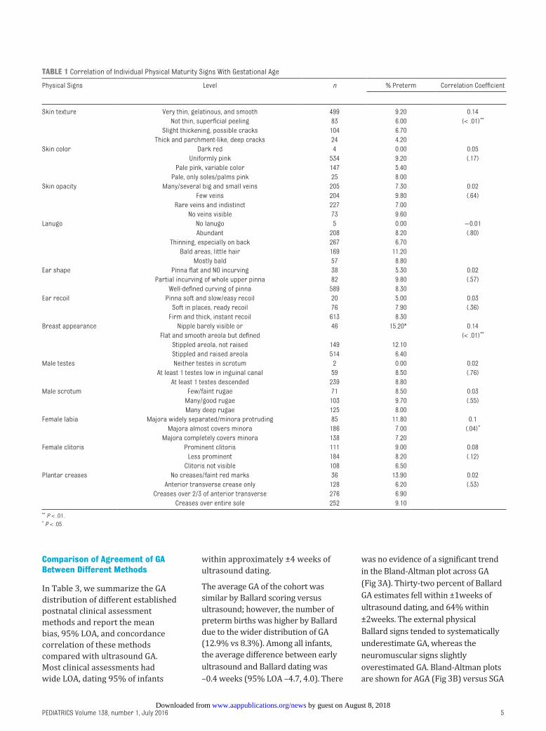
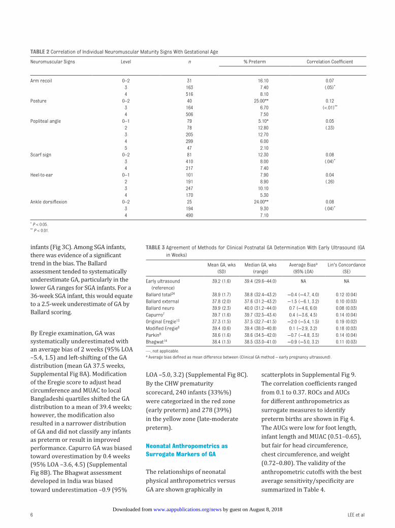
Correlation of Individual Physical and Neuromuscular Signs With GA
The relationship between individual
physical and neuromuscular signs
and GA is shown in Tables 1 and 2.
The correlation of GA with individual
physical signs was low for most
signs, but significant for skin texture,
breast appearance, and female labia.
GA was positively correlated with
the individual neuromuscular signs,
although the correlation coefficients
were also low. Posture, scarf sign,
arm recoil, and ankle dorsiflexion
were significantly correlated with GA.
We also examined the relationship
in the subset of SGA infants and
found significant correlation for
skin texture and posture; however,
correlation coefficients were similar
to infants appropriate for GA (AGA).
4
FIGURE 1Projahnmo Saving Lives at Birth Gestational Age Validation Flowchart.
FIGURE 2Distribution of GA by early ultrasound versus original Ballard score.
by guest on August 8, 2018www.aappublications.org/newsDownloaded from
PEDIATRICS Volume 138 , number 1 , July 2016
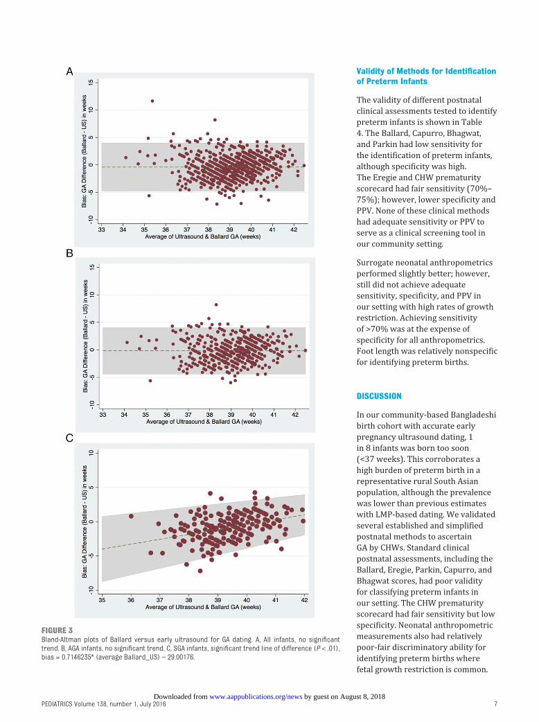
Comparison of Agreement of GA Between Different Methods
In Table 3, we summarize the GA
distribution of different established
postnatal clinical assessment
methods and report the mean
bias, 95% LOA, and concordance
correlation of these methods
compared with ultrasound GA.
Most clinical assessments had
wide LOA, dating 95% of infants
within approximately ±4 weeks of
ultrasound dating.
The average GA of the cohort was
similar by Ballard scoring versus
ultrasound; however, the number of
preterm births was higher by Ballard
due to the wider distribution of GA
(12.9% vs 8.3%). Among all infants,
the average difference between early
ultrasound and Ballard dating was
–0.4 weeks (95% LOA –4.7, 4.0). There
was no evidence of a significant trend
in the Bland-Altman plot across GA
(Fig 3A). Thirty-two percent of Ballard
GA estimates fell within ±1weeks of
ultrasound dating, and 64% within
±2weeks. The external physical
Ballard signs tended to systematically
underestimate GA, whereas the
neuromuscular signs slightly
overestimated GA. Bland-Altman plots
are shown for AGA (Fig 3B) versus SGA
5
TABLE 1 Correlation of Individual Physical Maturity Signs With Gestational Age
Physical Signs Level n % Preterm Correlation Coeffi cient
Skin texture Very thin, gelatinous, and smooth 499 9.20 0.14
Not thin, superfi cial peeling 83 6.00 (< .01)**
Slight thickening, possible cracks 104 6.70
Thick and parchment-like, deep cracks 24 4.20
Skin color Dark red 4 0.00 0.05
Uniformly pink 534 9.20 (.17)
Pale pink, variable color 147 5.40
Pale, only soles/palms pink 25 8.00
Skin opacity Many/several big and small veins 205 7.30 0.02
Few veins 204 9.80 (.64)
Rare veins and indistinct 227 7.00
No veins visible 73 9.60
Lanugo No lanugo 5 0.00 −0.01
Abundant 208 8.20 (.80)
Thinning, especially on back 267 6.70
Bald areas, little hair 169 11.20
Mostly bald 57 8.80
Ear shape Pinna fl at and NO incurving 38 5.30 0.02
Partial incurving of whole upper pinna 82 9.80 (.57)
Well-defi ned curving of pinna 589 8.30
Ear recoil Pinna soft and slow/easy recoil 20 5.00 0.03
Soft in places, ready recoil 76 7.90 (.36)
Firm and thick, instant recoil 613 8.30
Breast appearance Nipple barely visible or 46 15.20* 0.14
Flat and smooth areola but defi ned (< .01)**
Stippled areola, not raised 149 12.10
Stippled and raised areola 514 6.40
Male testes Neither testes in scrotum 2 0.00 0.02
At least 1 testes low in inguinal canal 59 8.50 (.76)
At least 1 testes descended 239 8.80
Male scrotum Few/faint rugae 71 8.50 0.03
Many/good rugae 103 9.70 (.55)
Many deep rugae 125 8.00
Female labia Majora widely separated/minora protruding 85 11.80 0.1
Majora almost covers minora 186 7.00 (.04)*
Majora completely covers minora 138 7.20
Female clitoris Prominent clitoris 111 9.00 0.08
Less prominent 184 8.20 (.12)
Clitoris not visible 108 6.50
Plantar creases No creases/faint red marks 36 13.90 0.02
Anterior transverse crease only 128 6.20 (.53)
Creases over 2/3 of anterior transverse 276 6.90
Creases over entire sole 252 9.10
** P < .01.* P < .05.
by guest on August 8, 2018www.aappublications.org/newsDownloaded from
LEE et al
infants (Fig 3C). Among SGA infants,
there was evidence of a significant
trend in the bias. The Ballard
assessment tended to systematically
underestimate GA, particularly in the
lower GA ranges for SGA infants. For a
36-week SGA infant, this would equate
to a 2.5-week underestimate of GA by
Ballard scoring.
By Eregie examination, GA was
systematically underestimated with
an average bias of 2 weeks (95% LOA
–5.4, 1.5) and left-shifting of the GA
distribution (mean GA 37.5 weeks,
Supplemental Fig 8A). Modification
of the Eregie score to adjust head
circumference and MUAC to local
Bangladeshi quartiles shifted the GA
distribution to a mean of 39.4 weeks;
however, the modification also
resulted in a narrower distribution
of GA and did not classify any infants
as preterm or result in improved
performance. Capurro GA was biased
toward overestimation by 0.4 weeks
(95% LOA –3.6, 4.5) (Supplemental
Fig 8B). The Bhagwat assessment
developed in India was biased
toward underestimation –0.9 (95%
LOA –5.0, 3.2) (Supplemental Fig 8C).
By the CHW prematurity
scorecard, 240 infants (33%%)
were categorized in the red zone
(early preterm) and 278 (39%)
in the yellow zone (late-moderate
preterm).
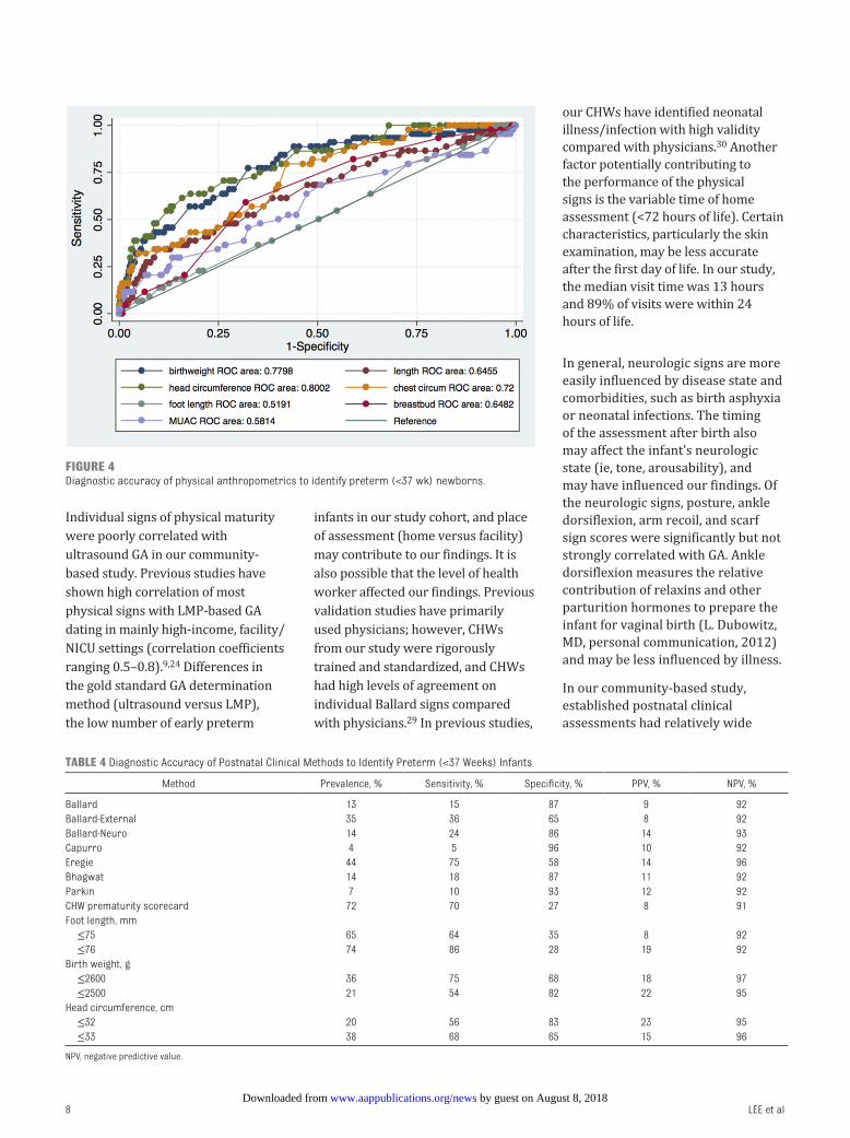
Neonatal Anthropometrics as Surrogate Markers of GA
The relationships of neonatal
physical anthropometrics versus
GA are shown graphically in
scatterplots in Supplemental Fig 9.
The correlation coefficients ranged
from 0.1 to 0.37. ROCs and AUCs
for different anthropometrics as
surrogate measures to identify
preterm births are shown in Fig 4.
The AUCs were low for foot length,
infant length and MUAC (0.51–0.65),
but fair for head circumference,
chest circumference, and weight
(0.72–0.80). The validity of the
anthropometric cutoffs with the best
average sensitivity/specificity are
summarized in Table 4.
6
TABLE 2 Correlation of Individual Neuromuscular Maturity Signs With Gestational Age
Neuromuscular Signs Level n % Preterm Correlation Coeffi cient
Arm recoil 0–2 31 16.10 0.07
3 163 7.40 (.05)*
4 516 8.10
Posture 0–2 40 25.00** 0.12
3 164 6.70 (<.01)**
4 506 7.50
Popliteal angle 0–1 79 5.10* 0.05
2 78 12.80 (.23)
3 205 12.70
4 299 6.00
5 47 2.10
Scarf sign 0–2 81 12.30 0.08
3 410 8.00 (.04)*
4 217 7.40
Heel-to-ear 0–1 101 7.90 0.04
2 191 8.90 (.26)
3 247 10.10
4 170 5.30
Ankle dorsifl exion 0–2 25 24.00** 0.08
3 194 9.30 (.04)*
4 490 7.10
* P < 0.05.** P < 0.01.
TABLE 3 Agreement of Methods for Clinical Postnatal GA Determination With Early Ultrasound (GA
in Weeks)
Mean GA, wks
(SD)
Median GA, wks
(range)
Average Biasa
(95% LOA)
Lin's Concordance
(SE)
Early ultrasound
(reference)
39.2 (1.6) 39.4 (29.6–44.0) NA NA
Ballard total24 38.9 (1.7) 38.8 (32.4–43.2) −0.4 (−4.7, 4.0) 0.12 (0.04)
Ballard external 37.8 (2.0) 37.6 (31.2–43.2) −1.5 (−6.1, 3.2) 0.10 (0.03)
Ballard neuro 39.9 (2.3) 40.0 (31.2–44.0) 0.7 (−4.6, 6.0) 0.08 (0.03)
Capurro7 39.7 (1.6) 39.7 (32.5–43.4) 0.4 (−3.6, 4.5) 0.14 (0.04)
Original Eregie13 37.3 (1.5) 37.5 (32.7–41.5) −2.0 (−5.4, 1.5) 0.19 (0.02)
Modifi ed Eregie8 39.4 (0.6) 39.4 (38.0–40.8) 0.1 (−2.9, 3.2) 0.18 (0.03)
Parkin9 38.6 (1.6) 38.6 (34.5–42.0) −0.7 (−4.8, 3.5) 0.14 (0.04)
Bhagwat14 38.4 (1.5) 38.5 (33.0–41.0) −0.9 (−5.0, 3.2) 0.11 (0.03)
—, not applicable.a Average bias defi ned as mean difference between (Clinical GA method – early pregnancy ultrasound).
by guest on August 8, 2018www.aappublications.org/newsDownloaded from
PEDIATRICS Volume 138 , number 1 , July 2016
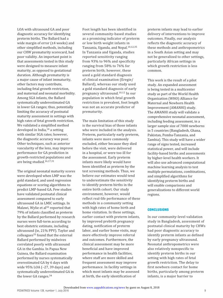
Validity of Methods for Identifi cation of Preterm Infants
The validity of different postnatal
clinical assessments tested to identify
preterm infants is shown in Table
4. The Ballard, Capurro, Bhagwat,
and Parkin had low sensitivity for
the identification of preterm infants,
although specificity was high.
The Eregie and CHW prematurity
scorecard had fair sensitivity (70%–
75%); however, lower specificity and
PPV. None of these clinical methods
had adequate sensitivity or PPV to
serve as a clinical screening tool in
our community setting.
Surrogate neonatal anthropometrics
performed slightly better; however,
still did not achieve adequate
sensitivity, specificity, and PPV in
our setting with high rates of growth
restriction. Achieving sensitivity
of >70% was at the expense of
specificity for all anthropometrics.
Foot length was relatively nonspecific
for identifying preterm births.
DISCUSSION
In our community-based Bangladeshi
birth cohort with accurate early
pregnancy ultrasound dating, 1
in 8 infants was born too soon
(<37 weeks). This corroborates a
high burden of preterm birth in a
representative rural South Asian
population, although the prevalence
was lower than previous estimates
with LMP-based dating. We validated
several established and simplified
postnatal methods to ascertain
GA by CHWs. Standard clinical
postnatal assessments, including the
Ballard, Eregie, Parkin, Capurro, and
Bhagwat scores, had poor validity
for classifying preterm infants in
our setting. The CHW prematurity
scorecard had fair sensitivity but low
specificity. Neonatal anthropometric
measurements also had relatively
poor-fair discriminatory ability for
identifying preterm births where
fetal growth restriction is common.
7
FIGURE 3Bland-Altman plots of Ballard versus early ultrasound for GA dating. A, All infants, no signifi cant trend. B, AGA infants, no signifi cant trend. C, SGA infants, signifi cant trend line of difference (P < .01), bias = 0.7146235* (average Ballard_US) – 29.00176.
by guest on August 8, 2018www.aappublications.org/newsDownloaded from
LEE et al
Individual signs of physical maturity
were poorly correlated with
ultrasound GA in our community-
based study. Previous studies have
shown high correlation of most
physical signs with LMP-based GA
dating in mainly high-income, facility/
NICU settings (correlation coefficients
ranging 0.5–0.8).9, 24 Differences in
the gold standard GA determination
method (ultrasound versus LMP),
the low number of early preterm
infants in our study cohort, and place
of assessment (home versus facility)
may contribute to our findings. It is
also possible that the level of health
worker affected our findings. Previous
validation studies have primarily
used physicians; however, CHWs
from our study were rigorously
trained and standardized, and CHWs
had high levels of agreement on
individual Ballard signs compared
with physicians.29 In previous studies,
our CHWs have identified neonatal
illness/infection with high validity
compared with physicians.30 Another
factor potentially contributing to
the performance of the physical
signs is the variable time of home
assessment (<72 hours of life). Certain
characteristics, particularly the skin
examination, may be less accurate
after the first day of life. In our study,
the median visit time was 13 hours
and 89% of visits were within 24
hours of life.
In general, neurologic signs are more
easily influenced by disease state and
comorbidities, such as birth asphyxia
or neonatal infections. The timing
of the assessment after birth also
may affect the infant’s neurologic
state (ie, tone, arousability), and
may have influenced our findings. Of
the neurologic signs, posture, ankle
dorsiflexion, arm recoil, and scarf
sign scores were significantly but not
strongly correlated with GA. Ankle
dorsiflexion measures the relative
contribution of relaxins and other
parturition hormones to prepare the
infant for vaginal birth (L. Dubowitz,
MD, personal communication, 2012)
and may be less influenced by illness.
In our community-based study,
established postnatal clinical
assessments had relatively wide
8
FIGURE 4Diagnostic accuracy of physical anthropometrics to identify preterm (<37 wk) newborns.
TABLE 4 Diagnostic Accuracy of Postnatal Clinical Methods to Identify Preterm (<37 Weeks) Infants
Method Prevalence, % Sensitivity, % Specifi city, % PPV, % NPV, %
Ballard 13 15 87 9 92
Ballard-External 35 36 65 8 92
Ballard-Neuro 14 24 86 14 93
Capurro 4 5 96 10 92
Eregie 44 75 58 14 96
Bhagwat 14 18 87 11 92
Parkin 7 10 93 12 92
CHW prematurity scorecard 72 70 27 8 91
Foot length, mm
≤75 65 64 35 8 92
≤76 74 86 28 19 92
Birth weight, g
≤2600 36 75 68 18 97
≤2500 21 54 82 22 95
Head circumference, cm
≤32 20 56 83 23 95
≤33 38 68 65 15 96
NPV, negative predictive value.
by guest on August 8, 2018www.aappublications.org/newsDownloaded from
PEDIATRICS Volume 138 , number 1 , July 2016
LOA with ultrasound GA and poor
diagnostic accuracy for identifying
preterm births. The Ballard had a
wide margin of error (±4 weeks) and
other simplified methods, including
our CHW prematurity scorecard, had
poor validity. An important point is
that assessments tested in this study
were designed to measure infant
maturity, as opposed to gestational
duration. Although prematurity is
a major cause of infant immaturity,
other factors may contribute,
including fetal growth restriction,
and maternal and neonatal morbidity.
Among SGA infants, the Ballard
systematically underestimated GA
in lower GA ranges; thus, potentially
limiting the accuracy of postnatal
maturity assessment in settings with
high rates of fetal growth restriction.
We validated a simplified method
developed in India, 14 a setting
with similar SGA rates; however,
the diagnostic accuracy was poor.
Other techniques, such as anterior
vascularity of the lens, may improve
performance for GA prediction in
growth-restricted populations and
are being studied.16, 17, 31
The original neonatal maturity scores
were developed when LMP was the
gold standard, and used regression
equations or scoring algorithms to
predict LMP-based GA. Few studies
have validated postnatal clinical
assessment compared to early
ultrasound GA in LMIC settings. In
Malawi, Wylie et al32 reported that
79% of infants classified as preterm
by the Ballard performed by research
nurses were full-term according to
best obstetric estimate, including
ultrasound (ie, 21% PPV). Taylor and
colleagues33 found that the external
Ballard performed by midwives
correlated poorly with ultrasound
GA in the Gambia. In Papua New
Guinea, the Ballard examination
performed by nurses systematically
overestimated GA by 6 days with
wide 95% LOA (–27, 39 days) and
systematically underestimated GA in
the lower GA ranges.34
Foot length has been identified in
several community-based studies
as a promising indicator of preterm
or low birth weight infants in
Tanzania, Uganda, and Nepal.10, 12, 35
In Tanzania and Uganda, studies
reported sensitivity ranging
from 93% to 96% and specificity
ranging from 58% to 76% for
preterm birth; however, these
used a gold standard diagnosis
of clinical examination (Eregie/
Ballard), whereas our study used
a gold standard diagnosis of early
pregnancy ultrasound.10, 12 In our
population in which fetal growth
restriction is prevalent, foot length
was not an accurate predictor of
prematurity.
The main limitation of this study
is the survival bias of those infants
who were included in the analysis.
Preterm, particularly early preterm,
infants were more commonly
excluded, either because they died
before the visit, were delivered
in a hospital, or were too ill for
the assessment. Early preterm
infants more likely would have
been identified as preterm by the
test screening methods. Thus, we
believe our estimates would tend
to underestimate the sensitivity
to identify preterm births in the
entire birth cohort. Our study
environment, however, would
reflect real-life performance of these
methods in a community setting
with high rates of home birth and
home visitation. In these settings,
earlier contact with preterm infants,
by improved prebirth pregnancy
dating, notification of preterm
labor, and earlier home visits, may
more effectively improve referral
and outcomes. Furthermore, the
clinical assessment may be more
beneficial and have improved
performance in health facilities,
where staff are more skilled and
frequent assessment may improve
performance. In facility settings in
which most infants may be assessed
at birth, the early identification of
preterm infants may lead to earlier
delivery of interventions to improve
outcomes. Finally, our analysis
reflects the diagnostic accuracy of
these methods and anthropometrics
in a South Asian setting and may
not be generalized to other settings,
particularly African settings in
which growth restriction is less
common.
This work is the result of a pilot
study. An expanded assessment
is being tested in a multicenter
study as part of the World Health
Organization (WHO) Alliance for
Maternal and Newborn Health
Improvement (AMANHI) study.
The AMANHI study will validate a
comprehensive neonatal assessment,
including feeding assessment, in a
larger sample size of 7000 newborns
in 5 countries (Bangladesh, Ghana,
Pakistan, Pemba-Tanzania, and
Zambia). This study will have a wider
range of signs tested, increased
statistical power, and will include
facility-based births and assessments
by higher-level health workers. It
will also use advanced computational
machine learning analysis to test
multiple permutations, combinations,
and simplified algorithms for
identifying preterm births and
will enable comparisons and
generalizations to different world
regions.
CONCLUSIONS
In our community-level validation
study in Bangladesh, assessment of
postnatal clinical maturity by CHWs
had poor diagnostic accuracy to
identify preterm infants as defined
by early pregnancy ultrasound.
Neonatal anthropometrics were
also relatively nonspecific to
identify preterm births in our
setting with high rates of fetal
growth restriction. The delay to
first newborn contact for home
births, particularly among preterm
infants, is a major barrier to
9 by guest on August 8, 2018www.aappublications.org/newsDownloaded from
LEE et al
improve preterm birth outcomes
in LMICs. There is an urgent need
to improve pregnancy dating
before birth, reduce delays to first
newborn contact, and develop
methods to feasibly and accurately
identify preterm births to improve
birth outcomes in settings of
highest need.
ACKNOWLEDGMENTS
We thank all the staff of the
Projahnmo team with special thanks
to those who implemented this
project, including Rina Paul, Eusuf
Ashraf, Monir Zaman, Mahmood
Rahman, Ataur Rahim, Tasima
Hussain, Nasreen Islam, field
supervisors, project officers, CHWs,
and Village Health Workers. We
thank our partners at WHO and the
AMANHI principal investigators
and teams: WHO: Rajiv Bahl and
Alex Manu, Pakistan: Fyezah Jehan,
Pemba: Sunil Sazawal, Zambia: David
Hamer, Ghana: Betty Kirkwood,
Lisa Hurt, Massachusetts General
Hospital: Blair Wylie. We thank
Lily Dubowitz, Jeanne Ballard, Kris
Karlsen (STABLE program), and
Jeanne Aby for providing their inputs
into the assessment and for sharing
training photographs, video, and/or
pictograms for trainings. We thank
Rachel Whelan, Lian Folger, and
Chelsea Clarke for their assistance
in formatting and preparing the
manuscript for submission. Finally,
we extend our appreciation to the
families, mothers, and infants who
participated in this study.
10
ABBREVIATIONS
AGA: appropriate for gestational
age
AMANHI: Alliance for Maternal
and Newborn Health
Improvement
AUC: area under the curve
BPD: biparietal diameter
CHW: community health worker
GA: gestational age
LMIC: low-and middle-income
country
LMP: last menstrual period
LOA: limits of agreement
MUAC: mid-upper arm
circumference
PPV: positive predictive value
ROC: receiver operating curve
SGA: small for gestational age
WHO: World Health Organization
revised the manuscript; Dr Christian helped conceptualize the design of the study protocols particularly related to pregnancy ultrasonography, provided input on
data analysis, and reviewed and revised the manuscript; Drs Labrique and Quaiyum helped conceptualize the design of the study, and reviewed and revised the
manuscript; Mr DasGupta helped design the data collection instruments, data management system, and reviewed and revised the manuscript; Dr Lokken helped
conceptualize the study procedures for ultrasonography, reviewed and provided quality control measures for ultrasound measures, and reviewed and revised
the manuscript; Dr Baqui helped conceptualize and design the study, obtain funding, provided input on data analysis, and reviewed and revised the manuscript;
and all authors approved the fi nal manuscript as submitted.
This trial has been registered at www. clinicaltrials. gov (identifi er NCT01572532).
DOI: 10.1542/peds.2015-3303
Accepted for publication Mar 30, 2016
Address correspondence to Anne CC Lee, MD, MPH, Brigham and Women’s Department of Pediatric Newborn Medicine, 75 Francis St, Thorn 229a, Boston, MA
02115. E-mail: [email protected]
PEDIATRICS (ISSN Numbers: Print, 0031-4005; Online, 1098-4275).
Copyright © 2016 by the American Academy of Pediatrics
FINANCIAL DISCLOSURE: The authors have indicated they have no fi nancial relationships relevant to this article to disclose.
FUNDING: This study is made possible through the generous support of the Saving Lives at Birth Round 1 partners: the US Agency for International Development,
the Government of Norway, the Bill & Melinda Gates Foundation, the World Bank, and Grand Challenges Canada. It was prepared by the Projahnmo research
group and does not necessarily refl ect the views of the Saving Lives at Birth Partners. The study was also funded by the Eunice Kennedy Shriver National Institute
of Child Health and Human Development (R01 HD066156–02). Funded by the National Institutes of Health (NIH).
POTENTIAL CONFLICT OF INTEREST: The authors have indicated they have no potential confl icts of interest to disclose.
COMPANION PAPER: A companion to this article can be found online at www. pediatrics. org/ cgi/ doi/ 10. 1542/ peds. 2016- 0734.
REFERENCES
1. Liu L, Johnson HL, Cousens S, et al; Child
Health Epidemiology Reference Group
of WHO and UNICEF. Global, regional, and
national causes of child mortality: an
updated systematic analysis for 2010
with time trends since 2000. Lancet.
2012;379(9832):2151–2161
2. Oza S, Lawn JE, Hogan DR, Mathers C,
Cousens SN. Neonatal cause-of-death
estimates for the early and late
neonatal periods for 194 countries:
2000–2013. Bull World Health Organ.
2015;93(1):19–28
3. Katz J, Lee AC, Kozuki N, et al;
CHERG Small-for-Gestational-Age-
Preterm Birth Working Group.
Mortality risk in preterm and
small-for-gestational-age infants
in low-income and middle-income
countries: a pooled country
analysis. Lancet. 2013;382(9890):
417–425
4. World Health Organization, The
Partnership for Maternal, Newborn,
and Child Health, Save the Children;
March of Dimes. Born Too Soon: the
Global Action Report on Preterm Birth.
by guest on August 8, 2018www.aappublications.org/newsDownloaded from

PEDIATRICS Volume 138 , number 1 , July 2016
Geneva, Switzerland: World Health
Organization; 2012
5. Dubowitz LM, Dubowitz V, Goldberg
C. Clinical assessment of gestational
age in the newborn infant. J Pediatr.
1970;77(1):1–10
6. Ballard JL, Khoury JC, Wedig K, Wang L,
Eilers-Walsman BL, Lipp R. New Ballard
Score, expanded to include extremely
premature infants. J Pediatr.
1991;119(3):417–423
7. Capurro H, Konichezky S, Fonseca
D, Caldeyro-Barcia R. A simplifi ed
method for diagnosis of gestational
age in the newborn infant. J Pediatr.
1978;93(1):120–122
8. Eregie CO. A new method for maturity
determination in newborn infants. J
Trop Pediatr. 2000;46(3):140–144
9. Parkin JM, Hey EN, Clowes JS. Rapid
assessment of gestational age at birth.
Arch Dis Child. 1976;51(4):259–263
10. Nabiwemba E, Marchant T, Namazzi G,
Kadobera D, Waiswa P. Identifying high-
risk babies born in the community
using foot length measurement at
birth in Uganda. Child Care Health Dev.
2013;39(1):20–26
11. Mukherjee S, Roy P, Mitra S, Samanta
M, Chatterjee S. Measuring new born
foot length to identify small babies in
need of extra care: a cross-sectional
hospital based study. Iran J Pediatr.
2013;23(5):508–512
12. Marchant T, Jaribu J, Penfold S,
Tanner M, Armstrong Schellenberg
J. Measuring newborn foot length
to identify small babies in need of
extra care: a cross sectional hospital
based study with community follow-up
in Tanzania. BMC Public Health.
2010;10:624
13. Eregie CO, Muogbo DC. A simplifi ed
method of estimating gestational age
in an African population. Dev Med Child
Neurol. 1991;33(2):146–152
14. Bhagwat VA, Dahat HB, Bapat NG.
Determination of gestational age of
newborns—a comparative study.
Indian Pediatr. 1990;27(3):272–275
15. Bindusha S, Rasalam CS, Sreedevi
N. Gestational age assessment of
newborn- clinical trial of a simplifi ed
method. Transworld Medical Journal.
2014;2(1):24–28
16. Narayanan I, Dua K, Gujral VV, Mehta
DK, Mathew M, Prabhakar AK. A simple
method of assessment of gestational
age in newborn infants. Pediatrics.
1982;69(1):27–32
17. Hittner HM, Hirsch NJ, Rudolph AJ.
Assessment of gestational age by
examination of the anterior vascular
capsule of the lens. J Pediatr.
1977;91(3):455–458
18. Lawn JE, Blencowe H, Oza S, et al;
Lancet Every Newborn Study Group.
Every newborn: progress, priorities,
and potential beyond survival. Lancet.
2014;384(9938):189–205
19. Baqui AH, El-Arifeen S, Darmstadt GL,
et al; Projahnmo Study Group. Effect
of community-based newborn-care
intervention package implemented
through two service-delivery strategies
in Sylhet district, Bangladesh: a
cluster-randomised controlled trial.
Lancet. 2008;371(9628):1936–1944
20. Arifeen SE, Mullany LC, Shah R, et
al. The effect of cord cleansing with
chlorhexidine on neonatal mortality in
rural Bangladesh: a community-based,
cluster-randomised trial. Lancet.
2012;379(9820):1022–1028
21. Lee AC, Quaiyum MA, Mullany LC, et al
Screening and treatment of maternal
genitourinary tract infections in early
pregnancy to prevent preterm birth
in rural Sylhet, Bangladesh: a cluster
randomized trial. BMC Pregnancy
Childbirth. 2015;15:326
22. Hadlock FP, Shah YP, Kanon DJ,
Lindsey JV. Fetal crown-rump
length: reevaluation of relation to
menstrual age (5–18 weeks) with high-
resolution real-time US. Radiology.
1992;182(2):501–505
23. Hadlock FP, Deter RL, Harrist RB,
Park SK. Fetal biparietal diameter: a
critical re-evaluation of the relation
to menstrual age by means of real-
time ultrasound. J Ultrasound Med.
1982;1(3):97–104
24. Ballard JL, Novak KK, Driver M. A
simplifi ed score for assessment of
fetal maturation of newly born infants.
J Pediatr. 1979;95(5 pt 1):769–774
25. Labrique AB, Shaikh S, West KP,
inventors; Alain Labrique, assignee.
Portable acrylic infant length board. US
patent 61/450, 9492011. February 2011
26. Labrique AB, Christian P, Klemm RD,
et al. A cluster-randomized, placebo-
controlled, maternal vitamin A or
beta-carotene supplementation trial
in Bangladesh: design and methods.
Trials. 2011;12:102
27. Villar J, Cheikh Ismail L, Victora
CG, et al; International Fetal and
Newborn Growth Consortium for
the 21st Century (INTERGROWTH-
21st). International standards for
newborn weight, length, and head
circumference by gestational age
and sex: the Newborn Cross-Sectional
Study of the INTERGROWTH-21st Project.
Lancet. 2014;384(9946):857–868
28. Lin LI. A concordance correlation
coeffi cient to evaluate reproducibility.
Biometrics. 1989;45(1):255–268
29. Lee Anne CC, Uddin J, Shah RML, et al
Validation of community health worker
clinical assessment of gestational age
in rural Bangladesh. In: Proceeding
from the Pediatric Academic Societies
Annual Meeting; May 4 - May 7, 2013;
Washington, DC
30. Baqui AHAS, Arifeen SE, Rosen HE, et al;
Projahnmo Study Group. Community-
based validation of assessment
of newborn illnesses by trained
community health workers in Sylhet
district of Bangladesh. Trop Med Int
Health. 2009;14(12):1448–1456
31. Skapinker R, Rothberg AD. Postnatal
regression of the tunica vasculosa
lentis. J Perinatol. 1987;7(4):279–281
32. Wylie BJ, Kalilani-Phiri L, Madanitsa
M, et al. Gestational age assessment
in malaria pregnancy cohorts: a
prospective ultrasound demonstration
project in Malawi. Malar J. 2013;12:183
33. Taylor RADF, Denison FC, Beyai
S, Owens S. The external Ballard
examination does not accurately
assess the gestational age of infants
born at home in a rural community
of The Gambia. Ann Trop Paediatr.
2010;30(3):197–204
34. Karl S, Li Wai Suen CSN, Unger HW,
et al. Preterm or not—an evaluation
of estimates of gestational age
in a cohort of women from rural
Papua New Guinea. PLoS One.
2015;10(5):e0124286
35. Mullany LC, Darmstadt GL, Khatry SK,
Leclerq SC, Tielsch JM. Relationship
between the surrogate anthropometric
measures, foot length and chest
circumference and birth weight among
newborns of Sarlahi, Nepal. Eur J Clin
Nutr. 2007;61(1):40–46
11 by guest on August 8, 2018www.aappublications.org/newsDownloaded from
DOI: 10.1542/peds.2015-3303 originally published online June 16, 2016; 2016;138;Pediatrics
Mohammed Quaiyum, Abdullah H Baqui and for the Projahnmo Study GroupAhmed, Parul Christian, Alain Labrique, Sushil K. DasGupta, R. Peter Lokken,
Anne CC Lee, Luke C. Mullany, Karima Ladhani, Jamal Uddin, Dipak Mitra, ParvezBangladesh
Validity of Newborn Clinical Assessment to Determine Gestational Age in
ServicesUpdated Information &
http://pediatrics.aappublications.org/content/138/1/e20153303including high resolution figures, can be found at:
Referenceshttp://pediatrics.aappublications.org/content/138/1/e20153303#BIBLThis article cites 29 articles, 3 of which you can access for free at:
Subspecialty Collections
http://www.aappublications.org/cgi/collection/public_health_subPublic Healthsubhttp://www.aappublications.org/cgi/collection/fetus:newborn_infant_Fetus/Newborn Infantfollowing collection(s): This article, along with others on similar topics, appears in the
Permissions & Licensing
http://www.aappublications.org/site/misc/Permissions.xhtmlin its entirety can be found online at: Information about reproducing this article in parts (figures, tables) or
Reprintshttp://www.aappublications.org/site/misc/reprints.xhtmlInformation about ordering reprints can be found online:
by guest on August 8, 2018www.aappublications.org/newsDownloaded from
DOI: 10.1542/peds.2015-3303 originally published online June 16, 2016; 2016;138;Pediatrics
Mohammed Quaiyum, Abdullah H Baqui and for the Projahnmo Study GroupAhmed, Parul Christian, Alain Labrique, Sushil K. DasGupta, R. Peter Lokken,
Anne CC Lee, Luke C. Mullany, Karima Ladhani, Jamal Uddin, Dipak Mitra, ParvezBangladesh
Validity of Newborn Clinical Assessment to Determine Gestational Age in
http://pediatrics.aappublications.org/content/138/1/e20153303located on the World Wide Web at:
The online version of this article, along with updated information and services, is
http://pediatrics.aappublications.org/content/suppl/2016/06/14/peds.2015-3303.DCSupplementalData Supplement at:
1073-0397. ISSN:60007. Copyright © 2016 by the American Academy of Pediatrics. All rights reserved. Print
the American Academy of Pediatrics, 141 Northwest Point Boulevard, Elk Grove Village, Illinois,has been published continuously since 1948. Pediatrics is owned, published, and trademarked by Pediatrics is the official journal of the American Academy of Pediatrics. A monthly publication, it
by guest on August 8, 2018www.aappublications.org/newsDownloaded from


![Long-term Behavioral Problems in Children With Severe …pediatrics.aappublications.org/content/pediatrics/138/5/...consciousness (Blantyre coma score [BCS] ≤2) and Plasmodium falciparum](https://img.pdfslide.us/doc/110x75/5ac1a5947f8b9a5a4e8d6456/long-term-behavioral-problems-in-children-with-severe-blantyre-coma-score-bcs.jpg)