Embed Size (px)
Citation preview
Accepted Manuscript
Validation of the Baveno Vi Criteria to Identify Low Risk Cirrhotic Patients notRequiring Endoscopic Surveillance for Varices
James Maurice, Edgar Brodkin, Frances Arnold, Annalan Navaratnam, HeidiPaine, Sabrina Khawar, Ameet Dhar, David Patch, James O’Beirne, RajMookerjee, Massimo Pinzani, Emmanouil Tsochatzis, Rachel H. Westbrook
PII: S0168-8278(16)30316-6DOI: http://dx.doi.org/10.1016/j.jhep.2016.06.021Reference: JHEPAT 6168
To appear in: Journal of Hepatology
Received Date: 22 January 2016Revised Date: 5 June 2016Accepted Date: 7 June 2016
Please cite this article as: Maurice, J., Brodkin, E., Arnold, F., Navaratnam, A., Paine, H., Khawar, S., Dhar, A.,Patch, D., O’Beirne, J., Mookerjee, R., Pinzani, M., Tsochatzis, E., Westbrook, R.H., Validation of the Baveno ViCriteria to Identify Low Risk Cirrhotic Patients not Requiring Endoscopic Surveillance for Varices, Journal ofHepatology (2016), doi: http://dx.doi.org/10.1016/j.jhep.2016.06.021
This is a PDF file of an unedited manuscript that has been accepted for publication. As a service to our customerswe are providing this early version of the manuscript. The manuscript will undergo copyediting, typesetting, andreview of the resulting proof before it is published in its final form. Please note that during the production processerrors may be discovered which could affect the content, and all legal disclaimers that apply to the journal pertain.
Title Page
VALIDATION OF THE BAVENO VI CRITERIA TO IDENTIFY LOW RISK CIRRHOTIC PATIENTS NOT REQUIRING ENDOSCOPIC SURVEILLANCE FOR VARICES James Maurice1,2; Edgar Brodkin1; Frances Arnold1; Annalan Navaratnam1; Heidi Paine2; Sabrina Khawar1; Ameet Dhar2; David Patch1; James O’Beirne1; Raj Mookerjee1,3; Massimo Pinzani1,3; Emmanouil Tsochatzis1,3; Rachel H Westbrook1
1 Department of Hepatology, Royal Free Hospital NHS Trust 2 Department of Hepatology, Imperial College Healthcare NHS Trust 3 Institute for Liver and Digestive Health, University College London Corresponding Author: Dr Rachel Westbrook Department of Hepatology, 8 South Office, Royal Free Hospital, Pond St, London NW3 2QG Tel: 020 7794 0500 ext 38097 Fax: 020 7472 6226 [email protected] Key Words: Portal hypertension, oesophageal varices, non- invasive investigations, transient elastography, cirrhosis Abbreviations: AUROC: Area under the receiver operator curve HVPG: Hepatic venous pressure gradient TE: Transient elastography LSM: Liver stiffness measurement kPa: Kilopascals OGD: Oesophagogastroduodenoscopy cACLD: Compensated Advanced Chronic Liver Disease LRV: Low risk varices HRV: High risk varices INR: International Normalised Ratio HCV: Hepatitis C ALD: Alcohol related liver disease NAFLD: Non alcoholic fatty liver disease HBV: Hepatitis B PPV: Positive predictive value NPV: Negative predictive value LR+: positive likelihood ratio LR-: negative likelihood ratio Word Count: 4078 6 Tables, 2 Figures, 1 Supplementary Figure Conflict of interest: Nothing to declare Financial support: there was no financial support given for this study. Author’s Contributions: JM: collected data, analysed data, wrote the manuscript, approved final manuscript.
EB, FA, AN, HP, SK: collected data and contributed to the drafting and final approval of the manuscript. AD, JO’B, DP, MP, RM, ET: provided data and contributed to the drafting and final approval of the manuscript. RW: Provided overall oversight of the study, analysed the data, contributed to the drafting and final approval of the manuscript.
Abstract
Background: The Baveno VI guidelines propose that cirrhotic patients with a liver stiffness
measurement (LSM) <20kPa and a platelet count >150000/µL can avoid screening
endoscopy as their combination is highly specific for excluding clinically significant varices.
The aim of the study was to validate these criteria.
Methods: Transient elastography data was collected from two institutions from 2006-2015.
Inclusion criteria were a LSM ≥10kPa and an upper gastrointestinal endoscopy within 12
months, with a diagnosis of compensated chronic liver disease. Exclusion criteria were
porto-mesenteric-splenic vein thrombosis and non-cirrhotic portal hypertension. Varices
were graded as low risk (grade <2) or high risk (grade ≥2).
Results: The study included 310 patients (169 (55%) hepatitis C, and 275 (89%) Child Pugh
A). Varices were present in 23% cases, with 5% prevalence of high risk varices. Overall
102/310 (33%) met the Baveno VI criteria. Within this group 11% had varices and 2% had
high risk varices, representing 2/15 (13%) of all high risk varices. The Baveno VI criteria
gave a sensitivity 0.87, specificity 0.34, positive predictive value 0.06, negative predictive
value 0.98, positive likelihood ratio 1.31 and negative likelihood ratio 0.39. The AUROC for
LSM and platelet count combined was 0.746.
Conclusions: The Baveno VI criteria performed well correctly identifiying 98% of patients
who could safely avoid endoscopy.
Word count: 213
Lay Summary
This study examines the effectives of a recent set of guidelines published by the Baveno VI
conference, which states that patients with chronic liver disease and a low liver stiffness
(<20kPa) and high platelet count (>150) are at low risk of having varices and do not need a
screening endoscopy. Varices are a complication of cirrhosis, confer a risk of serious
bleeding, and can be diagnosed and treated by endoscopy. Our study reviewed the clinical
records of patients who have had liver stiffness scans and endoscopy over a 9 year period
at two hospitals. The results show that only about 2% of patients who meet the Baveno VI
criteria will be miss classified as not having varices.
Introduction
Gastroesophageal varices occur as a consequence of portal hypertension and are a
major cause of morbidity and mortality due to the risk of haemorrhage. In cirrhosis raised
portal pressures initially develop as a result of advanced fibrosis and deranged liver
architecture, but as liver disease progresses additional haemodynamic factors, such as
splanchnic vasodilatation and hyperdynamic circulation, become increasingly important
(Vizzutti et al. 2007). Portal pressures have traditionally been measured using hepatic
venous pressure gradient (HVPG), and an HVPG ≥ 10mmHg confers increased risk of
developing gastroesophageal varices (Groszmann et al. 2005). HVPG has been shown to
correlate well with the presence and size of varices (Wadhawan et al. 2006), however
measuring portal pressures by HVPG is invasive and limited to the centres with the relevant
expertise.
Over the last decade transient elastography (TE) has become a widely used, non-
invasive measure of liver stiffness and fibrosis. Following initial studies showing its accuracy
in diagnosing significant fibrosis its clinical applications have been widened. The use of TE
as a surrogate marker of portal hypertension has been demonstrated by liver stiffness
measurement (LSM) correlating well with portal pressures up to a HVPG of 10-12mmHg
(Vizzutti et al. 2007)(Bureau et al. 2008). Subsequent data has shown that TE is of potential
benefit in the non- invasive diagnosis of varices, especially when TE is combined with other
markers such as platelet count and spleen size (Berzigotti et al. 2013).
A major limitation to implementing these tests into clinical practice for diagnosing
gastroesophageal varices has been an inadequate specificity. As a result the diagnostic
strength of non- invasive investigations such as TE have not yet been sufficient to replace
endoscopy in the diagnosis of varices (Shi et al. 2013) (EASL 2015), and all patients with
cirrhosis currently require routine surveillance with frequent oesophagogastroduodenoscopy
(OGD).
The promising sensitivity and negative predictive value of TE, especially in
combination with other non- invasive markers, means these investigations may be more
effective tools at identifying low risk cirrhotic patients who can be safely ‘ruled out’ of
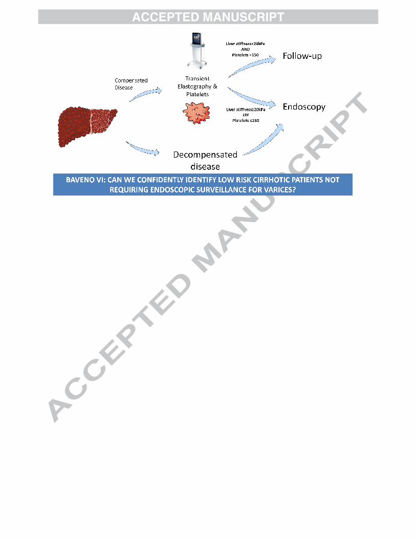
needing an endoscopy. The recent Baveno VI guidelines acknowledge this application and
recommend that in patients with compensated advanced chronic liver disease (cACLD) a
LSM <20Kpa and a platelet count > 150,000 cells/µL have a very low risk of having varices
requiring treatment and therefore do not require screening endoscopy. They advise
longitudinal follow-up of such patients by annual repetition of TE and platelet count with the
guidance that if liver stiffness increases or platelet count declines to within the
recommended values, these patients should undergo screening OGD (de Franchis 2015).
In this retrospective cross-sectional cohort study, we reviewed all patients over a nine
year period at two centres who have undergone clinical, laboratory, TE and endoscopic
evaluation of portal hypertension. The primary aim was to validate the recently proposed
Baveno VI criteria and assess their sensitivity at accurately identifying those patients who
can safely avoid screening endoscopy. Secondary aims were to assess if the criteria had
similar sensitivities across all aetiologies of chronic liver disease, given the majority of
published data is from patients with viral hepatitis, and to identify if alternative LSM or
platelet parameters should be recommended.
Methods
Study Population
This is a retrospective cohort study. Transient elastography data collected from two
institutions from November 2006 - September 2015 were analysed. All patients with a LSM
≥ 10kPa were selected. Additional inclusion criteria were an OGD within 12 months of TE,
and a diagnosis of chronic liver disease. Exclusion criteria were decompensated disease
(defined as Child Pugh C disease or Child Pugh B with evidence of ascites, encephalopathy
or previous variceal haemorrhage), current use of non- selective beta- blockers, porto-
mesenteric-splenic vein thrombosis, and non-cirrhotic portal hypertension. A sub-analysis of
all patients with OGD within 6 months of TE was also performed.
Transient Elastography
All TE was performed using Fibroscan© (Echosens, Paris) by experienced practitioners at
two large specialist centres who follow conventional practice. Patients were fasted for two
hours before the procedure. All patients were examined in the standard way with the right
lobe of the liver accessed by the patient lying in the dorsal cubitus position and maximal
abduction of the right arm. Ten valid measurements were obtained and a median LSM value
(kPa) generated. Inadequate LSM (defined by interquartile range >30% and success <60%)
were excluded.
Assessment of varices
All patients had an OGD within 12 months of the TE. Gastroesophageal varices were
defined as low risk varices (LRV) or high risk varices (HRV). For the purpose of this study all
varices that were described as < grade 2 were defined as LRV. Conversely all oesophageal
varices described as ≥ grade 2, and any gastric varices, were defined as HRV. This
differentiation was made as all varices classified as high risk in this way would be deemed
clinically significant and require treatment in standard clinical practice.
Laboratory Investigations
Laboratory investigations were collected, including platelets, bilirubin, alanine
aminotransferase, aspartate aminotransferase, albumin, international normalised ratio
(INR), sodium and creatinine.
Statistical analysis
Demographic and laboratory data was summarised and compared between patients with
and without HRV. Continuous variables were reported as medians with interquartile range,
and compared using Mann-Whitney test. The variables of LSM and platelet count were
compared to the binary outcome measure of the presence of HRV. Sensitivity, specificity,
positive predictive value (PPV), negative predictive value (NPV), positive likelihood ratio
(LR+) and negative likelihood ratio (LR-) were calculated as per the cut offs recommended
by Baveno VI (de Franchis 2015). AUROC values were generated for the presence of HRV,
using the variables of LSM, platelet count and the two variables combined.
Statistics were performed using the software packages SPSS.
Results
Study population
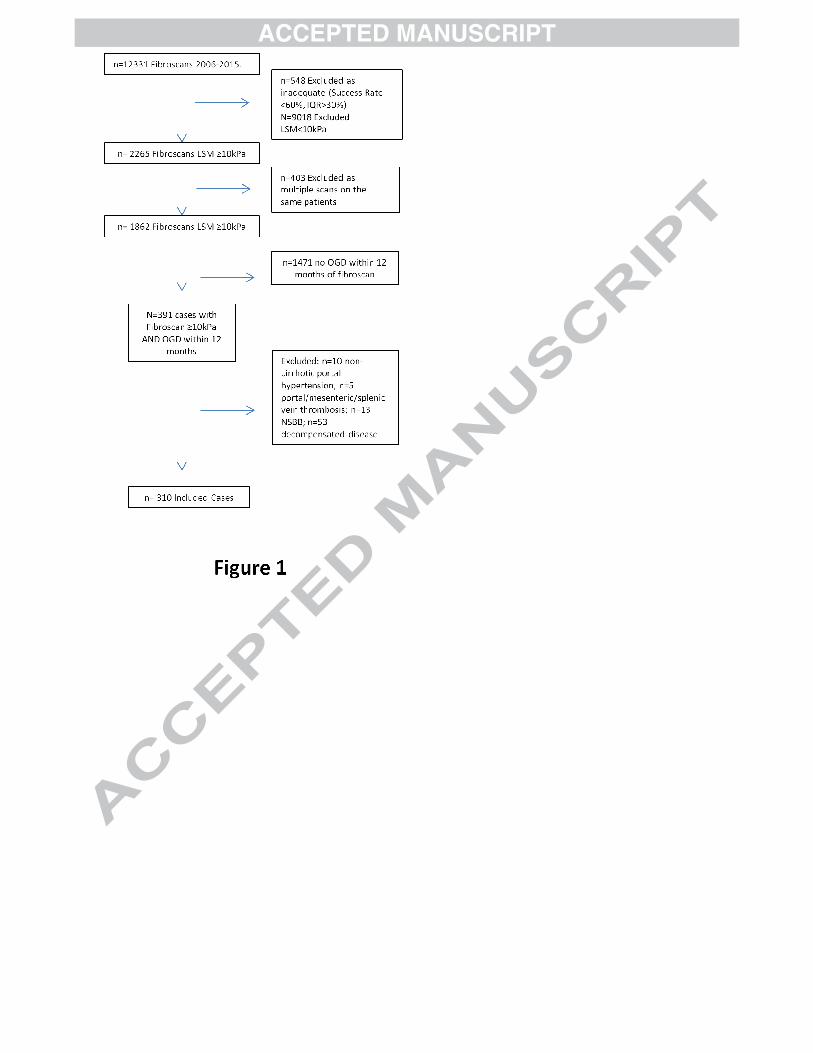
Over the study period 12331 transient elastography (TE) scans were performed. After
excluding inadequate scans, values <10kPa, and multiple scans on the same patient and
scans without an OGD within 12 months, 391 cases remained. Of these, a further 81 were
excluded: n=10 non-cirrhotic portal hypertension, n=5 portal/mesenteric/splenic vein
thrombosis, n=13 current use of non- selective beta blockers, n=53 decompensated
disease. In total 310 patients were included in the study (Figure 1).
Demographic data
Of the 310 cases that met the inclusion criteria for the study, 209 (67%) were male and 101
(33%) female. The aetiology of the underlying liver disease was hepatitis C ( HCV, n=169
(55%)), alcohol- related liver disease (ALD, n=40 (13%)), non-alcoholic fatty liver disease
(NAFLD, n=42 (14%)), Hepatitis B/D (HBV, n=24 (8%)), and other (n=35 (11%): ALD/HCV
(n=5), HBV/NAFLD (n=1), HBV/HCV (n=2), ALD/NAFLD (n=2), drug reaction (n=2),
cryptogenic (n=4), Gaucher’s/HCV (n=1), Gaucher’s (n=1), haemochromatosis (n=2),
haemochromatosis/HCV (n=1), sarcoidosis (n=1), sarcoidosis/HBV/HCV (n=1), autoimmune
hepatitis (n=2), primary biliary cholangitis (n=4), primary sclerosing cholangitis (n=5),
overlap syndrome (n=1)). One case with ALD had alcoholic hepatitis at the time of transient
elastography, with a LSM 16.8kPa. The majority of cases were Child Pugh A (n=275
(89%)), with 35 cases (11%) Child Pugh B. Median MELD score was 7. The above data is
summarised in Table 1.
Varices were present in 18% of the population studied (n=57); 15 patients (5%) had HRV.
Two cases had high risk stigmata with red wale signs. With respect to LSM, 167 (54%)
cases had a LSM <20kPa, and 143 (46%) cases had a LSM ≥ 20kPa. In laboratory tests
151 (49%) cases had platelets >150x103/ml, and 159 (51%) had platelets ≤150x103/ml
(Table 1, 2a and 2b).
Liver stiffness measurement as a predictor of varices
The median LSM in our cohort was 18.4kPa. As expected liver stiffness measurement was
significantly higher in patients with HRV than in those without HRV (26.0kPa vs 18.4kPa,
p<0.015). In the cases with LSM <20kPa, 23/167 (14%) had any varices, of which 5 (3%)
were HRV. Of the cases with LSM ≥20kPa, 49/143 (34%) had any varices, of which 10 (7%)
had HRV (Table 2a). Overall for identifying HRV, LSM cut- off of 20kPa had a sensitivity of
0.67, specificity 0.55, positive predictive value (PPV) 0.07, negative predictive value (NPV)
0.97, positive likelihood ratio (LR+) 1.48 and negative likelihood ratio (LR-) 0.61 (Table 3a).
The AUROC for LSM as an independent variable was 0.686 (Supplementary Fig 1).
Platelet value as a predictor of varices
The median platelet count in our cohort was 147000/µL, and was not significantly lower in
patients with HRV than in those without HRV. In cases with platelets >150000/µL, 22/151
(15%) had any varices, and of these 6 (4%) were HRV. In cases with platelets ≤150000/µL,
50/159 (31%) had any varices, of which 9 (6%) were HRV (Table 2a). Overall for identifying
HRV, platelet count cut-off of 150000/µL had a sensitivity of 0.60, specificity 0.49, PPV 0.06,
NPV 0.96, LR+ 1.18, LR- 0.81 (Table 3a). The AUROC for platelets as an independent
variable was 0.599 (Supplementary Fig 1).
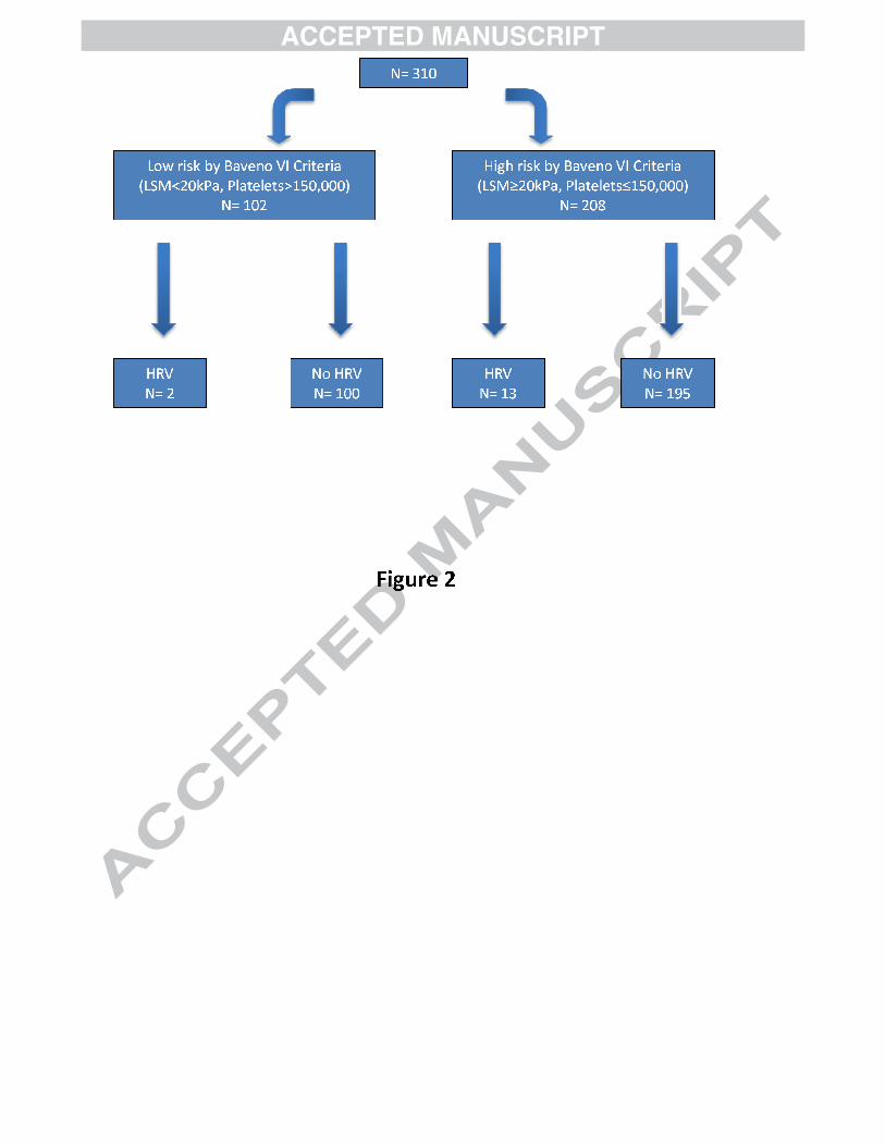
Baveno VI Criteria as a predictor of varices
The Baveno VI consensus guidelines combine LSM < 20kPa and platelet count
>150000/µL. In this cohort, 102/310 (33%) cases met these criteria, of whom 11 (11%) had
any varices and 2 (2%) had HRV. Among the 208/310 (67%) cases that fell outside of the
Baveno VI criteria, 61/208 (29%) had any varices and 13 (6%) had HRV (Table 2a).
Combining LSM and platelet count using the recommended cut-off values to detect HRV
gives a sensitivity 0.87, specificity 0.34, PPV 0.06, NPV 0.98, LR+ 1.31, LR- 0.39 (Table
3a). The AUROC for the combination of LSM and platelets was 0.746 (Supplementary Fig
1). Using the Baveno VI guideline 2/15 (13%) of HRV were missed (Figure ). The LSM and
platelet count of these cases were 16.8kPa / 380000/µL and 17.6kPa / 160000/µL
respectively. Both cases had grade 2 oesophageal varices and compensated cirrhosis
secondary to HCV.
Impact of aetiology on diagnostic accuracy
Out of the 15 cases with HRV, 11 had viral hepatitis and 2 cases had ALD and NAFLD. In a
sub analysis by aetiology, the Baveno VI guidelines in viral hepatitis had a sensitivity 0.82,
specificity 0.28, PPV 0.06, NPV 0.96, LR+ 1.13 and LR- 0.66; in NAFLD/ ALD, they had a
sensitivity 1.00, specificity 0.45, PPV 0.04, NPV 1.0, LR+ 1.82 and LR- 0.00 (Table 4).
AUROC for TE, platelets and the two variables combined in viral hepatitis was 0.633, 0.675
and 0.749 and in ALD/NAFLD were 0.924, 0.534 and 0.927 respectively (Supplementary
Fig 1).
Impact of Time between OGD and Transient Elastography
2
The above analysis was repeated in a population restricted to patients who had an OGD
within 6 months of transient elastography. This included a slightly smaller (n=219) cohort
with similarly mixed aetiology (HCV n=118). In this analysis, 66/219 patients met BAVENO
VI criteria for avoiding screening endoscopy, of whom 1 case had high risk varices. The
diagnostic performance of the BAVENO VI criteria was similar in this sub-group as for the
whole study population (Table 2b and 3b).
Discussion
In this large dual centre cross- sectional cohort study we validate the recently published
BAVENO VI guidelines for using non-invasive criteria in patients with cACLD to identify
patients who are at low risk of clinically significant varices and thus can safely avoid
screening endoscopy. We have demonstated that applying such criteria will reduce the
number of surveillance endoscopies by about 30%, but could incorrectly classify 2% of
patients. Thus adherence to these criteria may delay clinically effective prophylaxis against
variceal bleeding with non-selective beta-blockers in a small proportion of patients.
The study included 310 patients. Hepatitis C was the most common aetiology, but
with a prevalence of just 55% our cohort reflects a more heterogenous group compared to
many of the large studies in this field of predominantly HCV populations (Augustin et al.
2014)(Berzigotti et al. 2013).
The prevalence of all GOV was 23% (18% LRV and 5% HRV). The combination of
TE and platelet count with the cut-off values proposed by Baveno VI had a high NPV and
low LR-, in contrast to poor PPV and LR+, confirming these markers perform more strongly
at ‘ruling out’ rather than ‘ruling in’ HRV, in keeping with the guidelines. A total of 2/15
(13%) cases with HRV had platelets >150 and TE <20kPa and were miss-classified by
Baveno VI. Application of the guidelines will have excluded these patients from endoscopic
surveillance and delayed the introduction of appropriate primary prophylaxis. However, on
detailed examination of the two cases one had thalassaemia major and also a splenectomy
in 1975, which may explain the unusual finding of platelets in the upper limit of normal in the
context of cACLD. Careful consideration must therefore be given to co-morbidities which
may impact the validity of the proposed platelet cut-off.
Reassuringly only 2/102 (2%) cases meeting BAVENO criteria had HRV, therefore
the annual risk to a patient counselled in clinic based on a bleeding rate from varices of 15%
per year would be just 0.3% (Garcia-tsao et al. 2007). The guidelines however advise
annual assessment of TE and platelet count, followed by endoscopic surveillance of
patients who move out of the low risk category. In the miss-classified patients, sequential
annual platelet counts were 160000 – 195000 – 188000/µL (HCV, no known haematological
co-morbidities) with no further TE data, and 380- 298- 307- 337 (HCV with thalassaemia
major and previous splenectomy) with progression in liver stiffness from 16.8kPa to 20kPa
on repeat TE four years later. Further longitudinal prospective data will help define the
actual risk of bleeding by applying the Baveno VI criteria and if there is an increase
compared to current practice due to the inevitable small percentage of high risk varices that
will not be captured using non-invasive markers.
Applying the data from our cohort, a reduction in the LSM cut-off to 16.8kPa would
have resulted in the inclusion of both the miss-classified cases for endoscopic surveillance.
Incorporating this cut-off into the Baveno VI criteria would have correctly identified all
patients who could safely avoid screening endoscopy. This would be just below the median
LSM 18.4kPa in our cohort and similar to the mean LSM 17.6kPa in a study of compensated
cirrhotic patients with no cases of HRV (Augustin et al. 2014), but would result in an
additional 27 (13%) endoscopies in our cohort.
The rationale for the Baveno VI guidelines comes from evidence in a number of
studies demonstrating that non- invasive investigations such as TE and platelet count show
promise in the diagnosis of varices, but generally perform better at excluding rather than
diagnosing high risk varices. A study by Berzigotti et al in compensated cirrhotics showed
that TE, in combination with platelet count and spleen size, had a NPV 0.95 and LR- 0.13
compared to PPV 0.55 and LR+ 2.63 when the cut-off was set for a sensitivity of
90%(Berzigotti et al. 2013). Similarly Stefanescu et al showed that TE combined with
additional serological and radiological markers produced a NPV 1.0 and LR- 0.1 for high risk
oesophageal varices (Stefanescu et al. 2014). More recently Perezzo et al focussed on TE
and platelet count as in the Baveno VI guidelines in a prospective assessment 99 HCV
cirrhotic patients (80% Child Pugh A, 63% female): 14% had HRV, all of which were
appropriately classified by the Baveno VI criteria. Spleen stiffness did not improve the
performance of TE and platelet count to identify low risk patients (Perazzo et al. 2015).
Four aspects of our results should be commented on: firstly, these data suggest
there is a role for non- invasive markers in identifying patients at low risk of having clinically
significant varices who can safely avoid screening endoscopy. Our reported NPV of 0.98 is
similar to the NPV of troponin, which is widely implemented to exclude the diagnosis of
myocardial infarction (Al-Saleh et al. 2014). This presents an opportunity to reduce the
burden of unnecessary endoscopies for patients who often face many invasive
investigations through the course of their disease, but the poor PPV and LR+ show these
non- invasive tests cannot replace endoscopy in the diagnosis of varices and deciding
which patients warrant treatment with primary prophylaxis. Secondly, the guidelines offer
single cut-off values that do not account for underlying aetiology. They do acknowledge that
the majority of the work in this area has been in chronic hepatitis C, and the value of TE in
diagnosing clinically significant portal hypertension in other aetiologies remains to be
ascertained. Reassuringly in our small sub group of ALD and NAFLD cases no HRV were
missed by the Baveno VI criteria. This is in keeping with some recent studies which have
also investigated the use of TE for diagnosing gastroesophageal varices in heterogenous
populations(Ding et al. 2015) (Stefanescu et al. 2014). However, further research is needed
to validate these non-invasive markers in other aetiologies, particularly NAFLD and ALD.
Thirdly, the data leading to the current guidelines is drawn from populations with variable
disease severity and prevalence of varices. The overall prevalence of HRV in our cohort is
5%, and 2% in the cohort meeting the Baveno VI low risk criteria, which is higher than in a
study of carefully chosen compensated cirrhotics that found a 0% prevalence of HRV
(Augustin et al. 2014), but somewhat lower than in an earlier meta- analysis of TE in
detecting large varices that quoted higher prevalence rates ranging from 14.7 to 48% (Shi et
al. 2013). We agree with the Baveno VI statement that the guidelines should only be applied
to a population with early, well compensated disease with a low pre-test probability of
varices and therefore with a low chance of missing cases requiring treatment. Finally,
Baveno VI elected to use TE and platelet count alone. Many of the studies on the non-
invasive assessment of varices have used additional serological and radiological markers
such as spleen stiffness, platelet- spleen ratio, spleen size and Lok score, which can
improve the accuracy of diagnosing varices when used in combination with TE (Takuma et
al. 2013)(Stefanescu et al. 2014)(Berzigotti et al. 2013). While the results of our study are
promising for the simple, pragmatic use of just TE and platelet count, cases will be missed
which will have implications, albeit small, on bleeding risk. Most of these additional non-
invasive markers are readily available and could be useful in further refining the accuracy of
the diagnostic algorithm.
This study has some limitations. The retrospective design brings inherent limitations
of bias, which is a particular factor in the high number of cases that did not have an OGD
within 12 months of a transient elastography result demonstrating advanced fibrosis. We
think this is probably due to a clinical suspicion of mild disease, reflected in a lower median
LSM 15.3kPa in the patients not eventually included, compared to 17kPa in the study
population (p<0.001). Endoscopy was not performed simultaneously with the TE, with a
median duration of time between TE and endoscopy of 120 days. However, this is well
below the recommended annual surveillance frequency of high- risk patients without
varices, and repeating the analysis on only patients endoscoped within 6 months did not
significantly alter the results (Table 2b and 3b). Moreover, using the presence of varices as
an endpoint is limited by variable and subjective reporting of their size. However, this is a
real world study and initiation of primary prophylaxis is based on the endoscopic evidence of
high risk varices given the impractical nature of routinely measuring HVPG. Therefore,
identifying non-invasive techniques to identify patients without varices is directly relevant to
clinical practice. Finally, we elected to use a relatively conservative cut-off LSM value of
10kPa for our study cohort based on the Baveno VI definition of patients likely to have
compensated advanced chronic liver disease. This is below the accepted cut-off value
13.6kPa for cirrhosis in viral hepatitis, the most heavily studied cohort, and may have
reduced the prevalence of HRV and elevated the negative predictive value. However, the
HRV prevalence of 5% is higher than in other studies of compensated cirrhotics (Augustin et
al. 2014). Moreover, other causes of chronic liver disease such as non- alcoholic liver
disease are often poorly represented in this field of research, and the LSM criteria for
cirrhosis in non- viral aetiologies is less well defined.
Conclusion
Significant progress has been made in the field on non- invasive markers for diagnosing
HRV. Our data partly supports the Baveno VI statement that identifying low risk patients
who do not require surveillance endoscopy is a realistic goal with the current technologies,
which could produce a significant cost saving and beneficially impact on patient experience.
However, this data also highlights that a small proportion of cases will be miss-classified
and thus be denied proven prophylactic therapies for primary prevention of variceal
bleeding. The risk will be minimised by careful assessment for comorbidities that may affect
platelet count, and long-term follow-up with annual TE and platelet count to initiate timely
endoscopic surveillance in suitable patients. Confirmation by prospective, longitudinal data
collection will give further support to these recommendations.
Acknowledgements:
We would like to thank Talay Hakan, Heather Lewis and Louise Campbell for their help in
obtaining extensive transient elastography data in the two institutions. This study received
no external funding.
References
Al-Saleh, A. et al., 2014. Performance of the high-sensitivity troponin assay in diagnosing
acute myocardial infarction: systematic review and meta-analysis. CMAJ open, 2(3),
pp.E199–207. Available at:
http://cmajopen.ca/cgi/doi/10.9778/cmajo.20130074\nhttp://www.ncbi.nlm.nih.gov/pubm
ed/25295240\nhttp://www.pubmedcentral.nih.gov/articlerender.fcgi?artid=PMC418318.
Augustin, S. et al., 2014. Detection of early portal hypertension with routine data and liver
stiffness in patients with asymptomatic liver disease: A prospective study. Journal of
Hepatology, 60(3), pp.561–569.
Berzigotti, A. et al., 2013. Elastography, Spleen Size, and Platelet Count Identify Portal
Hypertension in Patients With Compensated Cirrhosis. Gastroenterology, 144(1),
pp.102–111.e1. Available at:
http://linkinghub.elsevier.com/retrieve/pii/S0016508512014667.
Bureau, C. et al., 2008. Transient elastography accurately predicts presence of significant
portal hypertension in patients with chronic liver disease. , (April), pp.1261–1268.
Ding, N.S. et al., 2015. Liver stiffness plus platelet count can be used to exclude high-risk
oesophageal varices. , pp.1–6.
EASL, 2015. EASL-ALEH Clinical Practice Guidelines: Non-invasive tests for evaluation of
liver disease severity and prognosis. Journal of Hepatology, 63, pp.237–264. Available
at: http://linkinghub.elsevier.com/retrieve/pii/S0168827815002597.
de Franchis, R., 2015. Expanding consensus in portal hypertension. Journal of Hepatology,
63(3), pp.743–752. Available at:
http://linkinghub.elsevier.com/retrieve/pii/S0168827815003499.
Garcia-tsao, G. et al., 2007. AASLD PRACTICE GUIDELINES Prevention and Management
of Gastroesophageal Varices and Variceal Hemorrhage in Cirrhosis. , pp.922–938.
Groszmann, R.J. et al., 2005. Beta-blockers to prevent gastroesophageal varices in patient
with cirrhosis. New England Journal of Medicine, 353, pp.2254–2261. Available at:
http://discovery.ucl.ac.uk/40547/.
Perazzo, H. et al., 2015. Reply to “Points to be considered when using transient
elastography for diagnosis of portal hypertension according to the Baveno’s VI
consensus”. Journal of hepatology, 63(4), pp.1049–50. Available at:
http://www.ncbi.nlm.nih.gov/pubmed/26212027.
Shi, K.-Q. et al., 2013. Transient elastography: a meta-analysis of diagnostic accuracy in
evaluation of portal hypertension in chronic liver disease. Liver international : official
journal of the International Association for the Study of the Liver, 33(1), pp.62–71.
Available at: http://www.ncbi.nlm.nih.gov/pubmed/22973991.
Stefanescu, H., Radu, C et al., 2014. Non-invasive ménage à trois for the prediction of
high-risk varices: stepwise algorithm using lok score, liver and spleen stiffness. Liver
international : official journal of the International Association for the Study of the Liver,
pp.1–9.
Takuma, Y. et al., 2013. Measurement of spleen stiffness by acoustic radiation force
impulse imaging identifies cirrhotic patients with esophageal varices. Gastroenterology,
144(1), pp.92–101.e2. Available at: http://www.ncbi.nlm.nih.gov/pubmed/23022955.
Vizzutti, F. et al., 2007. Liver stiffness measurement predicts severe portal hypertension in
patients with HCV-related cirrhosis. Hepatology, 45(5), pp.1290–1297. Available at:
http://ovidsp.ovid.com/ovidweb.cgi?T=JS&CSC=Y&NEWS=N&PAGE=fulltext&D=med5
&AN=17464971\nhttp://bf4dv7zn3u.search.serialssolutions.com.myaccess.library.utoro
nto.ca/?url_ver=Z39.88-
2004&rft_val_fmt=info:ofi/fmt:kev:mtx:journal&rfr_id=info:sid/Ovid:med5&rft.g.
Wadhawan, M. et al., 2006. Hepatic venous pressure gradient in cirrhosis: correlation with
the size of varices, bleeding, ascites, and child’s status. Digestive diseases and
sciences, 51(12), pp.2264–9. Available at:
http://www.ncbi.nlm.nih.gov/pubmed/17080245.
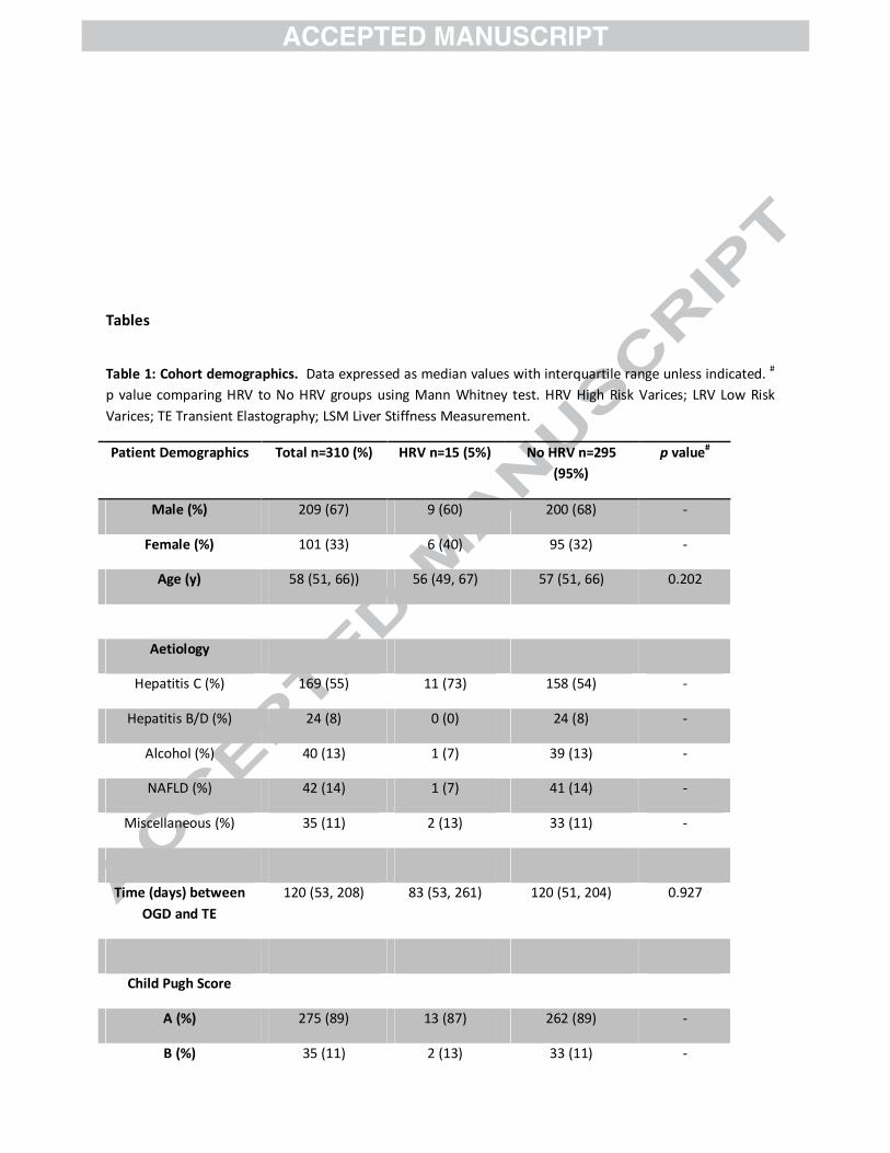
Tables
Table 1: Cohort demographics. Data expressed as median values with interquartile range unless indicated. #
p value comparing HRV to No HRV groups using Mann Whitney test. HRV High Risk Varices; LRV Low Risk
Varices; TE Transient Elastography; LSM Liver Stiffness Measurement.
Patient Demographics Total n=310 (%) HRV n=15 (5%) No HRV n=295
(95%)
p value#
Male (%) 209 (67) 9 (60) 200 (68) -
Female (%) 101 (33) 6 (40) 95 (32) -
Age (y) 58 (51, 66)) 56 (49, 67) 57 (51, 66) 0.202
Aetiology
Hepatitis C (%) 169 (55) 11 (73) 158 (54) -
Hepatitis B/D (%) 24 (8) 0 (0) 24 (8) -
Alcohol (%) 40 (13) 1 (7) 39 (13) -
NAFLD (%) 42 (14) 1 (7) 41 (14) -
Miscellaneous (%) 35 (11) 2 (13) 33 (11) -
Time (days) between
OGD and TE
120 (53, 208) 83 (53, 261) 120 (51, 204) 0.927
Child Pugh Score
A (%) 275 (89) 13 (87) 262 (89) -
B (%) 35 (11) 2 (13) 33 (11) -
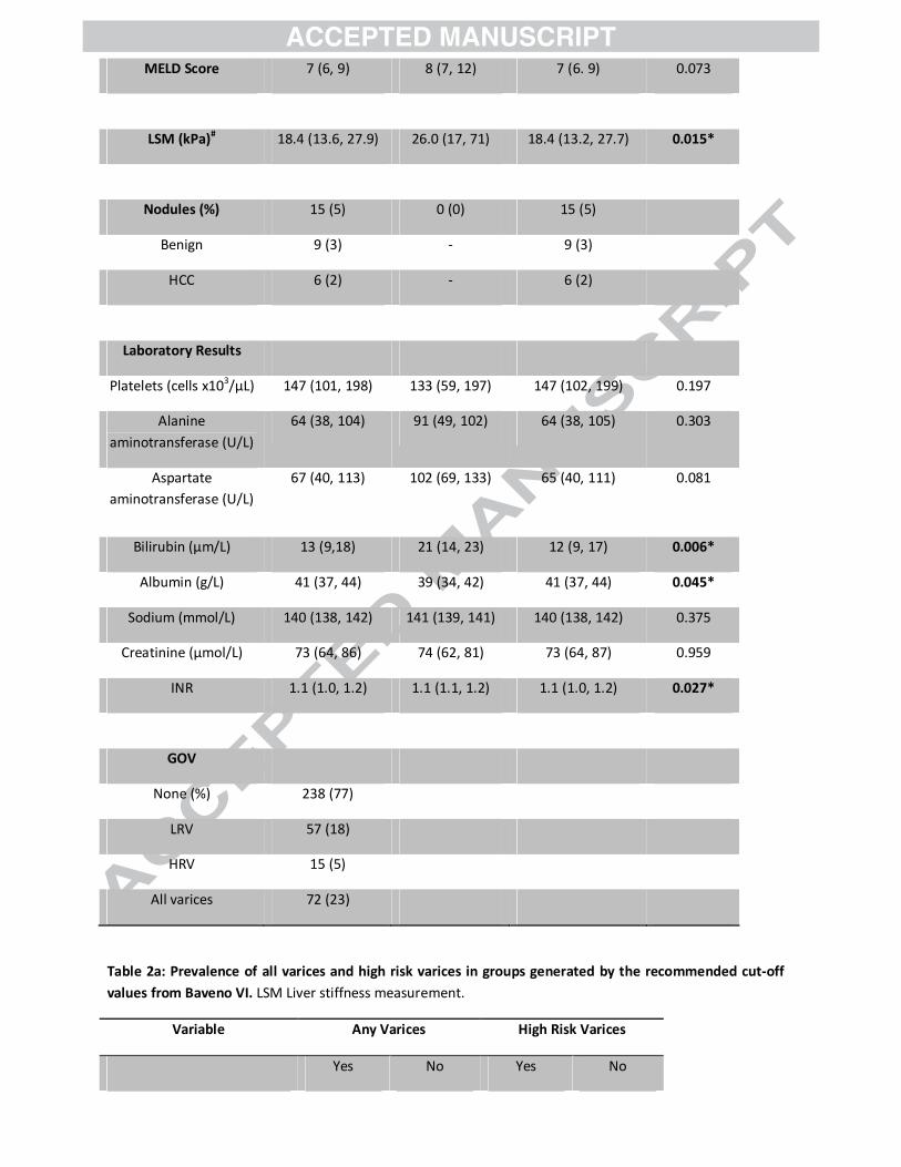
MELD Score 7 (6, 9) 8 (7, 12) 7 (6. 9) 0.073
LSM (kPa)# 18.4 (13.6, 27.9) 26.0 (17, 71) 18.4 (13.2, 27.7) 0.015*
Nodules (%) 15 (5) 0 (0) 15 (5)
Benign 9 (3) - 9 (3)
HCC 6 (2) - 6 (2)
Laboratory Results
Platelets (cells x103/μL) 147 (101, 198) 133 (59, 197) 147 (102, 199) 0.197
Alanine
aminotransferase (U/L)
64 (38, 104) 91 (49, 102) 64 (38, 105) 0.303
Aspartate
aminotransferase (U/L)
67 (40, 113) 102 (69, 133) 65 (40, 111) 0.081
Bilirubin (μm/L) 13 (9,18) 21 (14, 23) 12 (9, 17) 0.006*
Albumin (g/L) 41 (37, 44) 39 (34, 42) 41 (37, 44) 0.045*
Sodium (mmol/L) 140 (138, 142) 141 (139, 141) 140 (138, 142) 0.375
Creatinine (μmol/L) 73 (64, 86) 74 (62, 81) 73 (64, 87) 0.959
INR 1.1 (1.0, 1.2) 1.1 (1.1, 1.2) 1.1 (1.0, 1.2) 0.027*
GOV
None (%) 238 (77)
LRV 57 (18)
HRV 15 (5)
All varices 72 (23)
Table 2a: Prevalence of all varices and high risk varices in groups generated by the recommended cut-off
values from Baveno VI. LSM Liver stiffness measurement.
Variable Any Varices High Risk Varices
Yes No Yes No
LSM<20kPa (n=167) 23 144 5 162
LSM≥20kPa (n=143) 49 94 10 133
Platelets≤150x103/μL
(n=159)
50 109 9 150
Platelets>150x103/μL (n=
151)
22 129 6 145
Within Baveno VI criteria:
LSM<20kPa and
Platelets>150x103/μL
(n=102)
11 91 2 100
Outside Baveno VI criteria:
LSM≥20kPa and/or
Platelets≤150x103/μL(n=208)
61 147 13 195
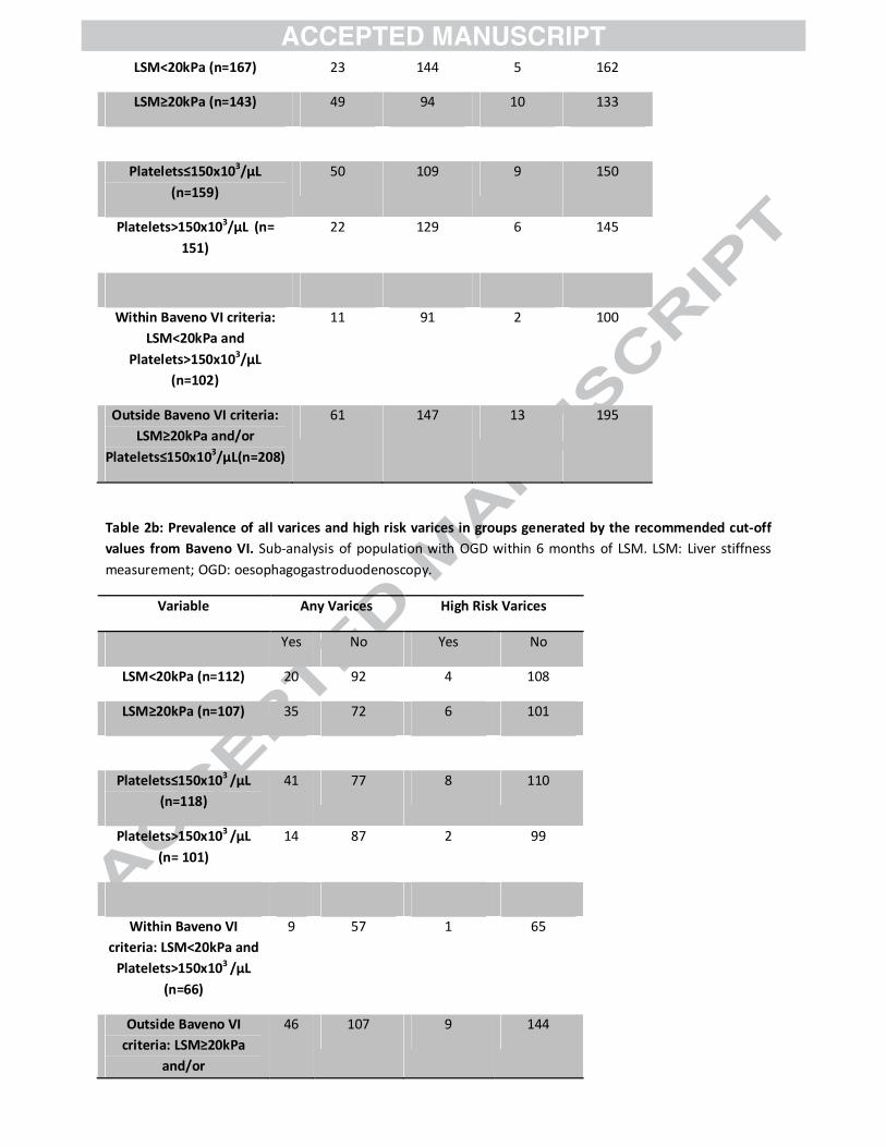
Table 2b: Prevalence of all varices and high risk varices in groups generated by the recommended cut-off
values from Baveno VI. Sub-analysis of population with OGD within 6 months of LSM. LSM: Liver stiffness
measurement; OGD: oesophagogastroduodenoscopy.
Variable Any Varices High Risk Varices
Yes No Yes No
LSM<20kPa (n=112) 20 92 4 108
LSM≥20kPa (n=107) 35 72 6 101
Platelets≤150x103
/μL
(n=118)
41 77 8 110
Platelets>150x103
/μL
(n= 101)
14 87 2 99
Within Baveno VI
criteria: LSM<20kPa and
Platelets>150x103 /μL
(n=66)
9 57 1 65
Outside Baveno VI
criteria: LSM≥20kPa
and/or
46 107 9 144
Platelets≤150x103 /μL
(n=153)
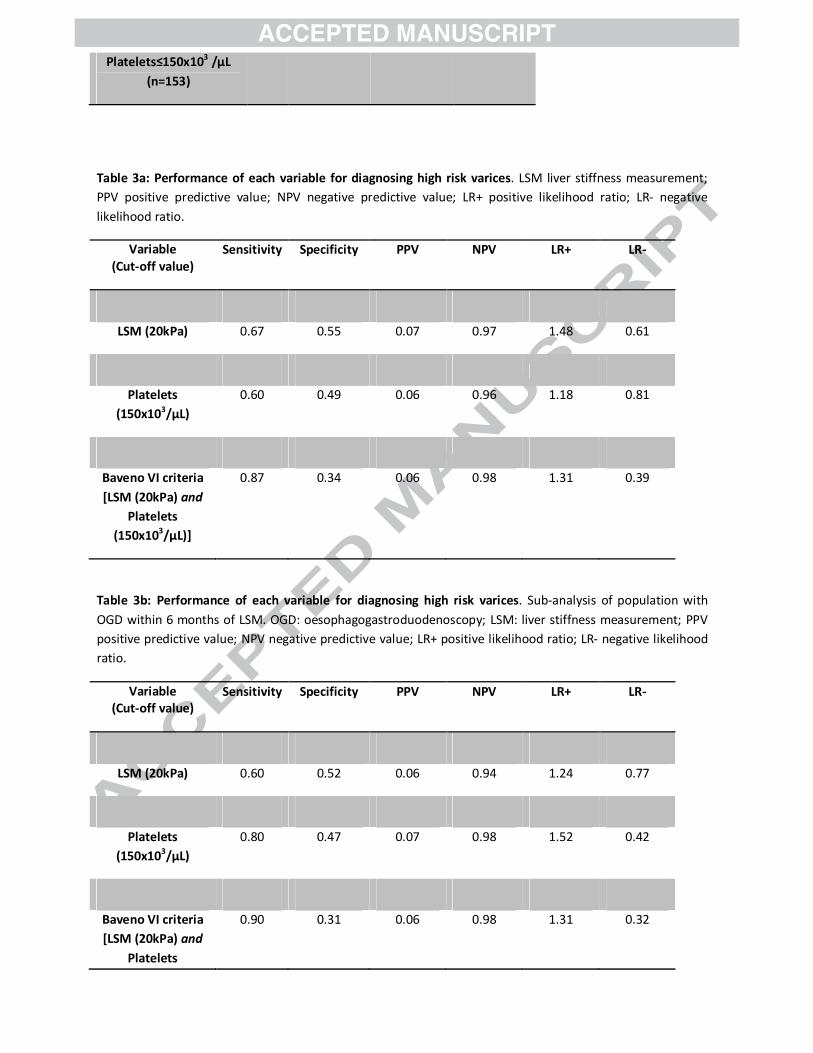
Table 3a: Performance of each variable for diagnosing high risk varices. LSM liver stiffness measurement;
PPV positive predictive value; NPV negative predictive value; LR+ positive likelihood ratio; LR- negative
likelihood ratio.
Variable
(Cut-off value)
Sensitivity Specificity PPV NPV LR+ LR-
LSM (20kPa) 0.67 0.55 0.07 0.97 1.48 0.61
Platelets
(150x103/μL)
0.60 0.49 0.06 0.96 1.18 0.81
Baveno VI criteria
[LSM (20kPa) and
Platelets
(150x103/μL)]
0.87 0.34 0.06 0.98 1.31 0.39
Table 3b: Performance of each variable for diagnosing high risk varices. Sub-analysis of population with
OGD within 6 months of LSM. OGD: oesophagogastroduodenoscopy; LSM: liver stiffness measurement; PPV
positive predictive value; NPV negative predictive value; LR+ positive likelihood ratio; LR- negative likelihood
ratio.
Variable
(Cut-off value)
Sensitivity Specificity PPV NPV LR+ LR-
LSM (20kPa) 0.60 0.52 0.06 0.94 1.24 0.77
Platelets
(150x103/μL)
0.80 0.47 0.07 0.98 1.52 0.42
Baveno VI criteria
[LSM (20kPa) and
Platelets
0.90 0.31 0.06 0.98 1.31 0.32
(150x103/μL)]
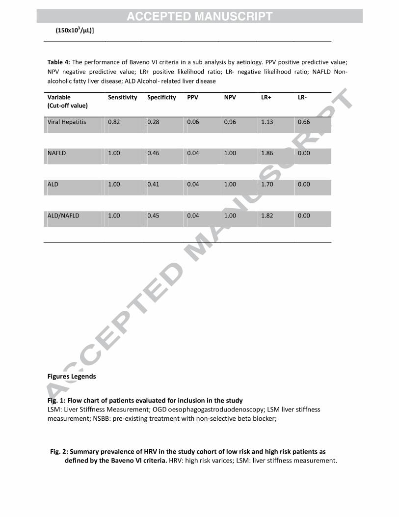
Table 4: The performance of Baveno VI criteria in a sub analysis by aetiology. PPV positive predictive value;
NPV negative predictive value; LR+ positive likelihood ratio; LR- negative likelihood ratio; NAFLD Non-
alcoholic fatty liver disease; ALD Alcohol- related liver disease
Variable
(Cut-off value)
Sensitivity Specificity PPV NPV LR+ LR-
Viral Hepatitis 0.82 0.28 0.06 0.96 1.13 0.66
NAFLD 1.00 0.46 0.04 1.00 1.86 0.00
ALD 1.00 0.41 0.04 1.00 1.70 0.00
ALD/NAFLD 1.00 0.45 0.04 1.00 1.82 0.00
Figures Legends
Fig. 1: Flow chart of patients evaluated for inclusion in the study LSM: Liver Stiffness Measurement; OGD oesophagogastroduodenoscopy; LSM liver stiffness
measurement; NSBB: pre-existing treatment with non-selective beta blocker;
Fig. 2: Summary prevalence of HRV in the study cohort of low risk and high risk patients as
defined by the Baveno VI criteria. HRV: high risk varices; LSM: liver stiffness measurement.