Embed Size (px)
Citation preview
Therapeutics, Targets, and Chemical Biology
Validation and Structural Characterization of theLEDGF/p75–MLL Interface as a New Target for theTreatment of MLL-Dependent Leukemia
Kate�rina �Cerm�akov�a1, Petr Tesina2,3, Jonas Demeulemeester1, Sara El Ashkar1, H�el�ene M�ereau4,Juerg Schwaller4, Pavlína �Rez�a�cov�a2,5, Vaclav Veverka2, and Jan De Rijck1
AbstractMixed lineage leukemia (MLL) fusion–driven acute leukemias represent a genetically distinct subset of
leukemias with poor prognosis. MLL forms a ternary complex with the lens epithelium–derived growth factor(LEDGF/p75) and MENIN. LEDGF/p75, a chromatin reader recognizing H3K36me3 marks, contributes to theassociation of the MLL multiprotein complex to chromatin. Formation of this complex is critical for thedevelopment of MLL leukemia. Available X-ray data represent only a partial structure of the LEDGF/p75–MLL–MENIN complex. Using nuclear magnetic resonance spectroscopy, we identified an additional LEDGF/p75–MLL interface, which overlaps with the binding site of known LEDGF/p75 interactors—HIV-1 integrase,PogZ, and JPO2. Binding of these proteins or MLL to LEDGF/p75 is mutually exclusive. The resolvedstructure, as well as mutational analysis, shows that the interaction is primarily sustained via two aromaticresidues of MLL (F148 and F151). Colony-forming assays in MLL–AF9þ leukemic cells expressing MLLinteraction-defective LEDGF/p75 mutants revealed that this interaction is essential for transformation.Finally, we show that the clonogenic growth of primary murine MLL-AF9–expressing leukemic blasts isselectively impaired upon overexpression of a LEDGF/p75-binding cyclic peptide CP65, originally developedto inhibit the LEDGF/p75–HIV-1 integrase interaction. The newly defined protein–protein interface thereforerepresents a new target for the development of therapeutics against LEDGF/p75–dependent MLL fusion–driven leukemic disorders. Cancer Res; 74(18); 5139–51. �2014 AACR.
IntroductionThe mixed lineage leukemia (MLL) gene, located on chro-
mosome 11q23, is frequently targeted by chromosomal trans-locations (see Krivtsov and Armstrong for a review; ref. 1).Balanced rearrangements result in the formation of new fusionproteins involved in childhood and adult de novo as well astherapy-related myeloid and lymphoblastic leukemias. Wild-type (WT) MLL is a histone methyltransferase associated with
the promoters of a large subset of active genes. More specif-ically, MLL maintains the expression of posterior HOXA genes(HOXA5, HOXA9, and HOXA10) and as such is essential forembryonic body plan formation and the self-renewal capacityof hematopoietic stem cells and immature progenitors (2, 3).
Typical MLL oncogenic fusions retain the MLL N-terminalregion. To date, more than 70 different N-terminal fusionsassociated with leukemia development have been identified,of which MLL–AF4, MLL–AF9, and MLL–ENL originate fromt(4;11), t(9;11), and t(11;19) translocations, respectively, andare the most prevalent (Fig. 1A; ref. 4). These fusion partnersare part of a complex converting promoter-proximal arrestedRNA Pol II into the elongation state (5), which suggests that asubset of MLL-mediated leukemias is triggered by aberranttranscriptional elongation. The mechanism whereby otherMLL fusion partners initiate cellular transformation is, how-ever, largely unknown. Recent studies also suggest that recip-rocal MLL fusion proteins may have an important role incancer development (6, 7) and that MLL fusions also need thefunctional activity of the WT MLL protein to maintain onco-genic transformation (8).
AlthoughMLL can bind directly toDNA via its AT-hooks andCXXC-domain, the MLL multiprotein complex is targeted tospecific genes through interaction with various cellular pro-teins inducing gene activation or repression (9). Among others,MLL forms a ternary complex with MENIN (MEN1) and the
1KU Leuven, Laboratory for Molecular Virology and Gene Therapy, Depart-ment of Pharmaceutical and Pharmacological Sciences, Leuven, Belgium.2Institute of Organic Chemistry and Biochemistry of the ASCR, v.v.i., Struc-turalBiology,Prague,CzechRepublic. 3CharlesUniversity inPrague,Depart-ment of Genetics and Microbiology, Faculty of Science, Prague, CzechRepublic. 4Department of Biomedicine, University Hospital and Children'sHospital Basel (UKBB) ZLF, Basel, Switzerland. 5Institute of MolecularGenetics of the ASCR, v.v.i., Structural Biology, Prague, Czech Republic.
Note: Supplementary data for this article are available at Cancer ResearchOnline (http://cancerres.aacrjournals.org/).
V. Veverka and J. De Rijck shared senior authorship for this article.
CorrespondingAuthors: JanDeRijck, KULeuven,Molecular Virology andGene Therapy, Kapucijnenvoer 33, VCTB þ5, 3000 Leuven, Belgium.Phone: 32-16-33-21-76; Fax: 32-16-33-63-36; [email protected]; and Vaclav Veverka, IOCB ASCR, Flemin-govo nam. 2, 16610 Prague, Czech Republic; Phone: 420-220-183-135;E-mail: [email protected]
doi: 10.1158/0008-5472.CAN-13-3602
�2014 American Association for Cancer Research.
CancerResearch
www.aacrjournals.org 5139
on February 14, 2019. © 2014 American Association for Cancer Research. cancerres.aacrjournals.org Downloaded from
Published OnlineFirst July 31, 2014; DOI: 10.1158/0008-5472.CAN-13-3602
lens epithelium–derived growth factor (LEDGF/p75) via theN-terminal MENIN bindingmotifs (MBM1, 2) and the LEDGF/p75 binding domain (LBD), respectively (Fig. 1A; refs. 10–12).Both MENIN and LEDGF/p75 are essential for MLL fusion–mediated transformation (11, 12). LEDGF/p75 is an epigeneticreader recognizing H3K36me3 marks via its PWWP domainand as such it preferentially associateswith the body of activelytranscribed genes (Fig. 1B; refs. 13–15). Alongside the MLLcomplex, LEDGF/p75 is known to target several other proteinsor protein complexes to chromatin including JPO2, the pogotransposable element with ZNF domain (PogZ), and humanimmunodeficiency virus type 1 (HIV-1) integrase (16–19). Inthis regard, LEDGF/p75 was shown to determine the HIVintegration site preference and was shown to be importantfor HIV-1 replication (20–22). Most LEDGF/p75-interactingproteins, including the MLL complex, interact with theLEDGF/p75 integrase binding domain (IBD; Fig. 1B). MENINfunctions as a scaffold protein stabilizing the interactionbetween MLL and LEDGF/p75. It was shown that replacementof the MLL–MENIN binding domain by the PWWP domain issufficient to rescue leukemic transformation in the absence ofMENIN (12).
We have previously shown that stable overexpression ofthe C-terminal fragment of LEDGF/p75 (aa 325–530,LEDGF325–530; Fig. 1B) containing the IBD, but lacking thePWWP domain, outcompetes endogenous LEDGF/p75 from
the MLL–MENIN complex and dramatically reduces clono-genic growth of hematopoietic stem cells immortalized byMLLfusion proteins both in vitro and in amousemodel (23). Similarresults were obtainedwith smallmolecules targeting theMLL–MENIN interaction (24). The existence of a ternary complexcomprising LEDGF/p75, MLL, and MENIN is supported by theavailable crystal structure (Supplementary Fig. S1A; ref. 25).This structure represents the assembly of close to full-lengthMENIN, the IBD of LEDGF/p75, and an MLL-derived peptidecomprising amino acids (aa) 4 to 135, which was furtherengineered by removal of unstructured regions (aa 16–22 and36–102; MLL4–135DD); Fig. 1A; Supplementary Fig. S1A). Huangand colleagues used isothermal titration calorimetry (ITC) toshow that theLEDGF/p75 IBDbinds theMLL–MENINcomplexwith high affinity, whereas neither MENIN nor MLL4–135DDalone stably associate with LEDGF/p75. In contrast, we wereable to detect an interaction between LEDGF/p75 and an N-terminal MLL-derived fragment (aa 1–160) in the absence ofMENIN using AlphaScreen technology (23). Moreover, wepreviously showed that a LEDGF/p75 IBD-derived peptidespanning aa 375 to 386 (LEDGF375–386) inhibited the LEDGF/p75–MLL1–160 interaction and leukemic transformation (Sup-plementary Fig. S1B; ref. 23). Because none of these LEDGF/p75residues are in direct contact with MLL in the publishedstructure, these data hint toward the existence of an additionalimportant interaction between MLL and LEDGF/p75.
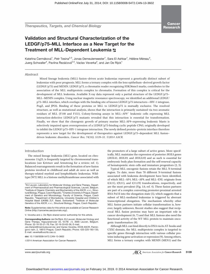
Figure 1. Schematic representation of the domain structure of MLL, MLL fusions, and LEDGF/75. A, schematic representation of the domain structure of WTMLL and N-terminal MLL fusion proteins. MLL contains several domains involved in chromatin binding—AT-Hooks, a cysteine rich domain (CxxC), four planthomeodomains (PHD) fingers, and a bromodomain (BD). TheMLL protein is expressed as a single polypeptide chain cleaved by taspase 1 into two fragments,which associate by noncovalent interactions between the FY-rich domain N-terminal (FYRN) and FY-rich domain C-terminal (FYRC). The suppressor ofvariegation enhancer of zeste and trithorax (SET) domain at the C-terminal end is responsible for the methyltranferase function. The breakpoint regionindicates the location where fusion partners are merged with part of the MLL protein. The N-terminal end of MLL contains two MBM1 and 2 (MBM) and theLBD. The structure of the MLL4–135DD peptide was determined in the published LEDGF/p75–MLL–MENIN ternary complex (PDB ID 3U88). The secondarystructure of resolved areas of MLL is indicated schematically. B, schematic representation of the domain structure of LEDGF/p75. At the N-terminal end,a Pro-Trp-Trp-Pro (PWWP) domain is situated, recognizing H3K36me3marks followed by the first charged region (CR1), the nuclear localization signal (NLS),two AT-Hooks, and two additional charged regions (CR2 and CR3). These domains are involved in chromatin recognition. The IBD, important for theinteraction of LEDGF/p75with theMLL–MENIN complex is situated at the C-terminal part of the protein. The currently known binding partners of LEDGF/p75are listed above the LEDGF/p75 cartoon. Methyl-CpG-binding protein 2 (MeCP2), neuro-oncological ventral antigen 1 (NOVA1), and TOX high mobilitygroup box family member 4 (TOX4) interact with the PWWP domain and HIV-1 integrase, the MLL–MENIN complex, JPO2, Pogo transposable element withZNF domain (PogZ), and Cdc7-activator of S-phase kinase (CDC7/ASK) interact with the IBD.
�Cerm�akov�a et al.
Cancer Res; 74(18) September 15, 2014 Cancer Research5140
on February 14, 2019. © 2014 American Association for Cancer Research. cancerres.aacrjournals.org Downloaded from
Published OnlineFirst July 31, 2014; DOI: 10.1158/0008-5472.CAN-13-3602
In this article, we identify and structurally characterize anovel LEDGF/p75–MLL interface. We show that the LEDGF/p75 IBD interacts with MLL via the same binding site as otherknown LEDGF/p75 binding proteins including HIV-1 inte-grase, PogZ, and JPO2 (16, 17, 26). In addition, we show thatLEDGF/p75 interacting peptides known to inhibit the LEDGF/p75–HIV-1 integrase interaction impair clonogenic growth ofprimary murine MLL fusion–expressing leukemic cells, whichvalidates this interface as a new therapeutic target (27).
Materials and MethodsGeneration of retroviral vectorsMurine stem cell virus–based (pMSCV) retroviral vector
production was performed as described previously (28). Pro-duction of Simian immunodeficiency virus (SIV)–derived vec-tors was performed as described earlier (29). Briefly, vesicularstomatitis virus glycoprotein pseudotyped SIV-based particleswere produced by poly(ethylenimine) transfection in 293T cellsusing the indicated transfer plasmids, the pAd-SIV3þ packag-ing plasmid (a kind gift from Didier N�egre, Ecole NormaleSup�erieure, Lyon, France) and the envelope expression plasmidpMD.G.
Culture and generation of THP1 cells expressingLEDGF/p75THP1 human MLL–AF9þ acute myeloid leukemia (AML)
cells were maintained in Roswell Park Memorial Institutesmedium (RPMI-1640; Gibco-BRL) supplemented with 10%heat-inactivated fetal bovine serum (FBS; Gibco, Invitrogen)and 50 mg/mL gentamycin (Gibco, Invitrogen). To create cellsstably expressing LEDGF/p75 or LEDGF/p75 L368A-K407D,cells were transduced with the respective SIV vectors or anempty control vector and selected with 0.5 mg/mL of hygro-mycin B (Clontech).
Generation and transduction of murine MLL–AF9-expressing AML cellsMurine MLL–AF9-expressing AML cells were generated by
transplantation of bone marrow cells stably transduced toexpress MLL–AF9 using a previously published protocolwith some modifications (30). In brief, bone marrow cellswere harvested from 8-week-old (FVB/Nx129/S1) F1 miceand enriched for progenitors using a lineage-marker-deple-tion progenitors kit (Cell Mag Kit; R&D Systems). These cellswere cultured for 24 hours in RPMI-1640 [10% FCS, 1%penicillin/streptomycin, 10 ng/mL of human interleukin-6(IL6), 6 ng/mL of murine IL3, and 100 ng/mL of murine stemcell factor] before transduction with a MSCV-derived retro-viral vector encoding MLL–AF9 by spinoculation (2,000 � g,90 minutes at 306 K) on two consecutive days. After thesecond spinoculation, 5 � 105 transduced bone marrow cellswere injected into the tail vein of lethally irradiated (950 rad)syngeneic recipients. As described previously and as shownin Supplementary Fig. S2, 100% of the mice developed AMLafter a median latency of 75 days. Leukemic cells wereharvested from the bone marrow of diseased mice andexpanded in medium containing growth factors until the
next round of transduction using the protocol describedabove with pMSCV eGFP-PGK-puro, pMSCV CP65 eGFP-PGK-puro, or its mutant variants. Cells were flow-sorted for eGFPexpression 24 hours after transduction before analysis.
Clonogenic growth assays in vitroMLL–AF9þ-expressing murine leukemic cells (described
above) or normal bone marrow progenitors cells were platedin methylcellulose containing 6 ng/mL IL3, 10 ng/mL IL6, and100 ng/mL murine stem cell factor (M3534; STEMCELL Tech-nologies) in the presence of 2 mg/mL puromycin. Five to 7 dayslater, the number of colonies was determined.
For the clonogenic growth assay with THP1-derived cells,cells were seeded in a 96-well plate (1 � 105 cells/well) andtransduced with a lentiviral vector expressing GFP and amiRNA targeting LEDGF/p75 or a GFP control vector. After24 hours, cells were selected with puromycin (4 mg/mL). Forty-eight hours after transduction, 5 � 103 cells were plated inmethylcellulose medium (M3534; STEMCELL Technologies)in the presence of hygromycin (0.5 mg/mL) and puromycin(4 mg/mL). Remaining cells were expanded in culture for 2 daysfor Western blot analysis. The number of colonies was deter-mined after 10 days.
Reverse transcription quantitative PCRqRT-PCR analysis of the HoxA9 mRNA expression level was
performed as described previously (23). The sequences of allused primer pairs are indicated in Supplementary Table S1.
NMR spectroscopy and structural calculationsAll nuclear magnetic resonance (NMR) data were acquired
at 298 K on a 600 MHz Bruker Avance spectrometer equippedwith triple-resonance (15N/13C/1H) cryoprobe. NMR spectrawere collected from0.35mL samples of 0.1mmol/L 15N-labeledLEDGF/p75 IBD for binding site mapping or 0.5 mmol/Lequimolar solution of 13C/15N-labeled LEDGF/p75 IBD-unla-beled MLL140–160 for resonance assignments and structuraldetermination of the complex in a 25 mmol/L HEPES buffer,pH 7.0, containing 100 mmol/L NaCl, 0.05% 2-mercaptoetha-nol, 5% D2O/95% H2O. A series of double- and triple-resonancespectrawere recorded to obtain essentially complete backboneand side-chain resonance assignments for the MLL140–160-bound 13C/15N-labeled LEDGF/p75 IBD (31, 32). The assign-ments for the bound MLL140–160 peptide were obtained using13C/15N-filtered homonuclear TOCSY andNOESY experiments.1H-1H distance constraints required to calculate the structureof the LEDGF/p75 IBD–MLL140–160 complex were derived from3D 15N/1H NOESY-HSQC, 13C/1H HSQC-NOESY, and in 2D13C/15N-filtered/edited NOESY spectra, which were acquiredwith an NOE mixing time of 120 milliseconds. Specific inter-action of the MLL-derived peptides with the LEDGF/p75IBD was monitored by changes induced in the positions ofsignals of 13C/15N-labeled LEDGF/p75 IBD in 3DHNCO spectra(33, 34).
The family of converged structures for the LEDGF/p75 IBD–MLL140–160 complex was initially calculated using Cyana 2.1, asdescribed previously (33). The combined automated NOEassignment and structure determination protocol (35) was
The LEDGF/p75–MLL Interface as Therapeutic Target for MLL
www.aacrjournals.org Cancer Res; 74(18) September 15, 2014 5141
on February 14, 2019. © 2014 American Association for Cancer Research. cancerres.aacrjournals.org Downloaded from
Published OnlineFirst July 31, 2014; DOI: 10.1158/0008-5472.CAN-13-3602
used to automatically assign the NOE cross-peaks identified in2D NOESY spectra, and to produce preliminary structures. Inaddition, backbone torsion angle constraints, generated fromassigned chemical shifts using the programTALOSþ (36), wereincluded in the calculations. Subsequently, five cycles of sim-ulated annealing combined with redundant dihedral angleconstraints were used to produce the sets of converged struc-tures with no significant restraint violations (distance and van
der Waals violations < 0.2 A�), which were further refined in
explicit solvent in YASARA (http://www.yasara.org/). Analysisand validation of the family of structures obtainedwere carriedout using the programs Molmol, iCING, and PyMol (37)
AlphaScreenAlphaScreen measurements were performed in a final vol-
ume of 25 mL in 384-well Optiwell microtiter plates (PerkinEl-mer). All components were diluted in assay buffer [25 mmol/LTris pH 7.4, 150 mmol/L NaCl, 1 mmol/L dithiothreitol , 0.1%(v/v) Tween-20, 1 mmol/LMgCl2, and 0.1% (w/v) bovine serumalbumin (BSA)]. The optimal binding concentration for eachprotein was determined in cross-titration experiments. Foravidity determinations, MLL1–160–GSTWT (first 160 aa ofMLLN-terminally fused to glutathione-S-transferase) and/or itsmutants were titrated against 0.3 nmol/L Flag-taggedLEDGF/p75 (Flag-LEDGF/p75). Flag-LEDGF/p75 and/or itsmutants were titrated against 10 nmol/L MLL111–160–GST. For50% inhibitory concentration (IC50) determinations, MBP-JPO2, MBP-PogZ, HIV-1 integrase, CP65-MBP constructs, ormaltose binding protein (MBP) were titrated against 1 nmol/LMLL1–160–GST and 0.3 nmol/L Flag-LEDGF/p75. In outcom-petition experiments, Flag-LEDGF/p75 was preincubated withMBP-JPO2,MBP-PogZ, HIV-1 integrase, CP65-MBP constructs,or MBP for 1 hour at 277 K. Subsequently, MLL1–160–GST wasadded at the indicated concentrations. When all proteins wereadded, the plate was incubated for 1 hour at 277 K. Glutathionedonor (20 mg/mL) and anti-Flag acceptor beads (PerkinElmer)were added, bringing all proteins to the indicated final con-centrations. After 1 hour of incubation at 293 K, the plate wasanalyzed in an EnVision Multi-label Reader in AlphaScreenmode (PerkinElmer). Results were analyzed in Prism 5.0(GraphPad software) after nonlinear regression with theappropriate equations: one-site specific binding, taking liganddepletion into account for the apparent Kd measurementsand sigmoidal dose-response with variable slope for the IC50
determination.
Peptide synthesisPeptides were synthesized by solid-phase synthesis in the
Laboratory of Medicinal chemistry, IOCB, ASCR v. v. i. (Prague,Czech Republic).
Statistical analysisMean values and SDs were calculated to estimate the degree
of variation as specified for each experiment. GraphPad Prism5.0 software (Pnsm Software Corp.) was used for statisticalanalysis, unless stated differently.
Additional methods can be found in the SupplementaryData.
ResultsCharacterization of the LEDGF/p75–MLL interface
We first examined whether the described LEDGF/p75–MLL–MENIN interface is sufficient to support the LEDGF/p75–MLL interaction in the absence of MENIN. Throughcoimmunoprecipitation and assessment of the myeloid trans-formation capacity of MLL fusion proteins, Yokoyama andCleary (12) suggested that MLL residue F129 (SupplementaryFig. S1C) is crucial to support MENIN-mediated binding ofLEDGF/p75 to MLL. We therefore introduced this mutationinto a recombinant protein comprising the first MLL1–160–GST, which contains both the LEDGF/p75 and the MBM1and 2 and compared its interaction with LEDGF/p75 to that ofthe WT variant. Integrity of all used recombinant proteinswas confirmed by SDS-PAGE (Supplementary Fig. S3). WTMLL1–160–GST readily interactedwith recombinant triple Flag-LEDGF/p75 in an in vitro AlphaScreen assay (Fig. 2A). Inter-estingly, this interaction was only marginally affected by theF129Amutation. The published crystal structure of the ternarycomplex indicates that next to F129, F133 also contributes tothe interaction with LEDGF/p75 (Supplementary Fig. S1C).Although the F129A, F133A double-mutant decreased theAlphaScreen signal, it failed to completely abolish the inter-action with LEDGF/p75 (Fig. 2A). However, mutational anal-ysis of conserved MLL residues outside the MLL fragmentresolved in the crystal structure revealed that a triple-mutation(E144Q-E146Q-F148A) abolished the interaction with LEDGF/p75 (Fig. 2A). This observation indicates the existence of anadditional LEDGF/p75–MLL interface.
We used NMR spectroscopy for a detailed characterizationof the LEDGF/p75–MLL interface. In particular, we followedthe changes in positions of backbone NMR signals of either 15Nor 13C/15N-labeled LEDGF/p75 IBD (aa 345–426) in the pres-ence of different synthetic MLL-derived peptides. The firstpeptide consisted of MLL aa 111 to 139 (MLL111–139) and thusincluded the a helical region resolved in the published ternarystructure. The second peptide comprised MLL aa 140 to 160(MLL140–160) not included in the resolved interface of theternary complex. A third peptide (aa 123 to 160; MLL123–160)combines residues from MLL140–160 with the amino acidresidues known to contribute to interaction with LEDGF/p75 (MLL F129 and F133). We compared the 2D 15N/1H HSQCspectrum of the 15N-labeled IBD in the absence or presence ofthe unlabeled MLL-derived peptides. MLL111–139 did notinduce significant shifts in positions of the IBD backboneamide signals (Supplementary Fig. S4A). This observation isin agreement with the results of Huang and colleagues (25),who were unable to detect a binary interaction between theIBD and MLL4–135DD in the absence of MENIN. In contrast,both MLL140–160 and MLL123–160 induced significant chemicalshift perturbations of the IBD backbone signals (Fig. 2B andSupplementary Fig. S3B, respectively). The specific shifts of IBDamino acid residues induced by addition of MLL140–160 orMLL123–160 were determined by comparison of the positionsof the IBD backbone signals (HN, N, C') in 3D HNCO spectra(Fig. 2C and Supplementary Fig. S4C, respectively). Overall,the chemical shift perturbations in the IBD backbone inducedby binding of MLL140–160 and MLL123–160 were found in two
�Cerm�akov�a et al.
Cancer Res; 74(18) September 15, 2014 Cancer Research5142
on February 14, 2019. © 2014 American Association for Cancer Research. cancerres.aacrjournals.org Downloaded from
Published OnlineFirst July 31, 2014; DOI: 10.1158/0008-5472.CAN-13-3602
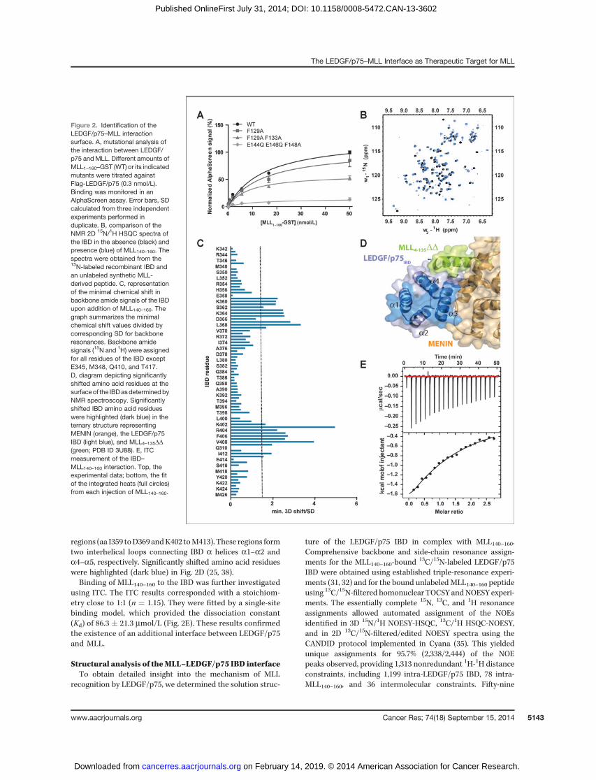
regions (aa I359 toD369 andK402 toM413). These regions formtwo interhelical loops connecting IBD a helices a1–a2 anda4–a5, respectively. Significantly shifted amino acid residueswere highlighted (dark blue) in Fig. 2D (25, 38).Binding of MLL140–160 to the IBD was further investigated
using ITC. The ITC results corresponded with a stoichiom-etry close to 1:1 (n ¼ 1.15). They were fitted by a single-sitebinding model, which provided the dissociation constant(Kd) of 86.3 � 21.3 mmol/L (Fig. 2E). These results confirmedthe existence of an additional interface between LEDGF/p75and MLL.
Structural analysis of theMLL–LEDGF/p75 IBD interfaceTo obtain detailed insight into the mechanism of MLL
recognition by LEDGF/p75, we determined the solution struc-
ture of the LEDGF/p75 IBD in complex with MLL140–160.Comprehensive backbone and side-chain resonance assign-ments for the MLL140–160-bound
13C/15N-labeled LEDGF/p75IBD were obtained using established triple-resonance experi-ments (31, 32) and for the bound unlabeledMLL140–160 peptideusing 13C/15N-filtered homonuclear TOCSY andNOESY experi-ments. The essentially complete 15N, 13C, and 1H resonanceassignments allowed automated assignment of the NOEsidentified in 3D 15N/1H NOESY-HSQC, 13C/1H HSQC-NOESY,and in 2D 13C/15N-filtered/edited NOESY spectra using theCANDID protocol implemented in Cyana (35). This yieldedunique assignments for 95.7% (2,338/2,444) of the NOEpeaks observed, providing 1,313 nonredundant 1H-1H distanceconstraints, including 1,199 intra-LEDGF/p75 IBD, 78 intra-MLL140–160, and 36 intermolecular constraints. Fifty-nine
Figure 2. Identification of theLEDGF/p75–MLL interactionsurface. A, mutational analysis ofthe interaction between LEDGF/p75 and MLL. Different amounts ofMLL1–160–GST (WT) or its indicatedmutants were titrated againstFlag-LEDGF/p75 (0.3 nmol/L).Binding was monitored in anAlphaScreen assay. Error bars, SDcalculated from three independentexperiments performed induplicate. B, comparison of theNMR 2D 15N/1H HSQC spectra ofthe IBD in the absence (black) andpresence (blue) of MLL140–160. Thespectra were obtained from the15N-labeled recombinant IBD andan unlabeled synthetic MLL-derived peptide. C, representationof the minimal chemical shift inbackbone amide signals of the IBDupon addition of MLL140–160. Thegraph summarizes the minimalchemical shift values divided bycorresponding SD for backboneresonances. Backbone amidesignals (15N and 1H) were assignedfor all residues of the IBD exceptE345, M348, Q410, and T417.D, diagram depicting significantlyshifted amino acid residues at thesurfaceof the IBDasdeterminedbyNMR spectroscopy. Significantlyshifted IBD amino acid residueswere highlighted (dark blue) in theternary structure representingMENIN (orange), the LEDGF/p75IBD (light blue), and MLL4–135DD(green; PDB ID 3U88). E, ITCmeasurement of the IBD–MLL140–160 interaction. Top, theexperimental data; bottom, the fitof the integrated heats (full circles)from each injection of MLL140–160.
The LEDGF/p75–MLL Interface as Therapeutic Target for MLL
www.aacrjournals.org Cancer Res; 74(18) September 15, 2014 5143
on February 14, 2019. © 2014 American Association for Cancer Research. cancerres.aacrjournals.org Downloaded from
Published OnlineFirst July 31, 2014; DOI: 10.1158/0008-5472.CAN-13-3602
converged structures for the LEDGF/p75 IBD–MLL140–160complex with no distance violations >0.2 A
�were obtained
from 100 random starting conformations using 1,451 NMR-derived structural constraints (�14.8 constraints per resi-due). The 20 lowest energy conformers were further refinedin explicit solvent in YASARA (39). Structural statistics forthe final water-refined set of structures are shown in Sup-plementary Table S2. The structures, NMR constraints andresonance assignments have been deposited in the ProteinData Bank (PDB, accession number 2msr) and BMRB data-base (accession number 25130).
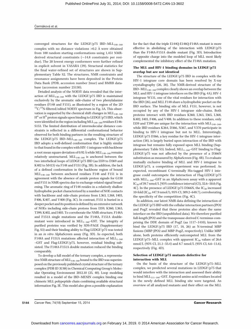
Detailed analysis of the NOESY data revealed that the inter-action of MLL140–160 with the LEDGF/p75 IBD is maintainedexclusively by the aromatic side-chains of two phenylalanineresidues (F148 and F151), as illustrated by a region of the 2D13C/15N-filtered/edited NOESY spectrum in Fig. 3A. This obser-vation is supported by the chemical shift changes in MLL140–160Ha orHNproton signals uponbinding to LEDGF/p75 IBD,whichwere identified in the region includingMLL140–160 residuesE146-S153. The limited distribution of intermolecular distance con-straints is reflected in a differential conformational behaviorobserved for both binding partners in the resulting structure ofthe LEDGF/p75 IBD–MLL140–160 complex. The LEDGF/p75IBD adopts a well-defined conformation that is highly similarto that found in thecomplexwithHIV-1 integrasewithbackbone
a rootmean square deviation of 0.92 A�whileMLL140–160 remains
relatively unstructured. MLL140–160 is anchored between thetwo interhelical loops of LEDGF/p75 IBD (aa I359 to D369 andK402 to M413) via F148 and F151 (Fig. 3B). In addition, the lackof a defined conformation in the backbone region of boundMLL140–160 between anchored residues F148 and F151 is inagreement with the absence of amide proton signals for G150and F151 in NMR spectra due to exchange-related signal broad-ening. The aromatic ring of F148 resides in a relatively shallowhydrophobic pocket characterized by a number ofNOE contactswith backbone and side-chain protons from L363, L368, I403,F406, K407, and V408 (Fig. 3C). In contrast, F151 is buried in adeeperpocketand itsposition isdefinedbyanextensivenetworkof NOEs including side-chain protons from I359, K360, L363,T399, K402, and I403. To corroborate the NMR structure, F148Aand F151A single mutations and the F148A, F151A double-mutant were introduced in MLL1–160–GST. The integrity ofpurified proteins was verified by SDS-PAGE (SupplementaryFig. S3) and their binding ability to Flag-LEDGF/p75 was testedin an in vitro AlphaScreen assay (Fig. 3D). As expected, bothF148A and F151A mutations affected interaction of MLL1–160–GST and Flag-LEDGF/p75, however, residual binding sub-sisted. The F148A-F151A double mutation reduced the bindingcomparably.
To develop a full model of the ternary complex, a representa-tiveNMR structure ofMLL140–160 bound to the IBDwas superim-posedon the previously published crystal structure of the ternarycomplex (PDB ID 3U88) in Chemical Computing Group's Molec-ular Operating Environment 2012.10 (25, 40). Loop modelingresulted in a model of the IBD–MENIN complex binding onechimeric MLL polypeptide chain combining available structuralinformation Fig. 3E. This model also gives a possible explanation
for the fact that the triple E144Q-E146Q-F148A mutant is moreeffective in abolishing of the interaction with LEDGF/p75than the F148A-F151A double mutant (Fig. 3D). Introductionof opposite charge into the modeled loop of MLL most likelycomplemented the inhibitory effect of the F148A mutation.
The MLL and HIV-1 binding domains in LEDGF/p75overlap but are not identical
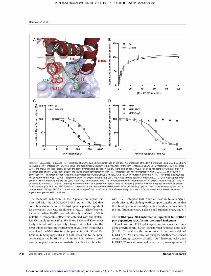
The structure of the LEDGF/p75 IBD in complex with theHIV-1 integrase core domain has been resolved by X-raycrystallography (26, 38). The NMR-derived structure of theIBD–MLL140–160 complex clearly shows an overlap between theMLL and HIV-1 integrase interfaces on the IBD (Fig. 4A). HIV-1integrase W131, one of the vital residues for interaction withthe IBD (26), andMLL F148 share a hydrophobic pocket on theIBD surface. The binding site of MLL F151, however, is notoccupied by any of the HIV-1 integrase side-chains. Bothproteins interact with IBD residues K360, L363, I365, L368,K402, I403, F406, and V408. In addition to these residues, onlyI359 and T399 are unique for the interaction with MLL140–160,while IBD residues K364, D366, N367, and V370 participate inbinding to HIV-1 integrase but not to MLL. Interestingly,LEDGF/p75 D366, a key residue for the HIV-1 integrase inter-action (38), is largely buried upon interaction of the IBD withintegrase but remains fully exposed upon MLL binding (Sup-plementary Table S3). Indeed, MLL1–160–GST binding to Flag-LEDGF/p75 was not affected by the presence of a D366Nsubstitution asmeasured by AlphaScreen (Fig. 4B). To evaluatemutually exclusive binding of MLL and HIV-1 integrase toLEDGF/p75, a competition experiment was performed. Asexpected, recombinant C-terminally His-tagged HIV-1 inte-grase could outcompete the interaction of Flag-LEDGF/p75with MLL1–160–GST in an AlphaScreen assay with a apparentIC50 of 39.7 nmol/L [95%confidence interval (CI), 32.6–50.0; Fig.4C]. In the presence of LEDGF/p75 D366N, the IC50 increased10-fold (IC50, 447.9 nmol/L; 95%CI, 309.2–648.7), corroboratingthe specificity of the competition (Fig. 4C).
In addition, our latest NMR data defining the interaction ofthe LEDGF/p75 IBDwith the cellular interaction partners JPO2and PogZ revealed that these proteins also share the sameinterface on the IBD (unpublished data). We therefore purifiedfull-length JPO2 and the transposase-derived C-terminus com-prising the DDE domain of PogZ (aa 1117–1410), known tobind the LEDGF/p75 IBD (17, 18, 26) as N-terminal MBPfusions (MBP-JPO2 and MBP-PogZ, respectively). Unlike MBPalone, both proteins efficiently outcompeted MLL from theLEDGF/p75–MLL complex with apparent IC50 values of 26.6nmol/L (95% CI, 21.1–33.4) and 8.7 nmol/L (95% CI, 6.6–11.6),respectively (Fig. 4D).
Selection of LEDGF/p75 mutants defective forinteraction with MLL
On the basis of the structure of the LEDGF/p75–MLLcomplex, we predicted several mutations in LEDGF/p75 thatwould interfere with the interaction and assessed their abilityto bind MLL111–160–GST. Exposed amino acid residues locatedin the newly defined MLL binding site were targeted. Anoverview of all analyzed mutants and their effect on the MLL
�Cerm�akov�a et al.
Cancer Res; 74(18) September 15, 2014 Cancer Research5144
on February 14, 2019. © 2014 American Association for Cancer Research. cancerres.aacrjournals.org Downloaded from
Published OnlineFirst July 31, 2014; DOI: 10.1158/0008-5472.CAN-13-3602
interaction is presented in Supplementary Tables S4 and S5.The integrity of purifiedWT Flag-LEDGF/p75 and interaction-defective mutants was verified by SDS-PAGE (SupplementaryFig. S3). Folding of these interaction-defective mutants was
additionally analyzed by differential scanning fluorimetry(DSF; Supplementary Fig. S5 and Supplementary Table S6).Themelting curves of all Flag-LEDGF/p75mutants used in thisstudy were comparable with that of WT Flag-LEDGF/p75.
Figure 3. Model of the ternary MLL–MENIN-IBD structure. A, regionfrom the 2D 13C/15N-filtered/editedNOESY spectrum obtained for theLEDGF/p75 IBD–MLL140–160complex. The spectrum showsintermolecular NOE contactsbetween protons from 13C/15N-labeled LEDGF/p75 IBD (y-axis)and unlabeled MLL140–160 complex(x-axis). The spectral analysisreveals that the interactionbetween the molecules isexclusively maintained by the twoaromatic side-chains of MLL F148and F151. B, converged structuresdetermined by NMR (PDB ID 2msr).The IBD is depicted in blue, MLL ingreen. MLL F148 and F151 arehighlighted in red. The structuresare overlaid on the LEDGF/p75 IBDsecondary structure elements. C,detailed view of the MLL140–160–LEDGF/p75 IBD interface. Thediagrams show a representativeNMR structure of MLL140–160(green) in complex with the LEDGF/p75 IBD (blue).MLL F148binds intoa hydrophobic pocket on the IBDsurface formed by L363, L368,I403, F406, K407, and V408 andMLL F151 is buried in a pocketformed by I359, K360, L363, T399,K402, and I403. D, the F148A andF151mutations affect LEDGF/p75–MLL interaction. Recombinant WT,F148A, or F151A single mutants ora F148A-F151A double mutant ofMLLwere titrated against 0.3 nmol/L LEDGF/p75 in an AlphaScreenexperiment. The E144Q-E146Q-F148A mutant was analyzed inparallel to enable the comparisonwith Fig. 2A. Error bars, SDscalculated from three independentexperiments performed induplicate. E, model of the MLL–MENIN–LEDGF/p75 ternarycomplex. The published crystalstructure of the ternary complex(PDB ID 3U88) and a representativeNMR structure were used as atemplate. MENIN is depicted inorange and the LEDGF/p75 IBD inblue. The MLL4–135DD peptidederived from the crystal structure isdepicted in lime green. The part ofMLL derived from the NMRmeasurements (MLLNMR) is shownin dark green and themodeled loop(MLLMod) in pale green. A detail ofthe interface is presented inSupplementary Fig. S6.
The LEDGF/p75–MLL Interface as Therapeutic Target for MLL
www.aacrjournals.org Cancer Res; 74(18) September 15, 2014 5145
on February 14, 2019. © 2014 American Association for Cancer Research. cancerres.aacrjournals.org Downloaded from
Published OnlineFirst July 31, 2014; DOI: 10.1158/0008-5472.CAN-13-3602
A moderate reduction in the AlphaScreen signal wasobserved with the LEDGF/p75 L368A mutant (Fig. 5A) thatcontributes to formation of the hydrophobic pocket importantfor interaction withMLL residue F148 (Fig. 3C). This effect wasincreased when K407D was additionally mutated (L368A-K407D). A comparable effect was obtained with the R404D-R405D double mutant (Fig. 5B). R404, R405, and K407 mostlikely interact with negatively charged side-chains in theflexible loop connecting the fragment ofMLL from the resolvedcrystal and the NMR structure (Supplementary Fig. S6; ref. 25).Residual binding may subsist in both cases due to the inter-action supported by MLL F151, F129, and F133. We also testeda subset of pointmutants known to be defective for interaction
with HIV-1 integrase (38). None of these mutations signifi-cantly affected the binding to MLL, supporting the notion thattheir binding domains overlap but involve different residues ofthe IBD (Supplementary Table S5 and Supplementary Fig. S7).
The LEDGF/p75–MLL interface is important for LEDGF/p75-dependent MLL fusion–mediated leukemias
Knockdown of LEDGF/p75 expression impaires the clono-genic growth of MLL fusion–transformed hematopoietic cells(12, 23). To evaluate the importance of the newly definedLEDGF/p75–MLL interface, we analyzed whether the reducedcolony-forming capacity of MLL–AF9þ leukemic cells uponLEDGF/p75 knockdown could be rescued by overexpression of
Figure 4. MLL, Jpo2, PogZ, and HIV-1 integrase share the same binding interface on the IBD. A, comparison of the HIV-1 integrase-, and MLL–LEDGF/p75interaction. HIV-1 integrase W131, I161, R166, and Q168 (red) are known to be important for the HIV-1 integrase–LEDGF/p75 interaction. HIV-1 integraseW131 and MLL F148 (dark green) occupy the same hydrophobic pocket on the IBD (light blue) surface. MLL F151 does not compete with any of HIV-1integrase side chains. D366 (dark blue) of the IBD is crucial for interaction with HIV-1 integrase, but not for interaction with MLL140–160. The structureof the IBD–HIV-1 integrase interfacewas previously described (PDB ID 2B4J). B, the LEDGF/p75D366Nmutation, defective for HIV-1 integrase binding, doesnot affect binding of MLL1–160–GST. Recombinant WT or D366N mutant Flag-LEDGF/p75 was titrated against 1 nmol/L MLL1–160–GST in an AlphaScreenassay. C, HIV-1 integrase impairs the LEDGF/p75–MLL interaction in vitro. The interaction between recombinant WT or D366N mutant Flag-LEDGF/p75(0.3 nmol/L) and MLL1–160–GST (1 nmol/L) was monitored in an AlphaScreen assay, while an increasing amount of HIV-1 integrase (HIV-IN) was added.D, Jpo2 and PogZ inhibit the LEDGF/p75–MLL interaction in vitro. Recombinant MBP,MBP-JPO2, orMBP-PogZ (aa 1117–1410) were titrated against a fixedconcentration of Flag-LEDGF (0.3 nmol/L) and MLL1–160–GST (1 nmol/L) in an AlphaScreen assay. Error bars, SDs calculated from three independentexperiments performed in duplicate.
�Cerm�akov�a et al.
Cancer Res; 74(18) September 15, 2014 Cancer Research5146
on February 14, 2019. © 2014 American Association for Cancer Research. cancerres.aacrjournals.org Downloaded from
Published OnlineFirst July 31, 2014; DOI: 10.1158/0008-5472.CAN-13-3602
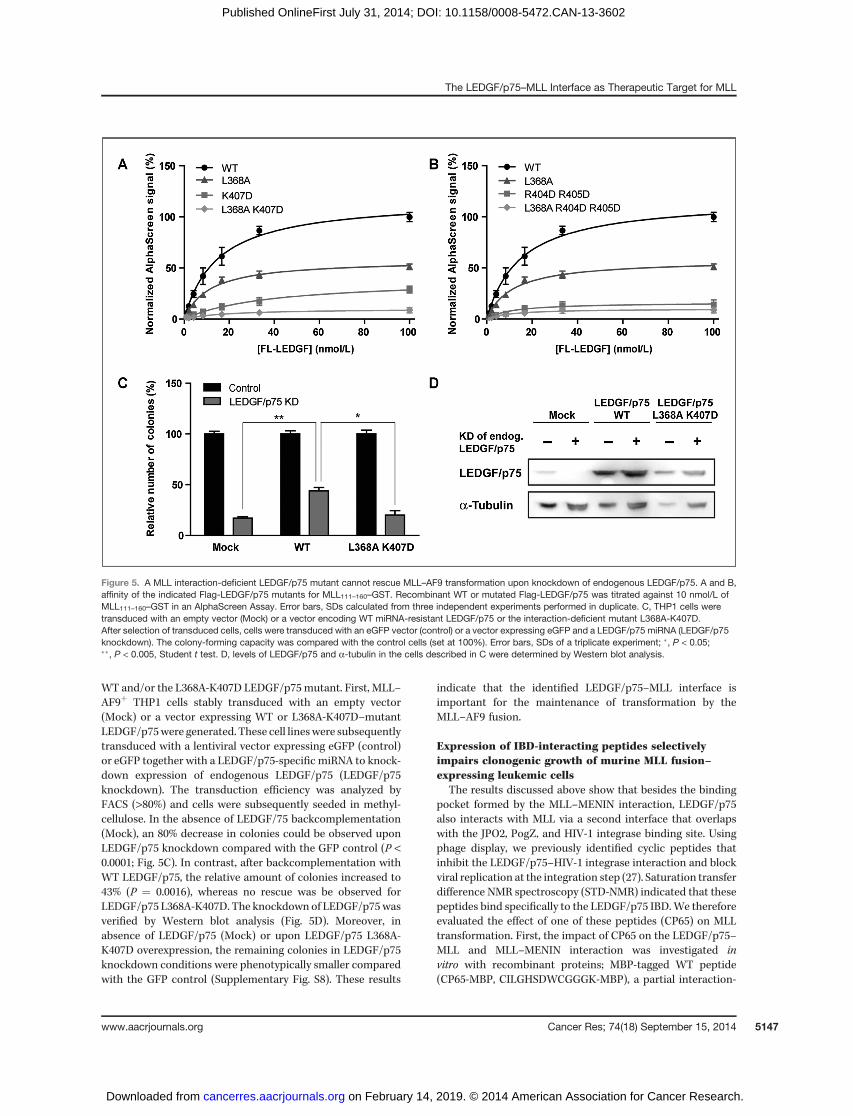
WT and/or the L368A-K407D LEDGF/p75mutant. First, MLL–AF9þ THP1 cells stably transduced with an empty vector(Mock) or a vector expressing WT or L368A-K407D–mutantLEDGF/p75were generated. These cell lineswere subsequentlytransduced with a lentiviral vector expressing eGFP (control)or eGFP together with a LEDGF/p75-specific miRNA to knock-down expression of endogenous LEDGF/p75 (LEDGF/p75knockdown). The transduction efficiency was analyzed byFACS (>80%) and cells were subsequently seeded in methyl-cellulose. In the absence of LEDGF/75 backcomplementation(Mock), an 80% decrease in colonies could be observed uponLEDGF/p75 knockdown compared with the GFP control (P <0.0001; Fig. 5C). In contrast, after backcomplementation withWT LEDGF/p75, the relative amount of colonies increased to43% (P ¼ 0.0016), whereas no rescue was be observed forLEDGF/p75 L368A-K407D. The knockdown of LEDGF/p75wasverified by Western blot analysis (Fig. 5D). Moreover, inabsence of LEDGF/p75 (Mock) or upon LEDGF/p75 L368A-K407D overexpression, the remaining colonies in LEDGF/p75knockdown conditions were phenotypically smaller comparedwith the GFP control (Supplementary Fig. S8). These results
indicate that the identified LEDGF/p75–MLL interface isimportant for the maintenance of transformation by theMLL–AF9 fusion.
Expression of IBD-interacting peptides selectivelyimpairs clonogenic growth of murine MLL fusion–expressing leukemic cells
The results discussed above show that besides the bindingpocket formed by the MLL–MENIN interaction, LEDGF/p75also interacts with MLL via a second interface that overlapswith the JPO2, PogZ, and HIV-1 integrase binding site. Usingphage display, we previously identified cyclic peptides thatinhibit the LEDGF/p75–HIV-1 integrase interaction and blockviral replication at the integration step (27). Saturation transferdifference NMR spectroscopy (STD-NMR) indicated that thesepeptides bind specifically to the LEDGF/p75 IBD.We thereforeevaluated the effect of one of these peptides (CP65) on MLLtransformation. First, the impact of CP65 on the LEDGF/p75–MLL and MLL–MENIN interaction was investigated invitro with recombinant proteins; MBP-tagged WT peptide(CP65-MBP, CILGHSDWCGGGK-MBP), a partial interaction-
Figure 5. A MLL interaction-deficient LEDGF/p75 mutant cannot rescue MLL–AF9 transformation upon knockdown of endogenous LEDGF/p75. A and B,affinity of the indicated Flag-LEDGF/p75 mutants for MLL111–160–GST. Recombinant WT or mutated Flag-LEDGF/p75 was titrated against 10 nmol/L ofMLL111–160–GST in an AlphaScreen Assay. Error bars, SDs calculated from three independent experiments performed in duplicate. C, THP1 cells weretransduced with an empty vector (Mock) or a vector encoding WT miRNA-resistant LEDGF/p75 or the interaction-deficient mutant L368A-K407D.After selection of transduced cells, cells were transduced with an eGFP vector (control) or a vector expressing eGFP and a LEDGF/p75 miRNA (LEDGF/p75knockdown). The colony-forming capacity was compared with the control cells (set at 100%). Error bars, SDs of a triplicate experiment; �, P < 0.05;��, P < 0.005, Student t test. D, levels of LEDGF/p75 and a-tubulin in the cells described in C were determined by Western blot analysis.
The LEDGF/p75–MLL Interface as Therapeutic Target for MLL
www.aacrjournals.org Cancer Res; 74(18) September 15, 2014 5147
on February 14, 2019. © 2014 American Association for Cancer Research. cancerres.aacrjournals.org Downloaded from
Published OnlineFirst July 31, 2014; DOI: 10.1158/0008-5472.CAN-13-3602
defective mutant (CP65mut-MBP, CILGHSDACGGGK-MBP),and a scrambled peptide (CP65scr-MBP, GCGLSCGWKGIHD-MBP) were produced and validated in an AlphaScreenassay. As shown in Supplementary Fig. S9A, WT CP65-MBPdisplaced Flag-LEDGF/p75 from MLL1–160–GST with approx-imately 6-fold lower apparent IC50 than CP65mut-MBP, whilethe CP65scr-MBP did not affect LEDGF/p75–MLL complexformation. As expected, none of these tagged peptides inter-fered with the interaction of MLL1–160–GST and His–TRX–tagged MENIN (Supplementary Fig. S9B).
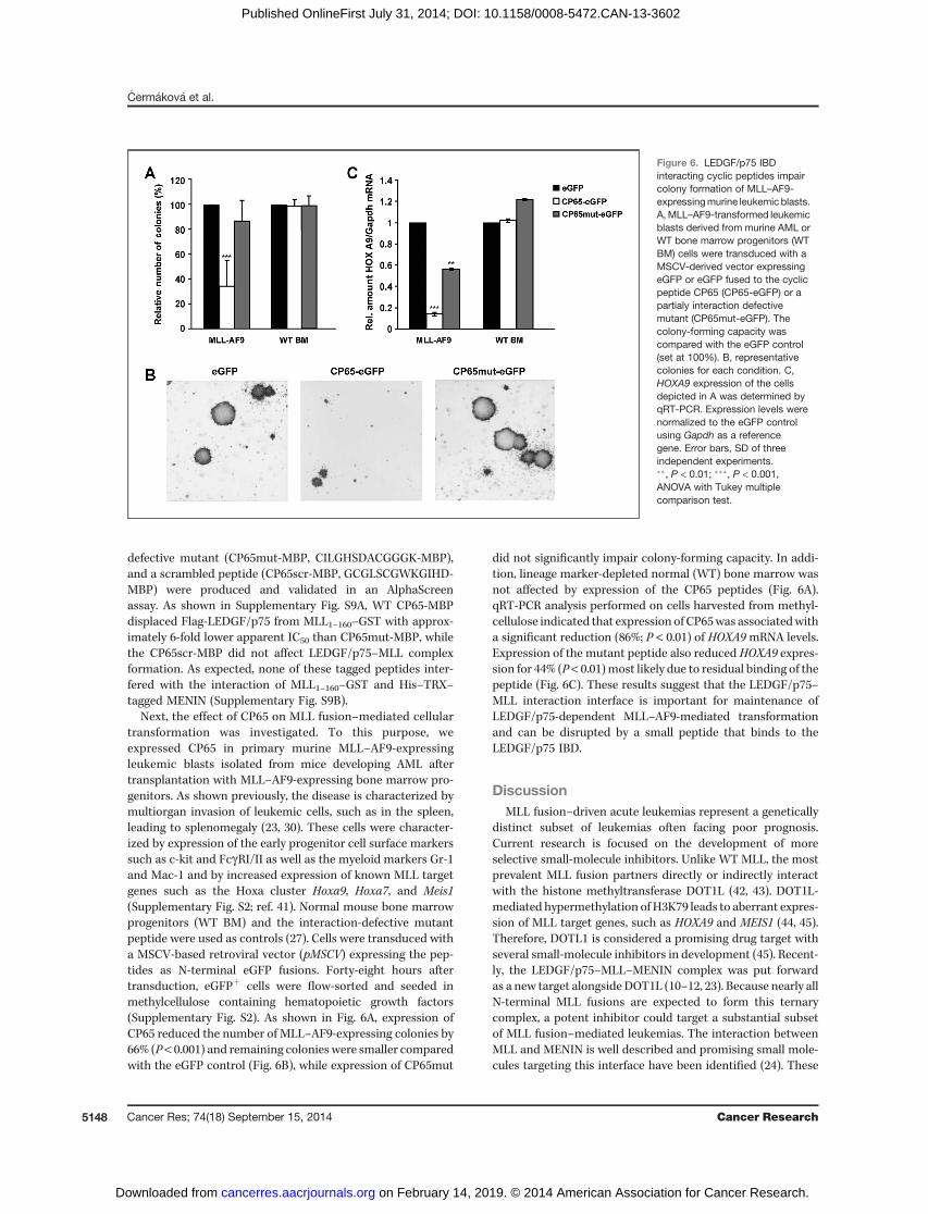
Next, the effect of CP65 on MLL fusion–mediated cellulartransformation was investigated. To this purpose, weexpressed CP65 in primary murine MLL–AF9-expressingleukemic blasts isolated from mice developing AML aftertransplantation with MLL–AF9-expressing bone marrow pro-genitors. As shown previously, the disease is characterized bymultiorgan invasion of leukemic cells, such as in the spleen,leading to splenomegaly (23, 30). These cells were character-ized by expression of the early progenitor cell surface markerssuch as c-kit and FcgRI/II as well as the myeloid markers Gr-1and Mac-1 and by increased expression of known MLL targetgenes such as the Hoxa cluster Hoxa9, Hoxa7, and Meis1(Supplementary Fig. S2; ref. 41). Normal mouse bone marrowprogenitors (WT BM) and the interaction-defective mutantpeptide were used as controls (27). Cells were transduced witha MSCV-based retroviral vector (pMSCV) expressing the pep-tides as N-terminal eGFP fusions. Forty-eight hours aftertransduction, eGFPþ cells were flow-sorted and seeded inmethylcellulose containing hematopoietic growth factors(Supplementary Fig. S2). As shown in Fig. 6A, expression ofCP65 reduced the number of MLL–AF9-expressing colonies by66% (P < 0.001) and remaining colonies were smaller comparedwith the eGFP control (Fig. 6B), while expression of CP65mut
did not significantly impair colony-forming capacity. In addi-tion, lineage marker-depleted normal (WT) bone marrow wasnot affected by expression of the CP65 peptides (Fig. 6A).qRT-PCR analysis performed on cells harvested from methyl-cellulose indicated that expression of CP65was associatedwitha significant reduction (86%; P < 0.01) of HOXA9mRNA levels.Expression of the mutant peptide also reduced HOXA9 expres-sion for 44% (P < 0.01)most likely due to residual binding of thepeptide (Fig. 6C). These results suggest that the LEDGF/p75–MLL interaction interface is important for maintenance ofLEDGF/p75-dependent MLL–AF9-mediated transformationand can be disrupted by a small peptide that binds to theLEDGF/p75 IBD.
DiscussionMLL fusion–driven acute leukemias represent a genetically
distinct subset of leukemias often facing poor prognosis.Current research is focused on the development of moreselective small-molecule inhibitors. Unlike WT MLL, the mostprevalent MLL fusion partners directly or indirectly interactwith the histone methyltransferase DOT1L (42, 43). DOT1L-mediated hypermethylation of H3K79 leads to aberrant expres-sion of MLL target genes, such as HOXA9 and MEIS1 (44, 45).Therefore, DOTL1 is considered a promising drug target withseveral small-molecule inhibitors in development (45). Recent-ly, the LEDGF/p75–MLL–MENIN complex was put forwardas a new target alongside DOT1L (10–12, 23). Because nearly allN-terminal MLL fusions are expected to form this ternarycomplex, a potent inhibitor could target a substantial subsetof MLL fusion–mediated leukemias. The interaction betweenMLL and MENIN is well described and promising small mole-cules targeting this interface have been identified (24). These
Figure 6. LEDGF/p75 IBDinteracting cyclic peptides impaircolony formation of MLL–AF9-expressingmurine leukemic blasts.A,MLL–AF9-transformed leukemicblasts derived from murine AML orWT bone marrow progenitors (WTBM) cells were transduced with aMSCV-derived vector expressingeGFP or eGFP fused to the cyclicpeptide CP65 (CP65-eGFP) or apartialy interaction defectivemutant (CP65mut-eGFP). Thecolony-forming capacity wascompared with the eGFP control(set at 100%). B, representativecolonies for each condition. C,HOXA9 expression of the cellsdepicted in A was determined byqRT-PCR. Expression levels werenormalized to the eGFP controlusing Gapdh as a referencegene. Error bars, SD of threeindependent experiments.��, P < 0.01; ���, P < 0.001,ANOVA with Tukey multiplecomparison test.
�Cerm�akov�a et al.
Cancer Res; 74(18) September 15, 2014 Cancer Research5148
on February 14, 2019. © 2014 American Association for Cancer Research. cancerres.aacrjournals.org Downloaded from
Published OnlineFirst July 31, 2014; DOI: 10.1158/0008-5472.CAN-13-3602
compounds bind to the central hydrophobic pocket of MENINand thereby destabilize the LEDGF/p75–MLL–MENIN ternarycomplex. However, it has not been shown whether thesecompounds can be used without inducing cytotoxic effects.In this regard, MENIN functions as a tumor suppressor inendocrine tissues. Mutations in MENIN can induce multipleendocrine neoplasia type I (MEN1) and some clinical muta-tions have been shown to abrogate the interaction withLEDGF/p75 (12). The tumor suppression function of MENINis related to its interactionwith JUND (46). Structural data haveindicated that the interaction of JUND with MENIN is highlysimilar to the MENIN–MLL interaction (25).Recently, we showed that MLL fusion–mediated transfor-
mation can be inhibited by outcompeting LEDGF/p75 from thecomplex without blocking the MLL–MENIN interaction (23).This study also demonstrated that the LEDGF/p75–MLL inter-face represents a potential target to treat MLL-associatedleukemias. Although only MLL–AF9 and MLL–ENL leukemiashave been tested for their LEDGF/p75 dependency, we expectthat like for MENIN inhibitors, all oncogenic MLL fusionsretaining the MLL N-terminus are potential targets (12, 23).In addition, it was recently shown that MLL fusion leukemiaalso requires the presence of the WT MLL protein offering anadditional target for LEDGF/p75–MLL inhibitors (8).In this report, we show that MLL interacts with LEDGF/
p75 via two distinct interfaces. The first interface is formedby the LEDGF/p75–MENIN complex as revealed in thepreviously published crystal structure (25). The interfacedescribed in this article overlaps with those established forinteraction of HIV-1 integrase, JPO2, and PogZ interactionwith the LEDGF/p75 IBD (unpublished data; refs. 16, 17).Structural NMR studies, including detailed characterizationof the interaction between LEDGF/p75 and MLL-derivedpeptides, revealed that the second interface is maintained bytwo aromatic side-chains of MLL (Fig. 3). MLL F148 occupiesa hydrophobic pocket on the IBD surface formed by L363,L368, I403, F406, K407, and V408 while MLL F151 is buried ina pocket formed by I359, K360, L363, T399, K402, and I403.Although the LEDGF/p75 IBD–MLL interface overlapswith that of HIV-1 integrase, the engaged IBD residues arenot identical (Supplementary Fig. S7 and SupplementaryTable S5).The LEDGF/p75–HIV-1 integrase interaction is considered a
novel target to treat HIV-1 infection. Small molecules disrupt-ing this interaction (LEDGINs) bind HIV-1 integrase and avoidcellular toxicity (47). Recently, we reported that cyclic peptidesand small molecules derived thereof that bind to the IBD,inhibit the interaction with HIV-1 integrase and viral replica-tion (27, 48). Taking into account that the LEDGF/p75–MLLand LEDGF/p75–HIV-1 integrase interfaces overlap, we eval-uated one of these peptides (CP65) as a potential inhibitor ofthe LEDGF/p75–MLL interaction. This peptide specificallyblocked the LEDGF/p75–MLL interaction and affected MLLfusion–dependent leukemic transformation, supporting theamenability of the LEDGF/p75–MLL interface for therapeutictargeting (Fig. 6). However, at this point, it is not possible todetermine whether CP65-mediated inhibition is exerted on theMLL fusion, WT MLL, or both of them.
Although earlier cellular experiments suggested that theLEDGF/p75–MLL interaction is dependent on the presenceof MENIN (12, 23), our in vitro data suggest that LEDGF/p75and MLL can interact in the absence of MENIN. Addition ofMENIN in the LEDGF/p75–MLL in vitro interaction assayinduces a 4-fold stimulation of the interaction (data notshown). It is still unclear how MENIN regulates the LEDGF/p75–MLL interaction and how the presence of MENINinfluences MLL targeting. In this regard, a recent study byArtinger and colleagues (49) revealed that expression ofsome MLL-regulated genes is not dependent on MENIN,which might reflect the presence of a LEDGF/p75–MENINindependent chromatin tethering mechanism. The possibil-ity that not all MLL target genes depend on the presence ofLEDGF/p75 has important implications when consideringthe LEDGF/p75–MLL interaction as a new therapeutic tar-get. Indeed, the function of the LEDGF/p75–MLL–MENINcomplex has been studied in the context of MLL-mediatedleukemic transformation but not in normal blood cell dif-ferentiation. The majority of LEDGF/p75 knockout mice dieprenatally and display a phenotype reminiscent of a Hoxagene knockout (22, 50). These results suggest that LEDGF/p75 is important for the function of MLL and to maintainHoxa gene expression during embryogenesis. In light offuture drug development, it will be of interest to evaluatethe role of a LEDGF/p75 for normal hematopoiesis.
Despite a potential role in blood cell differentiation, LEDGF/p75-dependent MLL-regulated genes are critical to maintainleukemic transformation (12, 23). In addition to the MLL–MENIN interaction, our results suggest that the LEDGF/p75–MLL interaction should also be considered as a target to treatLEDGF/p75-dependent MLL leukemia. The finding that theLEDGF/p75–MLL interface overlaps with the LEDGF/p75–HIV integrase interface implies that we can exploit knowledgeon the LEDGF/p75–HIV integrase interaction and drug devel-opment giving us a head start in the development of LEDGF/p75–MLL inhibitors.
Disclosure of Potential Conflicts of InterestNo potential conflicts of interest were disclosed.
Authors' ContributionsConception and design: K. �Cerm�akov�a, S. El Ashkar, J. Schwaller, V. Veverka,J. De RijckDevelopment of methodology: K. �Cerm�akov�a, S. El Ashkar, J. De RijckAcquisition of data (provided animals, acquired and managed patients,provided facilities, etc.): P. Tesina, H. M�ereau, J. Schwaller, V. Veverka, J. DeRijckAnalysis and interpretation of data (e.g., statistical analysis, biostatistics,computational analysis): K. �Cerm�akov�a, P. Tesina, J. Demeulemeester,H. M�ereau, J. Schwaller, V. Veverka, J. De RijckWriting, review, and/or revision of the manuscript: K. �Cerm�akov�a,P. Tesina, J. Demeulemeester, S. El Ashkar, H. M�ereau, J. Schwaller, P. �Rez�a�cov�a,V. Veverka, J. De RijckAdministrative, technical, or material support (i.e., reporting or orga-nizing data, constructing databases): K. �Cerm�akov�a, P. Tesina, J. De RijckStudy supervision: P. �Rez�a�cov�a, J. De Rijck
AcknowledgmentsThe authors thank Milan Kozisek (IOCB AS CR) for his help with collection,
processing, and analysis of ITC data, Martine Michiels (KU Leuven) for theexcellent technical assistance, and the Leuven Viral Vector Core for the vectorproduction.
The LEDGF/p75–MLL Interface as Therapeutic Target for MLL
www.aacrjournals.org Cancer Res; 74(18) September 15, 2014 5149
on February 14, 2019. © 2014 American Association for Cancer Research. cancerres.aacrjournals.org Downloaded from
Published OnlineFirst July 31, 2014; DOI: 10.1158/0008-5472.CAN-13-3602
Grant SupportThis work was supported by the Research Foundation Flanders (FWO;
G.0595.13 andKAN2012 1.5.108.12), the Flemish agency for Innovation by Scienceand Technology (IWT) SBO Interactomics (QPG-345523), the KU Leuvenresearch fund (IDO/12/2008), the Ministry of Education of the Czech Republic[LK11205 (programme "NAVRAT"), 7E08066, and Project InterBioMed LO1302],projects (RVO 61388963) and (68378050) awarded by the Academy of Sciences ofthe Czech Republic and Grant Agency of the Charles University in Prague (1400–243–253355), the Swiss
National Science Foundation (SNF-31003A-130661/1), the Swiss Cancer League(KFS-2778–02–2011), and the Gertrude von Meissner Foundation (Basel, Swit-zerland). K. �Cerm�akov�a and J. Demeulemeester are supported by the FWO.
The costs of publication of this article were defrayed in part by the payment ofpage charges. This article must therefore be hereby marked advertisement inaccordance with 18 U.S.C. Section 1734 solely to indicate this fact.
Received December 20, 2013; revised May 27, 2014; accepted June 10, 2014;published OnlineFirst July 23, 2014.
References1. Krivtsov AV, Armstrong SA. MLL translocations, histonemodifications
and leukaemia stem-cell development. Nat Rev Cancer 2007;7:823–33.
2. Jude CD, Climer L, Xu D, Artinger E, Fisher JK, Ernst P. Unique andindependent roles for MLL in adult hematopoietic stem cells andprogenitors. Cell Stem Cell 2007;1:324–37.
3. McMahonKA,HiewSY,Hadjur S, Veiga-FernandesH,Menzel U, PriceAJ, et al. Mll has a critical role in fetal and adult hematopoietic stem cellself-renewal. Cell Stem Cell 2007;1:338–45.
4. Meyer C, Hofmann J, Burmeister T, Groger D, Park TS, EmerencianoM, et al. TheMLL recombinome of acute leukemias in 2013. Leukemia2013;27:2165–76.
5. Lin C, Smith ER, Takahashi H, Lai KC, Martin-Brown S, Florens L, et al.AFF4, a component of the ELL/P-TEFb elongation complex and ashared subunit of MLL chimeras, can link transcription elongation toleukemia. Mol Cell 2010;37:429–37.
6. Bursen A, Schwabe K, Ruster B, Henschler R, Ruthardt M, Dinger-mann T, et al. The AF4.MLL fusion protein is capable of inducingALL in mice without requirement of MLL.AF4. Blood 2010;115:3570–9.
7. Emerenciano M, Kowarz E, Karl K, de Almeida Lopes B, Scholz B,BracharzS, et al. Functional analysis of the two reciprocal fusion genesMLL-NEBL and NEBL-MLL reveal their oncogenic potential. CancerLett 2013;332:30–4.
8. Thiel AT, BlessingtonP, ZouT, FeatherD,WuX,Yan J, et al.MLL–AF9–induced leukemogenesis requires coexpression of the wild-type Mllallele. Cancer Cell 2010;17:148–59.
9. Risner LE, Kuntimaddi A, Lokken AA, Achille NJ, Birch NW, SchoenfeltK, et al. Functional specificity of CpG DNA-binding CXXC domains inmixed lineage leukemia. J Biol Chem 2013;288:29901–10.
10. Grembecka J, Belcher AM, Hartley T, Cierpicki T. Molecular basis ofthe mixed lineage leukemia-menin interaction: implications for target-ing mixed lineage leukemias. J Biol Chem 2010;285:40690–8.
11. Yokoyama A, Somervaille TC, Smith KS, Rozenblatt-Rosen O,Meyerson M, Cleary ML. The menin tumor suppressor protein isan essential oncogenic cofactor for MLL-associated leukemogen-esis. Cell 2005;123:207–18.
12. YokoyamaA,ClearyML.Menincritically linksMLLproteinswithLEDGFon cancer-associated target genes. Cancer Cell 2008;14:36–46.
13. De Rijck J, Bartholomeeusen K, Ceulemans H, Debyser Z, Gijsbers R.High-resolutionprofiling of the LEDGF/p75chromatin interaction in theENCODE region. Nucleic Acids Res 2010;38:6135–47.
14. Eidahl JO, Crowe BL, North JA, McKee CJ, Shkriabai N, Feng L, et al.Structural basis for high-affinity binding of LEDGF PWWP to mono-nucleosomes. Nucleic Acids Res 2013;41:3924–36.
15. PradeepaMM,SutherlandHG,Ule J,GrimesGR,BickmoreWA.Psip1/Ledgf p52 binds methylated histone H3K36 and splicing factors andcontributes to the regulation of alternative splicing. PLoS Genet2012;8:e1002717.
16. Bartholomeeusen K, Christ F, Hendrix J, Rain JC, Emiliani S, BenarousR, et al.Lens epithelium-derived growth factor/p75 interacts with thetransposase-derived DDE domain of PogZ. J Biol Chem 2009;284:11467–77.
17. Bartholomeeusen K, De Rijck J, Busschots K, Desender L, Gijsbers R,Emiliani S, et al. Differential interaction of HIV-1 integrase and JPO2with the C terminus of LEDGF/p75. J Mol Biol 2007;372:407–21.
18. Cherepanov P, Maertens G, Proost P, Devreese B, Van Beeumen J,Engelborghs Y, et al. HIV-1 integrase forms stable tetramers and
associates with LEDGF/p75 protein in human cells. J Biol Chem2003;278:372–81.
19. Maertens G, Cherepanov P, Pluymers W, Busschots K, De Clercq E,D7ebyser Z, et al. LEDGF/p75 is essential for nuclear and chromo-somal targeting of HIV-1 integrase in human cells. J Biol Chem2003;278:33528–39.
20. Llano M, Saenz DT, Meehan A, Wongthida P, Peretz M, Walker WH,et al. An essential role for LEDGF/p75 in HIV integration. Science2006;314:461–4.
21. SchrijversR,DeRijck J, Demeulemeester J, AdachiN, VetsS,RonenK,et al. LEDGF/p75-independent HIV-1 replication demonstrates a rolefor HRP-2 and remains sensitive to inhibition by LEDGINs. PLoSPathog 2012;8:e1002558.
22. Shun MC, Raghavendra NK, Vandegraaff N, Daigle JE, Hughes S,Kellam P, et al. LEDGF/p75 functions downstream from preintegrationcomplex formation to effect gene-specific HIV-1 integration. GenesDev 2007;21:1767–78.
23. Mereau H, De Rijck J, Cermakova K, Kutz A, Juge S, DemeulemeesterJ, et al. Impairing MLL-fusion gene-mediated transformation by dis-secting critical interactions with the lens epithelium-derived growthfactor (LEDGF/p75). Leukemia 2013;27:1245–53.
24. Grembecka J, He S, Shi A, Purohit T, Muntean AG, Sorenson RJ, et al.Menin-MLL inhibitors reverse oncogenic activity of MLL fusion pro-teins in leukemia. Nat Chem Biol 2012;8:277–84.
25. Huang J, Gurung B, Wan B, Matkar S, Veniaminova NA, Wan K, et al.The same pocket inmenin binds bothMLL and JUNDbut has oppositeeffects on transcription. Nature 2012;482:542–6.
26. Cherepanov P, Ambrosio AL, Rahman S, Ellenberger T, Engelman A.Structural basis for the recognition between HIV-1 integrase andtranscriptional coactivator p75. Proc Natl Acad Sci U S A 2005;102:17308–13.
27. Desimmie BA, Humbert M, Lescrinier E, Hendrix J, Vets S, Gijsbers R,et al. Phage display-directed discovery of LEDGF/p75 binding cyclicpeptide inhibitors of HIV replication. Mol Ther 2012;20:2064–75.
28. Liu T, Jankovic D, Brault L, Ehret S, Baty F, Stavropoulou V, et al.Functional characterization of high levels of meningioma 1 as collab-orating oncogene in acute leukemia. Leukemia 2010;24:601–12.
29. Ibrahimi A, Vande Velde G, Reumers V, Toelen J, Thiry I, Vandeputte C,et al. Highly efficient multicistronic lentiviral vectors with peptide 2Asequences. Hum Gene Ther 2009;20:845–60.
30. Schwaller J, Frantsve J, Aster J, Williams IR, Tomasson MH, Ross TS,et al. Transformation of hematopoietic cell lines to growth-factorindependence and induction of a fatal myelo- and lymphoproliferativedisease in mice by retrovirally transduced TEL/JAK2 fusion genes.EMBO J 1998;17:5321–33.
31. Renshaw PS, Veverka V, Kelly G, Frenkiel TA, Williamson RA, GordonSV, et al. Sequence-specific assignment and secondary structuredetermination of the 195-residue complex formed by the Mycobac-terium tuberculosis proteins CFP-10 and ESAT-6. J Biomol NMR2004;30:225–6.
32. Veverka V, Lennie G, Crabbe T, Bird I, Taylor RJ, Carr MD. NMRassignment of the mTOR domain responsible for rapamycin binding. JBiomol NMR 2006;36:3.
33. Cheng X, Veverka V, Radhakrishnan A, Waters LC, Muskett FW,Morgan SH, et al. Structure and interactions of the human pro-grammed cell death 1 receptor. J Biol Chem 2013;288:11771–85.
34. Wilkinson IC, Hall CJ, Veverka V, Shi JY, Muskett FW, Stephens PE,et al. High resolution NMR-based model for the structure of a scFv-
�Cerm�akov�a et al.
Cancer Res; 74(18) September 15, 2014 Cancer Research5150
on February 14, 2019. © 2014 American Association for Cancer Research. cancerres.aacrjournals.org Downloaded from
Published OnlineFirst July 31, 2014; DOI: 10.1158/0008-5472.CAN-13-3602
IL-1beta complex: potential for NMR as a key tool in therapeuticantibody design and development. J Biol Chem 2009;284:31928–35.
35. Herrmann T, Guntert P, Wuthrich K. Protein NMR structure determi-nation with automated NOE assignment using the new softwareCANDID and the torsion angle dynamics algorithm DYANA. J Mol Biol2002;319:209–27.
36. Shen Y, Delaglio F, Cornilescu G, Bax A. TALOSþ: a hybrid method forpredicting protein backbone torsion angles fromNMR chemical shifts.J Biomol NMR 2009;44:213–23.
37. Doreleijers JF, Sousa da Silva AW, Krieger E, Nabuurs SB, Spronk CA,Stevens TJ, et al. CING: an integrated residue-based structure vali-dation program suite. J Biomol NMR 2012;54:267–83.
38. Cherepanov P, Sun ZY, Rahman S, Maertens G, Wagner G, EngelmanA. Solution structure of the HIV-1 integrase-binding domain in LEDGF/p75. Nat Struct Mol Biol 2005;12:526–32.
39. Harjes E, Harjes S, Wohlgemuth S, Muller KH, Krieger E, Herrmann C,et al. GTP-Ras disrupts the intramolecular complex of C1 and RAdomains of Nore1. Structure 2006;14:881–8.
40. MOE. Molecular Operating Environment. Montreal, QC, Canada:Chemical Computing Group Inc.; 2012.
41. Faber J, Krivtsov AV, StubbsMC,Wright R, Davis TN, van den Heuvel-Eibrink M, et al. HOXA9 is required for survival in human MLL-rear-ranged acute leukemias. Blood 2009;113:2375–85.
42. Mohan M, Lin C, Guest E, Shilatifard A. Licensed to elongate: amolecular mechanism for MLL-based leukaemogenesis. Nat RevCancer 2010;10:721–8.
43. Okada Y, Feng Q, Lin Y, Jiang Q, Li Y, Coffield VM, et al. hDOT1L linkshistone methylation to leukemogenesis. Cell 2005;121:167–78.
44. Bernt KM, Zhu N, Sinha AU, Vempati S, Faber J, Krivtsov AV, et al.MLL-rearranged leukemia is dependent on aberrant H3K79 methyla-tion by DOT1L. Cancer Cell 2011;20:66–78.
45. Daigle SR, Olhava EJ, Therkelsen CA, Majer CR, Sneeringer CJ,Song J, et al. Selective killing of mixed lineage leukemia cellsby a potent small-molecule DOT1L inhibitor. Cancer Cell 2011;20:53–65.
46. Agarwal SK, Guru SC, Heppner C, Erdos MR, Collins RM, Park SY,et al. Menin interacts with the AP1 transcription factor JunD andrepresses JunD-activated transcription. Cell 1999;96:143–52.
47. Christ F, Voet A, Marchand A, Nicolet S, Desimmie BA, Marchand D,et al. Rational design of small-molecule inhibitors of the LEDGF/p75-integrase interaction and HIV replication. Nat Chem Biol 2010;6:442–8.
48. Cavalluzzo C, Christ F, Voet A, Sharma A, Singh BK, Zhang KY, et al.Identification of small peptides inhibiting the integrase-LEDGF/p75interaction through targeting the cellular co-factor. J Pept Sci 2013;19:651–8.
49. Artinger EL, Mishra BP, Zaffuto KM, Li BE, Chung EK, Moore AW, et al.An MLL-dependent network sustains hematopoiesis. Proc Natl AcadSci U S A 2013;110:12000–5.
50. Sutherland HG, Newton K, Brownstein DG, Holmes MC, Kress C,Semple CA, et al. Disruption of Ledgf/Psip1 results in perinatal mor-tality and homeotic skeletal transformations. Mol Cell Biol 2006;26:7201–10.
www.aacrjournals.org Cancer Res; 74(18) September 15, 2014 5151
The LEDGF/p75–MLL Interface as Therapeutic Target for MLL
on February 14, 2019. © 2014 American Association for Cancer Research. cancerres.aacrjournals.org Downloaded from
Published OnlineFirst July 31, 2014; DOI: 10.1158/0008-5472.CAN-13-3602
2014;74:5139-5151. Published OnlineFirst July 31, 2014.Cancer Res Katerina Cermáková, Petr Tesina, Jonas Demeulemeester, et al. LeukemiaInterface as a New Target for the Treatment of MLL-Dependent
MLL−Validation and Structural Characterization of the LEDGF/p75
Updated version
10.1158/0008-5472.CAN-13-3602doi:
Access the most recent version of this article at:
Material
Supplementary
http://cancerres.aacrjournals.org/content/suppl/2014/08/01/0008-5472.CAN-13-3602.DC1
Access the most recent supplemental material at:
Cited articles
http://cancerres.aacrjournals.org/content/74/18/5139.full#ref-list-1
This article cites 49 articles, 15 of which you can access for free at:
Citing articles
http://cancerres.aacrjournals.org/content/74/18/5139.full#related-urls
This article has been cited by 6 HighWire-hosted articles. Access the articles at:
E-mail alerts related to this article or journal.Sign up to receive free email-alerts
Subscriptions
Reprints and
To order reprints of this article or to subscribe to the journal, contact the AACR Publications Department at
Permissions
Rightslink site. Click on "Request Permissions" which will take you to the Copyright Clearance Center's (CCC)
.http://cancerres.aacrjournals.org/content/74/18/5139To request permission to re-use all or part of this article, use this link
on February 14, 2019. © 2014 American Association for Cancer Research. cancerres.aacrjournals.org Downloaded from
Published OnlineFirst July 31, 2014; DOI: 10.1158/0008-5472.CAN-13-3602