Embed Size (px)
Citation preview
CO
PD
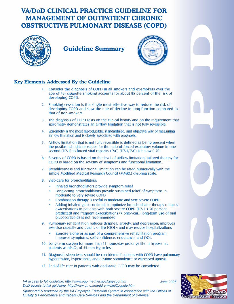
VA/DOD CLINICAL PRACTICE GUIDELINE FOR
MANAGEMENT OF OUTPATIENT CHRONIC
OBSTRUCTIVE PULMONARY DISEASE (COPD)
Guideline Summary
VA access to full guideline: http://www.oqp.med.va.gov/cpg/cpg.htmDoD access to full guideline: http://www.qmo.amedd.army.mil/pguide.htm
Sponsored & produced by the VA Employee Education System in cooperation with the Offices of Quality & Performance and Patient Care Services and the Department of Defense.
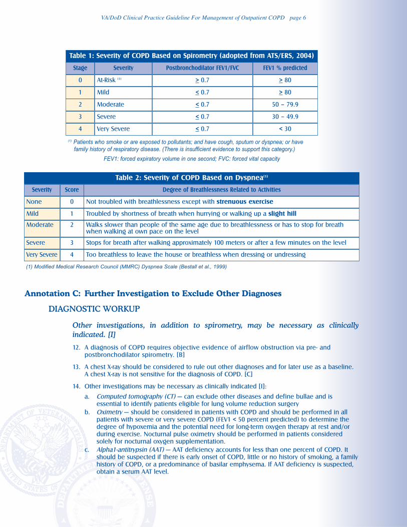
June 2007
Key Elements Addressed By the Guideline
1. Consider the diagnosis of COPD in all smokers and ex-smokers over the
age of 45; cigarette smoking accounts for about 85 percent of the risk of
developing COPD.
2. Smoking cessation is the single most effective way to reduce the risk of
developing COPD and slow the rate of decline in lung function compared to
that of non-smokers.
3. The diagnosis of COPD rests on the clinical history and on the requirement that
spirometry demonstrates an airflow limitation that is not fully reversible.
4. Spirometry is the most reproducible, standardized, and objective way of measuring
airflow limitation and is closely associated with prognosis.
5. Airflow limitation that is not fully reversible is defined as being present when
the postbronchodilator values for the ratio of forced expiratory volume in one
second (FEV1) to forced vital capacity (FVC) (FEV1/FVC) is below 0.70
6. Severity of COPD is based on the level of airflow limitation; tailored therapy for
COPD is based on the severity of symptoms and functional limitation.
7. Breathlessness and functional limitation can be rated numerically with the
simple Modified Medical Research Council (MMRC) dyspnea scale.
8. Step-Care for bronchodilators:
• Inhaled bronchodilators provide symptom relief
• Long-acting bronchodilators provide sustained relief of symptoms in
moderate to very severe COPD
• Combination therapy is useful in moderate and very severe COPD
• Adding inhaled glucocorticoids to optimize bronchodilator therapy reduces
exacerbations in patients with both severe COPD (FEV1 < 50 percent
predicted) and frequent exacerbations (> one/year); long-term use of oral
glucocorticoids is not recommended
9. Pulmonary rehabilitation reduces dyspnea, anxiety, and depression; improves
exercise capacity and quality of life (QOL); and may reduce hospitalizations
• Exercise alone or as part of a comprehensive rehabilitation program
improves symptoms, self-confidence, endurance, and QOL.
10. Long-term oxygen for more than 15 hours/day prolongs life in hypoxemic
patients withPaO2 of 55 mm Hg or less.
11. Diagnostic sleep tests should be considered if patients with COPD have pulmonary
hypertension, hypercapnia, and daytime somnolence or witnessed apneas.
12. End-of-life care in patients with end-stage COPD may be considered.
VA/DoD Clinical Practice Guideline For Management of Outpatient COPD page 2
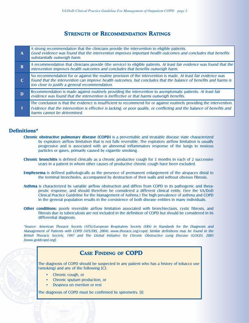
STRENGTH OF RECOMMENDATION RATINGS
AA strong recommendation that the clinicians provide the intervention to eligible patients.
Good evidence was found that the intervention improves important health outcomes and concludes that benefitssubstantially outweigh harm.
BA recommendation that clinicians provide (the service) to eligible patients. At least fair evidence was found that theintervention improves health outcomes and concludes that benefits outweigh harm.
CNo recommendation for or against the routine provision of the intervention is made. At least fair evidence wasfound that the intervention can improve health outcomes, but concludes that the balance of benefits and harms istoo close to justify a general recommendation.
D
I
Recommendation is made against routinely providing the intervention to asymptomatic patients. At least fairevidence was found that the intervention is ineffective or that harms outweigh benefits.
The conclusion is that the evidence is insufficient to recommend for or against routinely providing the intervention.
Evidence that the intervention is effective is lacking, or poor quality, or conflicting and the balance of benefits andharms cannot be determined.
Definitions*
Chronic obstructive pulmonary disease (COPD) is a preventable and treatable disease state characterized
by expiratory airflow limitation that is not fully reversible. The expiratory airflow limitation is usually
progressive and is associated with an abnormal inflammatory response of the lungs to noxious
particles or gases, primarily caused by cigarette smoking.
Chronic bronchitis is defined clinically as a chronic productive cough for 3 months in each of 2 successive
years in a patient in whom other causes of productive chronic cough have been excluded.
Emphysema is defined pathologically as the presence of permanent enlargement of the airspaces distal to
the terminal bronchioles, accompanied by destruction of their walls and without obvious fibrosis.
Asthma is characterized by variable airflow obstruction and differs from COPD in its pathogenic and thera-
peutic response, and should therefore be considered a different clinical entity. (See the VA/DoD
Clinical Practice Guideline for the Management of Asthma.) The high prevalence of asthma and COPD
in the general population results in the coexistence of both disease entities in many individuals.
Other conditions: poorly reversible airflow limitation associated with bronchiectasis, cystic fibrosis, and
fibrosis due to tuberculosis are not included in the definition of COPD but should be considered in its
differential diagnosis.
*Source: American Thoracic Society (ATS)/European Respiratory Society (ERS) in Standards for the Diagnosis and
Management of Patients with COPD (ATS/ERS, 2004). www.thoracic.org/copd; Similar definitions may be found in the
British Thoracic Society, 1997 and The Global Initiative for Chronic Obstructive Lung Disease (GOLD), 2005
[www.goldcopd.org].
CASE FINDING OF COPD
The diagnosis of COPD should be suspected in any patient who has a history of tobacco use
(smoking) and any of the following [C]:
• Chronic cough, or
• Chronic sputum production, or
• Dyspnea on exertion or rest
The diagnosis of COPD must be confirmed by spirometry. [I]
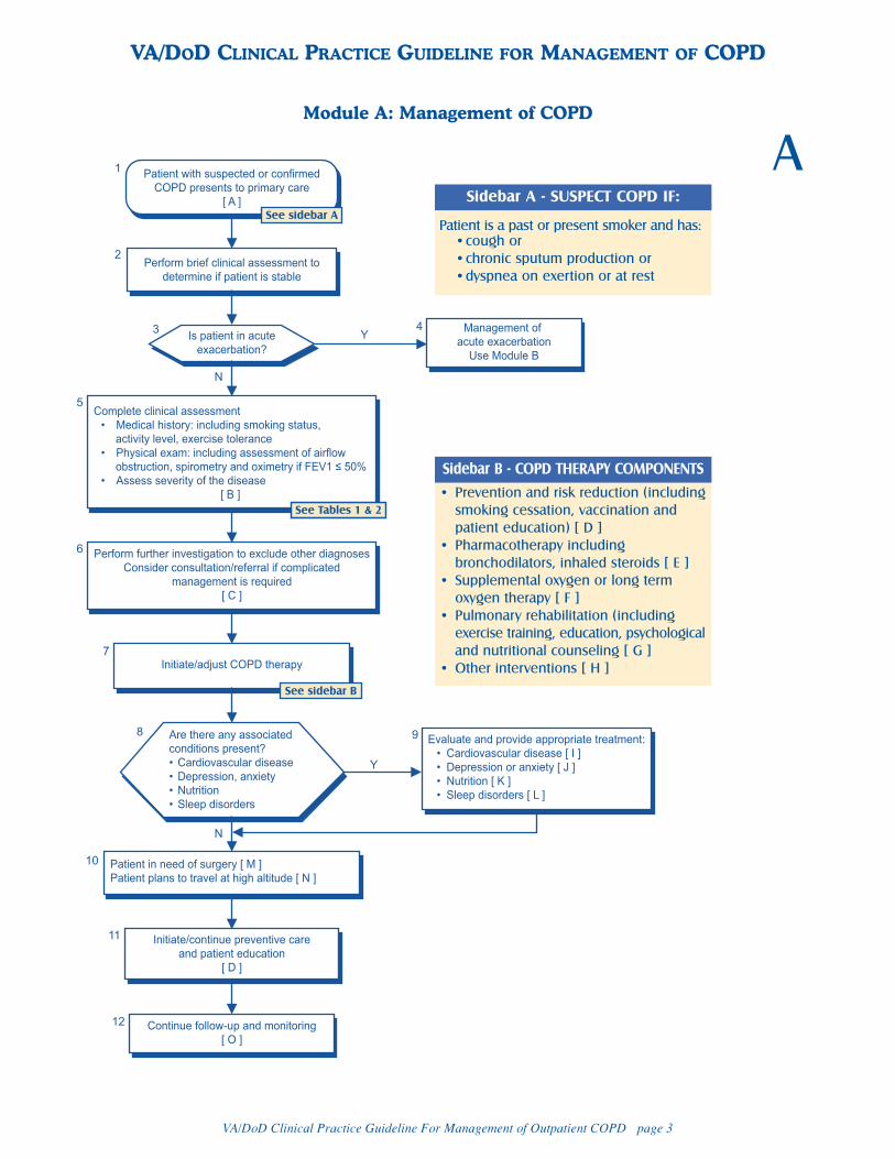
VA/DoD Clinical Practice Guideline For Management of Outpatient COPD page 3
Patient with suspected or confirmed
COPD presents to primary care
[ A ]
Perform brief clinical assessment to
determine if patient is stable
Is patient in acute
exacerbation?
Management of
acute exacerbation
Use Module B
Perform further investigation to exclude other diagnoses
Consider consultation/referral if complicated
management is required
[ C ]
Initiate/adjust COPD therapy
Initiate/continue preventive care
and patient education
[ D ]
Continue follow-up and monitoring
[ O ]
Complete clinical assessment
• Medical history: including smoking status,
activity level, exercise tolerance
• Physical exam: including assessment of airflow
obstruction, spirometry and oximetry if FEV1 ≤ 50%
• Assess severity of the disease
[ B ]
Are there any associated
conditions present?
• Cardiovascular disease
• Depression, anxiety
• Nutrition
• Sleep disorders
Patient in need of surgery [ M ]
Patient plans to travel at high altitude [ N ]
Evaluate and provide appropriate treatment:
• Cardiovascular disease [ I ]
• Depression or anxiety [ J ]
• Nutrition [ K ]
• Sleep disorders [ L ]
1
2
3 4
5
6
7
8 9
10
11
12
Y
N
Y
N
See sidebar A
See sidebar B
See Tables 1 & 2
ASidebar A - SUSPECT COPD IF:
Patient is a past or present smoker and has:•cough or
•chronic sputum production or
•dyspnea on exertion or at rest
Sidebar B - COPD THERAPY COMPONENTS
• Prevention and risk reduction (including
smoking cessation, vaccination and
patient education) [ D ]
• Pharmacotherapy including
bronchodilators, inhaled steroids [ E ]
• Supplemental oxygen or long term
oxygen therapy [ F ]
• Pulmonary rehabilitation (including
exercise training, education, psychological
and nutritional counseling [ G ]
• Other interventions [ H ]
VA/DOD CLINICAL PRACTICE GUIDELINE FOR MANAGEMENT OF COPD
Module A: Management of COPD
VA/DoD Clinical Practice Guideline For Management of Outpatient COPD page 4
MODULE A: MANAGEMENT OF COPD
ACTION STATEMENTS AND RECOMMENDATIONS
SCREENING
Annotation A: Patient with Suspected or Confirmed COPD
Presents to Primary Care
1. Persons with a history of smoking and the presence of cough or chronic sputum production or
dyspnea should be assessed for COPD with spirometry. [C]
CLINICAL ASSESSMENT
Annotation B: Clinical Assessment
HISTORY AND PHYSICAL EXAMINATION
All patients with known or suspected COPD should have a focused history and
physical examination to assess for the presence of airflow limitation. [I]
2. The following core elements of the medical history should be evaluated in patients with
suspected or proven COPD [I]:
a. Shortness of breath — patients should quantify their level of dyspnea (resting vs.
exertional). Early in the disease course, patients often complain of exertional dyspnea. As
the disease progresses, exercise tolerance worsens and patients may develop resting
dyspnea.
b. Cough — duration and character of the cough should be quantified. The presence of a
productive cough is a second clinical hallmark of COPD. This cough is typically initially
worse in the morning, but can be present throughout the day. An isolated nocturnal cough
is typically not characteristic of COPD. Chronic bronchitis is defined by the presence of a
persistent cough for at least 3 months for 2 or more consecutive years.
c. Sputum production — volume (amount) and character (color, thickness) of sputum
production should be qualified. Sputum production is required for a diagnosis of chronic
bronchitis.
d. Risk factor assessment — tobacco use, particularly cigarette smoking, is the primary risk
factor for developing COPD. Use should be quantified in pack-years (number of packs per
day x number of years = pack-years). A 10-pack year history of smoking is considered to be
the threshold for development of COPD. There is no comparable standard for pipes or
cigars that may also produce COPD. Environmental pollutant exposure and occupational
exposure to vapors, fumes, or irritants are important secondary risk factors.
e. Other important elements in the initial evaluation of COPD:
• Prior medical history of asthma, allergies, or recurrent respiratory illnesses (particularly
in childhood)
• Family history of COPD
• Self-reported history of prior COPD exacerbations and/or hospitalizations
• Presence of comorbid conditions, in particular coronary artery disease, congestive
heart failure, depression, and anxiety.
3. The following core elements of the physical examination should be evaluated in patients with
suspected or proven COPD [I]:
a. Vital signs — for patients with COPD, an assessment of pulse oximetry and body mass index
(BMI = kg/m2) should be included with the vital signs.
VA/DoD Clinical Practice Guideline For Management of Outpatient COPD page 5
b. Inspection — clinical observation should be performed to assess for the following elements:
• Chest wall morphology (e.g., ‘barrel-chest’); use of accessory muscles (e.g.,
‘suprasternal retractions’); pursed-lip breathing (surrogates that suggest airflow
limitation); and tracheal tug (sign of hyperinflation)
• Forced Expiratory Time — patients should be asked to completely empty their lungs
following a maximal inspiratory effort
• Central cyanosis (a surrogate for oxygen saturation); oxygen desaturation may be
present in the absence of cyanosis; cyanosis is indicative of severe desaturation
• Miscellaneous signs — jugular venous distension suggests elevated right heart
pressures; bilateral peripheral edema may suggest cor pulmonale.
c. Palpation/Percussion — these elements are often unhelpful in patients with COPD, but may
be helpful in diagnosing pulmonary hyperinflation.
d. Auscultation — the following elements should be noted on the cardiopulmonary
examination:
• Breath sounds are often diminished or distant in patients with COPD,
• A widened split second heart sound is suggestive of cor pulmonale.
SPIROMETRY AND REVERSIBILITY FOR DIAGNOSIS
Spirometry should be obtained in all stable patients suspected of or having a
diagnosis of COPD. [B]
4. Spirometry should be performed and documented in the medical record. [B]
5. A diagnosis of expiratory airflow limitation can be made if the post-bronchodilator FEV1/FVC or
FEV1/VC ratio is 0.70 or less. Where possible, value should be compared to age-related normal
values to avoid over diagnosis of COPD in the elderly. [I]
6. Reversibility should not be used to predict response to treatment or to distinguish between
COPD and asthma. [B]
7. Spirometry should be repeated if there is a clinically significant unexplained change in respi-
ratory symptoms. [I]
8. All patients presenting with airflow limitation at a relative early age (of the fourth to fifth
decade) or with a family history of COPD should be tested for alpha-1-antitrypsin deficiency. [I]
9. Oximetry should be considered in patients with COPD and should be performed in all patients
with severe or very severe COPD (FEV1 < 50 percent predicted) to determine the degree of
hypoxemia and the potential need for long-term oxygen therapy at rest and/or during exercise. [C]
ASSESSING SEVERITY OF THE DISEASE
COPD severity should be assessed on the basis of percentage of predicted FEV1 or
degree of dyspnea related to activities. [I]
10. The forced expiratory volume in one second (FEV1) should be used to stratify disease severity
by airflow limitation. [I] (See Table 1)
11. The Modified Medical Research Council (MMRC) Dyspnea Scale should be used to grade
severity of breathlessness according to the level of exertion required to elicit it and help
determine treatment. [C] (See Table 2)
VA/DoD Clinical Practice Guideline For Management of Outpatient COPD page 6
Annotation C: Further Investigation to Exclude Other Diagnoses
DIAGNOSTIC WORKUP
Other investigations, in addition to spirometry, may be necessary as clinically
indicated. [I]
12. A diagnosis of COPD requires objective evidence of airflow obstruction via pre- and
postbronchodilator spirometry. [B]
13. A chest X-ray should be considered to rule out other diagnoses and for later use as a baseline.
A chest X-ray is not sensitive for the diagnosis of COPD. [C]
14. Other investigations may be necessary as clinically indicated [I]:
a. Computed tomography (CT) — can exclude other diseases and define bullae and is
essential to identify patients eligible for lung volume reduction surgery
b. Oximetry — should be considered in patients with COPD and should be performed in all
patients with severe or very severe COPD (FEV1 < 50 percent predicted) to determine the
degree of hypoxemia and the potential need for long-term oxygen therapy at rest and/or
during exercise. Nocturnal pulse oximetry should be performed in patients considered
solely for nocturnal oxygen supplementation.
c. Alpha1-antitrypsin (AAT) — AAT deficiency accounts for less than one percent of COPD. It
should be suspected if there is early onset of COPD, little or no history of smoking, a family
history of COPD, or a predominance of basilar emphysema. If AAT deficiency is suspected,
obtain a serum AAT level.
Table 1: Severity of COPD Based on Spirometry (adopted from ATS/ERS, 2004)
(1) Patients who smoke or are exposed to pollutants; and have cough, sputum or dyspnea; or havefamily history of respiratory disease. (There is insufficient evidence to support this category.)
FEV1: forced expiratory volume in one second; FVC: forced vital capacity
Stage Severity Postbronchodilator FEV1/FVC FEV1 % predicted
0 At-Risk (1) > 0.7 > 80
1 Mild < 0.7 > 80
2 Moderate < 0.7 50 – 79.9
3 Severe < 0.7 30 – 49.9
4 Very Severe < 0.7 < 30
Table 2: Severity of COPD Based on Dyspnea(1)
(1) Modified Medical Research Council (MMRC) Dyspnea Scale (Bestall et al., 1999)
Severity Score Degree of Breathlessness Related to Activities
None 0 Not troubled with breathlessness except with strenuous exercise
Mild 1 Troubled by shortness of breath when hurrying or walking up a slight hill
Moderate 2 Walks slower than people of the same age due to breathlessness or has to stop for breathwhen walking at own pace on the level
Severe 3 Stops for breath after walking approximately 100 meters or after a few minutes on the level
Very Severe 4 Too breathless to leave the house or breathless when dressing or undressing
VA/DoD Clinical Practice Guideline For Management of Outpatient COPD page 7
d. Arterial blood gases — arterial blood gases should be done in patients with very severe
COPD (FEV1 < 30 percent predicted); signs of right heart failure (cor pulmonale);
polycythemia (hematocrit > 55 percent); or respiratory failure. Blood gases are an alternative
to pulse oximetry in patients being considered for O2 supplementation. Pulse oximetry can
determine arterial oxygen saturation, but pulse oximetry does not yield PCO2.
e. Full pulmonary function tests — lung volumes, carbon monoxide diffusing capacity and
flowvolume loops are not required for routine assessment but can provide additional
information useful for resolving diagnostic uncertainty and/or assessing surgical risk. A
reduced carbon monoxide diffusion capacity may suggest the presence of emphysema.
f. Exercise testing — exercise testing may be of value in patients with a disproportionate
degree of dyspnea for their FEV1. Exercise testing can quantify impairment and/or disability
and help to select patients able to safely undergo lung resection.
g. ECG — to assess cardiac status if pulmonary or nonpulmonary heart disease is suspected or
present.
h. Echocardiogram — to assess right and left cardiac status if cardiac dysfunction or disease is
suspected or present.
i. Sputum cultures — consider in patients with persistently purulent sputum or during
recurrent infectious exacerbations.
j. Complete blood count test should be done if anemia or polycythemia is suspected.
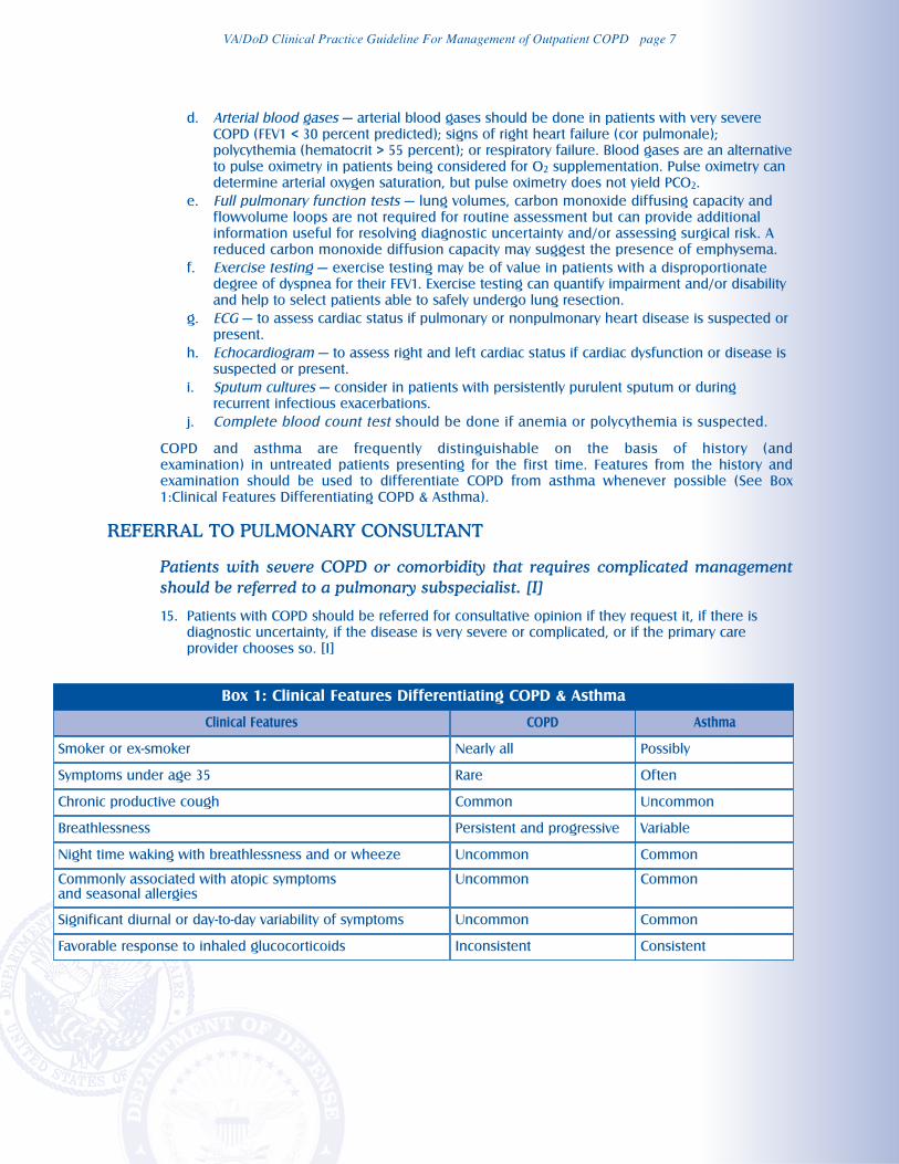
COPD and asthma are frequently distinguishable on the basis of history (and
examination) in untreated patients presenting for the first time. Features from the history and
examination should be used to differentiate COPD from asthma whenever possible (See Box
1:Clinical Features Differentiating COPD & Asthma).
REFERRAL TO PULMONARY CONSULTANT
Patients with severe COPD or comorbidity that requires complicated management
should be referred to a pulmonary subspecialist. [I]
15. Patients with COPD should be referred for consultative opinion if they request it, if there is
diagnostic uncertainty, if the disease is very severe or complicated, or if the primary care
provider chooses so. [I]
Box 1: Clinical Features Differentiating COPD & Asthma
Clinical Features COPD Asthma
Smoker or ex-smoker Nearly all Possibly
Symptoms under age 35 Rare Often
Chronic productive cough Common Uncommon
Breathlessness Persistent and progressive Variable
Night time waking with breathlessness and or wheeze Uncommon Common
Commonly associated with atopic symptoms and seasonal allergies
Uncommon Common
Significant diurnal or day-to-day variability of symptoms Uncommon Common
Favorable response to inhaled glucocorticoids Inconsistent Consistent
VA/DoD Clinical Practice Guideline For Management of Outpatient COPD page 8
Annotation D: Prevention and Risk Reduction
PATIENT EDUCATION
16. Patient should be educated about the disease, cause, therapy, and complications of COPD. [I]
SMOKING CESSATION
All patients must be screened for tobacco use and encouraged to stop smoking at
every visit, as smoking cessation is the only known intervention to reduce the decline
in FEV1. [A]
17. All patients should be counseled not to smoke and to avoid secondhand smoke. [A]
18. All smokers must be told that they need to quit smoking. [A]
19. All smokers should be assessed for willingness to quit. [C]
20. All smokers should be counseled on smoking cessation and be considered for medications that
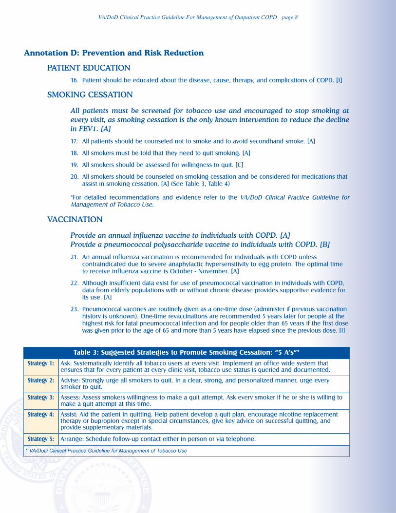
assist in smoking cessation. [A] (See Table 3, Table 4)
*For detailed recommendations and evidence refer to the VA/DoD Clinical Practice Guideline forManagement of Tobacco Use.
VACCINATION
Provide an annual influenza vaccine to individuals with COPD. [A]
Provide a pneumococcal polysaccharide vaccine to individuals with COPD. [B]
21. An annual influenza vaccination is recommended for individuals with COPD unless
contraindicated due to severe anaphylactic hypersensitivity to egg protein. The optimal time
to receive influenza vaccine is October - November. [A]
22. Although insufficient data exist for use of pneumococcal vaccination in individuals with COPD,
data from elderly populations with or without chronic disease provides supportive evidence for
its use. [A]
23. Pneumococcal vaccines are routinely given as a one-time dose (administer if previous vaccination
history is unknown). One-time revaccinations are recommended 5 years later for people at the
highest risk for fatal pneumococcal infection and for people older than 65 years if the first dose
was given prior to the age of 65 and more than 5 years have elapsed since the previous dose. [I]
Table 3: Suggested Strategies to Promote Smoking Cessation: “5 A’s”*
* VA/DoD Clinical Practice Guideline for Management of Tobacco Use
Strategy 1: Ask: Systematically identify all tobacco users at every visit. Implement an office wide system thatensures that for every patient at every clinic visit, tobacco use status is queried and documented.
Strategy 2: Advise: Strongly urge all smokers to quit. In a clear, strong, and personalized manner, urge everysmoker to quit.
Strategy 3: Assess: Assess smokers willingness to make a quit attempt. Ask every smoker if he or she is willing tomake a quit attempt at this time.
Strategy 4: Assist: Aid the patient in quitting. Help patient develop a quit plan, encourage nicotine replacementtherapy or bupropion except in special circumstances, give key advice on successful quitting, andprovide supplementary materials.
Strategy 5: Arrange: Schedule follow-up contact either in person or via telephone.
VA/DoD Clinical Practice Guideline For Management of Outpatient COPD page 9
THERAPY INTERVENTIONS FOR COPD
Annotation E: Pharmacotherapy Including Bronchodilators and
Inhaled Glucocorticoids
PHARMACOTHERAPY OF COPD
See Module C: Pharmacotherapy for specific recommendations (See Table 5, Table 6,
Figure 2). For dosage of selected COPD drug therapy (See Table C- 1-6)
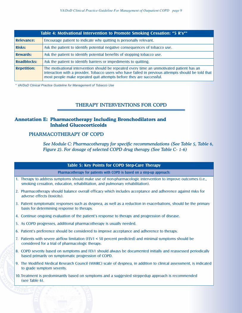
Table 4: Motivational Intervention to Promote Smoking Cessation: “5 R’s”*
Relevance: Encourage patient to indicate why quitting is personally relevant.
Risks: Ask the patient to identify potential negative consequences of tobacco use.
Rewards: Ask the patient to identify potential benefits of stopping tobacco use.
Roadblocks: Ask the patient to identify barriers or impediments to quitting.
Repetition: The motivational intervention should be repeated every time an unmotivated patient has an
interaction with a provider. Tobacco users who have failed in previous attempts should be told that
most people make repeated quit attempts before they are successful.
* VA/DoD Clinical Practice Guideline for Management of Tobacco Use
Table 5: Key Points for COPD Step-Care Therapy
Pharmacotherapy for patients with COPD is based on a step-up approach:
1. Therapy to address symptoms should make use of non-pharmacologic intervention to improve outcomes (i.e.,
smoking cessation, education, rehabilitation, and pulmonary rehabilitation).
2. Pharmacotherapy should balance overall efficacy which includes acceptance and adherence against risks for
adverse effects (toxicity).
3. Patient symptomatic responses such as dyspnea, as well as a reduction in exacerbations, should be the primary
basis for determining response to therapy.
4. Continue ongoing evaluation of the patient’s response to therapy and progression of disease.
5. As COPD progresses, additional pharmacotherapy is usually needed.
6. Patient’s preference should be considered to improve acceptance and adherence to therapy.
7. Patients with severe airflow limitation (FEV1 < 50 percent predicted) and minimal symptoms should be
considered for a trial of pharmacologic therapy.
8. COPD severity based on symptoms and FEV1 should always be documented initially and reassessed periodically
based primarily on symptomatic progression of COPD.
9. The Modified Medical Research Council (MMRC) scale of dyspnea, in addition to clinical assessment, is indicated
to grade symptom severity.
10.Treatment is predominantly based on symptoms and a suggested steppedup approach is recommended
(see Table 6).

VA/DoD Clinical Practice Guideline For Management of Outpatient COPD page 10
Asymptomatic No medication indicated -- Smoking cessation; influenza, and
other vaccinations
Step
A
Symptoms less than daily No scheduled medication
indicated
SABA➅
Smoking cessation; influenza, and
other vaccinations
B
Symptoms not controlled
with rescue therapy or
daily symptoms
Scheduled SAACor
Combination SABA + SAAC➂
SABA
➅Smoking cessation; influenza, and
other vaccinations
C
Symptoms not controlled
➁Combination SAAC + LABA
orLAAC
➃
SABA
➅Smoking cessation; influenza, and
other vaccinations
Consider Pulmonary Rehabilitation ➆
D
Symptoms not controlled
➁Combination LABA + LAAC
➃SABA
➅Smoking cessation; influenza, and
other vaccinationsRefer to Pulmonary Rehabilitation ➆
E
Exacerbations of morethan one per year andsevere disease (FEV1 < 50%)
Consider adding an inhaled
glucocorticoid
➄
SABA
➅Smoking cessation; influenza, and
other vaccinations
Refer to Pulmonary Rehabilitation ➆
F
SAAC – Short-acting anticholinergic; SABA – Short-acting beta-agonist; LABA – Long-acting inhaled beta-agonist; LAAC – Long-acting anticholinergic
1 Spirometry is essential to confirm the presence of airflow obstruction (low FEV1 and FEV1/VC ratio). Base therapy on symptoms, but consider alternatediagnoses (heart disease, pulmonary emboli, etc.) if out of proportion to spirometry.
➁ Use the lowest level of therapy that satisfactorily relieves symptoms and maximizes activity level. Assure compliance and proper use of medications beforeescalating therapy. It is unusual for patients with COPD with FEV1 above 70% to require therapy beyond short-acting bronchodilators; if these patients donot improve they should be considered for alternative diagnoses.
➂ Consider use of inhaler containing both a short-acting beta 2-agonist and an anticholinergic. Nighttime symptoms are frequently better controlled with along-acting inhaled beta 2-agonist.
➃ Consider adding a theophylline trial (slow release theophylline adjusted to the level of 5 to 12 µg/ml) with caution due to adverse effects. Nighttimerespiratory symptoms are frequently controlled, but theophylline may lead to insomnia. Discontinue if a benefit is not evident within several weeks.
➄ Consider high dose inhaled glucocorticoids in patients with severe COPD (FEV1 < 50 % predicted) and at least one exacerbation in the prior year. Acombination of a high dose inhaled glucocorticoid and a long-acting beta 2-agonist may help provide long-term maintenance for symptomatic COPDand improve quality of life (QOL). The use of oral glucocorticoids for maintenance therapy is discouraged.
➅ Short-acting inhaled beta 2-agonists (less than 12 puffs/day) may continue to be used as needed. Inhaled long-acting beta 2-agonists should not be usedas rescue therapy.
➆ Pulmonary rehabilitation should be offered to patients who, despite optimal medical therapy, have reduced exercise tolerance and/or dyspnea limiting exercise.
Symptoms 1 Maintenance Therapy ➁ Other InterventionsRescue therapy
Table 6: Step-Care Pharmacotherapy in COPD
Figure 2. Step-Care Pharmacotherapy in COPD
A Reduce risk factor(s): smoking cessation; influenza and other vaccinations
SABA when needed
Scheduled SAAC
OR
Combination SAAC + SAAB
B
C
D
E
Combination SAAC + LABA
OR
LAAC
LABA + LAAC + SABA when needed *
F
* Theophylline may be added at each step with caution regarding adverse effects.
SAAC – Short-acting anticholinergic; SABA – Short-acting beta-agonist; LABA – Long-acting inhaled beta-agonist; LAAC – Long-acting anticholinergic
Add inhaled glucocorticoids if repeated exacerbations
and FEV1 < 50%
+ SABA when needed*
+ SABA when needed*
VA/DoD Clinical Practice Guideline For Management of Outpatient COPD page 11
Annotation F: Supplemental and Long-term Oxygen Therapy
OXYGEN THERAPY
Patients with COPD should be periodically evaluated for the need of supplemental
oxygen. Supplemental oxygen for those exhibiting signs of tissue hypoxia may increase
survival of patients with severe COPD. Oxygen may also be used for exertional
hypoxemia or nocturnal hypoxemia.
24. Oximetry should be considered in patients with COPD and should be performed in all patients with
severe or very severe COPD (FEV1 < 50 percent predicted). [I]
25. Evaluation of nocturnal desaturation should be considered in patients with severe or very severe
COPD (FEV1 < 50 percent predicted) who exhibit unexplained findings indicating nocturnal
hypoxemia (e.g., polycythemia, pulmonary hypertension, and nocturnal restlessness). [I]
26. Oxygen therapy should be initiated in patients who have hypoxemia
(PaO2 ≤ 55 mm Hg and/or SaO2 ≤ 88 percent). [A]
27. Oxygen therapy should be initiated in patients who have hypoxemia
(PaO2 of 56 to 59 mm Hg or SaO2 ≤ 89 percent) and signs of tissue hypoxia such as hematocrit
above 55, pulmonary hypertension, or cor pulmonale. [A]
28. Oxygen therapy should be provided during exercise in stable patients with COPD with
exertional hypoxemia (SaO2 ≤ 88 percent). [B]
29. Oxygen therapy should be provided for nocturnal hypoxemia (SaO2 ≤ 88 percent). [I]
30. Patients who started to receive oxygen therapy while unstable or on suboptimal medical
therapy should be reevaluated within one to 3 months for need of long-term oxygen therapy
(LTOT). If repeated evaluation indicates a patient no longer qualifies for oxygen, cessation of
oxygen should be considered. [B]
31. Patients who continue to receive long-term oxygen therapy (LTOT) should be reevaluated at
least annually for continued need of LTOT. [I]
32. Patients prescribed oxygen should be cautioned about the potentially extreme fire hazard of
smoking or lighting cigarettes in the presence of oxygen. [I]
Annotation G Pulmonary Rehabilitation
PULMONARY REHABILITATION
Pulmonary rehabilitation should be offered to all patients with COPD who, despite
optimal medical therapy, have reduced exercise tolerance and/or dyspnea limiting
exercise. [A]
All patients with COPD with exertional symptoms should be offered a structured
program with exercise training to reduce dyspnea and improve exercise tolerance and
health-related QOL. [A]
Rehabilitation programs with education and self-management training reduce
healthcare use. [B]
VA/DoD Clinical Practice Guideline For Management of Outpatient COPD page 12
SELECTION OF PATIENTS
33. Pulmonary rehabilitation should be considered for patients with COPD who have dyspnea,
reduced exercise tolerance, a restriction in activities, or impaired health status. [A]
34. Pulmonary rehabilitation should be offered to all patients who consider themselves disabled by
COPD (Level 3 and above on the dyspnea scale). [B]
35. Pulmonary rehabilitation is recommended for patients with reduced exercise tolerance and
restricted activities because of dyspnea. [A]
EXERCISE TRAINING
36. The exercise program should be supervised and should provide cardiovascular reconditioning
with endurance and muscle strength training. [A]
37. The initial exercise program should be of sufficient length, duration, and frequency. [B]
38. Endurance training should be performed to improve physical endurance. [A]
39. Lower limb strength training should be performed to improve exercise tolerance (walking,
cycling); upper extremity training improves arm strength. [B]
40. In order to maintain benefits, subsequent exercise training is needed. [B]
41. As studies show conflicting results, respiratory muscle training is not recommended to be
part of a rehabilitation exercise program. [B]
EDUCATION AND SELF-MANAGEMENT
42. Patients with COPD with a prior hospitalization should be referred for pulmonary rehabilitation. [A]
43. Educational components and self-management programs should be included in rehabilitation
programs, as it can reduce COPD exacerbations, hospital admission, and length of stay. [B]
44. Self-management programs should include the following [B]:
a. Skills training to optimally control the disease
b. Education about medications and devices and how to use them properly
c. Instruction on how to deal with exacerbations
d. Other aspects of coping with the disease.
45. The benefit of education, psychosocial support, and nutritional therapy as a single intervention,
without exercise, are less well-documented. [I]
Annotation H: Other Interventions
MUCOLYTICS, ANTIOXIDANTS, AND ANTITUSSIVES
The use of mucolytics, antioxidants, or antitussive medications has little evidence of
any effect on lung function. [D]
46. N-acetylcysteine (NAC) is not recommended for patients with COPD for the purpose of cough
suppression. [D]
47. N-acetylcysteine (NAC) 600 mg by mouth every day may be considered to decrease the number
of exacerbations in selected patients with COPD with primarily chronic bronchitis who are not
on inhaled glucocorticoids. [B]
48. Antioxidants, such as alpha-tocopherol (contained in vitamin E preparations) or beta-carotene,
should not be administered to patients with COPD, as they have no significant effect on
phlegm, cough, or dyspnea. [D]
49. Antitussives are not indicated in stable COPD. [I]
VA/DoD Clinical Practice Guideline For Management of Outpatient COPD page 13
ALPHA L-ANTITRYPSIN AUGMENTATION THERAPY
Patients with COPD due to confirmed or suspected alpha1-antitrypsin (AAT)
deficiency should be referred to a pulmonary subspecialist. [C]
Alpha1-antitrypsin augmentation therapy should be considered in patients with severe
hereditary alpha1-antitrypsin (AAT) deficiency and established emphysema. [C]
50. Patients with COPD due to alpha1-antitrypsin (AAT) deficiency should be provided the usual
COPD therapy – smoking cessation, preventive vaccinations, bronchodilators, supplemental
oxygen if indicated, and pulmonary rehabilitation. [I]
51. Patients with severe alpha1-antitrypsin (AAT) deficiency who have stopped smoking and with
moderate to severe COPD (FEV1 30 to 60 percent predicted) should be considered for AAT
augmentation therapy. Furthermore, benefits are not clear for those with FEV1 either below 30
percent or above 60 percent predicted. [C]
52. Augmentation therapy is not indicated for patients without emphysema. [D]
LUNG VOLUME REDUCTION SURGERY
Consider lung volume reduction surgery (LVRS) in carefully selected patients with
very severe COPD who comply with selection criteria that have been used in studies
demonstrating benefit from LVRS. [A]
53. Referral for lung volume reduction surgery (LVRS) may be considered for patients with very
severe COPD if they meet the following criteria [A]:
a. High-resolution computed tomography (CT) confirming bilateral emphysema
b. Total lung capacity before rehabilitation and after treatment with bronchodilators is greater
than 100 percent predicted and residual volume is greater than 150 percent predicted
c. Post-bronchodilator FEV1 is less than 45 percent predicted
d. PaCO2 less than 60 mm Hg, and PaO2 greater than 45 mm Hg
e. Patient has completed a pulmonary rehabilitation program.
54. Lung volume reduction surgery (LVRS) should not be considered in patients whose FEV1 is less
than 20 percent predicted and who either have homogenous emphysema or carbon monoxide
diffusing capacity that is less than 20 percent or have non-upper lobe emphysema and high
baseline exercise capacity. [D]
55. Lung volume reduction surgery (LVRS) should only be performed in medical centers with
appropriately trained surgeons and availability of necessary equipment. [I]
LUNG TRANSPLANTATION
Consider lung transplantation as an option for carefully selected patients with very
severe COPD who comply with selection criteria and have no contraindications. [C]
56. Lung transplantation may be considered in selected patients with advanced COPD. The
choice of single lung transplantation (SLT) or bilateral lung transplantation (BLT) for COPD
remains controversial. [C]
VA/DoD Clinical Practice Guideline For Management of Outpatient COPD page 14
MANAGEMENT OF ASSOCIATED CONDITIONS
Annotation I: Evaluate and Provide Appropriate Treatment for
Cardiovascular Disease
PULMONARY HYPERTENSION AND COR PULMONALE IN COPD
Patients with pulmonary hypertension and/or cor pulmonale should be referred to a
specialist for the management of COPD and be provided long-term oxygen, if needed,
and optimized. [A]
57. Patients with diagnosed or suspected cor pulmonale should be referred to a pulmonary
subspecialist. [C]
58. Patients with pulmonary hypertension and/or cor pulmonale should be assessed for hypoxemia
and provided long-term oxygen, if needed. [A]
59. Bronchodilators should be optimized and edema treated cautiously with diuretics. [C]
60. The management of cardiovascular diseases in patients with COPD should follow existing
guidelines, including routine treatment with beta-blockers. [B]
Annotation J: Evaluate and Provide Appropriate Treatment for Depression
or Anxiety Mental Health (Depression and Anxiety)
Healthcare providers should be alert to the possibility of presence of depression in
patients with COPD and treat them according to depression guidelines.
61. Patients with COPD should be screened for depression and anxiety using validated screening
and assessment tools. [B]
62. Patients diagnosed with depression or anxiety should be treated with pharmacotherapy and
psychotherapy suitable for patients with COPD and the patient’s age. [B]
63. Sedative anxiolytic for the treatment of anxiety should be avoided in patients with severe COPD. [D]
See the VA/DoD Clinical Practice Guideline for Major Depressive Disorder.
Annotation K: Evaluate and Provide Appropriate Treatment for Nutrition
MALNUTRITION
Malnutrition and weight loss in patients with COPD carry a poor prognosis and
should be assessed and intervention considered.
64. Body Mass Index (BMI) should be monitored in patients with COPD. [B]
65. Patients who are losing weight over time (BMI ≤ 21 kg/m2) should be referred for dietary
evaluation and advice. [B]
66. Alternate causes of weight loss associated with COPD, such as lung cancer and lung infection,
should be considered. [I]
67. Dietary supplementation in combination with exercise and nutritional consultation should
be considered in the management of patients with COPD with weight loss or malnutrition. [B]
VA/DoD Clinical Practice Guideline For Management of Outpatient COPD page 15
Annotation L: Evaluate and Provide Appropriate Treatment for Sleep Disorders
SLEEP DISORDERS IN PATIENTS WITH COPD
All patients with COPD should be questioned about symptoms of sleep disturbance
and possible associated sleep apnea syndromes, such as snoring, witnessed apnea
during sleep, and excessive daytime sleepiness.
68. Patients with COPD should be evaluated for sleep disorders by using medical interview, which
should include standardized screening questionnaires for sleep disorders (e.g., insomnia,
sleep apnea). [I]
69. Patients complaining of insomnia should be managed in outpatient primary care and may be
treated with hypnotics cautiously. [I]
70. Patients with other sleep-related disorders (such as sleep apnea) should be referred to a
sleep specialist. [I]
Annotation M: Special Considerations for a Patient in Need of Surgery
SPECIAL CONSIDERATIONS FOR A PATIENT IN NEED OF SURGERY
The preoperative evaluation of a patient with COPD depends upon the type and
acuity of surgery and theseverity of COPD.
EMERGENCY SURGERY
71. Emergency surgeries should not be delayed pending preoperative consultation. [I]
LOW-RISK
72. Clinically stable patients with COPD who are undergoing minor procedures under local
anesthesia do not need preoperative testing. [I]
73. Clinically stable patients with mild to moderate COPD (FEV1 > 50 percent) who are undergoing
any operation under general anesthesia do not need preoperative testing. [I]
HIGH-RISK
74. Patients with severe COPD (FEV1 < 50 percent) undergoing any operation that is done under
general anesthesia should be considered for preoperative evaluation including pulmonary
function test, gas exchange, and chest X-ray. [I]
75. Patients with severe COPD (FEV1 < 50 percent) planned for high-risk surgery should be referred
to a pulmonary specialist. [I]
OPTIMIZATION OF PRE- AND POSTOPERATIVE CARE
76. Bronchodilator therapy should be optimized prior to planned surgery. [I]
77. Patients should be encouraged to quit smoking and instructed to stop smoking at least 6 to 8
weeks before surgery. [I]
78. Deep breathing, incentive spirometry, early mobilization, and adequate pain control should be
encouraged to reduce postoperative pulmonary complications in patients with COPD. [I]
79. Patients who are on oral glucocorticoids should receive stress doses of intravenous
glucocorticoids in the perioperative period to reduce the risk of adrenal insufficiency. [I]
80. Pulmonary consultation should be obtained prior to surgery in patients with an FEV1 below
35 percent predicted and in patients who are to undergo lung volume reduction surgery. [I]
VA/DoD Clinical Practice Guideline For Management of Outpatient COPD page 16
Annotation N: Special Considerations for a Patient Planning to
Travel at High Altitude
SPECIAL CONSIDERATIONS FOR A PATIENT PLANNING AIR TRAVEL
Patients with severe COPD who are on long-term oxygen therapy or have sea level
PO2 below 80 mm Hg should be evaluated pre-flight for supplementary oxygen during
air travel. [C]
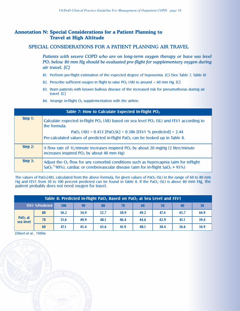
81. Perform pre-flight estimation of the expected degree of hypoxemia. [C] (See Table 7, Table 8)
82. Prescribe sufficient oxygen in flight to raise PO2 (Alt) to around ~ 60 mm Hg. [C]
83. Warn patients with known bullous disease of the increased risk for pneumothorax during air
travel. [C]
84. Arrange in-flight O2 supplementation with the airline.
Table 7: How to Calculate Expected In-flight PO2
Step 1: Calculate expected in-flight PO2 (Alt) based on sea level PO2 (SL) and FEV1 according to
the formula:
PaO2 (Alt) = 0.453 [PaO2SL] + 0.386 [FEV1 % predicted] + 2.44
Pre-calculated values of predicted in-flight PaO2 can be looked up in Table 8.
Step 2: A flow rate of 1L/minute increases inspired PO2 by about 20 mgHg (2 liter/minute
increases inspired PO2 by about 40 mm Hg)
Adjust the O2 flow for any comorbid conditions such as hypercapnia (aim for inflight
SaO2~90%), cardiac or cerebrovascular disease (aim for in-flight SaO2 > 95%)
Step 3:
The values of PaO2(Alt), calculated from the above formula, for given values of PaO2 (SL) in the range of 60 to 80 mm
Hg and FEV1 from 30 to 100 percent predicted can be found in Table 8. If the PaO2 (SL) is above 80 mm Hg, thepatient probably does not need oxygen for travel.
Table 8: Predicted In-flight PaO2 Based on PaO2 at Sea Level and FEV1
FEV1 %Predicted 100 90 80 70 60 50 40 30
PaO2 at
sea level
80
70
60
56.2 54.9 52.7 50.9 49.2 47.4 45.7 44.9
51.6 49.9 48.1 46.4 44.6 42.9 41.1 39.4
47.1 45.4 43.6 41.9 40.1 38.4 36.6 34.9
Dillard et al., 1989a
VA/DoD Clinical Practice Guideline For Management of Outpatient COPD page 17
FOLLOW-UP/MONITORING
Annotation O: Continue Follow-up and Monitoring
SCHEDULE FOLLOW-UP
Patients with moderate to severe COPD should be reevaluated at least once a year. [I]
85. Patients with COPD should be assessed on a periodic basis, based on the severity and
progression of their disease. [I]
86. Periodic evaluations of patients with COPD should include a review of their symptoms, their
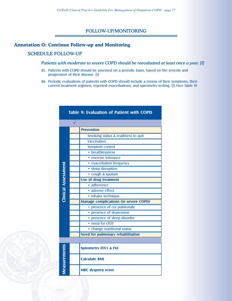
current treatment regimen, reported exacerbations, and spirometry testing. [I] (See Table 9)
Table 9: Evaluation of Patient with COPD
√
Clinic
al Assessm
ent
Prevention
Smoking status & readiness to quit
Vaccination
Symptom control
• breathlessness
• exercise tolerance
• exacerbation frequency
• sleep disruption
• cough & sputum
• adherence
Use of drug treatment
• adverse effect
• inhaler technique
Manage complications (in severe COPD)
• presence of cor pulmonale
• presence of depression
• presence of sleep disorder
• need for LTOT
• change nutritional status
Need for pulmonary rehabilitation
Measure
ments
Spirometry FEV1 & FVC
Calculate BMI
MRC dyspnea score
VA/DoD Clinical Practice Guideline For Management of Outpatient COPD page 18
PALLIATIVE CARE
Healthcare providers should assist patients with COPD and their families during
stable periods of health to promote discussion about advanced care planning,
including end-of-life care. [I]
The clinical care team will provide regular, ongoing assessments of distressing
symptoms (especially dyspnea) and actively seek to relieve suffering through a
comprehensive approach to the physical, psychological, social, and spiritual aspects. [I]
87. Healthcare providers should assess the needs of patients with COPD and their families for
advanced care planning and initiate advanced care in patients with poor prognosis (e.g.,
hospitalized with exacerbations). [I]
88. Patients with COPD and their families should be encouraged to participate in the planning and
management of their treatment to improve their ability to cope with COPD in the future. [I]
89. The referral of the patient and their family to appropriate expertise in palliative care to assist in
the relief of suffering may be considered when the patient/family’s needs require such or are
otherwise indicated. [I]
VA/DoD Clinical Practice Guideline For Management of Outpatient COPD page 19
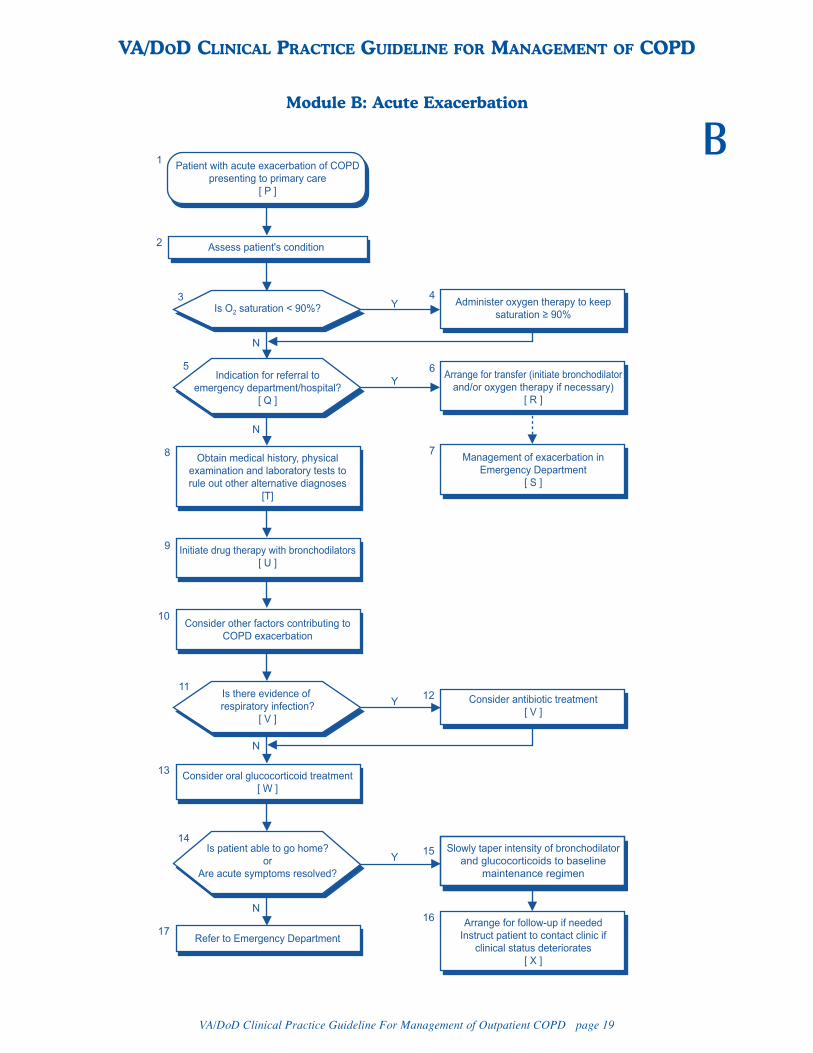
Patient with acute exacerbation of COPD
presenting to primary care
[ P ]
Assess patient's condition
Is O2 saturation < 90%?
Indication for referral to
emergency department/hospital?
[ Q ]
Obtain medical history, physical
examination and laboratory tests to
rule out other alternative diagnoses
[T]
Initiate drug therapy with bronchodilators
[ U ]
Consider other factors contributing to
COPD exacerbation
Is there evidence of
respiratory infection?
[ V ]
Consider oral glucocorticoid treatment
[ W ]
Is patient able to go home?
or
Are acute symptoms resolved?
Refer to Emergency Department
Administer oxygen therapy to keep
saturation ≥ 90%
Arrange for transfer (initiate bronchodilator
and/or oxygen therapy if necessary)
[ R ]
Management of exacerbation in
Emergency Department
[ S ]
Consider antibiotic treatment
[ V ]
Slowly taper intensity of bronchodilator
and glucocorticoids to baseline
maintenance regimen
Arrange for follow-up if needed
Instruct patient to contact clinic if
clinical status deteriorates
[ X ]
1
2
8
9
10
11
13
17
14
3
5
4
6
7
12
15
16
Y
Y
Y
Y
N
N
N
N
B
VA/DOD CLINICAL PRACTICE GUIDELINE FOR MANAGEMENT OF COPD
Module B: Acute Exacerbation
VA/DoD Clinical Practice Guideline For Management of Outpatient COPD page 20
MODULE B: MANAGEMENT OF COPD ACUTE EXACERBATION
ACTION STATEMENTS AND RECOMMENDATIONS
Annotation P: Patient with Acute Exacerbation of COPD
Presenting to Primary Care
PATIENT WITH ACUTE EXACERBATION
An exacerbation is a sustained worsening of the patient’s respiratory symptoms and function from his
or her usual stable state that is beyond normal day-to-day variations, and is acute in onset. Commonly
reported symptoms are worse breathlessness, cough, increased sputum production, and change in
sputum color. The change in the patient’s condition often necessitates a change in medication.
REFERRAL TO THE EMERGENCY DEPARTMENT
Annotation Q: Are There Indications for Referral to the
Emergency Department/Hospital?
CRITERIA FOR REFERRING TO THE EMERGENCY DEPARTMENT/HOSPITAL
More severe exacerbation or inadequate resources in the outpatient setting may
require evaluation and management of the patient in the emergency department or a
hospital setting. [I]
90. Patients evaluated for acute exacerbation of COPD should be considered for referral to the
emergency department or admission to the hospital if they present with any of the following
indications [I]:
a. Unstable vital signs
b. Impaired level of consciousness or altered mental status
c. Severe breathlessness
d. New or worsening hypoxemia (SaO2 < 90 percent)
e. Inadequate disease management resources at home
f. Lack of appropriate resources to evaluate or manage the patient in a clinic setting.
Annotation R: Arrange for Transfer to the Hospital
INITIATION OF SHORT-ACTING BRONCHODILATOR AND/OR OXYGEN
THERAPY IF NECESSARY
Early initiation of bronchodilator therapy and oxygen (in hypoxemic patients) is
appropriate prior to full assessment and treatment in the emergency department
or hospital.
91. Initial treatment for patients experiencing an initial acute exacerbation of COPD who have been
referred to the emergency department or admitted directly to the hospital should include [I]:
a. Short-acting bronchodilator, by nebulizer or metered dose inhaler, if readily available
b. Low flow oxygen therapy to maintain SAO2 at 90 percent.
VA/DoD Clinical Practice Guideline For Management of Outpatient COPD page 21
Annotation S: Management of Exacerbation in the Emergency Department
ASSESSMENT OF ACUTE EXACERBATION IN THE EMERGENCY DEPARTMENT
In the emergency department, patients experiencing an acute exacerbation of COPD should be
evaluated for the potential factors that contribute to the exacerbation. Assessment and treatment
should proceed simultaneously in these patients. The emergency department should have the ability
to perform these evaluations and treatments in a timely fashion. Increased respiratory symptoms in
COPD can be due to a number of cardiac or pulmonary causes. Appropriate management mandates
knowledge of the cause while simultaneously treating the severely ill patient.
MANAGEMENT OF ACUTE EXACERBATION IN OUTPATIENT SETTING
Annotation T: Obtain Medical History, Physical Examination,
and Laboratory Tests to Assess Severity, Rule Out
Alternatives, and Confirm Diagnosis
ASSESSMENT, TESTING, AND DIAGNOSIS
Patients with COPD with acute exacerbation should be assessed to confirm the
diagnosis, rule out other causes for worsening symptoms and determine the severity
of the exacerbation, and the priorities for treatment.
92. The diagnosis of acute exacerbation of COPD should be confirmed and other causes excluded
based upon clinical evaluation with additional diagnostic tests in selected cases. [I]
93. The severity of an exacerbation of COPD should be determined based upon medical history,
symptoms, physical examination, and pulmonary function tests. [I]
94. Medical history with a patient with acute exacerbation should include:
a. Onset, duration, and type of symptoms (cough, sputum production, dyspnea, fever,
decreased exercise tolerance, confusion, or acute mental status changes)
b. Current medication use
c. History of prior COPD exacerbations or hospitalizations (frequency, ICU admissions, and
prior intubation)
d. The severity of the underlying COPD
e. Presence of comorbid conditions; e.g., heart disease.
95. Physical examination with a patient with acute exacerbation should include:
a. Vital signs
b. Level of consciousness
c. A careful pulmonary examination
d. Cardiovascular examination
e. Oxygenation.
96. Laboratory testing that may be considered with a patient with acute exacerbation:
a. Oximetry (in all patients with moderate or worse COPD)
b. Arterial blood gas in patients with deteriorating clinical status
c. Spirometry, if available, in patients who are able to perform the test and for whom there is
baseline data available for comparison
d. Chest X-ray to exclude other causes if clinically suspected
e. ECG if clinically indicated.
97. Alternative causes of increased symptoms that need to be clinically excluded include:
VA/DoD Clinical Practice Guideline For Management of Outpatient COPD page 22
a. Congestive heart failure
b. Pneumonia
c. Pneumothorax
d. Pulmonary embolism
e. Cardiac ischemia
f. Cardiac arrhythmia
g. Upper airway infection; e.g., acute sinusitis
h. Upper airway obstruction
i. Pleural effusion
j. Recurrent aspiration
k. Noncompliance with medications
l. Inappropriate oxygen therapy
m. Adverse effects of medications; e.g., sedatives.
PHARMACOTHERAPY FOR
ACUTE EXACERBATION IN OUTPATIENT SETTINGS
Annotation U: Initiate Drug Therapy with Bronchodilators
BRONCHODILATORS
Provide relief of symptoms and improve FEV1 with short-acting inhaled
bronchodilator therapy. [B]
98. A short-acting bronchodilator (short-acting anticholinergic or short-acting beta 2-agonist) or a
combination of both, using a metered dose inhaler with a spacer or aerosol mobilization,
should be administered as soon as possible and as frequently as necessary. The choice of
agent should be made on the basis of individual assessment and initial response to therapy. [B]
99. Methyxanthines should be avoided either orally or systemically since these agents may lead to
side effects and have no proven efficacy in the setting of an acute exacerbation of COPD. [D]
Annotation V: Is There Evidence of Respiratory Infection?
ANTIBIOTICS
Prescribe a course of antibiotics for acute exacerbation of COPD if symptoms indicate
bacterial infection; choice of antibiotic agent may be based on the degree of complication
(number of exacerbations, FEV1, previous exposure to antibiotics, and cardiac disease).
100. COPD patients with acute exacerbation of COPD with at least two of the following will benefit
from antibiotic therapy [A]:
a. Increased sputum purulence (change in sputum color)
b. Increased sputum volume
c. Increased dyspnea.
101. Choice of antibiotic agents may be determined based on local bacterial resistance patterns. [C]
102. Choice of antibiotic agents may be determined based on the frequency of exacerbations in the
past 12 months, severity of underlying COPD, presence of cardiac disease, and recent (within 3
months) antibiotic exposure for each patient. [B]
103. For uncomplicated exacerbations of COPD, consider doxycycline, trimethoprim/sulfamethox-
azole, second generation cephalosporin. [C]
VA/DoD Clinical Practice Guideline For Management of Outpatient COPD page 23
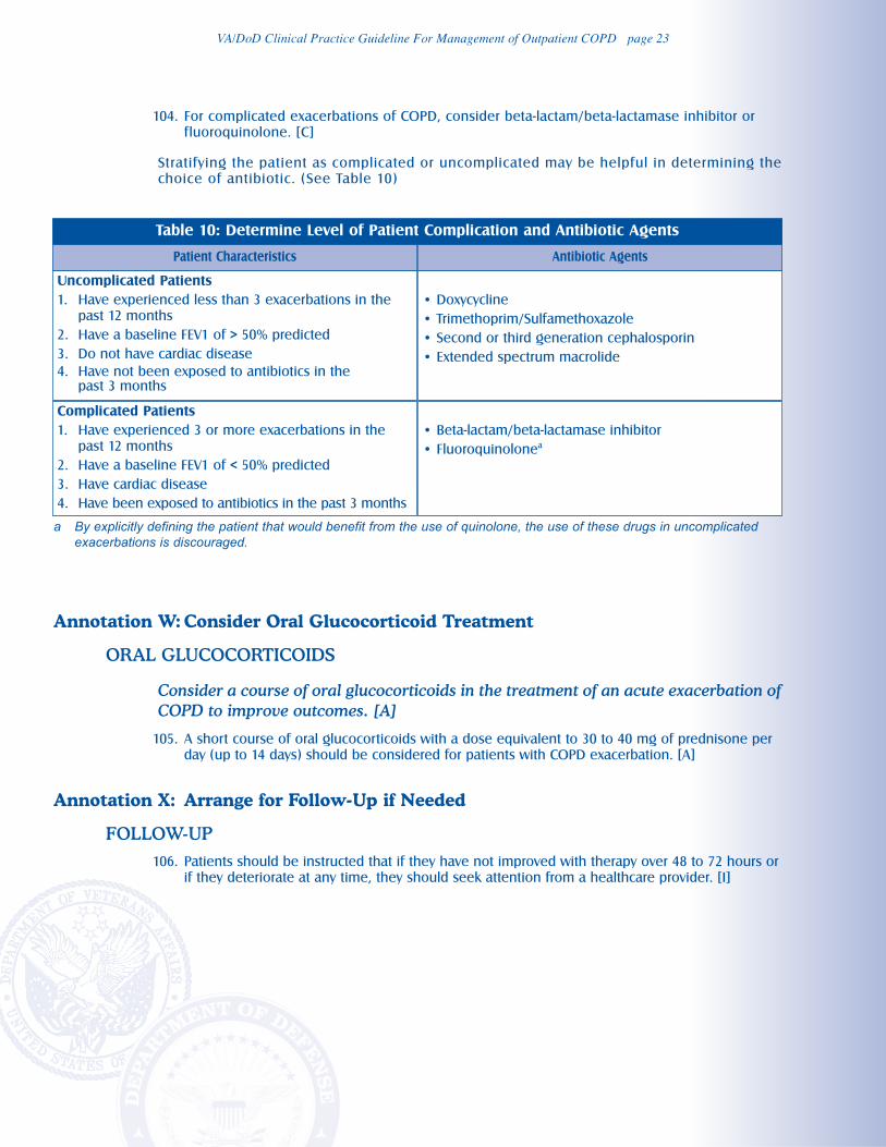
104. For complicated exacerbations of COPD, consider beta-lactam/beta-lactamase inhibitor or
fluoroquinolone. [C]
Stratifying the patient as complicated or uncomplicated may be helpful in determining the
choice of antibiotic. (See Table 10)
Annotation W: Consider Oral Glucocorticoid Treatment
ORAL GLUCOCORTICOIDS
Consider a course of oral glucocorticoids in the treatment of an acute exacerbation of
COPD to improve outcomes. [A]
105. A short course of oral glucocorticoids with a dose equivalent to 30 to 40 mg of prednisone per
day (up to 14 days) should be considered for patients with COPD exacerbation. [A]
Annotation X: Arrange for Follow-Up if Needed
FOLLOW-UP
106. Patients should be instructed that if they have not improved with therapy over 48 to 72 hours or
if they deteriorate at any time, they should seek attention from a healthcare provider. [I]
Table 10: Determine Level of Patient Complication and Antibiotic Agents
Patient Characteristics Antibiotic Agents
Uncomplicated Patients
1. Have experienced less than 3 exacerbations in the
past 12 months
2. Have a baseline FEV1 of > 50% predicted
3. Do not have cardiac disease
4. Have not been exposed to antibiotics in the past 3 months
• Doxycycline
• Trimethoprim/Sulfamethoxazole
• Second or third generation cephalosporin
• Extended spectrum macrolide
Complicated Patients
1. Have experienced 3 or more exacerbations in the
past 12 months
2. Have a baseline FEV1 of < 50% predicted
3. Have cardiac disease
4. Have been exposed to antibiotics in the past 3 months
• Beta-lactam/beta-lactamase inhibitor
• Fluoroquinolonea
a By explicitly defining the patient that would benefit from the use of quinolone, the use of these drugs in uncomplicatedexacerbations is discouraged.
VA/DoD Clinical Practice Guideline For Management of Outpatient COPD page 24
MODULE C: MANAGEMENT OF COPD PHARMACOTHERAPY
ACTION STATEMENTS AND RECOMMENDATIONS
Bronchodilators: Short-Acting Bronchodilators in Patients with
COPD (See Table C- 1, Table C- 2)
Consider using a maintenance short-acting anticholinergic and/or a maintenance short-
acting beta 2-agonist in patients whose symptoms adequately respond to these drugs.
107. Short-acting beta 2-agonists should be used as rescue therapy as needed. [A]
108. Short-acting bronchodilators may be considered for maintenance for patients with COPD, as
follows:
a. Short-acting anticholinergics (SAAC) or short-acting beta 2-agonists (SABA) to improve FEV1
and respiratory symptoms and reduce frequency of exacerbations [B]
b. Short-acting anticholingerics (SAAC) to improve QOL [B]
c. Insufficient evidence for short-acting beta 2-agonists (SABA) to improve QOL [I].
109. Since all chlorofluorocarbons (CFC) aerosols must be phased out, ipratropium CFC has been
replaced by ipratropium hydofluoroalkane (HFA). These two preparations may be considered in
usual doses to improve FEV1 in patients with COPD. [B]
Bronchodilators: Long-Acting Inhaled Beta 2-Agonists in Patients
with COPD (See Table C- 1, Table C-2)
Consider using a long-acting inhaled beta 2-agonist (LABA) to improve QOL or
respiratory symptoms such as dyspnea [A], and to reduce exacerbations [C].
110. Long-acting inhaled beta 2-agonists (LABA) should be considered for patients with COPD with
an FEV1 70 percent predicted or less to:
a. Improve FEV1 [B]
b. Improve persistent respiratory symptoms such as dyspnea, or impaired health-related quality
of life (QOL) [A]
c. Reduce exacerbations in patients who have had at least one exacerbation
in the previous year and required glucocorticoids, antibiotics, or
hospitalization [C].
111. In general, a long-acting inhaled beta 2-agonist (LABA) should not be substituted for a short-
acting anticholinergic (SAAC) with the expectation of improving respiratory symptoms, quality of
life (QOL), or exacerbations. [B]
Bronchodilators: Long-Acting Inhaled Anticholinergics in
Patients with COPD (See Table C- 1, Table C- 2)
Consider using a long-acting inhaled anticholinergic (LAAC) in patients with COPD to
improve respiratory symptoms and QOL or reduce moderate to severe exacerbations
[A]; or to improve FEV1 or reduce hospitalizations [B].
112. Long-acting anticholinergics (LAAC), compared to placebo or maintenance short-acting
anticholinergic (SAAC), should be considered for patients with COPD and an FEV1 65 percent
predicted or less to:
a. Improve persistent respiratory symptoms such as dyspnea or impaired quality of life (QOL) [A]

VA/DoD Clinical Practice Guideline For Management of Outpatient COPD page 25
b. Reduce moderate to severe COPD exacerbations (i.e., exacerbations requiring antibiotics
and/or oral or systemic glucocorticoids) [A]
c. Reduce COPD-related hospitalizations [B].
113. When a long-acting anticholinergic (LAAC) is used to improve patient outcomes in patients
taking a short-acting anticholinergic (SAAC), the SAAC should be discontinued. [I] However, the
use of a short-acting beta 2-agonist (SABA) as needed for rescue therapy should be continued.
114. In choosing long-acting bronchodilators, both long-acting anticholinergics (LAAC) and long-
acting beta 2-agonists (LABA) provide similar benefits; however, there may be more modest
improvement in FEV1 with LAAC. [B]
Combination Inhaled Bronchodilators
Combination bronchodilator therapy may be considered for patients with inadequate
response to single agents to improve FEV1 and to reduce symptoms and/or
exacerbations. [B]
115. When response to therapy with a short-acting beta agonist (SABA) is inadequate, consider the
use of regularly scheduled combination SABA + short-acting anticholinergic (SAAC) to improve
FEV1 and reduce exacerbations compared to treatment with the individual components. [B]
116. When response to regularly scheduled SAAC or combination of SABA + SAAC is inadequate,
consider the use of combination SAAC + long-acting beta 2-agonist (LABA) to improve FEV1 and
symptoms and reduce exacerbations compared to treatment with the individual components.
[B]
117. When response to a LABA + SAAC or a long-acting anticholinergic (LAAC) alone is inadequate,
consider the use of combination LABA + LAAC to improve FEV1. [B]
118. Consider the use of theophylline in addition to short-acting bronchodilators to improve FEV1. [B]
119. Consider the use of theophylline in addition to LABA to improve FEV1, symptoms, and quality of
life (QOL) compared to therapy with the individual components. [B]
120. There is insufficient evidence to recommend that certain combinations are superior to other
combinations, monotherapy with LAAC, or regimens including an inhaled glucocorticoid.
Therefore, treatment selection should be based on patient-specific variables. [I]
Inhaled Glucocorticoids (See Table C- 3)
Consider adding inhaled glucocorticoids to optimize bronchodilator therapy in
patients with COPD who have both severe disease (FEV1 < 50 percent predicted)
and who have had at least one exacerbation in the prior year, to reduce the
frequency of exacerbations. [A]
Alternatively, consider adding inhaled glucocorticoids in patients with severe COPD
(FEV1 < 50 percent predicted) to improve FEV1, respiratory symptoms, and QOL. [B]
121. Inhaled glucocorticoids are not recommended in patients with mild to moderate COPD
(FEV1 ≥ 50 percent predicted) as there is little evidence of efficacy. [D]
122. Combination of a long-acting beta 2-agonist (LABA) and inhaled glucocorticoid may be
considered in patients with severe COPD and at least one COPD exacerbation in the prior year
to decrease the incidence of COPD exacerbations compared to therapy with the individual
components. [A]
123. Combination of a long-acting beta 2-agonist (LABA) and inhaled glucocorticoid can be used in
symptomatic patients with severe COPD to improve FEV1 (approximately 0 to 100 ml), symptoms
and/or quality of life (QOL) [B]
VA/DoD Clinical Practice Guideline For Management of Outpatient COPD page 26
124. There is insufficient evidence to recommend a specific choice or optimal dose when starting
treatment with inhaled glucocorticoids. The doses used in efficacy trials or equivalent are
suggested (fluticasone proprionate 500 µg bid, budesonide 400 µg bid). [I]
125. Once treatment with inhaled glucocorticoids has been initiated, it is recommended to use
caution when stopping the medication, as discontinuation may lead to COPD exacerbation. [B]
126. Patients should be informed about the potential side effects of inhaled glucocorticoids (oral
candidiasis, bruising, adrenal suppression, cataracts, and osteoporosis). [B]
127. Treatment with inhaled glucocorticoids does not significantly affect the rate of decline in FEV1. [C]
128. Patients with COPD who are receiving oral or inhaled glucocorticoids should be evaluated for
bone loss and considered for prevention or treatment of osteoporosis. [I]
129. The risks of long-term treatment with glucocorticoids should be discussed with the patient. [I]
Theophylline (See Table C- 4, Table C- 5)
Theophylline can be added to improve pulmonary function, symptoms, or activities
in patients with COPD who do not achieve adequate symptom control with inhaled
bronchodilators. [A]
130. Patients with COPD who do not achieve adequate symptom control with inhaled
bronchodilators may be considered for adding theophylline therapy with an initial dose of 400
to 600 mg/day and a therapeutic target of blood level in the range 5 to 12 µg/ml). [A]
131. Blood levels should be carefully measured after initiation or change in dose. [I]
132. After the initial stability, repeat levels should be obtained when symptoms change, acute illness
develops, potentially interacting drugs are added, noncompliance is suspected, dose adjustments
are made, or symptoms suggestive of toxicity develop. [I]
133. If benefit has been demonstrated with a higher blood level (15 µg/ml of theophylline), careful
monitoring is required. The risk-to-benefit ratio increases above a concentration of 12µg/ml,
especially in older patients. [B]
134. Drug interactions with theophylline are common and may either increase or decrease
theophylline metabolism. All changes in medical regimens should be evaluated for potential
impact on theophylline levels. [C]
135. Theophylline should be continued only in patients who demonstrate a symptomatic benefit,
such as improved dyspnea or exercise tolerance. The improvement in function from theophylline
may not be evident in pulmonary function testing. However, therapy should be discontinued in
patients who demonstrate no subjective or objective improvement after several weeks of
theophylline therapy. [D]
VA/DoD Clinical Practice Guideline For Management of Outpatient COPD page 27
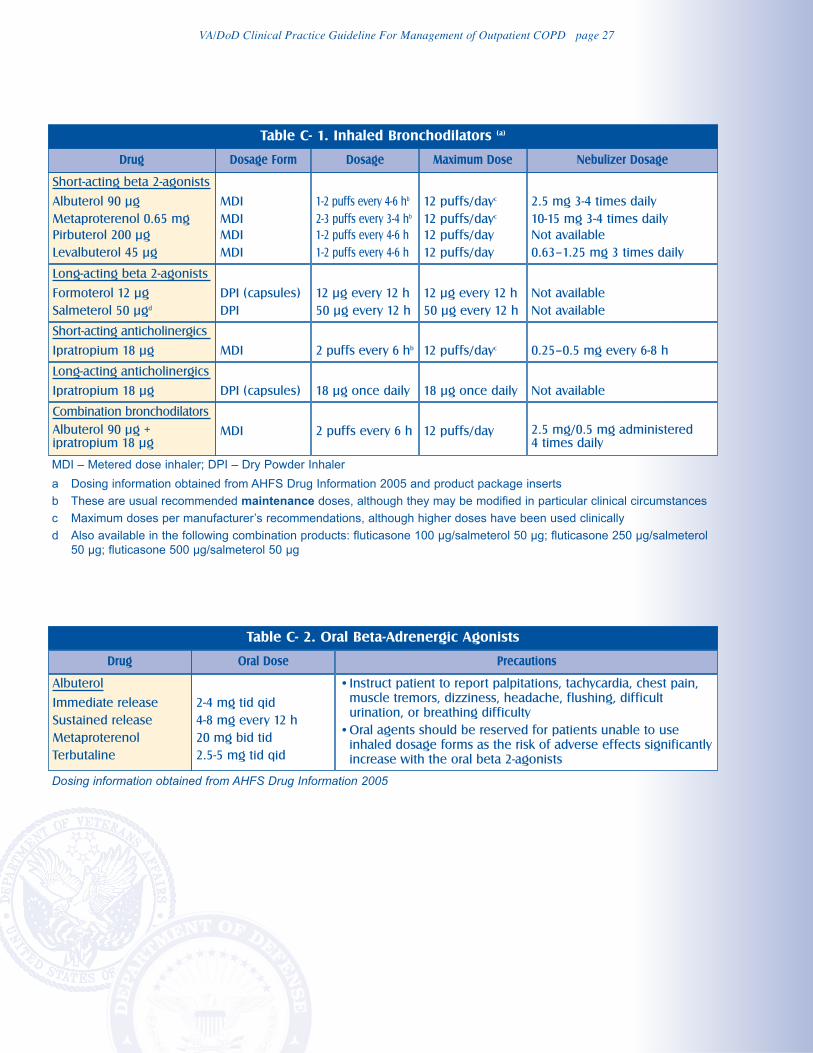
Table C- 1. Inhaled Bronchodilators (a)
Table C- 2. Oral Beta-Adrenergic Agonists
Drug Oral Dose Precautions
Albuterol
Immediate release
Sustained release
Metaproterenol
Terbutaline
2-4 mg tid qid
4-8 mg every 12 h
20 mg bid tid
2.5-5 mg tid qid
•Instruct patient to report palpitations, tachycardia, chest pain,
muscle tremors, dizziness, headache, flushing, difficult
urination, or breathing difficulty
•Oral agents should be reserved for patients unable to use
inhaled dosage forms as the risk of adverse effects significantly
increase with the oral beta 2-agonists
Drug Dosage Form Dosage Maximum Dose Nebulizer Dosage
Short-acting beta 2-agonists
Albuterol 90 µg
Metaproterenol 0.65 mg
Pirbuterol 200 µg
Levalbuterol 45 µg
MDI
MDI
MDI
MDI
1-2 puffs every 4-6 hb
2-3 puffs every 3-4 hb
1-2 puffs every 4-6 h
1-2 puffs every 4-6 h
12 puffs/dayc
12 puffs/dayc
12 puffs/day
12 puffs/day
2.5 mg 3-4 times daily
10-15 mg 3-4 times daily
Not available
0.63–1.25 mg 3 times daily
Long-acting beta 2-agonists
Formoterol 12 µg
Salmeterol 50 µgd
DPI (capsules)
DPI
12 µg every 12 h
50 µg every 12 h
12 µg every 12 h
50 µg every 12 h
Not available
Not available
Short-acting anticholinergics
Ipratropium 18 µg MDI 2 puffs every 6 hb 12 puffs/dayc 0.25–0.5 mg every 6-8 h
Long-acting anticholinergics
Ipratropium 18 µg DPI (capsules) 18 µg once daily 18 µg once daily Not available
Combination bronchodilators
Albuterol 90 µg + ipratropium 18 µg
MDI – Metered dose inhaler; DPI – Dry Powder Inhaler
a Dosing information obtained from AHFS Drug Information 2005 and product package inserts
b These are usual recommended maintenance doses, although they may be modified in particular clinical circumstances
c Maximum doses per manufacturer’s recommendations, although higher doses have been used clinically
d Also available in the following combination products: fluticasone 100 µg/salmeterol 50 µg; fluticasone 250 µg/salmeterol50 µg; fluticasone 500 µg/salmeterol 50 µg
MDI 2 puffs every 6 h 12 puffs/day 2.5 mg/0.5 mg administered 4 times daily
Dosing information obtained from AHFS Drug Information 2005
VA/DoD Clinical Practice Guideline For Management of Outpatient COPD page 28
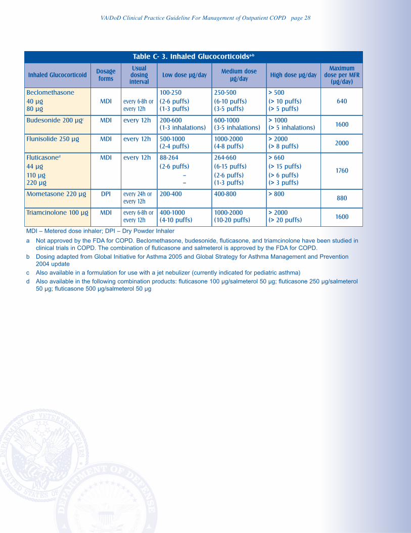
Table C- 3. Inhaled Glucocorticoidsa-b
Inhaled GlucocorticoidDosage
forms
Usualdosinginterval
Low dose µg/dayMedium dose
µg/dayHigh dose µg/day
Maximumdose per MFR
(µg/day)
Beclomethasone
40 µg
80 µg
MDI every 6-8h or
every 12h
100-250
(2-6 puffs)
(1-3 puffs)
250-500
(6-10 puffs)
(3-5 puffs)
> 500
(> 10 puffs)
(> 5 puffs)
640
Budesonide 200 µgc MDI every 12h 200-600
(1-3 inhalations)
600-1000
(3-5 inhalations)
> 1000
(> 5 inhalations)1600
Flunisolide 250 µg MDI every 12h 500-1000
(2-4 puffs)
1000-2000
(4-8 puffs)
> 2000
(> 8 puffs)2000
Fluticasoned
44 µg
110 µg
220 µg
MDI every 12h 88-264
(2-6 puffs)
–
–
264-660
(6-15 puffs)
(2-6 puffs)
(1-3 puffs)
> 660
(> 15 puffs)
(> 6 puffs)
(> 3 puffs)
1760
Mometasone 220 µg DPI every 24h or
every 12h
200-400 400-800 > 800880
Triamcinolone 100 µg
MDI – Metered dose inhaler; DPI – Dry Powder Inhaler
a Not approved by the FDA for COPD. Beclomethasone, budesonide, fluticasone, and triamcinolone have been studied inclinical trials in COPD. The combination of fluticasone and salmeterol is approved by the FDA for COPD.
b Dosing adapted from Global Initiative for Asthma 2005 and Global Strategy for Asthma Management and Prevention2004 update
c Also available in a formulation for use with a jet nebulizer (currently indicated for pediatric asthma)
d Also available in the following combination products: fluticasone 100 µg/salmeterol 50 µg; fluticasone 250 µg/salmeterol50 µg; fluticasone 500 µg/salmeterol 50 µg
MDI every 6-8h or
every 12h
400-1000
(4-10 puffs)
1000-2000
(10-20 puffs)
> 2000
(> 20 puffs)1600
VA/DoD Clinical Practice Guideline For Management of Outpatient COPD page 29
Figure 1. Time Course of COPD
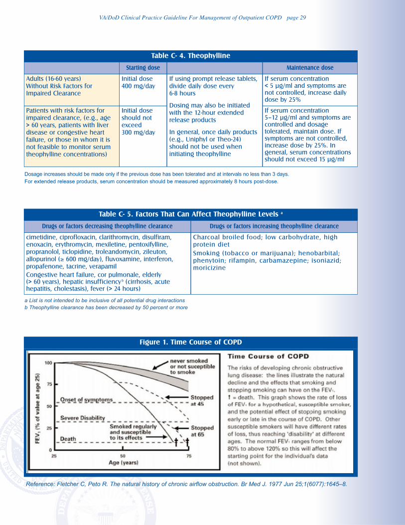
Reference: Fletcher C, Peto R. The natural history of chronic airflow obstruction. Br Med J. 1977 Jun 25;1(6077):1645–8.
Table C- 4. Theophylline
Table C- 5. Factors That Can Affect Theophylline Levels a
Drugs or factors decreasing theophylline clearance Drugs or factors increasing theophylline clearance
cimetidine, ciprofloxacin, clarithromycin, disulfiram,
enoxacin, erythromycin, mexiletine, pentoxifylline,
propranolol, ticlopidine, troleandomycin, zileuton,
allopurinol (≥ 600 mg/day), fluvoxamine, interferon,
propafenone, tacrine, verapamil
Congestive heart failure, cor pulmonale, elderly (> 60 years), hepatic insufficiency b (cirrhosis, acutehepatitis, cholestasis), fever (> 24 hours)
Charcoal broiled food; low carbohydrate, high
protein diet
Smoking (tobacco or marijuana); henobarbital;
phenytoin; rifampin, carbamazepine; isoniazid;
moricizine
Starting dose Maintenance dose
Adults (16-60 years)
Without Risk Factors for
Impaired Clearance
Initial dose
400 mg/day
If using prompt release tablets,
divide daily dose every
6-8 hours
Dosing may also be initiated
with the 12-hour extended
release products
In general, once daily products
(e.g., Uniphyl or Theo-24)
should not be used when
initiating theophylline
If serum concentration < 5 µg/ml and symptoms arenot controlled, increase dailydose by 25%
Patients with risk factors for
impaired clearance, (e.g., age
> 60 years, patients with liver
disease or congestive heart
failure, or those in whom it is
not feasible to monitor serum
theophylline concentrations)
Initial dose
should not
exceed
300 mg/day
If serum concentration 5–12 µg/ml and symptoms arecontrolled and dosagetolerated, maintain dose. Ifsymptoms are not controlled,increase dose by 25%. Ingeneral, serum concentrationsshould not exceed 15 µg/ml
Dosage increases should be made only if the previous dose has been tolerated and at intervals no less than 3 days.For extended release products, serum concentration should be measured approximately 8 hours post-dose.
a List is not intended to be inclusive of all potential drug interactionsb Theophylline clearance has been decreased by 50 percent or more
VA/DoD Clinical Practice Guideline For Management of Outpatient COPD page 30
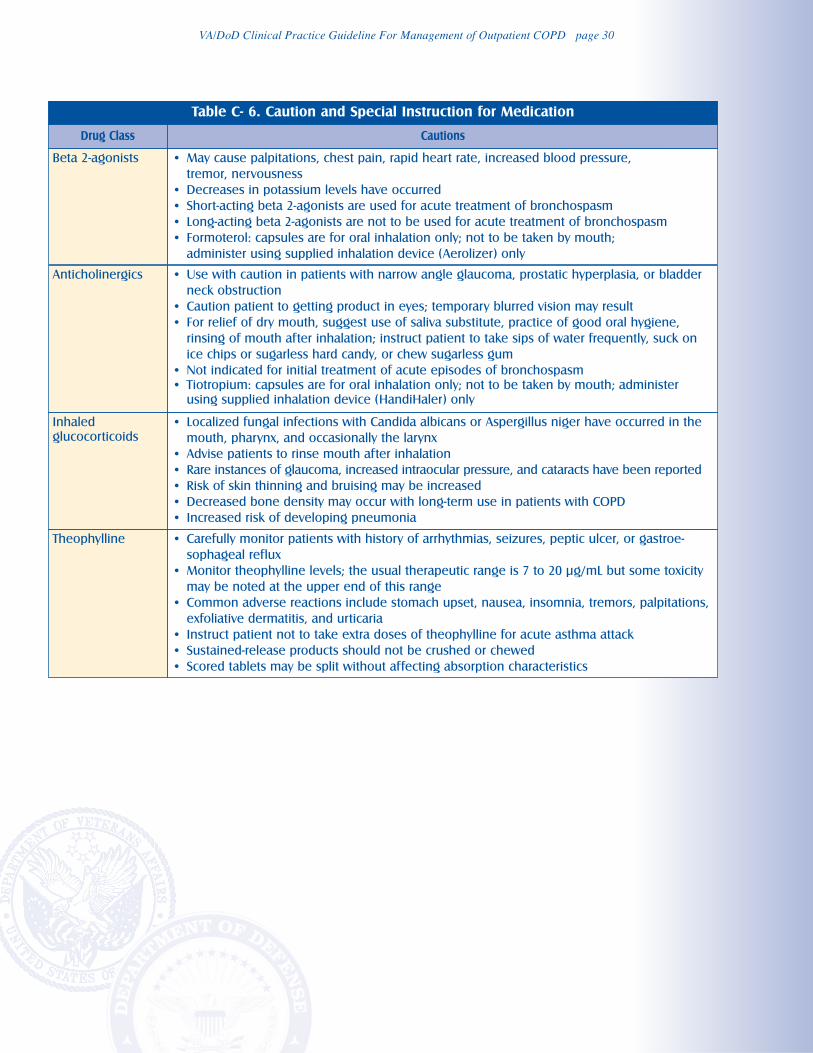
Table C- 6. Caution and Special Instruction for Medication
Drug Class Cautions
Beta 2-agonists • May cause palpitations, chest pain, rapid heart rate, increased blood pressure,
tremor, nervousness
• Decreases in potassium levels have occurred
• Short-acting beta 2-agonists are used for acute treatment of bronchospasm
• Long-acting beta 2-agonists are not to be used for acute treatment of bronchospasm
• Formoterol: capsules are for oral inhalation only; not to be taken by mouth;
administer using supplied inhalation device (Aerolizer) only
Anticholinergics • Use with caution in patients with narrow angle glaucoma, prostatic hyperplasia, or bladder
neck obstruction
• Caution patient to getting product in eyes; temporary blurred vision may result
• For relief of dry mouth, suggest use of saliva substitute, practice of good oral hygiene,
rinsing of mouth after inhalation; instruct patient to take sips of water frequently, suck on
ice chips or sugarless hard candy, or chew sugarless gum
• Not indicated for initial treatment of acute episodes of bronchospasm
• Tiotropium: capsules are for oral inhalation only; not to be taken by mouth; administer
using supplied inhalation device (HandiHaler) only
Inhaled
glucocorticoids
• Localized fungal infections with Candida albicans or Aspergillus niger have occurred in the
mouth, pharynx, and occasionally the larynx
• Advise patients to rinse mouth after inhalation
• Rare instances of glaucoma, increased intraocular pressure, and cataracts have been reported
• Risk of skin thinning and bruising may be increased
• Decreased bone density may occur with long-term use in patients with COPD
• Increased risk of developing pneumonia
Theophylline • Carefully monitor patients with history of arrhythmias, seizures, peptic ulcer, or gastroe-
sophageal reflux
• Monitor theophylline levels; the usual therapeutic range is 7 to 20 µg/mL but some toxicity
may be noted at the upper end of this range
• Common adverse reactions include stomach upset, nausea, insomnia, tremors, palpitations,
exfoliative dermatitis, and urticaria
• Instruct patient not to take extra doses of theophylline for acute asthma attack
• Sustained-release products should not be crushed or chewed
• Scored tablets may be split without affecting absorption characteristics
VA/DoD Clinical Practice Guideline For Management of Outpatient COPD page 31
ACRONYM LIST
AAT Alpha l-Antitrypsin
ATS American Thoracic Society
BMI Body Mass Index
CFC Chlorofluorocarbons
COPD Chronic Obstructive Pulmonary Disease
CT Computed Tomography
DPI Dry Powder Inhaler
ERS European Respiratory Society
FEV1 Forced Expiratory Volume in One Second
FVC Forced Vital Capacity
GOLD The Global Initiative for Chronic Obstructive Lung Disease
HFA Hydofluoroalkane
LAAC Long-Acting Anticholinergic
LABA Long-Acting Inhaled Beta 2-agonist
LTOT Long-Term Oxygen Therapy
LVRS Long Volume Reduction Surgery
MDI Metered Dose Inhalers
MMRC Modified Medical Research Council
MRC Medical Research Council
NAC N-Acetycysteine
QOL Quality of Life
SAAC Short-Acting Anticholinergic
SABA Short-Acting Beta 2-agonist
VC Vital Capacity