Embed Size (px)
Citation preview

PEER REVIEWED
Vaccine associated mucosa! disease case study: delllonstrating the illlportance of subsequent herd PI testing Myrna M. Miller,1 DVM, PhD; Donal O'Toole,1 MVB, PhD, FRCPath, DECVP; Jacqueline L. Cavender,1 MS; Todd E. Cornish,1 DVM, PhD, DACVP; Timothy G. Dawson,2 DVM; J. Maxwell Smylie,3 DVM; Jonathan H. Fox,1 DVM, PhD, DACVP; Kevin L. Hill,4 DVM; Donald L. Montgomery,1 DVM, PhD, DACVP; Maree Vasquez1; Brant A. Schumaker, 1 DVM, PhD 1Wyoming State Veterinary Laboratory, Department of Veterinary Sciences, College of Agriculture and Natural Sciences, University of Wyoming, Laramie, WY 82070 2Bi-State Large Animal Hospital, 10321 Polo Ranch Road, Cheyenne, WY 82003 3Smylie Animal Clinic, 93 West Richards Street, Douglas, WY 82633 4US Cattle Technical Services, Merck Animal Health, 447 N. Angel Street, Kaysville, UT 84037 Corresponding author: Dr. M. M. Miller ([email protected])
Abstract
Mucosal disease (MD) affected a single-source group of 461 recently purchased yearling steers shortly after vaccination with a multivalent vaccine containing 2 strains of modified-live bovine viral diarrhea virus (BVDV). Terminal disease affected 21 steers at 18 to 66 days post-vaccination, and was confirmed as mucosal disease in 3 carcasses examined postmortem. An additional 17 animals identified as persistently infected remained asymptomatic during the study. A laboratory study was undertaken to determine whether mucosal disease was triggered by a BVDV in the multivalent vaccine. Purified cytopathic BVDV isolates from 2 animals that died early in the outbreak were genetically identical (>99%) to the vaccine type 2 BVDV strain 125a in 3 genomic regions: 5'-untranslated region to the Npro, E2, and NS2/3. This genetic identity combined with the presence of a single peak outbreak occurring soon after vaccination suggests that the vaccine BVDV was the cause ofmucosal disease in these steers. The investigation underscores laboratory challenges in determining the role of vaccinal BVDV when mucosal disease affects multiple animals within 2 months of vaccination, and the need for lab~ratory testing to identify all persistently infected animals.
Key words: bovine viral diarrhea, BVD, mucosal disease, Pl, cattle, vaccine
Resume
La maladie des muqueuses a afflige un groupe de
84
461 bouvillons de l'annee provenant d'une seule source et achetes recemment peu de temps apres la vaccination avec un vaccin multivalent contenant deux souches de virus vivants modifies de la diarrhee virale bovine (BVDV). La maladie en phase terminale a affecte 21 bouvillons dans la periode de 18 a 66 jours suivant la vaccination et a ete confirmee comme la maladie des muqueuses apres l'examen post-mortem de trois carcasses. 11 y avait aussi 17 autres animaux immunotolerants qui demeurerent sans symptomes durant l'etude. Une etude en laboratoire a ete faite afin de determiner si la maladie des muqueuses a ete provoquee par l'un des virus BVDV du vaccin multivalent. Des isolats de virus BVDV cytopathiques purifies obtenus a partir de deux animaux qui moururent tot dans la flambee etaient genetiquement identiques (>99%) a la souche 125a du BVDV du type 2 dans le vaccin dans trois regions genomiques : la region 5' non traduite de NPro, E2 et NS2/3. Cette similitude genetique combinee a la presence d'une flambee a un seul pie qui prit place peu de temps apres la vaccination suggere que le vaccin BVDV etait la cause de la maladie des muqueuses chez ces bouvillons. Cette recherche met en lumiere les difficultes de determiner au laboratoire le role du BVDV vaccinal lorsque la maladie afflige plusieurs animaux moins ·de deux mois suivant la vaccination et le besoin de tests en laboratoire pour identifier les animaux immunotolerants.
Introduction and Background
Bovine viral diarrhea virus (BVDV) is a ubiquitous RNA virus of cattle and other ruminants. There are 2 genotypes, BVDVl and BVDV2, and multiple subgeno-
THE BOVINE PRACTITIONER-VOL. 47, NO. 2

types based on the comparison of the 5' untranslated region (UTR), and genomic regions for the Npro and E2 proteins. 7,46,53 Within each genotype there are 2 biotypes, cytopathic (cp) and noncytopathic (ncp), based on the characteristic of virus growth in cell culture. 8,43 BVDV-related illness is of great economic importance to producers due to respiratory disease, diarrhea, reproductive losses, and altered immune responses. 11,30
Reproductive losses are due to the ability of the virus to cross the placenta and infect the fetus. 4·23·32 This can result in abortion, malformations, congenital infections, or the live birth of a persistently infected (Pl), immunotolerant calf. 8 Persistent infections can only occur if the fetus is infected with ncp BVDV between 1 to 4 months of gestation. PI animals are often unthrifty, and many die before weaning. However, some PI calves are apparently healthy and go unrecognized without laboratory testing. They continuously shed large amounts of the virus and are the main source of disease transmission. 27
In feedlot cattle, direct contact with PI animals has been shown to result in decreased performance and increased fatalities. 30 This makes it critical to identify and cull PI cattle, as well as provide protective immunity through vaccination in order to control and eradicate BVDV in a herd.17,33,35
Mucosa! disease (MD) is an infrequent, often fatal disease in PI cattle that become co-infected with cp BVDV. 20,36 Experimental infections have been found to cause early- or late-onset MD. Early-onset MD results in disease within 2 to 3 weeks of cp BVDV challenge, and death within a few days. In these cases the cp BVDV virus re-isolated is identical to the challenge virus. When late-onset MD occurs, disease onset is months to years after challenge and the re-isolated cp virus has been found to be a recombination between the persisting ncp and the original cp BVDV. 5·10·18·22·36,37 The classical diagnostic criteria for MD is detection of both ncp and cp BVDV, and a distinctive clinicopathological ulcerative syndrome involving primarily the digestive tract.
Following its first description in 1946, 12 it was 4 decades before the pathogenesis of MD was partly described as severe disease due to the failure to mount an effective antibody response to a co-infection with an antigenically related cp biotype. 6,8,9,36,38 Natural and experimentally-induced cases of MD suggest that other immune mechanisms are also involved in the disease outcome. 6,1o,57 In many naturally occurring infections, the co-infecting cp virus originates from a spontaneous mutation in the endogenous ncp virus from a PI animal, resulting in the alternate biotype. 5 The co-infecting virus may also originate from infection with an exogenous cp virus, including modified-live virus vaccines.26·44 Concern about MD increased when the first live cp virus vaccine was released on the North American market in 1964. Early reports document MD occurring 1 to 4 weeks after
SUMMER 2013
administration of vaccines containing modified-live cp BVDV, but studies to show genetic relatedness between the vaccine and cp virus isolated from cases were not done. 16,26,39,41,51 Although lacking specificity, the term 'post-vaccinal MD' became established in the veterinary literature and implies that cases of MD which occur shortly after vaccination (2 to 4 weeks) are a result of the vaccine, on the principal of post hoc, ergo proper hoc ('after the fact, therefore because of the fact'). However, persistently infected cattle vaccinated with live BVDV vaccines do not always develop MD. In some cases this has been explained by the extent of antigenic relatedness of the cp and ncp BVDV pair. Given the frequency with which cattle are given vaccines containing live BVDV, a more specific term for MD triggered by vaccinal BVDV might be 'vaccine-precipitated MD.'Delayed-onset postvaccinal MD has also been reported due to endogenous ncp virus recombining with vaccine cp virus, and causing MD 3 months post-vaccination in the form of classic, late-onset MD.45
Routine diagnostic assays for BVDV detect virus antigens or nucleic acid by a variety of methods, including reverse transcription PCR (RT-PCR), virus isolation, enzyme-linked immunosorbent assay (ELISA), fluorescent antibody detection, or immunohistochemistry (IHC). The genotype can be determined by RT-PCR based on the conserved 5 prime untranslated region (5'-UTR). 46,48 Subgenotype determination requires sequencing of the 5'-UTR and/or the adjacent region coding for the protein Npro. 2·21·48·55 The highly variable E2 gene codes for the major surface glycoprotein. 13 This protein is the main antigenic determinant for inducing neutralizing antibodies. 29·52 The NS2/3 genetic region, also called p125, codes for the protein p54/p8O and is a region of variability, with examples of gene insertions or duplications reported.1,5o,54,55 This region is the genetic basis for many, but not all, changes resulting in the cp biotype. 28,42 The purpose of this investigation was to determine whether a MD outbreak was directly triggered by recent vaccination. To that end, our investigation focused on sequencing to determine the genetic relatedness of modified-live vaccine virus and cp and ncp virus isolated from MD cases from this outbreak.
Materials and Methods
Cattle Four-hundred and sixty-one short yearling Angus
cross steers were purchased via a video auction and transported directly from the ranch of origin to a backgrounder in southeastern Wyoming. All originated from a single beef herd, where they had received at weaning a multivalent vaccine with killed agents: infectious bovine rhinotracheitis virus (IBRV), noncytopathic BVDV types 1 and 2, and cytopathic type 1, parainfluenza 3
85
virus (PI3V), bovine respiratory syncytial virus (BRSV), and Histophilus somni. On arrival, 2 underweight, sickly animals were sent back to the source herd. The remaining 459 were divided between 2 adjacent pastures with shared water and fenceline between the groups. One day after arrival, all steers were vaccinated with a multivalent respiratory vaccineb containing modifiedlive IBRV, cytopathic BVDV types 1 and 2, PI3V, and BRSV antigens, and 2 avirulent live bacterial agents, Mannheimia haemolytica and Pasteurella multocida. The strains of BVDV in the vaccine were la (Singer) and 2a (125a, derived from the parent strain 125c). A clostridial bacterin-toxoid was given the following day: Clostridium chauvoei, septicum, nouyi, sordelli, perfringens Types C&D, and Histophilus somni. c Three weeks later, all steers except for those sorted into a sick pen (n=18, Figure 1, and Table 1, group 1) were revaccinated with a multivalent vaccined containing modified-live IBRV, BVDVl (cp Singer strain) and BVDV2 (cp 296 strain), PI3V, BRSV vaccine, and Histophilus somni bacterin.
Identification of Persistently Infected Steers All but 5 steers in the purchased group and 59 com
mingled steers (Table 1, group 2B) were tested for BVDV antigen 19 to 35 days after vaccination using ear notches and a commercial enzyme-linked immunosorbent assay (ELISA) kite following manufacturer's directions, or by examination of notches by immunohistochemistry.15 Of the remaining 5 steers, 2 were tested using serum and the ELISA kit at 19 days post-vaccination, and 3 steers died prior to testing. In non-PI cattle it is unlikely to detect modified-live BVDV vaccine virus from skin samples after vaccination.14 Confirmation of PI versus acute infection was made by virus detection at time of necropsy 1 to 5 weeks after initial testing, or by succumbing to death with signs attributed to MD.
Necropsy Eighteen steers testing positive for BVDV were ex
amined by necropsy and microscopically (Table 3). Three of these were examined after spontaneous death, and 15 were euthanized by barbiturater overdose immediately prior to necropsy. Tissues, serum, and whole blood were collected and tested for the presence ofBVDV via ELISA, virus isolation, and RT-PCR testing.
Virus Isolation The vaccine type 2 parent strain, BVDV2 125c,
was purchased from the National Veterinary Service Laboratoriesg and grown on bovine embryonic testicle
11/7&8/2011 11/8&10/2011
11/27/2011
~ ' ' ~ 12 sickly animals returned I BVD vaccine l ~
BVD booster vaccine
18 animals not responding to treatment to sick pen
24 lighter animals sent to separate facility
2MD 5 dx necrops
4MD 9 dx necropsy
12MD 3 dx necropsy
Commingled with 59 other cattle
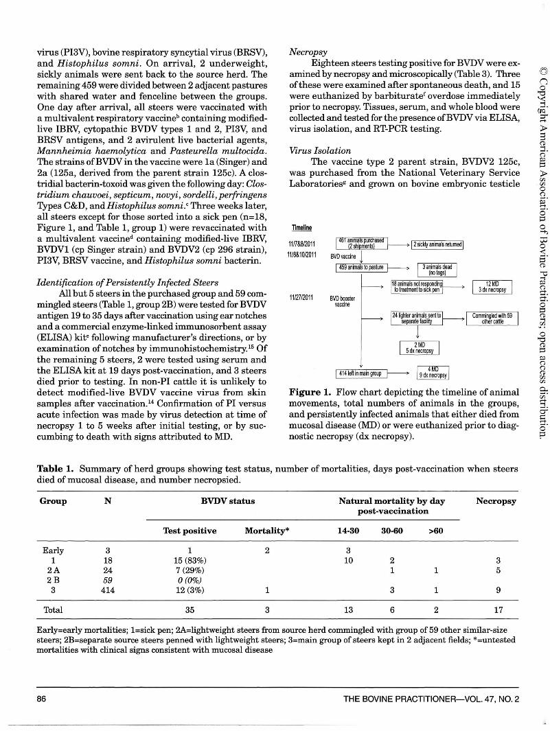
Figure 1. Flow chart depicting the timeline of animal movements, total numbers of animals in the groups, and persistently infected animals that either died from mucosal disease (MD) or were euthanized prior to diagnostic necropsy (dx necropsy).
Table 1. Summary of herd groups showing test status, number of mortalities, days post-vaccination when steers died of mucosal disease, and number necropsied.
Group N BVDVstatus Natural mortality by day Necropsy post-vaccination
Test positive Mortality* 14-30 30-60 >60
Early 3 1 2 3 1 18 15 (83%) 10 2 3
2A 24 7 (29%) 1 1 5 2B 59 0 (0%) 3 414 12 (3%) 1 3 1 9
Total 35 3 13 6 2 17
Early=early mortalities; l=sick pen; 2A=lightweight steers from source herd commingled with group of 59 other similar-size steers; 2B=separate source steers penned with lightweight steers; 3=main group of steers kept in 2 adjacent fields; *=untested mortalities with clinical signs consistent with mucosal disease
86 THE BOVINE PRACTITIONER-VOL. 47, NO. 2
(BeT) cells. Homogenized tissues diluted 1:10 in Bovarnick's media (sucrose, KH2PO4, K2HPO4, glutamic acid, phenol red), or blood and serum diluted 1:10 in Earle's media (199E),h were applied to semi-confluent BeT cells in 24 well plates. Plates were incubated at 98.6°F (37°C) for 1 hour in 5% CO2, washed lX and maintained in complete media 199E with 2% Pharma-grade FBS.i BeT cells and FBS had been previously found negative for BVDV by VI and RT-PCR. Cultures were observed daily for cytopathic effect. Cells were stained for BVDV on days 2 and 9 post-inoculation with monoclonal antibody 20.10.6,j followed by fluorescent-labeled goat anti-mouse antibody.k Cytopathic virus isolated from the first laboratory-diagnosed case of MD was purified by 4X plaque purification, and partially purified from the second diagnosed case (Table 3, calf numbers 3 and 15).
RNA Extraction Total RNA was extracted from tissue, blood, cell
culture isolates, or monovalent BVDV2 125a binary ethylenimine inactivated vaccine provided by the vaccine company using Trizol Reagent.1 The isolated RNA was eluted in RN ase free dH2O and frozen at -l12°F (-80°C) until used in RT-PCR assays. RNA for the vaccine type 1 (Singer c) and type 2 (125a) BVDV was also directly supplied from the company.
PCR and Sequencing To distinguish BVDVl and BVDV2, type-specific
PCR was run as previously described for the 5'-UTR.48
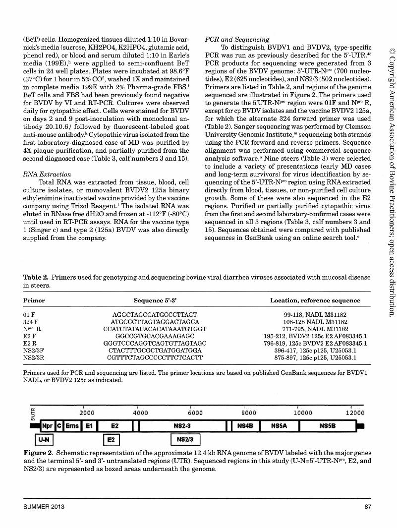
PCR products for sequencing were generated from 3 regions of the BVDV genome: 5'-UTR-Npro (700 nucleotides), E2 (625 nucleotides), and NS2/3 (502 nucleotides). Primers are listed in Table 2, and regions of the genome sequenced are illustrated in Figure 2. The primers used to generate the 5'UTR-Npro region were 0lF and Npro R, except for cp BVDV isolates and the vaccine BVDV2 125a, for which the alternate 324 forward primer was used (Table 2). Sanger sequencing was performed by Clemson University Genomic Institute,m sequencing both strands using the PCR forward and reverse primers. Sequence alignment was performed using commercial sequence analysis software.n Nine steers (Table 3) were selected to include a variety of presentations (early MD cases and long-term survivors) for virus identification by sequencing of the 5'-UTR-Npro region using RNA extracted directly from blood, tissues, or non-purified cell culture growth. Some of these were also sequenced in the E2 regions. Purified or partially purified cytopathic virus from the first and second laboratory-confirmed cases were sequenced in all 3 regions (Table 3, calf numbers 3 and 15). Sequences obtained were compared with published sequences in Geni3ank using an online search tool. 0
Table 2. Primers used for genotyping and sequencing bovine viral diarrhea viruses associated with mucosal disease in steers.
Primer
01 F 324F N pro R E2F E2R NS2/3F NS2/3R
Sequence 5'-3'
AGGCTAGCCATGCCCTTAGT ATGCCCTTAGTAGGACTAGCA
CCATCTATACACACATAAATGTGGT GGCCGTGCACGAAAGAGC
GGGTCCCAGGTCAGTGTTAGTAGC CTACTTTGCGCTGATGGATGGA CGTTTCTAGCCCCCTTCTCACTT
Location, reference sequence
99-118, NADL M31182 108-128 NADL M31182 771-795, NADL M31182
195-212, BVDV2 125c E2 AF083345.1 796-819, 125c BVDV2 E2 AF083345.1
396-417, 125c p125, U25053.l 875-897, 125c p125, U25053.1
Primers used for PCR and sequencing are listed. The primer locations are based on published GenBank sequences for BVDVl NADL, or BVDV2 125c as indicated.
li:r ~ 2000 4000
I . I "
6000 sooo 10000 120()0
-lf~prlcl&rm l 11 I 82 1 1 NS24 l'I NS48 I NSSA I NHI t--luJt 1 ~ NS?/3
Figure 2. Schematic representation of the approximate 12.4 kb RNA genome ofBVDV labeled with the major genes and the terminal 5'- and 3'- untranslated regions (UTR). Sequenced regions in this study (U-N=5'-UTR-Npro, E2, and NS2/3) are represented as boxed areas underneath the genome.
SUMMER 2013 87

Table 3. Persistently infected cattle by day of mortality and BVDV sequencing results.
Died/ Sequencing results
Calf no. DPV* euthanized Postmortem MD 5-UTR-Npro E2 NS2/3
lt 16 Died No PR 2t 18 Died No PR 3 19 Died Yes + NCP, 125c§ 125c§ 125c§ 4 23 Died No PR 5 23 Died No PR 6 24-29 Died No PR 7 25 Died No PR 8 26 Died No PR 9 27 Died No PR 10 28 Died No PR NCP 11 28 Died No PR 12 29 Died No PR 13 29 Died No PR 14t 32 Died No PR 15 38 Died Yes + NCP, 125c§ NCP, 125c§ 125c§ 16 39 Died No PR 17 47-49 Died No PR 18 48 Died No PR 19 48 Died No PR 20 49 Euthanized Yes + NCP NCP 21 49 Euthanized Yes No 22 49 Euthanized Yes No NCP NCP 23 66 Died No PR 24 66 Euthanized Yes No 25 66 Euthanized Yes No NCP NCP 26 66 Euthanized Yes No NCP 27 66 Euthanized Yes No 28 72 Euthanized No No 29 72 Euthanized No No 30 73 Euthanized Yes No 31 73 Euthanized Yes No 32 73 Euthanized Yes No 33 99 Died Yes UN NCP NCP 34 112 Euthanized Yes No 35 112 Euthanized Yes No NCP NCP 36 114 Euthanized Yes No 37 114 Euthanized Yes No 38 114 Euthanized Yes No
35 steers tested positive for BVDV by RT-PCR, VI, ELISA, or IHC, and 3 untested mortalities died with symptoms attributed to MD. All steers died of natural mortality or were euthanized and necropsied as part of the study. *=days post-vaccination, t=untested for BVDV, §=sequence result of purified or semi-purified cytopathic virus, MD=confirmed mucosal disease, PR=presumed mucosal disease mortality, +=macroscopic lesions consistent with !_\1:D, UN=undetermined, NCP=sequence ofnoncytopathic virus related (97%) to BVDV2 strain SH-28 (GenBank HQ258810.1), 125c=sequence with >99% identity with BVDV2 125c for regions U5'UTR-Npro, E2; GenBankAF038845.l, and NS2/3 (p125): GenBank U25053.1).
Results
Outbreak Description Within 10 days of purchase, some animals in the
purchased group began exhibiting nasal discharge and variably bloody diarrhea. This led to the entire purchase group being size/condition and health sorted.
88
Cattle noticeably sick with bloody diarrhea (n=15), lame (n=2), and suspect urolithiasis (n=l) were moved to an isolation pen (Table 1, group 1) and individually treated for coccidiosis with an oral drench.P Twenty-four lightweight steers (Table 1, group 2A) were moved to a separate facility where they were commingled with 59 other cattle (Table 1, group 2B) of similar size and age,
THE BOVINE PRACTITIONER-VOL. 47, NO. 2
(Q)
n 0
"'O '-< '"i ......
(JQ ~ ..-+-
> 8 (D '"i ...... (") ~ ~
> 00 00 0 (") ...... a ...... 0 ~ 0 1-i;
to 0 < ...... ~ (D
~ '"i ~ (") ..-+-...... ..-+-...... 0 ~ (D '"i 00
0 "'O (D
~ ~ (") (") (D 00 00
0.. ...... 00 ..-+-'"i ...... cr' I= ..-+-...... 0 p

sharing pens, fencelines, and water. A flow chart illustrating the disposition of groups is shown in Figure 1.
Two steers died between 2 and 3 weeks after purchase with clinical signs consistent with MD, acute or chronic BVDV, including depression, nasal and oral discharge, mucosal erosions in the oral cavity, and variable bloody diarrhea. 31•58 These were the first deaths in the consignment, and no laboratory samples were collected (Table 3, calf numbers 1 and 2). A third mortality was examined postmortem and had gross lesions typical of MD; it was positive for BVDV by VI and BVDV2 by type-specific RT-PCR (Table 3, calf number 3).
All remaining steers from the consignment, except for 1 additional mortality, were then tested for BVDV, and 34 (Table 3) were test-positive: 15 from the sick pen (15/18, 83%, Table 1, group 1), 7 from the group of sorted lightweight steers (7/24, 29% Table 1, group 2A), and 12 in the main herd (12/414, 3%, Table 1, group 3). None of the 59 commingled steers (Table 1, group 2B) tested BVDV-positive by ELISA.
Over the next 2 months 18 of the remaining BVDVpositive steers died on the farm with signs typical of MD, 10 of these within the 8 days following the initial diagnosis (Table 3). An epidemic curve illustrating the timing of the natural mortalities is shown in Figure 3. Most farm mortalities were not examined (n=16), 1 had confirmed MD based on typical postmortem examination findings (Table 3, calf number 15) and positive VI and RT-PCR, and 1 was undetermined due to predator scavenging, autolysis, and confounding grain overload (n=l, Table 3, calf number 33). The remaining 16 positive steers were free of signs of illness and were euthanized over the next 3 months and examined postmortem. Only 1 of these steers had mild gross lesions attributed to MD (Table 3, calf number 20). Of the 21 presumed or confirmed MD mortalities, 16 had received only the first BVDV-containing vaccine at the backgrounding facility.
Necropsy Of the steers necropsied, 3 had macroscopic lesions
consistent with MD (Table 3, calf numbers 3, 15, and 20). Steers numbered 3 and 15 died on days 19 and 38 after vaccination, while calf number 20 was euthanized on day 49 after the initial vaccination. The main findings were ulcers in the tongue, lips, oral cavity, esophagus, rumen, Peyer's patches, cecum, and colon (Figure 4). One animal had a secondary fungal infection of the intestinal ulcers, and another had marked enteritis with crypt necrosis. The confirmatory diagnosis of MD was based upon typical macroscopic and microscopic lesions, the identification of BVDV by RT-PCR, and isolation of cp and ncp virus. A fourth animal (calf number 33) in poor postmortem condition (predator scavenged and autolytic tissues) had lesions suggestive of MD or ruminal acidosis/grain overload. The remaining steers had minimal
SUMMER 2013
gross and histological lesions; no other bacterial or viral pathogens were found as significant factors.
PCR and Sequencing Blood and tissue samples from all of the necropsied
steers (n=18) were positive for BVDV2 by type-specific RT-PCR (data not shown). Sequences of ncp virus isolates and RNA directly from blood or tissues of 9 steers (Table 3) were identical to each other and most closely related (97% identity) to BVDV2 strain SH-28 (GenBank HQ258810.l) in the 5'-UTR-Npro and E2 regions (sequences submitted to GenBank; accession numbers KC596021, KC596022). We sequenced the 5'UTR-Npro
5 ;4 ~
~3 .. ~2 e zl
0 +,L,-,.,..,_,...,..,..,_,.,...,..,_~~-,---,'-r",-'r-r-r--rr-,L~ ",-r,-~~~-r,-,'c
15 19 23 27 31 35 39 43 47 51 55 59 63 Days post-vaccination
• Presumed MD • Confirmed MD
Figure 3. Epid~mic curve for the outbreak of mucosal disease (MD) in a Wyoming cattle herd. The natural mortalities presumed or confirmed by laboratory testing as MD are illustrated.
Figure 4. Images from the index case (Table 3, calf number 3) of mucosal disease following vaccination 19 days earlier; cytopathic BVDV2 125c BVDV was isolated from tissues. A. Multiple shallow ulcers on dorsum of tongue. B. Ulceration of gut-associated lymphoid tissue with adherent diphtheretic membrane. Histologically there was superimposed mycotic infection throughout this ulcerated lymphoid tissue. C. Focal ulceration of larynx overlying vocal ligament. D. Gingivitis with recession of gum line, exposing roots of cheek teeth.
89

region of the reference BVDV2 125c strain (submitted to GenBank; accession KC596020) to compare to the vaccine virus BVDV2 125a and our cp isolates. For cp BVDV isolates and vaccine BVDV2 125a, we used the alternate forward primer (Table 2, primer 324) for the 5'UTR-Npro product. This alternate primer overlaps the 0lF primer, but is shifted 3' by 10 nt. Based on our sequencing results, there is a single base deletion at the 3' end of the 324 primer, and it appears there are additional changes under the 0lF primer that prevent efficient amplification using this primer. Non-purified cp virus from 1 of these steers (Table 3, calf number 15) sequenced as BVDVl Singer when the 01 forward primer was used instead of the 324 primer.
The genomic regions of E2 and NS2/3 were sequenced to further verify the identity of the cp virus isolates from the first 2 cases (Table 3, calf numbers 3 and 15). These isolates were identical (>99%) to each other, to the vaccine BVDV2-125a, and to published BVDV1125c sequences in GenBank (Accession numbers: AF083345.l and U25053.1).
Virus Isolation Only ncp virus was isolated from blood or serum
samples, except for 1 steer from which both biotypes were obtained (data not shown). Viruses isolated from tissue samples collected at necropsy included both cp and ncp cell culture characteristics, and both biotypes could be identified in all animals necropsied (n=18) as late as 3 months after the outbreak without evidence of disease (data not shown). This agrees with previous reports finding long-term presence and shedding of cp virus in PI animals, even when co-infected with a heterologous virus.22 Such a co-infecting virus has been shown to be the source for genetic recombination with endogenous virus, resulting in late-onset MD.45
Discussion
Cattle persistently infected with BVDV that survive past weaning are at risk of dying from MD when co-infected with a cp virus. There are 3 main sources of cp BVDV causing MD: 1) spontaneous mutation of ncp virus resulting in the cp biotype, accounting for most episodes of MD in herds and includes recombination with field or vaccine virus, 2) exposure to an exogenous field strain cp BVDV, or 3) exposure to exogenous cp virus in a live vaccine. 3•5•40 Determining the source of cp virus causing MD goes beyond routine diagnostic testing, since it requires purification steps to separate the ncp and cp BVDV from cell cultures, followed by sequencing to identify the specific virus.3,24,25,34
The vaccine BVDV2 125a and cp isolates were difficult to amplify by RT-PCR in the 5'-UTR region, and required a different forward primer (Table 2, primer 324)
90
due to a polymorphism at the forward priming site. This was the likely cause of finding type 1 BVDV Singer strain in a non-purified cp isolate from 1 steer (Table 3, calf number 15) when this region was sequenced using the alternate (01) forward primer, while the E2 and NS2/3 regions gave sequences identical to reference BVDV2 125c and the vaccine. This calf had received 2 doses of vaccine containing type 1 BVDV Singer, vaccinated on days 1 and 21.
Our sequencing results demonstrate genetic identity (>99%) between the cp virus isolated from the first 2 diagnosed MD cases (Table 3, calf numbers 3 and 15) and the vaccine BVDV2 virus, indicating MD in these steers was probably directly triggered by the vaccine. There was not close genetic identify between the ncp and cp BVDV in the genomic regions that were sequenced, and antigenic relatedness was not tested in this study. Antigenically homologous virus pairs with failure to mount an antibody response have been found in many cases of MD;8 however, antigenically and genetically unrelated viruses have been found in experimental and naturally occurring cases ofMD.5•10•47•49•57 It is likely that a combination of viral and host factors determine the clinical outcome.
The temporal pattern of cases illustrated in the epidemic curve (Figure 3) was consistent with a pointsource exposure, with cases beginning 2 weeks after vaccination. Although many early mortalities were not examined by necropsy, the timing of the mortalities in PI steers with identical clinical signs supports a presumptive diagnosis ofMD. The short incubation from vaccination to mortalities is unlike the pattern that is seen in delayed-onset post-vaccine MD after genetic recombination occurs between a live vaccine and the endogenous ncp virus.3•45 lt is not possible to completely exclude the possibility of endogenous ncp virus mutation or recent exposure to field strains of cp BVDV2 125c. In either of these situations, the temporal pattern of cases should have exhibited delayed-onset or multiple peaks rather than a single cluster (within 2 months) after vaccination.
From this group of 459 steers, 35 tested positive for BVDV (1 by necropsy and 34 by ELISA or IHC), and 2 early and 1 late untested mortalities were presumed positive. All positive steers not dying of presumed MD on the farm were necropsied 1 to 5 weeks after initial testing and found to be BVDV positive by both PCR and VI, thereby confirming PI status. It is noteworthy that almost half(n=l 7) of the PI animals that had the same ncp virus based on the 5'-UTR-Npro and E2 regions did not develop MD as late as 3 months post-vaccination. Late onset, chronic, or transient MD has been reported, but the cause of the variable outcome from classic acute MD is not clear. 8•
10•18
This outbreak illustrates the potential risk that exists when PI cattle are vaccinated with cp modified-
THE BOVINE PRACTITIONER-VOL. 47, NO. 2

live virus vaccine. The occurrence of presumptive postvaccinal MD should trigger a search for additional persistently infected asymptomatic animals so that they can be eliminated from the cohort. Sorting of sickly and small animals was partially successful in separating PI animals from the rest of the herd, removing 25 of the 38 (66%) PI animals and reducing the group prevalence from 8% to 3%. However, sorting based on physical characteristics still missed 13 animals that would have contributed to BVD persistence within the herd had they not been detected by laboratory testing. The precipitation of MD by vaccination led to laboratory testing, identification, and culling of the other PI cattle, and likely prevented further BVD losses in the herd.
The benefit of vaccination against bovine respiratory complex agents, including BVDV, is well known. 35
The use of modified-live virus vaccine may be preferred over killed virus products when there is need to produce a faster and broader immune response. Despite direct contact with PI cattle shedding large amounts of virus, there was no noticeable evidence of acute BVD illness in non-PI steers. This may have been due to protection elicited by the vaccine, 19 though a controlled study with non-vaccinated na:ive controls would be needed to prove this point.
Conclusions
Collectively, the molecular and epidemiologic findings are consistent with a vaccine origin in this MD outbreak. Recognition of MD 2 to 4 weeks after vaccination with modified-live cp virus should prompt testing of the entire group to identify persistently infected animals so that they can be isolated or culled. It is more important to identify PI cattle than to establish whether vaccine triggered MD, since these PI animals shed a large amount of virus, and some of them may proceed to develop terminal disease due to mutations of the endogenous ncp BVDV. This case demonstrates the utility of a 3-pronged approach to BVDV biosecurity: 1) laboratory testing to identify PI animals, 2) separation or culling of positives, and 3) vaccination to protect na:ive animals.
Endnotes
avirashield 6 + H. Somni, Novartis Animal Health, Basel, Switzerland hVista Once, Merck Animal Health, Whitehouse, ME cVision® 7 Somnus, Intervet Inc., Millsboro, DE dExpress 5 HS, Boehringer Ingelheim Vetmedica, Inc., St. Joseph, MO eJdexx Herdchek BVDV test kit, Idexx Laboratories, Inc., Westbrook, ME
SUMMER 2013
f'Beuthanasia-D Special. Merck Animal Health, Summit, NJ gN ational Veterinary Service Laboratories, Ames IA hEarle's media (199E), Sigma, St. Louis, MO iPharma-grade FBS, PAA Laboratories Inc, Pasching Austria jMonoclonal antibody 20.10.6, Dr. Ed Dubovi, Cornell University, Ithaca, NY kFluorescein labeled goat anti-mouse antibody, Invitrogen, Carlsbad, CA 1Trizol Reagent, Gibco BRL, Gaithersburg, MD mClemson University Genomic Institute, Clemson, SC nSeqMan Pro, DNASTAR, Lasargene software, Madison, WI 0Basic Local Alignment Search Tool (BLAST). National Center for Biotechnology Information, National Library of Medicine. Online at http://blast.ncbi.nlm.nih.gov/ Blast.cgi PCorid, Merial Limited, Dublin, GA
Acknowledgements
We authors thank Paula Jaeger and Mercedes Thelen for histology and immunohistochemistry preparations, Breanna Bonner for assistance with necropsies, Jennifer McKenna for cell culture support, and Dr. Ed Dubovi for the gift of monoclonal antibody 20.10.6. The study was funded in part by Merck Animal Health. The authors declare no conflict of interest.
References
1. Balint A, Palfi V, Belak S, Baule C. Viral sequence insertions and a novel cellular insertion in the NS2 gene of cytopathic isolates of bovine viral diarrhea virus as potential cytopathogenicity markers. Virus Genes 2005;30:49-58. 2. Becher P, Orlich M, Kosmidou A, Konig M, Baroth M, Thiel HJ. Genetic diversity of pestiviruses: identification of novel groups and implications for classification. Virology 1999;262:64-71. 3. Becher P, Orlich M, Thiel HJ. RNA recombination between persisting pestivirus and a vaccine strain: generation of cytopathogenic virus and induction oflethal disease. J Virol 2001;75:6256-6264. 4. Bielefeldt-Ohmann H, Tolnay AE, Reisenhauer CE, Hansen TR, Smirnova N, Van Campen H. Transplacental infection with noncytopathic bovine viral diarrhoea virus types lb and 2: viral spread and molecular neuropathology. J Comp Pathol 2008;138:72-85. 5. Bolin SR. The pathogenesis of mucosal disease. Vet Clin North Am Food Anim Pract 1995;11:489-500. 6. Bolin SR, McClurkin AW, Cutlip RC, Coria MF. Severe clinical disease induced in cattle persistently infected with noncytopathic bovine viral diarrhea virus by superinfection with cytopathic bovine viral diarrhea virus. Am J Vet Res 1985;46:573-576. 7. Bolin SR, Ridpath JF. Prevalence of bovine viral diarrhea virus genotypes and antibody against those viral genotypes in fetal bovine serum. J Vet Diagn Invest 1998;10:135-139. 8. Brownlie J. The pathways for bovine virus diarrhoea virus biotypes in the pathogenesis of disease. Arch Virol Suppl 1991;3:79-96. 9. Brownlie J, Clarke MC, Howard CJ. Experimental production of fatal mucosal disease in cattle. Vet Rec 1984;114:535-536.
91

10. Bruschke CJ, HaghparastA, Hoek A, Rutten VP, Wentink GH, van Rijn PA, van Oirschot JT. The immune response of cattle, persistently infected with noncytopathic BVDV, after superinfection with antigenically semi-homologous cytopathic BVDV. ¼t Immunol Immunopathol 1998;62:37-50. 11. Campbell JR. Effect of bovine viral diarrhea virus in the feedlot. Vet Clin North Am Food Anim Pract 2004;20:39-50. 12. Childs T. X disease of cattle - Saskatchewan. Can J Comp Med Vet Sci 1946;10:316-319. 13. Collins ME, Desport M, Brownlie J. Bovine viral diarrhea virus quasispecies during persistent infection. Virology 1999;259:85-98. 14. Corbett EM, Grooms DL, Bolin SR. Evaluation of skin samples for bovine viral diarrhea virus by use ofreverse transcriptase polymerase chain reaction assay after vaccination of cattle with a modified-live bovine viral diarrhea virus vaccine. Am J ¼t Res 2012;73:319-324. 15. Cornish TE, van Olphen AL, Cavender JL, Edwards JM, Jaeger PT, Vieyra LL, Woodard LF, Miller DR, O'Toole D. Comparison of ear notch immunohistochemistry, ear notch antigen-capture ELISA, and huffy coat virus isolation for detection of calves persistently infected with bovine viral diarrhea virus. J Vet Diagn Invest 2005;17:110-117. 16. Deregt D. Introduction and history. In: Goyal SM, Ridpath JF, eds. Bovine viral diarrhea virus - diagnosis, management and control. Ames, IA: Blackwell Publishing, 2005;3-33. 17. Driskell EA, Ridpath JF. A survey of bovine viral diarrhea virus testing in diagnostic laboratories in the United States from 2004 to 2005. J Vet Diagn Invest 2006;18:600-605. 18. Edwards S, Wood L, Brockman S, lbata G. Clinical and virological observations of a mucosal disease outbreak with persistently-infected seropositive survivors. Arch Virol Suppl 1991;3:125-132. 19. Elam NA, Thomson DU, Gleghorn JF. Effects oflong- or short-term exposure to a calf identified as persistently infected with bovine viral diarrhea virus on feedlot performance of freshly weaned, transportstressed beef heifers. J Anim Sci 2008;86:1917-1924. 20. Evermann JF, Barrington GM. Clinical features. In: Goyal SM, Ridpath JF, eds. Bovine viral diarrhea virus - diagnosis, management, and control. Ames, Iowa: Blackwell Publishing, 2005;105-119. 21. Evermann JF, Ridpath JF. Clinical and epidemiologic observations of bovine viral diarrhea virus in the northwestern United States. Vet Microbiol 2002;89:129-139. 22. Fray MD, Clarke MC, Thomas LH, McCauley JW, Charleston B. Prolonged nasal shedding and viraemia of cytopathogenic bovine virus diarrhoea virus in experimental late-onset mucosal disease. Vet Rec 1998;143:608-611. . 23. Fredriksen B, Press CM, Sandvik T, Odegaard SA, Loken T. Detection of viral antigen in placenta and fetus of cattle acutely infected with bovine viral diarrhea virus. Vet Pathol 1999;36:267-275. 24. Fricke J, Gunn M, Meyers G. A family of closely related bovine viral diarrhea virus recombinants identified in an animal suffering from mucosal disease: new insights into the development of a lethal disease in cattle. Virology 2001;291:77-90. 25. Fritzemeier J, Greiser-Wilke I, Haas L, Pituco E, Moennig V, Liess B. Experimentally induced "late-onset" mucosal disease-characterization of the cytopathogenic viruses isolated. ¼t Microbiol 1995;46:285-294. 26. Fulton RW. Vaccines. In: Goyal SM, Ridpath JF, eds. Bovine viral diarrhea virus - diagnosis, management, and control. Ames, Iowa: Blackwell Publishing, 2005;209-222. 27. Fulton RW, Johnson BJ, Briggs RE, Ridpath JF, Saliki JT, Confer AW, Burge LJ, Step DL, Walker DA, Payton ME. Challenge with bovine viral diarrhea virus by exposure to persistently infected calves: protection by vaccination and negative results of antigen testing in non vaccinated acutely infected calves. Can J Vet Res 2006;70: 121-127. 28. Greiser-Wilke I, Haas L, Dittmar K, Liess B, Moennig V. RNA insertions and gene duplications in the nonstructural protein p125 region of pestivirus strains and isolates in vitro and in vivo. Virology 1993;193:977-980. 29. Harpin S, Talbot B, Mbikay M, Elazhary Y. Immune response to vaccination with DNA encoding the bovine viral diarrhea virus major glycoprotein gp53 (E2). FEMS Microbiol Lett 1997;146:229-234.
92
30. Hessman BE, Fulton RW, Sjeklocha DB, Murphy TA, Ridpath JF, Payton ME. Evaluation of economic effects and the health and performance of the general cattle population after exposure to cattle persistently infected with bovine viral diarrhea virus in a starter feedlot. Am J Vet Res 2009;70:73-85. 31. Hessman BE, Sjeklocha DB, Fulton RW, Ridpath JF, Johnson BJ, McElroy DR. Acute bovine viral diarrhea associated with extensive mucosal lesions, high morbidity, and mortality in a commercial feedlot. J Vet Diagn Invest 2012;24:397-404. 32. Hewicker-Trautwein M, Trautwein G, Frey HR, Liess B. Variation in neuropathogenicity in sheep fetuses transplacentally infected with non-cytopathogenic and cytopathogenic biotypes of bovine-virus diarrhoea virus. Zentralbl Veterinarmed B 1995;42:557-567. 33. Houe H. Epidemiology of bovine viral diarrhea virus. Vet Clin North Am Food Anim Pract 1995;11:521-547. 34. Jones LR, Zandomeni R, Web'er EL. Quasispecies in the 5' untranslated genomic region of bovine viral diarrhoea virus from a single individual. J Gen Virol 2002;83:2161-2168. 35. Kelling CL. Evolution of bovine viral diarrhea virus vaccines. Vet Clin North Am Food Anim Pract 2004;20:115-129. 36. Liebler-Tenorio EM. Pathogenesis. In: Goyal SM, Ridpath JF, eds. Bovine viral diarrhea virus - diagnosis, management, and control. Ames, Iowa: Blackwell Publishing, 2005;121-143. 37. Loehr Bl, Frey HR, Moennig V, Greiser-Wilke I. Experimental induction of mucosal disease: consequences of superinfection of persistently infected cattle with different strains of cytopathogenic bovine viral diarrhea virus. Arch Virol 1998;143:667-679. 38. McClurkin AW, Bolin SR, Coria MF. Isolation of cytopathic and noncytopathic bovine viral diarrhea virus from the spleen of cattle acutely and chronically affected with bovine viral diarrhea. J Am Vet Med Assoc 1985;186:568-569. 39. McKercher DG, Saito JK, Crenshaw GL, Bushnell RB. Complications in cattle following vaccination with a combined bovine viral diarrhea-infectious bovine rhinotracheitis vaccine. J Am ¼t Med Assoc 1968;152:1621-1624. 40. Meyers G, Thiel HJ. Molecular characterization of pestiviruses. Adv Virus Res 1996;47:53-118. 41. Peter CP, Tyler DE, Ramsey FK. Characteristics of a condition following vaccination with bovine virus diarrhea vaccine. J Am Vet Med Assoc 1967;150:46-52. 42. Qi F, Ridpath JF, Lewis T, Bolin SR, Berry ES. Analysis of the bovine viral diarrhea virus genome for possible cellular insertions. Virology 1992;189:285-292. 43. Ridpath JF. BVDV genotypes and biotypes: practical implications for diagnosis and control. Biologicals 2003;31:127-131. 44. RidpathJF. ImmunologyofBVDVvaccines.Biologicals 2013;41:14-19. 45. Ridpath JF, Bolin SR. Delayed onset postvaccinal mucosal disease as a result of genetic recombination between genotype 1 and genotype 2 BVDV. Virology 1995;212:259-262. 46. Ridpath JF, Bolin SR. Differentiation of types la, lb and 2 bovine viral diarrhoea virus (BVDV) by PCR. Mol Cell Probes 1998;12:101-106. 4 7. Ridpath JF, Bolin SR. Viral protein production in homogeneous and mixed infections of cytopathic and noncytopathic BVD virus. Arch Virol 1990;111:247-256. 48. Ridpath JF, Bolin SR, Dubovi EJ. Segregation of bovine viral diarrhea virus into genotypes. Virology 1994;205:66~74. 49. Ridpath JF, Lewis TL, Bolin SR, Berry ES. Antigenic and genomic comparison between non-cytopathic and cytopathic bovine viral diarrhoea viruses isolated from cattle that had spontaneous mucosal disease. J Gen Virol 1991;72 ( Pt 3):725-729. 50. Ridpath JF, Qi F, Bolin SR, Berry ES. Natural recombination in bovine viral diarrhea viruses. Arch Virol Suppl 1994;9:239-244. · 51. Rosner SF. Complications following vaccination of cattle against infectious bovine rhinotracheitis, bovine viral diarrhea - mucosal disease, and parainfluenza type 3. J Am Vet Med Assoc 1968;152:898-902.
THE BOVINE PRACTITIONER-VOL. 47, NO. 2

52. Stokstad M, Brownlie J, Collins ME. Analysis of variation of bovine viral diarrhoea virus E2 sequence following transplacental infection of cattle. Vet Microbial 2004;102:141-145. 53. Tajima M, Dubovi EJ. Genetic and clinical analyses of bovine viral diarrhea virus isolates from dairy operations in the United States of America. J Vet Diagn Invest 2005;17:10-15. 54. Tautz N, Meyers G, Stark R, Dubovi EJ, Thiel HJ. Cytopathogenicity of a pestivirus correlates with a 27-nucleotide insertion. J Viral 1996;70:7851-7858. 55. Vilcek S, Durkovic B, Kolesarova M, Paton DJ. Genetic diversity of BVDV: consequences for classification and molecular epidemiology. Prev Vet Med 2005;72:31-35; discussion 215-219.
SUMMER 2013
56. Vilcek S, Greiser-Wilke I, Nettleton P, Paton DJ. Cellular insertions in the NS2-3 genome region of cytopathic bovine viral diarrhoea virus (BVDV) isolates. Vet Microbial 2000;77:129-136. 57. Westenbrink F, Straver PJ, Kimman TG, de Leeuw PW. Development of a neutralising antibody response to an inoculated cytopathic strain of bovine virus diarrhoea virus. Vet Rec 1989;125:262-265. 58. Wilhelmsen CL, Bolin SR, Ridpath JF, Cheville NF, Kluge JP. Lesions and localization of viral antigen in tissues of cattle with experimentally induced or naturally acquired mucosa! disease, or with naturally acquired chronic bovine viral diarrhea. Am J Vet Res 1991;52:269-275.
93