Embed Size (px)
Citation preview
Equine “Recurrent” Uveitis is a
“Persistent” Problem in Horses Equine Ophthalmology Service
University of Florida
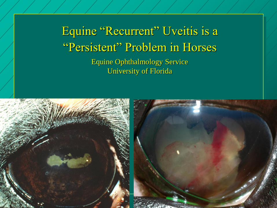
UVEITIS IN THE HORSE
UVEITIS is the
LEADING CAUSE
OF BLINDNESS IN
HORSES
Not a single disease:
SYNDROME, MANY
subsets! 199107
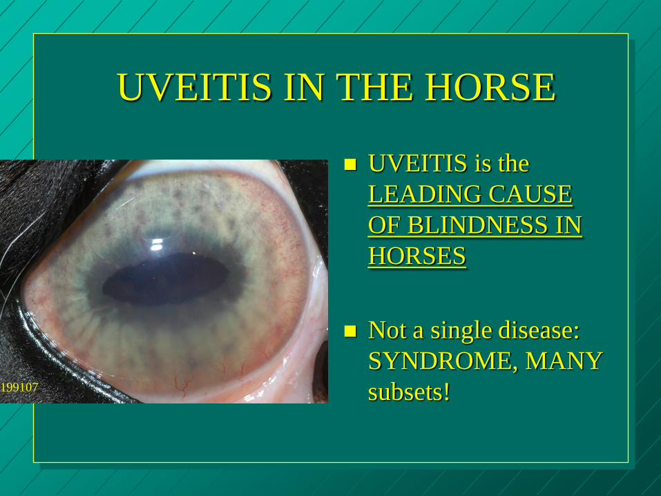
UVEITIS is like LAMINITIS…
– Variety of triggers
– Poorly understood,
but BAD for the
horse
– Variable response to
therapy
– Multiple tissues in
key functional area
involved Fastbridled
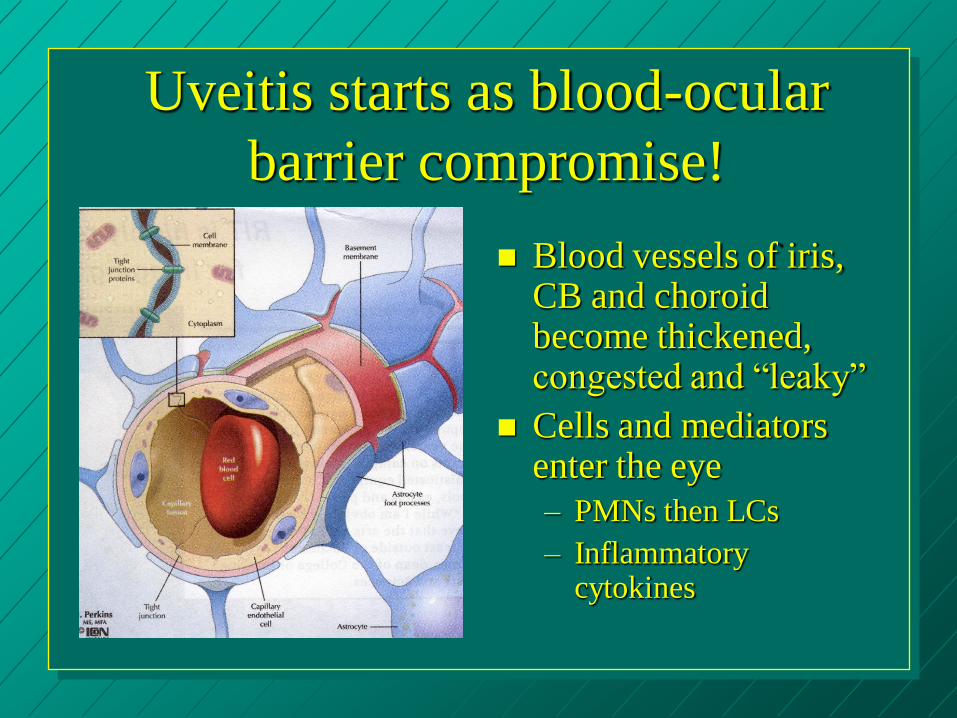
Uveitis starts as blood-ocular
barrier compromise!
Blood vessels of iris, CB and choroid become thickened, congested and “leaky”
Cells and mediators enter the eye
– PMNs then LCs
– Inflammatory cytokines
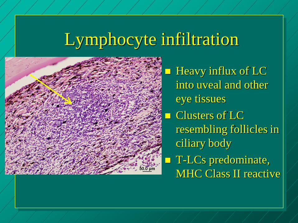
Lymphocyte infiltration
Heavy influx of LC
into uveal and other
eye tissues
Clusters of LC
resembling follicles in
ciliary body
T-LCs predominate,
MHC Class II reactive
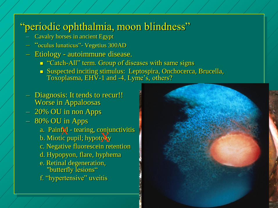
“periodic ophthalmia, moon blindness” – Cavalry horses in ancient Egypt
– “oculus lunaticus”- Vegetius 300AD
– Etiology - autoimmune disease. “Catch-All” term. Group of diseases with same signs
Suspected inciting stimulus: Leptospira, Onchocerca, Brucella, Toxoplasma, EHV-1 and -4, Lyme’s, others?
– Diagnosis: It tends to recur!! Worse in Appaloosas
– 20% OU in non Apps
– 80% OU in Apps a. Painful - tearing, conjunctivitis
b. Miotic pupil; hypotony
c. Negative fluorescein retention
d. Hypopyon, flare, hyphema
e. Retinal degeneration, "butterfly lesions“
f. “hypertensive” uveitis
X X
200555
Comments ERU prevalence in the USA is 1-8%
– 9.2 million horses in the USA (2005)
– 736,000 cases!!
ERU with leptospirosis is a bad form
ELA-A9 in German Warmbloods may be a heritable form of ERU
Appaloosas genetically predisposed. – UM011 microsatellite had greater 182 allele in Apps
with ERU.
– EqMHC1 microsatellite had greater 206 allele in Apps with ERU.
Acute and Chronic quiescent clinical phases

Homing and Molecular Mimicry in ERU
– Mucous membranes communicate!!!
– Antigens in the eye reach the lymphatic system, and vice versa!!
– Infectious agents may only activate ERU. Lepto antigen and horses.
– Self-antigens perpetuate the disease.
Bystander activation
Epitope (a single antigenic site on a protein against which an antibody reacts) spreading
– Shifts in immunoreactivity may cause the waxing/waning character of ERU
– Shifts in immune response to S-antigen and IRBP occur in horses with ERU
– These shifts occur in quiet clinical phases
The retina and vitreous have many T-cells.
– Th lymphocytes in the uveal tract.
Chorioretinitis occurs at all stages of ERU.
Pinealitis is present.
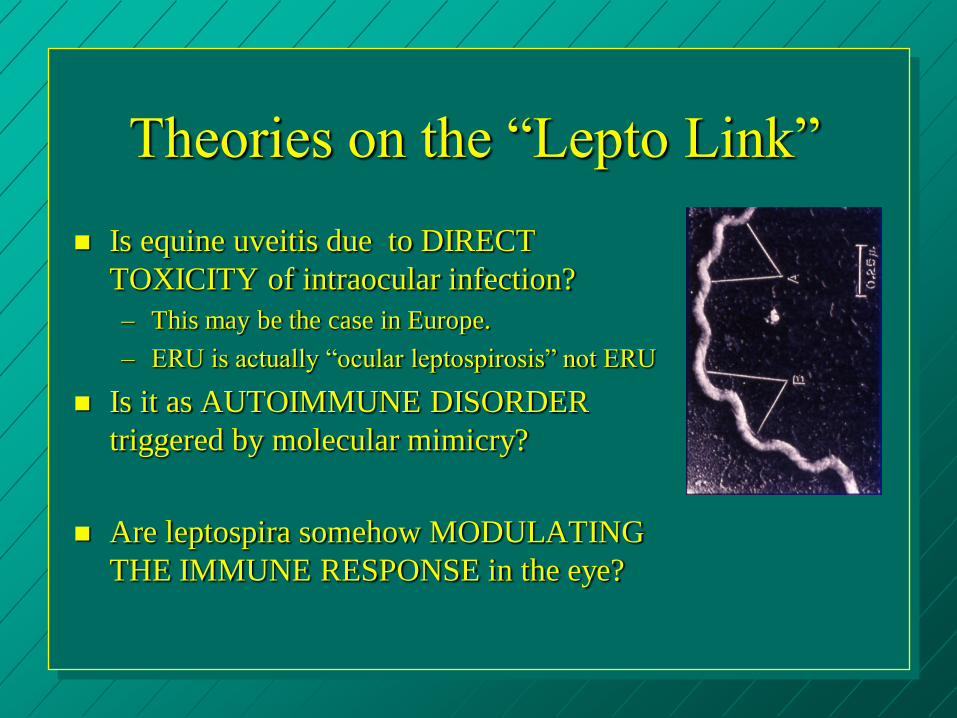
Theories on the “Lepto Link”
Is equine uveitis due to DIRECT
TOXICITY of intraocular infection?
– This may be the case in Europe.
– ERU is actually “ocular leptospirosis” not ERU
Is it as AUTOIMMUNE DISORDER
triggered by molecular mimicry?
Are leptospira somehow MODULATING
THE IMMUNE RESPONSE in the eye?
Testing for Leptospirosis
Most significant are L. pomona and L.
grippotyphosa L. pomona most important in USA
-Titers > 1:400 are significant
-Rarely will rising titer be found in paired
samples--sampling too late in course of disease.
Uveitis from lepto occurs later than the systemic
infection.
Some horses with lepto in eyes are seronegative!
Persistent Leptospirosis may sustain the auto-
immune attacks and be a subset of ERU.
– ERU Eyes: L. gryppotyphosa cultured from vitreous of
52% uveitis eyes in Germany and pomona from
aqueous humor of 20% (70% DNA+) uveitis eyes in
USA
Locally produced antibodies against Lepto cross
react with the cornea, lens and retina (S-antigen
and IRBP).
Not all horses positive for L. pomona have uveitis.
– The serologic evidence of pomona infection is more
frequent than the incidence of ERU.
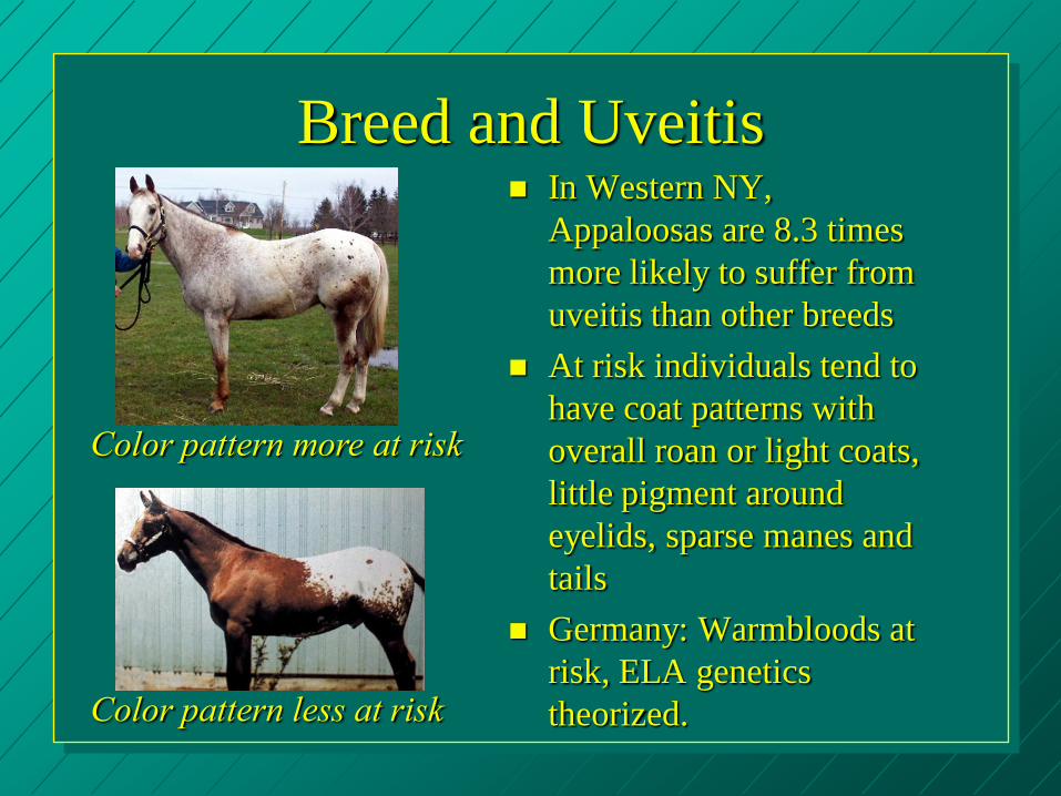
Breed and Uveitis In Western NY,
Appaloosas are 8.3 times
more likely to suffer from
uveitis than other breeds
At risk individuals tend to
have coat patterns with
overall roan or light coats,
little pigment around
eyelids, sparse manes and
tails
Germany: Warmbloods at
risk, ELA genetics
theorized.
Color pattern more at risk
Color pattern less at risk
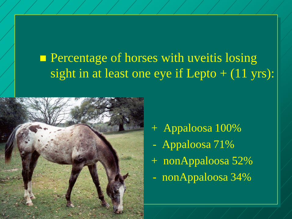
Percentage of horses with uveitis losing
sight in at least one eye if Lepto + (11 yrs):
+ Appaloosa 100%
- Appaloosa 71%
+ nonAppaloosa 52%
- nonAppaloosa 34%
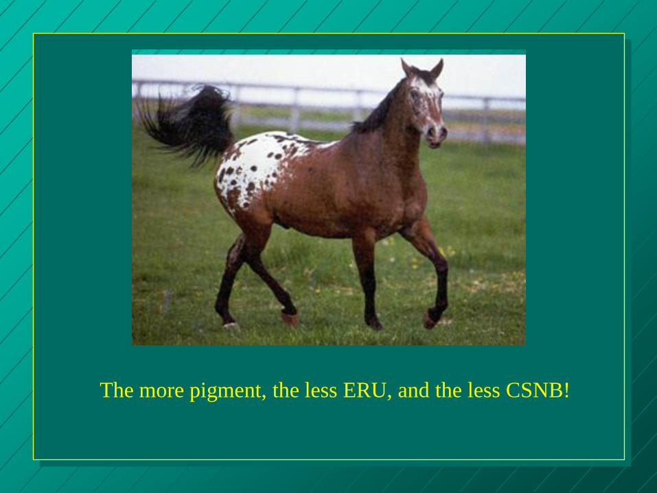
The more pigment, the less ERU, and the less CSNB!
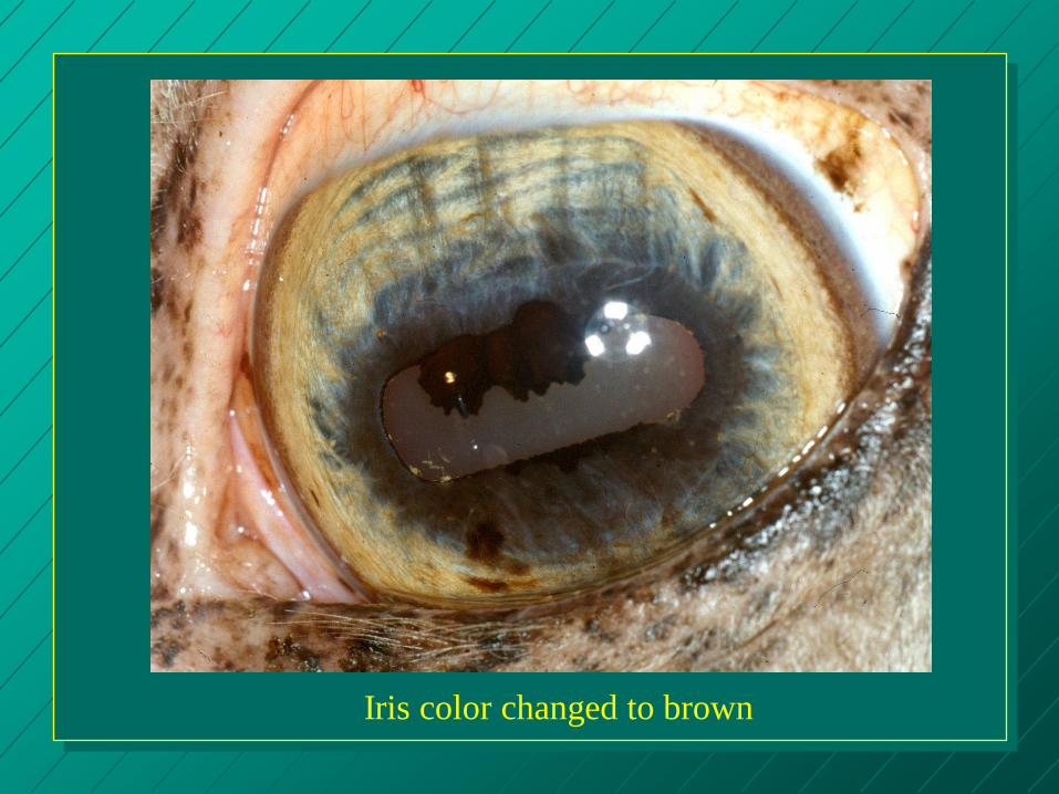
Iris color changed to brown
150952
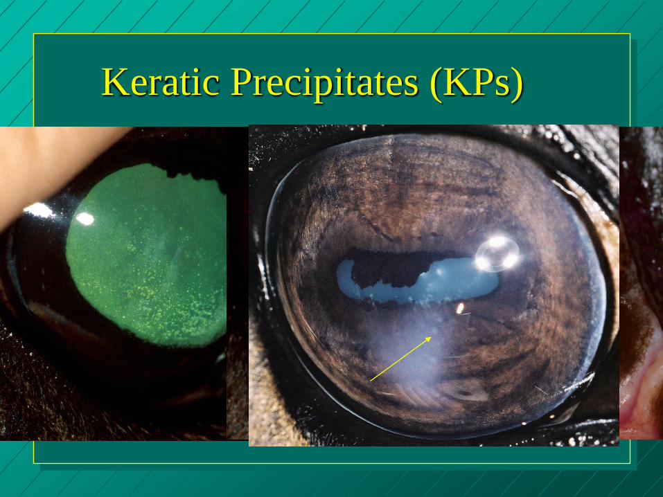
Keratic Precipitates (KPs)
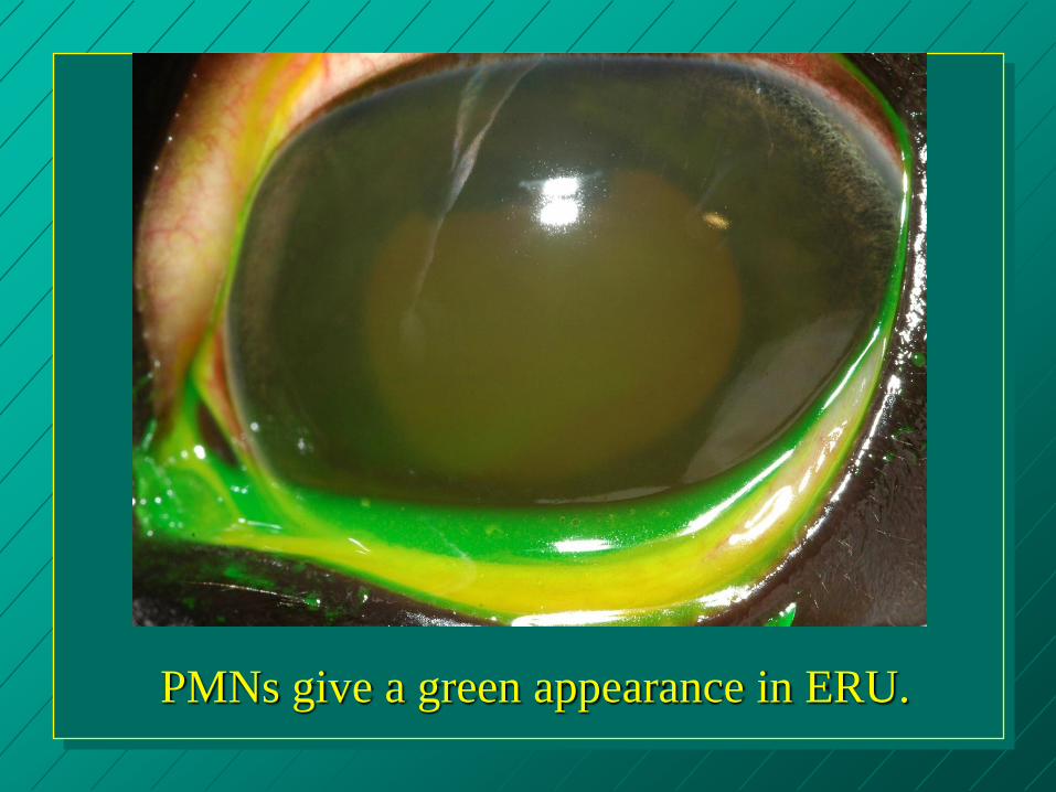
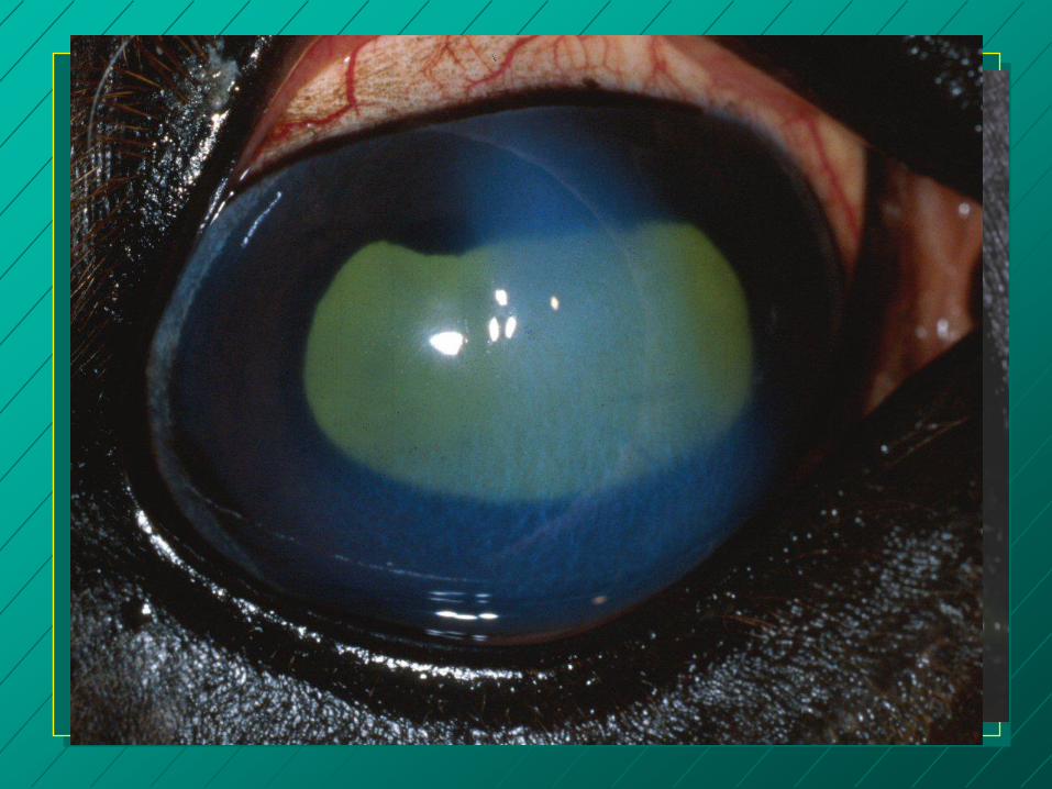
PMNs give a green appearance in ERU.
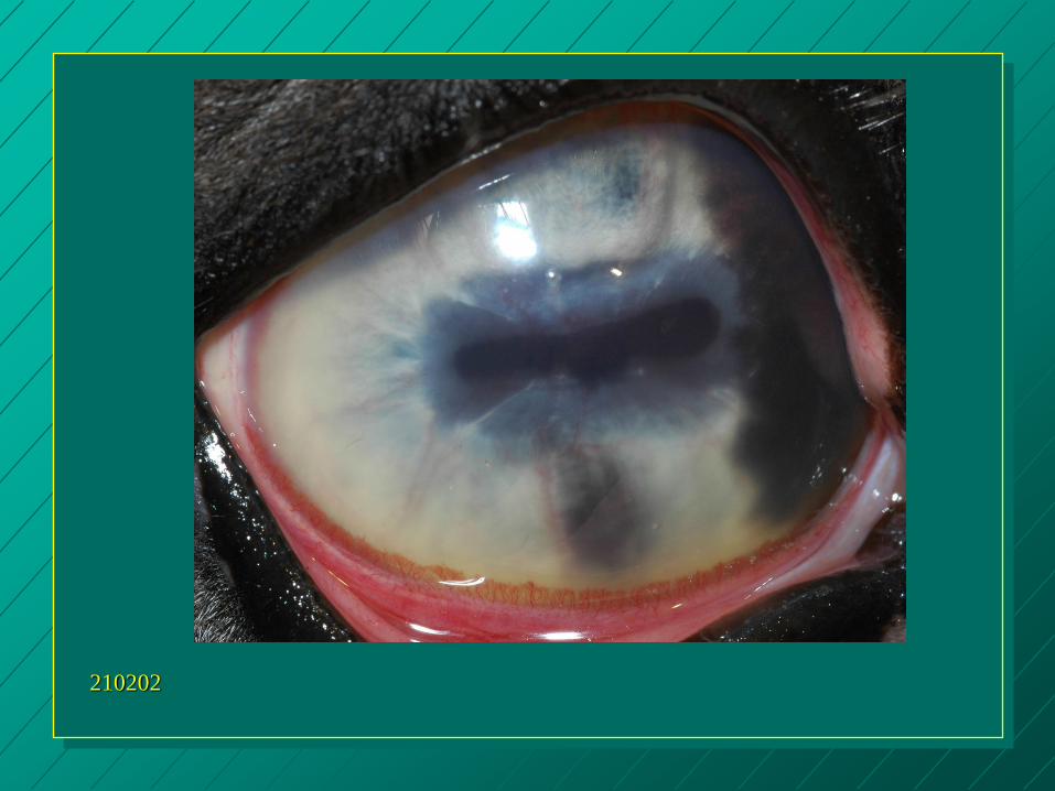
210202
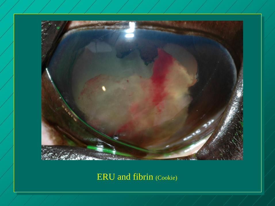
ERU and fibrin (Cookie)
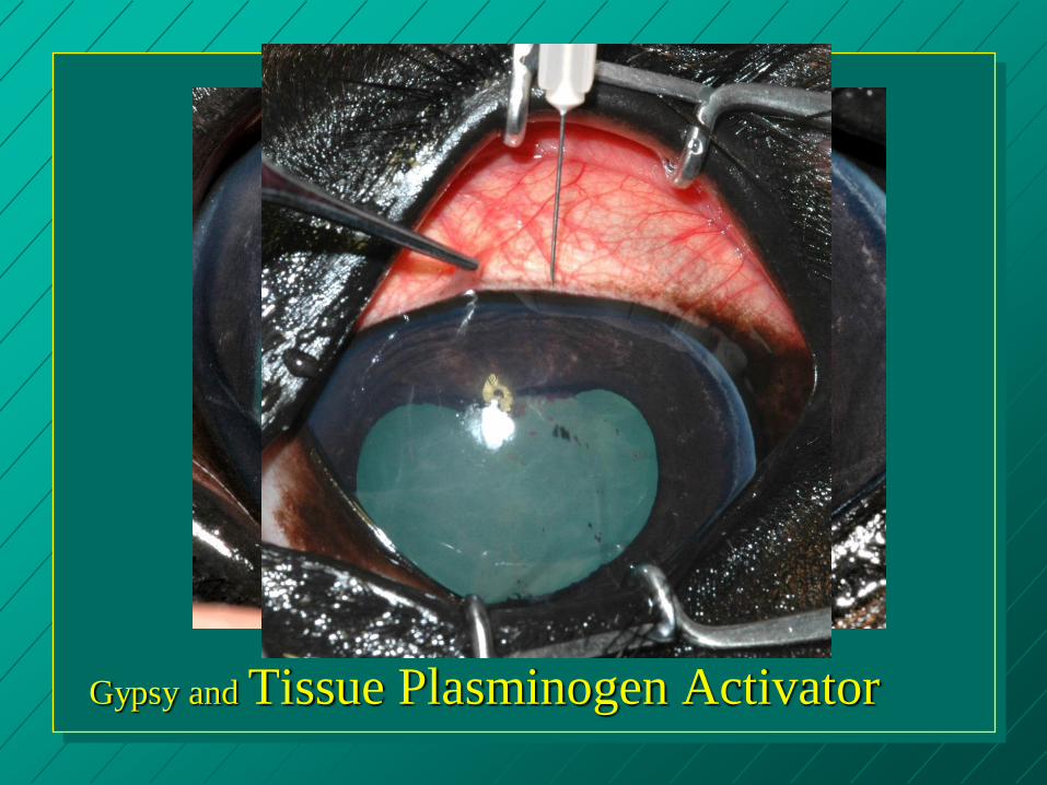
Gypsy and Tissue Plasminogen Activator
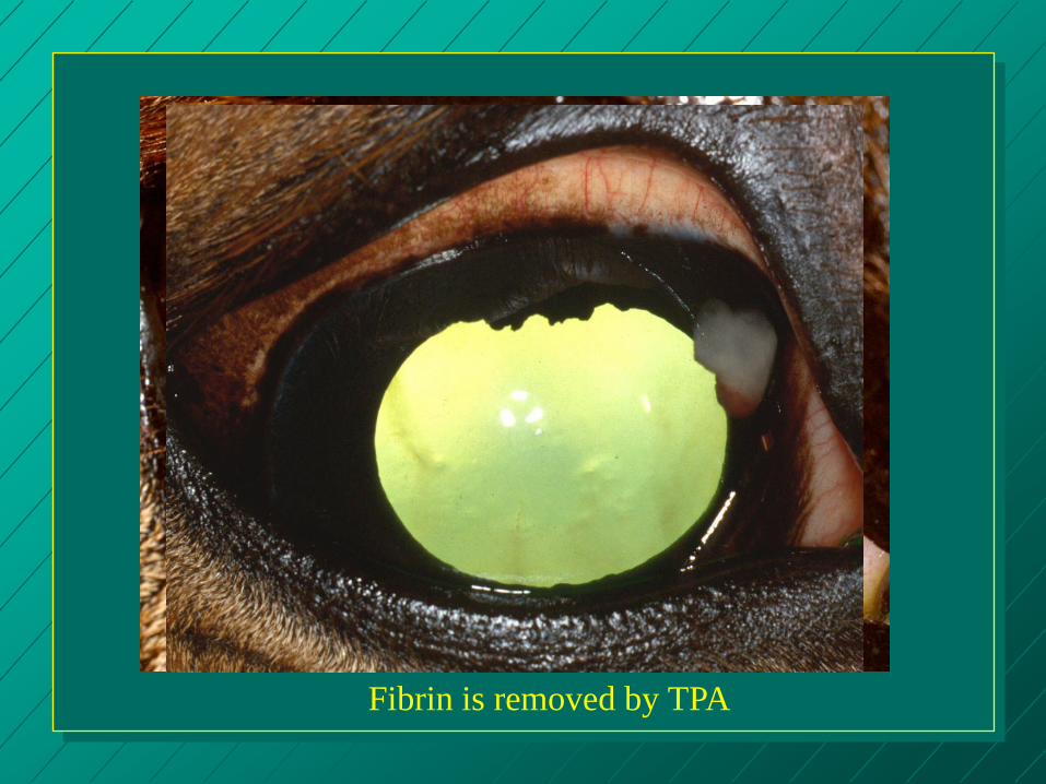
Fibrin is removed by TPA
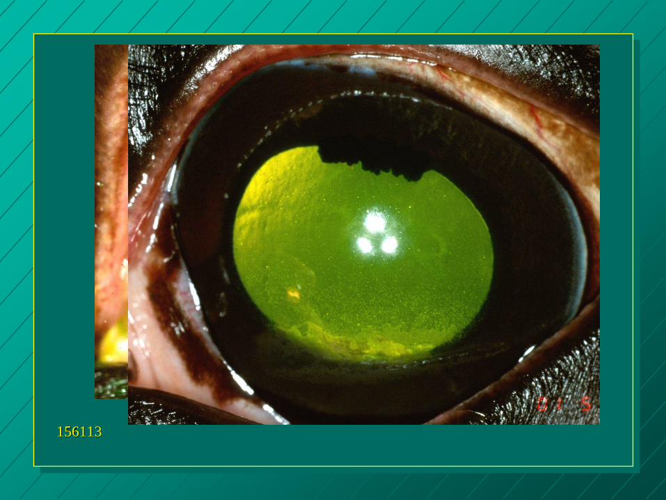
156113
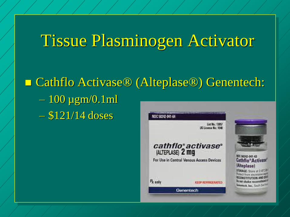
Tissue Plasminogen Activator
Cathflo Activase® (Alteplase®) Genentech:
– 100 µgm/0.1ml
– $121/14 doses
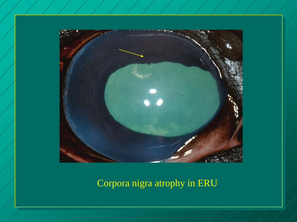
Corpora nigra atrophy in ERU
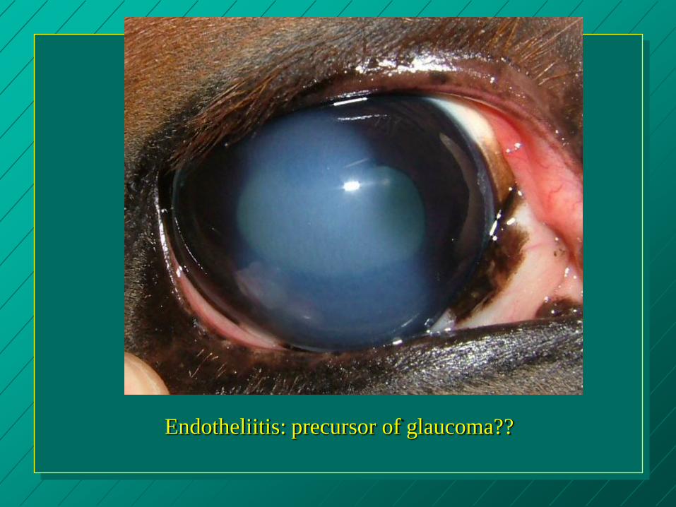
Endotheliitis: precursor of glaucoma??
Ventral edema from ERU is most typical
Vertical edema??
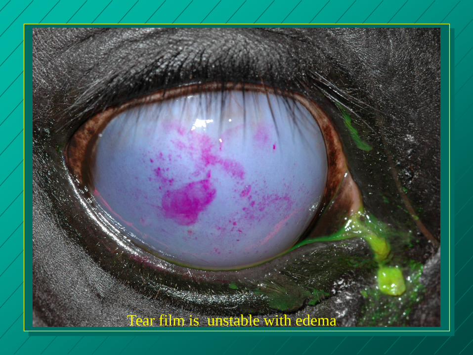
Tear film is unstable with edema
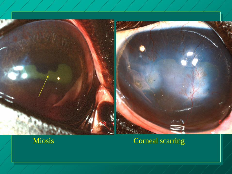
Miosis Corneal scarring
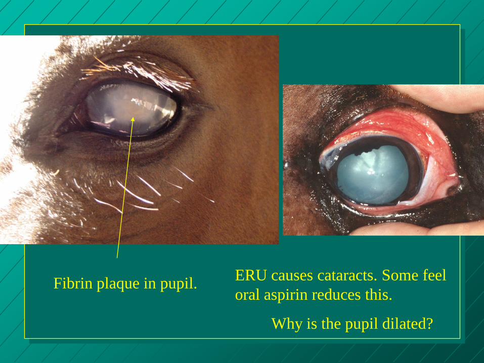
Fibrin plaque in pupil. ERU causes cataracts. Some feel
oral aspirin reduces this.
Why is the pupil dilated?
synechia
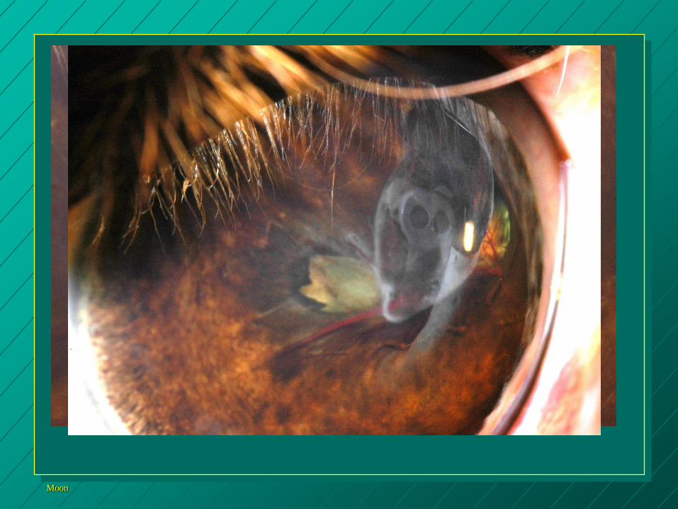
Moon
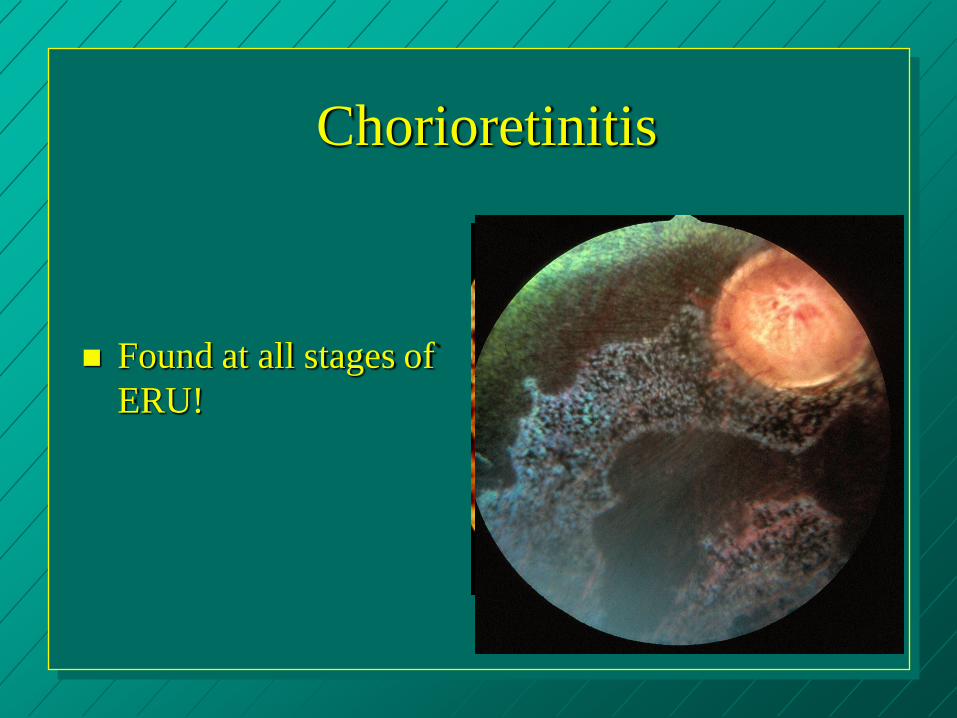
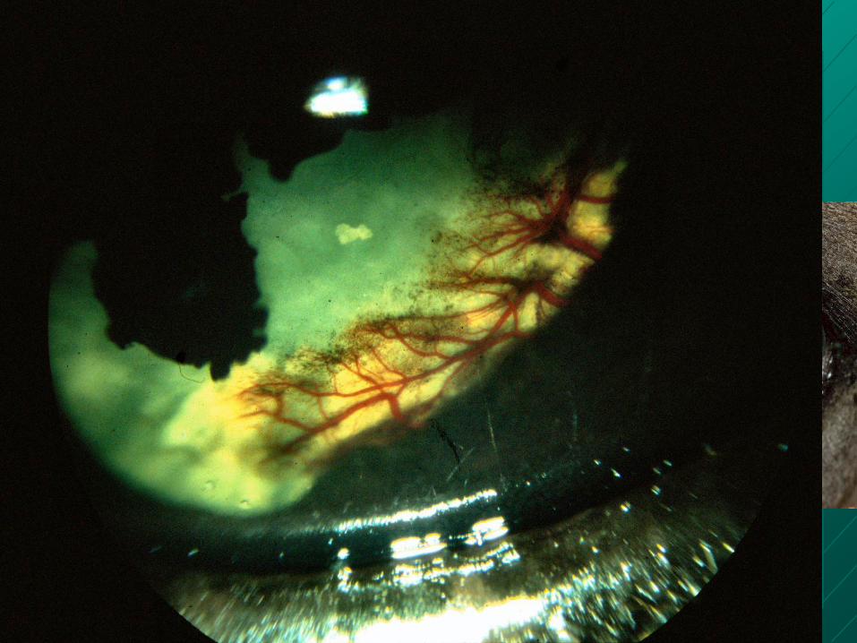
Chorioretinitis
Found at all stages of
ERU!
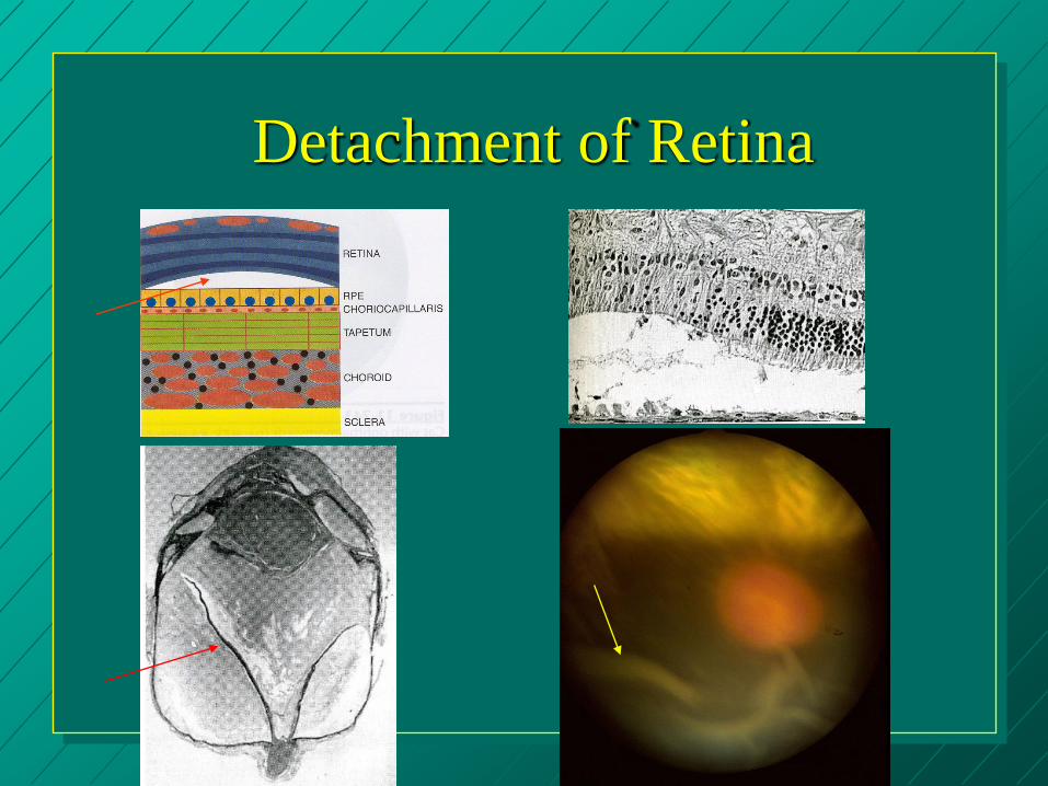
Detachment of Retina
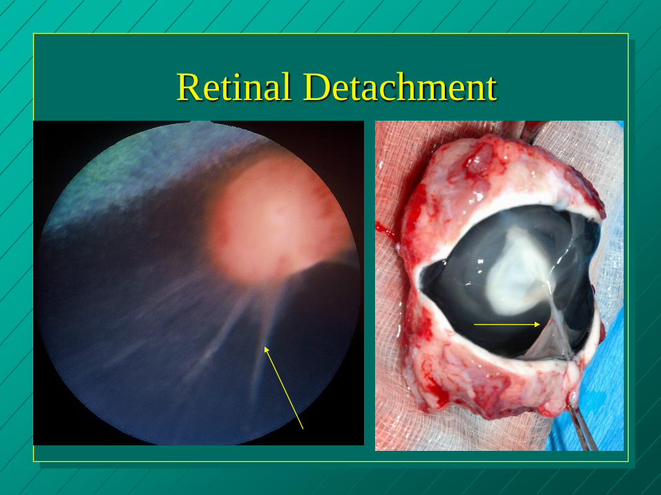
Retinal Detachment
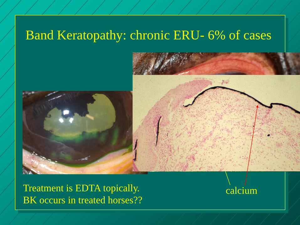
Band Keratopathy: chronic ERU- 6% of cases
calcium Treatment is EDTA topically.
BK occurs in treated horses??
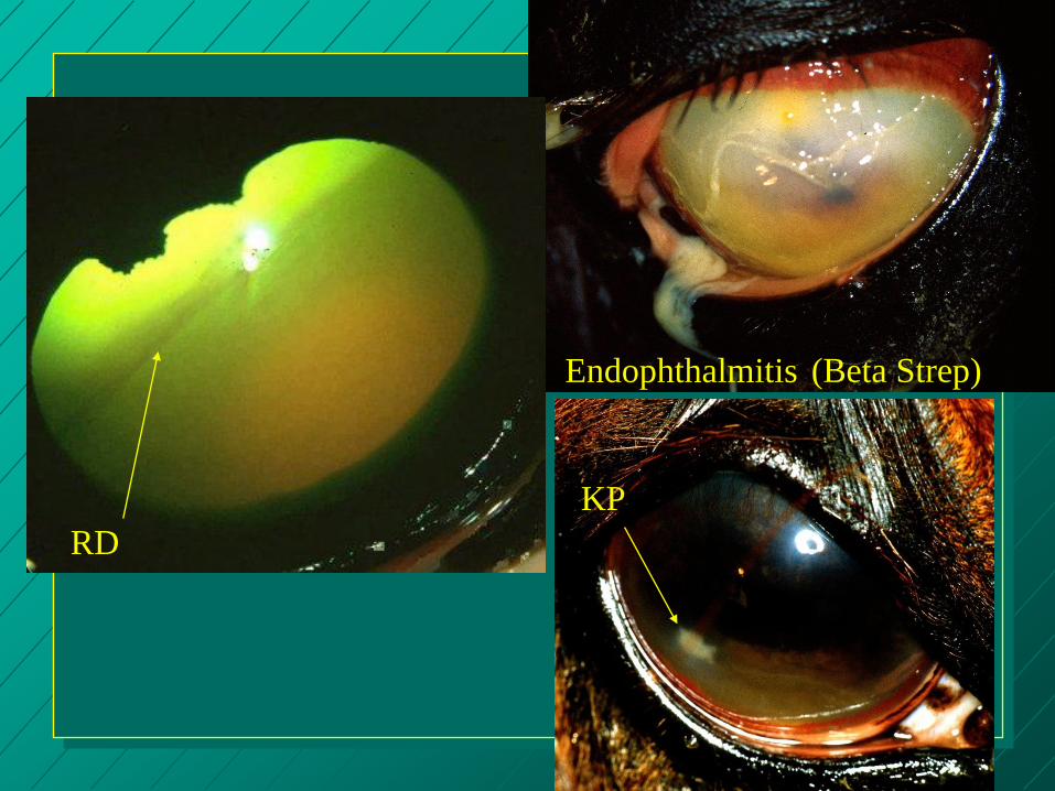
KP
Endophthalmitis (Beta Strep)
RD
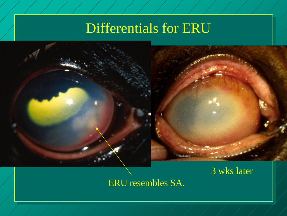
Differentials for ERU
ERU resembles SA.
3 wks later
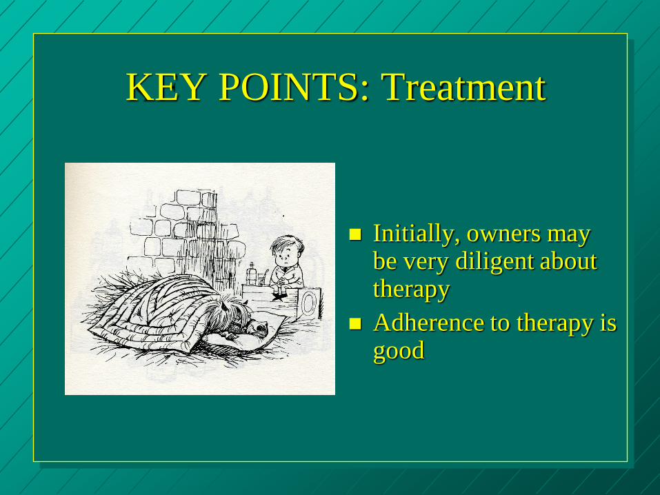
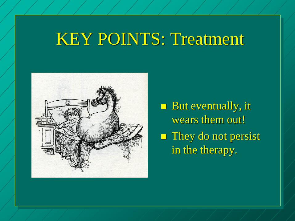
KEY POINTS: Treatment
Initially, owners may be very diligent about therapy
Adherence to therapy is good
KEY POINTS: Treatment
But eventually, it
wears them out!
They do not persist
in the therapy.
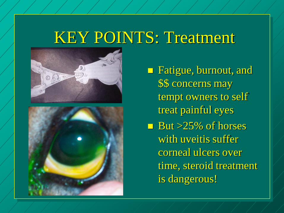
KEY POINTS: Treatment
Fatigue, burnout, and
$$ concerns may
tempt owners to self
treat painful eyes
But >25% of horses
with uveitis suffer
corneal ulcers over
time, steroid treatment
is dangerous!
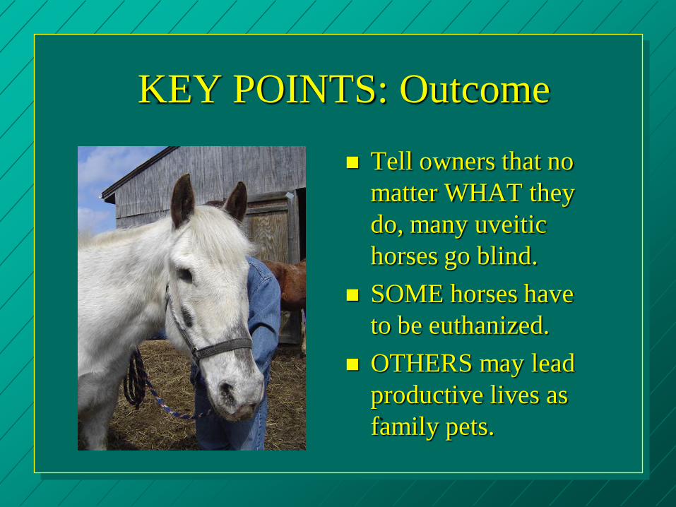
KEY POINTS: Outcome
Tell owners that no
matter WHAT they
do, many uveitic
horses go blind.
SOME horses have
to be euthanized.
OTHERS may lead
productive lives as
family pets.
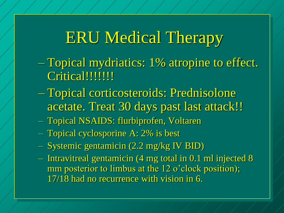
ERU Medical Therapy
– Topical mydriatics: 1% atropine to effect. Critical!!!!!!!
– Topical corticosteroids: Prednisolone acetate. Treat 30 days past last attack!!
– Topical NSAIDS: flurbiprofen, Voltaren
– Topical cyclosporine A: 2% is best
– Systemic gentamicin (2.2 mg/kg IV BID)
– Intravitreal gentamicin (4 mg total in 0.1 ml injected 8 mm posterior to limbus at the 12 o’clock position); 17/18 had no recurrence with vision in 6.
ERU Medical Therapy
– Systemic NSAIDS:
Flunixin meglumine: 0.5 mg/lb SID - BID initially
Phenylbutazone: 1-2 gm BID PO - 2nd choice
Aspirin: 10-40 mg/kg PO SID long term!!!!
– Methyl-Sulfonylmethane (MSM): 15 mg BID PO
– Systemic Corticosteroids:
Prednisolone/Prednisone: 0.75 mg/lb SID and
decrease dose
Dexamethasone: 0.05-0.2 mg/kg PO SID
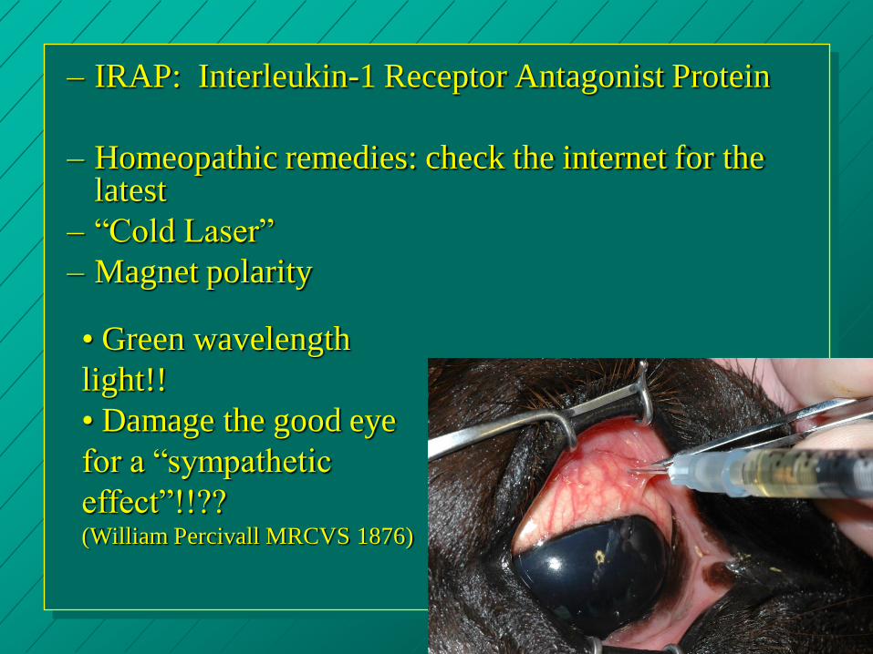
– IRAP: Interleukin-1 Receptor Antagonist Protein
– Homeopathic remedies: check the internet for the latest
– “Cold Laser”
– Magnet polarity
• Green wavelength
light!!
• Damage the good eye
for a “sympathetic
effect”!!?? (William Percivall MRCVS 1876)
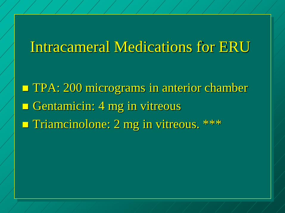
Intracameral Medications for ERU
TPA: 200 micrograms in anterior chamber
Gentamicin: 4 mg in vitreous
Triamcinolone: 2 mg in vitreous. ***
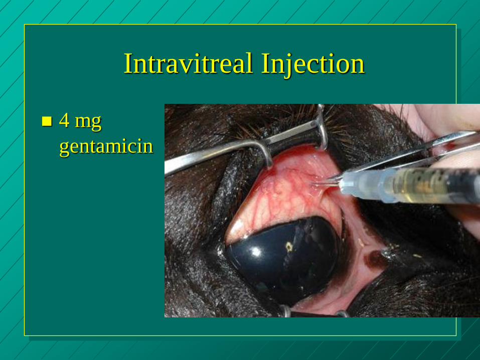
Intravitreal Injection
4 mg
gentamicin
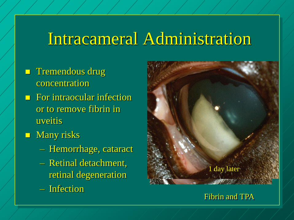
Intracameral Administration
Tremendous drug
concentration
For intraocular infection
or to remove fibrin in
uveitis
Many risks
– Hemorrhage, cataract
– Retinal detachment,
retinal degeneration
– Infection Fibrin and TPA
1 day later
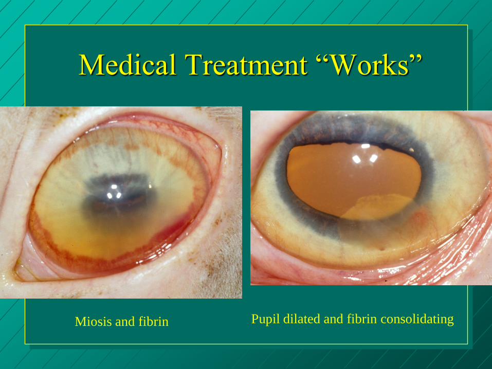
Medical Treatment “Works”
Pupil dilated and fibrin consolidating Miosis and fibrin
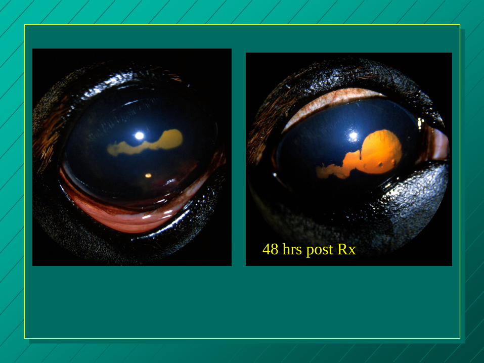
48 hrs post Rx
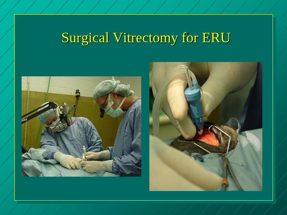
Surgical Vitrectomy for ERU
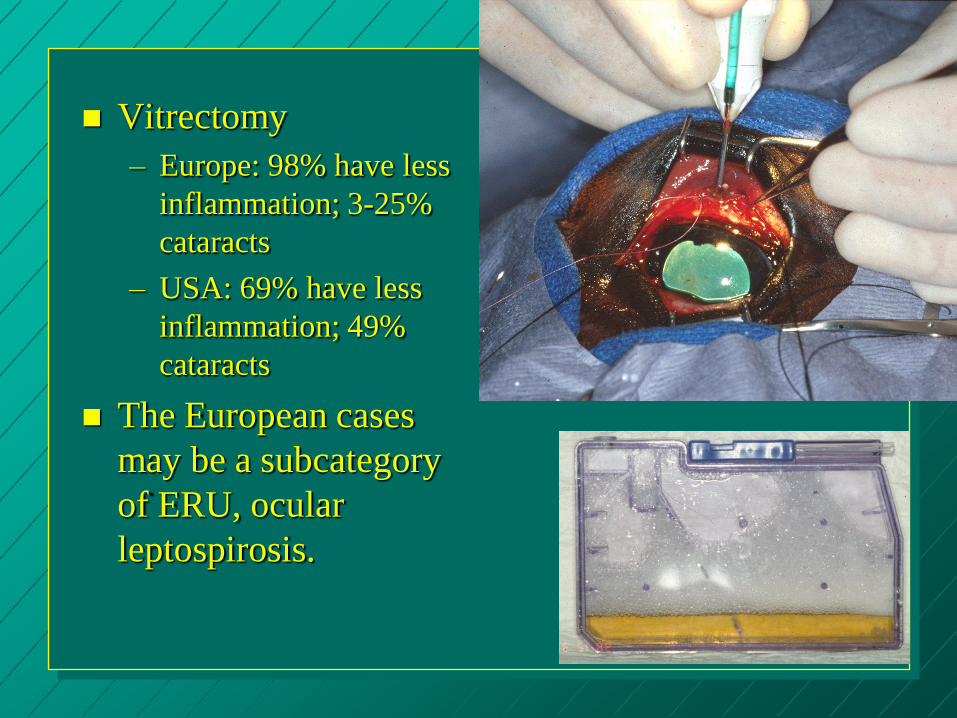
Vitrectomy
– Europe: 98% have less
inflammation; 3-25%
cataracts
– USA: 69% have less
inflammation; 49%
cataracts
The European cases
may be a subcategory
of ERU, ocular
leptospirosis.
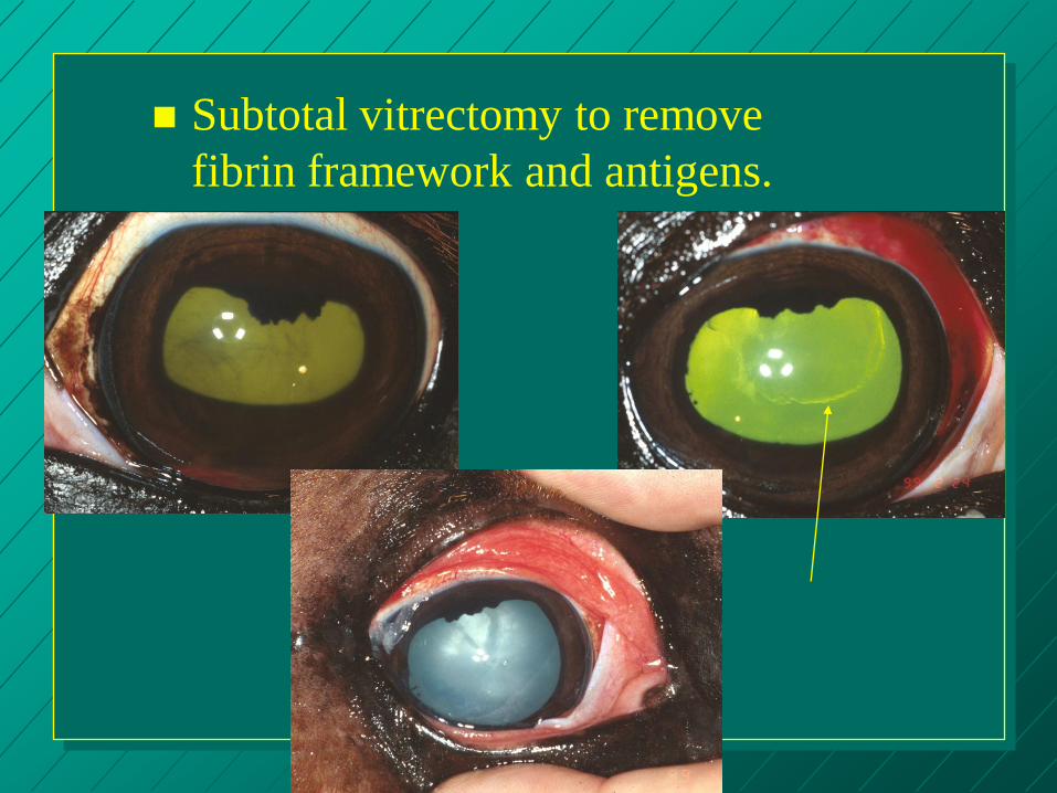
Subtotal vitrectomy to remove
fibrin framework and antigens.
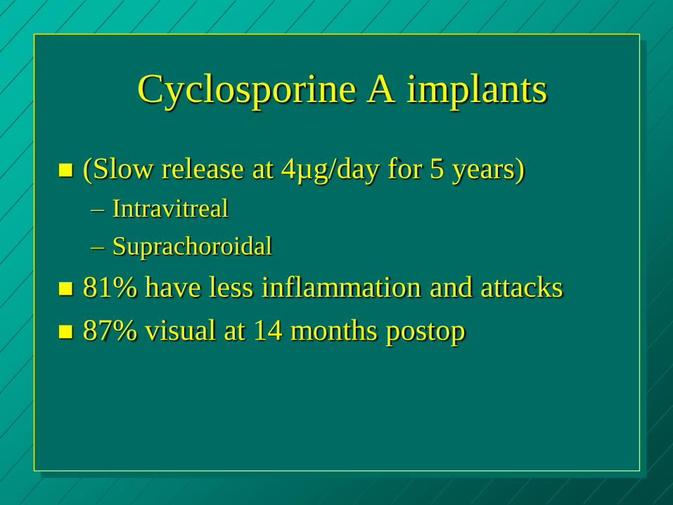
Cyclosporine A implants
(Slow release at 4µg/day for 5 years)
– Intravitreal
– Suprachoroidal
81% have less inflammation and attacks
87% visual at 14 months postop
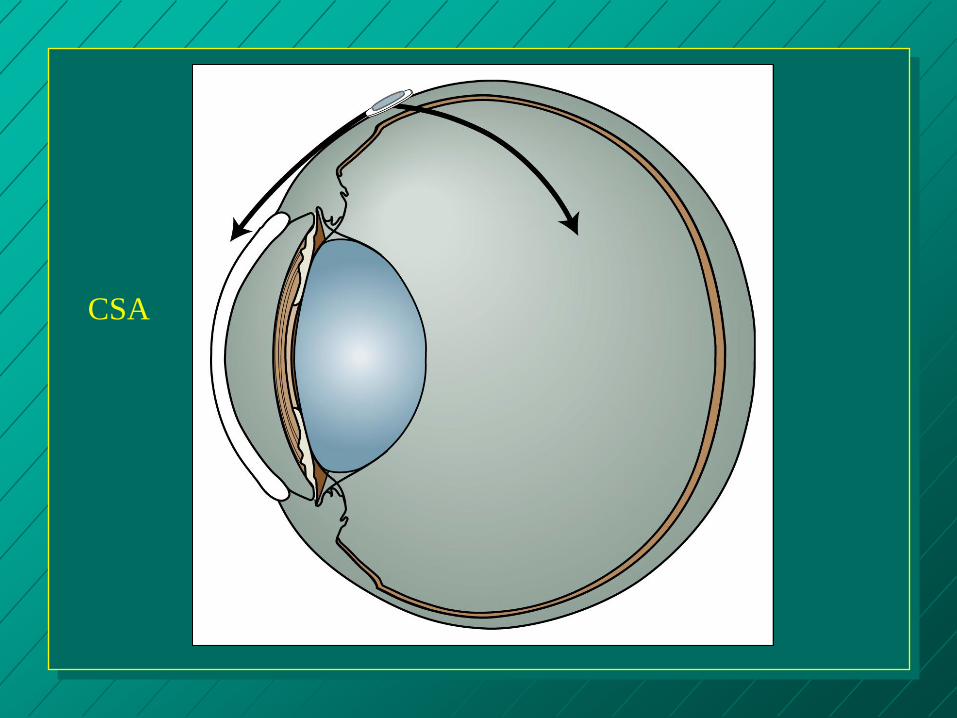
CSA