Embed Size (px)
Citation preview
UvA-DARE is a service provided by the library of the University of Amsterdam (http://dare.uva.nl)
UvA-DARE (Digital Academic Repository)
Zellweger spectrum disorders
Klouwer, F.C.C.
Link to publication
Citation for published version (APA):Klouwer, F. C. C. (2018). Zellweger spectrum disorders: From bench to bedside
General rightsIt is not permitted to download or to forward/distribute the text or part of it without the consent of the author(s) and/or copyright holder(s),other than for strictly personal, individual use, unless the work is under an open content license (like Creative Commons).
Disclaimer/Complaints regulationsIf you believe that digital publication of certain material infringes any of your rights or (privacy) interests, please let the Library know, statingyour reasons. In case of a legitimate complaint, the Library will make the material inaccessible and/or remove it from the website. Please Askthe Library: http://uba.uva.nl/en/contact, or a letter to: Library of the University of Amsterdam, Secretariat, Singel 425, 1012 WP Amsterdam,The Netherlands. You will be contacted as soon as possible.
Download date: 08 Sep 2018
2Zellweger spectrum disorders:Clinical overview and management approach
Femke C. C. Klouwer1, 2*, Kevin Berendse1, 2*, Sacha Ferdinandusse2, Ronald J. A. Wanders2, Marc Engelen1, Bwee Tien Poll-The1
* Equal contributors
1 Department of Pediatric Neurology, Emma Children’s Hospital, Academic Medical Center, University of Amsterdam, Amsterdam, The Netherlands
2 Laboratory Genetic Metabolic Diseases, Academic Medical Center, University of Amsterdam, Amsterdam, The Netherlands
Orphanet Journal of Rare Diseases (2015) 10:151
46 | Chapter 2
Abstract
Zellweger spectrum disorders (ZSDs) represent the major subgroup within the peroxisomal biogenesis disorders caused by defects in PEX genes. The Zellweger spectrum is a clinical and biochemical continuum which can roughly be divided into three clinical phenotypes. Patients can present in the neonatal period with severe symptoms or later in life during adolescence or adulthood with only minor features. A defect of functional peroxisomes results in several metabolic abnormalities, which in most cases can be detected in blood and urine. There is currently no curative therapy, but supportive care is available. This review focuses on the management of patients with a ZSD and provides recommendations for supportive therapeutic options for all those involved in the care for ZSD patients.
2
Clinical overview and management approach | 47
Background
The Zellweger spectrum disorders (ZSDs) are a heterogeneous group of autosomal recessive disorders characterized by a defect in peroxisome formation and are caused by mutations in one of 13 PEX genes 1–3. Because of the defect in peroxisome formation, multiple metabolic (both catabolic and anabolic) pathways are impaired resulting in metabolic abnormalities. Typically, ZSD patients accumulate very long chain fatty acids (VLCFAs), phytanic- and pristanic acid, C27-bile acid intermediates and pipecolic acid in plasma and have a deficiency of plasmalogens in erythrocytes 4. Clinically, ZSDs are highly heterogeneous, but the core features are: liver dysfunction, developmental delay and other neurological abnormalities, adrenocortical dysfunction and hearing- and vision impairment 5. Before the biochemical and molecular basis of ZSDs was known, they were clinically described as three distinct disorders: Zellweger syndrome (ZS), neonatal adrenoleukodystrophy (NALD) and infantile Refsum disease (IRD). These phenotypes are currently recognized as presentations within a clinical spectrum (with ZS being at the most severe end of the spectrum) which are now collectively referred to as ZSDs, in order to appreciate the wide variations in presentation 6. Recently, Heimler syndrome was recognized as a peroxisome biogenesis disorder within the Zellweger spectrum and added to the (very) mild end of the clinical spectrum 7. This review provides a clinical overview of Zellweger spectrum disorders and focuses on management of patients with a ZSD. New developments in the field of management are discussed.
Disease names and synonymsZellweger spectrum disorder/Zellweger syndrome spectrum/Zellweger syndrome/neonatal adrenoleukodystrophy/infantile Refsum disease/Heimler syndrome (ORPHA79189).
History and definitionBowen et al described a syndrome with failure to thrive, congenital glaucoma and craniofacial dysmorphic features with early death (before 2 years of age) 5. In 1965 Smith et al described two siblings with comparable multiple congenital malformations, but also polycystic kidneys and intrahepatic biliary dysgenesis 8. In 1967 Passarge et al introduced the term cerebro-hepato-renal syndrome. Since Hans Zellweger, a pediatrician, contributed two of the originally described patients it was later called Zellweger syndrome 9. It was not until 1973 that the causal link between ZS and peroxisomes was made, when Goldfischer et al described the absence of peroxisomes in hepatocytes and renal proximal tubules 10. Although the clinical presentation is different, the discovery of similar biochemical abnormalities revealed that the earlier described entities infantile Refsum disease and neonatal adrenoleukodystrophy were also peroxisomal disorders 11,12. Based on these findings, peroxisomes which were once considered unimportant organelles, were now connected to a group of diseases and became
48 | Chapter 2
the object of intensive scientifi c investigations. It turned out that peroxisomes are important organelles in the eukaryotic cell, and are involved in many catabolic and anabolic metabolic pathways 4,13. At present more than 15 diff erent peroxisomal disorders have been identifi ed. The genetic basis of ZSDs has largely been resolved and now includes 13 diff erent PEX genes 14,15. The group of diseases is now referred to as Zellweger spectrum disorders and include the old disease entities of ZS, NALD, IRD but also Heimler syndrome which was recently recognized as a ZSD 7,16.
EpidemiologyThe incidence of ZSDs is estimated to be 1 in 50.000 newborns in the United States 17. It is presumed that ZSDs occur worldwide, but the incidence may diff er between regions. For example, the incidence of (classic) Zellweger syndrome in the French-Canadian region of Quebec was estimated to be 1 in 12.000 18. A much lower incidence is reported in Japan, with an estimated incidence of 1 in 500.000 births 19. More accurate incidence data about ZSDs will become available in the near future, since newborn screening for X-linked adrenoleukodystrophy (X-ALD) will be implemented in several countries 20,21. The screening method is based on C26:0-lysophosphatidylcholine (C26:0-lysoPC) measurement in dried bloodspots using LC-MS/MS technology, which will also identify ZSD patients 22.
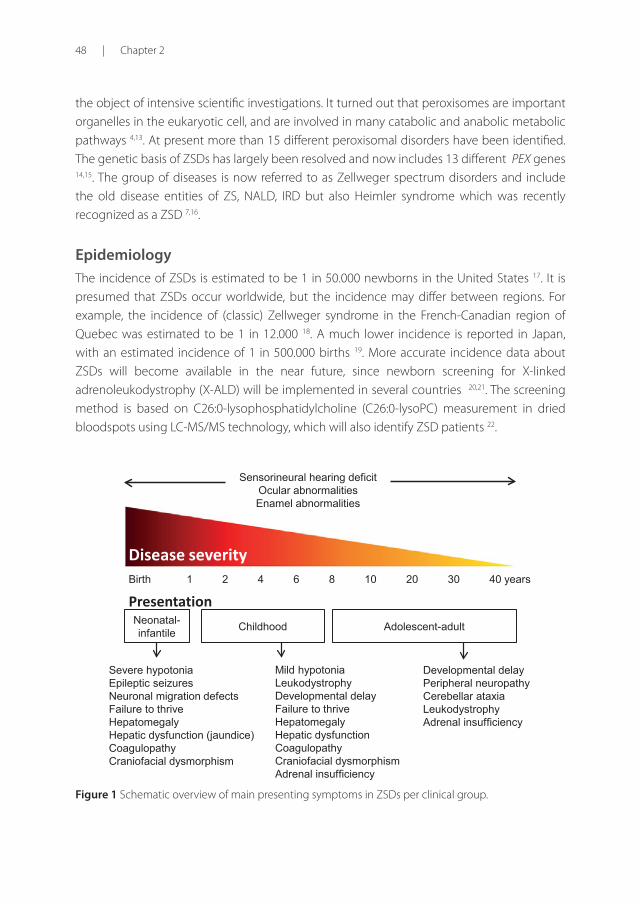
Birth 1 2 4 6 8 10 20 30 40 years
Neonatal-infantile Adolescent-adult Childhood
Presentation
Disease severity
Severe hypotoniaEpileptic seizuresNeuronal migration defectsFailure to thriveHepatomegalyHepatic dysfunction (jaundice)CoagulopathyCraniofacial dysmorphism
Sensorineural hearing deficitOcular abnormalitiesEnamel abnormalities
Mild hypotoniaLeukodystrophyDevelopmental delayFailure to thriveHepatomegalyHepatic dysfunction CoagulopathyCraniofacial dysmorphismAdrenal insufficiency
Developmental delayPeripheral neuropathyCerebellar ataxiaLeukodystrophyAdrenal insufficiency
Figure 1 Schematic overview of main presenting symptoms in ZSDs per clinical group.
2
Clinical overview and management approach | 49
Clinical features Patients with a ZSD can roughly be divided into three groups according to the age of presentation: the neonatal-infantile presentation, the childhood presentation and an adolescent-adult (late) presentation 23. An overview of the main presenting symptoms for these groups is summarized in Figure 1. The original classifi cation of ZS, NALD and IRD is less valuable now, especially since additional variant phenotypes suggestive for a disease spectrum have been identifi ed. For discussing prognosis and counseling patients or families this classifi cation may in some cases still be useful 24.
Neonatal-infantile presentation
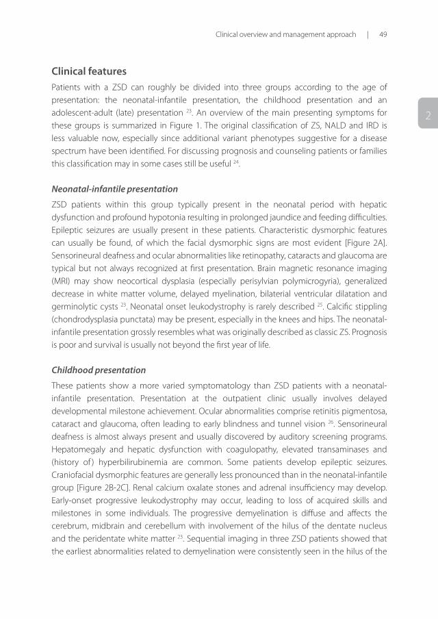
ZSD patients within this group typically present in the neonatal period with hepatic dysfunction and profound hypotonia resulting in prolonged jaundice and feeding diffi culties. Epileptic seizures are usually present in these patients. Characteristic dysmorphic features can usually be found, of which the facial dysmorphic signs are most evident [Figure 2A]. Sensorineural deafness and ocular abnormalities like retinopathy, cataracts and glaucoma are typical but not always recognized at fi rst presentation. Brain magnetic resonance imaging (MRI) may show neocortical dysplasia (especially perisylvian polymicrogyria), generalized decrease in white matter volume, delayed myelination, bilaterial ventricular dilatation and germinolytic cysts 23. Neonatal onset leukodystrophy is rarely described 25. Calcifi c stippling (chondrodysplasia punctata) may be present, especially in the knees and hips. The neonatal-infantile presentation grossly resembles what was originally described as classic ZS. Prognosis is poor and survival is usually not beyond the fi rst year of life.
Childhood presentation
These patients show a more varied symptomatology than ZSD patients with a neonatal-infantile presentation. Presentation at the outpatient clinic usually involves delayed developmental milestone achievement. Ocular abnormalities comprise retinitis pigmentosa, cataract and glaucoma, often leading to early blindness and tunnel vision 26. Sensorineural deafness is almost always present and usually discovered by auditory screening programs. Hepatomegaly and hepatic dysfunction with coagulopathy, elevated transaminases and (history of ) hyperbilirubinemia are common. Some patients develop epileptic seizures. Craniofacial dysmorphic features are generally less pronounced than in the neonatal-infantile group [Figure 2B-2C]. Renal calcium oxalate stones and adrenal insuffi ciency may develop. Early-onset progressive leukodystrophy may occur, leading to loss of acquired skills and milestones in some individuals. The progressive demyelination is diff use and aff ects the cerebrum, midbrain and cerebellum with involvement of the hilus of the dentate nucleus and the peridentate white matter 23. Sequential imaging in three ZSD patients showed that the earliest abnormalities related to demyelination were consistently seen in the hilus of the
50 | Chapter 2
D E F
A
B C
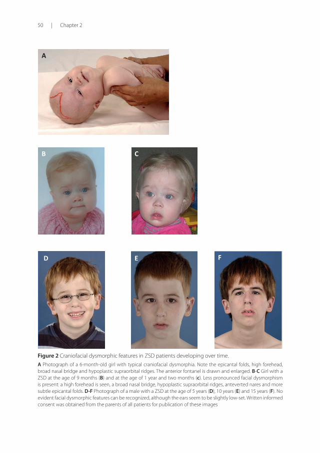
Figure 2 Craniofacial dysmorphic features in ZSD patients developing over time.
A Photograph of a 6-month-old girl with typical craniofacial dysmorphia. Note the epicantal folds, high forehead, broad nasal bridge and hypoplastic supraorbital ridges. The anterior fontanel is drawn and enlarged. B-C Girl with a ZSD at the age of 9 months (B) and at the age of 1 year and two months (c). Less pronounced facial dysmorphism is present: a high forehead is seen, a broad nasal bridge, hypoplastic supraorbital ridges, anteverted nares and more subtle epicantal folds. D-F Photograph of a male with a ZSD at the age of 5 years (D), 10 years (E) and 15 years (F). No evident facial dysmorphic features can be recognized, although the ears seem to be slightly low-set. Written informed consent was obtained from the parents of all patients for publication of these images
2
Clinical overview and management approach | 51
dentate nucleus and superior cerebellar peduncles, chronologically followed by the cerebellar white matter, brainstem tracts, parieto-occipital white matter, splenium of the corpus callosum and eventually involvement of the whole of the cerebral white matter 27. The above described rapid progressive leukodystrophy, in combination with other symptoms described here, resemble what was originally described as NALD. A small subgroup of patients develop a relatively late-onset white matter disease, but no patients with late-onset rapid progressive white matter disease after the age of five have been reported 28. Prognosis depends on what organ systems are primarily affected (i.e. liver) and the occurrence of progressive cerebral demyelination, but life expectancy is decreased and most patients die before adolescence.
Adolescent-adult presentation
Symptoms in this group are less severe, and diagnosis can be in late child- or even adulthood 29. Ocular abnormalities and a sensorineural hearing deficit are the most consistent symptoms. Craniofacial dysmorphic features can be present, but may also be completely absent [Figure 2D-2F]. Developmental delay is highly variable and some patients may have normal intelligence. Daily functioning ranges from completely independent to 24 hour care. It is important to emphasize that primary adrenal insufficiency is common and is probably under diagnosed 30. In addition to some degree of developmental delay, other neurological abnormalities are usually also present: signs of peripheral neuropathy, cerebellar ataxia and pyramidal tract signs. The clinical course is usually slowly progressive, although the disease may remain stable for (many) years 31. Slowly progressive, clinically silent leukoencephalopathy is common, but MRI may be normal in other cases 23.
Etiology and pathophysiology ZSDs are caused by mutations in one of the 13 different PEX genes. PEX genes encode proteins called peroxins and are involved in either peroxisome formation, peroxisomal protein import, or both. As a consequence, mutations in PEX genes cause a deficiency of functional peroxisomes. Cells from ZSD patients either entirely lack functional peroxisomes, or cells can show a reduced number of functional peroxisomes or a mosaic pattern (i.e. a mixed population of cells with functional peroxisomes and cells without) 1 32 33. Peroxisomes are involved in many anabolic and catabolic metabolic processes, like biosynthesis of ether phospholipids and bile acids, α- and β-oxidation of fatty acids and the detoxification of glyoxylate and reactive oxygen species. Dysfunctional peroxisomes therefore cause biochemical abnormalities in tissues, but also in readily available materials like plasma and urine 15 3 [summarized in Table 1]. There is a reasonable genotype-phenotype correlation 24. Approximately 60 percent of ZSD patients have biallelic PEX1 mutations and almost 90 different mutations in PEX1 have been reported so far 34. Detailed and up to date information about PEX gene mutations is available through the dbPEX gene database (http://www.dbpex.org).
52 | Chapter 2
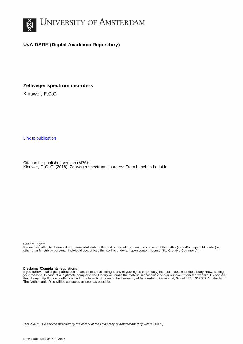
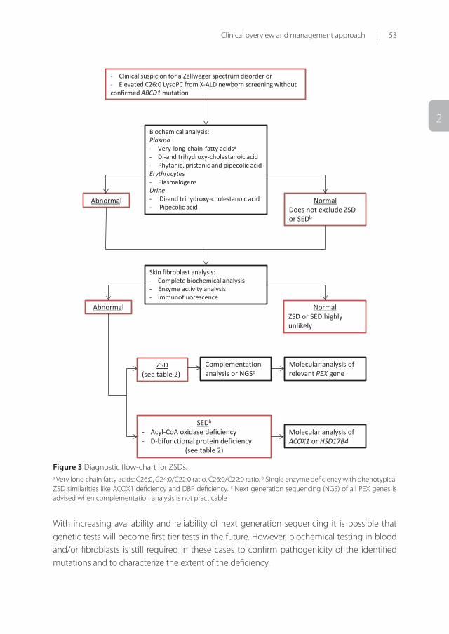
DiagnosisIf a ZSD is clinically suspected the first step to confirm the diagnosis is by biochemical testing in readily accessible materials like blood and urine. This testing includes measurement of VLCFAs, the peroxisomal bile acid intermediates di- and trihydroxycholestanoic acid (DHCA, THCA), the branched-chain fatty acids phytanic and pristanic acid, and pipecolic acid in plasma, plasmalogen levels in erythrocytes, and C26:0-lysoPC in dried blood spots. Additionally, bile acids and oxalic acid can be analyzed in urine 24. It is important to note that relatively mild ZSD patients may have (near) normal biochemical tests in plasma and urine 35 36 37. If clinical suspicion of a ZSD is high and peroxisomal parameters in blood and urine are normal, further testing in fibroblasts is recommended, including culturing the fibroblasts at 40°C 35. Further fibroblast testing is also required to differentiate between ZSDs and certain peroxisomal single enzyme deficiencies, and to perform complementation studies to pinpoint the defective PEX gene. Subsequent mutation analysis of the defective PEX gene is done in all patients to confirm the diagnosis. A diagnostic flowchart is provided [Figure 3].
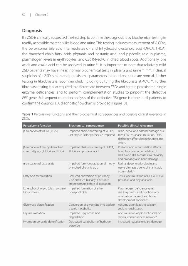
Table 1 Peroxisome functions and their biochemical consequences and possible clinical relevance in ZSDs.
Peroxisome function Biochemical consequence Possible clinical relevance
β-oxidation of VLCFA (≥C22) Impaired chain shortening of VLCFA, last step in DHA synthesis is impaired
Brain, nerve and adrenal damage due to VLCFA tissue accumulation, DHA deficiency affects brain function and vision.
β-oxidation of methyl-branched chain fatty acid, DHCA and THCA
Impaired chain shortening of DHCA, THCA and pristanic acid
Pristanic acid accumulation affects brain function, accumulation of DHCA and THCA causes liver toxicity and probably also brain damage.
α-oxidation of fatty acids Impaired (pre-)degradation of methyl branched phytanic acid
Retinal degeneration, brain and nerve damage due to phytanic acid accumulation
Fatty acid racemization Reduced convertion of pristanoyl-CoA and C27-bile acyl-CoAs into stereoisomers before β-oxidation
Tissue accumulation of DHCA, THCA, pristanic- and phytanic acid.
Ether phospholipid (plasmalogen) biosynthesis
Impaired formation of ether phospholipids
Plasmalogen deficiency gives rise to growth- and psychomotor retardation, cataract and bone development anomalies.
Glyoxylate detoxification Conversion of glyoxylate into oxalate, a toxic metabolite
Accumulation leads to calcium oxalate renal stones.
L-lysine oxidation Impaired L-pipecolic acid degradation
Accumulation of pipecolic acid, no clinical consequences known 38.
Hydrogen peroxide detoxification Decreased catabolism of hydrogen peroxide
Increased reactive oxidant damage.
2
Clinical overview and management approach | 53
- Clinical suspicion for a Zellweger spectrum disorder or - Elevated C26:0 LysoPC from X-ALD newborn screening without confirmed ABCD1 mutation
Biochemical analysis: Plasma - Very-long-chain-fatty acidsa
- Di-and trihydroxy-cholestanoic acid - Phytanic, pristanic and pipecolic acid Erythrocytes - Plasmalogens Urine - Di-and trihydroxy-cholestanoic acid - Pipecolic acid
Abnormal Normal Does not exclude ZSD or SEDb
Skin fibroblast analysis: - Complete biochemical analysis - Enzyme activity analysis - Immunofluorescence
Abnormal Normal ZSD or SED highly unlikely
ZSD (see table 2)
SEDb
- Acyl-CoA oxidase deficiency - D-bifunctional protein deficiency
(see table 2)
Molecular analysis of relevant PEX gene
Molecular analysis of ACOX1 or HSD17B4
Complementation analysis or NGSc
Figure 3 Diagnostic flow-chart for ZSDs. a Very long chain fatty acids: C26:0, C24:0/C22:0 ratio, C26:0/C22:0 ratio. b Single enzyme deficiency with phenotypical ZSD similarities like ACOX1 deficiency and DBP deficiency. c Next generation sequencing (NGS) of all PEX genes is advised when complementation analysis is not practicable
With increasing availability and reliability of next generation sequencing it is possible that genetic tests will become first tier tests in the future. However, biochemical testing in blood and/or fibroblasts is still required in these cases to confirm pathogenicity of the identified mutations and to characterize the extent of the deficiency.
54 | Chapter 2
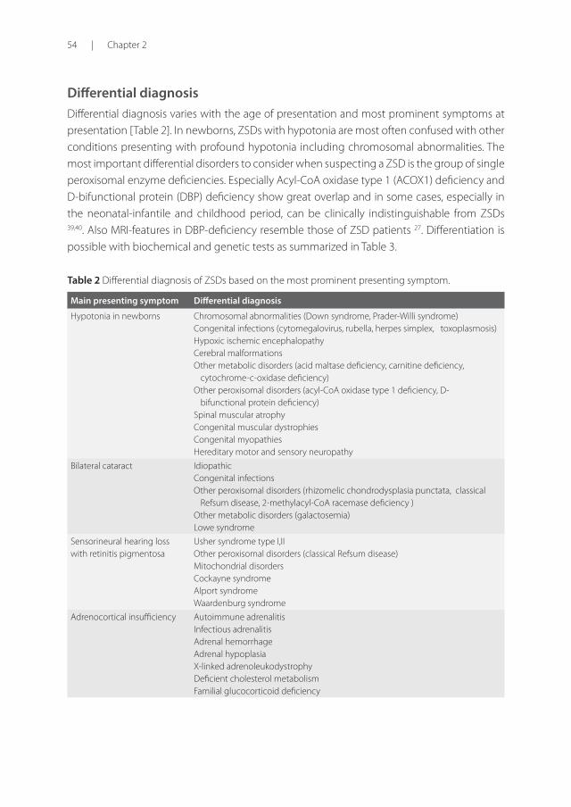
Differential diagnosisDifferential diagnosis varies with the age of presentation and most prominent symptoms at presentation [Table 2]. In newborns, ZSDs with hypotonia are most often confused with other conditions presenting with profound hypotonia including chromosomal abnormalities. The most important differential disorders to consider when suspecting a ZSD is the group of single peroxisomal enzyme deficiencies. Especially Acyl-CoA oxidase type 1 (ACOX1) deficiency and D-bifunctional protein (DBP) deficiency show great overlap and in some cases, especially in the neonatal-infantile and childhood period, can be clinically indistinguishable from ZSDs 39,40. Also MRI-features in DBP-deficiency resemble those of ZSD patients 27. Differentiation is possible with biochemical and genetic tests as summarized in Table 3. Table 2 Differential diagnosis of ZSDs based on the most prominent presenting symptom.
Main presenting symptom Differential diagnosis
Hypotonia in newborns Chromosomal abnormalities (Down syndrome, Prader-Willi syndrome) Congenital infections (cytomegalovirus, rubella, herpes simplex, toxoplasmosis)Hypoxic ischemic encephalopathy Cerebral malformations Other metabolic disorders (acid maltase deficiency, carnitine deficiency,
cytochrome-c-oxidase deficiency) Other peroxisomal disorders (acyl-CoA oxidase type 1 deficiency, D-
bifunctional protein deficiency)Spinal muscular atrophy Congenital muscular dystrophies Congenital myopathies Hereditary motor and sensory neuropathy
Bilateral cataract Idiopathic Congenital infections Other peroxisomal disorders (rhizomelic chondrodysplasia punctata, classical
Refsum disease, 2-methylacyl-CoA racemase deficiency )Other metabolic disorders (galactosemia) Lowe syndrome
Sensorineural hearing loss with retinitis pigmentosa
Usher syndrome type I,II Other peroxisomal disorders (classical Refsum disease)Mitochondrial disorders Cockayne syndrome Alport syndrome Waardenburg syndrome
Adrenocortical insufficiency Autoimmune adrenalitis Infectious adrenalitis Adrenal hemorrhage Adrenal hypoplasia X-linked adrenoleukodystrophyDeficient cholesterol metabolism Familial glucocorticoid deficiency
2
Clinical overview and management approach | 55
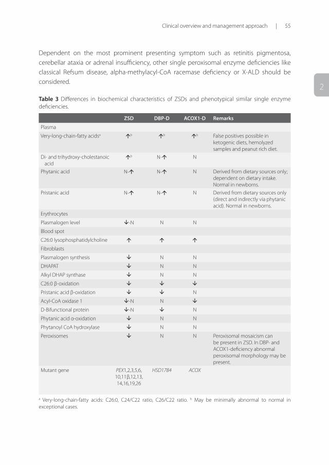
Dependent on the most prominent presenting symptom such as retinitis pigmentosa, cerebellar ataxia or adrenal insufficiency, other single peroxisomal enzyme deficiencies like classical Refsum disease, alpha-methylacyl-CoA racemase deficiency or X-ALD should be considered.
Table 3 Differences in biochemical characteristics of ZSDs and phenotypical similar single enzyme deficiencies.
ZSD DBP-D ACOX1-D Remarks
Plasma
Very-long-chain-fatty acidsa b b b False positives possible in ketogenic diets, hemolyzed samples and peanut rich diet.
Di- and trihydroxy-cholestanoic acid
b N- N
Phytanic acid N- N- N Derived from dietary sources only; dependent on dietary intake. Normal in newborns.
Pristanic acid N- N- N Derived from dietary sources only (direct and indirectly via phytanic acid). Normal in newborns.
Erythrocytes
Plasmalogen level -N N N
Blood spot
C26:0 lysophosphatidylcholine
Fibroblasts
Plasmalogen synthesis N N
DHAPAT N N
Alkyl DHAP synthase N N
C26:0 β-oxidation
Pristanic acid β-oxidation N
Acyl-CoA oxidase 1 -N N
D-Bifunctional protein -N N
Phytanic acid α-oxidation N N
Phytanoyl CoA hydroxylase N N
Peroxisomes N N Peroxisomal mosaicism can be present in ZSD. In DBP- and ACOX1-deficiency abnormal peroxisomal morphology may be present.
Mutant gene PEX1,2,3,5,6, 10,11β,12,13, 14,16,19,26
HSD17B4 ACOX
a Very-long-chain-fatty acids: C26:0, C24/C22 ratio, C26/C22 ratio. b May be minimally abnormal to normal in exceptional cases.
56 | Chapter 2
Genetic counseling and antenatal diagnosis Because of the poor outcome and high disease burden associated with the majority of ZSDs, genetic counseling should be offered to parents of affected children. Carriers can be offered prenatal- or preimplantation genetic diagnosis. Before prenatal genetic testing can be performed the familial pathogenic mutation(s) in one of the PEX genes need(s) to be identified 1. If the PEX mutations are unknown or cannot be detected, biochemical prenatal testing for ZSD is possible in chorionic villus biopsy material, cultured chorionic villus cells or cultured amniocytes. Biochemical prenatal testing can only be performed in case of clear biochemical abnormalities in cells from the index patient 15.
Clinical management and treatmentBecause no curative therapy for patients with a ZSD exists, intervention is supportive and based on symptoms. Past- and current supportive therapeutic options are summarized in Table 4.
Docosahexaenoic acid
Docosahexaenoic acid (DHA; C22:6ω3) is a long-chain polyunsaturated fatty acid important for retinal and brain function 41,42. Tetracosahexaenoic acid (C24:6ω3) undergoes one cycle of peroxisomal beta-oxidation to be converted to DHA 4, leading to reduced levels of DHA when peroxisomes are absent. Because ZSD patients often have low levels of DHA in membranes of erythrocytes, supplementation of DHA was suggested to be a possible therapy. Although some studies have claimed a beneficial effect of DHA supplementation 43 44, a randomized double-blind placebo controlled trial showed that DHA treatment leads to increased DHA levels in plasma, but no improvement of visual function and growth could be observed 45.
Lorenzo’s oil
Lorenzo’s oil (i.e. 4:1 mix of glyceryl trioleate and glyceryl trierucate) therapy was originally developed for the single peroxisomal enzyme deficiency X-ALD, and was shown to lower VLCFAs in plasma 46, but had no effect on disease progression 47 48. Some studies reported lowering of the VLCFA levels in plasma by Lorenzo’s oil in ZS babies 49 50. However, based on data of studies in X-ALD individuals, there is no reason to expect that Lorenzo’s oil will be beneficial for ZSD patients at this point.
Cholic acid
Cholic acid is a primary C24 bile acid, involved in for instance the absorption of fat-soluble vitamins. Cholic acid is formed from its precursor THCA by one peroxisomal beta-oxidation cycle. The peroxisomal C27-bile acid intermediates DHCA and THCA accumulate in ZSDs and
2
Clinical overview and management approach | 57
are considered to be more toxic than the primary C24 bile acids due to their altered physical properties and are believed to contribute to the liver disease in ZSDs (e.g. dysfunction and liver fibrosis) 51. The bile acid intermediates are only partly conjugated and are less well excreted than C24 bile acids contributing to cholestasis. We hypothesize that DHCA and THCA cross the blood-brain barrier and cause central nerve system damage. Several case reports have described a beneficial effect of cholic acid in ZS babies, supported by reduced urinary and plasma excretion of DHCA/THCA 52 53. Clinically there was increased growth and an increase in the levels of fat-soluble vitamins. Furthermore, bile acid treatment in mice was shown to improve hepatic disease 54. Limitations of the studies so far, however, are the small number of treated patients and short follow-up. Current evidence is insufficient to conclude that cholic acid treatment is beneficial for patients with a ZSD. The Food and Drug Administration recently approved cholic acid as a safe treatment for ZSD patients in the United States. However, efficiency should be demonstrated in large clinical trials before this treatment can be implemented.
Plasmalogen precursors
Due to a deficiency of the first peroxisomal steps in the biosynthesis of plasmalogens 55, ZSD patients may have low levels of plasmalogens. Plasmalogens play a critical role in cell membranes and as anti-oxidants 56. It was suggested that supplementation with precursors of plasmalogens (batyl alcohol) could be beneficial for ZSD patients, as import of these alkylglycerols proceeds normally. Several case reports have described an increase in erythrocyte plasmalogen levels after treatment and improvement of clinical symptoms in some patients 57 58 59. Although never studied systematically, ether lipid therapy could be of interest for ZSD.
Citrate
The toxic metabolite oxalate accumulates in plasma and urine from ZSD patients 4. This causes renal calcium oxalate stones. In a large cohort of Dutch ZSD patients a high prevalence of 83% of renal calcium oxalate stones was shown 60. For this reason, patients should be screened for the presence of high levels of oxalic acid in urine yearly. To prevent the formation of renal stones, patients with hyperoxaluria should start oral citrate treatment. Furthermore, sufficient fluid intake is recommended 61.
Supportive care
All ZSD patients need to be screened for adrenal insufficiency 30, epilepsy, low levels of fat-soluble vitamins, (partly) vitamin K dependent coagulopathy, high levels of phytanic acid, hearing or visual impairment and enamel hypoplasia. They should be treated according to the identified abnormalities, e.g. supplementation of cortisone, anti-epileptic drugs, vitamins
58 | Chapter 2
and/or a phytanic restricted diet. Because supplementation of cortisone is associated with severe side effects, such as growth suppression and osteoporosis 62, only patients with a true insufficiency (i.e. altered Synacthen test) should be treated. A phytanic acid restricted diet is only necessary when levels of phytanic acid are extremely high and is not recommended when levels are moderately increased, as sufficient intake of calories is more decisive. Hearing and visual impairment should be (partly) corrected by hearing aids and glasses, with ophthalmologic and audiological evaluations yearly. Enamel hypoplasia, present in nearly all patients, should be followed-up by a dentist 63 64. Some patients will need a gastrostomy to provide adequate intake of calories.
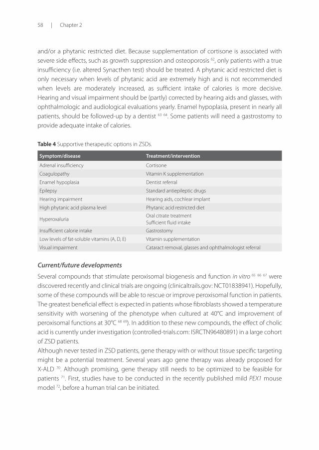
Table 4 Supportive therapeutic options in ZSDs.
Symptom/disease Treatment/intervention
Adrenal insufficiency Cortisone
Coagulopathy Vitamin K supplementation
Enamel hypoplasia Dentist referral
Epilepsy Standard antiepileptic drugs
Hearing impairment Hearing aids, cochlear implant
High phytanic acid plasma level Phytanic acid restricted diet
Hyperoxaluria Oral citrate treatment Sufficient fluid intake
Insufficient calorie intake Gastrostomy
Low levels of fat-soluble vitamins (A, D, E) Vitamin supplementation
Visual impairment Cataract removal, glasses and ophthalmologist referral
Current/future developments
Several compounds that stimulate peroxisomal biogenesis and function in vitro 65 66 67 were discovered recently and clinical trials are ongoing (clinicaltrails.gov: NCT01838941). Hopefully, some of these compounds will be able to rescue or improve peroxisomal function in patients. The greatest beneficial effect is expected in patients whose fibroblasts showed a temperature sensitivity with worsening of the phenotype when cultured at 40°C and improvement of peroxisomal functions at 30°C 68 69). In addition to these new compounds, the effect of cholic acid is currently under investigation (controlled-trials.com: ISRCTN96480891) in a large cohort of ZSD patients. Although never tested in ZSD patients, gene therapy with or without tissue specific targeting might be a potential treatment. Several years ago gene therapy was already proposed for X-ALD 70. Although promising, gene therapy still needs to be optimized to be feasible for patients 71. First, studies have to be conducted in the recently published mild PEX1 mouse model 72, before a human trial can be initiated.
2
Clinical overview and management approach | 59
An orthotopic liver transplantation was described in a single 6-month old ZSD patient and hepatocytes transplantation in another 4-year old patient 73 74. It resulted in decreased concentrations of VLCFAs and pipecolic acid, and improved bile acid profiles. However, the effect on long-term disease course has not been reported. Although bone marrow transplantation (BMT) is an established therapy for the cerebral childhood form of X-ALD 75, there are no reports describing BMT in ZSD patients. BMT would be of interest for those patients who develop leukodystrophy in infancy. However, with the current knowledge it is impossible to predict if patients will develop this rapid progressive leukodystrophy. Recently, a retrospective study revealed that patients with X-ALD still develop an adrenomyelopathy phenotype after BMT 76. Nevertheless, BMT could possibly be beneficial for a subgroup of patients within the ZSD spectrum, but first new techniques/markers that can predict whether or not patients will develop a severe progressive leukodystrophy have to be elucidated.
PrognosisAlthough a rough genotype-phenotype correlation exists for several PEX genes, such as PEX1 and PEX26 77 78, the severity and progression of the disease is difficult to predict for individual patients. This will become more relevant as newborn screening is implemented. As a consequence of newborn screening for X-ALD by C26:0-lysoPC in several countries ZSD will also be diagnosed at birth. Children with the severe phenotype (neonatal-infantile presentation with severe clinical symptoms) have a poor prognosis and these patients usually die within the first year of life. Patients that present in childhood or adolescence usually have a better prognosis, but can develop progressive liver disease or leukodystrophy and deteriorate. If progressive liver disease or leukodystrophy occurs prognosis is poor. The remaining milder individuals can reach adulthood without progression or with long periods of stabilization. When progression occurs, it is mainly related to peripheral neuropathy and pyramidal signs, while cognition remains stable 31.
Unresolved questions The effect of cholic acid is based upon case reports only, but within the coming years the clinical effects will be investigated in larger cohorts. In addition, results of ongoing trials will be published. An important limitation to consider when interpreting the data of these trials is the broad spectrum of severe and milder clinical phenotypes and associated biochemical variations within these cohorts. Furthermore, the natural course of the disease can lead to false conclusions, as peroxisomal metabolites were shown to fluctuate and decline with age 31. A large prospective natural history study is therefore needed. We and others, recently started collecting the data of a large prospective cohort of ZSD patients (clinicaltrails.gov: NCT01668186).
60 | Chapter 2
Second, plasma levels of peroxisomal metabolites do not correlate well with disease severity, as they generally decrease with age. Furthermore, therapies like DHA and Lorenzo’s oil improved plasma levels of DHA and C26:0, albeit no effect on the clinical phenotype has been observed. This is possibly related to differences in expression or activity of peroxisomes in the targeted tissue. Therefore, using plasma levels as a surrogate outcome in clinical trials is not recommended. New biochemical outcome parameters that correlate with disease progression are necessary, such as analysis of markers for peroxisomal dysfunctions in lymphocytes. The pathophysiology of ZSD is still poorly understood. Similar to the cerebral form of X-ALD, it is still not clear when or why ZSD patients develop severe rapid progressive leukodystrophy. The recently constructed mild PEX1 mouse model 72 and natural history studies will help to answer these questions.
Conclusions
Because of the recently implemented newborn screening, more medical doctors in different specialties (e.g. pediatricians, clinical geneticist and neurologists) will encounter patients with a ZSD. ZSDs are clinically heterogeneous with high morbidity in almost all patient and mortality in some. Although treatment is currently only symptomatic, it is important to initiate proper supportive therapy to improve quality of life of these patients.
Acknowledgements
This work was supported by a grant from ‘Metakids’, ‘Hersenstichting’ and ‘Stichting Steun Emma Kinderziekenhuis AMC’, The Netherlands. We would like to thank the parents of the patients displayed in this review for providing photographs and the permission for publication.
2
Clinical overview and management approach | 61
References1. Waterham, H. R. & Ebberink, M. S. Genetics and molecular basis of human peroxisome biogenesis
disorders. Biochim. Biophys. Acta 1822, 1430–41 (2012).
2. Fujiki, Y., Okumoto, K., Mukai, S., Honsho, M. & Tamura, S. Peroxisome biogenesis in mammalian cells. Front. Physiol. 5, 307 (2014).
3. Braverman, N. E., D’Agostino, M. D. & Maclean, G. E. Peroxisome biogenesis disorders: Biological, clinical and pathophysiological perspectives. Dev. Disabil. Res. Rev. 17, 187–96 (2013).
4. Wanders, R. J. A. & Waterham, H. R. Biochemistry of mammalian peroxisomes revisited. Annu. Rev. Biochem. 75, 295–332 (2006).
5. Bowen, P., Lee, C. S., Zellweger, H. & Lindenberg, R. A familial syndrome of multiple congenital defects. Bull. Johns Hopkins Hosp. 114, 402–14 (1964).
6. Poll-The, B. T. et al. Infantile Refsum disease: an inherited peroxisomal disorder. Comparison with Zellweger syndrome and neonatal adrenoleukodystrophy. Eur. J. Pediatr. 146, 477–83 (1987).
7. Ratbi, I. et al. Heimler Syndrome Is Caused by Hypomorphic Mutations in the Peroxisome-Biogenesis Genes PEX1 and PEX6. Am. J. Hum. Genet. 97(4):535-45 (2015).
8. Smith, D. W., Opitz, J. M. & Inhorn, S. L. A syndrome of multiple developmental defects including polycystic kidneys and intrahepatic biliary dysgenesis in 2 siblings. J. Pediatr. 67, 617–24 (1965).
9. Opitz, J. M. et al. The Zellweger Syndrome (cerebro-hepato-renal syndrome). Birth Defects Orig. Art. Set 2, 144–158 (1969).
10. Goldfischer, S. et al. Peroxisomal and mitochondrial defects in the cerebro-hepato-renal syndrome. Science 182, 62–4 (1973).
11. Poulos, A., Sharp, P. & Whiting, M. Infantile Refsum’s disease (phytanic acid storage disease): a variant of Zellweger’s syndrome? Clin. Genet. 26, 579–86 (1984).
12. Kelley, R. I. & Moser, H. W. Hyperpipecolic acidemia in neonatal adrenoleukodystrophy. Am. J. Med. Genet. 19, 791–5 (1984).
13. Van Veldhoven, P. P. Biochemistry and genetics of inherited disorders of peroxisomal fatty acid metabolism. J. Lipid Res. 51, 2863–2895 (2010).
14. Reuber, B. E. et al. Mutations in PEX1 are the most common cause of peroxisome biogenesis disorders. Nat. Genet. 17, 445–8 (1997).
15. Wanders, R. J. A. & Waterham, H. R. Peroxisomal disorders I: biochemistry and genetics of peroxisome biogenesis disorders. Clin. Genet. 67, 107–33 (2005).
16. Collins, C. S. & Gould, S. J. Identification of a common PEX1 mutation in Zellweger syndrome. Hum. Mutat. 14, 45–53 (1999).
17. Gould, S., Raymond, G. & Valle, D. in The Metabolic and Molecular Bases of Inherited Disease 3181–218 (McGraw-Hill, 2001).
18. Levesque, S. et al. A founder mutation in the PEX6 gene is responsible for increased incidence of Zellweger syndrome in a French Canadian population. BMC Med. Genet. 13, 72 (2012).
19. Shimozawa, N. et al. Genetic heterogeneity of peroxisome biogenesis disorders among Japanese patients: evidence for a founder haplotype for the most common PEX10 gene mutation. Am. J. Med. Genet. A 120A, 40–3 (2003).
20. Haynes, C. A. & De Jesús, V. R. The stability of hexacosanoyl lysophosphatidylcholine in dried-blood spot quality control materials for X-linked adrenoleukodystrophy newborn screening. Clin. Biochem. 48, 8–10 (2014).

62 | Chapter 2
21. Vogel, B. H. et al. Newborn screening for X-linked adrenoleukodystrophy in New York State: Diagnostic protocol, surveillance protocol and treatment guidelines. Mol. Genet. Metab. 114, 599–603 (2015).
22. Hubbard, W. C. et al. Newborn screening for X-linked adrenoleukodystrophy (X-ALD): validation of a combined liquid chromatography-tandem mass spectrometric (LC-MS/MS) method. Mol. Genet. Metab. 97, 212–20 (2009).
23. Poll-The, B. T. & Gärtner, J. Clinical diagnosis, biochemical findings and MRI spectrum of peroxisomal disorders. Biochim. Biophys. Acta 1822, 1421–9 (2012).
24. Steinberg, S. J., Raymond, G. V, Braverman, N. E. & Moser, A. B. Peroxisome Biogenesis Disorders, Zellweger Syndrome Spectrum. GeneReviews (2012).
25. Poll-The, B. T. et al. Peroxisome biogenesis disorders with prolonged survival: phenotypic expression in a cohort of 31 patients. Am. J. Med. Genet. A 126A, 333–8 (2004).
26. Hamel, C. Retinitis pigmentosa. Orphanet J. Rare Dis. 1, 40 (2006).
27. van der Knaap, M. S. et al. MRI as diagnostic tool in early-onset peroxisomal disorders. Neurology 78, 1304–8 (2012).
28. Barth, P. G. et al. Late onset white matter disease in peroxisome biogenesis disorder. Neurology 57, 1949–1955 (2001).
29. Moser, A. B. et al. Phenotype of patients with peroxisomal disorders subdivided into sixteen complementation groups. J. Pediatr. 127, 13–22 (1995).
30. Berendse, K., Engelen, M., Linthorst, G. E., van Trotsenburg, A. S. P. & Poll-The, B. T. High prevalence of primary adrenal insufficiency in Zellweger spectrum disorders. Orphanet J. Rare Dis. 9, 133 (2014).
31. Berendse, K. et al. Zellweger spectrum disorders: clinical manifestations in patients surviving into adulthood. J. Inherit. Metab. Dis. 39(1):93-106 (2015).
32. Pineda, M. et al. Diagnosis and follow-up of a case of peroxisomal disorder with peroxisomal mosaicism. J. Child Neurol. 14, 434–9 (1999).
33. Gootjes, J. et al. Identification of the molecular defect in patients with peroxisomal mosaicism using a novel method involving culturing of cells at 40 degrees C: implications for other inborn errors of metabolism. Hum. Mutat. 24, 130–9 (2004).
34. Ebberink, M. S. et al. Genetic classification and mutational spectrum of more than 600 patients with a Zellweger syndrome spectrum disorder. Hum. Mutat. 32, 59–69 (2011).
35. Zeharia, A. et al. A novel PEX12 mutation identified as the cause of a peroxisomal biogenesis disorder with mild clinical phenotype, mild biochemical abnormalities in fibroblasts and a mosaic catalase immunofluorescence pattern, even at 40 degrees C. J. Hum. Genet. 52, 599–606 (2007).
36. Steinberg, S. J. et al. A PEX10 defect in a patient with no detectable defect in peroxisome assembly or metabolism in cultured fibroblasts. J. Inherit. Metab. Dis. 32, 109–19 (2009).
37. Ebberink, M. S. et al. Identification of an unusual variant peroxisome biogenesis disorder caused by mutations in the PEX16 gene. J. Med. Genet. 47, 608–15 (2010).
38. Vallat, C. et al. Major hyperpipecolataemia in a normal adult. J. Inherit. Metab. Dis. 19, 624–6 (1996).
39. Ferdinandusse, S. et al. Clinical, biochemical, and mutational spectrum of peroxisomal acyl-coenzyme A oxidase deficiency. Hum. Mutat. 28, 904–12 (2007).
40. Ferdinandusse, S. et al. Clinical and biochemical spectrum of D-bifunctional protein deficiency. Ann. Neurol. 59, 92–104 (2006).
2
Clinical overview and management approach | 63
41. Horrocks, L. A. & Yeo, Y. K. Health benefits of docosahexaenoic acid (DHA). Pharmacol. Res. 40, 211–25 (1999).
42. Birch, E. E., Hoffman, D. R., Uauy, R., Birch, D. G. & Prestidge, C. Visual acuity and the essentiality of docosahexaenoic acid and arachidonic acid in the diet of term infants. Pediatr. Res. 44, 201–9 (1998).
43. Martínez, M. et al. Therapeutic effects of docosahexaenoic acid ethyl ester in patients with generalized peroxisomal disorders. Am. J. Clin. Nutr. 71, 376S–85S (2000).
44. Noguer, M. T. & Martinez, M. Visual follow-up in peroxisomal-disorder patients treated with docosahexaenoic Acid ethyl ester. Invest. Ophthalmol. Vis. Sci. 51, 2277–85 (2010).
45. Paker, a M. et al. Docosahexaenoic acid therapy in peroxisomal diseases: results of a double-blind, randomized trial. Neurology 75, 826–30 (2010).
46. Moser AB, Borel J, Odone A, Naidu S, Cornblath D, Sanders DB, M. H. A new dietary therapy for adrenoleukodystrophy: biochemical and preliminary clinical results in 36 patients. Ann Neurol. 240–249 (1987).
47. Patrick Aubourg, Catherine Adamsbaum, Marie-Claude Lavallard-Rousseau, Francis Rocchiccioli, Nathalie Cartier, Isabelle Jambaque, Christine Jakobezak, Anne Lemaitre, Francois Boureau, Claude Wolf, and P.-F. B. A Two-Year Trial of Oleic and Erucic Acids (‘Lorenzo’s Oil’) as Treatment for Adrenomyeloneuropathy. NEJM 745–752 (1993).
48. van Geel, B. M. et al. Progression of abnormalities in adrenomyeloneuropathy and neurologically asymptomatic X-linked adrenoleukodystrophy despite treatment with ‘Lorenzo’s oil’. J. Neurol. Neurosurg. Psychiatry 67, 290–299 (1999).
49. Tanaka, K., Shimizu, T., Ohtsuka, Y., Yamashiro, Y. & Oshida, K. Early dietary treatments with Lorenzo’s oil and docosahexaenoic acid for neurological development in a case with Zellweger syndrome. Brain Dev. 29, 586–9 (2007).
50. Arai, Y. et al. Effect of dietary Lorenzo’s oil and docosahexaenoic acid treatment for Zellweger syndrome. Congenit. Anom. (Kyoto). 48, 180–2 (2008).
51. Ferdinandusse, S., Denis, S., Dacremont, G. & Wanders, R. J. A. Toxicity of peroxisomal C27-bile acid intermediates. Mol. Genet. Metab. 96, 121–8 (2009).
52. Setchell, K. D. R. et al. Oral bile acid treatment and the patient with zellweger syndrome. Hepatology 15, 198–207 (1992).
53. Maeda, K. et al. Oral Bile Acid Treatment in Two Japanese Patients With Zellweger Syndrome. 227–230 (2002). doi:10.1097/01.MPG.0000018760.79867.53
54. Keane, M. H. et al. Bile acid treatment alters hepatic disease and bile acid transport in peroxisome-deficient PEX2 Zellweger mice. Hepatology 45, 982–97 (2007).
55. de Vet, E. C. & van den Bosch, H. Alkyl-dihydroxyacetonephosphate synthase. Cell Biochem. Biophys. 32 Spring, 117–21 (2000).
56. Braverman, N. E. & Moser, A. B. Functions of plasmalogen lipids in health and disease. Biochim. Biophys. Acta 1822, 1442–52 (2012).
57. Holmes, R. D., Wilson, G. N. & Hajra, a. Oral ether lipid therapy in patients with peroxisomal disorders. J. Inherit. Metab. Dis. 10, 239–241 (1987).
58. Das, A. K., Holmes, R. D., Wilson, G. N. & Hajra, A. K. Dietary ether lipid incorporation into tissue plasmalogens of humans and rodents. Lipids 27, 401–405 (1992).
59. Wilson, G. N. et al. Zellweger syndrome: diagnostic assays, syndrome delineation, and potential therapy. Am. J. Med. Genet. 24, 69–82 (1986).
64 | Chapter 2
60. van Woerden, C. S. et al. High incidence of hyperoxaluria in generalized peroxisomal disorders. Mol. Genet. Metab. 88, 346–50 (2006).
61. Leumann, E., Hoppe, B., Neuhaus, T. & Blau, N. Efficacy of oral citrate administration in primary hyperoxaluria. Nephrol. Dial. Transplant 10 Suppl 8, 14–6 (1995).
62. Buchman, Alan L. M.D., M. S. P. H. Side Effects of Corticosteroid Therapy. Journal of Clinical Gastroenterology 289–294 (2001).
63. Lertsirivorakul, J., Wongswadiwat, M. & Treesuwan, P. Oral manifestations and dental management of a child with Zellweger syndrome. Spec. Care Dent. 34, 46–50 (2012).
64. Acharya, B. S., Ritwik, P., Velasquez, G. M. & Fenton, S. J. Medical-dental findings and management of a child with infantile Refsum disease: a case report. Spec. Care Dentist. 32, 112–7 (2012).
65. Zhang, R. et al. Recovery of PEX1-Gly843Asp peroxisome dysfunction by small-molecule compounds. Proc. Natl. Acad. Sci. U. S. A. 107, 5569–74 (2010).
66. Berendse, K. et al. Arginine improves peroxisome functioning in cells from patients with a mild peroxisome biogenesis disorder. Orphanet J. Rare Dis. 8, 138 (2013).
67. Wei, H., Kemp, S., McGuinness, M. C., Moser, a B. & Smith, K. D. Pharmacological induction of peroxisomes in peroxisome biogenesis disorders. Ann. Neurol. 47, 286–96 (2000).
68. Imamura, a et al. Temperature-sensitive mutation in PEX1 moderates the phenotypes of peroxisome deficiency disorders. Hum. Mol. Genet. 7, 2089–94 (1998).
69. Shimozawa, N. et al. Nonsense and temperature-sensitive mutations in PEX13 are the cause of complementation group H of peroxisome biogenesis disorders. Hum. Mol. Genet. 8, 1077–83 (1999).
70. Cartier, N. et al. Lentiviral hematopoietic cell gene therapy for X-linked adrenoleukodystrophy. Methods Enzymol. 507, 187–98 (2012).
71. Toscano, M. G. et al. Physiological and tissue-specific vectors for treatment of inherited diseases. Gene Ther. 18, 117–27 (2011).
72. Hiebler, S. et al. The Pex1-G844D mouse: a model for mild human Zellweger spectrum disorder. Mol. Genet. Metab. 111, 522–32 (2014).
73. Sokal, E. M. et al. Hepatocyte transplantation in a 4-year-old girl with peroxisomal biogenesis disease: technique, safety, and metabolic follow-up. Transplantation 76, 735–8 (2003).
74. Van Maldergem, L. et al. Orthotopic liver transplantation from a living-related donor in an infant with a peroxisome biogenesis defect of the infantile Refsum disease type. J. Inherit. Metab. Dis. 28, 593–600 (2005).
75. Patrick Aubourg, M.D., Stéphane Blanche, M.D., Isabelle Jambaqué, Ph.D., Francis Rocchiccioli, Ph.D., Gabriel Kalifa, M.D., Catherine Naud-Saudreau, M.D., Marie-Odile Rolland, Ph.D., Mariane Debré, M.D., Jean-Louis Chaussain, M.D., Claude Griscelli, M.D., M. D. Reversal of Early Neurologic and Neuroradiologic Manifestations of X-Linked Adrenoleukodystrophy by Bone Marrow Transplantation. N. Engl. J. Med. 1860–1866 (1990).
76. van Geel, B. M. et al. Hematopoietic cell transplantation does not prevent myelopathy in X-linked adrenoleukodystrophy: a retrospective study. J. Inherit. Metab. Dis. 38, 359–61 (2015).
77. Bader, P. I., Dougherty, S., Cangany, N., Raymond, G. & Jackson, C. E. Infantile Refsum disease in four Amish sibs. Am. J. Med. Genet. 90, 110–114 (2000).
78. Rosewich, H., Ohlenbusch, a & Gärtner, J. Genetic and clinical aspects of Zellweger spectrum patients with PEX1 mutations. J. Med. Genet. 42, e58 (2005).