Embed Size (px)
Citation preview
UvA-DARE is a service provided by the library of the University of Amsterdam (http://dare.uva.nl)
UvA-DARE (Digital Academic Repository)
The phosphoketolase pathway in Lactobacillus pentosus.
Posthuma, C.C.
Link to publication
Citation for published version (APA):Posthuma, C. C. (2001). The phosphoketolase pathway in Lactobacillus pentosus. Enschede: Ipskamp
General rightsIt is not permitted to download or to forward/distribute the text or part of it without the consent of the author(s) and/or copyright holder(s),other than for strictly personal, individual use, unless the work is under an open content license (like Creative Commons).
Disclaimer/Complaints regulationsIf you believe that digital publication of certain material infringes any of your rights or (privacy) interests, please let the Library know, statingyour reasons. In case of a legitimate complaint, the Library will make the material inaccessible and/or remove it from the website. Please Askthe Library: http://uba.uva.nl/en/contact, or a letter to: Library of the University of Amsterdam, Secretariat, Singel 425, 1012 WP Amsterdam,The Netherlands. You will be contacted as soon as possible.
Download date: 18 Jun 2018

Chapter
General introduction
CHAPTER
1.1 LACTIC ACID BACTERIA
Fermentation is one of the traditional methods for food preservation, and dates back from about 6000 BC. The most probable basis for the development of fermented food was through food that spoiled during storage, giving rise to a product that was acceptable for consumption [128]. Many of these food products are prepared by the addition of so-called starter cultures, defined cultures that contain strains with known properties, in order to control the fermentation process as much as possible. Lactic acid bacteria play an important role in the production of several types of food products (listed in Table 1.1).
The metabolism of sugars by lactic acid bacteria leads to the production of compounds like lactate, ethanol, diacetyl and carbon dioxide, that influence the taste, flavour or texture of the fermented products. At the same time, fermentation results in the production of acids like lactate and acetate, which together with the resulting decrease in pH, prevent the growth of food spoilage and pathogenic microorganisms, like Listeria and Clostridium. Some lactic acid bacteria also produce bacteriocins, small peptides with bacteriostatic or bactericidal properties against mainly Gram-positive bacteria.
Some species from the genus Lactobacillus that naturally occur in the human and animal gastro-intestinal tract, have been claimed to stimulate the mucosal immune system and to be beneficial for human health [24]. The colonization capacities of these so-called probiotic strains are investigated by many laboratories and much research is performed to study their health effects as well as to investigate the possibilities to use manipulated strains of lactic acid bacteria as oral vaccine delivery vehicles (for reviews: see [92] and [155]).
Since lactic acid bacteria play such an important role in the production of food and may contribute to human health, many laboratories are interested in their genetic and metabolic properties. A good insight in the regulation of fermentation pathways which result in the formation of flavour compounds might help to construct starter strains that are better adapted to the fermentation conditions and /o r produce more or other flavour compounds, to improve product quality. Redirection of metabolic pathways has already been shown to be possible in for instance Lactococcus lactis or Lactobacillus plantarurrc Inactivation of lactate dehydrogenase (LDH) leads to the increased production of acetate, formate, acetoin and ethanol at the expense of the formation of lactate, which is the main end-product in the parent strains (for reviews about metabolic engineering in lactic acid bacteria: see [17] and [52]).
GENERAL INTRODUCTION 11
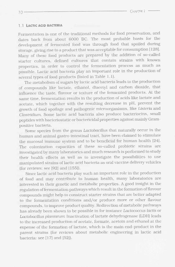
Products Lactic acid bacteria
Dairy products
Gouda- type cheeses
Swiss-type cheeses
Fermented milk and yoghurts
Fermented meat
Sausages
Fermented vegetables
Sauerkraut (white cabbage)
Soy sauce
Pickles, cucumbers
Olives
Other products of plant origin
Sourdough bread
Silage
Wines and ciders
Lactococcus /actis subsp. cremoris and subsp. /actis,
Leuconosfoc lactis subsp. cremoris
Lactobacillus delbrueckii subsp. /act/'s, Lactobaci//us
helveticus
Streptococcus thermophilus, Lactobacillus
delbrueckii subsp. bulgaricus, Lactobacillus
acidophilus, Lactobacillus casei, Lactobacillus kefir,
Lactococcus lactis subsp. diacetylactis
Lactobacillus sakei, Lactobacillus curvatus,
Pediococcus acidilactici, Leuconosfoc carnosus
Leuconostoc mesenteroides, Lactobacillus
plantarum, Lactobacillus pentosus, Lactobacillus
bavaricus
Tetragenococcus halophilus (formerly ca l led:
Pediococcus halophilus)
Lactobacillus plantarum, Lactobacillus pentosus,
Pediococcus pentosaceus, Leuconostoc
mesenteroides
Leuconostoc mesenteroides, Lactobacillus pentosus
Lactobacillus sanfransisco, Lactobacillus brevis
Lactobacillus plantarum
Oenococcus oen/ (formerly ca l led: Leuconostoc
oenos), Lactobacillus plantarum
Table 1 . 1 . An overview of food products in which fermentat ion by lactic acid bacteria
is used.
12 CHAPTER
CO O
ja
0> t X J CO
0 CO O
ja 0 3
-O U_ X Œ Û Q _ J Q
Q_
/ <t < \'*— O
2 ™ ^°-i * <
CD
0 ra 0 0 a 0
0 —^ 3 "V> er
0 —^ 3
/ V g & O = Q_ 9 s:
O CD z 0 Q_ 2
CS -r^r
O >— <u X
o m o E o
GENERAL INTRODUCTION 13
1.2 S U G A R M E T A B O L I S M I N LACTIC A C I D BACTERIA
Lactic acid bacteria are aero tolerant organisms, which preferably grow under anaerobic or micro-aerobic conditions. For growth, carbohydrates together with nucleotides, amino acids and vitamins are needed in the medium, since many biosynthetic pathways are deficient [60]. During fermentation of carbohydrates, metabolic energy is stored by substrate level phosphorylation, by phosphoglycerate kinase and pyruvate kinase which are operative in the glycolytic pathway or by acetate kinase during the conversion of acetyl-phosphate to acetate. An additional way to generate energy is by the formation of a proton motive force during the efflux of solutes over the cell membrane, by which a net charge is translocated. The proton motive force is used to synthesize ATP by a membrane-located ATPase [63, 90]. For instance, lactate that has accumulated in the cytoplasm as a result of sugar fermentation can exit from the cell in symport with protons in L. locus resulting in the net translocation of protons and positive charge outside of the cells. Also malate/ lactate antiport results in the generation of a proton motive force in this organism, if it is combined with the decarboxylation of malate in the cytoplasm during which reaction a proton is consumed [63].
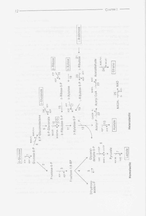
There are several pathways for sugar metabolism in lactic acid bacteria, which are summarized in Figure 1.1. During so-called homolactic fermentation, glucose is metabolized into pyruvate via the glycolytic or Embden-Meyerhoff-Parnas (EMP) pathway. Under anaerobic conditions in the presence of excess glucose, pyruvate is mainly converted by LDH into lactate. The conversion of pyruvate into lactate regenerates the NAD+ that is consumed during the conversion of glucose into pyruvate (see Figure 1.1,
Figure 1.1. Overview of sugar fermentation in lactic acid bacteria. On the left side, homolactic fermentation via the Embden-Meyerhoff-Parnas pathway; on the right side, heterolactic fermentation via the phosphoketolase pathway. Both the 'acetate-branch' and the 'ethanol-branch' of the phosphoketolase pathway were drawn in the Figure. Dashed arrow indicates that more than one enzyme is involved in the conversion from one substrate into the other. The other enzymes are indicated with numbers. 1, glucokinase or phosphorylation during PEP-dependent PTS transport; 2, glucose 6-phosphate isomerase; 3, 6-phosphofructokinase; 4, fructose bisphosphate aldolase; 5, triose-phosphate isomerase; 6, glyceraldehyde 3-phosphate dehydrogenase, phosphoglycerate kinase, phosphoglyceromutase, enolase, pyruvate kinase; 7, lactate dehydrogenase; 8, glucose 6-phosphate dehydrogenase; 9, lactonase; 10, 6-phosphogluconate dehydrogenase; 1 1, D-ribulose 5-phosphate 3-epimerase; 12, xylulose 5-phosphate phosphoketolase; 1 3, acetate kinase; 1 4, gluconate kinase phosphorylation during PEP-dependent PTS transport; 1 5, ribose 5-phosphate isomerase; 1 6, ribose kinase; 1 7, xylulose kinase; 1 8, xylose isomerase; 1 9, L-ribulose 5-phosphate 4-epimerase; 20, ribulose kinase; 2 1 , arabinose isomerase; 22, phosphotransacetylase; 23, acetaldehyde dehydrogenase; 24, alcohol dehydrogenase; 25, NADH oxidase.
j 4 CHAPTER I
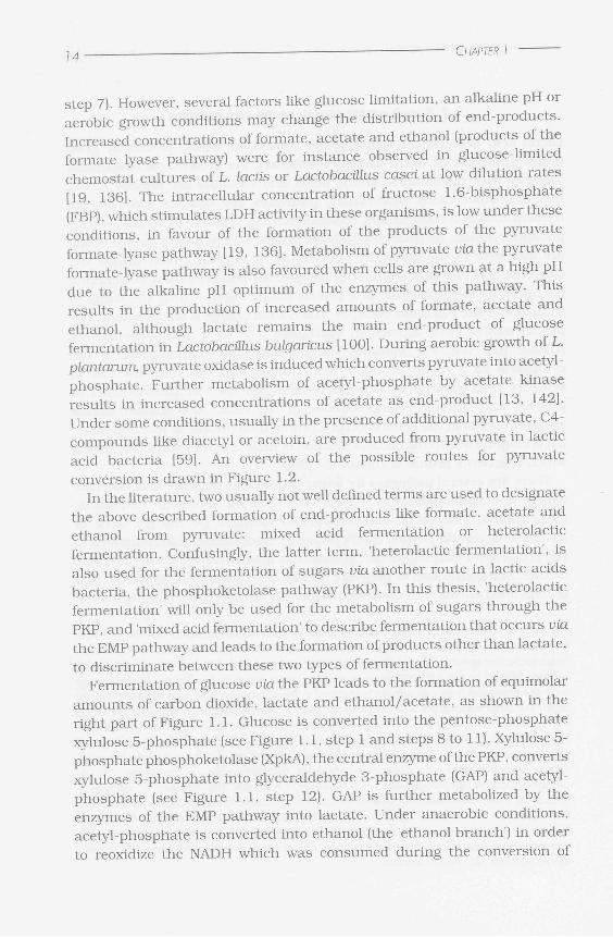
step 7). However, several factors like glucose limitation, an alkaline pH or aerobic growth conditions may change the distribution of end-products. Increased concentrations of formate, acetate and ethanol (products of the formate lyase pathway) were for instance observed in glucose-limited chemostat cultures of L. lactis or Lactobacillus casei at low dilution rates [19, 136]. The intracellular concentration of fructose 1,6-bisphosphate (FBP), which stimulates LDH activity in these organisms, is low under these conditions, in favour of the formation of the products of the pyruvate formate-lyase pathway [19, 136]. Metabolism of pyruvate via the pyruvate formate-lyase pathway is also favoured when cells are grown at a high pH due to the alkaline pH optimum of the enzymes of this pathway. This results in the production of increased amounts of formate, acetate and ethanol, although lactate remains the main end-product of glucose fermentation in Lactobacûlus bulgaricus [100]. During aerobic growth of L. plantarunx pyruvate oxidase is induced which converts pyruvate into acetyl-phosphate. Further metabolism of acetyl-phosphate by acetate kinase results in increased concentrations of acetate as end-product [13, 142]. Under some conditions, usually in the presence of additional pyruvate, C4-compounds like diacetyl or acetoin, are produced from pyruvate in lactic acid bacteria [59]. An overview of the possible routes for pyruvate
conversion is drawn in Figure 1.2. In the literature, two usually not well defined terms are used to designate
the above described formation of end-products like formate, acetate and ethanol from pyruvate: mixed acid fermentation or heterolactic fermentation. Confusingly, the latter term, 'heterolactic fermentation', is also used for the fermentation of sugars via another route in lactic acids bacteria, the phosphoketolase pathway (PKP). In this thesis, 'heterolactic fermentation' will only be used for the metabolism of sugars through the PKP, and 'mixed acid fermentation' to describe fermentation that occurs via the EMP pathway and leads to the formation of products other than lactate, to discriminate between these two types of fermentation.
Fermentation of glucose ufathe PKP leads to the formation of equimolar amounts of carbon dioxide, lactate and ethanol/acetate, as shown in the right part of Figure 1.1. Glucose is converted into the pentose-phosphate xylulose 5-phosphate (see Figure 1.1, step 1 and steps 8 to 11). Xylulose 5-phosphate phosphoketolase (XpkA), the central enzyme of the PKP, converts xylulose 5-phosphate into glyceraldehyde 3-phosphate (GAP) and acetyl-phosphate (see Figure 1.1, step 12). GAP is further metabolized by the enzymes of the EMP pathway into lactate. Under anaerobic conditions, acetyl-phosphate is converted into ethanol (the ethanol branch') in order to reoxidize the NADH which was consumed during the conversion of
GENERAL INTRODUCTION
glucose into xylulose 5-phosphate, whereas mainly acetate is produced under aerobic conditions [ 13]. In the presence of oxygen, an NADH oxidase is induced, which allows the bacteria to convert acetyl-phosphate into acetate by acetate kinase, during which reaction an ATP is formed. Thus, during heterofermentation in the presence of oxygen, two ATP molecules are produced per glucose like during fermentation via the EMP pathway. In contrast, fermentation via the PKP under anaerobic conditions gains only one ATP per glucose.
Pentoses, like xylose, arabinose and ribose, are metabolized by their appropriate enzymes into xylulose 5-phosphate, which is further degraded in the PKP (see Figure 1.1). The ability of heterofermentative lactic acid bacteria to grow on a certain pentose is dependent on the availability of the enzymes for transport and conversion of the compound into xylulose 5-phosphate [59]. Pentose metabolism via the PKP under anaerobic conditions
Lactate
NADH- NAD NADH NAD
Diacetyl — ^ ^ — > Acetoin -^—^—> 2.3-Butanediol
CoASH
Acetyl CoA
10 12 CO;
*= 7^T NAD NADH;
Pyruvate n Pyruvate — s
N > Acetolactate
CO,
CO;
Formate
Acetyl-P * ^ > Acetyl CoA 5
ADP
ATP «
NADH;
Acetate
13 -V» NAD
0; H;0;
Acetaldehyde
N A D ( P ) H ;
NAD(P)
Ethanol
Figure 1.2. An overview of the possible routes for pyruvate conversion in homofermentative lactic acid bacteria. 1, Lactate dehydrogenase; 2, pyruvate oxidase; 3, acetate kinase; 4, pyruvate formate lyase; 5, phosphotransacetylase; 6, acetaldehyde dehydrogenase; 7, alcohol dehydrogenase; 8, diacetyl synthase; 9, acetolactate synthase; 10, diacetyl reductase; 11, acetolactate decarboxylase; 12, 2,3-butanediol dehydrogenase; 13, NADH oxidase.
Ribose-5-P
Acetyl-P
)
Acetate
Glyceral-dehyde-3-P
Sedohep-tulose-7-P
Glyceral-dehyde-3-P
NAD Np 2 ADP
NADH. . v 2 ATP
Pyruvate
N A D H î
NAD
Lactate
CHAPTER
D-Glucose
ATP A
ADP -J,
Glucose-6-P
Fructose-6-P
'~
Acetyl-P
Erythrose-4-P ADp .
ATP
Acetate
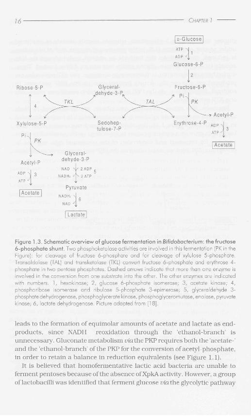
Figure 1.3. Schematic overview of glucose fermentation in Bifidobacterium: the fructose 6-phosphate shunt. Two phosphoketolase activities are involved in this fermentation (PK in the Figure): for cleavage of fructose 6-phosphate and for cleavage of xylulose 5-phosphate. Transaldolase (TAL) and transketolase (TKL) convert fructose 6-phosphate and erythrose 4-phosphate in two pentose phosphates. Dashed arrows indicate that more than one enzyme is involved in the conversion from one substrate into the other. The other enzymes are indicated with numbers. 1, hexokinase; 2, glucose 6-phosphate isomerase; 3, acetate kinase; 4, phosphoribose isomerase and ribulose 5-phosphate 3-epimerase; 5, glyceraldehyde 3-phosphate dehydrogenase, phosphoglycerate kinase, phosphoglyceromutase, enolase, pyruvate kinase; 6, lactate dehydrogenase. Picture adapted from [18].
leads to the formation of equimolar amounts of acetate and lactate as end-products, since NADH reoxidation through the 'ethanol-branch' is unnecessary. Gluconate metabolism via the PKP requires both the 'acetate-' and the 'ethanol-branch' of the PKP for the conversion of acetyl-phosphate, in order to retain a balance in reduction equivalents (see Figure 1.1).
It is believed that homofermentative lactic acid bacteria are unable to ferment pentoses because of the absence of XpkA activity. However, a group of lactobacilli was identified that ferment glucose ufathe glycolytic pathway
GENERAL INTRODUCTION Î 7
bu t is also able to use pentoses as a sole energy source [59]. These lactobacilli, amongst which are Lactobacillus plantarum, L. casei and Lactobacillus pentosus, possess an inducible XpkA. This group of lactobacilli is called facultative heterofermentative lactobacilli to discriminate them from obligate homo- (only fermentation via the EMP) and obligate heterofermentative (only fermentation via the PKP) lactobacilli [59].
1.3 GLUCOSE METABOLISM I N BIFIDOBACTERIUM
Phosphoketolase activity is also involved in sugar metabolism of Bifidobacterium, a genus that physiologically resembles the lactic acid bacteria, since lactate and acetate are the main end-products of fermentation [44]. For this reason, the genus Bifidobacteriumhas for a long time been considered to belong to the lactic acid bacteria. However, phylogenetic classification has identified the bifidobacteria as a distinct group of GC-rich bacteria which is only very distantly related to the genuine genera of lactic acid bacteria, like Lactococcus, Lactobacillus and Leuconostoc, which belong to the AT-rich branch of Gram-positive bacteria [145].
The fermentation of glucose in Bifidobacterium occurs via a pathway which is specific for this group of bacteria, the so-called fructose 6-phosphate shunt , in which two phosphoketolase activities are involved (see Figure 1.3). The genus Bifidobacterium belongs thus to the few bacteria in which phosphoketolase activity has been reported. The first phosphoketolase that is active in glucose fermentation converts fructose 6-phosphate into erythrose 4-phosphate and acetyl-phosphate, the second one catalyses the phosphorolytic cleavage of xylulose 5-phosphate like described for XpkA in heterolactic fermentation in lactobacilli [18, 107]. The fructose 6-phosphate shunt results in the formation of acetate and lactate at a molar ratio of 3 to 2. Fructose 6-phosphate phosphoketolases have been partially purified from several Bifidobacterium species [119]. Some of the fructose 6-phosphate phosphoketolases appeared to be specific for both xylulose 5-phosphate and fructose 6-phosphate, whereas others catalyse only the cleavage of fructose 6-phosphate [119]. In some Bifidobacterium species, both phosphoketolase activities might be catalysed by the same enzyme.
1.4 THE PENTOSE PHOSPHATE PATHWAY
The presence of the PKP as a glucose or pentose degrading pathway is only known in lactic acid bacteria. In most other organisms, pentoses and under certain circumstances also glucose is metabolized via the pentose
j 8 • CHAPTER 1
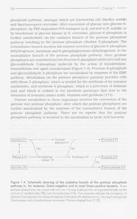
phosphate pathway, amongst which are Escherichia coli, Baciüus subtilis and Saccharomyces cerevisiae. After conversion of glucose into glucose 6-phosphate, by PEP-dependent PTS transport in E. coli and in B. subtilis, or by hexokinase or glucose kinase in S. cerevisiae, glucose 6-phosphate is further metabolized via the oxidative branch of the pentose phosphate pathway resulting in the pentose phosphate ribulose 5-phosphate. The nonoxidative branch involves the enzyme activities of glucose 6-phosphate dehydrogenase, lactonase and 6-phosphogluconate dehydrogenase. In the nonoxidative branch of the pentose phosphate pathway, three pentose phosphates are converted into two fructose 6-phosphate molecules and one glyceraldehyde 3-phosphate molecule by the action of transketolase, transaldolase and again transketolase (Figure 1.4). Fructose 6-phosphate and glyceraldehyde 3-phosphate are metabolized by enzymes of the EMP pathway. Metabolism via the pentose phosphate pathway provides cells with ribose 5-phosphate, which is needed for the synthesis of for instance nucleotides, and erythrose 4-phosphate, which is a precursor of shikimic acid and which is utilized in the pyridoxin pathways that lead to the formation of aromatic amino acids, vitamins and pyridoxin [171].
Pentose metabolism in these organisms involves the conversion of the pentose into pentose phosphate, after which the pentose phosphates are further metabolized by the enzymes of the nonoxidative branch of the pentose phosphate pathway. There are no reports that the pentose phosphate pathway is involved in the metabolism in lactic acid bacteria.
Glyceral- —• |Fructose-6-P| dehyde-3-P
Sedohep- • z -* Erythrose-4-P -?—• |Fructose-6-P| tulose-7-P /
TKL
|Xy lu lose 5-P | z • |Glyceraldehyde-3-P
Figure 1.4. Schematic drawing of the oxidative branch of the pentose phosphate pathway in, for instance, Gram-negative and in most Gram-positive bacteria. Three pentose phosphates are converted into two fructose 6-phosphate and glyceraldehyde by the actions of transketolase (TKL) and transaldolase (TAL). Two enzymes catalyse the conversion of xylulose 5-phosphate into ribose phosphate (indicated with dashed arrow): ribulose 5-phosphate 3-epimerase and phosphoribose isomerase. Picture adapted from [1 12].
GENERAL INTRODUCTION 79
1 .6 XYLOSE METABOLISM
The organism to which the research described in this thesis refers, L. pentosus, is a facultative heterofermentative Lactobacillus. As a consequence, it possesses the enzymes for both homolactic fermentation via the EMP pathway and for heterofermentation via the PKP. The latter pathway is mainly used for the fermentation of pentoses, like for instance xylose. The ability of L. pentosus to grow on xylose is a rather unique property for Lactobacillus, since only a few Lactobacillus species are able to utilize this carbohydrate.
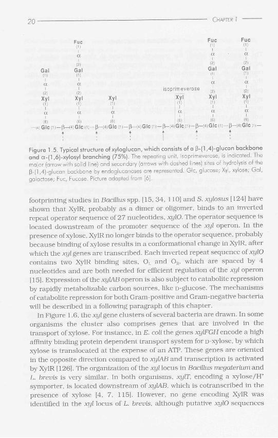
Xylose is a naturally occurring sugar, mostly occurring as part of hemicellulose polymers like xylan and xyloglucan. Xylan, consisting of ß-1,4-linked D-xylosyl residues, is abundan t in nature, mainly found in secondary plant cell walls. Xyloglucan polysaccharides, which have a ß-1,4-glucan backbone substi tuted with a-l,6-xylosyl residues, play an important role in determining the semi-rigid structure of the primary cell wall. Degradation of the polymers is catalysed by ß-xyloside degrading enzymes, like xylanases and _8-xylosidases, and various ß-l,4-endoglu-canases. These enzymes are present in ruminai and soil bacteria such as Bacillus species or in filamentous fungi. A picture of xyloglucan is shown in Figure 1.5. For reviews about the degradation of hemicelluloses, see Warren [152] and Wong etal. [159].
The monosaccharide D-xylose can be utilized by a number of bacterial species. The metabolism involves the transport into the cell, after which xylose is isomerized to D-xylulose, which is phosphorylated to form D-xylulose 5-phosphate. The conversion of xylose uiaxylulose into xylulose 5-phosphate is catalysed by xylose isomerase and xylulose kinase. The genes encoding these enzymes have been cloned and sequenced from E. coli [69], Salmonella typhimurium [120], Bacillus spp. [45, 71 , 103, 111], Staphylococcus xylosus [124], Klebsiella pneumoniae [27], Thermoanaerobac-ter ethanolicus [25], Tetragenococcus halophilus [134], Lactobacillus brevis [4] and L. pentosus [75]. In all these organisms, the xylose isomerase encoding gene, xylA, and the gene encoding the xylulose kinase, xylB, are clustered in an operon. Expression of the genes is induced in the presence of xylose in the growth medium by a regulator protein encoded by xylR, which is part of the xyl gene cluster in most xylose utilizing bacteria. XylR functions as an activator in the Gram-negative bacteria E. coli [126] and S. typhimurium [120] and as a repressor in Gram-positive bacteria like Bacillus spp. [65, 103, 110], L. pentosus [73] and S. xylosus [124].
In E. coli XylR binds in the presence of xylose to an adenine-rich operator sequence IA which is located upstream of the promoter sequence [ 126], thereby activating transcription of the operon. Gel-mobility and DNA-
20
Fuc (1)
a
Gal (1)
i
(2)
Gal (1)
i
a a
(2)
Xyl (1)
i
(2)
Xyl (1)
i
Xyl (1)
i
a a i
a i
(6)
—(4)Glc(i)-(6)
- ß — ( 4 ) G I C (1) +
-ß -(6)
- (4)GICP)-ß-
CHAPTER
Fuc Fuc ID ID
i i a a i i
(2) (2)
Gal Gal m (M
i i a a i i
(2) (2) isoprimeverose
Xyl Xyl Xyl m (1) (M
i i a a a ! I I
(6) (6) (6)
-(4)G)C(D—ß—(4)|G1C|(1)— P—<4)GIC<1)—ß—fflGICÜ) —
t ; ! Figure 1.5. Typical structure of xyloglucan, which consists of a ß-(l ,4)-glucan backbone and a-(l,6)-xylosyl branching (75%). The repeating unit, isoprimeverose, is indicated. The major (arrow with solid line) and secondary (arrows with dashed lines) sites of hydrolysis of the ß-(l,4)-glucan backbone by endoglucanases are represented. Glc, glucose; Xyl, xylose; Gal, galactose; Fuc, Fucose. Picture adapted from [6].
footprinting studies in Bacillus spp. [15, 34, 110] and S. xylosus [124] have shown tha t XylR, probably as a dimer or oligomer, binds to an inverted repeat operator sequence of 27 nucleotides, xylO. The operator sequence is located downstream of the promoter sequence of the xyl operon. In the presence of xylose, XylR no longer binds to the operator sequence, probably because binding of xylose results in a conformational change in XylR, after which the xyl genes are transcribed. Each inverted repeat sequence of xylO contains two XylR binding sites, O L and OR , which are spaced by 4 nucleotides and are both needed for efficient regulation of the xyl operon [15]. Expression of the xylAB operon is also subject to catabolite repression by rapidly metabolizable carbon sources, like D-glucose. The mechanisms of catabolite repression for both Gram-positive and Gram-negative bacteria will be described in a following paragraph of this chapter.
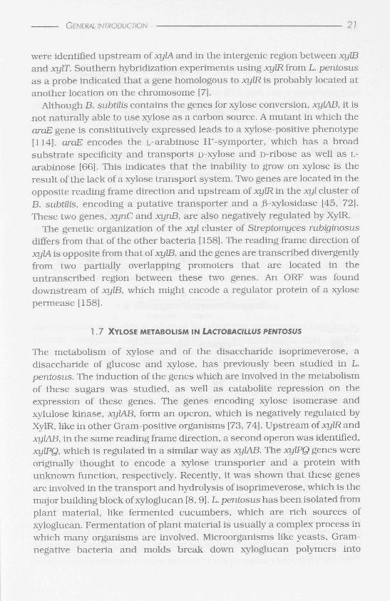
In Figure 1.6, the xyigene clusters of several bacteria are drawn. In some organisms the cluster also comprises genes that are involved in the transport of xylose. For instance, in E. coli the genes xylFGH encode a high affinity binding protein dependent transport system for D-xylose, by which xylose is translocated at the expense of an ATP. These genes are oriented in the opposite direction compared to xylAB and transcription is activated by XylR [ 126]. The organization of the xyl locus in Bacillus megatertum and L. brevis is very similar. In both organisms, xylT, encoding a xylose/H+
symporter, is located downstream of xylAB, which is cotranscribed in the presence of xylose [4, 7, 115]. However, no gene encoding XylR was identified in the xyl locus of L. brevis, although putative xylO sequences
GENERAL INTRODUCTION
were identified upstream of xylA and in the intergenic region between xyLB and xylT. Southern hybridization experiments using xylR from L. pentosus as a probe indicated that a gene homologous to xylR is probably located at another location on the chromosome [7].
Although B. subtûis contains the genes for xylose conversion, xylAB, it is not naturally able to use xylose as a carbon source. A mutant in which the araE gene is constitutively expressed leads to a xylose-positive phenotype [114]. araE encodes the L-arabinose H+-symporter, which has a broad substrate specificity and transports D-xylose and D-ribose as well as L-arabinose [66]. This indicates that the inability to grow on xylose is the result of the lack of a xylose transport system. Two genes are located in the opposite reading frame direction and upstream of xylR in the xyl cluster of B. subtüis, encoding a putative transporter and a j3-xylosidase [45, 72]. These two genes, xynC and xynB, are also negatively regulated by XylR.
The genetic organization of the xyl cluster of Streptomyces rubiginosus differs from that of the other bacteria [158]. The reading frame direction of xylA is opposite from that of xylB. and the genes are transcribed divergently from two partially overlapping promoters that are located in the untranscribed region between these two genes. An ORF was found downstream of xylB, which might encode a regulator protein of a xylose permease [158].
1.7 XYLOSE METABOLISM I N LACTOBACILLUS PENTOSUS
The metabolism of xylose and of the disaccharide isoprimeverose, a disaccharide of glucose and xylose, has previously been studied in L. pentosus. The induction of the genes which are involved in the metabolism of these sugars was studied, as well as catabolite repression on the expression of these genes. The genes encoding xylose isomerase and xylulose kinase, xylAB, form an operon, which is negatively regulated by XylR, like in other Gram-positive organisms [73, 74]. Upstream of xyLRand xylAB, in the same reading frame direction, a second operon was identified, xylPQ, which is regulated in a similar way as xylAB. The xylPQ genes were originally thought to encode a xylose transporter and a protein with unknown function, respectively. Recently, it was shown that these genes are involved in the transport and hydrolysis of isoprimeverose, which is the major building block of xyloglucan [8,9]. L. pentosus has been isolated from plant material, like fermented cucumbers, which are rich sources of xyloglucan. Fermentation of plant material is usually a complex process in which many organisms are involved. Microorganisms like yeasts, Gram-negative bacteria and molds break down xyloglucan polymers into
22 CHAPTER
activation
E. coli
binding
© of xylose ,—,
xylB xylA * " ^" xylF xylG xylH xylR kinase isomerase binding protein dependent activator
transport system
megaterium
^ = ^x. xylR 'm* xylA xylB xylT repressor isomerase kinase xylose/H
symporter
S. subtilis
* " xynC xynB xylR ^" xylA xylB putative ß-xylosidase repressor isomerase kinase
transporter
L. brevis
S. rubiginosus
L. pentosus
% — > f l r = ^ % :>£ ^ xylA xylB w xylT
isomerase kinase xylose/H symporter
<::::::=^<===^t=>tt-'ORF xylB xylA
? kinase isomerase
xylP ' xylQ xylR w xylA xylB 1 isoprimeverose a-xylosidase repressor ' isomerase kinase
permease J,
repression O binding of xylose
0 Figure 1.6. Organizat ion of xyl loci which are described in the text. Expression of the xyl
genes of E. coli is activated by XylR in the presence of xylose, whereas the xyl genes of the other
organisms are repressed by XylR. No xylR has been characterized from L brevis and from S.
rubiginosus yet. Arrows indicate transcriptional start sites, stemloop structures depict
transcriptional termination sites.
oligomers like isoprimeverose. The capacity of L. pentosus to hydrolyse isoprimeverose into D-glucose and D-xylose and subsequently ferment both monosaccharides may be related to the natural habitat of this organism in plant material [8].
GENERAL INTRODUCTION 23
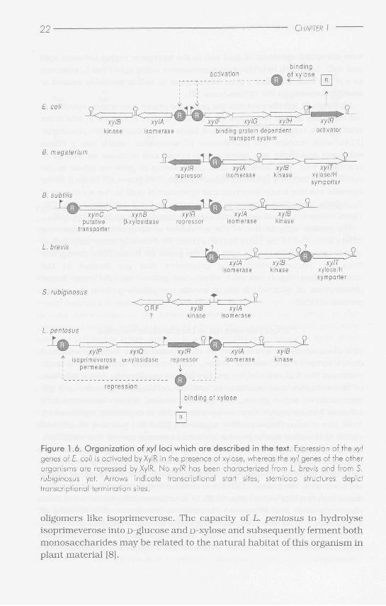
The observation that mutan t s of L. pentosus that were affected in mannose phosphoenol pyruvate (PEP)-dependent phosphotransferase system (PTS) activity were unable to grow on xylose as the sole energy source, led to the idea that the mannose PTS and not a specific xylose permease might be involved in xylose transport [10]. The PTS is a so-called group translocation system for the uptake of carbohydrates, which catalyses the concomitant transport and phosphorylation of sugars in both Gram-positive and Gram-negative bacteria. A phosphoryl group is successively transferred from PEP to Enzyme I, the histidine-containing protein (HPr) and the sugar-specific Enzyme II, which then phosphorylates the sugar (see Figure 1.7). The EIIman complex consists of two integral membrane proteins, EIICman and EIIDman, which probably bind and transport the carbohydrate into the cytoplasm, and of two cytoplasmic proteins, EIIAman and EIIBman, of which the latter phosphorylates the transported carbohydrate (for a review about PTS, see [91]). The D-mannose PTS in L. pentosus t ransports both glucose and mannose [6]. The membrane component of the mannose PTS, EIICDman, is believed to recognize D-xylose and transport it by facilitated diffusion in L. pentosus. Xylose is not phosphorylated during the translocation [10].
1.8 CATABOLITE REPRESSION IN BACTERIA
Bacteria have to be able to adapt to the changing environment they live in. Carbon catabolite repression (CR), the phenomenon that rapidly metabolizable carbon sources repress the expression of catabolic genes that are involved in the metabolism of other carbon sources, is one of the mechanisms by which bacteria respond to the availability of multiple carbon and energy sources in their environment. The mechanisms of CR have been studied in both Gram-negative and Gram-positive bacteria, using the enteric bacterium E. coli and the low GC containing B. subtilis as model organisms, respectively. In both the Gram-negative and Gram-positive bacteria, the enzymes of the PTS play an important role in sensing the environmental availability of sugars. The presence of many rapidly metabolizable compounds, like glucose, fructose or mannose, amongst others, results in the repression of the transcription of other genes.
In E. cofi transcription of many catabolic genes is activated by binding of a complex formed by adenosine cyclic-3'-5'-monophosphate (cAMP) and the cAMP receptor protein (CRP) to a DNA consensus sequence in the promoter region (reviewed by [99] and [104]).The concentration of cAMP is tightly regulated in two ways: by transcriptional regulation of cya the gene encoding adenylate cyclase which catalyses the formation of cAMP, and by activation of adenylate cyclase activity by the phosphorylated form of EIIAglc,
24 CHAPTER
one of the enzymes of the glucose PTS [91]. In the absence of PTS substrates, the enzymes of the PTS are mainly phosphorylated, resulting in increased cAMP concentrations and activation of the transcription of catabolic genes.
Inducer exclusion, in which the transcription of catabolic genes is prevented by inhibiting the transport or early metabolism of the inducer, is another important mechanism of CR in E. colt The non-phosphorylated form of EIIAglc binds to sugar permeases that are specific for lactose, maltose, melibiose or raffinose thereby inhibiting their activity. Inhibition of glycerol metabolism is exerted by binding of EIIAglc to the glycerol kinase resulting in the inactivation of glycerol kinase: the inducer glycerol 3-phosphate is not produced and no induction of the glp genes takes place. EIIA0, only interacts with the target proteins in the presence of their substrates, like lactose or glycerol [91]. The relative effects of the cAMP/CRP mechanism and of inducer exclusion on CR of certain catabolic genes might be dependent on the growth conditions. For instance, it was shown that inducer exclusion was the predominant form of CR by glucose on the lactose metabolism in E. coli [54],
Unlike in E. coli no CRP protein was found and no cAMP is present in detectable amounts in B. subtilis, nor does addition of cAMP influence CR [89]. CR in Gram-positive bacteria was studied mainly in B. subtilis and it has been shown that the predominant mechanism of CR is mediated by the catabolite control protein A (CcpA). The expression of many catabolic genes and opérons in B. subtilis is subject to CR mediated by CcpA, like the bglPH operon (transport and hydrolysis of aryl-ß-glucosides), the cunyE gene (a-amylase), the hut operon (histidine utilization), the gnt operon (gluconate utilization), the lev operon (transport of fructose and degradation of levan), the iol operon (inositol catabolism) and the already described xyn operon, encoding a putative transporter and ß-xylosidase, and the xyl operon, involved in xylose metabolism.
The regulator protein CcpA belongs to the GalR/LacI family of bacterial regulators containing a helix-turn-helix motif near the amino-terminus of the protein that is involved in DNA binding [153]. CcpA is able to bind at a specific DNA operator sequence, the catabolite responsive element (ere). These operators are palindromic sequences of 14 nucleotides, which can be located within promoter regions (like for the bglPH operon and the amyE gene), downstream of the promoter within the coding sequence (like observed for the xyl and gnt operon), or upstream of the promoter (like in the lev operon) [132]. Binding of CcpA to the ere sequence would lead to prevention of transcription initiation or of transcription elongation. The expression of some genes, however, is activated upon binding of CcpA to the
GENERAL INTRODUCTION 25
ere sequence, as discussed further in the General Introduction. Site-directed mutagenesis of the amyO sequence, the ere from amyE, led to the identification of a consensus sequence: TGWAANCGNTNWCA, in which N is any base and W indicates an adenine or thymine [ 154].
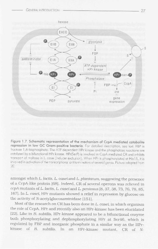
CcpA-mediated CR is linked to PTS activity by HPr in B. subtilis and B. megaterium HPr is a small protein of about 9 kDa which, in contrast to HPr from Gram-negative bacteria, can be phosphorylated at two sites: His 15 by Enzyme I from the PTS, like in Gram-negative bacteria, and at Ser46 by an ATP-dependent HPr-kinase. The ATP-dependent HPr-kinase is activated by metabolic intermediates, of which FBP is the most important, and is inhibited by inorganic phosphate [33, 55, 64, 98], Dephosphorylation of Ser46 in the presence of inorganic phosphate is catalysed by a phosphatase which has been shown in B. subtilis to be the same enzyme as the HPr-kinase [33, 64], HPr(SerP) (HPr which is phosphorylated at Ser46) interacts with CcpA, thereby increasing the affinity of CcpA for ere sequences [56]. Direct binding of the HPr(SerP)/CcpA complex to the operator sequence has been shown in vitro for several catabolic opérons from B. subtilis like the xyl operon. the xyn operon, the gnt operon [29, 31 , 39, 56, 83 , 94] and for amyO, the operator sequence from amyE [61]. The complex comprises a CcpAdimer and two molecules of HPr(SerP) [56]. Binding of the complex to a ere sequence interferes with transcription initiation or transcription elongation [51, 132].
A second corepressor with an amino acid sequence showing high similarity to that of HPr has been identified by analysis of the complete genome sequence of B. subtilis [32]. Crh (catabolite repression HPr) exhibits 45% identical residues to HPr, bu t the active site His 15 of HPr is replaced by a glutamine in Crh [32]. However, the Ser residue at position 46 is conserved in Crh and can be phosphorylated by the HPr-kinase [32]. Crh(SerP) has been shown to be involved in CR of several genes like the hut, lev, xyn and iol opérons, where mutation of the crh gene in addition to the ptsHl mutation, in which Ser46 of HPr is replaced by a non-phosphorylatable alanyl residue, leads to a complete relief of CR [31, 32, 80, 169]. The ptsHl mutation alone still results in repression of these genes. In vitro binding assays showed that a complex of CcpA with Crh(SerP) binds to the ere sequences of the xyn operon of B. subtilis [31]. In Figure 1.7, a schematic representation of CcpA-mediated CR is drawn.
A protein similar to CcpA was found in B. subtilis and called CcpB, which is involved in CR of the xyl and the gnt opérons [11]. However, its function has not been clarified yet, nor have other reports been published that show the involvement of CcpB in CR of other catabolic genes.
CR is closely linked to the metabolic activity and PTS activity in Gram-positive bacteria, by the roles of the metabolite FBP and the
26 CHAPTER 1
phosphorylation state of the PTS protein HPr. As described above, FBP has a stimulatory effect on the phosphorylation of HPr by HPr^kinase, on the complex formation of HPr(SerP) and Crh(SerP) to CcpA and on the binding of these complexes to the ere sequence. The metabolism of rapidly metabolizable carbon sources generates intracellular glycolytic intermediates like FBP [28] which stimulate CR on catabolic genes. Phosphorylation of HPr at His 15, the PEP-dependent phosphorylation which is involved in PTS activity, prevents interaction of HPr with CcpA [20]. During transport of a PTS sugar, His 15 of HPr will be largely unphosphorylated since it is constantly used for the phosphorylation of the sugar, which facilitates complex formation between HPr(SerP) and CcpA and thus the exertion of CR. If a non-PTS sugar is transported, however, the His 15 of HPr will remain phosphorylated, leading to a relief of CR under these circumstances.
In some cases, CcpA is involved in an activation mechanism, like for the ackA and ptaA genes of B. subtilis, which encode acetate kinase and phosphotransacetylase, respectively. Mutations in ccpA or in ere sequences that are located upstream of the transcriptional start sites resulted in a loss of activation of ackA or ptaA in the presence of glucose [42, 94, 143]. For this stimulation, also HPr(SerP) and Crh(SerP) are involved [94, 143], indicating that the same components are involved in this regulation as in CR. Indeed, footprinting experiments showed the binding of a complex of CcpA with HPr(SerP) or with Crh(SerP) to the ere sequences of the acs gene and the ptaA gene [94, 168]. Also genes involved in glycolysis, like the gap-gene (encoding glyceraldehyde 3-phosphate dehydrogenase) and the pgk operon (containing the genes encoding phosphoglycerate kinase, triose-phosphate isomerase, phosphoglycerate mutase and enolase) are activated by CcpA in B. subtilis [139]. These results indicate that CcpA probably is a general regulator protein and is not only involved in CR of catabolic genes.
HPr(HisP) and Ell of the PTS are also involved in a dual regulation mechanism of transcriptional regulator proteins in B. subtilis, for instance the activator protein LevR (controlling the lev operon), and the antiterminator proteins LicT (involved in the expression of the bglPH operon and LicS, encoding ß-l,3-l,4-glucanase) and SacT (sacPA, encoding a sucrose permease and a phosphosucrase). For reviews, see [102, 130, 131].
1.9 CATABOLITE REPRESSION IN LACTIC ACID BACTERIA
CR repression in lactic acid bacteria has been less extensively studied than CR in B. subtilis, but it is generally believed that it follows the same mechanism. Polyclonal antibodies raised against CcpA from B. subtilis were shown to react with cell extracts from many low GC Gram-positive strains,
GENERAL INTRODUCTION 27
hexose
antiterm inator ( E N A )
PEP
glycolysis
ATP-dependent HPr kinase
Phosphatase +
pyruvate
ifl^tser-ia
I Pi
F B P H + K ^
ere
CcpA
gene expression
Figure 1.7. Schematic representation of the mechanism of CcpA mediated catabolite repression in low GC Gram-positive bacteria. For detailed description, see text. FBP is fructose 1,6-bisphosphate. The ATP dependent HPr kinase and the phosphatase reactions are catalysed by a bifunctional HPr kinase. HPr(SerP) is involved in CcpA mediated CR and inhibits transport of maltose in L casei (inducer exclusion). When HPr is phosphorylated at Hisl5, it is involved in activation of the transcriptional antiterminators of several genes. Picture adapted from [6].
amongst which L. lactis, L. casei and L. plantarunx suggesting the presence of a CcpA-like protein [68]. Indeed, CR of several opérons was relieved in ccpA mutants of L. lactis, L. casei and L. pentosus [8, 37, 38, 73, 76, 78, 85, 167]. In L. casei HPr mutan t s showed a relief in repression by glucose on the activity of N-acetylglucosaminidase [151].
Most of the research on CR has been done in L. casei in which organism the role of CcpA, HPr and recently also an HPr-kinase has been elucidated [22]. Like in B. subtiLis, HPr-kinase appeared to be a bifunctional enzyme both phosphorylating and dephosphorylating HPr at Ser46, which is regulated by FBP and inorganic phosphate in a similar way as the HPr-kinase of B. subtiLis. In an HPr-kinase mutant , CR of N-
28 CHAPTER
acetylglucosaminidase had disappeared, indicating that HPr-kinase plays a central role in CR [22]. However, a direct interaction of HPr(SerP) with CcpA, or binding of the CcpA/HPr(SerP) complex to a ere sequence has not been shown yet in lactic acid bacteria. Neither has a protein similar to HPr, like Crh in B. subtilis, been identified.
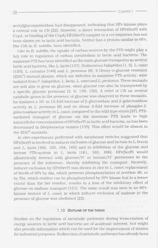
Like in B. subtilis, the uptake of carbon sources by the PTS might play a key role in regulation of carbon metabolism in lactic acid bacteria. The mannose PTS has been identified as the main glucose transporter in several lactic acid bacteria, like L. lactis [137], Pediococcus halophuus [ 1, 2], L. casei [150], L. curvatus [149] and L. pentosus [6]. 2-Deoxy-D-glucose resistant (2DGR) mutant strains, which are defective in mannose PTS activity, were isolated from P. halophilus, L. lactis, L. casei and L. pentosus. These mutan t s are still able to grow on glucose, since glucose can also be transported by a specific glucose permease [2, 6, 138, 150]. A relief of CR on several catabolic genes in the presence of glucose was observed in these mutants , for instance a 10- to 15-fold increase of ß-glucosidase and ß-galactosidase activity in L. pentosus [6] and an about 4-fold increase of phospho-ß-galactosidase activity in L. casei, compared to the wild type strain [37]. PTS-mediated transport of glucose via the mannose PTS leads to high intracellular concentrations of HPr(SerP) in lactic acid bacteria, as has been determined in Streptococcus mutans [135]. This effect would be absent in the 2DGR-mutants.
In vitro experiments performed with membrane vesicles suggested that HPr(SerP) is involved in inducer exclusion of glucose and lactose in L. brevis and L. lactis [160, 162, 164, 165] and in inhibition of the glucose and lactose PTS-system in L. (actis [161, 163, 166]. HPr(SerP) would allosterically interact with glucose/H+ or lactose/H+ permeases in the presence of the substrate, thereby inhibiting the transport. Recently, inducer exclusion by HPr(SerP) was shown in vivo in L. caset replacement of Ser46 of HPr by Ala, which prevents phosphorylation at position 46, or by Thr, which residue can be phosphorylated by HPr kinase but to a lesser extent than the Ser residue, results in a loss of the inhibitory effect of glucose on maltose transport [151]. The same result was seen in an HPr-kinase mutan t of L. casei, in which inducer exclusion of maltose in the presence of glucose was abolished [22].
1.10 OUTLINE OF THE THESIS
Studies on the regulation of metabolic pathways during fermentation of energy sources in lactic acid bacteria are of academic interest, bu t might also provide information which can be used for the improvement of strains for industrial purposes. Redirection of metabolic pathways has already been
GENEPAL INTRODUCTION 2 9
shown to be possible in some lactic acid bacteria, for instance in L. lactis where inactivation of lactate dehydrogenase led to an increase of the production of acetate, formate, acetoin and ethanol at the expense of lactate.
In earlier research the regulation of the five genes of the xylose regulon, involved in isoprimeverose and D-xylose catabolism, was analysed in L. pentosus. The induction of these genes in the presence of xylose, as well as the effect of CR by glucose on the expression of the xyl regulon have been studied. This thesis describes the investigation of the PKP, the pathway by which xylose, after being converted into xylulose 5-phosphate, is further metabolized. The PKP is a general pathway for sugar fermentation in lactic acid bacteria, by which pentoses, like xylose, arabinose and ribose, bu t also other metabolites like gluconate and in some cases glucose are metabolized.
In Chapter 2 the purification of xylulose 5-phosphate phosphoketolase (XpkA), the central enzyme of the PKP, is reported and the subsequent isolation, characterization and sequencing of the XpkA encoding gene, xpkA. The gene can be used to study the regulation of xpkA in L. pentosus. Enzyme activity assays showed that XpkA is an inducible enzyme, which activity was found after growth on energy sources that are fermented via the PKP. Growth on sugars that are metabolized via the glycolytic pathway resulted in low XpkA activities. The results of the assays also suggested that XpkA regulation is subject to CR by glucose and fructose. It was shown in an xpkA knock-out strain that an active XpkA is essential for growth of L. pentosus on pentoses and gluconate.
Acetate kinase catalyses the conversion of acetyl-phosphate into acetate, which is one of the end-products of fermentation via the PKP. The cloning and sequencing of the acetate kinase encoding gene, ackA, is described in Chapter 3. Acetate kinase was constitutively synthesized during growth on all tested energy sources in L. pentosus. The lowest acetate kinase activities were found after growth on glucose, which is probably due to CR by glucose. An ackA knock-out mutan t of L. pentosus was constructed, in which acetate kinase was inactivated. The mutan t had lost its ability to grow on energy sources that are fermented via the PKP.
In Chapter 4 several Lactobacillus strains were screened for the presence of XpkA. XpkA activity was detected in lactobacilli during heterofermentative growth via the PKP: after growth on glucose or ribose in obligate heterofermentative lactobacilli and after growth on ribose but not on glucose in facultative heterofermentative bacteria. The presence of XpkA activity correlated with the synthesis of a protein of the size of L. pentosus XpkA {approximately 90 kDa). On the basis of a Southern blot analysis of chromosomal DNA of several Lactobacillus strains, an indication for a
o 0 CHAPTER I
second gene with similarity to xpkA was found in L. pentosus and in the
related strain L. plantamm. A database search with the amino acid sequence of XpkA from L.
pentosus revealed that an ORF encoding a protein with 39 to 60% identical residues with XpkA is present in various organisms. None of these organisms had been reported to contain phosphoketolase activity. The data in Chapter 5 describe our efforts to reveal the function of one of these ORFs: slr0453 from Synechocystis. The protein encoded by slr0453 was synthesized under certain conditions in Synechocystis, however phosphoketolase activity could not be detected.
In the Summary and Concluding Remarks, all results are summarized and some perspectives for further research are discussed.

![RESEARCH Open Access Optimization of bacteriocin ......iron-chelating compounds, antibiotics, hydrogen perox-ide, organic acids and bacteriocins [17,18]. Bacteriocins are small peptides](https://img.pdfslide.us/doc/110x75/60e08de7842bb1363819093a/research-open-access-optimization-of-bacteriocin-iron-chelating-compounds.jpg)