Embed Size (px)
Citation preview
UvA-DARE is a service provided by the library of the University of Amsterdam (http://dare.uva.nl)
UvA-DARE (Digital Academic Repository)
Ritonavir
Gisolf, E.H.
Link to publication
Citation for published version (APA):Gisolf, E. H. (2000). Ritonavir.
General rightsIt is not permitted to download or to forward/distribute the text or part of it without the consent of the author(s) and/or copyright holder(s),other than for strictly personal, individual use, unless the work is under an open content license (like Creative Commons).
Disclaimer/Complaints regulationsIf you believe that digital publication of certain material infringes any of your rights or (privacy) interests, please let the Library know, statingyour reasons. In case of a legitimate complaint, the Library will make the material inaccessible and/or remove it from the website. Please Askthe Library: https://uba.uva.nl/en/contact, or a letter to: Library of the University of Amsterdam, Secretariat, Singel 425, 1012 WP Amsterdam,The Netherlands. You will be contacted as soon as possible.
Download date: 27 Aug 2019
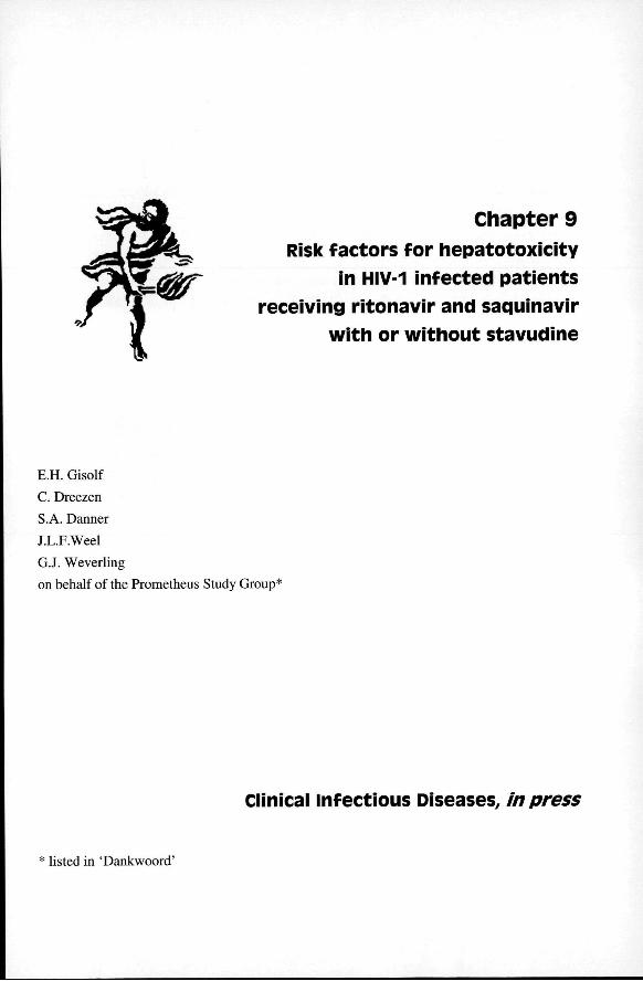
Chapterr 9 Riskk factors for hepatotoxicity
inn Hiv-1 infected patients receivingg ritonavir and saquinavir
withh or without stavudine
E.H.. Gisolf
C.. Dreezen
S.A.. Danner
J.L.F.Weel l
G.J.. Weverling
onn behalf of the Prometheus Study Group*
Clinicall infectious Diseases, in press
** listed in 'Dankwoord'
ChapterChapter 9
Summary Summary
Introduction:: Liver enzyme elevations (LEE) are commonly observed following the start of
combinationn antiretroviral therapy (ARVT) for HIV infection.
Methods:: 208 HIV-infected patients were randomized to ritonavir (RTV) 400 mg BID,
saquinavirr (SQV) 400 mg BID with or without stavudine (d4T) 40 mg BID. LEE was defined
ass having an increase in AST/ALT of > 5x upper limit of normal and >100 U/L increase from
baseline.. We investigated potential risk factors for LEE following treatment with RTV/SQV
withh or without d4T using Cox's proportional hazard model.
Results:: Eighteen (9%) patients developed LEE during the 48 week follow-up. Multivariate
analysis,, adjusted for baseline ALT and AST, showed that HBsAg positivity (RR 8.8,
95%CI:3.3-23.1)) and the use of d4T (RR 4.9,95%CI: 1.5-16.0) were the only two significant
riskrisk factors for developing LEE. After LEE occurred, ALT/AST concentrations decreased
>50%% in 13/14 patients, who continued ARVT during LEE.
Conclusions:: LEE during ARVT is more frequently observed in HIV-infected patients, who
aree HBsAg positive. Furthermore, the use of d4T, in combination with RTV/SQV, was a risk
factorr for development of LEE, independent from hepatitis serology. In this study, it appeared
safee to continue ARVT during LEE. However, more data from larger studies are required to
confirmm this finding.
110 0
HepatotoxicityHepatotoxicity during antiretroviral therapy
Introduction Introduction
Liverr enzyme elevation (LEE) has been reported to be a potential side effect of most
antiretrovirall agents used for the treatment of the human immunodeficiency virus (HIV)
infectionn [1]. Since the introduction of highly active antiretroviral therapy (HAART) with
triplee drug combination regimens, severe elevations in liver enzymes have been observed
moree frequently. LEE may be drug-induced. However, LEE seems to appear more often in
HIV-infectedd patients who are co-infected with hepatitis B virus (HBV) and/or hepatitis C
viruss (HCV) [2-9].
Co-infectionss of HTV and HBV and/or HCV are common. Up to 84% of intravenous drug
userss with HTV infection and 83% of homosexuals with HIV infection have markers of past or
currentt HBV infection; 81% and 14%, respectively, have markers of past or current HCV
infectionn [10].
Manyy questions related to the clinical management of patients who develop LEE during the
usee of antiretroviral agents remain unanswered. First, the risk of developing hepatotoxicity has
nott properly been quantified. Furthermore, it is unknown whether certain antiretroviral drugs,
classess of antiretroviral drugs or combinations of antiretroviral drugs are more prone to
predisposee for LEE. Finally, there are no guidelines for deciding to continue or discontinue
HAARTT once LEE has been observed.
Thee aim of this study was to describe hepatotoxicity in HIV-infected patients, treated with
ritonavirr (RTV) and saquinavir (SQV) with or without stavudine (d4T). We searched for risk
factorss for LEE in this study population and described the clinical management of and the
outcomee in these patients.
MaterialsMaterials and methods
Patientss and data collection
Thee Prometheus study was an open label, randomized controlled multicenter trial in 208 HIV-
11 infected patients in the Netherlands and Belgium. Patients were randomized between
Januaryy 1997 and January 1998 to receive orally RTV 400 mg twice daily (BID) plus SQV
4000 mg BID with or without d4T 40 mg BID (30 mg BID if body weight was below 60 kg).
Participantss had to be protease inhibitor (PI)- and d4T-naive before the start of the study. They
hadd an indication for initiation of antiretroviral treatment or there was a reason to change
currentt treatment. Participants were 18 years of age or older and had a Karnofsky score > 60.
Antiretrovirall drugs, used before the study, had to be discontinued before start of the study
medication.. There were no exclusion criteria for patients with elevated liver enzymes other
111 1
ChapterChapter 9
thann that patients who had a clinical condition or laboratory abnormality that was, in the
investigator'ss opinion, incompatible with the use of study medication were excluded.
Patientss were allowed to intensify their treatment with, if possible, two new reverse
transcriptasee inhibitors if serum HIV-RNA concentration did not drop to < 400 copies/ml at
weekk 12, confirmed at week 18, or if serum HIV-RNA became detectable again.
Thee following baseline characteristics were collected: gender, age, weight, height, prior
antiretrovirall drug use, CDC classification, transmission risk group, CD4- and CD8-
lymphocytee count, serum HIV-1 RNA, the presence of hepatitis surface antigen (HBsAg) and
anti-hepatitiss C antibody (anti-HCV) in serum. During the study, patients were evaluated at
weekk 0, 2, 4, 8, 12, 18, 24, 36 and 48. After week 48, follow up on the clinical outcome of
LEEE was continued for patients, who consented for this extended follow up. The occurrence
off HIV-related events as classified according to the Centers for Disease Control and
Preventionn (CDC) 1993 guidelines [11], other clinical events, use of antiretroviral medication,
usee of co-medication, laboratory safety parameters (among others: asparate aminotransferase
(AST),, alanine aminotransferase (ALT), alkaline phosphatase (AF), and ^-glutamyl transferase
(GGT))) and laboratory efficacy parameters (serum HIV-1 RNA (Amplicor HIV Monitor Test,
Rochee Diagnostic Systems Inc., Branchburg, New Jersey, USA), CD4+- and CD8+-
lymphocytee counts) were recorded every visit.
Definitionn of HBV and HCV serologic categories
Patientss were considered to have a chronic HBV infection when HBsAg could be detected at
baselinee [12]. Earlier studies reported that HCV infection has a very high rate of persistence
[13].. Therefore, when antibodies against HCV were present at baseline, patients were
consideredd to have a chronic HCV infection. Subsequently, the following categories were
distinguished:: patients with positive HBsAg- or anti-HCV-serology ('chronic hepatitis B' or
'chronicc hepatitis C') and patients without serological markers for chronic HBV and HCV
infectionn or with serological signs of prior HBV infection ('no chronic hepatitis').
Definitionn of LEE
AA clinically relevant LEE was defined as having a transaminase elevation (ALT and/or AST)
greaterr or equal than five times the upper limit of normal according to AIDS Clinical Trials
Groupp (ACTG) criteria [14]. In addition, the absolute increase needed to be at least 100 U/L
comparedd to an individual's baseline value, to avoid misclassification of patients with high
baselinee transaminase values and only minor transaminase elevation. Patients were regarded
112 2
HepatotoxicityHepatotoxicity during antiretroviral therapy
ass having a LEE if they had experienced at least one LEE while they were using the study
medication.. Improvement of transaminase elevation was defined as experiencing a decrease in
thee ALT to less then 50% of the ALT concentration at the time LEE first was observed, if
LEEE was based on elevated ALT; the same applied for AST.
Statisticall analysis
Potentiall risk factors of LEE were explored by univariate Cox proportional hazard analysis,
enteringg the time to LEE as dependent variable. In case no LEE occurred patients were
censoredd at their last observation during the study. Parameters considered as potential predic-
torss of LEE were: age, gender, weight, pre-treatment with reverse transcriptase inhibitors
(RTIs),, HIV transmission category, stage of HIV disease (CDC classification), baseline AST,
baselinee ALT, HIV-1 RNA, CD4+ cell count, CD8+ cell count, change in CD4+ cell count
betweenn week 0 and week 12, change in CD8+ cell count between week 0 and 12, results of
HBsAgg and anti-HCV tests. Besides variables with p-values less than 0.10 in the univariate
model,, baseline AST and ALT were a priori entered in the multivariate model, as the study
endpointt LEE was based on ALT and/or ALT levels. Differences between groups were
consideredd significant at a p < 0.05 level. All reported p-values are two sided. Analyses were
performedd using SAS, version 6.12 (SAS Institute, Cary, North Carolina, USA).
Results Results
Patientss characteristics
AA total of 208 HIV-1 infected subjects participated in this study. Participants were
predominantlyy homosexual males, with a median serum HIV-1 RNA of 4.47 logio copies/ml
andd a median CD4+ cell count of 255 cells/mm3 (Table 1). Patients were regarded to have a
chronicc hepatitis B if they had tested positive for hepatitis B surface antigen (HBsAg) and to
havee a chronic hepatitis C if they had tested positive for anti-HCV at baseline. Results of the
HBsAgg test at baseline (week 0 or week -2) were available in 195/208 subjects (94%). Results
off HCV serology were available in 201/208 subjects (97%). Twenty-five (12%) patients were
HBsAgg positive, 17 (8%) were anti-HCV positive, two of these patients were both HBsAg and
anti-HCVV positive. Patients with missing data were regarded as having no chronic hepatitis.
Ass some patients with missing data may have positive HBsAg or anti-HCV, this assumption
wil ll give a conservative approach when comparing patients with and without chronic
hepatitis.. Patients with chronic hepatitis were more likely to have higher baseline ALT or
AST,, were more likely to have acquired HTV by intravenous drug use and to have used
113 3
ChapterChapter 9
antiretrovirall medication before start of the study (Table 1). Patients assigned to treatment
withh RTV/SQV/d4T were equally distributed between the two groups, reflecting the
randomizationn in this study.
Tablee 1 Baseline characteristics
N N
treatment t
ALT T
AST T
gender r
priorr ARVT
HIVV transmission
riskrisk group
timee since first
positivee HIV-test
CDCC classification
age e
weight t
serumm HIV- l RNA
CD4++ cell count
CD8++ cell count
RTV/SQV/d4T T
RTV/SQV V
U/L,, median (IQR)
U/L,, median (IQR)
males s
females s
pre-treated d
naive e
homosexuall contacts
heterosexuall contacts/
endemicc area
IVV drugs
other r
unknown n
years,, median (IQR)
A A
B B
C C
years,, median (IQR)
kg,, median (IQR)
logg io copies/ml, median
(IQR) )
/mm3,, median (IQR)
/mm3,, median (IQR)
Chronicc hepatitis*
40 0
18(45) )
222 (55)
366 (25-55)
322 (27-45)
35(88) )
5(12) )
27(68) )
13(32) )
200 (50)
5(13) )
11(28) )
3(8) )
1(3) )
3.4(1.3-7.8) )
15(37) )
17(43) )
8(20) )
36(31-46) )
72(66-81) )
4.33 (3.8-4.7)
275(190-355) )
815(625-1140) )
Noo chronic hepatitis5
168 8
86(51) )
82(49) )
288 (21-42)
255 (20-34)
1422 (85)
26(15) )
711 (42)
977 (58)
114(68) )
388 (22)
0(0) )
13(8) )
3(2) )
2.44 (0.5-6.0)
788 (47)
511 (30)
399 (23)
377 (32-45)
722 (64-80)
4.55 (4.0-5.0)
250(115-390) )
835(655-1150) )
p-value e
.60 0
.01 1
.002 2
.81 1
.005 5
.04 4
.20 0
<.001 1
1.0 0
.04 4
.38 8
.19 9
.83 3
.57 7
.51 1
.12 2
.22 2
.80 0
== patients, who were HBsAg or anti-HCV positive; § = patients Numberss are n (%), unless otherwise specified; _
withh negative or missing serology for HBsAg and anti-HCV; SQV = saquinavir; RTV = ritonavir;
d4T=stavudine;; ARVT = antiretroviral therapy
Liverr enzyme elevations
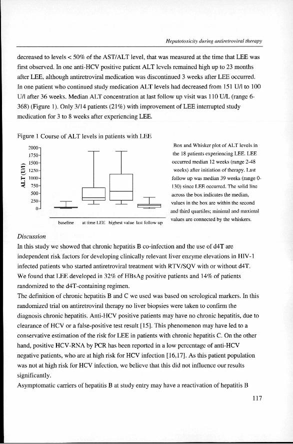
Eighteenn (9%) patients developed LEE, after a median of 12 weeks since start of the study
(range:: 2 to 48 weeks). LEE was observed earlier in the RTV/SQV arm than in the
114 4
HepatotoxicityHepatotoxicity during antiretroviral therapy
RTV/SQV/d4TT arm (median 9 versus 14 weeks, respectively, p=.01). The ALT levels were
mediann 358 U/l (range 150-1890) at the time LEE was first observed. Ten patients
experiencedd a grade 3 and eight patients experienced a grade 4 toxicity at that time point. The
mediann highest ALT level measured during LEE was 574 U/l (range 150-1890). Seven
patientss experienced a grade 3 and eleven patients experienced a grade 4 toxicity (Figure 1).
Thee highest ALT levels per patient during the study were significantly higher in the group
randomizedd to d4T (median 69 U/l in the RTV/SQV/d4T group (n=104), 51 U/l in the
RTV/SQVV group (n=104), p=.03). These highest levels occurred later in the RTV/SQV/d4T-
groupp (median 18 weeks, mean 19 weeks) than in the RTV/SQV-group (median 4 weeks,
meann 13 weeks, p=.002).
Patientss with chronic hepatitis who were treated with RTV/SQV/d4T had 45% chance to
developp LEE during the study period. Patients with chronic hepatitis B who were treated with
RTV/SQVV alone got LEE in 21%. In patients without chronic hepatitis B, LEE was observed
inn 9% and 2% of patients treated with RTV/SQV/d4T or RTV/SQV, respectively.
Patientss were allowed to intensify their initial treatment with reverse transcriptase inhibitors if
serumm HIV-RNA concentration did not drop to below 400 copies/ml at week 12, confirmed at
weekk 18, or if serum HIV-RNA became detectable again. Thirty-one patients intensified study
medicationn as stated in the protocol: 28 (27%) in the RTV/SQV arm and three (3%) in
RTV/SQV/d4TT arm. Median follow-up after intensification was 26 weeks. Most patients
(21/31)) added d4T plus 3TC to their initial regimen. No LEE was observed after
intensificationn of study medication.
Riskk factors for LEE
Inn univariate Cox regression analyses, HBsAg positivity (OR 7.3,95% Confidence Interval
(CI):: 2.9-18.7, p=.0001) and a higher baseline ALT concentration (RR 1.1,95% CI: 1.0-1.2,
p=.03)) were significant risk factors for development of LEE. The use of d4T (RR 2.7,95% CI:
1.0-7.7)) showed a trend (p=.06). Anti-HCV positivity, gender, age, weight, CDC-status, HIV
transmissionn risk group, prior antiretroviral treatment, baseline AST concentrations, CD4+
andd CD8+ cell counts at baseline, CD4+ or CD8+ increases from week 0 to week 12 and
baselinee HIV-1 RNA levels were not predictive for the development of LEE in univariate
analysiss (Table 2). Multivariate analysis showed that HBsAg positivity (RR 8.8, 95% CI: 3.3-
23.1)) and randomization to the d4T containing regimen (RR 4.9,95% CI: 1.5-16.0) were the
onlyy two significant risk factor for developing a LEE, adjusted for baseline ALT and AST
levelss (Table 2).
115 5
ChapterChapter 9
Tablee 2 Cox proportional hazard ratios for baseline characteristics of LEE during treatment
withh RTV/SQV with or without d4T
riskrisk factor
HBsAgg +
RTV/SQV/d4TT arm
ALTT concentration*
ASTT concentration*
anti-HCVV +
genderr male
priorr ARVT
HIVV transmission risk group
homosexual l
heterosexual l
intravenouss drugs use
other r
CDCC classification
A A
B B
C C
age8 8
weight* *
log10HIV-lRN A A
CD4++ lymphocyte count**
CD8++ lymphocyte count**
CD4++ increase > 50/mm3^
CD8++ increase > 100/mmw
univariatee analysis
hazardd ratio (95% CI)
7.33 (2.9-18.7)
2.7(1.0-7.7) )
1.11 (1.0-1.2)
1.11 (1.0-1.3)
1.8(0.4-8.0) )
0.99 (0.2-3.9)
0.7(0.3-1.7) )
0.88 (0.3-2.0)
0.5(0.1-2.1) )
1.3(0.2-10.1) )
0.9(0.1-6.5) )
0.99 (0.4-2.4)
1.4(0.6-3.7) )
0.77 (0.2-2.3)
1.0(0.9-1.0) )
1.0(1.0-1.0) )
1.2(0.6-2.2) )
1.0(0.8-1.3) )
1.11 (1.0-1.2)
1.2(0.4-3.5) )
0.7(0.3-1.9) )
p-value e
.0001 1
.06 6
.03 3
.09 9
.42 2
.87 7
.40 0
.59 9
.32 2
.77 7
.89 9
.91 1
.46 6
.50 0
.73 3
.58 8
.66 6
.88 8
.19 9
.80 0
.54 4
multivariatee analysis
hazardd ratio (95% CI)
8.8(3.3-23.1) )
4.8(1.5-16.0) )
1.2(1.0-1.5) )
0.8(0.5-1.3) )
p-value e
.0001 1
.01 1
.11 1
.47 7
HBsAgg = hepatitis B antigen; anti-HCV = hepatitis C antibody; AST = asparate aminotransferase; ALT = alanine
aminotransferase;; ARVT = antiretroviral therapy; * = per 10 U/L increase in baseline AST or ALT concentrations; §
== per year increase in baseline age; # = per kilogram increase in baseline weight; ** = per 100 cells/mm increase
inn baseline CD4 or CD8 lymphocyte counts; §§ = change in absolute CD4/8 lymphocyte counts from week 0 to
weekk 12
Followw up and clinical management after LEE
Thee median follow-up after developing LEE was 40 weeks (range 0-130 weeks). In one
patientt no follow up was available from the time of LEE. One patient was only followed until
1.55 weeks after LEE occurred, at which time point ALT had already decreased 50%. In 14/16
patientss with at least 14 weeks of follow up since LEE ALT and/or AST concentrations
116 6
HepatotoxicityHepatotoxicity during antiretroviral therapy
decreasedd to levels < 50% of the AST/ALT level, that was measured at the time that LEE was
firstt observed. In one anti-HCV positive patient ALT levels remained high up to 23 months
afterr LEE, although antiretroviral medication was discontinued 3 weeks after LEE occurred.
Inn one patient who continued study medication ALT levels had decreased from 151 U/l to 100
U/ll after 36 weeks. Median ALT concentration at last follow up visit was 110 U/L (range 6-
368)) (Figure 1). Only 3/14 patients (21%) with improvement of LEE interrupted study
medicationn for 3 to 8 weeks after experiencing LEE.
Figuree 1 Course of ALT levels in patients with LEE
H H
i i
2000 0
1750 0
1500 0
1250 0
1000 0
750 0
500 0
250 0
0 0
baselinee at time LEE highest value last follow up
Boxx and Whisker plot of ALT levels in
thee 18 patients experiencing LEE. LEE
occurredd median 12 weeks (range 2-48
weeks)) after initiation of therapy. Last
followw up was median 39 weeks (range 0-
130)) since LEE occurred. The solid line
acrosss the box indicates the median,
valuesvalues in the box are within the second
andd third quartiles; minimal and maximal
valuess are connected by the whiskers.
Discussion Discussion
Inn this study we showed that chronic hepatitis B co-infection and the use of d4T are
independentt risk factors for developing clinically relevant liver enzyme elevations in HIV-1
infectedd patients who started antiretroviral treatment with RTV/SQV with or without d4T.
Wee found that LEE developed in 32% of HBsAg positive patients and 14% of patients
randomizedd to the d4T-containing regimen.
Thee definition of chronic hepatitis B and C we used was based on serological markers. In this
randomizedd trial on antiretroviral therapy no liver biopsies were taken to confirm the
diagnosiss chronic hepatitis. Anti-HCV positive patients may have no chronic hepatitis, due to
clearancee of HCV or a false-positive test result [15]. This phenomenon may have led to a
conservativee estimation of the risk for LEE in patients with chronic hepatitis C. On the other
hand,, positive HCV-RNA by PCR has been reported in a low percentage of anti-HCV
negativee patients, who are at high risk for HCV infection [16,17]. As this patient population
wass not at high risk for HCV infection, we believe that this did not influence our results
significantly. .
Asymptomaticc carriers of hepatitis B at study entry may have a reactivation of hepatitis B
117 7
ChapterChapter 9
duringg antiretroviral treatment. We included these asymptomatic carriers, who were anti-HBs
and/orr anti-HBc positive in the 'no chronic hepatitis'-group in this study. These elements may
havee caused an underestimation of the true risk for LEE in patients with chronic hepatitis B.
Patientss with chronic hepatitis had higher baseline ALT or AST concentrations when
comparedd to patients without chronic hepatitis, reflecting their underlying liver disease. They
weree more likely to have acquired HIV by intravenous drug use, as especially the prevalence
off hepatitis C was high in this risk group. Patients with chronic hepatitis were more often pre-
treatedd before start of the study.
Ourr data confirm the results of cohort studies [2,3] and anecdotal reports [4-8]. These studies
alreadyy suggested that liver enzyme elevations during antiretroviral treatment are more
commonn in HIV-infected patients co-infected with hepatitis B. However, we did not find an
associationn between anti-HC V positivity and occurrence of LEE.
Recentlyy published papers on the incidence of hepatotoxicity in cohort studies reported
comparablee incidences of LEE [18,19], although the observed median time to LEE was longer
inn these studies. Patients in this randomized trial were evaluated intensively in the first 6
months,, which could have led to earlier recognition of LEE.
Sulkowskii et al. found a high incidence of LEE in patients treated with ritonavir (30%), which
wass much higher when compared to our study population (9%), of which all were treated with
ritonavir.ritonavir. An explanation for this difference can be found in the different study population.
Moreover,, a bias in the choice of antiretroviral therapy could have occurred, as their analyses
weree done in a non-randomized cohort. In other studies [2,3,19] the association between
ritonavirritonavir use and a high incidence of LEE is not confirmed.
Hepatotoxicityy is a well known phenomenon during antiretroviral therapy. Immune
reconstitutionn induced by antiretroviral therapy can cause disease activation of chronic or
previouss latent infections, such as hepatitis B or C [4,20]. In this study, LEE was highly
associatedd with the presence of hepatitis B surface antigen. LEE occurred most frequently in
thee first three months of treatment and no LEE was observed after intensification of therapy
withh d4T and 3TC. Patients who intensified treatment during the study were not different from
otherr patients with respect to baseline ALT/AST levels, hepatitis serology or pre-treatment
status.. These observations support the hypothesis of hepatotoxicity due to immune restoration.
However,, the difference between the two study arms cannot be explained by a difference in
immunee restoration. The treatment groups did not differ in their CD4+- and CD8+ lymphocyte
responsee to therapy [21]. Moreover, the change in CD4+- or CD8+ lymphocyte counts were
nott associated with the occurrence of LEE. Direct toxic effects of antiretroviral drugs can also
118 8
HepatotoxicityHepatotoxicity during antiretroviral therapy
causee LEE. Nucleoside analogue RTIs are known to cause mitochondrial toxicity [1].
Althoughh the nucleoside analogue RTT d4T is seldom described as a hepatotoxic drug in the
currentt prescribed dose [22,23], we found that d4T was an independent risk factor for
developmentt of LEE in this study. Of note is that the highest ALT levels per patient occurred
laterr (around week 18) in patients on RTV/SQV/d4T, when compared to patients on
RTV/SQVV alone (week 12).
Inn all but one patients, who continued antiretroviral medication, ALT/AST concentrations
decreasedd to < 50% of the ALT/AST level, thatt was measured at the time that LEE was first
observed.. This suggests that antiretroviral medication can be continued safely during LEE.
However,, more data from larger studies are required to confirm this finding.
Inn conclusion, clinical relevant liver enzyme elevations during antiretroviral combination
therapyy are more frequently observed in HIV-infected patients with concurrent chronic
hepatitiss B. We also found that the use of d4T, in combination with RTV/SQV, was an
independentt risk factor for LEE.
Acknowledgements Acknowledgements
Thee authors want to thank the technicians of the Clinical Virology Unit and the Department of
Retroo virology of the Academic Medical Center, and the study participants.
References References 1.. Notermans DW, van Leeuwen R, Lange JMA: Treatment of HIV infection. Tolerability of commonly
usedd antiretroviral agents. Drug Safety 1996,15:176-187.
2.. den Brinker M, Wit FWNM, Wertheim-van Dillen PME, et al. HBV and HCV co-infection predispose
forr HAART-associated hepatotoxicity. 4th International Congress on Drug Therapy in HIV
Infection,Infection, Glascow November 8-12 1998.
3.. Rodriguez-Rosado R, Garcia-Samaniego J, Soriano V: Hepatotoxicity after introduction of highly
activee antiretroviral therapy. AIDS 1998,12:1256
4.. Carr A, Cooper DA: Restoration of immunity to chronic hepatitis B infection in HIV-infected patient
onn protease inhibitor. Lancet 1997, 349:995-996.
5.. Brau N, Leaf HL, Wieczorek L, Margolis DM: Severe hepatitis in three AIDS patients treated with
indinavir.. Lancet 1997, 349:924-925.
6.. Vento S, Garofano T, Renzini C, Casali F, Ferraro T, Concia E: Enhancement of hepatitis C virus
replicationn and liver damage in HIV co-infected patients on antiretroviral combination therapy.
AIDSAIDS 1998,12:116-117.
7.. Jeurissen FJF, Schneider MME, Borleffs JCC: Is the combination of hepatitis and indinavir potentially
dangerous?? AIDS 1998,12:441-442.
8.. Mastroianni CM, Trinchieri V, Santopadre P, et al: Acute clinical hepatitis in an HTV-seropositive
119 9
ChapterChapter 9
hepatitiss B carrier receiving protease inhibitor therapy. AIDS 1998,12:1939-1930.
9.. Arribas JR, Ibanez C, Ruiz-Antoran B, et al: Acute hepatitis in HIV-infected patients during ritonavir
treatmentt AIDS 1998,12:1722-1724.
10.. Weinstock DM, Merrick S, Malak SA, Jacobs J, Sepkowitz KA: Hepatitis C in an urban population
infectedd with the immunodeficiency virus. AIDS 1999,13:2593-2595.
11.. Centers for Disease Control: 1993 revised classification system for HTV infection and expanded
surveillancee case definition for AIDS among adolescents and adults. MMWR 1992, 41(No.RR-
17):1-19. .
12.. Lee WM; Hepatitis B virus infection. N.EnglJ.Med. 1997, 337:1733-1745.
13.. Sharara AI, Hunt CM, Hamilton JD: Hepatitis C. Ann.Intern.Med. 1996,125:658-668.
14.. ACTG criteria, division of AIDS. Table for grading severity of adult adverse experiences. 1992.
15.. Villano SA, Vlahov D, Nelson KE, Colin S, Thomas DL: Persistence of viremia and the importance of
long-termm follow-up after acute hepatitis C infection. Hepatology 1999, 29:908-914.
16.. Caramelo C, Bartolome J, Albalate M, et al: Undiagnosed hepatitis C infection in hemodialysis
patients:: value of HCV RNA and liver enzyme levels. Kidney Int. 1996, 50:2027-2031.
17.. Thio CL, Nolt KR, Astemborski J, Vlahov D, Nelson KE, Thomas DL: Screening for hepatitis C virus
inn human immumnodeficiency virus-infected individuals. J.ClinMicrobiol. 2000,38:575-577.
18.. Sulkowski MS, Thomas DL, Chaisson RE, Moore RD: Hepatotoxicity associated with antiretroviral
therapyy in adults infected with human immunodeficiency virus and the role of hepatitis C or B
viruss infection. JAMA 2000, 283:74-80.
19.. Savez M, Vandentorren S, Daucourt V, et al: Severe hepatic cytolysis: incidence and risk factors in
patientss treated by antiretroviral combinations Aquitane Cohort, France, 1996-1998. AIDS
1999,13:F115-F121. .
20.. John M, Flexman J, French MAH: Hepatitis C virus-associated hepatitis following treatment of HIV-
infectedd patients with protease inhibitors: an immune restoration disease? AIDS 1998,12:2289-
2293. .
21.. Gisolf EH, Jurriaans S, Pelgrom J, et al: Treatment with ritonavir/saquinavir or
ritonavir/saquinavir/stavudineritonavir/saquinavir/stavudine in HTV-1 infected individuals. The effect of treatment
intensification.. AIDS 2000,14:405-413.
22.. Spruance SL, Pavia AT , Mellors JW, et al: Clinical efficacy of monotherapy with stavudine compared
withh zidovudine in HIV-infected, zidovudine-experienced patients. Ann.Intern.Med. 1997,
126:355-363. .
23.. Lenzo NP, Garas BA, French MA: Hepatic steatosis and lactic acidosis associated with stavudine
treatmentt in an HIV patient: a case report. AIDS 1997,11:1294-1296.
120 0