Embed Size (px)
Citation preview

UvA-DARE is a service provided by the library of the University of Amsterdam (http://dare.uva.nl)
UvA-DARE (Digital Academic Repository)
Reflux disease and achalasia: Failure of the gatekeeper
Rohof, W.O.A.
Link to publication
Citation for published version (APA):Rohof, W. O. A. (2013). Reflux disease and achalasia: Failure of the gatekeeper
General rightsIt is not permitted to download or to forward/distribute the text or part of it without the consent of the author(s) and/or copyright holder(s),other than for strictly personal, individual use, unless the work is under an open content license (like Creative Commons).
Disclaimer/Complaints regulationsIf you believe that digital publication of certain material infringes any of your rights or (privacy) interests, please let the Library know, statingyour reasons. In case of a legitimate complaint, the Library will make the material inaccessible and/or remove it from the website. Please Askthe Library: http://uba.uva.nl/en/contact, or a letter to: Library of the University of Amsterdam, Secretariat, Singel 425, 1012 WP Amsterdam,The Netherlands. You will be contacted as soon as possible.
Download date: 29 Aug 2018

Reflux disease and AchalasiaFailure of the Gatekeeper
W.O.A. Rohof

Reflux disease and Achalasia, Failure of the Gatekeeper
Thesis, University of Amsterdam
ISBN: 978-94-6191-673-0
Cover design: Thomas Buijs
Lay-out: Ipskamp drukkers
Printed by: Ipskamp Drukkers
Copyright © W.O.A. Rohof, Amsterdam, the Netherlands, 2013
The research described in this thesis was performed at the:
Department of Gastroenterology and Hepatology, Academic Medical Center, Amsterdam
Department of Nuclear Medicine, Academic Medical Center, Amsterdam
The printing of this thesis was financially supported by Reckitt Benckiser, Unisensor AG, Crospon
inc, Nederlandse Vereniging voor Gastroenterologie, Stichting Wetenschappelijk Onderzoek
Interne Geneeskunde OLVG, MMS international, Olympus Nederland BV, FMH medical BV,
Chipsoft, Ferring BV, Tramedico BV, Zambon BV.
Reflux disease and AchalasiaFailure of the Gatekeeper
ACADEMISCH PROEFSCHRIFT
ter verkrijging van de graad van doctor
aan de universiteit van Amsterdam
op gezag van de Rector Magnificus
prof. dr. D.C. van den Boom
ten overstaan van een door het college van promoties ingestelde commissie
in het openbaar te verdedigen in de Agnietenkapel
op donderdag 4 april 2013 om 10:00 uur
door
Wout Olav Anne Rohof
geboren te Oss
Promotiecommissie:
Promotor: Prof. dr. G.E.E. Boeckxstaens
Overige Leden: Prof. dr. A.J.P.M. Smout
Prof. dr. P.J. Kahrilas
Prof. dr. M.A. Benninga
Prof. dr. J.B.L. Hoekstra
Prof. dr. E.J. Kuipers
Dr. M.I. van Berge Henegouwen
Faculteit der Geneeskunde

TAble oF conTenTs
chapter 1 General introduction 9
Part I: Gastroesophageal reflux diseasechapter 2 Study on the mechanisms underlying proton pump inhibitor resistance
in gastroesophageal reflux disease
Submitted
31
chapter 3 High resolution esophageal pressure topography is superior to
conventional sleeve manometry for the detection of transient lower
esophageal sphincter relaxations associated with a reflux event
Neurogastroenterol Motil. 2011;23(5):427-32
51
chapter 4 Localization of mGluR5, GABAB, GABAA, and cannabinoid receptors
on the vago-vagal reflex pathway responsible for transient lower
esophageal sphincter relaxation in humans: an immunohistochemical
study.
Neurogastroenterol Motil. 2012;24(4):383-391
65
chapter 5 The effects of a novel metabotropic glutamate receptor 5 antagonist
(AZD2066) on transient lower oesophageal sphincter relaxations and
reflux episodes in healthy volunteers
Aliment Pharmacol Ther. 2012;35(10):1231-42
85
chapter 6 Proton pump inhibitors reduce the size and acidity of the acid pocket
Submitted
107
chapter 7 Effect of azithromycin on acid reflux, hiatus hernia and proximal acid
pocket in the postprandial period
Gut. 2012 Dec;61(12):1670-7
123
chapter 8 An alginate-antacid formulation targets the acid pocket to reduce acid
reflux in gastroesophageal reflux disease.
Submitted
143
Part II: Achalasiachapter 9 Pneumodilation versus laparoscopic Heller myotomy for idiopathic
achalasia
N Engl J Med. 2011;364(19):1807-16.
159
chapter 10 Outcomes of Treatment for Achalasia depend on Manometric Subtype
Gastroenterology, 2012 Dec 28, epub ahead of print
175
chapter 11 Esophageal stasis on a timed barium esophagogram predicts recurrent
symptoms in patients with long-standing achalasia
Am J Gastroenterol. 2013 Jan;108(1):49-55.
193
chapter 12 Efficacy of treatment for patients with achalasia depends on the
distensibility of the esophagogastric junction
Gastroenterology. 2012;143(2):328-35
209
chapter 13 Screening for dysplasia in idiopathic achalasia using Lugol staining: A
prospective study in 138 patients
Submitted
227
chapter 14 Summary, discussion and future perspectives 243
Nederlandse samenvatting 257
Contributing authors 267
List of publications 271
Dankwoord 275
Curriculum vitae 281


Chapter 1General introduction
In part adapted from:
Pathophysiology and management of gastroesophageal reflux disease
Wout Rohof, David Hirsch, Guy Boeckxstaens
Minerva Gastroenterol Dietol. 2009 Sep;55(3):289-300.
New insights in pathophysiology and management of achalasia
Wout Rohof, Guy Boeckxstaens
J Pediatr Gastroenterol Nutr. 2011 Dec;53 Suppl 2:S17-9.
Treatment of the patient with achalasia
Wout Rohof, Guy Boeckxstaens
Curr Opin Gastroenterol. 2012 Jul;28(4):389-94.
10
The esophagogastric junction
The junction between the esophagus and stomach is a highly specialized region, composed of
the lower esophageal sphincter (LES) and crural diaphragm.1 Together these structures have to
reassure that a bolus of food can enter the stomach, whereas reflux of gastric contents across the
esophagogastric junction (EGJ) into the esophagus should be prevented. Yet, the high-pressure
zone should allow retrograde passage of gastric contents into the esophagus in the occasion of
vomiting or venting of accumulated air during belching.2
The LES is a specialized thickened region of the circular muscle layer of the distal esophagus,
extending over an axial distance of 3-4 cm. By generating a myogenic tonic resting pressure higher
than the intragastric pressure, the LES provides sufficient protection against the pressure gradient
between the stomach and esophagus.2 The latter results from the fact that abdominal pressure is
higher than thoracic pressure. However, during episodes of acute abdominal pressure increments,
such as inspiration, straining, coughing, sneezing or laughing, this pressure gradient increases
well above LES pressure. Hence, an additional compensatory mechanism is required. This task
is fulfilled by the second component of the EGJ or the crural diaphragm.1,3 The crural diaphragm
forms a canal through which the esophagus enters the abdomen and is anchored to the LES by
the phreno-esophageal ligament. Since the two components are anatomically superimposed,
contraction of the striated muscle of the crural diaphragm during inspiration or straining exerts a
pressure on the LES, leading to a dynamic and powerful increase in EGJ pressure.1 Hence, the LES
and crural diaphragm are considered the internal and external sphincter of the EGJ, and together
these structures act in concert to prevent gastroesophageal reflux.3,4
Conversely, the EGJ has to be able to relax briefly upon swallowing to allow passage of ingested
food towards the stomach. This task is fulfilled by the deglutitive inhibition which is mediated
by the vagal inhibitory pathway.5 In short, efferent stimuli travel from the dorsal motor nucleus
of the vagal nerve to the myenteric plexus in the sphincter. In the plexus efferent neurones can
either increase or decrease LES tone by stimulation of inhibitory or excitatory motor neurones
respectively.6 The postganglionic inhibitory myenteric neurones innervating the LES are nitrergic
in nature, and act by releasing nitric oxide.7,8 Swallow-induced relaxation of the LES results from
activation of the inhibitory motor innervation of the sphincter, thereby allowing passage of a food
bolus or saliva.
Thus, the complicated task of the EGJ is to prevent gastroesophageal reflux while passage through
the sphincter has to be permitted and stasis of food, fluids and saliva avoided. Dysfunction of
this “gatekeeper” may result in the two typical examples of esophageal motility disorders, i.e.
gastroesophageal reflux disease (GERD) and achalasia. The pathophysiology and management of
these disorders represent the topic of the two parts presented in this thesis.
11
General introduction | Chapter 1
PART I: Gastroesophageal reflux disease
When reflux of gastric contents into the esophagus causes troublesome symptoms and/or
complications, it is referred to as GERD. 9 GERD is one of the most common digestive diseases in
the Western world, with typical symptoms such as heartburn, regurgitation or retrosternal pain
reported by 15-20% of the general population.10 The majority of patients has mild to moderate
complaints. Nevertheless, increased exposure of the esophageal epithelium to noxious gastric
contents may lead to complications such as erosive esophagitis, Barrett’s esophagus, peptic
strictures and even esophageal carcinoma.11,12
Given the high prevalence of GERD, understanding of the pathophysiology is of great importance.
The pathophysiology is multifactorial, including dysfunction of the EGJ, gastric and esophageal
motility, differences in the composition of the refluxate and the presence of visceral hypersensitivity.
Clearly, GERD most commonly is the result of incompetence of the gatekeeper between the
stomach and the esophagus. The two most important factors contributing to this incompetence
are the physiological occurrence of transient lower esophageal sphincter relaxations (TLESRs) and
the anatomical distortion of the LES and crural diaphragm, i.e. a hiatal hernia.
Transient lower esophageal sphincter relaxations
Transient lower esophageal sphincter relaxations (TLESRs) are the predominant mechanisms
underlying gastroesophageal reflux, both in normal subjects and in GERD patients.13,14 A TLESR is
a vago-vagally mediated motor pattern triggered by activation of vagal afferents in the cardia of
the stomach by various stimuli, of which gastric distension is the most important.15 In response
to gastric distention, vagal afferents are activated triggering neurons in the dorsal motor nucleus
of the vagus nerve to initiate the specific motor pattern underlying TLESRs. The latter are
characterized by a rapid relaxation of the LES, esophageal shortening and inhibition of the crural
diaphragm, believed to be the physiological mechanism by which the stomach vents gas.16 The
frequency of TLESRs in GERD patients is not different from that of normal subjects 17. However,
the occurrence of acid reflux during a TLESR is twice as high in GERD patients compared to healthy
controls.18 Until recently, it was unclear why reflux should be more acidic in GERD patients, mainly
in patients with a hiatal hernia.
Hiatal hernia
In the presence of a hiatal hernia, the capacity of the EGJ to prevent reflux is hampered, mainly as
a result of the migration of the stomach through the diaphragmatic hiatus into the mediastinum
separating the high pressure zones of the LES and the crural diaphragm. In addition, the hiatal sac
can function as a reservoir from fluid can re-reflux into the esophagus after swallowing or during
periods of low sphincter pressure.

12
A hiatal hernia is associated with more severe erosive esophagitis and Barrett’s esophagus.19-21 This
increase in esophageal injury is due to a prolonged acid exposure time, which in its turn results
from a larger number of reflux episodes in patients with hiatal hernia than in those without, and
a prolonged acid clearance time.20,21 In contrast to earlier believes, a hiatal hernia is a dynamic
entity- as through axial movement of the LES through the diaphragmatic hiatus, the length of the
hiatal hernia can differ in time. When the LES and diaphragm are spatially separated, the rate of
reflux episodes is almost doubled compared to the period when there is no hiatal hernia.22 The
increase in reflux episodes is mainly explained by increased reflux due to other mechanisms than
TLESRs. Indeed, half of the reflux episodes in GERD patients with a hiatal hernia occur during
swallowing or straining.23 Moreover, during spatial separation, the rate of acid reflux episodes
during a TLESR is doubled compared to the rate without spatial separation,22 most likely due to
alteration of the position of the gastric acid pocket.
Acid pocket
Most reflux episodes occur after a meal, when the stomach is filled with ingested food. In contrast
to the believe that meal ingestion buffers gastric acid, acid reflux episodes occur even in the early
postprandial period.24 Fletcher et al. elegantly showed that gastric acid floats on top of the meal
acting as a reservoir from which acid can enter the esophagus during episodes of opening of the
EGJ.25 Using a gradual pull-through pH-metry, they discovered a highly acidic zone of approximately
2 cm near the EGJ in the postprandial state. This gastric acid pocket accounted for the lower pH of
the refluxate compared to the gastric postprandial pH.25
Recently, the existence of the acid pocket was confirmed using scintigraphy in both healthy subjects
and GERD patients.26 GERD patients have larger acid pockets whereas the proximal extent of the
acid pocket is closer to the LES in patients than in healthy subjects. Most importantly, Beaumont et
al. demonstrated that the major risk factor for acid reflux is the presence of a hiatal hernia and the
position of the acid pocket relative to the diaphragm.24 Clearly, if the acid pocket extends into the
hiatal opening or is located above the diaphragm, the pocket is the source of refluxate, resulting
in a five-fold increased risk of having acid reflux.24 Moreover, in patients with a large hiatal hernia
it was demonstrated that the hiatal sac can function as a reservoir from which fluid can re-reflux
into the esophagus during swallowing and straining.24,27,28 This explains the increased risk to have
acidic gastroesophageal reflux during a TLESR, when the LES relaxes after swallowing or when LES
pressure is low in patients with a hiatal hernia.26
Perception of reflux episodes
There is a striking discrepancy between the actual and perceived number of reflux episodes.
Although it is well established that reflux episodes underlie the typical symptoms of GERD,
i.e. heartburn and regurgitation, it is important to emphasize that not all reflux episodes are

13
General introduction | Chapter 1
perceived. Using 24h pH and multi intraluminal impedance monitoring, Bredenoord et al. showed
in a group of GERD patients off proton pump inhibitors (PPIs) that symptoms only occurred during
203 of 1807 reflux episodes (11%).29 Possible factors that increase the likelihood of perception are
episodes with a larger pH drop, a high proximal extent of the refluxate, a lower nadir pH and a
longer clearance time.29 The actual symptom perception occurs in the central nervous system and
is determined by various factors, including visceral sensitivity, central sensitivity and psychological
factors.
Treatment
The current choice of treatment of GERD is undoubtedly acid suppression, for which PPIs are
most frequently used. PPIs have been abundantly shown to reduce the number of acid reflux
episodes and esophageal acid exposure. Thereby, PPIs reduce symptoms of heartburn, achieve a
high rate of mucosal healing6,7 and normalize quality of life of patients with GERD. Of note, the use
of PPIs is safe; although chronic use can result in side effects such as osteoporosis and increased
risk for intestinal infections.8,9 Moreover, in approximately 30% of patients PPI-treatment fails to
completely resolve symptoms.31 Especially treatment in non erosive reflux disease patients can be
difficult with failure rates up to 40% 32, emphasizing that there is certainly a need for improvement
of the management of GERD. Because of the high prevalence of GERD, refractory GERD symptoms
are a large clinical problem and press a high burden on current health care facilities. Furthermore,
therapeutic options for this patient group are limited with only laparoscopic fundoplication as an
alternative treatment.
Reflux related symptoms resistant to PPI treatment are mainly associated to weakly acidic reflux
episodes.34,35 The mechanisms that contribute to refractory symptoms are however incompletely
understood. Suggested mechanisms include heightened perception of esophageal sensation,
increased reflux rate or volume and persistent impaired mucosal integrity.34-39 To what extent
these mechanisms interact and contribute to PPI resistant symptoms has not been prospectively
evaluated in PPI responders versus non-responders. In chapter 2 esophageal sensitivity, mucosal
integrity, and postprandial reflux parameters were compared in patients with refractory symptoms
to patients without GERD symptoms during PPI treatment.
Obviously, there is still a need for improvement of GERD therapy. In this thesis we have focussed
on two treatment strategies: First, the inhibition of TLESRs is a potential target in GERD therapy,
especially in patients with symptoms resulting from non-acid reflux. Inhibitors of TLESRs, i.e.
reflux inhibitors, would in contrast to PPIs not only reduce acid reflux, but all reflux episodes,
irrespective of the chemical composition. Second, as the acid pocket is the most important source
of postprandial reflux episodes in GERD, it represents an alternative therapeutic target.
14
Therapy aimed at TLESRs
As reflux mainly occurs during TLESRs, this motor pattern is an interesting target for new drug
development. For most of these studies the number of TLESRs is used as the primary outcome
variable, which emphasizes the importance of accurate detection of TLESRs. Previously TLESRs
were scored using strict criteria designed for water perfused manometry with a sleeve sensor.40
Recently, high resolution manometry has been introduced, which uses closely spaced pressure
sensors, spanning from the pharynx to the gastric lumen, incorporating both esophageal
sphincters. The higher spatial resolution of high resolution manometry in combination with the
use of an isocontour plot provides a better understanding of the complex functional anatomy
of the esophageal sphincters and peristalsis.41 In chapter 3 we studied to what extent this new
technique is superior to conventional manometry to depict TLESRs.
Several neurotransmitters along the pathway of transmission of TLESRs have been identified.
Subsequently, it has been shown that several (ant-)agonists of these receptors indeed reduce
the number of TLESRs, and thereby reduce the number of reflux episodes. Of these, the
γ-aminobutyric acid type B (GABAB) receptor and the metabotropic glutamate receptor type 5
(mGluR5) are the most promising. The GABAB receptor agonist baclofen has proven to inhibit 60%
of TLESRs and reflux episodes in healthy volunteers.42 Four weeks of baclofen in GERD patients
showed a significant reduction in acid exposure and symptoms.43 However, baclofen generates
significant central side effects as sedation, dizziness and nausea in a large part of patients, as the
GABAB receptor is also abundant in the central nervous system. Central side effects were also
observed during treatment with other reflux inhibitors, such as GABAA-agonists and cannabinoid-
agonists.44,45 Instead, more peripherally acting compounds might still reduce TLESRs, but without
central side effects. However, the exact location of the receptors involved in TLESR neural pathway
has not been evaluated in humans. To optimise bench to bedside translation, we aimed to analyse
the presence of the mGluR5, GABAB, GABAA and cannabinoid receptors along the pathway of
transmission of TLESRs. To this end we performed immunohistochemical stainings for these
receptors in human nodose ganglion, the nucleus of the solitary tract, the dorsal motor nucleus of
the vagal nerve, and the myenteric plexus of the LES in chapter 4.
The metabotropic glutamate receptor is structurally related to the GABAB receptor. Agonists of
the mGluR5 subtype have shown to be potent inhibitors of TLESRs in ferrets and dogs.46,47 A study
with riluozole, an aspecific inhibitor of excitatory amino acids (glutamate, aspartate), showed
a significant reduction of TLESRs evoked by gastric balloon distension.48 Interestingly, mGluR5
antagonism has been shown to reduce visceral pain, enhancing the potential benefit for patients
with visceral hypersensitivity.49 In chapter 5 we investigated the effect of AZD2066, a novel
selective, non-competitive antagonist of mGluR5 on the frequency of TLESRs and determined its
safety and tolerability profile.

15
General introduction | Chapter 1
Therapy aimed at the acid pocket
As previously discussed, the position of the pocket relative to diaphragm is largely determined by
the presence of a hiatal hernia. Especially in patients with a large hiatal hernia, the acid pocket is
frequently located above the diaphragm, facilitating the occurrence of acid reflux events.24,27 Hence,
the acid pocket represents a unique therapeutic target. For example, drugs affecting the position
or acidity of the acid pocket may alter acid exposure and represent an alternative approach to
treat GERD. Although the therapeutic importance of PPIs is generally accepted, it remains unclear
to what extent these drugs affect the size, pH and position of the acid pocket, contributing to their
mechanism of action. Therefore we studied the effect of PPI on the characteristics of the acid
pocket in chapter 6, and related this to the effect of PPIs on reflux episodes.
The position of the acid pocket relative to the crural diaphragm is an important determinant of the
acidity of the refluxate. Interestingly, 74-85% of reflux episodes are acidic when the acid pocket is
located above or at the level of the diaphragm. In contrast, only 7-20% of reflux episodes are acidic
if the acid pocket is located below the diaphragm.24 Prokinetic agents like macrolides increase
gastric emptying and in addition increase proximal stomach tone and LES pressure.51These
properties make these compounds interesting candidates to alter the acid pocket position. In a
recent study in lung transplant patients, Azithromycin, a macrolide similar in structure and function
to erythromycin,52 reduced the rate of - mainly acid- reflux episodes, suggesting a potential effect
on the acid pocket.53 In chapter 7 we determined the effect of azithromycin on acid reflux, hiatus
hernia and proximal acid pocket in the postprandial period in GERD patients.
An alternative approach to macrolides is the use of alginates. Alginates are natural polysaccharide
polymers isolated from brown seaweed. On contact with gastric acid, they precipitate into a low
density viscous gel or raft of near neutral pH in a matter of seconds.55,56 With the pH change,
the sodium bicarbonate contained in the alginate-antacid formulation releases carbon dioxide,
which is then trapped in the alginate gel causing it to float to the top of the gastric contents like
a ‘raft’.55,56 Hence, alginate-based formulations with sodium bicarbonate may induce direct and
immediate neutralisation of the acid pocket. The raft of the original alginate based formulation
(Gaviscon) remains in the stomach for up to 4 hours.56 Potentially, this raft floats on top of the
acid pocket, thereby altering the position of the acid pocket.57 In chapter 8 we aimed to visualize
the location of Gaviscon relative to the acid pocket and to assess the effect of alginates-antacid
formulations on reflux parameters and the position of the acid pocket. We compared the outcome
parameters of the alginate-based formulation to antagel, a commonly used antacid.

16
PART II: Achalasia
Achalasia is a primary esophageal motor disorder characterized by the absence of peristalsis
and a defective relaxation of the LES, resulting in impaired bolus transport and stasis of food
in the esophagus.2 The incidence of achalasia is approximately 1 per 100.000 persons per year,
with a peak around the 5th decade of life.58,59 Typical symptoms of achalasia include dysphagia,
regurgitation and weight loss.
Pathophysiology
The pathophysiology of achalasia is still incompletely understood, but histological examination
reveals a significant decrease in the number of myenteric neurons in the distal esophagus and at
the level of the LES.60 Why these neurons gradually disappear in patients with achalasia remains
however unclear. In the past decade, evidence has accumulated suggesting that achalasia may be
an immune-mediated inflammatory disorder. More detailed examination of resection specimens
shows infiltration of myenteric ganglia with CD3/CD8 positive lymphocytes expressing activation
markers.61,62 In addition, IgM antibodies and evidence of complement activation was shown within
myenteric ganglia.63 Finally, antibodies against myenteric neurons have been repeatedly shown in
serum of achalasia patients,64,65 especially in patients with a specific HLA genotype, namely those
carrying the DQA1*0103 and DQB1*0603 alleles.66 These findings all point towards an immune-
mediated origin of the myenteric ganglionitis observed in achalasia. The exact stimulus initiating
this immune response or the antigen targeted remains however to be identified. One of the
potential triggers for the development of immune-mediated diseases is an infection. Especially in
auto-immune diseases, characterized by immune-mediated inflammation to self antigens, viral or
bacterial infections may trigger the cascade of events in genetic susceptible patients.67 Similarly,
Facco et al. provided evidence that human herpes simplex virus type 1 (HSV-1) could be the
infectious trigger leading to immune-mediated destruction of esophageal neurons in achalasia.68
They demonstrated activation of T cells of achalasia patients by HSV-1 antigens leading to T cell
proliferation and cytokine production. The knowledge that HSV-1 is a neurotropic virus with a
predilection for the squamous epithelium could also explain why the loss of neurons in achalasia
is region-specific and largely limited to the LES and esophagus (in some patients extending to the
proximal stomach). Based on these data, the hypothesis is now forwarded that achalasia may
be an auto-immune disorder due to an aberrant immune response in genetically susceptible
individual triggered by a viral infection.
Diagnosis
A patient with achalasia typically presents with dysphagia for solids and liquids, regurgitation
of undigested food, weight loss and retrosternal pain. The first diagnostic step is to rule out
anatomical lesions using endoscopy or radiology. In early stages, both endoscopy and radiology

17
General introduction | Chapter 1
may be completely normal. In advanced cases, endoscopy may reveal a dilated esophagus with
retained food and some increased resistance at the EGJ. Radiological examination may show a
typical ‘bird-beak’ image at the junction, with a dilated esophageal body, sometimes with an air-
fluid level. It is now generally accepted that esophageal manometry, preferably high resolution
manometry is the gold standard to diagnose achalasia.69 Manometry typically shows an aperistaltic
esophageal body, sometimes with elevated intra-esophageal pressure due to stasis of food and
saliva, in combination with incomplete relaxation of the LES upon deglutition. Based on the residual
esophageal wave pattern on high resolution manometry, 3 manometric can be identified: in type
I only minimal contractility is observed in the esophageal body; in type II, intermittent periods of
compartmentalized esophageal pressurization is recorded; and in type III, spastic contractions are
measured in the distal esophagus.70
Treatment
To date, mainly due to a lack in pathophysiological insight, treatment is confined to mechanical
disruption of the LES, rather than restoring esophageal motility. Treatment modalities available
for this purpose include mainly pneumatic dilation (PD) and laparoscopic Heller myotomy (LHM).
Pneumodilation disrupts the LES by forceful inflation of an air-filled balloon. For this technique,
a non-compliant Rigiflex balloon (Boston Scientific, Nanterre, France) is inserted over an
endoscopically placed guide wire, and positioned at the level of the LES. Under fluoroscopic
guidance, the balloon is inflated until the waist caused by the impression of the EGJ is completely
obliterated. Usually, a graded distension protocol with increasing balloon sizes (30, 35 and 40 mm)
is used, leading to success rates of 70-80%. Treatment success further increases to more than 90%
when redilation is allowed in case of recurrent symptoms.71-73
During LHM, the esophagogastric junction is laparoscopically approached and both muscle layers
of the LES are cleaved with an extension of the incision of 2-3 cm over the proximal stomach.74
A Heller myotomy is usually combined with an anti-reflux procedure, lowering the incidence of
gastroesophageal reflux disease after treatment from 32 to 8.8%.75 In a recent meta-analysis
including 3086 patients by Campos et al., success rates were as high as 89% (77-100%) after a
mean follow up of 35 months. However, similar to pneumatic dilation, treatment success rates of
LHM decline with time to 60%, as demonstrated in several studies with a follow up of 6-10 years. 76-78
For many years, repeated endoscopic PD has been the treatment of choice. With the introduction
of laparoscopic surgery, however, the enthusiasm for the surgical approach has markedly increased.
It has to be emphasized though that comparison of success rates reported in literature for PD and
LHM is difficult as different outcome measures are used. Moreover, data are rather conflicting:

18
a retrospective longitudinal study of 1181 patients with a follow up of 10 years showed that
patients treated with PD had to undergo retreatment more often than those who had LHM (64%
vs 38%).79 In contrast, a cross-sectional follow up study by Vela et al, showed similar success rates
for PD and LHM.(76) Finally and most importantly, randomized studies with sufficient statistical
power comparing these two major treatment options were lacking. Therefore we conducted a
randomised controlled trial in 5 European countries in which PD and LHM were compared as initial
treatment for idiopathic achalasia (chapter 9).
By defining risk factors for failure/success, it might be possible to design an individualized therapy
for the patient with achalasia. For instance, in patients treated with PD, redilation is more often
needed in younger and male patients (<40 years),80 suggesting that LHM should be preferentially
offered to younger, male patients with a low surgical risk.73 In addition, data have been reported
that clinical outcome is determined by the manometric subtype. Recently, Pandolfino et al.
identified three subtypes of achalasia based on the residual esophageal pressure wave pattern.70
In 83 patients mainly treated with PD, success rates were significantly higher for type II achalasia
(96%) compared to type I (56%) and type III (29%) achalasia. In a subsequent study reporting on
246 patients treated with LHM, the differences in treatment success between the subtypes were
confirmed, with success rates of 85%, 95% and 70% for type I, II and III respectively.81 Potentially,
achalasia subtype classification could be used to determine the choice of treatment. However,
currently available studies had a different definition of treatment success and patients were not
followed up prospectively.70,81,82 The study population of the randomized European trial is however
ideally suited for this purpose. In chapter 10 we determined whether the subtype of achalasia
could determine the type of treatment.
Follow up
Follow up of patients with achalasia is not only important to obtain optimal symptom control,
but also to prevent possible complications such as esophageal decompensation.73,74 Furthermore,
patients with achalasia have an increased risk to develop dysplasia and eventually even esophageal
squamous cell carcinoma.83,84 Current guidelines do not advice on follow up of patients with
achalasia. 85,86 However, several studies have proposed to perform regular follow up visits to decide
on retreatment, preferably based on a functional test such as a timed barium esophagogram.71,87
Earlier studies have demonstrated that treatment success of PD and LHM gradually decreases
in patients with longstanding disease (≥10 yr) to 40-60%.72,79,88,89 Importantly, however,
additional treatment both after initial PD or LHM has satisfactory results with success rates of
60-80% and improvement of esophageal emptying.72,74,76,90 The decision to retreat patients may
be a clinical challenge, especially as patients get used to certain level of symptoms or adapt
their diet. Furthermore, symptoms and functional data such as stasis on a barium swallow or

19
General introduction | Chapter 1
LES pressure not always match. Timely detection of patients in need for additional treatment
may be important to avoid long-term complications such as esophageal decompensation.74,91
However, current guidelines do not advice on how follow up of patients with achalasia should
be performed.85,86 Currently, several objective risk factors for the need for retreatment during
follow up are known. For instance, patients with a LES pressure >10 mmHg after treatment as
determined during esophageal manometry have an increased risk for retreatment. Therefore,
several centres determine LES pressure after therapy to assess the need for additional treatment.
A second potential parameter is to determine esophageal stasis on esophagograms after ingestion
of barium after treatment. Vaezi et al have demonstrated that 90% patients with esophageal
stasis need additional treatment within a year, even in case of few or no symptoms shortly after
initial treatment.92 In chapter 11 we determined which of these tests best predicts the need
for retreatment in patients with longstanding achalasia, and therefore should be advocated as
objective tool to decide on retreatment during follow up.
The absence of correlation between LES pressure and stasis on a timed barium swallow is
incompletely understood. A significant proportion of patients with persistent symptoms has a low
or even absent LES pressure.93-95 Interestingly, these patients often have incomplete esophageal
emptying on a timed barium esophagogram and a significant proportion of these patients benefits
from additional treatment. As shown by Pandolfino et al. flow across the EGJ is mainly determined
by its distensibility in response to increased intraluminal pressure.96 We hypothesized that in
achalasia, although LES pressure will definitely contribute, other factors such as fibrosis due to
previous treatment or natural history of the disease may impair distensibility and subsequently
EGJ opening. Reduced distensibility even in the absence of LES pressure may therefore explain
why esophageal emptying on timed barium esophagogram may be impaired. In chapter 12, we
determined EGJ distensibility in patients with achalasia using the Endo functional luminal imaging
probe (EndoFLIP) during volume controlled distensions.97,98 We compared its ability to predict
clinical success with that of LES pressure and esophageal emptying.
Another potential long-term complication of achalasia is the development of esophageal squamous
cell carcinoma. Several studies have shown that the relative risk of developing esophageal
carcinoma with achalasia is increased, ranging from 0- to 50-fold.83,84,99 Recently, a long-term
prospective trial demonstrated a hazard ratio of 28 for esophageal carcinoma in patients with
achalasia compared to matched controls.83 In line with this, we observed a mortality rate of 19%
due to esophageal carcinoma in a cohort of patients with longstanding achalasia in a retrospective
study.88 Mainly as symptoms of esophageal carcinoma are misinterpreted as exacerbation of
symptoms related to achalasia, the diagnosis is mostly made in a late and advanced stage of
disease, illustrating the need for early detection of dysplastic lesions. Conventional endoscopy of

20
the esophagus is however not sensitive to detect dysplasia. Hence, most lesions are detected in
an advanced stage.83 Potentially, the aid of Lugol’s staining could be used to detect early dysplastic
lesions. For instance, Lugol’s staining is shown to have sensitivity as high as 96% for detection of
dysplastic lesions in squamous epithelium.100 In chapter 13 we determined if Lugol’s staining could
be used as a tool for repetitive screening in patients with achalasia.
In summary, the main focus of this thesis was to improve our understanding in the pathophysiology
and treatment of dysfunction of the esophagogastric junction. Many challenging questions
have arisen for both GERD and achalasia. In part I we focussed on a more pathophysiologically
orientated treatment in GERD, with TLESRs as a target for new therapy. Subsequently we studied
the effect of several medical treatments such as acid suppression, prokinetics and alginates on the
formation and position of the postprandial acid pocket. In part II, the results of a randomized study
comparing the two standard treatments for achalasia are presented, and we determined whether
subgroups of patients could benefit from one of the two treatments. Lastly we investigated the
follow up protocol, by determining the best follow up test and the need for a screening program
to timely detect esophageal carcinoma. In the general discussion (chapter 14) the main findings
of the studies presented in this thesis, the implications for current treatment and research in the
future are discussed.
21
General introduction | Chapter 1
ReFeRence lIsT
1. Mittal RK, Balaban DH. The esophagogastric junction. N Engl J Med 1997;336(13):924-32.
2. Boeckxstaens GE. The lower oesophageal sphincter. Neurogastroenterol Motil 2005;17 suppl 1:13-21.
3. Mittal RK. The crural diaphragm, an external lower esophageal sphincter: a definitive study. Gastroenterology 1993;105(5):1565-7.
4. Miller L, Dai Q, Korimilli A et al. Use of endoluminal ultrasound to evaluate gastrointestinal motility. Dig Dis 2006;24(3-4):319-41.
5. Goyal RK, Padmanabhan R, Sang Q. Neural circuits in swallowing and abdominal vagal afferent-mediated lower esophageal sphincter relaxation. Am J Med 2001;111 suppl 8A:95S-105S.
6. Yuan S, Costa M, Brookes SJ. Neuronal pathways and transmission to the lower esophageal sphincter of the guinea Pig. Gastroenterology 1998;115(3):661-71.
7. Yamato S, Saha JK, Goyal RK. Role of nitric oxide in lower esophageal sphincter relaxation to swallowing. Life Sci 1992;50(17):1263-72.
8. Sivarao DV, Mashimo HL, Thatte HS et al. Lower esophageal sphincter is achalasic in nNOS(-/-) and hypotensive in W/W(v) mutant mice. Gastroenterology 2001;121(1):34-42.
9. Kahrilas PJ, Shaheen NJ, Vaezi MF et al. American Gastroenterological Association Medical Position Statement on the management of gastroesophageal reflux disease. Gastroenterology 2008;135(4):1383-91, 1391.
10. Dent J, El-Serag HB, Wallander MA et al. Epidemiology of gastro-oesophageal reflux disease: a systematic review. Gut 2005;54(5):710-7.
11. Lagergren J, Bergstrom R, Lindgren A et al. Symptomatic gastroesophageal reflux as a risk factor for esophageal adenocarcinoma. N Engl J Med 1999;340(11):825-31.
12. Champion G, Richter JE, Vaezi MF et al. Duodenogastroesophageal reflux: relationship to pH and importance in Barrett’s esophagus. Gastroenterology 1994;107(3):747-54.
13. Dent J, Dodds WJ, Friedman RH et al. Mechanism of gastroesophageal reflux in recumbent asymptomatic human subjects. J Clin Invest 1980;65(2):256-67.
14. Dodds WJ, Dent J, Hogan WJ et al. Mechanisms of gastroesophageal reflux in patients with reflux esophagitis. N Engl J Med 1982;307(25):1547-52.
15. Page AJ, Blackshaw LA. An in vitro study of the properties of vagal afferent fibres innervating the ferret oesophagus and stomach. J Physiol 1998;512 ( Pt 3):907-16.
16. Mittal RK, Holloway RH, Penagini R et al. Transient lower esophageal sphincter relaxation. Gastroenterology 1995;109(2):601-10.
17. Sifrim D, Holloway R. Transient lower esophageal sphincter relaxations: how many or how harmful? Am J Gastroenterol 2001;96(9):2529-32.
18. Trudgill NJ, Riley SA. Transient lower esophageal sphincter relaxations are no more frequent in patients with gastroesophageal reflux disease than in asymptomatic volunteers. Am J Gastroenterol 2001;96(9):2569-74.
19. Bredenoord AJ, Hemmink GJ, Smout AJ. Relationship between gastro-oesophageal reflux pattern and severity of mucosal damage. Neurogastroenterol Motil 2009;21(8):807-12.
20. Jones MP, Sloan SS, Rabine JC et al. Hiatal hernia size is the dominant determinant of esophagitis presence and severity in gastroesophageal reflux disease. Am J Gastroenterol 2001;96(6):1711-7.

22
21. Jones MP, Sloan SS, Jovanovic B et al. Impaired egress rather than increased access: an important independent predictor of erosive oesophagitis. Neurogastroenterol Motil 2002;14(6):625-31.
22. Bredenoord AJ, Weusten BL, Timmer R et al. Intermittent spatial separation of diaphragm and lower esophageal sphincter favors acidic and weakly acidic reflux. Gastroenterology 2006;130(2):334-40.
23. van Herwaarden MA, Samsom M, Smout AJ. Excess gastroesophageal reflux in patients with hiatus hernia is caused by mechanisms other than transient LES relaxations. Gastroenterology 2000;119(6):1439-46.
24. Beaumont H, Bennink RJ, de JJ et al. The position of the acid pocket as a major risk factor for acidic reflux in healthy subjects and patients with GORD. Gut 2010;59(4):441-51.
25. Fletcher J, Wirz A, Young J et al. Unbuffered highly acidic gastric juice exists at the gastroesophageal junction after a meal. Gastroenterology 2001;121(4):775-83.
26. Beaumont H, Boeckxstaens GE. Scintigraphic imaging of the acid pocket: an enlarged pocket with acid coating the distal esophagus in GERD patients with hiatal hernia. Gut. In press.
27. Mittal RK, Lange RC, McCallum RW. Identification and mechanism of delayed esophageal acid clearance in subjects with hiatus hernia. Gastroenterology 1987;92(1):130-5.
28. Sloan S, Kahrilas PJ. Impairment of esophageal emptying with hiatal hernia. Gastroenterology 1991;100(3):596-605.
29. Bredenoord AJ, Weusten BL, Curvers WL et al. Determinants of perception of heartburn and regurgitation. Gut 2006;55(3):313-8.
30. Vela MF, Camacho-Lobato L, Srinivasan R et al. Simultaneous intraesophageal impedance and pH measurement of acid and nonacid gastroesophageal reflux: effect of omeprazole. Gastroenterology 2001;120(7):1599-606.
31. Fass R, Shapiro M, Dekel R et al. Systematic review: proton-pump inhibitor failure in gastro-oesophageal reflux disease--where next? Aliment Pharmacol Ther 2005;22(2):79-94.
32. Bate CM, Griffin SM, Keeling PW et al. Reflux symptom relief with omeprazole in patients without unequivocal oesophagitis. Aliment Pharmacol Ther 1996;10(4):547-55.
33. Vela MF, Camacho-Lobato L, Srinivasan R et al. Simultaneous intraesophageal impedance and pH measurement of acid and nonacid gastroesophageal reflux: effect of omeprazole. Gastroenterology 2001;120(7):1599-606.
34. Zerbib F, Duriez A, Roman S et al. Determinants of gastro-oesophageal reflux perception in patients with persistent symptoms despite proton pump inhibitors. Gut 2008;57(2):156-60.
35. Mainie I, Tutuian R, Shay S et al. Acid and non-acid reflux in patients with persistent symptoms despite acid suppressive therapy: a multicentre study using combined ambulatory impedance-pH monitoring. Gut 2006;55(10):1398-402.
36. Knowles CH, Aziz Q. Visceral hypersensitivity in non-erosive reflux disease. Gut 2008;57(5):674-83.
37. Fass R, Sifrim D. Management of heartburn not responding to proton pump inhibitors. Gut 2009;58(2):295-309.
38. Emerenziani S, Sifrim D, Habib FI et al. Presence of gas in the refluxate enhances reflux perception in non-erosive patients with physiological acid exposure of the oesophagus. Gut 2008;57(4):443-7.
39. van MH, Farre R, Sifrim D. Esophageal dilated intercellular spaces (DIS) and nonerosive reflux disease. Am J Gastroenterol 2008;103(4):1021-8.
40. Holloway RH, Penagini R, Ireland AC. Criteria for objective definition of transient lower esophageal sphincter relaxation. Am J Physiol 1995;268(1 Pt 1):G128-G133.

23
General introduction | Chapter 1
41. Fox MR, Bredenoord AJ. Oesophageal high-resolution manometry: moving from research into clinical practice. Gut 2008;57(3):405-23.
42. Lidums I, Lehmann A, Checklin H et al. Control of transient lower esophageal sphincter relaxations and reflux by the GABA(B) agonist baclofen in normal subjects. Gastroenterology 2000;118(1):7-13.
43. Ciccaglione AF, Marzio L. Effect of acute and chronic administration of the GABA B agonist baclofen on 24 hour pH metry and symptoms in control subjects and in patients with gastro-oesophageal reflux disease. Gut 2003;52(4):464-70.
44. Beaumont H, Jonsson-Rylander AC, Carlsson K et al. The role of GABA(A) receptors in the control of transient lower oesophageal sphincter relaxations in the dog. Br J Pharmacol 2008;153(6):1195-202.
45. Beaumont H, Jensen J, Carlsson A et al. Effect of Delta(9)-tetrahydrocannabinol, a cannabinoid receptor agonist, on the triggering of transient lower oesophageal sphincter relaxations in dogs and humans. Br J Pharmacol 2008.
46. Frisby CL, Mattsson JP, Jensen JM et al. Inhibition of transient lower esophageal sphincter relaxation and gastroesophageal reflux by metabotropic glutamate receptor ligands. Gastroenterology 2005;129(3):995-1004.
47. Jensen J, Lehmann A, Uvebrant A et al. Transient lower esophageal sphincter relaxations in dogs are inhibited by a metabotropic glutamate receptor 5 antagonist. Eur J Pharmacol 2005;519(1-2):154-7.
48. Hirsch DP, Tytgat GN, Boeckxstaens GE. Is glutamate involved in transient lower esophageal sphincter relaxations? Dig Dis Sci 2002;47(3):661-6.
49. Lindstrom E, Brusberg M, Hughes PA et al. Involvement of metabotropic glutamate 5 receptor in visceral pain. Pain 2008;137(2):295-305.
50. Chiba N, De Gara CJ, Wilkinson JM et al. Speed of healing and symptom relief in grade II to IV gastroesophageal reflux disease: a meta-analysis. Gastroenterology 1997;112(6):1798-810.
51. Koutsoumbi P, Epanomeritakis E, Tsiaoussis J et al. The effect of erythromycin on human esophageal motility is mediated by serotonin receptors. Am J Gastroenterol 2000;95(12):3388-92.
52. Abu-Gharbieh E, Vasina V, Poluzzi E et al. Antibacterial macrolides: a drug class with a complex pharmacological profile. Pharmacol Res 2004;50(3):211-22.
53. Mertens V, Blondeau K, Pauwels A et al. Azithromycin reduces gastroesophageal reflux and aspiration in lung transplant recipients. Dig Dis Sci 2009;54(5):972-9.
54. Kwiatek MA, Roman S, Fareeduddin A et al. An alginate-antacid formulation (Gaviscon Double Action Liquid) can eliminate or displace the postprandial ‘acid pocket’ in symptomatic GERD patients. Aliment Pharmacol Ther 2011;34(1):59-66.
55. Tytgat GN, Simoneau G. Clinical and laboratory studies of the antacid and raft-forming properties of Rennie alginate suspension. Aliment Pharmacol Ther 2006;23(6):759-65.
56. Mandel KG, Daggy BP, Brodie DA et al. Review article: alginate-raft formulations in the treatment of heartburn and acid reflux. Aliment Pharmacol Ther 2000;14(6):669-90.
57. Zentilin P, Dulbecco P, Savarino E et al. An evaluation of the antireflux properties of sodium alginate by means of combined multichannel intraluminal impedance and pH-metry. Aliment Pharmacol Ther 2005;21(1):29-34.
58. Podas T, Eaden J, Mayberry M et al. Achalasia: a critical review of epidemiological studies. Am J Gastroenterol 1998;93(12):2345-7.
24
59. Gennaro N, Portale G, Gallo C et al. Esophageal Achalasia in the Veneto Region: Epidemiology and Treatment : Epidemiology and Treatment of Achalasia. J Gastrointest Surg 2010.
60. Goldblum JR, Rice TW, Richter JE. Histopathologic features in esophagomyotomy specimens from patients with achalasia. Gastroenterology 1996;111(3):648-54.
61. Clark SB, Rice TW, Tubbs RR et al. The nature of the myenteric infiltrate in achalasia: an immunohistochemical analysis. Am J Surg Pathol 2000;24(8):1153-8.
62. Goldblum JR, Whyte RI, Orringer MB et al. Achalasia. A morphologic study of 42 resected specimens. Am J Surg Pathol 1994;18(4):327-37.
63. Storch WB, Eckardt VF, Junginger T. Complement components and terminal complement complex in oesophageal smooth muscle of patients with achalasia. Cell Mol Biol (Noisy -le-grand) 2002;48(3):247-52.
64. Storch WB, Eckardt VF, Wienbeck M et al. Autoantibodies to Auerbach’s plexus in achalasia. Cell Mol Biol (Noisy -le-grand) 1995;41(8):1033-8.
65. Moses PL, Ellis LM, Anees MR et al. Antineuronal antibodies in idiopathic achalasia and gastro-oesophageal reflux disease. Gut 2003;52(5):629-36.
66. Ruiz-de-Leon A, Mendoza J, Sevilla-Mantilla C et al. Myenteric antiplexus antibodies and class II HLA in achalasia. Dig Dis Sci 2002;47(1):15-9.
67. Abbas AK, Lichtman AH, Pillai S. Cellular and molecular immunology. Saunders Elsevier, 2007.
68. Facco M, Brun P, Baesso I et al. T cells in the myenteric plexus of achalasia patients show a skewed TCR repertoire and react to HSV-1 antigens. Am J Gastroenterol 2008;103(7):1598-609.
69. Kahrilas PJ. Esophageal motor disorders in terms of high-resolution esophageal pressure topography: what has changed? Am J Gastroenterol 2010;105(5):981-7.
70. Pandolfino JE, Kwiatek MA, Nealis T et al. Achalasia: a new clinically relevant classification by high-resolution manometry. Gastroenterology 2008;135(5):1526-33.
71. Zerbib F, Thetiot V, Richy F et al. Repeated pneumatic dilations as long-term maintenance therapy for esophageal achalasia. Am J Gastroenterol 2006;101(4):692-7.
72. Hulselmans M, Vanuytsel T, Degreef T et al. Long-term outcome of pneumatic dilation in the treatment of achalasia. Clin Gastroenterol Hepatol 2010;8(1):30-5.
73. Richter JE, Boeckxstaens GE. Management of achalasia: surgery or pneumatic dilation. Gut 2011.
74. Zaninotto G, Costantini M, Rizzetto C et al. Four hundred laparoscopic myotomies for esophageal achalasia: a single centre experience. Ann Surg 2008;248(6):986-93.
75. Campos GM, Vittinghoff E, Rabl C et al. Endoscopic and surgical treatments for achalasia: a systematic review and meta-analysis. Ann Surg 2009;249(1):45-57.
76. Vela MF, Richter JE, Khandwala F et al. The long-term efficacy of pneumatic dilatation and Heller myotomy for the treatment of achalasia. Clin Gastroenterol Hepatol 2006;4(5):580-7.
77. Snyder CW, Burton RC, Brown LE et al. Multiple preoperative endoscopic interventions are associated with worse outcomes after laparoscopic Heller myotomy for achalasia. J Gastrointest Surg 2009;13(12):2095-103.
78. Chen Z, Bessell JR, Chew A et al. Laparoscopic cardiomyotomy for achalasia: clinical outcomes beyond 5 years. J Gastrointest Surg 2010;14(4):594-600.
25
General introduction | Chapter 1
79. Lopushinsky SR, Urbach DR. Pneumatic dilatation and surgical myotomy for achalasia. JAMA 2006;296(18):2227-33.
80. Richter JE. A young man with a new diagnosis of achalasia. Clin Gastroenterol Hepatol 2008;6(8):859-63.
81. Salvador R, Costantini M, Zaninotto G et al. The preoperative manometric pattern predicts the outcome of surgical treatment for esophageal achalasia. J Gastrointest Surg 2010;14(11):1635-45.
82. Pratap N, Kalapala R, Darisetty S et al. Achalasia cardia subtyping by high-resolution manometry predicts the therapeutic outcome of pneumatic balloon dilatation. J Neurogastroenterol Motil 2011;17(1):48-53.
83. Leeuwenburgh I, Scholten P, Alderliesten J et al. Long-term esophageal cancer risk in patients with primary achalasia: a prospective study. Am J Gastroenterol 2010;105(10):2144-9.
84. Eckardt AJ, Eckardt VF. Editorial: Cancer surveillance in achalasia: better late than never? Am J Gastroenterol 2010;105(10):2150-2.
85. Spechler SJ. AGA technical review on treatment of patients with dysphagia caused by benign disorders of the distal esophagus. Gastroenterology 1999;117(1):233-54.
86. Vaezi MF, Richter JE. Diagnosis and management of achalasia. American College of Gastroenterology Practice Parameter Committee. Am J Gastroenterol 1999;94(12):3406-12.
87. Gerson LB. Pneumatic dilation or myotomy for achalasia? Gastroenterology 2007;132(2):811-3.
88. West RL, Hirsch DP, Bartelsman JF et al. Long term results of pneumatic dilation in achalasia followed for more than 5 years. Am J Gastroenterol 2002;97(6):1346-51.
89. Eckardt VF, Gockel I, Bernhard G. Pneumatic dilation for achalasia: late results of a prospective follow up investigation. Gut 2004;53(5):629-33.
90. Guardino JM, Vela MF, Connor JT et al. Pneumatic dilation for the treatment of achalasia in untreated patients and patients with failed Heller myotomy. J Clin Gastroenterol 2004;38(10):855-60.
91. Richter JE. Modern management of achalasia. Curr Treat Options Gastroenterol 2005;8(4):275-83.
92. Vaezi MF, Baker ME, Achkar E et al. Timed barium oesophagram: better predictor of long term success after pneumatic dilation in achalasia than symptom assessment. Gut 2002;50(6):765-70.
93. Katz PO, Richter JE, Cowan R et al. Apparent complete lower esophageal sphincter relaxation in achalasia. Gastroenterology 1986;90(4):978-83.
94. Mearin F, Malagelada JR. Complete lower esophageal sphincter relaxation observed in some achalasia patients is functionally inadequate. Am J Physiol Gastrointest Liver Physiol 2000;278(3):G376-G383.
95. Amaravadi R, Levine MS, Rubesin SE et al. Achalasia with complete relaxation of lower esophageal sphincter: radiographic-manometric correlation. Radiology 2005;235(3):886-91.
96. Ghosh SK, Kahrilas PJ, Lodhia N et al. Utilizing intraluminal pressure differences to predict esophageal bolus flow dynamics. Am J Physiol Gastrointest Liver Physiol 2007;293(5):G1023-G1028.
97. Beaumont H, Gondrie JJ, McMahon BP et al. Stepwise radiofrequency ablation of Barrett’s esophagus preserves esophageal inner diameter, compliance, and motility. Endoscopy 2009;41(1):2-8.

26
98. Kwiatek MA, Pandolfino JE, Hirano I et al. Esophagogastric junction distensibility assessed with an endoscopic functional luminal imaging probe (EndoFLIP). Gastrointest Endosc 2010;72(2):272-8.
99. Zaninotto G, Rizzetto C, Zambon P et al. Long-term outcome and risk of oesophageal cancer after surgery for achalasia. Br J Surg 2008;95(12):1488-94.
100. Dawsey SM, Fleischer DE, Wang GQ et al. Mucosal iodine staining improves endoscopic visualization of squamous dysplasia and squamous cell carcinoma of the esophagus in
Linxian, China. Cancer 1998;83(2):220-31.


PART IGastroesophageal reflux disease


Chapter 2Study on the mechanisms underlying PPI
resistance in GERD patientsWout Rohof, Roelof Bennink, Hugo de Jonge, Guy Boeckxstaens
Submitted

32
Part I | Gastroesophageal reflux disease
AbsTRAcT
Background and aims:
Approximately 30% of patients with gastroesophageal reflux disease (GERD) have symptoms
resistant to treatment with proton pump inhibitors (PPIs). Several mechanisms such as esophageal
hypersensitivity, increased mucosal permeability and possibly the position of the gastric acid pocket
have been suggested to underlie a partial response to PPI. To what extent these mechanisms
interact and contribute to PPI resistant symptoms has however not been investigated.
Methods:
In eighteen GERD patients (9 PPI responders and 9 PPI partial responders), esophageal sensitivity,
mucosal permeability and postprandial reflux parameters were determined during PPI use.
Esophageal sensitivity for distension was measured by gradual balloon inflation at 5 and 15 cm
above the LES. Mucosal permeability of 4 esophageal biopsies was determined in Ussing chambers
by measuring the transepithelial electrical resistance (TEER) and transmucosal flux of fluorescein.
Postprandial reflux parameters were determined using concurrent high-resolution manometry/
pH-impedance following a standardized meal. In addition, the acid pocket was visualized using
scintigraphy.
Results:
PPI partial responders had more reflux episodes with a higher mean proximal extent, compared
to PPI responders. Additionally, PPI partial responders were more sensitive to balloon distension,
both in the upper and the lower esophagus. No difference in the rate of postprandial acid reflux or
in the pH of the acid pocket (PPI responders 3.7 ± 0.7 vs. PPI partial responders 4.2 ± 0.4 p=0.54)
was observed. Additionally, the position of the acid pocket was similar. Permeability of esophageal
mucosa did not differ, as demonstrated by a similar TEER and flux of fluorescein.
Discussion:
Mucosal permeability and the position of the acid pocket are similar in PPI partial responders and
responders and are therefore less likely to explain persistent symptoms. In contrast, esophageal
sensitivity to distension and the number of reflux episodes with a higher proximal extent are
increased in PPI partial responders compared to PPI responders. Based on these results, we
suggest that PPI resistant symptoms are most likely explained by increased proximal reflux in a
hypersensitive esophagus.
33
Partial PPI failure in GERD | Chapter 2
Introduction
Gastroesophageal reflux disease (GERD) is a common chronic condition, with some 20 per
cent of the population in Western countries experiencing typical symptoms such as heartburn
or regurgitation at least once a week.1 The current choice of treatment is undoubtedly acid
suppression, especially as healing of the mucosa is achieved within 8 weeks of treatment with
proton pump inhibitors (PPIs) in the large majority of patients (>90%).2 However, in up to 30%
of patients, PPI therapy fails to completely resolve symptoms. Additionally, it appears that only
less than 50% of patients with GERD are satisfied with their medical treatment.3 Given the high
prevalence of GERD, PPI resistant symptoms represent a significant clinical problem and press a
high burden on current health care.
The cause of symptoms unresponsive to PPIs is incompletely understood. To this end, Zerbib et
al studied 1273 reflux episodes during PPI use and their relationship to symptoms. Of note, up
to 60% of PPI resistant symptoms were not associated with acid reflux episodes, a phenomenon
detected in 50% of patients.4,5 In the remaining patients, symptoms were mainly associated to
weakly acidic reflux episodes with proximal extent of the refluxate as most important determinant
of symptom perception.4,5
Several mechanisms that may contribute to symptom perception and thereby to PPI resistant
symptoms have been suggested. First, increased perception of a variety of stimuli, including
acid, esophageal distention, electrical stimulation and temperature, also referred to as visceral
hypersensitivity, has been repeatedly reported.6 Second, impaired mucosal integrity or dilated
intercellular spaces (DIS) have been proposed to underlie PPI resistant symptoms.3,7-9 The apical
membranes and junctional complexes of the cell prevent the diffusion of noxious refluxed
luminal contents from penetrating into the esophageal mucosa.7 Acid, bile acids and acid-
pepsin can however damage mucosal integrity, potentially contributing to increased perception
and symptoms such as heartburn. Calabrese et al demonstrated that DIS, a marker of impaired
mucosal integrity, can be restored by PPI treatment, a finding associated with relief of symptoms.10
Finally, we recently reported that the gastric acid pocket is a major player in the pathogenesis
of GERD.11,12 The contribution of differences in acidity, position or size of the acid pocket in PPI
resistant symptoms however remains unclear.
Based on the above, visceral hypersensitivity, impaired mucosal integrity and differences in the
acid pocket may all contribute to PPI resistant symptoms in GERD patients. To what extent these
mechanisms are present within the same patient or are rather predominant and characteristic for
a certain subpopulation of PPI partial responders has not been addressed in detail. Therefore, we
determined esophageal sensitivity and mucosal integrity, visualized the acid pocket and assessed
postprandial reflux parameters in PPI responders and partial responders to obtain a better and

34
Part I | Gastroesophageal reflux disease
integrated picture of the mechanisms underlying PPI resistant symptoms in GERD patients. Clearly,
this insight is of great importance to better target these symptoms and develop more effective
treatments.
MeThods
Patients
We included 18 patients with GERD, of whom 9 were PPI responders (5 males, 59 years) and 9
were patients with refractory symptoms on PPI (2 males, 52 years). The latter category of patients
was classified as partial PPI responders. A diagnosis of GERD was based on the observation of
esophagitis during endoscopy and/or a pathological acid exposure, both in combination with
typical reflux symptoms off PPI. In GERD patients with complete response, typical reflux symptoms
were absent during PPI use. For partial PPI responders the relation between reflux symptoms and
reflux episodes had to be proven by a positive symptom association probability (>95%). The study
was approved by the Medical Ethics Committee of the Academic Medical Center. Written informed
consent was obtained from all subjects before enrolment in the study.
Study protocol
Study subjects underwent 3 measurements on 3 different study days with at least 3 days in
between. (Figure 1.) On the first study day esophageal sensitivity for balloon distension and
infusion of weakly acid (pH 4.0) and saline (pH 7.0) was determined. Sensitivity to distension
and infusions were tested in the distal and proximal esophagus. For all distensions and infusions
patients reported pain/discomfort using visual analogue scale (VAS) scores, on a scale from 0 to
100 mm.
On the second study day we performed concurrent high resolution manometry (HRM) and pH-
impedance monitoring to detect postprandial reflux episodes. In addition, 350 MBq technetium-
99m (99mTc)-pertechnetate was injected intravenously to scintigraphically visualize the acid
pocket.13,14 Then, a HRM catheter and pH-impedance catheter were inserted transnasally.
After positioning of the catheters, patients were positioned in upright position in front of the
scintigraphy camera. First, a baseline fasting recording was obtained during 5 minutes. Then
patients consumed a standardized meal in 10 minutes consisting of 200 ml orange juice and two
pancakes with jam (510 kcal). After the meal, scintigraphic, HRM and pH-impedance recordings
were performed for 105 minutes.

35
Partial PPI failure in GERD | Chapter 2
Figure 1 | The 3 study days are presented in this image. Figure 1.1 demonstrates the esophageal sensitivity measurement. A manometry catheter with a balloon and infusion port is introduced. Sensitivity for balloon distension and infusion of neutral (pH7.0) and weakly acid (pH 4.0) solution is tested at 5 cm and 15 cm above the LES. Figure 1.2 represents a schematic view of the second study day set-up. Two radionuclide markers are attached to the HRM catheter (blue dots), which can be observed on the scintigraphic image, on the proximal and distal end of the acid pocket. A representative image of the HRM and pH-impedance recording is shown underneath, with a mixed reflux episode during a TLESR in a patient with a hiatal hernia. The LES is marked with A, and the upper and lower borders of the crural diaphragm are marked with B and C respectively. On the third study day (Figure 3.3) 4 esophageal biopsies are mounted in Ussing chambers. Paracellular permeability is determined by measuring fluorescein concentration on the serosal side 30 minutes after adding 0.5 mg/mL fluorescein on the mucosal side. Transepithelial electrical resistance is determined by using Ohm’s law after measuring voltage deflection induced by a bipolar constant current of 20 µA.
On the third study day patients underwent upper endoscopy with a therapeutic endoscope
(Olympus Endoscopy, Tokyo, Japan). After regular inspection of the duodenum, stomach and
esophagus, 4 large jaw biopsies (3.7 mm) were taken 5 cm above the Z-line. Mucosal integrity
of these biopsies was determined in Ussing chamber experiments. All measurements were
performed during PPI use.
Esophageal sensitivity
Esophageal sensitivity was determined using a manometry catheter with an inflatable balloon
and a side hole for infusion (Figure 1). The proximal border of the lower esophageal sphincter was
determined based on the manometric recordings of the esophagus, LES and stomach obtained

36
Part I | Gastroesophageal reflux disease
using a perfused 10-channel silicone rubber assembly (DentSleeve International Ltd, Mississauga,
Ontario, Canada). Sensitivity was determined in the distal and proximal esophagus, defined as 5
and 15 cm above the proximal border of the LES. First, the balloon was inflated to 0, 5, 10 and 15
mL of air in a random order. Subsequently, 150 mL of neutral and weakly acid (pH 4.0) solution
were infused for 3 minutes (50 mL per minute). Patients reported a VAS score to pain or discomfort
ranging from 0 to 100 for all balloon sizes and infusions in the proximal and distal esophagus.
For balloon distension, the areas under the curve and the mean VAS score of the 4 distensions
were determined in each patient, and compared for the 2 groups. The VAS scores of weakly acid
infusion in the proximal and distal esophagus were compared for the 2 groups.
Postprandial reflux and acid pocket protocol
We performed concurrent high resolution manometry (HRM) and pH-impedance monitoring to
detect postprandial reflux episodes, and scintigraphy to determine the size and position of the
postprandial acid pocket to localize the acid pocket.
High resolution manometry was performed using a 21 lumen water perfused HRM catheter (MMS,
Enschede, the Netherlands). Eleven distal side holes were positioned at 1 cm intervals, and the
10 proximal side holes were spaced at 3 cm intervals. The side holes were perfused with distilled
water at 0.15 mL⁄min, using a pneumohydraulic capillary perfusion pump (MMS, Enschede, the
Netherlands) and hydraulic flow restrictors. The working channel had a diameter of 0.9 mm with
the opening located at the distal tip of the catheter. Pressure sensors were zeroed before insertion
and data was collected and analyzed with a MMS Solar system (MMS, Enschede, the Netherlands).
We used a standard combined pH-impedance catheter (Unisensor, Attikon, Switzerland), containing
6 pairs of impedance electrodes and 1 ISFET pH sensor that allowed impedance recordings at 3,
5, 7, 9, 15 and 17 cm above the upper border of the LES and pH recording at 5 cm above the LES.
The HRM-catheter was positioned with the 3 most distal sensors located in the stomach, and the
pH-sensor of the pH-impedance catheter was positioned 5 centimetres above the upper border
of the LES.
Thirty minutes prior to the start of these measurements, 350 MBq of 99mTc-pertechnetate was
injected intravenously. After a meal, 99mTc-pertechnetate is secreted by parietal cells similar to
chloride ions. We previously validated that the postprandial pooling of 99mTc-pertechnetate in the
proximal stomach represents the gastric acid pocket.13,15 The acid pocket can therefore be observed
on scintigraphy, also during PPI use.16 After the standardized meal recordings were performed for
105 minutes. Patients were studied in the upright position and scintigraphic images were made
in posterior view. At the end of the measurements the acid pocket was aspirated through the
working channel of the HRM-catheter, guided by the radionuclide marker on the distal tip of the
catheter on scintigraphy.
37
Partial PPI failure in GERD | Chapter 2
Dynamic scintigraphic images were acquired on a gamma camera system (Diacam; Siemens
Medical Solutions, IL, USA), equipped with a low-energy all purpose collimator. Dynamic recordings
were made for 2 hours (720 views, 10 s/view, 120 min total acquisition time). The acid pocket
was observed as a pooling of nuclear activity in the proximal stomach. Scintigraphic images were
processed on a Hermes processing station (Hermes Medical Solutions, Stockholm, Sweden) for
further analysis.
To determine the exact location of the pocket relative to the crural diaphragm, 2 sealed markers
impregnated with topical 99mTc-pertechnetate were attached to the HRM-catheter. The nuclear
markers attached to the HRM catheter were observed as clear dots on the proximal and distal side
of the acid pocket. We detected each reflux episode and determined the acidity of the refluxate
and the proximal extent. Subsequently we used the two markers as demonstrated in Figure 1.2 to
localize the acid pocket relative to the crural diaphragm, similar to earlier studies.14 The position of
the acid pocket was classified as under, at the level or above the diaphragm.
Ussing chamber experiments
During endoscopy 4 biopsies were obtained using a large biopsy forceps for Ussing chamber
studies. The biopsies were taken at 5 cm from the Z-line. In patients with erosive reflux disease
the biopsies were taken at macroscopically normal mucosa. The biopsies were immediately
transferred to Ussing chambers in ice-cold oxygenated modified Meyler buffer composed of 105
mM NaCl, 4.7 mM KCl, 1.3 mM CaCl2, 1.0 mM MgCl2, 20.0 mM NaHCO3, 0.4 mM Na2HPO4, 0.3 mM
Na2HPO4 and 10.0 mM HEPES, pH 7.4. Biopsies were mounted in special biopsy holders with a
diameter of 2 mm and a square area of 0.0314 cm2. Tissue was bathed in the modified Meyler
buffer, while being continuously gassed with carbogen (95% O2 - 5% CO2). The tissue was kept at
37 °C using hot water jackets.
Two sets of electrodes connected to a dual voltage clamp (World Precision Instruments, Berlin,
Germany) were used to short-circuit the tissue, allowing the direct measurement of voltage
deflection induced by a bipolar constant current of 20 µA. The transepithelial electrical resistance
(TEER) was calculated according to Ohm’s law. Transepithelial permeability for small molecules was
used as a second measure of mucosal integrity, by adding fluorescein (376 Da) at a concentration
of 0.5 mg/ml to the mucosal side of the biopsy and measuring it at the serosal side. The serosal
bath was sampled before the addition of fluorescein, to serve as a zero value.
After a 15 minute acclimatisation period, we performed two mucosal integrity tests in a random
order. For the first test we replaced the luminal buffer with a solution of modified Meyler buffer
containing fluorescein at a concentration of 0.5 mg/ml. After thirty minutes TEER and the serosal
side was sampled to determine the flux of fluorescein. In the other integrity test, we aimed to

38
Part I | Gastroesophageal reflux disease
study the effect of short acid exposure on esophageal mucosa. For this purpose we replaced the
luminal buffer with acidified modified Meyler buffer (pH 2.0) for five minutes. We subsequently
replaced luminal and serosal buffer for modified Meyler buffer containing fluorescein (0.5 mg/mL)
and Meyler buffer respectively. After 30 minutes the serosal bath was sampled once more.
The fluorescein concentration in the samples was measured with a fluorescence plate reader
(BioTek Synergy, BioTek, Winooski, VT, USA) using an excitation wavelength of 485 nm and an
emission wavelength of 538 nm. The transepithelial permeability of tissue is expressed as nmol/
cm2. Transepithelial permeability and TEER were compared for the two groups of patients, and for
with and without acid exposure.
Statistical analysis
Statistical analysis was performed using SPSS 18.0 (IBM Corporation, Somers, NY, United States).
Data are presented as median [IQR]. Data were tested using a Mann-Whitney U test in case of
unpaired data, and the Wilcoxon signed rank test in case of paired data. Comparison of proportions
was performed using Fisher’s exact testing. Pearson’s and Spearman’s correlation were used for
correlations, in case of parametric and non-parametric data respectively. All p-values were two-
tailed and a p-value < 0.05 was considered as statistically significant.
ResulTs
Patients
In all 18 patients esophageal sensitivity measurements and the postprandial reflux protocol were
completed. In 2 patients (1 responder, 1 partial responder) endoscopy could not be performed,
such that no biopsies were available for Ussing experiments.
No significant difference was found in ages and sex between complete and partial responders
(Age: 59 ± 4.9 year vs. 52 ±6.4 years, p=0.39; Sex 5 male vs. 2 male, p=0.34). PPIs used were
omeprazole, pantoprazole and esomeprazole (8, 5 and 5 patients respectively), and dosage varied
from 20 mg to 40 mg and to 40 mg bid (1, 7 and 8 patients respectively).
Mucosal integrity
No differences were observed in basal TEER, TEER after 30 minutes and mucosal permeability
of fluorescein between PPI responders and partial responders (Table 1). We did observe a good
inverse correlation between basal TEER and the flux of fluorescein after 30 minutes (r=-0.63,
p=0.01) demonstrating that a lower epithelial resistance leads to a more permeable mucosa.

39
Partial PPI failure in GERD | Chapter 2
Partial PPI responders
(n=8)
PPI responders (n=8)
P
BasalTEER (Ω.cm2) 87 [63-103] 84 [63-97] 0.86
30 minutes TEER (Ω.cm2)Fluorescein flux (nmol/cm2)
87 [60-107]30 [10-207]
81 [64-99]40 [20-246
0.920.86
30 minutes post pH 2.0TEER (Ω.cm2)Fluorescein flux (nmol/cm2)
87 [63-110]152 [22-428]*
84 [71-97]249 [34-439]*
0.920.73
TAble 1 | Esophageal permeability to fluorescein is comparable in PPI responders and partial PPI responders after 30 minutes and 30 minutes after short acid exposure. No difference in TEER was observed for partial and complete PPI responders. Permeability to fluorescein is increased by exposure to luminal acid (* p=0.03 vs. permeability without exposure to luminal acid). Data are presented as median [IQR] and tested using a Mann Whitney U test
Subsequently, we aimed to study the effect of short acid exposure on esophageal mucosa. For
this purpose we replaced the luminal buffer with acidified modified Meyler buffer (pH 2.0). A
five minute period of luminal acidification significantly increased esophageal permeability to
fluorescein during the following 30 minutes from 37 [16-209] to 249 [31-317] nmol/cm2 (p=0.03.
Signed rank test). However, no statistical difference in response to acidification in esophageal
permeability and TEER was found between partial responders compared to complete responders
(Table 1).
Esophageal sensitivity
As we hypothesized that visceral hypersensitivity is an important determinant of persistent
symptoms, we determined esophageal sensitivity using infusion of neutral and weakly acid
solution and balloon distension in the distal and proximal esophagus. As demonstrated in Figure
2, esophageal sensitivity for balloon distension in both the distal and proximal esophagus is
significantly increased in partial PPI responders compared to complete responders (Table 2.). The
proximal esophagus was more sensitive to balloon distension than the distal esophagus (p<0.0001,
Wilcoxon signed rank test). This was observed both in patients with a complete and partial PPI
response (p=0.01 and p<0.01 respectively, Wilcoxon signed rank test).
In the distal esophagus, no significant difference was observed in reported VAS scores after a 3
minute neutral or weakly acid infusion between partial and complete PPI responders (Table 2.). In
the proximal esophagus reported VAS scores tended to be higher in partial responders compared
to complete PPI responders, mainly for a neutral solution, but no statistical significance was
reached (p=0.08, Table 2.).

40
Part I | Gastroesophageal reflux disease
FIGuRe 2 | Sensitivity to distension is significantly higher in partial responders compared to complete responders, both in the distal and proximal esophagus. Area under the curve values were compared using a Mann-Whitney-U test. The upper esophagus is more sensitive to balloon distension than the distal esophagus (median AUC 488 [144-757] vs. 246 [63-468], p<0.001, Wilcoxon signed rank test). This was observed for responders (p=0.01) as well as for partial responders (p<0.01).
Sensitivity to distension
0 5 10 150
20
40
60
80
100
PPI respondersPartial PPI responders
p<.01
Balloon volume in mL
VAS
scor
e
Area under the curve
PPI responders Partial PPI responders0
200
400
600
800 p<.01
AUC
VAS
sco
re
Sensitivity to distension
0 5 10 150
20
40
60
80
100
PPI respondersPartial PPI responders
p<.05
Balloon volume in mL
VAS
scor
e
Area under the curve
PPI responders Partial PPI responders0
500
1000
1500 p<.05
AUC
VAS
sco
re
Distal esophagus
Proximal esophagus

41
Partial PPI failure in GERD | Chapter 2
Partial PPI responders
(n=9)
PPI responders (n=9)
P
Proximal esophagus (15 cm above LES)Area under the curve of VAS scores following distension
VAS pH 7.0VAS pH 4.0
640 [415-843]
29 [4-51]26 [3-52]
145 [107-496]
3 [2-8]3 [2-13]
0.01
0.080.14
Distal esophagus (5 cm above LES)Area under the curve of VAS scores following distension
VAS pH 7.0VAS pH 4.0
463 [270-591]
12 [5-46]12 [4-62]
65 [54-245]
3 [2-13]4 [2-13]
0.003
0.140.11
TAble 2 | Esophageal sensitivity to distension is significantly higher in partial PPI responders compared to complete responders, both in the proximal and in the distal esophagus. In the distal esophagus, no significant difference were observed in reported VAS scores after a 3 minute neutral or weakly acid infusion between partial and complete PPI responders. In the proximal esophagus reported VAS scores mainly for a neutral solution tended to be higher in partial responders compared to complete PPI responders, but no statistical significance was reached. Data are presented as median [IQR] and tested using a Mann Whitney U test.
Postprandial reflux
The number of reflux episodes was significantly higher in partial responders compared to complete
responders (13 [9.0-17] vs. 6.0 [4.0-10] p<0.01). In line with previous studies, the proximal extent
was significantly higher in patients with partial response compared to patients with complete
response on PPIs (11 [10-12] vs. 9.0 [7.0-10] cm p<0.05, Figure 3.). Interestingly, 8 of 9 partial PPI
responders had a mean proximal extent of ≥ 10 cm, compared to only 2 of 9 complete responders
(p=0.02, Fisher’s exact).
The number of acid reflux episodes and the rate of acid reflux were similarly low for partial and
complete responders (1.5 [0-8] vs. 2.0 [0-3.0], p=0.89 and 8.0 [0-48] % vs. 20 [0-30] %, p=0.96). In
line, no difference in the esophageal acid exposure time was observed (0.3 [0-6.7] % vs. 0.4 [0-2.3]
%, p=0.67).
Acid pocket
We recently reported that the gastric acid pocket is a major player in the pathogenesis of GERD.
Therefore we compared the pH, position and size of the acid pocket in responders and partial
responders. The acid pocket was scintigraphically visible in 16 of 18 patients within 15 minutes after
meal ingestion. In 2 patients (1 responder, 1 partial responder) no pooling of 99mTc-pertechnetate
in the proximal stomach was observed.

42
Part I | Gastroesophageal reflux disease
FIGuRe 3 | Partial PPI responders had significantly more reflux episodes compared with complete responders. In addition, the mean proximal extent of reflux episodes was significantly higher. No differences in the rate of acid reflux, the pH of the acid pocket and the rate of reflux episodes in which the acid pocket was locate below the diaphragm were observed.
As a more proximal position of the acid pocket is an important risk factor for acid reflux, we
analyzed the position of the acid pocket in complete and partial responders. Also on PPIs acid
reflux occurred mainly when the acid pocket was located above (17 of 35 reflux episodes [49%]) or
at the level (36 of 79 [46%]) of the diaphragm, but seldom when the pocket was located below the
Reflux episodes
PPI responders Partial PPI responders0
20
40
60 p<.01
Num
ber
of r
eflu
x ep
isod
es
Acid reflux
PPI responders Partial PPI responders0
20
40
60
80
100
p=.96
Rat
e of
aci
d re
flux
(%)
Acid pocket pH
PPI responders Partial PPI responders0
2
4
6
8 p=.40
pH
Acid pocket below diaphragm
PPI responders Partial PPI responders0
20
40
60
80
100p=.54
65% 56%
Rat
e of
ref
lux
epis
odes
(%)
Proximal extent
PPI responders Partial PPI responders0
4
8
12
16 p<.05
Prox
imal
ext
ent i
n cm
A B
C
D E

43
Partial PPI failure in GERD | Chapter 2
diaphragm (10 of 139 [7.2%]). However, no difference in position of the acid pocket was observed
between partial responders versus complete responders (localisation below diaphragm: 56 ± 10%
vs. 65 ± 10%, p=0.54, Figure 3.).
At the end of the measurement, the radioactive material was aspirated via the working channel
of the HRM catheter. Scintigraphic activity in the proximal stomach disappeared after aspiration
in both patient groups. No difference was observed in the pH of the aspirated acid pocket fluid
(Figure 3.).
Interaction of mechanisms
To determine the interaction of mechanisms underlying PPI resistant symptoms in GERD patients
we evaluated the potential interactions between the different outcome parameters. Interestingly,
we found a significant correlation between both upper and lower esophageal sensitivity and the
mean proximal extent of refluxate. (Figure 4.) As demonstrated in Figure 4, 8 of 9 (89%) partial
PPI responders had both high proximal extent and high sensitivity, whereas 7 of 9 complete
responders had a low proximal extent and a low sensitivity. Moreover, the number of reflux
episodes significantly correlated with distal esophageal sensitivity to distension (r=0.53, p<0.05),
demonstrating that a higher number of reflux episodes might lead to a more sensitive esophagus.
FIGuRe 4 | Patients with a partial response to PPIs had a higher proximal extent of refluxate and were hypersensitive to esophageal distension compared to patients with a complete symptomatic response after PPIs. As demonstrated in Figure 4, 8 of 9 partial PPI responders had both high proximal extent and high sensitivity, marked by the blue circle, whereas 7 of 9 complete responders had a low proximal extent and a low sensitivity (orange circle).
Previous studies in patients off PPI demonstrated a clear inverse relation of acid exposure and
mucosal integrity.17,18 Therefore we correlated postprandial acid exposure and markers of mucosal
integrity (i.e. flux of fluorescein and TEER). No relation was found between esophageal acid
0 200 400 600 800
6
9
12
15
Partial PPI respondersPPI responders
AUC VAS score
Prox
imal
ext
ent i
n cm

44
Part I | Gastroesophageal reflux disease
exposure on PPIs and the basal TEER or flux. However, it should be noted that our study was
performed in the postprandial period with a median acid exposure of only 0.3 [0-2.4] % of time.
dIscussIon
Visceral hypersensitivity, impaired mucosal integrity and differences in the acid pocket have been
hypothesized to contribute to PPI resistant symptoms in GERD patients. In the current study we
demonstrated that mucosal permeability and the position of the acid pocket are similar in PPI
partial responders and responders and are therefore less likely to explain persistent symptoms.
In contrast, esophageal sensitivity to distension and the number of reflux episodes with a higher
proximal extent are increased in PPI partial responders compared to PPI responders. This is the
first study to demonstrate that PPI resistant symptoms are most likely explained by increased
proximal reflux in a hypersensitive esophagus, independent of increased esophageal permeability.
Clearly, this insight can be of great importance to better target these symptoms and develop more
effective treatments.
Refractory symptoms to PPIs have become the most common cause for presentation of GERD
patients in third referral gastrointestinal practices, and occur in approximately 30% of patients.19
Most patients with true GERD have at least a partial response to PPIs. Moreover, PPIs have a good
safety profile, are widely available and are very effective in the treatment of reflux esophagitis. 2,20 Therefore, acid suppression by means of PPIs will remain the mainstay of GERD therapy, and
additional studies should mainly focus on the treatment of refractory symptoms during PPI use.
Hence, we compared the possible mechanisms underlying PPI refractory symptoms in patients
with partial response to patients with PPI responders.
Previous studies in animal models and healthy volunteers have clearly demonstrated that noxious
components of the refluxate such as acid, pepsin and bile acids reduce mucosal resistance and
lead to impaired mucosal integrity.8,21 In the current study we indeed confirmed that mucosal
permeability of esophageal biopsies of GERD patients on PPI is increased by short episodes of acid
exposure.9,8,22 However, in contrast to previous studies,7-9,23 we did not find a significant difference
in TEER and flux of fluorescein between partial and complete responders to PPIs. In line, Ribolsi et
al. recently demonstrated that patients with PPI resistant symptoms have comparable impedance
baselines, a validated measure of mucosal integrity, to patients with complete response.22 Based on
these data we conclude that although mucosal permeability indeed increases after acid exposure,
differences in mucosal permeability are unlikely to play a major role in PPI resistant symptoms.

45
Partial PPI failure in GERD | Chapter 2
As hypothesized by Kahrilas et al, a reflux event can trigger a chemo-sensitive or a mechano-
sensitive signal from the esophagus; the first related to its acidity, the latter related to its volume. 24 Symptoms caused by acid reflux and mediated by chemo-stimulation are very responsive to a
PPI, while symptoms caused by esophageal distension and mediated by mechano-stimulation are
not.24 In line, we here demonstrate that PPI resistant symptoms are indeed most likely triggered
by mechanical stimulation of the esophagus, i.e. by reflux-induced distension of the proximal
esophagus. We demonstrated that the refluxate in partial responders extended more proximal
in the esophagus compared to responders, suggesting that the volume of the refluxate is an
important determinant of PPI resistant symptoms. Moreover, we showed that the rate of acid
reflux events and the pH and position of the acid pocket did not differ between responders and
partial responders. In line, Emerenziani et al. observed that irrespective of acidity, the frequency
of symptomatic reflux events in the proximal oesophagus was more than twofold that in the distal
esophagus in patients with GERD.25 On PPI, symptoms only result from acidic reflux in a minority
of patients (11%)4,26,27 confirming that the acidity of the refluxate is not a major determinant of PPI
resistant symptoms. Based on these data, we conclude that the proximal extent or the volume of
the refluxate rather than its chemical composition (i.e. acidity) is one of the main determinants of
symptom perception during acid suppression.
Most interestingly, patients with PPI resistant symptoms also revealed increased sensitivity
to distension, both in the proximal and distal esophagus. Of note, 89% of partial responders in
our study had both reflux episodes with a high proximal extent in combination with increased
sensitivity to distension, in contrast to only 11% of complete responders. The mechanisms
underlying the development of the esophageal hypersensitivity to distension however remain
largely unknown, but frequent exposure of the esophageal mucosa to gastric contents prior to
symptoms may be involved.28,29 Clearly, reflux of larger volumes into the proximal esophagus will
more likely trigger symptoms if sensitivity to distension is increased, thus explaining the origin of
PPI resistant symptoms, and confirming the hypothesis that symptoms of mechanical origin are
resistant to acid suppression.30
Accepting that visceral hypersensitivity is an important contributory factor to persistent symptoms
on PPI, one might argue that pain modulation might be beneficial in these patients. In a recent
study Viazis et al compared citalopram 20 mg to placebo in 75 patients with refractory GERD
symptoms on twice daily PPI. Interestingly, success rate of treatment was significantly higher in the
patients receiving citalopram (62%, n=39) compared to patients receiving placebo (33%, n=36).31
Previous studies in patients with hypertensive esophageal disorders such as non-cardiac chest
pain and esophageal spasm have clearly demonstrated that SSRIs reduce visceral hypersensitivity,
and that this is associated with a reduction in symptoms.32-34 Based on the data from the current
46
Part I | Gastroesophageal reflux disease
study in combination with earlier studies, patients with refractory symptoms might possible
benefit from pain modulator treatment. Additional studies to determine the effect and the mode
of action are however still warranted.
A potential shortcoming of this study is the number of patients included (n=18). However, in this
small population we were able perform multiple invasive measurements, enabling us to not only
compare groups, but also to find interactions between the different physiological parameters.
Especially as we carefully selected patients with refractory symptoms based on a positive symptom
association during 24hr impedance-pHmetry, we are confident though that our results are sound
and are clinically relevant.
In conclusion, we suggest that PPI resistant symptoms are most likely explained by increased
proximal reflux in a hypersensitive esophagus. Hence, our data support the hypothesis that
agents reducing esophageal hypersensitivity may be an effective approach to reduce PPI resistant
symptoms in GERD.
47
Partial PPI failure in GERD | Chapter 2
ReFeRence lIsT
1. Dent J, El-Serag HB, Wallander MA et al. Epidemiology of gastro-oesophageal reflux disease: a systematic review. Gut 2005;54(5):710-7.
2. Chiba N, De Gara CJ, Wilkinson JM et al. Speed of healing and symptom relief in grade II to IV gastroesophageal reflux disease: a meta-analysis. Gastroenterology 1997;112(6):1798-810.
3. Fass R, Sifrim D. Management of heartburn not responding to proton pump inhibitors. Gut 2009;58(2):295-309.
4. Mainie I, Tutuian R, Shay S et al. Acid and non-acid reflux in patients with persistent symptoms despite acid suppressive therapy: a multicentre study using combined ambulatory impedance-pH monitoring. Gut 2006;55(10):1398-402.
5. Zerbib F, Duriez A, Roman S et al. Determinants of gastro-oesophageal reflux perception in patients with persistent symptoms despite proton pump inhibitors. Gut 2008;57(2):156-60.
6. Knowles CH, Aziz Q. Visceral hypersensitivity in non-erosive reflux disease. Gut 2008;57(5):674-83.
7. Barlow WJ, Orlando RC. The pathogenesis of heartburn in nonerosive reflux disease: a unifying hypothesis. Gastroenterology 2005;128(3):771-8.
8. Woodland P, Al-Zinaty M, Yazaki E et al. In vivo evaluation of acid-induced changes in oesophageal mucosa integrity and sensitivity in non-erosive reflux disease. Gut 2012.
9. Farre R, Blondeau K, Clement D et al. Evaluation of oesophageal mucosa integrity by the intraluminal impedance technique. Gut 2011;60(7):885-92.
10. Calabrese C, Bortolotti M, Fabbri A et al. Reversibility of GERD ultrastructural alterations and relief of symptoms after omeprazole treatment. Am J Gastroenterol 2005;100(3):537-42.
11. Beaumont H, Bennink RJ, de JJ et al. The position of the acid pocket as a major risk factor for acidic reflux in healthy subjects and patients with GORD. Gut 2010;59(4):441-51.
12. Rohof WO, Bennink RJ, de Ruigh AA et al. Effect of azithromycin on acid reflux, hiatus hernia and proximal acid pocket in the postprandial period. Gut 2012.
13. Beaumont H, Bennink RJ, de JJ et al. The position of the acid pocket as a major risk factor for acidic reflux in healthy subjects and patients with GORD. Gut 2010;59(4):441-51.
14. Rohof WO, Bennink RJ, de Ruigh AA et al. Effect of azithromycin on acid reflux, hiatus hernia and proximal acid pocket in the postprandial period. Gut 2012.
15. Kuiken S, Van Den Elzen B, Tytgat G et al. Evidence for pooling of gastric secretions in the proximal stomach in humans using single photon computed tomography. Gastroenterology 2002;123(6):2157-8.
16. Rohof W.O., Bennink, R., and Boeckxstaens, G. Effect of PPIs on the size, position and acidity of the postprandial acid pocket. Gastroenterology 142[5 Suppl 1], S92. 1-5-2012.
17. Farre R, van MH, De VR et al. Short exposure of oesophageal mucosa to bile acids, both in acidic and weakly acidic conditions, can impair mucosal integrity and provoke dilated intercellular spaces. Gut 2008;57(10):1366-74.
18. Kessing BF, Bredenoord AJ, Weijenborg PW et al. Esophageal acid exposure decreases intraluminal baseline impedance levels. Am J Gastroenterol 2011;106(12):2093-7.
48
Part I | Gastroesophageal reflux disease
19. Tsoukali E, Sifrim D. The role of weakly acidic reflux in proton pump inhibitor failure, has dust settled? J Neurogastroenterol Motil 2010;16(3):258-64.
20. Massoomi F, Savage J, Destache CJ. Omeprazole: a comprehensive review. Pharmacotherapy 1993;13(1):46-59.
21. Farre R, van MH, De VR et al. Short exposure of oesophageal mucosa to bile acids, both in acidic and weakly acidic conditions, can impair mucosal integrity and provoke dilated intercellular spaces. Gut 2008;57(10):1366-74.
22. Ribolsi M, Emerenziani S, Borrelli O et al. Impedance baseline and reflux perception in responder and non-responder non-erosive reflux disease patients. Scand J Gastroenterol 2012.
23. Sifrim D, Zerbib F. Diagnosis and management of patients with reflux symptoms refractory to proton pump inhibitors. Gut 2012;61(9):1340-54.
24. Kahrilas PJ, Jonsson A, Denison H et al. Regurgitation is less responsive to acid suppression than heartburn in patients with gastroesophageal reflux disease. Clin Gastroenterol Hepatol 2012;10(6):612-9.
25. Emerenziani S, Sifrim D, Habib FI et al. Presence of gas in the refluxate enhances reflux perception in non-erosive patients with physiological acid exposure of the oesophagus. Gut 2008;57(4):443-7.
26. Tutuian R, Vela MF, Hill EG et al. Characteristics of symptomatic reflux episodes on Acid suppressive therapy. Am J Gastroenterol 2008;103(5):1090-6.
27. Boeckxstaens GE, Smout A. Systematic review: role of acid, weakly acidic and weakly alkaline reflux in gastro-oesophageal reflux disease. Aliment Pharmacol Ther 2010;32(3):334-43.
28. Bredenoord AJ, Weusten BL, Curvers WL et al. Determinants of perception of heartburn and regurgitation. Gut 2006;55(3):313-8.
29. Zerbib F, Duriez A, Roman S et al. Determinants of gastro-oesophageal reflux perception in patients with persistent symptoms despite proton pump inhibitors. Gut 2008;57(2):156-60.
30. Kahrilas PJ, Jonsson A, Denison H et al. Regurgitation is less responsive to acid suppression than heartburn in patients with gastroesophageal reflux disease. Clin Gastroenterol Hepatol 2012;10(6):612-9.
31. Viazis N, Keyoglou A, Kanellopoulos AK et al. Selective serotonin reuptake inhibitors for the treatment of hypersensitive esophagus: a randomized, double-blind, placebo-controlled study. Am J Gastroenterol 2012;107(11):1662-7.
32. Lee H, Kim JH, Min BH et al. Efficacy of venlafaxine for symptomatic relief in young adult patients with functional chest pain: a randomized, double-blind, placebo-controlled, crossover trial. Am J Gastroenterol 2010;105(7):1504-12.
33. Nguyen TM, Eslick GD. Systematic review: the treatment of noncardiac chest pain with antidepressants. Aliment Pharmacol Ther 2012;35(5):493-500.
34. Cannon RO, III, Quyyumi AA, Mincemoyer R et al. Imipramine in patients with chest pain despite normal coronary angiograms. N Engl J Med 1994;330(20):1411-7.


Chapter 3High Resolution Esophageal Pressure
Topography is superior to conventional sleeve manometry for the detection of Transient Lower Esophageal Sphincter
Relaxations associated with a reflux event.Wout Rohof, Guy Boeckxstaens, David Hirsch
Neurogastroenterol Motil. 2011;23(5):427-32

52
Part I | Gastroesophageal reflux disease
AbsTRAcT
Background and aims:
Transient lower esophageal sphincter relaxations (TLESRs) are the main mechanism underlying
gastroesophageal reflux and are detected during manometric studies using well defined criteria.
Recently, high resolution esophageal pressure topography (HREPT) has been introduced and is
now considered the new standard to study esophageal and lower esophageal sphincter (LES)
function. In the present study we performed a head-to-head comparison between HREPT and
conventional sleeve manometry for the detection of TLESRs.
Methods:
A setup with two synchronized MMS-solar systems was used. A solid state HREPT catheter, a
water perfused sleeve catheter and a multi intraluminal impedance pH (MII-pH) catheter were
introduced in ten healthy volunteers (M6F4, age 19-56). Subjects were studied 0.5hr before and
3hrs after ingestion of a standardized meal. Tracings were blinded and analyzed by the three
authors according to the TLESR criteria.
Results:
In the HREPT mode 157 TLESRs were scored, versus 143 during sleeve manometry (P=0.08). 123
TLESRs were scored by both techniques. Of all TLESRs (177), 138 were associated with reflux
(78%). HREPT detected significantly more TLESRs associated with a reflux event (132 vs 119,
P=0.015) resulting in a sensitivity for detection of TLESRs with reflux of 96% compared to 86%
respectively. Analysis of the discordant TLESRs associated with reflux showed that TLESRs were
missed by sleeve manometry due to low basal LES pressure (N=5), unstable pharyngeal signal
(N=4) and residual sleeve pressure > 2mmHg (N=10).
Conclusion:
HREPT is superior to sleeve manometry for the detection of TLESRs associated with reflux.
However, rigid HREPT criteria are awaited.

53
HRM for detection of TLESRs | Chapter 3
InTRoducTIon
Gastroesophageal reflux disease (GERD) is a condition characterized by reflux of gastric contents
causing troublesome symptoms and/ or complications.1 It is widely accepted that transient lower
esophageal sphincter relaxations (TLESRs) are the main mechanism underlying the occurrence
of gastroesophageal reflux, both in patients with GERD and healthy subjects.2, 3 TLESRs occur
as frequent in healthy volunteers as in patients with GERD.4 However, in patients TLESRs are
more often accompanied by acid reflux, presumably at least partly due to the position of the
gastric acid pocket.5 A TLESR is a vago-vagal reflex triggered by various stimuli, of which gastric
distention is probably the most important.6 After activation, the specific motor pattern underlying
TLESRs is triggered, causing a relaxation of the lower esophageal sphincter (LES), esophageal
shortening, and inhibition of the crural diaphragm.7 New pharmacological agents inhibiting TLESRs
are currently evaluated in clinical trials. These reflux inhibitors may prove a more physiological
treatment for GERD, or more likely can be used as an add-on therapy in combination with proton
pump inhibitors.8, 9
Objective criteria for detection of TLESRs were described in a keystone article of Holloway et al. 10 Criteria were defined by distinguishing LES relaxations during dry and water swallows from LES
relaxations with acid reflux, which were detected by pH-metry. The criteria were designed for
water perfused manometry with a sleeve sensor and have been widely used since then. Until
recently sleeve manometry was considered the gold standard to detect TLESRs. As a sleeve sensor
records the highest pressure along a 6 cm membrane covering a perfused chamber positioned at
the level of the LES, reliable recording of sphincter pressure even in case of axial movement during
peristalsis, TLESRs or respiration is guaranteed. However, the sensitivity of the sleeve sensor to
detect TLESRs has been questioned. In a review by Dent, it was hypothesized that the influence of
TLESRs as the mechanism for reflux episodes in patients with GERD is probably underestimated
in actual literature.11 Especially during periods of low LES pressure and during periods of frequent
swallowing, TLESRs can easily be missed when criteria are strictly applied. Furthermore, the inter-
and intra observer variability for sleeve manometry are only reasonable, and depend on the
quality of the tracing.12
The development of high resolution esophageal pressure topography (HREPT) has improved our
insight in esophageal motility. HREPT uses closely spaced pressure sensors, spaced 1 cm apart,
spanning from the pharynx to the gastric lumen, incorporating both esophageal sphincters. The
higher spatial resolution of HREPT provides a better understanding of the complex functional
anatomy of the esophageal sphincters and peristalsis.13 Furthermore, the combination of the
increased resolution and the isocontour or Clouse plot, a spatiotemporal plot in which a colour
represents a pressure, warrants a completely different view of esophageal motility, and simplifies
54
Part I | Gastroesophageal reflux disease
the analysis for untrained physicians.14 In clinical practice it has been shown that HREPT increases
diagnostic yield in patients with dysphagia and achalasia, and is therefore considered the new
standard to study esophageal motility. 15, 16 13 HREPT has already demonstrated its value in the
complex motor event of TLESRs by showing the esophageal shortening preceding the opening of
the LES.17
Bredenoord et al. showed that closely spaced water perfused side holes at the level of the LES
were as accurate in the detection of TLESRs as a sleeve sensor.18 However, to what extent the
technique using the isocontour plot mode is superior to depict TLESRs remains to be studied.
Therefore, the aim of this study is to compare HREPT with sleeve manometry for the detection of
TLESRs.
MATeRIAls And MeThods
Subjects
Studies were performed in 10 healthy volunteers (median age 23 (range 19-56), 6 males). All
subjects were free of any gastrointestinal symptoms; none had a history of gastrointestinal surgery
or took any medication known to influence gastrointestinal motility at time of the study. Each
subject gave written informed consent to participate in the study, which was approved by the
Medical Ethical Committee of the Academic Medical Center.
Study Protocol
All studies were performed after an overnight fast. Either the HREPT catheter or the sleeve
manometry catheter was inserted first through a topically anaesthetized nostril. The HREPT
catheter was placed with the most distal 5 sensors positioned in the stomach. The sleeve
catheter was placed with the sleeve straddling the LES. To exclude interference with LES function
manometry catheters were tested separately first before insertion of the other manometry
catheter through the other nostril. No interference between the two catheters was seen regarding
the nadir pressure, relaxation rate and relaxation time of the LES. (table 1) Subsequently, the MII-
pH catheter was placed. TLESRs were studied during a basal period of 30min and 3-hour TLESR-
recordings after a standardized meal, consisting of two pancakes with jam and 200 mL of orange
juice (510 Kcal). During the study subjects were in a semi-recumbent position.
Recording methods
Two separate MMS GI Solar systems (MMS, Enschede, The Netherlands) were used in a parallel
setup, after synchronisation for time. Sleeve manometry was performed using a 10-lumen
silicone rubber assembly (Dentsleeve International Ltd, Mississauga, ON, Canada) with a 6-cm
long reversed perfused sleeve sensor incorporated at its distal end to monitor LES pressure. Side

55
HRM for detection of TLESRs | Chapter 3
holes monitored pressure in the stomach (3 cm below the distal margin of the sleeve), just at the
proximal margin of the sleeve and at 4, 9, 14, 19 and 24 cm above the sleeve. Four pharyngeal
side holes were available to monitor swallows, of which one was selected for this purpose. The
side holes and the sleeve were perfused with degassed distilled water at 0.2 mL⁄min, using a
pneumohydraulic capillary perfusion pump (Dentsleeve Pty, Belair, South Australia) and hydraulic
flow restrictors (Dentsleeve International Ltd). HREPT recordings were performed using a solid
state high-resolution catheter (Unisensor, Attikon, Switzerland) with 36 solid state pressure
transducers spaced 1 cm apart. Pressure sensors were zeroed before insertion, and the sample
rate was 50 Hz. A combined MII-pH catheter (VersaFlex, Alpine Biomed, Skovlunde, Denmark),
containing 6 pairs of impedance electrodes and 1 antimony pH sensor with internal reference was
used. The design of the catheter allowed impedance recordings at 3, 5, 7, 9, 15 and 17 cm above
the upper border of the LES and pH recording at 5 cm above the LES. Impedance and pH data were
sampled at a frequency of 50 and 2 Hz, respectively. The pH electrode was calibrated using buffers
solutions of pH 4,0 and pH 7,0 (Medtronic A/S, Skovlunde, Denmark) before each study.
Data analysis
All investigators (WR, GB, DH) analyzed manometry tracings separately after blinding. A TLESR
was scored when there was agreement between at least two out of the three investigators.
TLESRs were evaluated according to previously published criteria12 : (1) absence of swallowing
for 4 s before to 2 s after the onset of LES relaxation, (2) relaxation rate of ≥ 1 mmHg⁄ s, (3) time
from onset to complete relaxation of ≤ 10 s, and (4) nadir pressure of ≤ 2 mmHg. LES relaxations
associated with a swallow and fulfilling the above mentioned criteria 2, 3 and 4 that lasted more
than 10 s were included as TLESR. According to the modified criteria described by Holloway et
al. inhibition of the crural diaphragm and a prominent after contraction were also taken into
account..12 All criteria for sleeve manometry were also applied to HREPT. For determination of
nadir pressure and relaxation rate the vSleeve feature of MMS software was used. This part of
the software integrates all pressure values to one tracing mimicking the function of the sleeve,
after visual location of the upper and lower limit of the LES. Esophageal shortening is attributable
to longitudinal muscle contraction, and can be seen during peristalsis, TLESRs and esophageal
spasm. HREPT can be used to visualize esophageal shortening as was shown in previous studies.17,
19 Esophageal shortening is defined as the axial movement of the upper border of the LES, which
can be determined during a TLESR.20
A reflux event was defined as either pure liquid, pure gas or a mixture of liquid and gas detected by
impedance. Liquid reflux was defined as a fall in impedance of ³ 40 % of baseline impedance starting
at the most distal segment and propagating retrograde to at least the next measuring segment.
Pure gas reflux was defined as a rapid (>3000 W/s) rise in impedance, occurring simultaneously in

56
Part I | Gastroesophageal reflux disease
at least two impedance sites, in the absence of swallowing. Mixed reflux was defined as gas reflux
occurring during or immediately before liquid reflux. 21A reflux episode as recorded by impedance
was classified as acid reflux if a pH fall from above to below 4 occurred.
Statistical analysis
Statistical analysis was performed using SPSS 16.0 software. Parametric data are presented as
mean ± SEM and non-parametric data as median (interquartile range). For statistical analysis,
McNemar test was used for paired analysis of dichotomous variables, and Fisher’s exact was used
for dichotomous variables with cells < 5. Paired t-test was used for paired data, and student’s
t-test for unpaired data. Spearman’s Rho was used for correlations. Inter-observer agreement was
calculated by using the mean of percentages of agreement of two authors. P-value < 0.05 was
considered as statistically significant.
ResulTs
TLESRs
A total of 177 TLESRs was detected by either recording method, of which 122 were detected by
both methods. HREPT detected more TLESRS compared to sleeve manometry, although this did
not reach statistical significance (156 vs 143 P = 0.10). Median number of TLESRs per HV was 13.5
(10 – 20.5) using HREPT, and 12.5 (8.5-20.5) using sleeve manometry. The number of TLESRs in
HREPT per healthy volunteer correlates well with the number of TLESRs in sleeve manometry (r =
0.88 P<0.001). Nadir pressure during TLESRs is lower using sleeve manometry compared to HREPT
(mean 0.1 ± 0.1 mmHg vs 0.6 ± 0.15 mmHg P<0.01)(Table1). TLESR duration is equal in HREPT and
sleeve manometry (18.5 ± 0.6 s vs 17.3 ± 0.6 s, P=0.06). (Table 1) Mean percentage of agreement
for sleeve manometry was 81% (78-83%) and 80% (75-86%) for HREPT.
nadir (mmhg) Relaxation rate(mmhg/s)
Relaxationtime (s)
esophageal shortening (cm)
Wet swallows separate catheters
HREPT 2.1 ± 0.38 4.7 ± 0.52 6.6 ± 0.31 1.3 ± 0.21
Sleeve 1.4 ± 0.36 4.5 ± 0.72 5.4 ± 0.25 X
Wet swallowsboth catheters
HREPT 1.2 ± 0.27 4.5 ± 0.79 5.8 ± 0.23 1.2 ± 0.13
Sleeve 2.0 ± 0.39 3.9 ± 0.32 5.8 ± 0.33 X
TLESRs HREPT 0.8 ± 0.12 3.8 ± 0.14 18.5 ± 0.55 3.0 ± 0.11
Sleeve 0.1 ± 0.08 4.2 ± 0.19 17.3 ± 0.51 X
TAble 1 | Wet swallow and TLESR characteristics. LES characteristics during wet swallows do not differ with one or with two catheters.

57
HRM for detection of TLESRs | Chapter 3
FIGuRe 1 | Refluxate characteristics, the large majority of the reflux episodes contain liquid, as episodes are either completely liquid or mixed with gas (left). Most reflux episodes are weakly acidic (right).
TLESRs associated with Reflux
A total of 143 reflux episodes were detected by MII-pH, of which 138 occurred during a TLESR
(97%). Of all TLESRs recorded by both methods (177), 78% was accompanied by a reflux event.
TLESRS accompanied by reflux were significantly more detected by HREPT (132) compared to
sleeve manometry (119) (P=0.015). The sensitivity for the detection of TLESRs associated with
reflux increases from 86% for sleeve manometry to 96% for HREPT, in comparison to all reflux
episodes during TLESRs detected. The refluxate acidity and content characteristics are shown in
Figure 1. Of all reflux episodes 16% was acid, 76% was weakly acid and 8% was non acid. There was
no difference in the number of TLESRs with an acid reflux episode detected (21 for HRM vs 18 for
sleeve manometry (P=NS)). TLESRs that were detected by both techniques but not accompanied
by reflux (N=14) were not different from TLESRs accompanied by reflux regarding nadir pressure
or TLESR duration.
Analysis of ‘missed’ TLESRs
Nineteen TLESRs accompanied by reflux were missed by sleeve manometry. Analysis of these
episodes in sleeve manometry showed that 1. the sleeve sensor did not reach a nadir pressure of
2 mmHg (n=10)(Figure 2), 2. TLESRs occurred during periods of low basal LES pressure and were
missed (n=5) (Figure 3) 3. unstable pharyngeal signal or multiple swallows influenced the tracing
(n=4). Six TLESRs accompanied by reflux were missed by HREPT due to 1. an unclear start of the
TLESR relative to the onset of a swallow (n=2) 2.a nadir pressure above 2 (n=2) 3 a relaxation rate
of < 1 mmHg(n=1) 4 erroneously missed by two of three authors (n=1). Nadir pressure of more
than 2 mmHg during a TLESR with reflux was more seen using sleeve manometry then using HREPT
(10 vs 2, P<0.05). During low LES pressure, TLESRs with reflux were more often missed by sleeve
manometry than HREPT (5 vs 0, P<0.05).
Refluxate Characteristics
Content0
50
100
150
MixedLiquidGas
Num
ber
of R
eflu
x Ep
isod
es
Acidity0
50
100
150
Weakly AcidAcidNon Acid
Num
ber
of R
eflu
x Ep
isod
es
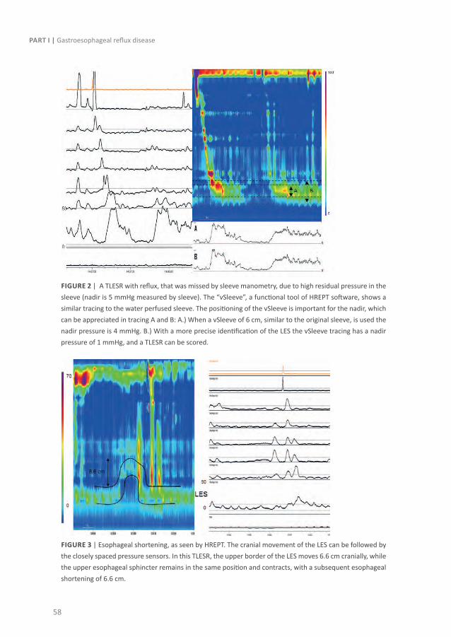
58
Part I | Gastroesophageal reflux disease
FIGuRe 2 | A TLESR with reflux, that was missed by sleeve manometry, due to high residual pressure in the sleeve (nadir is 5 mmHg measured by sleeve). The “vSleeve”, a functional tool of HREPT software, shows a similar tracing to the water perfused sleeve. The positioning of the vSleeve is important for the nadir, which can be appreciated in tracing A and B: A.) When a vSleeve of 6 cm, similar to the original sleeve, is used the nadir pressure is 4 mmHg. B.) With a more precise identification of the LES the vSleeve tracing has a nadir pressure of 1 mmHg, and a TLESR can be scored.
FIGuRe 3 | Esophageal shortening, as seen by HREPT. The cranial movement of the LES can be followed by the closely spaced pressure sensors. In this TLESR, the upper border of the LES moves 6.6 cm cranially, while the upper esophageal sphincter remains in the same position and contracts, with a subsequent esophageal shortening of 6.6 cm.

59
HRM for detection of TLESRs | Chapter 3
Esophageal shortening
Mean shortening during TLESRs detected with HREPT was 3.0 ± 0.11 cm compared to1.2 ± 0.10
cm during wet swallows (P<0.001). (Figures 3 and 4) The maximum esophageal shortening during
peristalsis after wet swallows was 3 cm, versus 7 cm for TLESRS. Esophageal shortening was greater
during TLESRs with reflux then during those without (3.1 ± 0.11 cm vs 2.5 ± 0.26 cm P<0.05).
TLESRs with reflux missed by sleeve manometry had a mean shortening of 3.1 ± 0.30 cm, and a
shortening of more than 2 cm was seen in 13 out of 19 of these TLESRs.
FIGuRe 4 | Esophageal Shortening.
dIscussIon
This study compared HREPT and sleeve manometry for the detection of TLESRs. The detection rate
of all TLESRs was comparable for HREPT and sleeve manometry. However, TLESRs accompanied by
a reflux event were significantly more detected using HREPT. Based on these findings, we suggest
that HREPT is the method of choice to detect TLESRs.
Reflux events are best detected by impedance, and their acidity characterised by pH-metry.22
MII-pH monitoring detects acidic, weakly acidic reflux, and gas reflux, and is therefore currently
considered the gold standard for the detection of reflux events.22, 23 In this study, MII-pH was used
to compare HREPT to sleeve manometry. We showed that the sensitivity to detect TLESRs with
reflux increased from 86% for sleeve manometry to 96% for HREPT. Our results are comparable to
the study of Bredenoord et al. that compared closely spaced water perfused sideholes to a sleeve
sensor for the detection of TLESRs.18 In that study, criteria were slightly modified and were applied
Esophageal Shortening
0
1
2
3
4
5
6
7
*
Wet Swallows TLESRs
Esop
hage
al s
hort
enin
g le
ngth
(cm
)

60
Part I | Gastroesophageal reflux disease
to the two tracings with the highest pressure values at the level of the LES. In recent studies
performed with HREPT, the original criteria as defined by Holloway et al. for sleeve manometry
were used for detection of TLESRs. 10, 24-26 The comparison in our study shows that the use of these
criteria leads to a comparable detection rate, and thus can indeed be used for HREPT.
The four main conditions in which HREPT is superior to sleeve manometry to detect TLESRs are
1. an increased resolution at the level of the LES that reveals LES opening in detail 2. an increased
detection during periods of low basal LES pressure, 3. the detection of esophageal shortening, and
4. the improved detection of reflux during a TLESRs (increased esophageal pressure resulting from
reflux). In the first condition, a high residual LES pressure is seen in TLESRs with reflux detected
by HREPT but missed by sleeve manometry. Detailed analysis of the HREPT tracings revealed
that the EGJ relaxation during a TLESR is not homogeneous with some parts maintaining some
residual pressure above 2 mmHg. Indeed, the gastric sling or the crural diaphragm do not always
open instantaneously, and completely relax only during a short period of time. Furthermore,
contraction of the lowest part of the esophagus while the LES is relaxed can be picked up by the
sleeve sensor. As the sleeve detects the highest pressure along its length, this small segment of
persistent pressure will result in a residual pressure at sleeve manometry above the cut off value
of 2 mmHg and thus will not be scored as a TLESR. To confirm this hypothesis, we used the virtual
sleeve function (vSleeve). This part of the software integrates all pressure values at the level of
the LES to one tracing, mimicking the function of the sleeve. As shown in figure 2 a similar tracing
to the original sleeve sensor tracing is seen, with a similar residual LES pressure above 2 mmHg,
when a vSleeve of 6 cm is used. The second condition in which HREPT is superior to the sleeve is
during periods of low LES pressure. (figure 3) Detection of TLESRs is indeed more difficult during
episodes of low LES pressure, and moreover, the criterion that pressure has to drop faster than 1
mmHg per second is more difficult to fulfil. The advantages of HREPT may therefore be even more
pronounced in GERD patients, who more often have a low basal LES pressure. The third condition
leading to improved detection of TLESRs by HREPT is the detection of esophageal shortening.
Shortening can be clearly appreciated during TLESRs and wet swallows. In contrast to swallowing,
esophageal shortening during a TLESR is much more pronounced, even up to 7 cm (Figures 3
and 4). Comparable findings have been described previously by Pandolfino et al. 20. Esophageal
shortening facilitated recognition of TLESRs in 13 out of 19 events with reflux missed by sleeve
manometry.
The last condition which leads to an improved detection rate is the pressure increase in the
esophageal body during a reflux event. Although there is some debate whether this pressure
increase is a consequence of esophageal shortening or the occurrence of reflux, in healthy
volunteers both are only seen during TLESRs. In 13 out of 19 TLESRs with reflux missed by

61
HRM for detection of TLESRs | Chapter 3
conventional manometry there was a pressure increase in the esophageal body, whereas this
pressure increase (common cavity) was only seen in 4 of these TLESRs in conventional manometry.
TLESRs are therefore more easily picked up, and this explains why especially TLESRs with reflux are
better detected by HREPT. Both esophageal shortening and esophageal pressurisation are however
no criteria for detection of TLESRs. Therefore, specific criteria for HREPT are still awaited. It needs
to be emphasized that there is no gold standard for the detection of TLESRs, which complicates the
validation of a new technique. In this study we have assumed that events only detected by HREPT
that met Holloway criteria and have a reflux episode were TLESRs. Supportive to our assumption
is the clear esophageal shortening during these events, which is only seen during TLESRs as was
shown in multiple studies 20, 27
In conclusion, detection rate for TLESRS is equal for HREPT and sleeve manometry, and this study
thereby shows that HREPT is suitable for detection. The increase in detection rate for TLESRs with
reflux is mainly explained by the higher spatial resolution at the level of the LES and the precise
positioning of the vSleeve, the increased detection rate during low basal LES pressure and the
visualisation of esophageal shortening and gastroesophageal reflux. The additional value of HREPT
for the detection of TLESRs in patients was not investigated in this article. Patients with GERD and
healthy volunteers are regarded to have comparable numbers of TLESRs, but one can hypothesize
that the beneficial effect of HREPT is even larger in GERD patients, due to a lower basal LES
pressure. Further studies on this topic are awaited to confirm this assumption.

62
Part I | Gastroesophageal reflux disease
ReFeRence lIsT
1. Kahrilas PJ, Shaheen NJ, Vaezi MF, Hiltz SW, Black E, Modlin IM, Johnson SP, Allen J, Brill JV. American Gastroenterological Association Medical Position Statement on the management of gastroesophageal reflux disease. Gastroenterology 2008;135:1383-1391, 1391.
2. Dent J, Dodds WJ, Friedman RH, Sekiguchi T, Hogan WJ, Arndorfer RC, Petrie DJ. Mechanism of gastroesophageal reflux in recumbent asymptomatic human subjects. J Clin Invest 1980;65:256-267.
3. Dodds WJ, Dent J, Hogan WJ, Helm JF, Hauser R, Patel GK, Egide MS. Mechanisms of gastroesophageal reflux in patients with reflux esophagitis. N Engl J Med 1982;307:1547-1552.
4. Sifrim D, Holloway R. Transient lower esophageal sphincter relaxations: how many or how harmful? Am J Gastroenterol 2001;96:2529-2532.
5. Beaumont H, Bennink R, de JJ, Boeckxstaens G. The position of the acid pocket as a major risk factor for acidic reflux in healthy subjects and GERD patients. Gut 2009.
6. Page AJ, Blackshaw LA. An in vitro study of the properties of vagal afferent fibres innervating the ferret oesophagus and stomach. J Physiol 1998;512 ( Pt 3):907-916.
7. Babaei A, Bhargava V, Korsapati H, Zheng WH, Mittal RK. A unique longitudinal muscle contraction pattern associated with transient lower esophageal sphincter relaxation. Gastroenterology 2008;134:1322-1331.
8. Boeckxstaens GE, Tytgat GN. More pathophysiologically oriented treatment of GORD? Lancet 2002;359:1267-1268.
9. Boeckxstaens GE. Reflux inhibitors: a new approach for GERD? Curr Opin Pharmacol 2008;8:685-689.
10. Holloway RH, Penagini R, Ireland AC. Criteria for objective definition of transient lower esophageal sphincter relaxation. Am J Physiol 1995;268:G128-G133.
11. Dent J. Pathogenesis of gastro-oesophageal reflux disease and novel options for its therapy. Neurogastroenterol Motil 2008;20 Suppl 1:91-102.
12. Holloway RH, Boeckxstaens G, Penagini R, Sifrim D, Smout AJ, Kristensson C, Ruth M. Objective Definition and Detection of Transient Lower Esophageal Relaxation Revisited: Is There Room for Improvement? Gastroenterology, Vol.136, Issue 5, S1, A527 ed. 2009.
13. Fox MR, Bredenoord AJ. Oesophageal high-resolution manometry: moving from research into clinical practice. Gut 2008;57:405-423.
14. Grubel C, Hiscock R, Hebbard G. Value of spatiotemporal representation of manometric data. Clin Gastroenterol Hepatol 2008;6:525-530.
15. Pandolfino JE, Ghosh SK, Rice J, Clarke JO, Kwiatek MA, Kahrilas PJ. Classifying esophageal motility by pressure topography characteristics: a study of 400 patients and 75 controls. Am J Gastroenterol 2008;103:27-37.
16. Ghosh SK, Pandolfino JE, Rice J, Clarke JO, Kwiatek M, Kahrilas PJ. Impaired deglutitive EGJ relaxation in clinical esophageal manometry: a quantitative analysis of 400 patients and 75 controls. Am J Physiol Gastrointest Liver Physiol 2007;293:G878-G885.
17. Pandolfino JE, Zhang QG, Ghosh SK, Han A, Boniquit C, Kahrilas PJ. Transient lower esophageal sphincter relaxations and reflux: mechanistic analysis using concurrent fluoroscopy and high-resolution manometry. Gastroenterology 2006;131:1725-1733.

63
HRM for detection of TLESRs | Chapter 3
18. Bredenoord AJ, Weusten BL, Timmer R, Smout AJ. Sleeve sensor versus high-resolution manometry for the detection of transient lower esophageal sphincter relaxations. Am J Physiol Gastrointest Liver Physiol 2005;288:G1190-G1194.
19. Bredenoord AJ, Weusten BL, Timmer R, Smout AJ. Intermittent spatial separation of diaphragm and lower esophageal sphincter favors acidic and weakly acidic reflux. Gastroenterology 2006;130:334-340.
20. Pandolfino JE, Zhang QG, Ghosh SK, Han A, Boniquit C, Kahrilas PJ. Transient lower esophageal sphincter relaxations and reflux: mechanistic analysis using concurrent fluoroscopy and high-resolution manometry. Gastroenterology 2006;131:1725-1733.
21. Sifrim D, Holloway R, Silny J, Xin Z, Tack J, Lerut A, Janssens J. Acid, nonacid, and gas reflux in patients with gastroesophageal reflux disease during ambulatory 24-hour pH-impedance recordings. Gastroenterology 2001;120:1588-1598.
22. Sifrim D, Castell D, Dent J, Kahrilas PJ. Gastro-oesophageal reflux monitoring: review and consensus report on detection and definitions of acid, non-acid, and gas reflux. Gut 2004;53:1024-1031.
23. Kahrilas PJ, Sifrim D. High-resolution manometry and impedance-pH/manometry: valuable tools in clinical and investigational esophagology. Gastroenterology 2008;135:756-769.
24. Pandolfino JE, Ghosh SK, Zhang Q, Han A, Kahrilas PJ. Upper sphincter function during transient lower oesophageal sphincter relaxation (tLOSR); it is mainly about microburps. Neurogastroenterol Motil 2007;19:203-210.
25. Kuribayashi S, Massey BT, Hafeezullah M, Perera L, Hussaini SQ, Tatro L, Darling RJ, Franco R, Shaker R. Terminating motor events for TLESR are influenced by the presence and distribution of refluxate. Am J Physiol Gastrointest Liver Physiol 2009;297:G71-G75.
26. Babaei A, Bhargava V, Mittal RK. Upper Esophageal Sphincter during Transient Lower Esophageal Sphincter Relaxation: Effects of Reflux Content & Posture. Am J Physiol Gastrointest Liver Physiol 2010.
27. Kwiatek MA, Post J, Pandolfino JE, Kahrilas PJ. Transient lower oesophageal sphincter relaxation in achalasia: everything but LOS relaxation. Neurogastroenterol Motil 2009;21:1294-e123.


Chapter 4Localisation of mGluR5, GABAB, GABAA and
cannabinoid receptors on the vago-vagal reflex pathway responsible for transient
lower esophageal sphincter relaxation in humans: An immunohistochemical study.
Wout Rohof, Eleonora Aronica, Hanneke Beaumont, Dirk Troost, Guy Boeckxstaens
Neurogastroenterol Motil. 2012;24(4):383-391

66
Part I | Gastroesophageal reflux disease
AbsTRAcT
Background and aims:
Transient lower esophageal sphincter relaxations (TLESRs) are the predominant mechanisms
underlying gastroesophageal reflux. TLESRs are mediated by a vago-vagal reflex, which can be
blocked by interaction with metabotropic Glutamate Receptor 5 (mGluR5), γ-aminobutyric acid
type B (GABAB), γ-aminobutyric acid type A (GABAA) and cannabinoid (CB) receptors. However, the
distribution of these receptors in the neural pathway underlying the triggering of TLESRs has not
been evaluated in humans.
Methods:
Using immunohistochemistry we investigated the distribution of mGluR5, GABAA, GABAB, CB1 and
CB2 receptors in the human nodose ganglion, the brain stem and the myenteric plexus of the
esophagus.
Results:
MGluR5, GABAB, CB1 and CB2 receptors are abundantly expressed in neurons of the myenteric
plexus of the LES, nodose ganglion cell bodies and nerve fibres, the dorsal motor nucleus and
nucleus of the solitary tract in the brain stem. GABAA receptors are expressed in the same regions
except in the nodose ganglion and myenteric plexus of the LES.
Conclusion:
Human mGluR5, GABAA,B , and CB1,2 receptors are abundantly expressed along the vago-vagal
neural pathway and involved in the triggering of TLESRs. These findings are in line with the central
side effects observed during treatment with reflux inhibitors such as GABAB receptor agonists and
mGluR5 antagonists, but also suggest that peripherally acting compounds may be effective.

67
Localisation of mGlur5, GABA and cannabinoid receptors | Chapter 4
InTRoducTIon
Gastroesophageal reflux disease (GERD) is a condition characterized by reflux of gastric contents
causing troublesome symptoms, complications or both.1 In Western countries, GERD is one of
the most common gastrointestinal diseases affecting approximately 10–20% of adults. 2 Transient
lower esophageal sphincter relaxations (TLESR) are the predominant mechanisms underlying
gastro-esophageal reflux, both in normal subjects and in GERD patients. 3, 4
TLESR are vago-vagally mediated motor patterns triggered by activation of vagal afferents in the
cardia of the stomach by various stimuli, of which gastric distentions are the most important. 5 The
cell bodies of the vagal afferents are located in the nodose ganglion synapsing in the nucleus of the
solitary tract (NTS) in the brain stem. Subsequently, neurons in the dorsal motor nucleus (DMV) of
the vagus nerve are activated thereby initiating the specific motor pattern underlying TLESRs, i.e.
relaxation of the lower esophageal sphincter (LES), esophageal shortening, and inhibition of the
crural diaphragm. 6 (Figure 1)
Figure 1 | Schematic representation of the neural pathway involved in the triggering of TLESRs. (DMV dorsal motor nucleus of the vagal nerve, NTS nucleus of the solitary tract, LES lower esophageal sphincter)

68
Part I | Gastroesophageal reflux disease
TLESRs and particularly the receptors involved in the underlying neural pathway have been
identified as a novel therapeutic target. Animal data indicate that γ-aminobutyric acid type B
(GABAB) receptors and metabotropic glutamate receptor 5 (mGluR5) are expressed along this
pathway, in particular in the nodose ganglion and NTS of the ferret.7 More importantly, recent
clinical studies have clearly demonstrated that GABAB-receptor agonists baclofen and lesogaberan
and mGluR5 antagonists inhibit TLESRs thereby reducing the number of reflux episodes and
symptoms in healthy subjects and GERD patients. 8-12 Activation of GABAA receptors, known to
mediate fast postsynpatic inhibition, also leads to an inhibition of TLESRs in dogs, but the effect
of GABAA agonists on the inhibition of TLESRs has not been studied in humans.13 Lastly, Δ9-
tetrahydrocannabinol, a receptor agonist of cannabinoid 1 and 2 receptor (CB1, CB2) reduced the
number of TLESRs in humans and dogs. 14
Compounds targeting TLESRs, also referred to as reflux inhibitors, are currently evaluated as an
add-on treatment for GERD patients that only partially respond to acid suppression with proton
pump inhibitors. One of the main problems so far are central side effects such dizziness and
nausea, most likely due to their interaction with receptors located in the central nervous system. 10, 15, 16 However, the exact location of the receptors involved in TLESR neural pathway has not been
evaluated in humans. Evidences from animal models, including receptor distribution, may differ
significantly from the human situation; therefore an insight into the distribution of these receptors
in human tissue is of utmost importance in optimising bench to bedside translation. Therefore,
the aim of this study was to analyse the presence of the mGluR5, GABAB, GABAA and cannabinoid
receptors in human nodose ganglion, DMV, NTS and the myenteric plexus of the lower esophageal
sphincter.
MeThods
Tissue samples
Brain and nodose ganglion tissues were obtained post-mortem from patients (5 male, median
age 72 yrs, range 45-82) with prior informed consent for brain autopsy and for the use of human
brain tissue for research. All autopsies were performed within 12 hours after death. Full thickness
esophageal specimens (n=4) were obtained from surgical esophageal resections. Clinical data
are presented in table 1. None of the patients had a reported history of any neurodegenerative
diseases. Tissues were fixed in paraformaldehyde and embedded in paraffin. Cerebellum and
cortex control tissue was obtained from the tissue bank of the department of neuropathology of
the Academic Medical Centre.

69
Localisation of mGlur5, GABA and cannabinoid receptors | Chapter 4
Tissue number Age sex cause of death
Nodose Ganglion 1 73 M Pulmonary Embolism
2 65 M Myocardial infarction
3 72 F Colorectal cancer with liver metastasis
4 81 M Pneumonia caused by carcinoma
5 72 M Myocardial infarction
Medulla Oblongata 1 45 F Mesenterial ischemia with complicated surgery
2 77 F Myocardial infarction
3 75 F Pancreatic adenocarcinoma, no brain metastasis
4 54 M Pneumonia, respiratory insufficiency
5 71 F Cardiomyopathy with decompensation
Esophagus 1 66 M Squamous cell carcinoma midesophagus
2 53 F Squamous cell carcinoma midesophagus
3 56 M Adenocarcinoma esophagoagastric junction
4 59 M Adenocarcinoma esophagoagastric junction
Table 1
Immunohistochemistry and antibody characterisation
Paraffin-embedded tissues were sectioned coronally with sections of 6 μm. Sections were
mounted on pre-coated glass slides. Representative sections of all specimens were processed for
hematoxylin and eosin and/or Nissl stains, to assure that specimens were of normal histology.
The sections were deparaffinised using xylene, re-hydrated and incubated with 1% H2O2 diluted in
methanol for 20 min. Slides were then washed with phosphate-buffered saline (PBS) (10 mM, pH
7.4) and heated in sodium citrate buffer (0.01 M, pH 6.0) for 10 minutes for antigen retrieval. After
washing with PBS, the slides were incubated for 30 minutes in 10% normal goat serum followed
by incubation with the primary antibodies overnight at 4 °C. Antibodies were visualized using the
Powervision peroxidase system (Immunologic, Duiven, the Netherlands) and 3,3-diaminobenzidine
(Sigma, St Louis, MO, USA) for a brownish staining. Hematoxylin was used as a counterstain, and
sections were subsequently dehydrated and mounted. As a negative control, slides were incubated
without the primary antibody. Human cerebellum and cortex were used as positive control tissue
when appropriate. The primary antibodies used in the study are summarized in Table 2. Studies in
which the specificity of the antibodies was confirmed in humans are listed in the table.
Evaluation of immunohistochemical staining
Immunoreactivity of neurons, nerve fibres or ganglion cells was evaluated in the nodose
ganglion, NTS, DMV and myenteric plexus. Localisation of the NTS and DMV was confirmed by an
experienced neuropathologist (DT). The relative density of labelling was classified as absent (-),

70
Part I | Gastroesophageal reflux disease
rare (±), moderate (+) or extensive (++). All slides were scored independently by two observers
and a consensus score was obtained. Additionally, immunopositive cells in nodose ganglion were
counted (mean ± standard deviation [SD]). Sections were digitized using an Olympus microscope
equipped with a DP-10 digital camera (Olympus, Japan). The images were imported into Adobe
Photoshop for minor adjustments of brightness and sharpness.
Antigen Primary antibody source dilution specificity
mGluR5 Polyclonal rabbit Upstate 1:100 Aronica, Boer 44 45
GABABR1, ab1531 Polyclonal guinea pig Chemicon International
1:200 Waldvogel, Aronica 27 46
GABABR2, ab28792 Polyconcal rabbit Santa Cruz Biotechnology
1:75 Torashima 28
GABAAα1Mab 339 Monoclonal mouse Chemicon 1:500 Waldvogel, Aronica 46, 47
GABAA β2/3 Mab341 Polyclonal mouse Chemicon 1:150 Waldvogel 47
CB1 Polyclonal rabbit Abcam 1:100 Aronica 48
CB2 Polyclonal rabbit Cayman 1:100 Aronica 48
Table 2
ResulTs
Nodose ganglion
The presence of ganglion cells was confirmed using Nissl staining in all nodose ganglion sections
(n=5). Immunostaining for mGluR5 and for GABAB R1 and GABAB R2 was scattered throughout the
nodose ganglion and mainly expressed in ganglion cells and moderately in nerve fibres (Figure 2).
No immunoreactivity for GABAAα1 was observed whereas normal staining was observed in brain
cortex, i.e. diffuse and strong neurophil immunoreactivity throughout the cortex. In contrast,
immunoreactivity for GABAA β2/3 was detected in ganglion cells, as shown in figures 2I and 2J.
Extensive staining of both CB1 and CB2 was observed with a prominent immunoreactivity of
ganglion cells and moderate staining of nerve fibres.
Medulla oblongata
Nuclei in the medulla oblongata were indentified using Nissl and haematoxylin and eosin (H&E)
staining. A representative overview of a section stained for mGluR5 is shown (figure 3A). Both in
the NTS and DMV, expression of mGluR5 was observed in nerve fibres and in neurons while the
solitary tract remained unstained. Other stained regions comprised the olivary nuclei, but staining
was less prominent compared to the cranial nerve nuclei like the nucleus of the hypoglossus nerve.
A summary of staining data is given in table 3.

71
Localisation of mGlur5, GABA and cannabinoid receptors | Chapter 4
Figure 2 | Immunostainings of the nodose ganglion. In panel A and B, mGluR5 immunoreactivity is detected in neuronal cell bodies and nerve fibres (A and B). GABAB R1 (C and D) and GABAB R2 (E and F) have a comparable staining pattern in ganglion cells and in nerve fibres. In contrast to GABAAα1 (G and H), GABAA β2/3 receptor subunits (I and J) are present in the nodose ganglion. Strong and specific labelling of ganglion cells is observed for both cannabinoid receptors (CB1 (K and L) and CB2 (M and N)). Scale bar: A,C,G,I, K and M = 200 μm, B, D and J = 70 μm, F, L and N = 40 μm.

72
Part I | Gastroesophageal reflux disease
Figure 3 | Images A to G show representative stainings of the medulla oblongata at the level of the upper medulla. Immunoreactivity for mGluR5 can be observed in A, B and C with different magnification, revealing mGluR5 immunoreactive neurons in the DMV. GABAB R1 immunreactivity on neurons and nerve fibres in the DMV is shown in image D, and reactivity for GABAB R2 is shown in the DMV (E) and at a larger magnification (F) . A typical plasma membrane and dendrite staining is observed for GABAAα1 subunits in the NTS (G) with a larger magnification in H. Comparably, extensive staining of axons and dendrites is observed for CB1 (I) and CB2 (J) in DMV. Scale bars: A 400 μm BG 150 μm CDEIJ 50 μm FH 20 μm. Abbreviations: DMV: dorsal motor nucleus of the vagus, NTS: nucleus of the solitary tract, Sol: Solitary tract, VL: Ventrolateral solitary nucleus, DFr: dorsal fringe of the dorsal nucleus of the vagus, MFr: medial fringe of the dorsal nucleus of the vagus, In: Intercalated nucleus of the vagus.
73
Localisation of mGlur5, GABA and cannabinoid receptors | Chapter 4
nodose ganglion nTs dMV Myenteric plexus
Ganglion cells
Nerve fibres
Cell counts (SD)
Neurons Nerve fibres
Neurons Nerve fibres
Neurons
mGluR5 ++ + 72 (12) +/- + + ++ ++
GABAB R1 ++ + 70 (10) + ++ + + +
GABAB R2 ++ + 73 (12) +/- +/- + + ++
GABAA α1 - - 0 (0) + ++ +/- ++ -
GABAA β2/3 ++ + 52 (6) - + +/- + +
CB 1 ++ + 64 (13) + +/- + + ++
CB 2 ++ + 66 (11) + + ++ + ++
Table 3 | The relative density of labelling was classified as absent (-), rare (±), moderate (+) or extensive (++).
In control cerebellum, GABABR1 and GABABR2 co-staining was observed along with a cellular
pattern similar to that previously described. Positive stainings of Purkinje cell and molecular layers
were also observed. In the medulla oblongata, GABAB receptors were localized in the DMV and
NTS mostly on the cell bodies, as shown in figures 3D and 3E. Moderate staining was observed in
the solitary tract, whilst a profound staining in the olivary nucleus and in the dorsal olivary nucleus
was observed.
A typical staining pattern of the plasma membrane and dendrites was observed in GABAAα1 and
CB1 and CB2 stainings, as shown in figures 3G-3J. Cannabinoid receptors were observed in the
DMV and NTS and other nuclei in the brain stem, comparable to GABAB receptor distribution,
whereas staining in the solitary tract could not be detected.
Myenteric plexus
Four LES resection specimens were studied in which the myenteric plexus could easily be identified
between the two muscle layers. The ganglion cells in the myenteric plexus clearly expressed
mGluR5, GABAB receptor subunits and both cannabinoid receptors (Figure 4). In contrast, no
staining for GABAAα1 was observed in any of the esophageal resection specimens. Additionally,
immunolabeling of nerve fibres was observed for GABAA β2/3 (I and J) and CB1 (L).
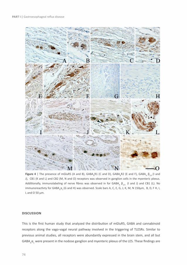
74
Part I | Gastroesophageal reflux disease
Figure 4 | The presence of mGluR5 (A and B), GABABR1 (C and D), GABABR2 (E and F), GABAA β2/3 (I and J), CB1 (K and L) and CB2 (M, N and O) receptors was observed in ganglion cells in the myenteric plexus. Additionally, immunolabeling of nerve fibres was observed in for GABAA β2/3 (I and J) and CB1 (L). No immunoreactivty for GABAAα1 (G and H) was observed. Scale bars A, C, E, G, J, K, M, N 150μm, B, D, F H, I, L and O 50 μm.
dIscussIon
This is the first human study that analyzed the distribution of mGluR5, GABA and cannabinoid
receptors along the vago-vagal neural pathway involved in the triggering of TLESRs. Similar to
previous animal studies, all receptors were abundantly expressed in the brain stem, and all but
GABAAα1 were present in the nodose ganglion and myenteric plexus of the LES. These findings are
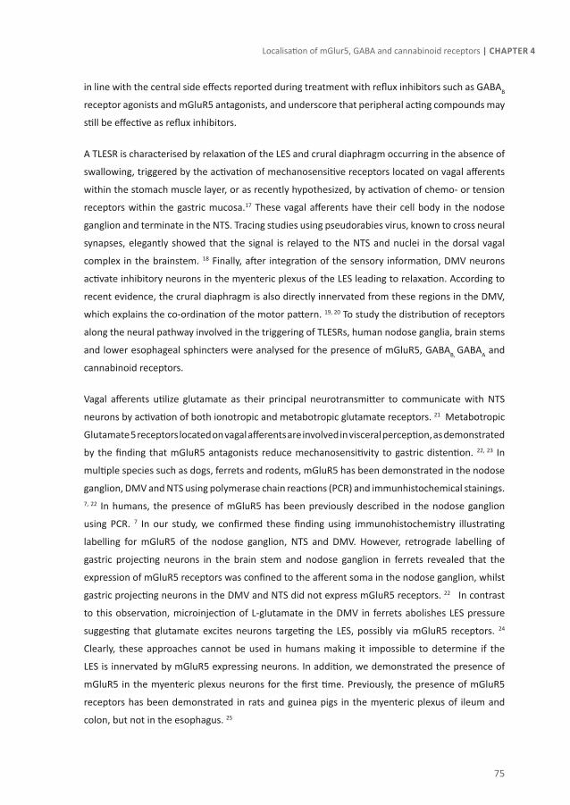
75
Localisation of mGlur5, GABA and cannabinoid receptors | Chapter 4
in line with the central side effects reported during treatment with reflux inhibitors such as GABAB
receptor agonists and mGluR5 antagonists, and underscore that peripheral acting compounds may
still be effective as reflux inhibitors.
A TLESR is characterised by relaxation of the LES and crural diaphragm occurring in the absence of
swallowing, triggered by the activation of mechanosensitive receptors located on vagal afferents
within the stomach muscle layer, or as recently hypothesized, by activation of chemo- or tension
receptors within the gastric mucosa.17 These vagal afferents have their cell body in the nodose
ganglion and terminate in the NTS. Tracing studies using pseudorabies virus, known to cross neural
synapses, elegantly showed that the signal is relayed to the NTS and nuclei in the dorsal vagal
complex in the brainstem. 18 Finally, after integration of the sensory information, DMV neurons
activate inhibitory neurons in the myenteric plexus of the LES leading to relaxation. According to
recent evidence, the crural diaphragm is also directly innervated from these regions in the DMV,
which explains the co-ordination of the motor pattern. 19, 20 To study the distribution of receptors
along the neural pathway involved in the triggering of TLESRs, human nodose ganglia, brain stems
and lower esophageal sphincters were analysed for the presence of mGluR5, GABAB, GABAA and
cannabinoid receptors.
Vagal afferents utilize glutamate as their principal neurotransmitter to communicate with NTS
neurons by activation of both ionotropic and metabotropic glutamate receptors. 21 Metabotropic
Glutamate 5 receptors located on vagal afferents are involved in visceral perception, as demonstrated
by the finding that mGluR5 antagonists reduce mechanosensitivity to gastric distention. 22, 23 In
multiple species such as dogs, ferrets and rodents, mGluR5 has been demonstrated in the nodose
ganglion, DMV and NTS using polymerase chain reactions (PCR) and immunhistochemical stainings. 7, 22 In humans, the presence of mGluR5 has been previously described in the nodose ganglion
using PCR. 7 In our study, we confirmed these finding using immunohistochemistry illustrating
labelling for mGluR5 of the nodose ganglion, NTS and DMV. However, retrograde labelling of
gastric projecting neurons in the brain stem and nodose ganglion in ferrets revealed that the
expression of mGluR5 receptors was confined to the afferent soma in the nodose ganglion, whilst
gastric projecting neurons in the DMV and NTS did not express mGluR5 receptors. 22 In contrast
to this observation, microinjection of L-glutamate in the DMV in ferrets abolishes LES pressure
suggesting that glutamate excites neurons targeting the LES, possibly via mGluR5 receptors. 24
Clearly, these approaches cannot be used in humans making it impossible to determine if the
LES is innervated by mGluR5 expressing neurons. In addition, we demonstrated the presence of
mGluR5 in the myenteric plexus neurons for the first time. Previously, the presence of mGluR5
receptors has been demonstrated in rats and guinea pigs in the myenteric plexus of ileum and
colon, but not in the esophagus. 25

76
Part I | Gastroesophageal reflux disease
For an appropriately functioning GABAB receptor, both GABABR1 and GABABR2 receptor subtypes
are needed. 26 GABAB receptors produce slow prolonged inhibitory signals, and therefore agonism
of GABAB receptors on the vago-vagal reflex arch results in a reduction of the number of TLESRs. 24, 26 The presence of GABAB receptors in the nodose ganglion was previously demonstrated in
rats and ferrets, as well as their presence in the brain stem of rodents and humans. 16, 24 27 Our
study confirms the presence of both receptor subtypes in human nodose ganglion, NTS and
DMV. In addition, we demonstrate the presence of GABAB receptors in the myenteric plexus,
comparable to a recent study by Torashima et al. 28 These authors demonstrated that both GABAB
receptor subtypes are present in the myenteric plexus and form heterodimers. This suggests that
the myenteric plexus is a possible site of action of GABAB agonists, thereby contributing to the
reduction in TLESRs. 11
In contrast to mGluR5 receptors, GABAB receptors are highly expressed in LES-projecting neurons
in the DMV. 24 In line with this, injection of baclofen into the DMV inhibits the firing rate of vagal
motor neurons that are responsive to gastric distension. 29 Interestingly, the DMV is composed of
heterogeneous neuronal subpopulations in terms of both membrane as well as pharmacological
properties. 30 These subpopulations can further be distinguished based on their peripheral
targets, at least in rats. 21, 30 Notably, activation of GABAB-receptors in one neuronal subpopulation
projecting to the stomach leads to an increase in vagal excitatory cholinergic drive mediating an
increase in rhythmic contractions, whereas activation of another neuronal subpopulation leads
a decrease in tonic vagal drive to NANC inhibitory neurons resulting in an increase in gastric
pressure. 30 To what extent this also applies to human remains unclear and cannot be concluded
from our data.
Next, we evaluated the distribution of CB1 receptors, as blockade of these receptors has been
shown to inhibit TLESRs in humans. 14 CB1 receptors have a comparable distribution and function
as GABAB receptors. 31 Comparable to rats and ferrets, CB1 receptors were present in the human
nodose ganglion, DMV and NTS. 31-34 Interestingly, in animals ganglionectomy or vagotomy did
not lead to an alteration receptor expression of DMV or NTS neurons. As vagal motor neurons
degenerate after vagotomy, these findings support an intrinsic interneuronal localization of
immunoreactive terminals in the brainstem.34 In line with this, no staining of CB1 receptors was
observed in dorsal motor nucleus neurons projecting to the LES and gastric fundus identified using
retrograde labelling studies in ferrets. 32 Although we cannot directly extrapolate animal data to
humans, these data suggest that it is questionable whether these neurons in the DMV and NTS in
humans project to the LES.
In contrast to previous immunohistochemical evidences 34, 35, a more recent study by van Sickle
showed that CB2 receptors are expressed in rat and ferret brainstem. 36 In line with our data in

77
Localisation of mGlur5, GABA and cannabinoid receptors | Chapter 4
humans, expression in the brain was most prominent in the DMV, although van Sickle et al report
that the levels of CB2 were much lower compared to the spleen. Due to the high levels of CB2
receptors in the spleen, agonism of CB2 receptors leads to immunosuppression. Therefore, this
receptor is probably not a suitable candidate for the reduction of TLESRs in GERD. 36
GABAA induces fast synaptic ionotropic inhibition upon activation, and the receptor consists of
various subunits. 13, 26 In contrast to GABAB receptors, no GABAAα1 were present in the myenteric
plexus and nodose ganglion, which is comparable to the absence of these receptors in the
periphery in dogs. 13 The lack of effect of peripherally acting GABAA antagonists on TLESR rates
in dogs substantiates the absence of peripheral GABAA receptors on the vago-vagal reflex arch. 13
To date, clinical studies have indeed demonstrated the favourable effect of GABAB agonism
and mGluR5 antagonism on the number of TLESRs, reflux episodes and symptoms in humans
compared to placebo. 9-12, 37 However, currently there are no reflux inhibitors available for clinical
use except baclofen, which also has sustainable side effects. 8, 15 The same holds true for mGluR5
antagonists inducing central side effects such as dizziness and vertigo in 16% to 12% of patients. 37
These data are in line with the profound distribution of both GABA and mGluR5 in the brain stem,
cortex and cerebellum. Therefore, centrally acting agents are anticipated to have sustainable side
effects, emphasizing the need for development of peripherally acting agents. In view of these
central side effects, pharmaceutical companies have developed peripherally acting compounds. As
shown in the present study, these receptors are abundantly present in the periphery. Futhermore,
functional evidence has established a peripheral site of action of baclofen on GABAB receptors
and of mGluR5 antagonists on mGlur5 receptors in gastric vagal afferents, suggesting that reflux
inhibitors targeting these receptors may have the potential to block TLESRs. 7, 38, 39
Two strategies specifically targeting peripheral receptors can be considered: First, polarized
compounds that do not pass the blood barrier and thus do not enter the central nervous system
can be used. It should be emphasized though that the blood brain barrier at the level of the DMV
and NTS is leaky and thus polarized compounds could also – at least partly – have an effect on
receptors in this region. 40, 41 Second, targeting specific subpopulations of receptors or transporters
could also affect the TLESR reducing properties or side effect profile of a compound. For instance,
lesogaberan, a GABAB agonist reducing the number of TLESRs and GERD symptoms 12, is taken up
by the GABA transporter (GAT), and is therefore sequestrated by CNS neurons leading to reduced
intracerebral levels. 42 Baclofen however does not bind to GAT most likely explaining the finding
that lesogaberan has fewer CNS side effects than baclofen. Still, Lesogaberan effectively reduces
the number of TLESRs leading to a significantly better symptom relief than placebo. 12
78
Part I | Gastroesophageal reflux disease
Our study shows the expression of several receptors along the entire neural pathway in human
tissue. Studies in laboratory animals are usually hard to translate to the human situation, and
therefore conflicting results in animal and clinical studies often arise.43 however the limitation of
our study is that the target organ of the neurons cannot be determined. To this end, anterograde
and retrograde labelling is required which unfortunately is not feasible in humans due to ethical
concerns.
In conclusion, this study shows that mGluR5, GABAA,B , and CB1,2 receptors, which are the targets
of known reflux inhibitors, are present throughout the vago-vagal reflex pathway involved
in the triggering of TLESRs. These findings are in line with the central side effects observed
during treatment with GABAB receptor agonists and mGluR5 antagonists, but also suggest that
peripherally acting compounds may be effective as reflux inhibitors.
79
Localisation of mGlur5, GABA and cannabinoid receptors | Chapter 4
ReFeRence lIsT
1. Kahrilas PJ, Shaheen NJ, Vaezi MF. American Gastroenterological Association Institute technical review on the management of gastroesophageal reflux disease. Gastroenterology 2008;135:1392-1413, 1413.
2. Dent J, El-Serag HB, Wallander MA, Johansson S. Epidemiology of gastro-oesophageal reflux disease: a systematic review. Gut 2005;54:710-717.
3. Dent J, Dodds WJ, Friedman RH, Sekiguchi T, Hogan WJ, Arndorfer RC, Petrie DJ. Mechanism of gastroesophageal reflux in recumbent asymptomatic human subjects. J Clin Invest 1980;65:256-267.
4. Dodds WJ, Dent J, Hogan WJ, Helm JF, Hauser R, Patel GK, Egide MS. Mechanisms of gastroesophageal reflux in patients with reflux esophagitis. N Engl J Med 1982;307:1547-1552.
5. Page AJ, Blackshaw LA. An in vitro study of the properties of vagal afferent fibres innervating the ferret oesophagus and stomach. J Physiol 1998;512 ( Pt 3):907-916.
6. Mittal RK, Holloway RH, Penagini R, Blackshaw LA, Dent J. Transient lower esophageal sphincter relaxation. Gastroenterology 1995;109:601-610.
7. Page AJ, Young RL, Martin CM, Umaerus M, O’Donnell TA, Cooper NJ, Coldwell JR, Hulander M, Mattsson JP, Lehmann A, Blackshaw LA. Metabotropic glutamate receptors inhibit mechanosensitivity in vagal sensory neurons. Gastroenterology 2005;128:402-410.
8. Vela MF, Tutuian R, Katz PO, Castell DO. Baclofen decreases acid and non-acid post-prandial gastro-oesophageal reflux measured by combined multichannel intraluminal impedance and pH. Aliment Pharmacol Ther 2003;17:243-251.
9. Keywood C, Wakefield M, Tack J. A proof-of-concept study evaluating the effect of ADX10059, a metabotropic glutamate receptor-5 negative allosteric modulator, on acid exposure and symptoms in gastro-oesophageal reflux disease. Gut 2009;58:1192-1199.
10. Beaumont H, Smout A, Aanen M, Rydholm H, Lei A, Lehmann A, Ruth M, Boeckxstaens G. The GABA(B) receptor agonist AZD9343 inhibits transient lower oesophageal sphincter relaxations and acid reflux in healthy volunteers: a phase I study. Aliment Pharmacol Ther 2009;30:937-946.
11. Boeckxstaens GE, Beaumont H, Mertens V, Denison H, Ruth M, Adler J, Silberg DG, Sifrim D. Effects of lesogaberan on reflux and lower esophageal sphincter function in patients with gastroesophageal reflux disease. Gastroenterology 2010;139:409-417.
12. Boeckxstaens GE, Beaumont H, Hatlebakk JG, Silberg DG, Bjorck K, Karlsson M, Denison H. A novel reflux inhibitor lesogaberan (AZD3355) as add-on treatment in patients with GORD with persistent reflux symptoms despite proton pump inhibitor therapy: a randomised placebo-controlled trial. Gut 2011 Sep;60(9):1182-8.
13. Beaumont H, Jonsson-Rylander AC, Carlsson K, Pierrou S, Ahlefelt M, Branden L, Jensen J, Boeckxstaens GE, Lehmann A. The role of GABA(A) receptors in the control of transient lower oesophageal sphincter relaxations in the dog. Br J Pharmacol 2008;153:1195-1202.
14. Beaumont H, Jensen J, Carlsson A, Ruth M, Lehmann A, Boeckxstaens G. Effect of delta9-tetrahydrocannabinol, a cannabinoid receptor agonist, on the triggering of transient lower oesophageal sphincter relaxations in dogs and humans. Br J Pharmacol 2009;156:153-162.

80
Part I | Gastroesophageal reflux disease
15. Koek GH, Sifrim D, Lerut T, Janssens J, Tack J. Effect of the GABA(B) agonist baclofen in patients with symptoms and duodeno-gastro-oesophageal reflux refractory to proton pump inhibitors. Gut 2003;52:1397-1402.
16. Rohof W.O., Lei A., Boeckxstaens G. Effect of a novel metabotropic glutamate receptor 5 antagonist (AZD2066) on transient lower esophageal sphincter relaxations and reflux episodes in healthy males. Gastroenterology Vol. 140, Issue 5, Supplement 1, Page S-66.
17. van Wijk MP, Benninga MA, Davidson GP, Haslam R, Omari TI. Small volumes of feed can trigger transient lower esophageal sphincter relaxation and gastroesophageal reflux in the right lateral position in infants. J Pediatr 2010;156:744-8, 748.
18. Niedringhaus M, Jackson PG, Pearson R, Shi M, Dretchen K, Gillis RA, Sahibzada N. Brainstem sites controlling the lower esophageal sphincter and crural diaphragm in the ferret: a neuroanatomical study. Auton Neurosci 2008;144:50-60.
19. Niedringhaus M, Jackson PG, Evans SR, Verbalis JG, Gillis RA, Sahibzada N. Dorsal motor nucleus of the vagus: a site for evoking simultaneous changes in crural diaphragm activity, lower esophageal sphincter pressure, and fundus tone. Am J Physiol Regul Integr Comp Physiol 2008;294:R121-R131.
20. Young RL, Page AJ, Cooper NJ, Frisby CL, Blackshaw LA. Sensory and motor innervation of the crural diaphragm by the vagus nerves. Gastroenterology 2010;138:1091-1101.
21. Browning KN, Travagli RA. Plasticity of vagal brainstem circuits in the control of gastric function. Neurogastroenterol Motil 2010;22:1154-1163.
22. Young RL, Page AJ, O’Donnell TA, Cooper NJ, Blackshaw LA. Peripheral versus central modulation of gastric vagal pathways by metabotropic glutamate receptor 5. Am J Physiol Gastrointest Liver Physiol 2007;292:G501-G511.
23. Frisby CL, Mattsson JP, Jensen JM, Lehmann A, Dent J, Blackshaw LA. Inhibition of transient lower esophageal sphincter relaxation and gastroesophageal reflux by metabotropic glutamate receptor ligands. Gastroenterology 2005;129:995-1004.
24. McDermott CM, Abrahams TP, Partosoedarso E, Hyland N, Ekstrand J, Monroe M, Hornby PJ. Site of action of GABA(B) receptor for vagal motor control of the lower esophageal sphincter in ferrets and rats. Gastroenterology 2001;120:1749-1762.
25. Nasser Y, Keenan CM, Ma AC, McCafferty DM, Sharkey KA. Expression of a functional metabotropic glutamate receptor 5 on enteric glia is altered in states of inflammation. Glia 2007;55:859-872.
26. White JH, Wise A, Main MJ, Green A, Fraser NJ, Disney GH, Barnes AA, Emson P, Foord SM, Marshall FH. Heterodimerization is required for the formation of a functional GABA(B) receptor. Nature 1998;396:679-682.
27. Waldvogel HJ, Billinton A, White JH, Emson PC, Faull RL. Comparative cellular distribution of GABAA and GABAB receptors in the human basal ganglia: immunohistochemical colocalization of the alpha 1 subunit of the GABAA receptor, and the GABABR1 and GABABR2 receptor subunits. J Comp Neurol 2004;470:339-356.
28. Torashima Y, Uezono Y, Kanaide M, Ando Y, Enjoji A, Kanematsu T, Taniyama K. Presence of GABA(B) receptors forming heterodimers with GABA(B1) and GABA(B2) subunits in human lower esophageal sphincter. J Pharmacol Sci 2009;111:253-259.
29. Partosoedarso ER, Blackshaw LA. Selectivity of GABAB receptors in reducing peripheral and central vagal sensory transduction. Gastroenterology 2000;118 ed. S1:A173.
30. Browning KN, Travagli RA. Mechanism of action of baclofen in rat dorsal motor nucleus of the vagus. Am J Physiol Gastrointest Liver Physiol 2001;280:G1106-G1113.
81
Localisation of mGlur5, GABA and cannabinoid receptors | Chapter 4
31. Tsou K, Brown S, Sanudo-Pena MC, Mackie K, Walker JM. Immunohistochemical distribution of cannabinoid CB1 receptors in the rat central nervous system. Neuroscience 1998;83:393-411.
32. Partosoedarso ER, Abrahams TP, Scullion RT, Moerschbaecher JM, Hornby PJ. Cannabinoid1 receptor in the dorsal vagal complex modulates lower oesophageal sphincter relaxation in ferrets. J Physiol 2003;550:149-158.
33. Burdyga G, Varro A, Dimaline R, Thompson DG, Dockray GJ. Expression of cannabinoid CB1 receptors by vagal afferent neurons: kinetics and role in influencing neurochemical phenotype. Am J Physiol Gastrointest Liver Physiol 2010;299:G63-G69.
34. Van Sickle MD, Oland LD, Ho W, Hillard CJ, Mackie K, Davison JS, Sharkey KA. Cannabinoids inhibit emesis through CB1 receptors in the brainstem of the ferret. Gastroenterology 2001;121:767-774.
35. Derbenev AV, Stuart TC, Smith BN. Cannabinoids suppress synaptic input to neurones of the rat dorsal motor nucleus of the vagus nerve. J Physiol 2004;559:923-938.
36. Van Sickle MD, Duncan M, Kingsley PJ, Mouihate A, Urbani P, Mackie K, Stella N, Makriyannis A, Piomelli D, Davison JS, Marnett LJ, Di M, V, Pittman QJ, Patel KD, Sharkey KA. Identification and functional characterization of brainstem cannabinoid CB2 receptors. Science 2005;310:329-332.
37. Zerbib F, Bruley d, V, Roman S, Tutuian R, Galmiche JP, Mion F, Tack J, Malfertheiner P, Keywood C. Randomised clinical trial: effects of monotherapy with ADX10059, a mGluR5 inhibitor, on symptoms and reflux events in patients with gastro-oesophageal reflux disease. Aliment Pharmacol Ther 2011;33:911-921.
38. Page AJ, Blackshaw LA. GABA(B) receptors inhibit mechanosensitivity of primary afferent endings. J Neurosci 1999;19:8597-8602.
39. Blackshaw LA, Smid SD, O’Donnell TA, Dent J. GABA(B) receptor-mediated effects on vagal pathways to the lower oesophageal sphincter and heart. Br J Pharmacol 2000;130:279-288.
40. Gross PM, Wall KM, Pang JJ, Shaver SW, Wainman DS. Microvascular specializations promoting rapid interstitial solute dispersion in nucleus tractus solitarius. Am J Physiol 1990;259:R1131-R1138.
41. Cottrell GT, Ferguson AV. Sensory circumventricular organs: central roles in integrated autonomic regulation. Regul Pept 2004;117:11-23.
42. Lehmann A, Antonsson M, Aurell-Holmberg A, Blackshaw LA, Branden L, Elebring T, Jensen J, Karrberg L, Mattsson JP, Nilsson K, Oja SS, Saransaari P, Unge SV. Differential in vitro and in vivo profiles of substituted 3-aminopropylphosphinate and 3-aminopropyl(methyl)phosphinate GABA(B) receptor agonists as inhibitors of transient lower esophageal sphincter relaxation. Br J Pharmacol 2011 Sep 26. doi: 10.1111/j.1476-5381.2011.01682.x. [Epub ahead of print]
43. Perel P, Roberts I, Sena E, Wheble P, Briscoe C, Sandercock P, Macleod M, Mignini LE, Jayaram P, Khan KS. Comparison of treatment effects between animal experiments and clinical trials: systematic review. BMJ 2007;334:197.
44. Aronica E, Catania MV, Geurts J, Yankaya B, Troost D. Immunohistochemical localization of group I and II metabotropic glutamate receptors in control and amyotrophic lateral sclerosis human spinal cord: upregulation in reactive astrocytes. Neuroscience 2001;105:509-520.
45. Boer K, Troost D, Timmermans W, Gorter JA, Spliet WG, Nellist M, Jansen F, Aronica E. Cellular localization of metabotropic glutamate receptors in cortical tubers and subependymal giant cell tumors of tuberous sclerosis complex. Neuroscience 2008;156:203-215.

82
Part I | Gastroesophageal reflux disease
46. Aronica E, Redeker S, Boer K, Spliet WG, Van Rijen PC, Gorter JA, Troost D. Inhibitory networks in epilepsy-associated gangliogliomas and in the perilesional epileptic cortex. Epilepsy Res 2007;74:33-44.
47. Waldvogel HJ, Kubota Y, Fritschy J, Mohler H, Faull RL. Regional and cellular localisation of GABA(A) receptor subunits in the human basal ganglia: An autoradiographic and immunohistochemical study. J Comp Neurol 1999;415:313-340.
48. Zurolo E, Iyer AM, Spliet WG, Van Rijen PC, Troost D, Gorter JA, Aronica E. CB1 and CB2 cannabinoid receptor expression during development and in epileptogenic developmental pathologies. Neuroscience 2010;170:28-41.


Chapter 5Randomized clinical trial: effect of a
novel metabotropic glutamate receptor 5 antagonist (AZD2066) on transient lower
esophageal sphincter relaxations and reflux episodes in healthy volunteers
Wout Rohof, Aaltje Lei, David Hirsch, Lars Ny, Magnus Astrand, Mark Berner Hansen, Guy Boeckxstaens
Aliment Pharmacol Ther. 2012;35(10):1231-42

86
Part I | Gastroesophageal reflux disease
suMMARY
Background:
Selective metabotropic glutamate receptor 5 (mGluR5) antagonists inhibit transient lower
esophageal sphincter relaxations (TLESRs) in animals and acid reflux in humans.
Aim:
To assess the effect of single doses of the mGluR5 antagonist AZD2066 on TLESRs and reflux in
humans.
Methods:
Healthy male volunteers received AZD2066 13 mg and placebo (part A), or AZD2066 2 mg and
AZD2066 6 mg and placebo (part B), in a randomized crossover study. Postprandial manometry/
pH-impedance measurements were taken after each dose.
Results:
13 individuals completed part A of the study and 19 individuals completed part B. There was a
significant reduction in the geometric mean number of TLESRs (27%; P = 0.02) and the geometric
mean number of reflux episodes (51%; P = 0.01) in subjects receiving AZD2066 13 mg compared
with placebo. Adverse events in participants receiving AZD2066 13 mg were mostly related to the
nervous system (dizziness [3/13]; disturbance in attention [3/13]). Adverse events were reversible
and of mild intensity. There were no serious adverse events. The effects of AZD2066 appeared
dose-dependent, with smaller reductions in TLESRs and reflux episodes (relative to placebo) and
fewer adverse events observed for AZD2066 2 mg and AZD2066 6 mg compared with AZD2066
13 mg.
Conclusions:
mGluR5-mediated inhibition of TLESRs may be a useful approach for inhibiting gastroesophageal
reflux.

87
Effect of AZD2066 on TLESRs and reflux | Chapter 5
InTRoducTIon
Gastroesophageal reflux disease (GERD) is a chronic disease caused by reflux of gastric contents
into the esophagus.1, 2 In Western countries, approximately 10–20% of adults are thought to be
affected by GERD symptoms, with lower rates of around 5% reported in Asian populations.3, 4 The
mainstay of GERD treatment is acid suppression, most commonly achieved with the use of proton
pump inhibitors (PPIs).5, 6 However, there is mounting evidence that reflux symptoms persist in a
high proportion (20–30%) of patients despite PPI therapy,7-10 and that they are associated with
decreased psychological and physical well-being.11
Transient lower esophageal sphincter relaxations (TLESRs) are considered to be a major mechanism
for generating reflux in healthy individuals and in patients with GERD.12, 13 Pharmacological
inhibition of TLESRs is a promising new therapeutic approach because it has the potential to
reduce most types of reflux. This is particularly relevant for patients who have troublesome reflux
symptoms despite acid suppression from PPI use, because weakly acidic and weakly alkaline reflux
are thought to play a role in symptom generation in this group.14 TLESRs are triggered in response
to the activation of stretch receptors in the stomach,15 and are thought to be mediated by a vago-
vagal reflex pathway.16, 17 The gamma-aminobutyric acid type B (GABAB) receptor acts at several
points along this pathway, and recent clinical studies demonstrate that inhibition of TLESRs using
the GABAB-receptor agonist lesogaberan reduces the number of reflux episodes and symptoms.18,
19
Inhibition of TLESRs may also be achieved via other pathways. In particular, metabotropic
glutamate receptor 5 (mGluR5) expressed in the soma of gastric vagal afferents appears to play
an important role in triggering TLESRs.20 Selective antagonists of mGluR5 have been shown to
inhibit TLESRs in animal studies21, 22,21, 22 and to reduce acid reflux and improve clinical symptoms in
patients with GERD.23 However, the effect of mGluR5 inhibition on the number of reflux episodes
has not been assessed in humans in relation to TLESR frequency.
AZD2066 is a novel chemical substance that has been pharmacologically characterized as a selective,
non-competitive antagonist of mGluR5, and is being evaluated for the treatment of neuropathic
pain, depression and GERD. The primary objective of this pharmacodynamic study was to measure
the effect of AZD2066 on the frequency of TLESRs in healthy volunteers. Secondary objectives
included assessing the effect of AZD2066 on the frequency of TLESRs, number and type of reflux
episodes, esophageal acid exposure, lower esophageal sphincter (LES) pressure and number of
swallows, and to determine its pharmacokinetic characteristics and safety and tolerability profile.
88
Part I | Gastroesophageal reflux disease
MeThods
Study participants
To be enrolled, study participants were required to be healthy men, aged 18–45 years, with a body
mass index (BMI = weight/height2) of 19–30 kg/m2. Full details of the inclusion/exclusion criteria
implemented during the study are presented in supplementary Table 1. At the 21-day pre-entry
visit, each subject had a medical examination which included a full laboratory screen, a physical
examination (general appearance, cardiovascular, lungs, abdomen, skin, head and neck, lymph
nodes, thyroid, musculoskeletal/extremities and reflexes), a 12-lead resting ECG recording, blood
pressure measurement and a pregnancy test for females. Tests for Hepatitis B and C, HIV and drug
abuse were also performed at the pre-entry visit, and participants also underwent a structured
psychiatric interview using the Mini International Neuropsychiatric Interview. Participants had to
have clinically normal electrocardiogram, pulse and blood pressure measurements and laboratory
values at the pre-entry visit, and normal laboratory values before administering the first dose at
the second visit.
This study was conducted in accordance with the Declaration of Helsinki and the International
Conference on Harmonisation Guidelines for Good Clinical Practice, and was approved by the
Institutional Review Board associated with the study centre (Department of Gastroenterology,
Academic Medical Centre, Amsterdam, Netherlands). Participants provided written informed
consent prior to the study. This study is registered on a publicly accessible website (ClinicalTrials.
gov Identifier: NCT00813306).
Study design and study drugs
This was a double-blind, randomized, single-centre, phase 1, crossover study (2008–2009)
conducted in two parts, each involving a different set of participants. Part A assessed the impact of
AZD2066 on gastroesophageal sphincter function and reflux variables using a single dose (13 mg)
considered to have an acceptable safety and tolerability profile, based on an interim analysis of a
multiple ascending dose study (study code: DO475C00002). Each participant received 130 mL of
0.1 mg/mL oral AZD2066 (13 mg) solution and 130 mL of oral placebo solution, each on different
days separated by a 7–28-day washout period (Figure 1, study part A).
Part B of the study explored the dose–response relationship for AZD2066 using two lower single
doses (2 mg and 6 mg). Each participant received 60 mL of 0.1 mg/mL oral AZD2066 (6 mg)
solution, 20 mL of 0.1 mg/mL oral AZD2066 (2 mg) solution made up to 60 mL with oral placebo
solution and 60 mL oral placebo solution each on different days. A washout period of 7–28 days
was initially planned for both parts of the study. However, in part A of the study, AZD2066 (5–6
nmol at Cmax) was detected in the plasma of 2 subjects in the placebo treated group after treatment

89
Effect of AZD2066 on TLESRs and reflux | Chapter 5
with AZD2066 and a washout period of 8 days. The washout period between each treatment in
part B of the study was thus increased to 10–28-days (Figure 1, study part B).
Figure 1 | Randomized, crossover design used for parts A and B of the study.
In both parts of the study, the first dose (AZD2066 or placebo) was administered ≤21 days after
the pre-entry visit. Food and fluids were not allowed after midnight on the night before dose
administration. Participants received a standardized breakfast (700 kcal, of which 219 kcal [31.3%]
were from fat) 30 min after administration of AZD2066 or placebo, which was to be consumed
within 15 min. Esophageal manometric and pH-impedance measurements were collected from
participants (while in a semi-recumbent position) for 3 h 45 min, so that 3 h of data collection
occurred in the postprandial period. Participants also had a follow-up visit 5–7 days after the last
study period.
Assignment and blinding
The principal investigator (GB) assigned individuals to unique enrolment numbers, determined
subject eligibility, and assigned eligible participants to unique randomization numbers. A
randomized dosing regimen allocation list was generated by AstraZeneca R&D Mölndal using a
validated computer program, and the randomization was performed within blocks of consecutive
patient numbers. The study was double-blinded so that both investigators and participants were
unaware of who received which dosing regimen. Treatment codes were not broken until the
evaluability of the data from each participant had been established and documented.
Pharmacodynamic assessments
Manometric recordings of the pharynx, esophagus, LES and stomach were obtained using a
perfused 12-channel silicone rubber assembly (DentSleeve International Ltd, Mississauga, Ontario,
90
Part I | Gastroesophageal reflux disease
Canada). The manometric catheter was positioned with the proximal border of the sleeve 1 cm
above the LES. Manometric readings from dosing until the end of the 3-h postprandial period were
used to assess the following.
i) Number of TLESRs, defined according to previously described criteria,24 and their time of
onset (primary outcome in relation to AZD2066 13 mg; secondary outcome in relation to
AZD2066 2mg and AZD2066 6 mg).
ii) LES pressure (recorded every 15 min and expressed as the mean difference between the end
expiration LES pressure and the end-expiration intragastric pressure over 1 min). Mean LES
pressure during the 3-hour postprandial period was calculated from measurements taken
15 min after dose administration and then every 15 min until 3 h after the meal (3 h 45 min
in total) (secondary outcome).
iii) Number of swallows (defined as a fast increase in pressure in the pharyngeal channel, clearly
distinguishable from baseline activity) (secondary outcome).
For the impedance-pH readings, a catheter, comprising several electrode pairs measuring
intraluminal impedance, and an antimony pH sensor were used. The catheter was positioned so
that the pH sensor was located 5 cm above the LES. Impedance-pH readings from dosing until the
end of the 3-h postprandial period were used to assess the following:
i) Number of reflux episodes, defined as a period when impedance decreased to below 50% of
baseline (liquid episode) or increased to above 150% of baseline (gas episode), propagating
aborally from the most distal channel (secondary outcome).
ii) Number of reflux episodes that were acidic (intraesophageal pH <4 [or a drop of ≥1 pH unit
if pH is already <4] lasting more than 5 s), weakly acidic (intraesophageal pH 4.0–6.5 lasting
more than 5 s) and weakly alkaline (intraesophageal pH ≥6.5 lasting more than 5 s) (secondary
outcome).
iii) Percentage of time with an esophageal pH <4 (secondary outcome).
Pharmacokinetic, safety and tolerability assessments
Blood samples (2 mL) were collected at frequent intervals for 12 h after each dose of AZD2066
to determine the pharmacokinetic profile (secondary outcome). Plasma AZD2066 concentrations
were determined by liquid chromatography and mass spectrometry (quantification limit:
1.00 nmol⁄L). Adverse events occurring in the first 24 h after dose administration were considered
to be during active dosing.
91
Effect of AZD2066 on TLESRs and reflux | Chapter 5
Statistical analysis
For part A of the study, it was determined that complete data from a minimum of 10 subjects
would be required to detect a true effect of a 50% reduction in the number of TLESRs (primary
variable). This was based on using a one-sided t-test at a significance threshold of 5%, with 90%
power and a within subject standard deviation of 0.5 for the difference (placebo versus AZD2066)
in the log number of TLESRs. The null hypothesis (H0) was that the difference in the log number of
TLESRs is ≤ 0. This was tested against the alternative hypothesis (H1) that the difference is > 0. It
was determined that at least 14 subjects would need to be enrolled to ensure that a minimum of
10 subjects would complete the study. The results of part A of the study revealed the true effect of
AZD2066 on TLESR frequency to be lower than anticipated (i.e. there was a 27% reduction in the
number of TLESRs). Based on detecting a true effect of a 27% reduction in the number of TLESRs, it
was calculated that at least 18 participants would need to complete part B of the study to provide
adequate statistical power based on the same parameters used in part A. It was determined that
at least 22 subjects would need to be enrolled to ensure this level of completion.
Pharmacodynamic and pharmacokinetic variables were analysed for all participants not affected
by major protocol violations (per protocol data). Major protocol violations were defined as those
that would have lead to concerns over the validity of the analysis and included: entering the study
despite not meeting study entry criteria; continuing the study despite meeting criteria for being
withdrawn; receiving the incorrect treatment or dose; taking prohibited concomitant medications
during the study and severe non-compliance with the study protocol. All participants who received
at least one dose of AZD2066 or placebo and for whom post-dose information was available were
included in the safety analysis.
Geometric means were used to compare treatment effects where possible because this method
of calculating the mean is less biased by extreme values than the arithmetic mean. Mean total
differences in the effect of AZD2066 and placebo could only be estimated as geometric mean
ratios if data were able to be log-transformed i.e. if the value of the variable being assessed was ≥ 1
for every patient. Values for LES pressure and the number of TLESRs, reflux episodes and swallows
that occurred during the 0–3 hour postprandial period were able to be log-transformed in the
analysis. Analyses for log-transformable data were performed using analysis of variance (ANOVA)
with treatment, period and sequence as fixed effects, and participant’s ID as a random effect.
Carry-over effects were not specifically analysed because they cannot be distinguished from
treatment, sequence and period effects in 2 x 2 crossover studies,25 and only 3 of these factors
can be assessed at the same time in the ANOVA model (i.e. to estimate treatment, sequence
and period effects it must be assumed that the carry-over effect is zero). However, in line with
recommendations, the washout period was based on the pharmacokinetics profile of AZD2066

92
Part I | Gastroesophageal reflux disease
and sequence and period effects were accounted for as a proxy for carry-over effects (i.e. carry-
over effects are unlikely if sequence and period effects are not detected).25
Confidence intervals (CIs) for the true mean were calculated in the logarithmic scale based on
the mean square error obtained in the ANOVA. The limits were transformed back to the original
scale to give a CI for the ratio of geometric means between dose regimens. The number of acidic,
weakly acidic, pure liquid and mixed gas/liquid reflux episodes that occurred in the 0–3 hour
postprandial period, and values for the % time with esophageal pH <4, were not able to be log-
transformed (i.e. not all values in each dataset were ≥ 1). These variables were thus compared
between dose regimens in terms of arithmetic mean differences for which 95% CIs were calculated
using Student’s t-distribution.
ResulTs
Participant flow and follow-up
A total of 16 participants enrolled in part A of the study, of whom three did not meet the inclusion,
exclusion and/or screening criteria (described above) at the pre-entry visit. The remaining 13
participants were randomized to the dose regimens and all 13 completed part A. Of 24 participants
enrolled in part B, two did not meet the inclusion, exclusion and/or screening criteria the pre-entry
visit. The remaining 22 participants were randomized to the dose regimens. Two discontinued
after the first treatment visit in part B (included in the safety analysis only): one because of severe
non-compliance and one because of involvement in the conduct of the study. Another participant
discontinued before administration of the first dose because of a fainting episode.
Impedance data for one patient (required to calculate the number of TLESRs and reflux episodes)
were excluded from the AZD2066 6 mg analysis owing to poor quality tracings.
Participants’ characteristics at baseline
All 13 participants that completed part A of the study were white men. The mean age of the group
was 22.1 years (range, 19–27 years). The mean BMI was 22.4 (Standard deviation [SD]: 2.1) and
the median BMI was 22.6 (range, 19.2–26.6). Of the 19 participants who completed part B of the
study, 18 were white men and one was a black man. The mean age of the group was 25.5 years
(range, 18–32 years). The mean BMI was 24.2 (SD: 2.1) and the median BMI was 24.1 (range,
20.7–28.7). All participants were judged as being healthy at study enrolment based on normal
results from the physical examination and investigations.

93
Effect of AZD2066 on TLESRs and reflux | Chapter 5
Analysis
Pharmacodynamic results: effect of AZD2066 on TLESRs
There was substantial variation between participants in terms of the number of TLESRs that
occurred in both Part A (Figure 2A) and part B (Figure 3A) of the study. A significant 27% reduction
was observed in the geometric mean number of postprandial TLESRs that occurred when
participants received AZD2066 13 mg compared with when they received placebo (Table 1). At the
individual time points, the greatest reductions in the number of TLESRs were observed 0–1 h and
1–2 h after food intake for AZD2066 13 mg relative to placebo (Figure 2B). In part B of the study, no
significant reduction in the mean number of TLESRs was observed during the 0–3-h postprandial
period when participants received AZD2066 2 mg (Table 2) or AZD2066 6 mg (Table 3) compared
with when they received placebo, and this was reflected across all of the individual time points
(Figure 3B).
Figure 2 | Number of TLESRs with 13 mg AZD2066 or placebo (A) presented as individual patient data for the 0–3 hours after the standardized meal consumed 45 min after dose administration (B) presented as geometric means for individual time periods before and after the standardized meal consumed 45 min after dose administration. Error bars show 95% confidence intervals.

94
Part I | Gastroesophageal reflux disease
Geometric mean Placebo AZd2066 13 mg
Geometric mean ratio (95% cI)
P value
Number of TLESRs (n = 13) 19.0 14.0 0.73 (0.53, 1.00) 0.02
Number of reflux episodes (n = 13) 13.0 6.2 0.49 (0.29, 0.85) 0.01
LES pressure (n = 13) 9.8 10.0 1.05 (0.83, 1.32) 0.33
Number of swallows (n = 13) 233.0 242.0 0.92 (0.77, 1.10) 0.34
Arithmetic mean Arithmetic mean difference (95% cI)
P value
Number of acid reflux episodes (n = 13) 8.3 5.3 –3.05 (–5.60, –0.5) 0.01
Number of weakly acidic reflux episodes (n = 13) 4.1 3.5 –0.57 (–3.97, 2.83) 0.36
Pure liquid reflux episodes (n = 13) 8.7 6.6 –2.06 (–6.30, 2.18) 0.15
Mixed gas/liquid reflux episodes (n = 13) 4.4 2.4 –1.98 (–4.82, 0.86) 0.08
% time with esophageal pH <4 (n = 13) 2.5 3.7 –1.20 (–2.59, 0.27) 0.10
TLESR, transient lower esophageal sphincter; LES, lower esophageal sphincter; CI, confidence interval
Table 1 | Pharmacodynamic effects of AZD2066 13 mg compared with placebo 0–3 h after food intake (study part A).
Figure 3 | Number of reflux episodes with 13 mg AZD2066 or placebo (A) presented as individual patient data for the 0–3 hours after the standardized meal consumed 45 min after dose administration (B) presented as geometric means for individual time periods before and after the standardized meal consumed 45 min after dose administration. Error bars show 95% confidence intervals.Pharmacodynamic results: effect of AZD2066 on reflux episodes

95
Effect of AZD2066 on TLESRs and reflux | Chapter 5
Geometric mean Placebo AZd2066 6 mg
Geometric mean ratio (95% cI)
P value
Number of TLESRs (n = 18) 16.0 16.0 0.98 (0.82, 1.17) 0.41
Number of reflux episodes (n = 16) 13.0 7.9 0.81 (0.60, 1.10) 0.08
LES pressure (n = 19) 12.0 12.0 0.96 (0.79, 1.18) 0.65
Number of swallows (n = 19) 212.0 206.0 0.97 (0.82, 1.16) 0.74
Arithmetic mean Arithmetic mean difference (95% cI)
P value
Number of acid reflux episodes (n = 16) 7.8 6.1 –1.74 (–4.45, 0.96) 0.10
Number of weakly acidic reflux episodes (n = 16) 3.3 3.5 0.18 (–1.94, 2.30) 0.57
Pure liquid reflux episodes (n = 16) 5.2 6.3 1.09 (–1.38, 3.55) 0.81
Mixed gas/liquid reflux episodes (n = 16) 6.1 3.4 –2.63 (–5.03, –0.23) 0.02
% time with esophageal pH <4 (n = 18) 3.8 6.3 2.54 (–0.84, 5.92) 0.14
TLESR, transient lower esophageal sphincter; LES, lower esophageal sphincter; CI, confidence interval
Table 2 | Pharmacodynamic effects of AZD2066 6 mg compared with placebo 0–3 h after food intake (study part B).
Geometric mean Placebo AZd2066 2 mg
Geometric mean ratio (95% cI)
P value
Number of TLESRs (n = 19) 16.0 16.0 0.99 (0.83, 1.18) 0.45
Number of reflux episodes (n = 19) 13.0 11.0 0.87 (0.61, 1.26) 0.23
LES pressure (n = 19) 12.0 14.0 1.15 (0.94, 1.40) 0.08
Number of swallows (n = 19) 212.0 225.0 1.06 (0.89, 1.25) 0.50
Arithmetic mean Arithmetic mean difference (95% cI)
P value
Number of acid reflux episodes (n = 19) 7.8 7.7 –0.12 (–2.64, 2.41) 0.46
Number of weakly acidic reflux episode s (n = 19) 3.3 3.7 0.37 (–1.61, 2.36) 0.65
Pure liquid reflux episodes (n = 19) 5.2 7.4 2.26(–0.04, 4.57) 0.97
Mixed gas/liquid reflux episodes (n = 19) 6.1 4.0 –2.06 (–4.32, 0.19) 0.04
% time with esophageal pH <4 (n = 19) 3.8 5.1 1.30 (–1.94, 4.55) 0.42
TLESR, transient lower esophageal sphincter; LES, lower esophageal sphincter; CI, confidence interval
Table 3 | Pharmacodynamic effects of AZD2066 2 mg compared with placebo 0–3 h after food intake (study part B).
There was also substantial variation in the total number of reflux episodes that occurred during
both parts of the study (Figure 4A and Figure 5A). There was a significant 51% reduction in the
geometric mean number of reflux episodes that occurred during the 0–3-h postprandial period
when participants received AZD2066 13 mg compared with when they received placebo (Table 1).
The 13 mg dose of AZD2066 appeared to reduce the number of reflux episodes relative to placebo
both before and after food intake, with the greatest effects observed at the 1–2- and 2–3-h
postprandial time points (Figure 4B).

96
Part I | Gastroesophageal reflux disease
Figure 4 | Number of TLESRs with 2 mg AZD2066, 6 mg AZD2066 or placebo (A) presented as individual patient data for the 0–3 hours after the standardized meal consumed 45 min after dose administration (B) presented as geometric means for individual time periods before and after the standardized meal consumed 45 min after dose administration. Error bars show 95% confidence intervals.
The initial analysis of data from part B of the study showed a significant 39% reduction in the
geometric mean number of reflux episodes during the 0–3-h postprandial period for AZD2066 6
mg compared with placebo (geometric mean ratio, 0.61; 95% CI, 0.41–0.90; P = 0.01). However,
closer inspection revealed that this effect was mainly driven by two high responders. Exclusion
of these outliers changed the estimated mean reduction in reflux episodes for AZD2066 6 mg to
a non-significant decrease of 19% compared with placebo (Table 2). Compared with when they
received placebo, participants did not have a significant reduction in the geometric mean number
of postprandial reflux episodes when they received AZD2066 2 mg (Table 3). The mean number of
reflux episodes that occurred at the individual time points for AZD2066 6 mg, AZD2066 2 mg and
placebo are presented in Figure 5B.

97
Effect of AZD2066 on TLESRs and reflux | Chapter 5
Figure 5 | Number of reflux episodes with 2 mg AZD2066, 6 mg AZD2066 or placebo (A) presented as individual patient data for the 0–3 hours after the standardized meal consumed 45 min after dose administration (B) presented as geometric means for individual time periods before and after the standardized meal consumed 45 min after dose administration. Error bars show 95% confidence intervals.
Pharmacodynamic results: effect of AZD2066 on different types of reflux
In part A of the study, the significantly lower geometric mean number of reflux episodes that
occurred when participants received AZD2066 13 mg compared with when they received placebo
was largely accounted for by a statistically significant reduction in the mean number of acid reflux
episodes (Table 1). The mean number of acid reflux episodes was not significantly reduced when
participants received AZD2066 6 mg (Table 2) or AZD2066 2 mg (Table 3) compared with when
they received placebo, although AZD2066 6 mg reduced the mean number of acid reflux episodes
to a larger extent relative to placebo than did AZD2066 2 mg. None of the AZD2066 doses in part
A or B of the study significantly reduced the mean number of weakly acidic reflux episodes (Table
2 and Table 3). Analyses were not performed for weakly alkaline reflux episodes owing to their
infrequent nature.

98
Part I | Gastroesophageal reflux disease
In part A of the study, the mean numbers of pure liquid and mixed gas/liquid reflux episodes
occurring during the postprandial period were lower when participants received AZD2066 13 mg
than when they received placebo (Table 1), although these effects were not statistically significant.
In part B of the study, the mean number of mixed gas/liquid reflux episodes that occurred in
participants receiving AZD2066 6 mg (Table 2) and AZD2066 2 mg (Table 3) was significantly
lower than in those receiving placebo. Pure gas reflux episodes were not analysed owing to the
infrequent nature of these events.
Pharmacodynamic results: relationship between TLESRs and reflux episodes
When patients received AZD2066 13 mg in part A of the study, 43% of TLESRs were temporally
related to reflux episodes, compared with 56% of TLESRs when they received placebo, although
this difference was not statistically significant. In part B, the proportion of TLESRs temporally
related to reflux episodes was 56% for AZD2066 2 mg and 48% for AZD2066 6 mg, compared with
55% for placebo.
In part A of the study, the proportion of reflux episodes temporally related to TLESRs was 65%
when participants received AZD2066 13 mg compared with 68% when they received placebo.
In part B, 73% and 72% of reflux episodes were temporally related to TLESRs when participants
received AZD2066 2 mg and AZD2066 6 mg, respectively, compared with 80% when they received
placebo. The proportion of acid reflux episodes temporally related to TLESRs was 82.4% when
patients received AZD2066 13 mg compared with 86.4% when they received placebo, while the
proportion of weakly acidic reflux episodes related to TLESRs was 61.1% for AZD2066 13 mg and
83.7% for placebo.
Pharmacodynamic results: effect of AZD2066 on esophageal pH
In part A of the study, time with esophageal pH <4 was reduced when participants received AZD2066
13 mg compared with when they received placebo, although this effect was not significant (Table
1). In part B of the study, no effect relative to placebo was observed for AZD2066 6 mg (Table 2)
and AZD2066 2 mg (Table 3) on time with esophageal pH <4 .
Pharmacodynamic results: effect of AZD2066 on LES pressure and swallowing
There were no significant differences relative to placebo in the mean LES pressure or in the mean
number of swallows for any of the dose regimens investigated (Tables 1–3).
Sequence, period and carry-over effects
For the reflux variables assessed, no significant sequence or period effects were detected,
thus indicating an absence of carry-over effects. A significant sequence effect was detected for
AZD2066 13 mg in relation to TLESRs in part A of the study (P = 0.02), indicating that sequence,
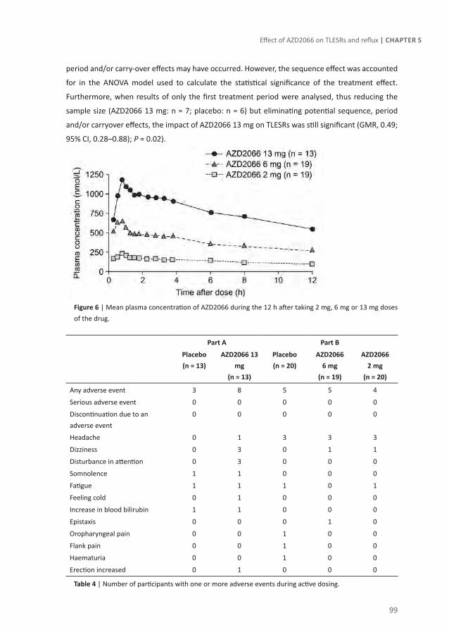
99
Effect of AZD2066 on TLESRs and reflux | Chapter 5
period and/or carry-over effects may have occurred. However, the sequence effect was accounted
for in the ANOVA model used to calculate the statistical significance of the treatment effect.
Furthermore, when results of only the first treatment period were analysed, thus reducing the
sample size (AZD2066 13 mg: n = 7; placebo: n = 6) but eliminating potential sequence, period
and/or carryover effects, the impact of AZD2066 13 mg on TLESRs was still significant (GMR, 0.49;
95% CI, 0.28–0.88); P = 0.02).
Figure 6 | Mean plasma concentration of AZD2066 during the 12 h after taking 2 mg, 6 mg or 13 mg doses of the drug.
Part A Part b
Placebo(n = 13)
AZd2066 13 mg
(n = 13)
Placebo (n = 20)
AZd2066 6 mg
(n = 19)
AZd2066 2 mg
(n = 20)
Any adverse event 3 8 5 5 4
Serious adverse event 0 0 0 0 0
Discontinuation due to an adverse event
0 0 0 0 0
Headache 0 1 3 3 3
Dizziness 0 3 0 1 1
Disturbance in attention 0 3 0 0 0
Somnolence 1 1 0 0 0
Fatigue 1 1 1 0 1
Feeling cold 0 1 0 0 0
Increase in blood bilirubin 1 1 0 0 0
Epistaxis 0 0 0 1 0
Oropharyngeal pain 0 0 1 0 0
Flank pain 0 0 1 0 0
Haematuria 0 0 1 0 0
Erection increased 0 1 0 0 0
Table 4 | Number of participants with one or more adverse events during active dosing.

100
Part I | Gastroesophageal reflux disease
Safety and tolerability assessments
The adverse events experienced during the study are reported in Table 2. All adverse events were
mild in intensity and reversible. No serious adverse events were reported during the active dosing
periods, and there were no discontinuations due to adverse events.
In part A of the study, eight of the 13 participants reported an adverse event when they received
AZD2066 13 mg compared with three when they received placebo. Most of the adverse events
reported when participants were receiving AZD2066 13 mg were related to the nervous system,
the most common being dizziness (3/13) and disturbance in attention (3/13). Only one nervous
system disorder (somnolence) was reported when participants were receiving placebo. In part B
of the study, the number of participants reporting adverse events was similar for AZD2066 2 mg
(5/20) and AZD2066 6 mg (4/19) compared with placebo (5/20). Headache was the most common
adverse event reported during dosing with AZD2066 2 mg and AZD2066 6 mg (3/20 and 3/19,
respectively), and the same number of headaches was reported when these participants were
taking placebo (3/20).
dIscussIon
In this randomized, double-blind, crossover study conducted in healthy volunteers, a single oral
13 mg dose of AZD2066 was found to reduce the numbers of TLESRs, reflux episodes and acid
reflux episodes significantly compared with placebo.
The overall frequency of TLESRs was greater during the 3-h postprandial period than in the
preprandial period, and differences in the number of TLESRs and reflux episodes between
AZD2066 and placebo were generally more pronounced during the postprandial period. These
observations are consistent with previous findings that TLESRs can be activated by food intake via
stretch receptors in the stomach.15
A dose dependent effect was seen with AZD2066: only the AZD2066 13 mg dose significantly
reduced the geometric mean number of TLESRs (27%) and geometric mean number of reflux
episodes (51%) compared with placebo, and there was a trend towards a greater effect for AZD2066
6 mg than for AZD2066 2 mg. A possible exception to the dose dependent effects of AZD2066 was
the counterintuitive observation of a significant reduction relative to placebo in mixed gas/liquid
reflux when participants received AZD2066 2 mg and AZD2066 6 mg, but not when they received
the higher AZD2066 13 mg dose. However, a similar reduction in the absolute number of mixed
gas/liquid reflux episodes relative to placebo was observed for AZD2066 13 mg and AZD2066 6

101
Effect of AZD2066 on TLESRs and reflux | Chapter 5
mg, both of which reduced mixed gas/liquid reflux to a greater extent that AZD2066 2 mg. The
lack of a statistically significant effect for AZD2066 13 mg in relation to mixed gas/liquid reflux may
have been because of the smaller sample size used in part A of the study (AZD2066 13 mg n = 13)
versus part B (AZD2066 2 mg: n = 19; AZD2066 6 mg: n = 16).
This is not the first study to show that pharmacological inhibition of TLESRs reduces the number
of reflux episodes in humans. Activation of inhibitory GABAB receptors using baclofen, a drug
indicated for spasticity, has been shown to reduce the number of TLESRs and reflux episodes in
healthy volunteers and in patients with GERD.26-28 In more recent studies, inhibition of TLESRs
using the novel GABAB agonist lesogaberan reduced the number of reflux episodes in healthy men
and in patients with symptomatic GERD despite PPI treatment.18, 19 The current study is, however,
the first to demonstrate that the reduced frequency of reflux episodes previously shown to be
associated with inhibition of mGluR5 in humans is related to the ability to reduce the number of
TLESRs.
Interestingly, AZD2066 13 mg reduced the geometric mean number of reflux episodes in this study
by 51%, despite a reduction in the geometric mean number of TLESRs of only 27%. Indeed, the
number of TLESRs temporally related to reflux episodes was reduced (although not significantly)
in participants receiving AZD2066 13 mg compared with those receiving placebo (43% vs 56%).
Lesogaberan has also been shown to reduce the number of reflux episodes to a greater extent
than the number of TLESRs. In the case of lesogaberan, this difference may be partially explained
by a concomitant increase in mean LES pressure, which has been shown to reduce reflux via a
mechanism that is unrelated to TLESRs.13 However, AZD2066 was not found to increase LES
pressure in the current study, and the mechanism by which AZD2066 reduces the number of
TLESRs generating reflux remains unclear. One possibility is that some TLESRs were not detected in
the current study. While this is consistent with the observation that only 65–80% of reflux episodes
were temporally related to TLESRs, it is difficult to envisage why a smaller proportion of TLESRs
would be detected in participants receiving AZD2066 13 mg than in those receiving placebo. It is
also possible that inhibition of mGluR5 may influence both the quantity and quality of TLESRs.
Thus, in the presence of AZD2066, LES relaxations may occur that meet the criteria for a TLESR but
do not generate reflux with sufficient properties to meet the definition of a reflux episode (i.e. a
drop in impedance to <50% of baseline). Other risk factors for acid reflux, such as the position of
the acid pocket,29 may also be affected by blocking mGluR5, although to our knowledge no studies
have investigated this.
Another interesting observation from this study was that acid reflux, and not weakly acidic or
weakly alkaline reflux, was significantly reduced in participants receiving AZD2066 compared with
those receiving placebo. A similarly disproportionate effect on acid reflux has been seen for both

102
Part I | Gastroesophageal reflux disease
lesogaberan and baclofen.18, 27 It has been postulated that this may be due to the greater effect
of these drugs on liquid and mixed liquid/gas reflux compared with pure gas reflux, the former
being more often associated with acid reflux.18 In this study, AZD2066 13 mg reduced liquid and
mixed liquid/gas reflux more than pure gas reflux. However, pure gas reflux made up a very small
proportion (~6%) of the total number of reflux episodes observed in this study. It therefore seems
unlikely that the inability of AZD2066 to inhibit pure gas reflux could account for its lack of effect
on weakly acidic reflux, which accounted for approximately one-third of the total number of reflux
episodes. There is some evidence to suggest that GABAB agonists may also alter the amount and
distribution of acid in the stomach30, 31.29 Given that GABAB and mGluR5 receptors are both involved
in regulating TLESRs, it is conceivable that mGluR5 may also help regulate digestive components
in the stomach, such as acid. It should also be noted that the proportion of weakly acidic reflux
episodes that were unrelated to TLESRs increased from 13.6% when patients received placebo to
38.9% when patients received AZD2066 13 mg. The lack of effect of AZD2066 13 mg on weakly
acidic reflux may thus be attributed to an increase in the proportion of these events that occur
independently of TLESRs, although the mechanism behind this is unclear.
In conclusion, the novel mGluR5 antagonist AZD2066 reduces the number of postprandial reflux
episodes that occur in healthy individuals, and this effect is correlated with a reduction in the
frequency of TLESRs. In addition, AZD2066 appeared to have an acceptable safety and tolerability
profile in this study. However, it should be noted that the GABAB-receptor agonist lesogaberan
was previously shown to reduce the geometric mean number of TLESRs in healthy volunteers
to a greater extent (36%) than AZD2066 did in the current study.19 Lesogaberan has since been
discontinued owing to a lack of evidence for a clinically significant effect as an add-on to PPI
therapy in patients with reflux symptoms despite PPI treatment. Nevertheless, the results of
the current study reinforce the importance of TLESRs in generating reflux and provide another
potential avenue for the development of novel therapeutic agents for GERD (ClinicalTrials.gov
Identifier: NCT00813306).

103
Effect of AZD2066 on TLESRs and reflux | Chapter 5
ReFeRences
1. Armstrong D. Systematic review: persistence and severity in gastroesophageal reflux disease. Aliment Pharmacol Ther 2008;28:841–53.
2. Vakil N, Veldhuyzen van Zanten S, Kahrilas P, et al. The Montreal definition and classification of gastro-esophageal reflux disease (GERD) – a global evidence-based consensus. Am J Gastroenterol 2006;101:1900–20.
3. Dent J, El-Serag HB, Wallander MA, et al. Epidemiology of gastro-oesophageal reflux disease: a systematic review. Gut 2005;54:710–17.
4. Wong BCY, Kinoshita Y. Systematic review on epidemiology of gastroesophageal reflux disease in Asia. Clin Gastroenterol Hepatol 2006;4:398–407.
5. Dent J. Review article: from 1906 to 2006 – a century of major evolution of understanding of gastro-oesophageal reflux disease. Aliment Pharmacol Ther 2006;24:1269–81.
6. van Pinxteren B, Numans ME, Bonis PA, et al. Short-term treatment with proton pump inhibitors, H2-receptor antagonists and prokinetics for gastro-oesophageal reflux disease-like symptoms and endoscopy negative reflux disease. Cochrane Database Syst Rev 2001:CD002095.
7. Armstrong D, Pare P, Pericak D, et al. Symptom relief in gastroesophageal reflux disease: a randomized, controlled comparison of pantoprazole and nizatidine in a mixed patient population with erosive esophagitis or endoscopy-negative reflux disease. Am J Gastroenterol 2001;96:2849–57.
8. Fass R. Proton-pump inhibitor therapy in patients with gastro-oesophageal reflux disease: putative mechanisms of failure. Drugs 2007;67:1521–30.
9. Hatlebakk JG, Hyggen A, Madsen PH, et al. Heartburn treatment in primary care: randomised, double blind study for 8 weeks. BMJ 1999;319:550–3.
10. Meineche-Schmidt V, Hauschildt Juhl H, Ostergaard JE, et al. Costs and efficacy of three different esomeprazole treatment strategies for long-term management of gastro-oesophageal reflux symptoms in primary care. Aliment Pharmacol Ther 2004;19:907–15.
11. El-Serag H, Becher A, Jones R. Systematic review: persistent reflux symptoms on proton pump inhibitor therapy in primary care and community studies. Aliment Pharmacol Ther 2010;32:720–37.
12. Dent J, Dodds WJ, Friedman RH, et al. Mechanism of gastroesophageal reflux in recumbent asymptomatic human subjects. J Clin Invest 1980;65:256–67.
13. Dodds WJ, Dent J, Hogan WJ, et al. Mechanisms of gastroesophageal reflux in patients with reflux esophagitis. N Engl J Med 1982;307:1547–52.
14. Boeckxstaens GE, Smout A. Systematic review: role of acid, weakly acidic and weakly alkaline reflux in gastroesophageal reflux disease. Aliment Pharmacol Ther 2010;32:334–43.
15. Penagini R, Carmagnola S, Cantu P, et al. Mechanoreceptors of the proximal stomach: Role in triggering transient lower esophageal sphincter relaxation. Gastroenterology 2004;126:49–56.
16. Blackshaw LA. Receptors and transmission in the brain-gut axis: potential for novel therapies. IV. GABA(B) receptors in the brain-gastroesophageal axis. Am J Physiol Gastrointest Liver Physiol 2001;281:G311–5.
17. Hirsch DP, Tytgat GN, Boeckxstaens GE. Transient lower oesophageal sphincter relaxations--a pharmacological target for gastro-oesophageal reflux disease? Aliment Pharmacol Ther 2002;16:17–26.
18. Boeckxstaens GE, Beaumont H, Mertens V, et al. Effects of lesogaberan on reflux and lower esophageal sphincter function in patients with gastroesophageal reflux disease. Gastroenterology 2010;139:409–17.
104
Part I | Gastroesophageal reflux disease
19. Boeckxstaens GE, Rydholm H, Lei A, et al. Effect of lesogaberan, a novel GABA(B)-receptor agonist, on transient lower esophageal sphincter relaxations in male subjects. Aliment Pharmacol Ther 2010;31:1208–17.
20. Young RL, Page AJ, O’Donnell TA, et al. Peripheral versus central modulation of gastric vagal pathways by metabotropic glutamate receptor 5. Am J Physiol Gastrointest Liver Physiol 2007;292:G501–11.
21. Frisby CL, Mattsson JP, Jensen JM, et al. Inhibition of transient lower esophageal sphincter relaxation and gastroesophageal reflux by metabotropic glutamate receptor ligands. Gastroenterology 2005;129:995–1004.
22. Jensen J, Lehmann A, Uvebrant A, et al. Transient lower esophageal sphincter relaxations in dogs are inhibited by a metabotropic glutamate receptor 5 antagonist. Eur J Pharmacol 2005;519:154–7.
23. Keywood C, Wakefield M, Tack J. A proof of concept study evaluating the effect of ADX10059, a metabotropic glutamate receptor-5 negative allosteric modulator, on acid exposure and symptoms in gastro-esophageal reflux disease. Gut 2009;58:1192–9.
24. Holloway RH, Penagini R, Ireland AC. Criteria for objective definition of transient lower esophageal sphincter relaxation. Am J Physiol 1995;268:G128–33.
25. Senn S. Cross-over Trials in Clinical Research Second Edition ed. Chichester: Wiley 2002.
26. Lidums I, Lehmann A, Checklin H, et al. Control of transient lower esophageal sphincter relaxations and reflux by the GABA(B) agonist baclofen in normal subjects. Gastroenterology 2000;118:7–13.
27. Vela MF, Tutuian R, Katz PO, et al. Baclofen decreases acid and non-acid post-prandial gastro-oesophageal reflux measured by combined multichannel intraluminal impedance and pH. Aliment Pharmacol Ther 2003;17:243–51.
28. Zhang Q, Lehmann A, Rigda R, et al. Control of transient lower oesophageal sphincter relaxations and reflux by the GABA(B) agonist baclofen in patients with gastro-oesophageal reflux disease. Gut 2002;50:19–24.
29. Beaumont H, Bennink RJ, de Jong J, et al. The position of the acid pocket as a major risk factor for acidic reflux in healthy subjects and patients with GORD. Gut 2010;59:441–51.
30. del Genio G, del Genio A, Brusciano L, et al. Laparoscopic cardioplasty to avoid esophageal resection in patient not responsive to Heller Myotomy. Annals of Thoracic Surgery 2007;83:2235–8.
31. Del Tacca M, Blandizzi C, Bernardini MC. Central GABAA excitatory and GABAB inhibitory receptors regulate gastric acid secretion in rats. Eur J Pharmacol 1990;177:189–94.


Chapter 6Proton pump inhibitors reduce the size and
acidity of the acid pocketWout Rohof , Roelof Bennink, Guy Boeckxstaens
Submitted
108
Part I | Gastroesophageal reflux disease
AbsTRAcT
Background and aims:
The gastric acid pocket is increasingly recognized as reservoir from which acid reflux events
originate. Its position is a major determinant of acid reflux during transient relaxations of the
lower esophageal sphincter. To what extent changes in position, size and acidity of the acid
pocket contribute to the therapeutic effect of proton pump inhibitors (PPIs) is unknown. The
aim of this study was therefore to determine the effect of PPI treatment on the acid pocket in
gastroesophageal reflux disease (GERD) patients.
Methods:
Thirty-six GERD patients (off PPI: n=18; on PPI: n=18, 19M 15F age 55 yrs) underwent concurrent
high resolution manometry (HRM) and pH-impedance following a standardized meal. The acid
pocket was visualized using scintigraphy after intravenous administration of 99mTc-pertechnetate.
The size of the acid pocket was measured and its position was determined relative to the diaphragm
using radionuclide markers on the HRM catheter. At the end of the study, the acid pocket was
aspirated to determine the pH.
Results:
The number of reflux episodes was comparable on and off PPI, but the rate of acid reflux episodes
was significantly reduced on PPI. On PPI, the acid pocket was smaller and more frequently located
below the diaphragm compared to off PPI. The pH of the acid pocket was significantly lower off
PPI (n=6) compared to on PPI (n=16) (0.9 [range 0.7 to 1.2] vs. 4.0 [range 1.6 - 5.9] p<0.001). The
pH of the acid pocket correlated significantly with the nadir pH of the refluxate (r=0.72, p<0.01)
Conclusion:
Also during PPI treatment, the acid pocket most likely represents the reservoir from which reflux
occurs, and that the effect of PPIs on the size, acidity and position of the acid pocket contributes
to the clinical success of PPIs in GERD.
109
The effect of PPIs on the acid pocket | Chapter 6
InTRoducTIon
Despite the buffering effect of the ingested food, most acid reflux episodes and associated
symptoms occur in the postprandial period. This phenomenon appears to be a paradox, but is
explained by the postprandial formation of a floating pool of acid on top of the ingested food.1
From this so-called acid pocket, highly acidic liquid can reflux into the esophagus during transient
lower esophageal sphincter relaxations or other episodes promoting gastroesophageal reflux,
both in patients with gastroesophageal reflux disease (GERD) and healthy subjects. Hence, the acid
pocket can be considered as the reservoir from which acidic fluid refluxes into the esophagus.1-3
We previously demonstrated that the position of the acid pocket largely depends on the presence
of a hiatus hernia and is a major determinant of the risk to have acid reflux. Notably, when the
acid pocket is located below the diaphragm or in the gastric fundus, only 11% of episodes is acidic,
while the majority of episodes is acidic when the pocket is located in the hiatus or even above
the diaphragm (62% and 91% respectively), underlining the importance of the acid pocket in the
pathophysiology of GERD.2,4
Proton pump inhibitors (PPIs) have been abundantly shown to reduce the number of acid reflux
episodes and esophageal acid exposure. Previously it was thought that the reduction in acid
production in the stomach by PPIs would lead to less reflux episodes. However, studies with pH-
impedance demonstrated a similar number of reflux episodes (non-acid + acid) on and off PPIs.5
On PPIs, acid reflux is only observed in 20% of reflux episodes, and most reflux episodes are weakly
acidic or alkaline. Currently, the effect of PPIs on the acid pocket is unknown. As PPIs reduce acid
secretion, the acidity and volume (and indirectly position) of the acid pocket may be altered,
potentially contributing to the reduction in acidic reflux events. More insight into the effect of PPIs
on the acid pocket could improve insight in persistent symptoms on PPIs, and thereby potentially
lead to better treatment options. Therefore, the aim of this study was to determine the effect of
PPIs on the size, position and acidity of the acid pocket. If the acid pocket is still present during PPI
therapy, this might represent a target for further therapy.
PATIenTs And MeThods
Patients
The study was performed in 36 patients with proven GERD, defined by the presence of esophagitis
observed during upper endoscopy and/or impedance-pH-metry with an acid exposure of >4.5%,
in combination with typical GERD symptoms.6 None of the patients had undergone previous
gastrointestinal surgery or was taking medication known to influence esophageal motor function.

110
Part I | Gastroesophageal reflux disease
The study was approved by the Medical Ethics Committee of the Academic Medical Center,
Amsterdam, the Netherlands. Written informed consent was obtained from all subjects before
enrolment in the study.
Study set-up and protocol
Eighteen patients on PPI were compared to 18 patients off PPI. In the latter group of patients, PPIs
were stopped at least 7 days before the study day. All subjects were studied after an overnight
fast. Reflux episodes were detected using concurrent HRM and pH-impedance monitoring, and
scintigraphy was performed to determine the size of the acid pocket and the position of the
pocket relative to the crural diaphragm. Thirty minutes prior to the start of these measurements,
350 MBq of 99mTc-pertechnetate was injected intravenously. Pertechnetate behaves as a chloride
ion, and is secreted by the parietal cells of the stomach.2,7,8 Using scintigraphy, the formation of
the acid pocket can be observed after the meal as pooling of 99mTc-pertechnetate in the proximal
stomach as validated previously.2
Figure 1 | Figure 1 represents a spatiotemporal plot of the esophagus, alongside the catheter set-up and a representative scintigraphic image. Two radionuclide markers are attached to the HRM catheter (marked by yellow dots), which can be observed on the scintigraphic image (II), marked by white arrows. The spatiotemporal plot of the HRM and pH-impedance recording (I) shows a mixed reflux episode during a TLESR in a patient with a large hiatal hernia. The LES is marked with A, and the upper and lower borders of the crural diaphragm are marked with B and C respectively. The distances from A, B and C to the upper and lower marker are measured. In addition, the distance from the markers to acid pocket is measured in the scintigraphic image. Using these distances, the position of the acid pocket is classified as below the diaphragm, at the level of the diaphragm or above the diaphragm. In this example, the acid pocket is located below the diaphragm. The pH of the refluxate is weakly acidic (pH5.8).
111
The effect of PPIs on the acid pocket | Chapter 6
To determine the exact location of the high resolution manometry (HRM) catheter during
scintigraphy, 2 sealed markers impregnated with topical 99mTc-pertechnetate were attached to the
catheter before introduction, one to the distal end of the catheter and one between sensor 11
and 12. These markers were used to integrate manometry and scintigraphy as described in detail
below. The HRM catheter and the pH-impedance catheter were inserted through an anaesthetized
nostril. The HRM catheter was positioned with the most distal sensors located in the stomach,
and the pH-sensor of the pH-impedance catheter was positioned 5 centimetres above the upper
border of the LES. (Figure 1)
Patients were positioned in the upright position in front of the scintigraphy camera. After a
baseline fasting recording of 5 minutes, patients consumed a standardized meal consisting of 200
ml orange juice and two pancakes with jam (510 kcal). After the meal, concurrent scintigraphic,
HRM and pH-impedance recordings were acquired for 105 minutes. At the end of the protocol, the
acid pocket was aspirated through the 0.9 mm working channel of the HRM catheter. The marker
at the distal tip of HRM catheter was used to position the opening of the working channel at the
level of the acid pocket and to accurately aspirate the fluid of the pocket. The pH of the fluid was
measured before storage.
Recording methods
High resolution manometry was performed using a 21 lumen water perfused HRM catheter (MMS,
Enschede, the Netherlands) with a diameter of 4.0 mm. Eleven distal side holes were positioned at
1 cm intervals, and the 10 proximal side holes were spaced at 3 cm intervals as depicted in Figure
1. The side holes were perfused with distilled water at 0.15 mL⁄min, using a pneumohydraulic
capillary perfusion pump (MMS, Enschede, the Netherlands) and hydraulic flow restrictors. The
working channel had a diameter of 0.9 mm with the opening located at the distal tip of the
catheter. Pressure sensors were zeroed before insertion and data was collected and analyzed with
a MMS Solar system (MMS, Enschede, the Netherlands).
We used a combined pH-impedance catheter (Unisensor, Attikon, Switzerland), containing 6 pairs
of impedance electrodes and 1 ISFET pH sensor that allowed impedance recordings at 3, 5, 7, 9,
15 and 17 cm above the upper border of the LES and pH recording at 5 cm above the LES. The pH
electrode was calibrated before each study using buffers solutions of pH 4.0 and 7.0 (Medtronic
A/S, Skovlunde, Denmark).
Dynamic scintigraphic images were acquired on a gamma camera system (Diacam; Siemens
Medical Solutions, Illinois, USA), equipped with a low-energy all purpose collimator. Dynamic
recordings were made for 2 hours (720 views, 10 s/view, 120 min total acquisition time). Every
acquisition was processed on a Hermes processing station (Hermes Medical Solutions, Stockholm,
Sweden) for further analysis.
112
Part I | Gastroesophageal reflux disease
Data analysis
Scintigraphic images were analysed using calibrated Hermes software. The size and nuclear counts
of the acid pocket were determined by drawing an area of interest (ROI) over the acid pocket in
the proximal stomach in images taken at 10 minutes intervals. The acid pocket could be visualized
starting from 15 minutes postprandially. Background activity measured in an area of equal size
adjacent to the stomach was deducted from activity counts.
Each liquid or mixed reflux event detected by impedance was classified as acidic reflux when
pH < 4, as weakly acidic when pH ≥ 4 and pH < 7 and as non-acidic when pH ≥ 7. Impedance
segments were used to determine proximal extent of refluxate. A liquid reflux was defined as a fall
in impedance of ≥ 40% of baseline impedance starting at the most distal segment and propagating
retrograde to at least the next measuring segment. Mixed reflux was defined as gas reflux occurring
during or immediately before liquid reflux.
For each reflux episode, HRM recordings were used to determine the exact distance from the
crural diaphragm to the markers on the catheter for the position of the acid pocket and to the
LES for hiatal hernia size. The lower and upper border of the crural diaphragm and the middle of
the LOS zone were determined. (Figure 1.) Hiatal hernia size was defined as the distance from the
lower end of the diaphragm to the middle of the LOS.(9)
For each reflux event, the position of the acid pocket was determined relative to the diaphragm.
For this purpose, HRM recordings were used to determine the exact position of the crural
diaphragm and LES, relative to the markers on the catheter. (Figure 1.) Subsequently, calibrated
Hermes software was used to measure the distance of the proximal end of the gastric acid pocket
to the markers on the catheters on one image prior to the reflux event to calculate the distance
between the acid pocket and the crural diaphragm. Measurements of the distances between the
acid pocket and the diaphragm were made through a straight line in a planar posterior view of the
stomach. On scinitigraphy, the pocket was identified as a clear pool of radiolabelled activity in the
proximal stomach. With these distances, the exact location of the acid pocket relative to the crural
diaphragm could be calculated. The position of the acid pocket was classified in three categories:
below the diaphragm, at the level of the diaphragm or above the level of the diaphragm.
Statistical analysis
Statistical analysis was performed using SPSS 19.0 (IBM Corporation, Somers, NY, United States).
Data are presented as mean ± SEM when parametric or median [IQR] when non-parametric.
Parametric data were compared using a Student’s t-test, and non-parametric data using a Mann-
Whitney U test. Comparison of proportions was performed using Fisher’s exact testing. Pearson’s

113
The effect of PPIs on the acid pocket | Chapter 6
correlation was used for correlations. All p-values were two-tailed and a p-value < 0.05 was
considered as statistically significant.
ResulTs
Patients
All 36 patients completed the study protocol. Eight patients on PPI used omeprazole, 5 used
pantoprazole and 5 used esomeprazole. Dosage varied from 20 mg (1 patient), 40 mg (7 patients)
to 40 mg bid (8 patients). In the group of patients that discontinued PPI use for 1 week, 7 patients
normally used bid PPI. Patient characteristics of the two patient groups are shown in Table 1. No
difference was observed in age and sex in the two patient groups. In addition, hiatal hernia size as
measured using HRM recordings was similar.
off PPI (n=18) on PPI (n=18) P-value
Age (yrs) 55 ± 1.4 55 ± 4.0 0.93
SexMaleFemale
135
711
0.09
Hiatal hernia size (cm) 2.7 ± 0.2 2.4 ± 0.4 0.34
PPI bid 7 8
Persistent symptoms (SAP >95%) 8 9
Table 1 | Patient characteristics
off PPI (n=18) on PPI (n=18) P
Postprandial reflux events (nr) 14 ± 2.1 15 ± 2.6 0.81
Acid reflux events (nr) 4.5 [2.0-14] 2.0 [0-3.0] <0.05
Acid reflux events (%) 51 ± 7.9 21 ± 5.9 <0.01
Weakly acid reflux (nr) 5.0 [3.0-6.5] 9.0 [7.0-18] <0.01
Weakly acid reflux (%) 49 ± 7.9 79 ± 5.9 <0.01
Esophageal acid exposure (%) 11 ± 3.5 2.2 ± 1.0 <0.05
Proximal extent (cm) 10 ± 0.6 9.9 ± 0.5 0.80
Table 2
Reflux episodes
The total postprandial number of reflux episodes was similar in patients on PPI compared to
those off PPI (15 ± 2.6 vs. 14 ± 2.1, p=0.81, Table 2.). As expected, the number of acid reflux
episodes was reduced on PPI (4.5 [2.0-14] vs. 2.0 [0-3.0], p=0.04), while the number of weakly acid
reflux episodes was increased on PPI (5.0 [3.0-6.5] vs. 9.0 [7.0-18], p<0.01). In line, PPI treatment
114
Part I | Gastroesophageal reflux disease
decreased esophageal acid exposure (11 ± 3.5% vs. 2.2 ± 1.0%, p=0.02), and significantly increased
nadir pH of refluxate (1.2 [1.0-3.5] vs. 3.2 [2.8-4.1], p=0.02). No difference in proximal extent of
refluxate was observed between on and off PPI (10 ± 0.6 cm vs. 9.9 ± 0.5 cm, p=0.80).
The effect of PPIs on the acid pocket
All patients off PPI showed an acid pocket, compared to 16 out of 18 patients on PPI. (p=0.49) In 2
patients on PPI, no pooling of 99mTc-pertechnetate in the proximal stomach was observed. Proton
pump inhibitors significantly reduced the size of the acid pocket (off PPI: 15 ± 0.9 cm² vs. on PPI:
10 ± 1.3 cm², p<0.01, Figure 2.). In addition, the mean number of counts in the acid pocket was
significantly lower on PPI, suggesting that the secretion of 99mTc-pertechnetate into the pocket was
reduced by PPIs (2762 ± 265 vs. 1566 ± 210 counts/10s, 43%, p<0.001, Figure 2). The acid pocket
size significantly correlated with the number of counts, demonstrating that the reduction in 99mTc-
pertechnetate secretion by PPI treatment leads to a smaller acid pocket (r=0.76, p<0.001).
At the end of the study, a clear acid pocket was visualised scintigraphically in the proximal stomach
in all patients off PPI and in 16 of 18 patients on PPI. To confirm that this scintigraphic activity
indeed resulted from radiolabelled acid pooled on top of the meal, the radioactive material was
aspirated through the working channel (mean 60 mL of transparent mucoid fluid). Scintigraphic
activity in the proximal stomach disappeared after aspiration in both patient groups. The pH of
the aspirated acid pocket fluid was significantly higher during PPI treatment (0.9 [0.8-1.1] vs. 3.9
[2.8-5.1], p<0.001, Figure 3a).
Acid pocket as a source of refluxate
To support the assumption that the acid pocket is de reservoir from which reflux occurs, we
determined the relationship between the pH of the acid pocket and the pH of the reflux episodes.
Interestingly, the pH of the aspirate was strongly and significantly correlated with the pH of
the reflux events (r=0.72, p<0.01) (Figure 3.) Especially when the pocket is located above the
diaphragm, the relation between the pH of the pocket and refluxate is excellent (r=0.88, p<0.001).
In line, we found a strong inverse correlation between the acid pocket pH and the rate of acid reflux
episodes (r=-0.79, p<0.01) and esophageal acid exposure time (r=-0.72, p<0.01), demonstrating
that an increase in acid pocket pH leads to a reduction in the rate of acid reflux and esophageal
acid exposure.
115
The effect of PPIs on the acid pocket | Chapter 6
Figure 2 | In Figure 2A and B the size and nuclear activity of the acid pocket are depicted off and on PPI. As demonstrated in Figure 2A, the size of the acid pocket is significantly smaller on PPI compared to off PPI during the entire postprandial period (area under the curve p<.01). In addition, the number of counts in the area of interest is significantly lower on PPI compared to off PPI (p<.01). This demonstrates that less gastric secretion is present in the acid pocket during treatment with PPI. Tested using a Student’s t-test on area under the curve values.
Figure 3 | As demonstrated in Figure 3A, the nadir pH of reflux episodes was significantly increased by PPIs. Proton pump inhibitors also significantly increased the pH of the acid pocket (3B). As demonstrated in figure 3C, the pH of the acid pocket had a good correlation with the nadir pH of refluxate (r=0.72).
As a more proximal position of the acid pocket is an important risk factor for acid reflux, we
analyzed the effect of PPI treatment on the position of the acid pocket prior to reflux episodes.
Compared to off PPIs, the pocket was more often located below the diaphragm in patients on PPIs
(40±6.5% vs. 60±6.9% of reflux episodes, p=0.04) (Figure 4). When the acid pocket was located
below the diaphragm, the risk for acidic reflux was only 15 ± 5.5% off PPI and 7.0 ± 3.0% on PPI
(p=0.20). When the acid pocket was located at the level of the diaphragm, so in the hiatus, 76
± 6.7% of reflux events off PPI was acidic, whereas 46 ± 12% of reflux episodes was acidic on
PPI (p=0.11). Finally, when the acid pocket was located above the diaphragm, the refluxate was
acidic in 95 ± 3.9% of reflux episodes off PPI, compared to 49 ± 7.5% of reflux episodes on PPI
(p<0.01). (Figure 4) The reduced rate of acid reflux episodes when the pocket is located above the
diaphragm is probably explained by the increased pH of the acid pocket on PPIs. This is supported
Acid pocket size
30 40 50 60 70 80 90 100
110
120
0
5
10
15
20
On PPIOff PPI
Time in minutes
p<.01
Size
in c
m2
Nuclear acitivity
30 40 50 60 70 80 90 100
110
120
0
1000
2000
3000
4000
On PPIOff PPI
Time in minutes
p<.01
Cou
nts
A B
Nadir pH of reflux episode
Off PPI On PPI0
1
2
3
4
5
6
7 p<.05
pH
Acid pocket pH
Off PPI On PPI01234567 p<.01
n=6 n=16
pH
Correlation of pocketand refluxate
0 1 2 3 4 5 6 70
1
2
3
4
5
6
7
On PPIOff PPI
r=.72, p<.01
Nadir refluxate pH
Acid
poc
ket p
H
A CB

116
Part I | Gastroesophageal reflux disease
by the excellent correlation (r=0.88) of the pH of the pocket and refluxate when the pocket is
located above the diaphragm. In contrast, no correlation of the pH of the acid pocket and refluxate
is found when the pocket is located below the diaphragm (r=0.41 p=0.08)
We hypothesized that the smaller size of the pocket on PPIs may contribute to a more distal
position. Indeed, the rate of acid pocket located below the diaphragms correlated with the count
number and size of the acid pocket (r=0.47, P<0.01). Subsequently we analysed whether changes
in hiatal hernia size contributed to the more distal position on PPI. However, hiatal hernia size
was similar on and off PPI therapy (2.7 ± 0.2 cm 2.4 ± 0.4 cm, p=0.34). In addition, we found no
difference in spatial separation rate of the hiatus hernia (70 ±7.0% vs. 56±10%, p=0.20)
Figure 4 | The position of the acid pocket is an important determinant in the acidity of the refluxate. On PPI the acid pocket is significantly more often located below the diaphragm compared to off PPI (*<0.05, Student’s t-test). As demonstrated in the right image, a more proximal position of the acid pocket leads to a higher acid reflux rate, also on PPI. However, the risk for acid reflux is significantly lower on PPI compared to off PPI when the pocket is located above the diaphragm. This is probably attributable to the higher pH (>4 in 50%) of the acid pocket on PPI.
Acid reflux episodes on PPI
Acid reflux episodes still occurred during PPI treatment. From earlier studies, it is known that
acid reflux more often leads to heartburn compared to non-acid reflux episodes. Therefore we
compared the acidity, size and position of the acid pocket in acid reflux episodes to non-acid reflux
episodes (pH>4). For this analysis, only patients on PPI were analysed. Notably, we found that
esophageal acid exposure time is 4.0 ± 2.0 % when the acid pocket was acidic, compared to 0.2 ±
0.4% when the pocket was not acidic. Due to the low number of patients (8 vs. 8), this difference
was not significant (p=0.17). Also during PPI use, acidic reflux episodes occurred primarily when
the pocket was located above (17 of 35 reflux episodes [49%]) or at the level of the diaphragm (35
of 74 [46%]) compared to when the pocket was below the diaphragm (10 of 134 [7%], Figure 4).
Position of the acid pocket
Off PPI
On PPI0
20
40
60
80
100
Below diaphragmAt the level of diaphragmAbove diaphragm
*
% o
f ref
lux
epis
odes
Acid reflux and acid pocket position
Below th
e diap
hragm
At the l
evel
of the d
iaphrag
m
Above th
e diap
hragm
0
20
40
60
80
100Off PPIOn PPI
*
Acid
ref
lux
rate
(%)

117
The effect of PPIs on the acid pocket | Chapter 6
These data suggest that the risk for acid reflux is mainly determined by the pH of the pocket. When
the pocket is acidic, acid reflux mainly occurs when the pocket is located at the level or above the
diaphragm, i.e. when the acid pocket is the source of refluxate.
dIscussIon
In the present study, we demonstrated that also during PPI treatment gastric secretions
accumulate on top of the ingested food and form a pocket. Proton pump inhibitors do not alter
the number of reflux events, but increase the pH of the acid pocket which in its turn leads to the
increased pH of reflux events. When the pocket is not acidic, the risk for acidic reflux events is
negligible, irrespective of its position. When the pocket is acidic, the position of the pocket is a
major determinant of acid reflux during transient relaxations of the lower esophageal sphincter,
also during acid suppression. Taken together, these data demonstrate that also during PPI use, the
acid pocket can act as a reservoir from which liquid refluxes into the esophagus, and that mainly
the change in the pH of the acid pocket leads to the reduction in acid reflux events.
After meal intake, the acid pocket escapes the buffering effect of the ingested food and floats in
the proximal stomach. There, this pocket functions as a reservoir from which acid refluxes into
the oesophagus. If the pocket is located above the diaphragm or in the hiatus, acid reflux is highly
likely to occur (70-90%), in contrast to when the pocket is located below the diaphragm (10%).2
Moreover, it was demonstrated that modulation of the acid pocket to a more distal position leads
to less acidic reflux events, confirming that the acid pocket is a potential therapeutic target in
the treatment of GERD.4 Not only H+ ions are taken up and secreted into the stomach by parietal
cells, but also 99mTc-pertechnetate when injected intravenously.8 As a result, the acid pocket
can be visualized using 99mTc-pertechnetate on scintigraphy.8,11 We previously validated that
postprandial pooling of 99mTc-pertechnetate in the gastric cardia indeed represents the gastric acid
pocket.2 Moreover, aspiration from this pocket revealed highly acidic fluid and nuclear activity
scintigraphically disappeared after aspiration.2 Hence, this technique is ideally suited to visualize
the acid pocket, and determine the effect of treatment on the pocket. In the current study, we
determined how PPIs affect properties of the acid pocket and determined whether this contributes
to the therapeutic effect of these drugs.
Interestingly, we demonstrated for the first time that also during acid suppression, a postprandial
‘acid’ pocket is formed in the large majority of GERD patients (89%). This might be explained
by incomplete blocking of acid secretion by PPIs. As demonstrated in early in vitro studies with
omeprazole, PPIs do not fully suppress acid production in parietal cells.12,13 Moreover, in vivo, acid
118
Part I | Gastroesophageal reflux disease
inhibition is never complete because of continued synthesis of new proton pumps.14 Ongoing acid
secretion into the pocket is supported by a pH of the pocket below 4 in 50% of our patients on
PPI. On the other hand, the acid pocket not only consists of acid secreted by the parietal cell. It
is known that 99mTc-pertechnetate is also secreted by mucoid cells. For instance, in patients with
severe atrophic gastritis with achlorhydria, thus in absence of parietal cells, 99mTc-pertechnetate
is still secreted into the gastric lumen.15 Comparably, in GERD patients with suppression of acid
secretion, the observed formation of a large postprandial pocket with a pH of 4-6 is therefore
probably also in part attributable to secretion by mucoid cells. Either way, based on the findings
from this study we can conclude that also during acid suppression, a pocket of gastric secretions
floats on top of the ingested food.
Although the acid pocket was still present, PPIs significantly influenced its properties. The most
important effect of PPIs on the pocket is the reduction of its acidity. Aspiration of the pocket on
PPIs revealed fluid with a pH of 4.0 (range 1.6-5.9), compared to highly acidic fluid with a pH of
0.9 (range 0.7-1.2) off PPI. Importantly, the pH of the acid pocket correlated well with the nadir pH
of refluxate (r=0.72) and inversely with the rate of acid reflux (r=-0.78). Moreover, when the pH
of the aspirated pocket was over 4, the esophageal acid exposure was very low (0.2%). Especially
when the pocket is located above the diaphragm, the correlation between the pH of the pocket
and refluxate is excellent (r=0.88, p<0.001). Taken together, this confirms that PPIs significantly
increase the pH of the acid pocket, which largely determines the pH of the refluxate.
Previously, we demonstrated that if the pocket is located in the hiatus or in the hiatal sac, the
pocket functions as a reservoir from which acid refluxes into the oesophagus.2,4 In the current
study we showed that also on PPI, significantly more acid reflux episodes occur when the acid
pocket is located at the level or above the diaphragm compared to below the diaphragm (46% and
49% compared to 7% respectively). This demonstrates that also on PPI a more proximal position
of the acid pocket is associated with acid reflux events. Interestingly, on PPIs, the pocket was
more often located below the diaphragm compared to off PPIs (60% vs. 40%). The difference in
acid pocket position cannot be explained by altered gastric motility, as PPIs have no impact on
upper gastrointestinal motility.16 Furthermore, in the current study we did not find an effect from
PPIs on hiatal hernia size. In contrast, the effect of PPIs on the position may be explained by the
effect on acid pocket size. As previously demonstrated, off PPIs, the acid pocket in GERD patients
is larger compared to controls.3 Hence, the pocket extends very close to, or even across, the
squamocolumnar junction in GERD patients off.2,3 As measured by scintigraphy, PPIs reduced the
nuclear counts and thus the secretion of 99mTc-pertechnetate into the acid pocket by 43%, leading
to a reduction in size of 33%. As a smaller acid pocket is associated with a more distal position, we
argue that acid suppression results in a smaller and more distally located acid pocket, which in its
turn leads to less acid reflux events.

119
The effect of PPIs on the acid pocket | Chapter 6
In approximately 30% of patients PPI-therapy fails to resolve symptoms, either partially or
completely. As demonstrated by Zerbib et al, remaining heartburn during PPI use is more often
caused by acid reflux than by weakly acid reflux. In addition, Karamanolis et al demonstrated that
16% and 32% of the symptomatic subjects on double dose and standard dose PPIs, respectively,
have abnormal pH tests.17 If one accepts that the acid pocket is still the source of the refluxate
for acid reflux during PPI use, therapeutic strategies directly intervening with the pH of the acid
pocket may possibly prove effective in preventing persistent heartburn on PPI. However, in a
recent systematic review handling 6 studies with in total 174 GERD patients with persistent reflux
symptoms during PPI use, it was demonstrated that more than 80% of reflux-related symptoms
were attributable to weakly acid reflux. Only a minority of symptoms is caused by acid reflux
(17%). Therefore, obviously, a strategy to prevent all sorts of reflux episodes (acid and non-acid)
such as fundoplication or possibly reflux inhibitors would prove a better solution for reflux-related
symptoms, irrespective whether reflux originates from the acid pocket.
A possible shortcoming of this study is that this study did not have a cross-over design. However,
due to the radiation burden, a cross-over design was not approved by the ethics committee. To
compensate, a larger number of GERD patients was included compared to previous studies of
similar design.2,4 Furthermore, baseline characteristics such as hiatal hernia size, PPI dosage and
number of patients with persistent symptoms were matched. Therefore we are confident that our
study was adequately powered and patient groups were comparable.
In conclusion, the effect of PPIs of the acid pocket largely contributes to their effectiveness in the
treatment of GERD. During acid suppression the pocket still acts as a reservoir for liquid reflux. As
the pH of the acid pocket is increased, the pH of the refluxate is as well. In patients with ongoing
acid reflux on PPI, the acid pocket represents a potential target for additional therapy.
120
Part I | Gastroesophageal reflux disease
ReFeRence lIsT
1. Fletcher J, Wirz A, Young J et al. Unbuffered highly acidic gastric juice exists at the gastroesophageal junction after a meal. Gastroenterology 2001;121(4):775-83.
2. Beaumont H, Bennink RJ, de JJ et al. The position of the acid pocket as a major risk factor for acidic reflux in healthy subjects and patients with GORD. Gut 2010;59(4):441-51.
3. Clarke AT, Wirz AA, Manning JJ et al. Severe reflux disease is associated with an enlarged unbuffered proximal gastric acid pocket. Gut 2008;57(3):292-7.
4. Rohof WO, Bennink RJ, de Ruigh AA et al. Effect of azithromycin on acid reflux, hiatus hernia and proximal acid pocket in the postprandial period. Gut 2012.
5. Vela MF, Camacho-Lobato L, Srinivasan R et al. Simultaneous intraesophageal impedance and pH measurement of acid and nonacid gastroesophageal reflux: effect of omeprazole. Gastroenterology 2001;120(7):1599-606.
6. Kahrilas PJ, Quigley EM. Clinical esophageal pH recording: a technical review for practice guideline development. Gastroenterology 1996;110(6):1982-96.
7. O’Connor MK, O’Connell R, Keane FB et al. The relationship between technetium 99m pertechnetate gastric scanning and gastric contents. Br J Radiol 1983;56(671):817-22.
8. Kuiken S, Van Den Elzen B, Tytgat G et al. Evidence for pooling of gastric secretions in the proximal stomach in humans using single photon computed tomography. Gastroenterology 2002;123(6):2157-8.
9. Rohof WO, Bennink RJ, de Ruigh AA et al. Effect of azithromycin on acid reflux, hiatus hernia and proximal acid pocket in the postprandial period. Gut 2012.
10. Bickel JG, Witten TA, Killian MK. Use of pertechnetate clearance in the study of gastric physiology. Gastroenterology 1972;63(1):60-6.
11. Taylor TV, Bone D, Torrance B. A non-invasive test of gastric function. Br J Surg 1977;64(10):702-8.
12. Fellenius E, Berglindh T, Sachs G et al. Substituted benzimidazoles inhibit gastric acid secretion by blocking (H+ + K+)ATPase. Nature 1981;290(5802):159-61.
13. Kittang E, Aadland E, Schjonsby H. Effect of omeprazole on the secretion of intrinsic factor, gastric acid and pepsin in man. Gut 1985;26(6):594-8.
14. Massoomi F, Savage J, Destache CJ. Omeprazole: a comprehensive review. Pharmacotherapy 1993;13(1):46-59.
15. Chaudhuri TK. Can 99mTc-pertechnetate be used to assess the secretion of gastric acid in pernicious anemia? J Nucl Med 1977;18(2):121-2.
16. Michalek W, Semler JR, Kuo B. Impact of acid suppression on upper gastrointestinal pH and motility. Dig Dis Sci 2011;56(6):1735-42.
17. Karamanolis G, Vanuytsel T, Sifrim D et al. Yield of 24-hour esophageal pH and bilitec monitoring in patients with persisting symptoms on PPI therapy. Dig Dis Sci 2008;53(9):2387-93.
18. McColl KE, Clarke A, Seenan J. Acid pocket, hiatus hernia and acid reflux. Gut 2010;59(4):430-1.


Chapter 7Effect of Azithromycin on acid reflux, hiatus
hernia and proximal acid pocket in the postprandial period
Wout Rohof, Roel Bennink , Annemijn de Ruigh, David Hirsch, Aeilko Zwinderman, Guy Boeckxstaens
Gut. 2012 Dec;61(12):1670-7
124
Part I | Gastroesophageal reflux disease
AbsTRAcT
Background and Aims:
The risk for acidic reflux is mainly determined by the position of the gastric acid pocket. We
hypothesized that compounds affecting proximal stomach tone might reduce gastro-oesophageal
reflux by changing the acid pocket position. Therefore, we studied the effect of Azithromycin (Azi)
on acid pocket position and acid exposure in patients with gastro-oesophageal reflux disease
(GERD).
Methods:
Nineteen patients with GERD were included of whom 7 had a large hiatal hernia (≥3cm)(L-HH) and
12 had a small or no hiatal hernia (S-HH). Patients were randomized to Azi 250 mg/day or placebo
during 3 days in a cross-over manner. On each study day, reflux episodes were detected using
concurrent HRM and pH-impedance monitoring after a standardized meal. The acid pocket was
visualized using scintigraphy, and its position was determined relative to the diaphragm.
Results:
Azi reduced the number of acid reflux events (placebo 8.0±2.2 vs Azi 5.6±1.8, p<0.01) and
postprandial acid exposure (placebo 10.5±3.8% vs Azi 5.9±2.5%, p<0.05), in all patients without
affecting the total number of reflux episodes. Acid reflux occurred mainly when the acid pocket
was located above or at the level of the diaphragm, in contrast to below the diaphragm. Treatment
with Azi reduced hiatal hernia size and resulted in a more distal position of the acid pocket
compared to placebo (below the diaphragm 39% vs 29%, p=0.03). Azi reduced the rate of acid
reflux episodes in S-HH patients (38% to 17%) to a larger extent than in L-HH patient (69% to 62%,
p=0.04).
Conclusion:
Azi leads to a reduction in acid reflux episodes and esophageal acid exposure. This effect was
associated with a smaller hiatal hernia size and a more distal position of the acid pocket, further
indicating the importance of the acid pocket in the pathogenesis of GERD.
125
The effect of Azithromycin on the acid pocket | Chapter 7
InTRoducTIon
Gastroesophageal reflux is a physiological phenomenon in which gastric contents flow back
into the esophagus. When reflux causes symptoms such as heartburn or regurgitation and/ or
esophageal damage, it is referred to as gastro esophageal reflux disease (GERD), a very common
chronic condition affecting 10-20% of the adult population.1,2
Most reflux episodes occur after a meal, when the stomach is filled with ingested food. In contrast
to the believe that meal ingestion buffers gastric acid, acid reflux episodes occur even in the early
postprandial period.3 Fletcher et al elegantly showed that gastric acid floats on top of the meal
acting as a reservoir from which acid can enter the esophagus during episodes of opening of the
esophagogastric junction. This unbuffered pool of gastric acid is referred to as the gastric acid
pocket.4
The position of the acid pocket relative to the crural diaphragm is an important determinant of
the acidity of the refluxate. We previously showed that 74-85% of reflux episodes is acidic when
the acid pocket is located above or at the level of the diaphragm. In contrast, only 7-20% of reflux
episodes is acidic if the acid pocket is located below the diaphragm.3 The position of the pocket
relative to diaphragm on the other hand is largely determined by presence of a hiatal hernia.
Especially in patients with a large HH, the acid pocket is frequently located above the diaphragm,
facilitating the occurrence of acid reflux events.3,5 This insight implies that drugs affecting the
position of the acid pocket (hiatal hernia) may alter acid exposure.
Prokinetic agents like macrolides increase gastric emptying and in addition increase proximal
stomach tone and LES pressure, presumably via a cholinergic pathway mediated by motilin
receptors or possibly by serotonin receptors.6 These properties make these compounds interesting
candidates to alter the acid pocket position. Recently, Mertens et al. indeed reported that the
macrolide Azithromycin (Azi), a macrolide similar in structure and function to erythromycin7,
reduced the rate of reflux episodes in lung transplant patients.8 Interestingly, Azi mainly affected
the rate of acidic reflux leaving the number of weakly acid reflux events unchanged. As the acidity
of the refluxate is mainly determined by the position of the acid pocket, we hypothesized that this
action of Azi was mediated by a reduction in hiatal hernia size thereby leading to an alteration
in the position of the acid pocket. To evaluate this hypothesis, we examined the effect of Azi on
gastroesophageal reflux and the acid pocket position in patients with GERD,

126
Part I | Gastroesophageal reflux disease
PATIenTs And MeThods
Patients
The study was performed in 19 patients with proven GERD, defined by the presence of esophagitis
observed during upper endoscopy and/or pH-metry with an acid exposure of > 4.5%, in combination
with typical GERD symptoms.9 None of the patients had undergone previous gastrointestinal
surgery or was taking medication known to influence esophageal motor function. Hiatal hernia
size was measured by high resolution manometry (HRM), and patients were divided in large HH
(≥3cm) or small or no HH (<3cm) according to the size of the hiatal hernia. One patient withdrew
consent after the first study day due to discomfort. The study was approved by the Medical Ethics
Committee of the Academic Medical Center. Written informed consent was obtained from all
subjects before enrolment in the study.
Study design
The study had a randomized double blind, crossover design. Patients were randomised to Azi 250
mg or Placebo by the pharmacy where the code was stored until the end of the study. Both the
investigators and the patients were blinded to the treatment. Study medication was taken during a
three day course ending on the study day, with comparable dosing of Azi used in the earlier study
by Mertens et al.8 Acid suppressive medication was stopped at least 7 days before each study day.
The study day protocol was repeated after a minimal wash-out period of two weeks.
Study day protocol
Subjects were studied after an overnight fast of at least 12 h. Reflux episodes were detected using
concurrent HRM and pH-impedance monitoring, and scintigraphy was performed to localize the
acid pocket relative to the crural diaphragm.
At 30 minutes before insertion of the catheters, 350 MBq 99mTc-pertechnetate was injected
intravenously. Pertechnetate behaves as a chloride ion, and is secreted by the parietal cells of the
stomach.10 Using this technique, acid distribution in the stomach can be visualized scintigraphically
as validated previously.3
Before introduction of the HRM catheter, two sealed markers impregnated with 99mTc-pertechnetate
were attached to the catheter, one to the distal end of the catheter and one between sensor 13
and 14 to visualize the exact location of the catheter during scintigraphy. These markers were used
to integrate manometry and scintigraphy as described below in more detail.
The HRM catheter and the pH-impedance catheter were inserted transnasally. The HRM catheter
was positioned with the most distal 5 sensors positioned in the stomach. The pH-sensor of the pH/

127
The effect of Azithromycin on the acid pocket | Chapter 7
impedance catheter was placed 5 centimetres above the upper border of the LES. Subsequently,
the patients were positioned in upright position in front of the scintigraphy camera. First, a baseline
fasting recording was obtained during 5 minutes. Then subjects consumed a standardized meal in
10 minutes consisting of 200 ml orange juice and two pancakes with jam (510 kcal). After the meal
scintigraphic, HRM and pH-impedance recordings were performed for 105 minutes.
Recording methods
High resolution manometry was performed using a solid state high-resolution manometry
catheter (Unisensor, Attikon, Switzerland) with 36 solid state pressure transducers spaced 1 cm
apart. Pressure sensors were zeroed before insertion. The sample rate was 50 Hz and data was
collected and analyzed with the MMS Solar system (MMS, Enschede, the Netherlands)
For pH-impedance measurement a Unisensor pH-impedance catheter (Unisensor, Attikon,
Switzerland) containing one ISFET pH sensor and 8 impedance electrodes was used, allowing
impedance recordings at 3, 5, 7, 9, 15 and 17 cm above the upper border of the lower esophageal
sphincter (LES). Data was collected using the MMS Solar system. Before each study the pH
electrode was calibrated with pH 4.0 and pH 7.0 solutions (Medtronic A/S, Skovlunde, Denmark).
Dynamic scintigraphic images were acquired on a gamma camera system (Diacam; Siemens
Medical Solutions, Illinois, USA), equipped with a low-energy all purpose collimator. Dynamic
recordings were made for 2 hours (720 views, 10 s/view, 120 min total acquisition time). Every
acquisition was processed on a Hermes processing station (Hermes Medical Solutions, Stockholm,
Sweden) for further analysis.
Data analysis
Reflux episodes were detected using pH-impedance and each liquid or mixed reflux event was
defined as acidic reflux when pH < 4, as weakly acidic when pH ≥ 4 and pH < 7 and as non-acidic
when pH ≥ 7. A liquid reflux was defined as a fall in impedance of ≥ 40% of baseline impedance
starting at the most distal segment and propagating retrograde to at least the next measuring
segment. Pure gas reflux was defined as a rapid (>3000 Ω s−1) rise in impedance, occurring
simultaneously in at least two impedance sites, in the absence of swallowing. Mixed reflux was
defined as gas reflux occurring during or immediately before liquid reflux. Esophageal clearance
time and proximal extent of refluxate was determined for each reflux episode.
For each reflux episode, HRM recordings were used to determine the exact distance from the
crural diaphragm to the markers on the catheter for the position of the acid pocket and to the
LES for hiatal hernia size. The lower and upper border of the crural diaphragm and the middle
of the LES zone were determined. (Figure 1.) Hiatal hernia size was defined as the distance from
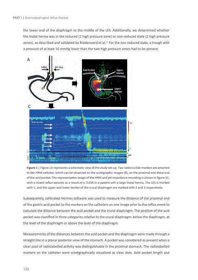
128
Part I | Gastroesophageal reflux disease
the lower end of the diaphragm to the middle of the LES. Additionally, we determined whether
the hiatal hernia was in the reduced (1 high pressure zone) or non-reduced state (2 high pressure
zones), as described and validated by Bredenoord et al.11 For the non reduced state, a trough with
a pressure of at least 10 mmHg lower than the two high pressure zones had to be present.
Figure 1 | Figure 1A represents a schematic view of the study set-up. Two radionuclide markers are attached to the HRM catheter, which can be observed on the scintigraphic images (B), on the proximal and distal end of the acid pocket. The representative image of the HRM and pH-impedance recording is shown in figure 1C, with a mixed reflux episode as a result of a TLESR in a patient wth a large hiatal hernia. The LES is marked with 1, and the upper and lower border of the crural diaphragm are marked with 2 and 3 respectively.
Subsequently, calibrated Hermes software was used to measure the distance of the proximal end
of the gastric acid pocket to the markers on the catheters on one image prior to the reflux event to
calculate the distance between the acid pocket and the crural diaphragm. The position of the acid
pocket was classified in three categories relative to the crural diaphragm: below the diaphragm, at
the level of the diaphragm or above the level of the diaphragm.
Measurements of the distances between the acid pocket and the diaphragm were made through a
straight line in a planar posterior view of the stomach. A pocket was considered as present when a
clear pool of radiolabelled activity was distinguishable in the proximal stomach. The radiolabelled
markers on the catheter were scintigraphically visualised as clear dots. Acid pocket length and

129
The effect of Azithromycin on the acid pocket | Chapter 7
width were determined for each reflux event by measuring the distance from the proximal to the
distal end of the radiolabelled activity for the length and between both lateral ends for the width
respectively.
For each reflux episode the underlying mechanism was classified using previously described
criteria (12) as a TLESR, swallow induced, low LES pressure, abdominal straining or with unknown
cause. Briefly, a TLESR was defined as a decrease in LES pressure with a rate of ≥ 1 mmHg/s, with
a duration of 10 seconds, a nadir pressure ≤ 2mmHg, and absence of swallowing from 4 seconds
before to 2 seconds after the start of the relaxation. Swallow induced reflux was defined similarly
to TLESRs but with the presence of a swallow. Low LES pressure was defined as a period of at least
30 seconds with end-expiratory LES pressure of ≤ 3mmHg. Abdominal straining was detected as a
sharp and brief increase in gastric pressure of at least 30 mmHg with a simultaneous elevation in
esophageal pressures.
Statistical analysis
Based on data from previous studies3,13 a mean number of postprandial acid reflux episodes of
10 per study day was assumed in patients with GERD. To detect a difference of 30% in acid reflux
episodes with a statistical power of 80% and a two-sided alpha of 0.05, a total of 16 patients was
required in a cross-over design. Thirty percent reduction in reflux episodes is a clinically relevant
difference and is observed in studies with a comparable design.14,15 To compensate for drop-outs,
we decided to include 19 patients.
Statistical analysis was preformed using SPSS 20.0 software (IBM corporation, Somers, NY, United
States). Data are presented as mean ± SEM. Continuous data were compared using a paired t-test
or with a linear mixed model (LMM) in case of multiple measurements per patients. Comparisons
of proportions were performed using a generalised estimating equations (GEE) logistic regression
model, with subject as clustering factor, and treatment as independent variable. Pearson’s
correlation was used for correlations. All p-values were two-tailed and a p-value < 0.05 was
considered as statistically significant.
ResulTs
Patients
Nineteen patients with GERD (13 male; median age 56 yrs, range 45-67 yrs) were included. HRM
analysis showed that 7 patients had a large hiatal hernia (L-HH) (≥ 3 cm) and 12 had a small hiatal
hernia (S-HH) (< 3 cm).
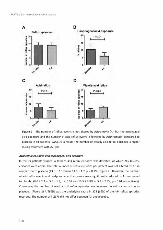
130
Part I | Gastroesophageal reflux disease
Figure 2 | The number of reflux events is not altered by Azitromcyin (A), but the esophageal
acid exposure and the number of acid reflux events is lowered by Azithromycin compared to
placebo in all patients (B&C). As a result, the number of weakly acid reflux episodes is higher
during treatment with AZI (D).
Acid reflux episodes and esophageal acid exposure
In the 19 patients studied, a total of 494 reflux episodes was detected, of which 245 (49.6%)
episodes were acidic. The total number of reflux episodes per patient was not altered by Azi in
comparison to placebo (13.8 ± 2.0 versus 14.0 ± 1.7, p = 0.79) (Figure 2). However, the number
of acid reflux events and postprandial acid exposure were significantly reduced by Azi compared
to placebo (8.0 ± 2.2 vs 5.6 ± 1.8, p < 0.01 and 10.5 ± 3.8% vs 5.9 ± 2.5%, p = 0.01 respectively).
Conversely, the number of weakly acid reflux episodes was increased in Azi in comparison to
placebo. (Figure 2) A TLESR was the underlying cause in 328 (66%) of the 494 reflux episodes
recorded. The number of TLESRs did not differ between Azi and placebo.
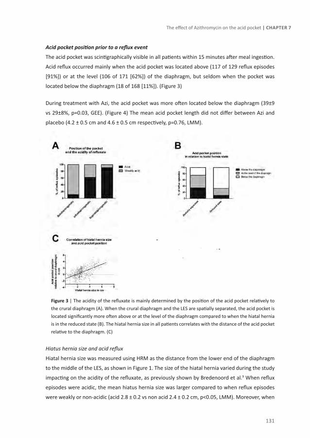
131
The effect of Azithromycin on the acid pocket | Chapter 7
Acid pocket position prior to a reflux event
The acid pocket was scintigraphically visible in all patients within 15 minutes after meal ingestion.
Acid reflux occurred mainly when the acid pocket was located above (117 of 129 reflux episodes
[91%]) or at the level (106 of 171 [62%]) of the diaphragm, but seldom when the pocket was
located below the diaphragm (18 of 168 [11%]). (Figure 3)
During treatment with Azi, the acid pocket was more often located below the diaphragm (39±9
vs 29±8%, p=0.03, GEE). (Figure 4) The mean acid pocket length did not differ between Azi and
placebo (4.2 ± 0.5 cm and 4.6 ± 0.5 cm respectively, p=0.76, LMM).
Figure 3 | The acidity of the refluxate is mainly determined by the position of the acid pocket relatively to the crural diaphragm (A). When the crural diaphragm and the LES are spatially separated, the acid pocket is located significantly more often above or at the level of the diaphragm compared to when the hiatal hernia is in the reduced state (B). The hiatal hernia size in all patients correlates with the distance of the acid pocket relative to the diaphragm. (C)
Hiatus hernia size and acid reflux
Hiatal hernia size was measured using HRM as the distance from the lower end of the diaphragm
to the middle of the LES, as shown in Figure 1. The size of the hiatal hernia varied during the study
impacting on the acidity of the refluxate, as previously shown by Bredenoord et al.9 When reflux
episodes were acidic, the mean hiatus hernia size was larger compared to when reflux episodes
were weakly or non-acidic (acid 2.8 ± 0.2 vs non acid 2.4 ± 0.2 cm, p<0.05, LMM). Moreover, when

132
Part I | Gastroesophageal reflux disease
the hiatus hernia was in non-reduced state (spatial separation between diaphragm and LES), the
risk for acid reflux was significantly increased compared to in the reduced state (59 ± 7% compared
to 30 ± 8% of reflux events respectively, p<0.001, GEE). The distance of the acid pocket to the crural
diaphragm correlated significantly with hiatal hernia size (r=0.61, p<0.0001 Pearson’s correlation),
with a larger hiatal hernia size thus leading to a more proximal position of the acid pocket. (Figure
3) In the reduced state, hiatal hernia size was never more than 2.0 cm.
Proximal extent and esophageal clearance time
In the total group, the proximal extent of liquid and mixed reflux episodes varied between 5 and 17
cm. During treatment with Azi, the proximal extent was significantly reduced compared to placebo
(9.9 ± 0.50 cm vs 8.6 ± 0.44 cm, p<0.01). As acid exposure is also determined by clearance of the
refluxate, we assessed the effect of AZI on acid clearance time. Mean clearance time was not
significantly altered by AZI (13 ± 1.5 s with Azi vs 17 ± 2.1 s with placebo (p=0.10)).
Small versus large hiatal hernia
Motilides increase proximal stomach tone thereby reducing the volume of the proximal stomach.
We hypothesized that this would affect the position of acid pocket, particularly in patients with a
small HH, by reducing the size of the hiatal hernia. In patients with a large HH, however, the acid
pocket is continuously positioned above the diaphragm,16 reducing the potential of Azi to affect
the position of the acid pocket. Based on previous findings, patients were divided in large HH
(≥3cm) or small or no HH (<3cm) according to the size of the hiatal hernia. Seven patients had a
large HH whereas 12 had a small HH.
The total number of reflux episodes is comparable in the two groups (249 in L-HH patients, and
245 in S-HH patients). In L-HH patients, the acid pocket was located above the diaphragm in 45% of
reflux episodes, and 99 (91%) of these reflux episodes were acidic. In S-HH patients however, the
acid pocket was mostly located below (63%) or at the level (29%) of the diaphragm.
In large hiatal hernia patients Azi reduces the rate of acid reflux from 69% to 62%. In small hiatal
hernia patients, Azi reduces the rate of acid reflux from 38% to 17%, which is a significantly larger
effect compared to in large hiatal hernia patients. (p<0.05, GEE), (Figure 5)
In S-HH patients treatment with Azi led to a significantly smaller mean hiatal hernia size prior to
reflux episodes compared to placebo (2.5 ± 0.3 cm vs 2.1 ± 0.2 cm, p<0.05 LMM). In line with this,
the hiatus hernia was more often in the reduced state during Azi treatment compared to during
placebo. (Figure 5) As a result, the acid pocket in S-HH patients was more often located below the
diaphragm during treatment (69 ± 8% vs 53 ± 8% of reflux episodes respectively, p=0.01,GEE).
(Figure 4)

133
The effect of Azithromycin on the acid pocket | Chapter 7
Figure 4 | Most acid reflux episodes occur starting from 10-15 minutes postprandial till 60 minutes postprandial (A). In this period, the acid pocket has its most proximal position. Azithromycin leads overall to a more distal position of the acid pocket compared to placebo (B). In figure 3C the acid pocket position relative to the crural diaphragm is shown when the subdivision in small and large hiatal hernia is made, demonstrating that the observed effect on altered acid pocket position mainly results from patients with a small hiatal hernia.
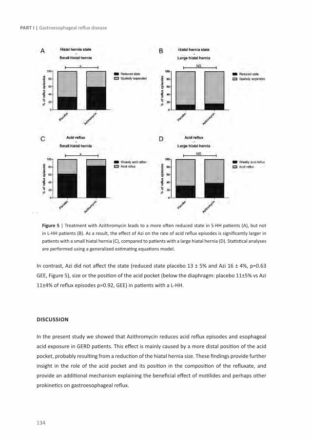
134
Part I | Gastroesophageal reflux disease
Figure 5 | Treatment with Azithromycin leads to a more often reduced state in S-HH patients (A), but not in L-HH patients (B). As a result, the effect of Azi on the rate of acid reflux episodes is significantly larger in patients with a small hiatal hernia (C), compared to patients with a large hiatal hernia (D). Statistical analyses are performed using a generalized estimating equations model.
In contrast, Azi did not affect the state (reduced state placebo 13 ± 5% and Azi 16 ± 4%, p=0.63
GEE, Figure 5), size or the position of the acid pocket (below the diaphragm: placebo 11±5% vs Azi
11±4% of reflux episodes p=0.92, GEE) in patients with a L-HH.
dIscussIon
In the present study we showed that Azithromycin reduces acid reflux episodes and esophageal
acid exposure in GERD patients. This effect is mainly caused by a more distal position of the acid
pocket, probably resulting from a reduction of the hiatal hernia size. These findings provide further
insight in the role of the acid pocket and its position in the composition of the refluxate, and
provide an additional mechanism explaining the beneficial effect of motilides and perhaps other
prokinetics on gastroesophageal reflux.
135
The effect of Azithromycin on the acid pocket | Chapter 7
After meal intake, gastric acid floats on top of the ingested food also referred to as the acid
pocket. We recently provided evidence supporting the hypothesis that this pocket functions
as a reservoir from which acid refluxes into the esophagus, especially if the pocket is located
above the diaphragm or in the hiatus.3 The present study confirms that the risk for having acid
reflux is strongly determined by the position of the acid pocket relative to the diaphragm. We
indeed found that only 11% of the reflux episodes were acidic when the acid pocket is located
below the diaphragm, in contrast to 91% when the pocket is positioned above. As we previously
demonstrated that the position of the acid pocket is largely determined by the presence of a
hiatal hernia, accurate recording of the position of the hiatal hernia is of great importance when
evaluating the interaction between hiatal hernia, acid pocket and reflux. This is particularly of
importance in patients with a small or no hiatal hernia, as concurrent pH-impedance and HRM
recording elegantly showed that a hiatal hernia is a dynamic entity, i.e. it appears and disappears
in time, thereby largely determining the risk to have acidic reflux.11,17 When the hiatal hernia is in
the reduced state, i.e. no separation between LES and diaphragm, the risk to have acidic reflux
is 2 to 4-fold lower compared to when the hernia is in non-reduced state.11,17 Using the same
methodology, but combined with continuous recording of the acid pocket, we were able to study
the dynamics of the acid pocket (in contrast to pH pull-through studies) and hiatus hernia in great
detail during a prolonged period of time. Hence, we could demonstrate that spatial separation
of the LES and crural diaphragm (hiatal hernia size) is indeed larger prior to acid reflux events
compared to weakly acid reflux events, and that a reduction in hiatal hernia size results in a
more distal position of the acid pocket relative to the diaphragm. When the hiatal hernia is in the
reduced state, the acid pocket is more often located below the diaphragm, and the risk for acidic
reflux is lower (30%) compared to when the hernia is in a non-reduced state (59%). These data
confirm that the position of the acid pocket and the size of the hiatal hernia are closely related and
are major risks factors to have acid reflux or GERD.
Clinically, the relation between GERD and a hiatal hernia is evident as hiatal hernia size correlates
excellent with esophageal acid exposure and esophageal damage.12,18-20 Accepting that the position
of the acid pocket largely determines acidity of the refluxate, drugs reducing the hiatus hernia or
move the acid pocket more distally should result in reduced acid exposure. To this end, GERD
patients were treated with Azi, a broad spectrum antibiotic with prokinetic properties, previously
shown to reduce acid reflux in lung transplant patients.8 In the present study, we showed that
also in GERD patients Azi treatment decreased the number of acid reflux episodes and acid reflux
exposure, but increased non-acid reflux. Hence, the total number of reflux events remained
unchanged, in line with the finding that Azi had no effect on TLESRs, the main mechanism
underlying GOR.12 Instead, Azi significantly reduced hiatal hernia size. This effect was mainly
observed in patients with a small hiatal hernia (<3 cm). This was confirmed by the observation
136
Part I | Gastroesophageal reflux disease
that hiatal hernia was in a reduced state in 59% of reflux episodes during Azi compared to 33%
during placebo in these patients. In patients with a large hiatal hernia (≥3 cm) Azi however failed to
affect the hiatal hernia (reduced vs separated) or alter the position of the acid pocket, most likely
explaining the larger effect on acid reflux rates in patients with a small hiatal hernia.
The exact mechanism by which Azi affects the hiatal hernia and acid pocket position is unclear.
Motilides such as Azi and erythromycin act on motilin receptors on nerves and smooth muscle cells
mediating an overall prokinetic effect.6,12In the proximal stomach and distal esophagus motilides
therefore enhance motility leading to increased proximal gastric tone and LES pressure, presumably
via a cholinergic pathway.22-24 One could speculate that increased tone reduces proximal stomach
volume, thereby reducing hiatal hernia size forcing the gastric content and acid pocket more
distally. This effect is mainly observed in patients with a small HH, as illustrated by the increase in
the reduced state of the HH. In contrast, in patients with a large HH, the separation of the LES and
the crural diaphragm is too large for the HH to be reverted into the reduced state.25-27 Alternatively,
Azi accelerates gastric emptying and improves mixing of stomach contents, potentially affecting
acid pocket properties.24,28 Lastly, reduction in acid secretion by Azi could have contributed to the
reduced number of acidic reflux events. However, no effect of Azi on acid pocket width and length
was observed in our study. It should be emphasized though that our scintigraphic technique is
not sensitive enough to detect subtle changes in acid secretion. Hence, we cannot exclude this
possibility. A limitation of our study is that we only analysed one dose of Azi, However, due to the
nature of the recording methods and the burden of these techniques, we choose to use one low
dose comparable to the earlier study on this subject.8 Another possible limitation of our study is
that we did not assess gastric emptying.
Our findings provide additional evidence for the importance of the position of the gastric acid
pocket as a risk factor for acidic reflux. In addition, our study confirms that the gastric acid pocket
may represent a possible target for GERD therapy.28-30 Finally, we provide evidence that distal
migration of the acid pocket and increased time of the hiatal hernia in the reduced state are
new potential explanations for the effect of prokinetics on acid exposure, although the effect of
affecting the pocket position was rather limited and primarily observed in patients with a small
hiatal hernia. The effect of motilin agonists and other prokinetics on gastroesophageal parameters
has been investigated more extensively in earlier studies showing a reduction in proximal extent
of the refluxate, accelerated gastric emptying and increased LES pressure as potential mechanisms
explaining the reduction in acid exposure.8,31-35 Although a 30% reduction in acid reflux episodes and
esophageal acid exposure as observed in our study is a large beneficial effect, earlier therapeutic
studies with prokinetics have demonstrated a rather small effect on GERD symptoms.36-39 Our
results indeed provide evidence for a new potential explanation of the effect of prokinetics on

137
The effect of Azithromycin on the acid pocket | Chapter 7
acid exposure, but whether treatment with prokinetics in GERD leads to a reduction in symptoms
remains to be studied.
In theory, therapeutic strategies directly intervening with the composition of the acid pocket may
possibly prove more efficient in preventing acid reflux events. Recent studies indeed show that
antacid-alginate formulations effectively eliminate or displace the acid pocket, by formation of a
raft in the proximal stomach.30,40 We hypothesize that this intervention will be successful in S-HH
as well as in L-HH patients, as antacid-alginate formulations reduce acid reflux events in patients
with and without a hiatal hernia.41
In conclusion, this study demonstrates that Azi reduces the number of acid reflux events and
esophageal acid exposure in GERD patients, especially in patients with a small HH. We hypothesize
that this effect results from relocation of the acid pocket to a more distal position and a concomitant
reduction in hiatal hernia. These data indicate that modulation of the position of the acid pocket
has an impact on the acidity of the refluxate, further confirming the importance of the acid pocket
in the pathogenesis of GERD.
138
Part I | Gastroesophageal reflux disease
ReFeRence lIsT
1. Kahrilas PJ, Shaheen NJ, Vaezi MF et al. American Gastroenterological Association Medical Position Statement on the management of gastroesophageal reflux disease. Gastroenterology 2008;135(4):1383-91, 1391.
2. Dent J, El-Serag HB, Wallander MA et al. Epidemiology of gastro-oesophageal reflux disease: a systematic review. Gut 2005;54(5):710-7.
3. Beaumont H, Bennink RJ, de Jong J et al. The position of the acid pocket as a major risk factor for acidic reflux in healthy subjects and patients with GORD. Gut 2010;59(4):441-51.
4. Fletcher J, Wirz A, Young J et al. Unbuffered highly acidic gastric juice exists at the gastroesophageal junction after a meal. Gastroenterology 2001;121(4):775-83.
5. Mittal RK, Lange RC, McCallum RW. Identification and mechanism of delayed esophageal acid clearance in subjects with hiatus hernia. Gastroenterology 1987;92(1):130-5.
6. Koutsoumbi P, Epanomeritakis E, Tsiaoussis J et al. The effect of erythromycin on human esophageal motility is mediated by serotonin receptors. Am J Gastroenterol 2000;95(12):3388-92.
7. Abu-Gharbieh E, Vasina V, Poluzzi E et al. Antibacterial macrolides: a drug class with a complex pharmacological profile. Pharmacol Res 2004;50(3):211-22.
8. Mertens V, Blondeau K, Pauwels A et al. Azithromycin reduces gastroesophageal reflux and aspiration in lung transplant recipients. Dig Dis Sci 2009;54(5):972-9.
9. Kahrilas PJ, Quigley EM. Clinical esophageal pH recording: a technical review for practice guideline development. Gastroenterology 1996;110(6):1982-96.
10. Kuiken S, Van Den Elzen B, Tytgat G et al. Evidence for pooling of gastric secretions in the proximal stomach in humans using single photon computed tomography. Gastroenterology 2002;123(6):2157-8.
11. Bredenoord AJ, Weusten BL, Timmer R et al. Intermittent spatial separation of diaphragm and lower esophageal sphincter favors acidic and weakly acidic reflux. Gastroenterology 2006;130(2):334-40.
12. van Herwaarden MA, Samsom M, Smout AJ. Excess gastroesophageal reflux in patients with hiatus hernia is caused by mechanisms other than transient LES relaxations. Gastroenterology 2000;119(6):1439-46.
13. Boeckxstaens GE, Beaumont H, Mertens V et al. Effects of lesogaberan on reflux and lower esophageal sphincter function in patients with gastroesophageal reflux disease. Gastroenterology 2010;139(2):409-17.
14. Beaumont H, Smout A, Aanen M et al. The GABA(B) receptor agonist AZD9343 inhibits transient lower oesophageal sphincter relaxations and acid reflux in healthy volunteers: a phase I study. Aliment Pharmacol Ther 2009;30(9):937-46.
15. Rohof WO, Lei A, Boeckxstaens GE. Effect of a novel metabotropic glutamate receptor 5 antagonist (AZD2066) on transient lower esophageal sphincter relaxations and reflux episodes in healthy males. Gastroenterology Suppl 1. 1-5-2011.
16. Beaumont H, Bennink RJ, de JJ et al. The position of the acid pocket as a major risk factor for acidic reflux in healthy subjects and patients with GORD. Gut 2010;59(4):441-51.
17. Scheffer RC, Bredenoord AJ, Hebbard GS et al. Effect of proximal gastric volume on hiatal hernia. Neurogastroenterol Motil 2010;22(5):552-6, e120.
139
The effect of Azithromycin on the acid pocket | Chapter 7
18. Jones MP, Sloan SS, Rabine JC et al. Hiatal hernia size is the dominant determinant of esophagitis presence and severity in gastroesophageal reflux disease. Am J Gastroenterol 2001;96(6):1711-7.
19. van Herwaarden MA, Samsom M, Smout AJ. The role of hiatus hernia in gastro-oesophageal reflux disease. Eur J Gastroenterol Hepatol 2004;16(9):831-5.
20. Sgouros SN, Mpakos D, Rodias M et al. Prevalence and axial length of hiatus hernia in patients, with nonerosive reflux disease: a prospective study. J Clin Gastroenterol 2007;41(9):814-8.
21. Arts J, Caenepeel P, Verbeke K et al. Influence of erythromycin on gastric emptying and meal related symptoms in functional dyspepsia with delayed gastric emptying. Gut 2005;54(4):455-60.
22. Coulie B, Tack J, Peeters T et al. Involvement of two different pathways in the motor effects of erythromycin on the gastric antrum in humans. Gut 1998;43(3):395-400.
23. Sifrim D, Matsuo H, Janssens J et al. Comparison of the effects of midecamycin acetate and azithromycin on gastrointestinal motility in man. Drugs Exp Clin Res 1994;20(3):121-6.
24. Annese V, Janssens J, Vantrappen G et al. Erythromycin accelerates gastric emptying by inducing antral contractions and improved gastroduodenal coordination. Gastroenterology 1992;102(3):823-8.
25. Kahrilas PJ, Lin S, Chen J et al. The effect of hiatus hernia on gastro-oesophageal junction pressure. Gut 1999;44(4):476-82.
26. Pandolfino JE, Kim H, Ghosh SK et al. High-resolution manometry of the EGJ: an analysis of crural diaphragm function in GERD. Am J Gastroenterol 2007;102(5):1056-63.
27. Pandolfino JE, Shi G, Trueworthy B et al. Esophagogastric junction opening during relaxation distinguishes nonhernia reflux patients, hernia patients, and normal subjects. Gastroenterology 2003;125(4):1018-24.
28. Boecxstaens V, Bisschops R, Blondeau K et al. Modulation of the postprandial acid and bile pockets at the gastro-oesophageal junction by drugs that affect gastric motility. Aliment Pharmacol Ther 2011;33(12):1370-7.
29. Vo L, Simonian HP, Doma S et al. The effect of rabeprazole on regional gastric acidity and the postprandial cardia/gastro-oesophageal junction acid layer in normal subjects: a randomized, double-blind, placebo-controlled study. Aliment Pharmacol Ther 2005;21(11):1321-30.
30. Kwiatek MA, Roman S, Fareeduddin A et al. An alginate-antacid formulation (Gaviscon Double Action Liquid) can eliminate or displace the postprandial ‘acid pocket’ in symptomatic GERD patients. Aliment Pharmacol Ther 2011;34(1):59-66.
31. Bruley des Varannes, Parys V, Ropert A et al. Erythromycin enhances fasting and postprandial proximal gastric tone in humans. Gastroenterology 1995;109(1):32-9.
32. Ruth M, Hamelin B, Rohss K et al. The effect of mosapride, a novel prokinetic, on acid reflux variables in patients with gastro-oesophageal reflux disease. Aliment Pharmacol Ther 1998;12(1):35-40.
33. Kim YS, Kim TH, Choi CS et al. Effect of itopride, a new prokinetic, in patients with mild GERD: a pilot study. World J Gastroenterol 2005;11(27):4210-4.
34. van Herwaarden MA, Samsom M, Van Nispen CH et al. The effect of motilin agonist ABT-229 on gastro-oesophageal reflux, oesophageal motility and lower oesophageal sphincter characteristics in GERD patients. Aliment Pharmacol Ther 2000;14(4):453-62.

140
Part I | Gastroesophageal reflux disease
35. Netzer P, Schmitt B, Inauen W. Effects of ABT-229, a motilin agonist, on acid reflux, oesophageal motility and gastric emptying in patients with gastro-oesophageal reflux disease. Aliment Pharmacol Ther 2002;16(8):1481-90.
36. van Herwaarden MA, Samsom M, Van Nispen CH et al. The effect of motilin agonist ABT-229 on gastro-oesophageal reflux, oesophageal motility and lower oesophageal sphincter characteristics in GERD patients. Aliment Pharmacol Ther 2000;14(4):453-62.
37. Champion G, Richter JE, Singh S et al. Effects of oral erythromycin on esophageal pH and pressure profiles in patients with gastroesophageal reflux disease. Dig Dis Sci 1994;39(1):129-37.
38. Talley NJ, Verlinden M, Geenen DJ et al. Effects of a motilin receptor agonist (ABT-229) on upper gastrointestinal symptoms in type 1 diabetes mellitus: a randomised, double blind, placebo controlled trial. Gut 2001;49(3):395-401.
39. van PB, Sigterman KE, Bonis P et al. Short-term treatment with proton pump inhibitors, H2-receptor antagonists and prokinetics for gastro-oesophageal reflux disease-like symptoms and endoscopy negative reflux disease. Cochrane Database Syst Rev 2010;(11):CD002095.
40. Fox M, Kant R, Kaufman E et al.. The Mechanism of Reflux Suppression by Alginates Assessed by Magnetic Resonance Imaging and Manometry. Gastroenterology 140[5 Suppl1], S576-S577. 2011.
41. Zentilin P, Dulbecco P, Savarino E et al. An evaluation of the antireflux properties of sodium alginate by means of combined multichannel intraluminal impedance and pH-metry. Aliment Pharmacol Ther 2005;21(1):29-34.


Chapter 8An alginate-antacid formulation (Gaviscon
Double Action) targets the acid pocket to reduce acid reflux in symptomatic GERD
patients Wout Rohof, Roel Bennink, André Smout, Guy Boeckxstaens
Submitted

144
Part I | Gastroesophageal reflux disease
AbsTRAcT
Background & Aims:
Alginate rafts may directly target the acid pocket, an important source of postprandial acid reflux,
but this has never been visualized in vivo. This study determined the location of an alginate formu-
lation in relation to the acid pocket and the corresponding effects on reflux parameters and acid
pocket positioning in symptomatic patients with gastroesophageal reflux disease (GERD).
Methods:
In this randomized controlled study, 16 GERD patients with a large hiatal hernia received either 111In-labeled alginate-antacid (Gaviscon Double Action Liquid) or antacid (Antagel) after a standard
meal. The relative positions of labeled alginate and acid pocket were analyzed for two hours using
scintigraphy and reflux episodes were detected using concurrent high-resolution manometry and
pH-impedance monitoring.
Results:
The alginate-antacid labeling co-localized with the acid pocket. The number of acid reflux episodes
(antacid 15 [5-50 vs. alginate-antacid 3.5 [0-6.5], p=0.03 was significantly reduced, whereas the
time to acid reflux was significantly increased (antacid 14 minutes [9-23] vs. alginate-antacid 63
minutes [23-92], p=0.01). The acid pocket was more frequently located below the diaphragm af-
ter alginate-antacid treatment than with antacid (below diaphragm 71% [27-78] vs. 21% [10-51]
respectively; p=0.08). Sub-diaphragmatic positioning of the acid pocket negatively correlated with
acid reflux (r=-0.76, p<0.001).
Conclusions:
The alginate-antacid raft co-localized with the postprandial acid pocket and displaced it below the
diaphragm resulting in significant suppression of postprandial acid reflux. This demonstrates the
importance of the acid pocket in GERD pathogenesis and establishes alginate-antacid as an appro-
priately targeted treatment for postprandial acid reflux.

145
Alginates target the acid pocket in GERD patients | Chapter 8
InTRoducTIon
Gastroesophageal reflux disease (GERD) is a common condition where backflow of gastric con-
tents into the esophagus causes symptoms and/or esophageal damage.1-3 The majority of acidic
gastroesophageal reflux and symptoms, such as heartburn, occur after eating, which is somewhat
paradoxical given that the buffering effect of food causes a reduction in intra-gastric acidity.
An explanation for this paradox was provided by Fletcher et al. who were the first to detect an
unbuffered pool of acid that floats on top of ingested food, referred to as the gastric acid pocket.4
The acid pocket develops as a result of poor mixing of newly secreted acid and food in the proximal
stomach, which remains relatively quiescent after a meal compared to the distal stomach.5 We
recently reported that the position of the acid pocket relative to the crural diaphragm is an im-
portant determinant of the acidity of the refluxate.6 In GERD, proximal extension of the acid pocket
above the diaphragm increases the risk for acid reflux.6,7 The acid pocket is therefore an important
source of postprandial acid in GERD and, as such, represents a unique therapeutic target.8
Emerging evidence suggests that alginates may act through direct targeting of the acid pocket.
Alginates are natural polysaccharide polymers which, on contact with gastric acid, precipitate into
a low-density viscous gel of near-neutral pH within minutes.9, 10 The change in pH triggers the
sodium bicarbonate in the formulation to release carbon dioxide, which becomes trapped in the
alginate gel, causing it to float to the top of the gastric contents like a ‘raft’.9, 10 MRI shows that
alginate rafts form in the proximal stomach close to the esophagogastric junction (EGJ), precisely
where the acid pocket develops.11 Furthermore, a commercial alginate-antacid formulation, Gavis-
con Double Action Liquid (Reckitt Benckiser Healthcare, Hull, UK) has been shown to shift the
postprandial acidified segment in the proximal stomach away from the EGJ in the majority of
GERD patients tested.12 The raft of Gaviscon Double Action is reported to remain in the stomach,
on top of the meal, for more than four hours,13 while the acid pocket is reported to remain for up
to 120 minutes.6, 14 It is attractive to hypothesize that the mode of action of alginates allows direct
capping and displacement of the acid pocket, isolating it from the EGJ and reducing postprandial
acid reflux events.12 However, the position of the alginate raft in relation to the acid pocket has
never been visualized in vivo.
The aim of this study is to visualize the position of the alginate raft after administration of Gaviscon
Double Action in GERD patients and to concurrently assess the effect on reflux parameters and
position of the acid pocket compared with a commonly used antacid control.

146
Part I | Gastroesophageal reflux disease
MATeRIAls And MeThods
Subjects
The study was performed in 16 patients with proven GERD, defined by the presence of esophagitis
observed during upper endoscopy and/or pH-metry with an acid exposure of >4.5%, in combina-
tion with typical GERD symptoms.15 None of the subjects had previous gastrointestinal surgery or
was taking medication known to influence esophageal motor function. Hiatal hernia size was mea-
sured by high-resolution manometry (HRM), and patients with small or no hiatal hernia (<3 cm)
were excluded. Patients with confirmed long segment Barrett’s epithelium, or who were unable
to stop the use of proton pump inhibitors for one week, or who had participated in another study
using radioactivity within the last year were also excluded. The study was approved by the medical
ethics committee of the Academic Medical Center. Written informed consent was obtained from
all subjects before enrolment in the study.
Study medication
Sodium alginate-bicarbonate (Gaviscon Double Action Liquid) is an oral liquid suspension, directly
acting without absorption into the systemic circulation. The medication is a combination of two
antacids (calcium carbonate and sodium bicarbonate) and sodium alginate. Each half-maximal
10 mL dose contains 500 mg sodium alginate, 213 mg sodium bicarbonate and 325 mg calcium
carbonate with a neutralizing capacity of approximately 10mEq H+. Gaviscon was labeled with 1
MBq indium-111 (111In)-chloride according to the established labeling procedure.16, 17 Antagel is a
common antacid drug with a neutralizing capacity of 30mEq H+. Each 10 mL dose contains 200 mg
magnesium hydroxide and 400mg aluminum oxide.
Study design
In this parallel designed study, 16 GERD patients were randomized to 111In-labeled alginate-antacid
(n=8) or antacid (n=8). Reflux episodes were detected using concurrent HRM and pH impedance
monitoring, and scintigraphy was performed to localize the alginate-antacid raft and the acid pock-
et relative to the crural diaphragm (Figure 1). Acid-suppressive medication was stopped at least
7 days before the study day. Patients were allowed to use antacids except for on the study day.
Subjects fasted overnight for at least 12 hours. On the day of study 350 MBq technetium-99m
(99mTc)-pertechnetate was injected intravenously. Tc-pertechnetate behaves as a chloride ion, and
is secreted by the parietal cells of the stomach.18 When this technique is used, acid distribution in
the stomach can be visualized scintigraphically, as validated previously.6
Before introduction of the HRM catheter, two sealed markers impregnated with 99mTc-pertechne-
tate were attached to the catheter to visualize the exact location of the catheter during scintigra-
phy. This makes it possible to integrate the distances from the manometry and the scintigraphy,

147
Alginates target the acid pocket in GERD patients | Chapter 8
to relate the acid pocket to the crural diaphragm.14 The HRM catheter and the pH-impedance
catheter were inserted trans-nasally, through the pharynx and esophagus, into the stomach. The
pH sensor of the pH-impedance catheter was placed 5 cm above the upper border of the lower
esophageal sphincter (LES). Subsequently, the patients were positioned in an upright position in
front of the scintigraphy camera. After positioning of the catheters, 5 min of baseline fasting mea-
surements were obtained. Subjects then consumed a standardized meal within 10 min, consisting
of 200 ml orange juice and two pancakes with jam (510 kcal). Thirty minutes after starting the
measurement patients drank 10 ml of Gaviscon Double Action or 10 ml of Antagel. After 2 h of
measurement catheters were removed (Figure1).
Figure 1 | Study design HRM: high-resolution manometry
Recording methods
HRM was performed using a 21 lumen water-perfused HRM catheter (MMS, Enschede, the Neth-
erlands). Eleven distal side holes were positioned at 1 cm intervals, and the 10 proximal side holes
were spaced at 3 cm intervals. The side holes were perfused with distilled water at 0.15 mL⁄min,
using a pneumohydraulic capillary perfusion pump (MMS, Enschede, the Netherlands) and hy-
draulic flow restrictors. For pH-impedance measurement a Unisensor pH-impedance catheter
(Unisensor, Attikon, Switzerland) containing one ISFET pH sensor and eight impedance electrodes
was used, allowing impedance recordings at 3, 5, 7, 9, 15 and 17 cm above the upper border of
the LES. The sample rate was 50 Hz, and data were collected and analyzed with the MMS Solar sys-
tem (MMS, Enschede, the Netherlands). Before each study the pH electrode was calibrated with
pH 4.0 and pH 7.0 solutions (Medtronic A/S, Skovlunde, Denmark). Dynamic scintigraphic images
were acquired on a gamma camera system (Diacam; Siemens Medical Solutions, Illinois, USA),
equipped with a medium-energy collimator. Dynamic recordings were made in posterior view for
2 h (720 views, 10 s/view, 120 min total acquisition time). Gamma radiation of 99mTc-pertechnetate
and 111In have different energy levels, and can therefore be detected separately. Every acquisition

148
Part I | Gastroesophageal reflux disease
was processed on a Hermes processing station (Hermes Medical Solutions, Stockholm, Sweden)
for further analysis.
Data analysis
An acid pocket was considered as present when a clear pool of radiolabelled activity was distin-
guishable in the proximal stomach. The radiolabelled markers on the catheter were scintigraphi-
cally visualized as clear dots. Reflux episodes were detected by pH impedance and each liquid or
mixed reflux event was defined as acidic reflux when pH<4, as weakly acidic when pH≥4 and pH<7.
HRM recordings were used to determine the exact distance from the crural diaphragm to the
markers on the catheter for the position of the acid pocket. The lower and upper border of the cru-
ral diaphragm and the middle of the LES zone were determined. Subsequently, calibrated Hermes
software was used to measure the distance from the proximal end of the gastric acid pocket to the
markers on the catheters on one image to calculate the distance between the acid pocket and the
crural diaphragm. The position of the acid pocket was classified in three categories relative to the
crural diaphragm: below the diaphragm, at the level of the diaphragm or above the diaphragm.
Measurements of the distances between the acid pocket and the diaphragm were made through
a straight line in a planar posterior view of the stomach.
Statistical analysis
Based on the mean numbers of acid reflux episodes reported previously,19 a sample size of 7 per
group in a parallel design will have 80% power to detect a difference in means of 5.8 (e.g. a first
condition mean of 8.4 and a second condition mean of 2.6), assuming a standard deviation of dif-
ferences of 3.5, using a Student t-test with a 0.05 two-sided significance level. To compensate for
potential drop-outs, 16 patients with GERD were included. Statistical analysis was performed using
SPSS 19.0 software for Windows (IBM, New York, USA). Results are presented as mean ± SEM in
the case of a normal distribution or as median [interquartile range] for variables with a skewed dis-
tribution. For statistical analysis, the Mann-Whitney test was used. Correlations were determined
using Spearman’s correlation coefficient.
Figure 2 | Scintigraphic images of 99mTc-pertechnetate-labeled acid pocket (A) and 113mIndium-labeled alginate-antacid (B) and the two scintigraphic recordings superimposed (C).

149
Alginates target the acid pocket in GERD patients | Chapter 8
ResulTs
Relative localization of the alginate-antacid raft and the acid pocket in vivo
Scintigraphy allowed independent visualization of the 99mTc-pertechnetate acid pocket and 111mIn-
dium-labeled alginate raft (Figure 2; panels A and B). The radiolabeled acid pocket was scintigraph-
ically visible in all patients within 15 min of meal ingestion. The indium labeled alginate-antacid
was directly visible after administration. When the two simultaneous scintigraphic recordings
were superimposed, the alginate raft co-localized precisely with the acid pocket (Figure 2; panel
c). The acid pocket and raft were visible for the entire length of the study.
Reflux episodes and esophageal acid exposure
The 16 GERD patients included in the study had a large hiatus hernia (≥3cm as seen on endos-
copy or HRM) resulting in a high rate of acid reflux episodes. A total of 266 reflux episodes were
detected, of which 138 (52%) episodes were acidic. Compared with the antacid, the total number
of reflux episodes was reduced with alginate-antacid ingestion (median 21 [15-27] vs 14 [8.5-17],
respectively; p=0.05) (Figure 3).
Figure 3 | The mean number of reflux events (A), acid reflux episodes (B) and esophageal acid exposure (C) were reduced by alginate-antacid compared with antacid but the mean number of weakly acid reflux episodes was higher with alginate-antacid (D).

150
Part I | Gastroesophageal reflux disease
The number of acid reflux episodes and the rate of acid reflux were also significantly lower after
treatment with alginate-antacid, compared with antacid (15 (5.5-20) vs 3.5 (0-6.5), p=0.03 and
68% [40-79] vs 21% [0-44], p=0.02,) and there was a trend towards reduced esophageal acid ex-
posure (7.4 [2.1-22] vs 0.2 [0-4.1]; p=0.08) (Figure 3). Conversely, the number of weakly acid reflux
episodes were not significantly different between alginate-antacid and antacid-treated patients
(antacid 6 [6.0-7.5] vs. alginate-antacid 8 [6.0-13.0]; P=0.38) (Figure 3).
Buffering time and time to acid reflux
A pH sensor was continuously positioned in the acid pocket to allow comparison of buffering time
after administration of alginate-antacid and antacid. The buffer time (amount of time that the acid
pocket pH>4) was not significantly different after antacid and alginate-antacid ingestion (3.5 min
[0.3-4.5] vs. 6 min [3-12], respectively; p=0.13) and the mean pH of the acid pockets were similar
(2.1 [2.0-2.4) vs. 2.5 [1.7-2.8], respectively; p=0.71). However, compared with antacid, the time
to acid reflux (antacid 14 min [9-23] vs. alginate-antacid 63 min [23-92]; p=0.01) and the mean
pH of reflux episodes were significantly increased with alginate-antacid (antacid 3.5 [2.0-5.0] vs.
alginate-antacid 5.0 [4.1-6.2]; p<0.05) (Figure 4).
Figure 4 | The buffering capacity (A) was similar for antacid and alginate-antacid. However, the mean time to acid reflux (B) and the mean pH of reflux episodes (C) were significantly increased with alginate-antacid compared with antacid.
Acid pocket position and acid reflux events
Acid reflux occurred mainly when the acid pocket was located above (78/95 reflux episodes; 82%),
or at the level of (47/78 [60%]) of the diaphragm, but seldom when the pocket was located be-
low the diaphragm (13/88; 15%). There was a trend towards a higher rate of sub-diaphragmatic
positioning of the acid pocket with alginate-antacid, compared with antacid (71% [27-78] vs. 21%
(10-51) respectively p=0.08) (Figure 5; panel A). The rate of sub-diaphragmatic positioning of the
acid pocket had a significant negative correlation with the rate of acid reflux (r=-0.76, p<0.001),
that is, acid pocket positioning below the diaphragm corresponds to a lower rate of acid reflux
(Figure 5; panel B).
A b c
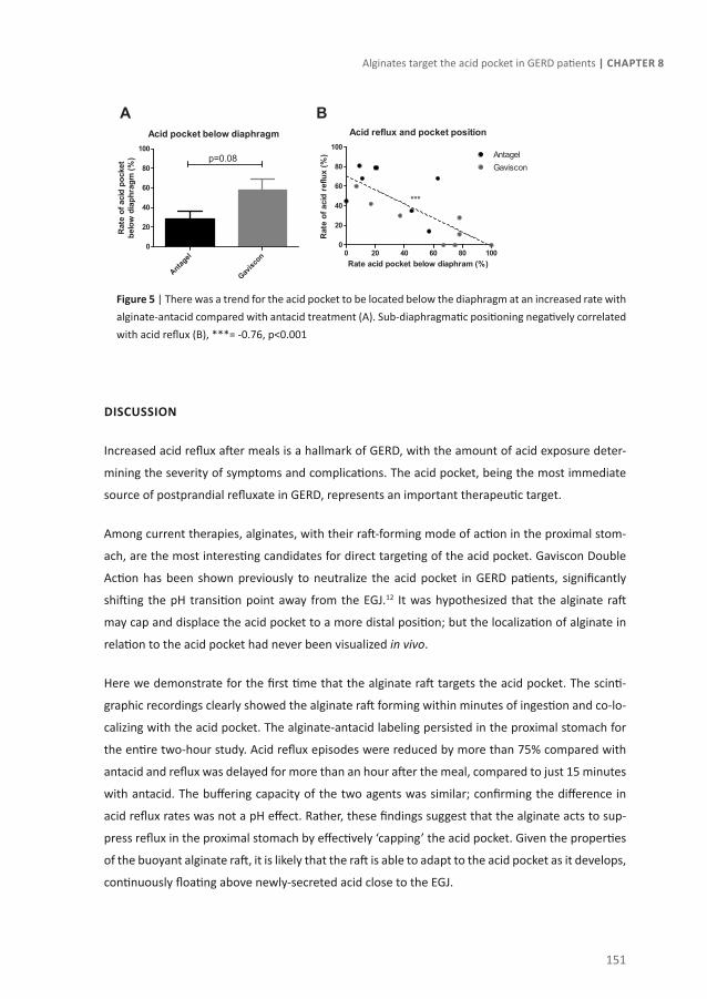
151
Alginates target the acid pocket in GERD patients | Chapter 8
Figure 5 | There was a trend for the acid pocket to be located below the diaphragm at an increased rate with alginate-antacid compared with antacid treatment (A). Sub-diaphragmatic positioning negatively correlated with acid reflux (B), ***= -0.76, p<0.001
dIscussIon
Increased acid reflux after meals is a hallmark of GERD, with the amount of acid exposure deter-
mining the severity of symptoms and complications. The acid pocket, being the most immediate
source of postprandial refluxate in GERD, represents an important therapeutic target.
Among current therapies, alginates, with their raft-forming mode of action in the proximal stom-
ach, are the most interesting candidates for direct targeting of the acid pocket. Gaviscon Double
Action has been shown previously to neutralize the acid pocket in GERD patients, significantly
shifting the pH transition point away from the EGJ.12 It was hypothesized that the alginate raft
may cap and displace the acid pocket to a more distal position; but the localization of alginate in
relation to the acid pocket had never been visualized in vivo.
Here we demonstrate for the first time that the alginate raft targets the acid pocket. The scinti-
graphic recordings clearly showed the alginate raft forming within minutes of ingestion and co-lo-
calizing with the acid pocket. The alginate-antacid labeling persisted in the proximal stomach for
the entire two-hour study. Acid reflux episodes were reduced by more than 75% compared with
antacid and reflux was delayed for more than an hour after the meal, compared to just 15 minutes
with antacid. The buffering capacity of the two agents was similar; confirming the difference in
acid reflux rates was not a pH effect. Rather, these findings suggest that the alginate acts to sup-
press reflux in the proximal stomach by effectively ‘capping’ the acid pocket. Given the properties
of the buoyant alginate raft, it is likely that the raft is able to adapt to the acid pocket as it develops,
continuously floating above newly-secreted acid close to the EGJ.

152
Part I | Gastroesophageal reflux disease
Consistent with previous findings, non-acid reflux events were not reduced.19 Despite a similar
buffering capacity, the acidity of the refluxate was significantly reduced with alginate-antacid com-
pared with antacid. This discrepancy between acid pocket pH and refluxate pH is most likely due
to additional effects by the alginate on the position of the acid pocket. In healthy volunteers the
pocket is highly acidic but gastroesophageal refluxate is usually weakly acidic.20 In GERD patients
there is proximal migration of the acid pocket and increased rates of acid reflux.6 Therefore, dis-
placement of the acid pocket distally in GERD patients would be expected to create a situation
similar to healthy volunteers; a shift from acidic to weakly acidic events, owing to the fact that
the acid pocket is no longer the most immediate source of refluxate. Our previous observations
in GERD patients with large hiatus hernia show that the acid pocket frequently extends into the
hiatal sac above the diaphragm and that this significantly increases the risk for acid reflux.6 This
may explain why hiatal hernia size correlates closely with esophageal acid exposure and esopha-
geal damage.21-24
In a previous study we found that the prokinetic agent azithromycin is able to alter the acid pocket
position from supra-diaphragmatic to sub-diaphragmatic in patients with a small hiatus hernia,
but that this effect was eliminated by a large hiatus hernia.8 The data presented here confirms that
the raft-forming mode of action of alginates is not compromised by a large hiatus hernia (≥3 cm).
Furthermore, the rate at which the acid pocket was located below the diaphragm was more than
three times greater with alginate-antacid compared with antacid (although not reaching statisti-
cally significance, p=0.08). This confirms that the alginate-antacid raft can displace the acid pocket
distally, out of the hiatal sac, and builds on the previous data of Kwiatek et al. who showed signif-
icant distal shifting of the pH transition point.12 Sub-diaphragmatic positioning of the acid pocket
had a significant negative correlation with acid reflux confirming that the effect of alginate-antacid
on acid pocket position contributes to the observed reduction in acid reflux.
Further experimentation using a larger population of patients and/or a maximal dose of Gaviscon
are needed to establish the outcomes that were approaching significance, such as esophageal acid
exposure and acid pocket positioning below the diaphragm (p=0.08 for both). It would also be in-
teresting to determine if distal displacement of the acid pocket plays a role in the reduction of acid
reflux seen in patients with no hiatus hernia,19 and to confirm whether the observed reduction in
acid reflux suppression translates to a reduction in GERD symptoms. This seems likely considering
a previous systematic review of randomized, placebo-controlled trials that demonstrated a benefit
in symptomatic improvement over placebo of 60% with alginate-antacid compared with an 11%
improvement for anatacid.25
In summary, Gaviscon Double Action forms a robust and persistent alginate raft that caps the
acid pocket, displacing it distally and reducing acid reflux events. These findings demonstrate the

153
Alginates target the acid pocket in GERD patients | Chapter 8
value of directly targeting the acid pocket with a non-systemic, alginate-antacid formulation for
the treatment of postprandial acid reflux in GERD. This may have clinical implications for patients
who suffer primarily from postprandial reflux, who may be able to adequately control their symp-
toms with optimized use of alginate-antacid. Alginates may also benefit patients with insufficient
response to PPIs. We have observed that the acid pocket persists after PPI treatment in GERD
patients,26 and thus, may play a role in the residual symptoms that can occur despite PPI therapy.
If this is the case, direct capping of the acid pocket would be expected to benefit these patients.
In support of this, a recent study in patients with non-erosive reflux disease (NERD) demonstrated
that supplementing PPI therapy with alginate more than doubled the number of patients who
experienced complete reflux symptom relief.27

154
Part I | Gastroesophageal reflux disease
ReFeRences
1. Vakil N, van Zanten SV, Kahrilas P, et al. The Montreal definition and classification of gas-troesophageal reflux disease: a global evi-dence-based consensus. Am J Gastroenterol 2006;101:1900-20; quiz 1943.
2. Kahrilas PJ. GERD pathogenesis, pathophysiolo-gy, and clinical manifestations. Cleve Clin J Med 2003;70 Suppl 5:S4-19.
3. Boeckxstaens GE. Alterations confined to the gastro-oesophageal junction: the relationship between low LOSP, TLOSRs, hiatus hernia and acid pocket. Best Pract Res Clin Gastroenterol 2010;24:821-9.
4. Fletcher J, Wirz A, Young J, et al. Unbuffered highly acidic gastric juice exists at the gastro-esophageal junction after a meal. Gastroenter-ology 2001;121:775-83.
5. Sauter M, Curcic J, Menne D, et al. Measuring the interaction of meal and gastric secretion: a combined quantitative magnetic resonance im-aging and pharmacokinetic modeling approach. Neurogastroenterol Motil 2012;24:632-e273.
6. Beaumont H, Bennink RJ, de Jong J, et al. The position of the acid pocket as a major risk factor for acidic reflux in healthy subjects and patients with GORD. Gut 2010;59:441-51.
7. Clarke AT, Wirz AA, Manning JJ, et al. Severe reflux disease is associated with an enlarged unbuffered proximal gastric acid pocket. Gut 2008;57:292-7.
8. Rohof WO, Bennink RJ, de Ruigh AA, et al. Effect of azithromycin on acid reflux, hiatus hernia and proximal acid pocket in the postprandial period. Gut 2012.
9. Tytgat GN, Simoneau G. Clinical and laboratory studies of the antacid and raft-forming prop-erties of Rennie alginate suspension. Aliment Pharmacol Ther 2006;23:759-65.
10. Mandel KG, Daggy BP, Brodie DA, et al. Review article: alginate-raft formulations in the treat-ment of heartburn and acid reflux. Aliment Pharmacol Ther 2000;14:669-90.
11. Fox M, Kant R, Kaufman E, et al. The mechanism of reflux suppression by alginates visualized by magnetic resonance imaging and manometry. Poster presentation. Digestive Disease Week, May 2011.
12. Kwiatek MA, Roman S, Fareeduddin A, et al. An alginate-antacid formulation (Gaviscon Dou-ble Action Liquid) can eliminate or displace the postprandial ‘acid pocket’ in symptom-atic GERD patients. Aliment Pharmacol Ther 2011;34:59-66.
13. Hampson FC, Jolliffe IG, Bakhtyari A, et al. Algi-nate-antacid combinations: raft formation and gastric retention studies. Drug Dev Ind Pharm 2010;36:614-23.
14. Rohof WO, Bennink RJ, de Ruigh AA, et al. Effect of azithromycin on acid reflux, hiatus hernia and proximal acid pocket in the postprandial period. Gut 2012;61:1670-7.
15. Kahrilas PJ, Quigley EM. Clinical esophageal pH recording: a technical review for practice guideline development. Gastroenterology 1996;110:1982-96.
16. Taylor G, Warren S, Kellaway I, et al. Gastric Res-idence of Gaviscon Advance and Liquid Gavis-con in healthy volunteers. J Pharm & Pharmacol 1997;49(Suppl4):73.
17. Davies N, Farr S, Kellaway I, et al. A comparison of the gastric retention of alginate containing tablet formulations with and without the inclu-sion of excipient calcium ions. Int J Pharmaceut 1994;105:97-101.
18. Kuiken S, Van Den Elzen B, Tytgat G, et al. Ev-idence for pooling of gastric secretions in the proximal stomach in humans using single pho-ton computed tomography. Gastroenterology 2002;123:2157-8; author reply 2158.

155
Alginates target the acid pocket in GERD patients | Chapter 8
19. Zentilin P, Dulbecco P, Savarino E, et al. An eval-uation of the antireflux properties of sodium alginate by means of combined multichannel intraluminal impedance and pH-metry. Aliment Pharmacol Ther 2005;21:29-34.
20. Trudgill NJ, Riley SA. Transient lower esoph-ageal sphincter relaxations are no more fre-quent in patients with gastroesophageal reflux disease than in asymptomatic volunteers. Am J Gastroenterol 2001;96:2569-74.
21. van Herwaarden MA, Samsom M, Smout AJ. Excess gastroesophageal reflux in patients with hiatus hernia is caused by mechanisms other than transient LES relaxations. Gastroenterolo-gy 2000;119:1439-46.
22. van Herwaarden MA, Samsom M, Smout AJ. The role of hiatus hernia in gastro-oesopha-geal reflux disease. Eur J Gastroenterol Hepatol 2004;16:831-5.
23. Jones MP, Sloan SS, Rabine JC, et al. Hiatal hernia size is the dominant determinant of esophagitis presence and severity in gastro-esophageal reflux disease. Am J Gastroenterol 2001;96:1711-7.
24. Sgouros SN, Mpakos D, Rodias M, et al. Prev-alence and axial length of hiatus hernia in patients, with nonerosive reflux disease: a prospective study. J Clin Gastroenterol 2007;41:814-8.
25. Tran T, Lowry AM, El-Serag HB. Meta-analysis: the efficacy of over-the-counter gastro-oesoph-ageal reflux disease therapies. Aliment Pharma-col Ther 2007;25:143-53.
26. Rohof W, Bennink R, Boeckxstaens G. Effect of PPIs on the Size, Position and Acidity of the Postprandial Acid Pocket. Gastroenterology 2012;142:S-92.
27. Manabe N, Haruma K, Ito M, et al. Efficacy of adding sodium alginate to omeprazole in pa-tients with nonerosive reflux disease: a ran-domized clinical trial. Dis Esophagus 2011.


PART IIAchalasia

Chapter 9Pneumodilation versus laparoscopic Heller
myotomy for idiopathic achalasiaGuy Boeckxstaens, Vito Annese, Stanislas Bruley des Varannes,
Stanislas Chaussade, Mario Costantini, Antonello Cuttitta, J. Ignasi Elizalde, Uberto Fumagalli, Marianne Gaudric, Wout Rohof,
André J Smout, Jan Tack, Aeilko Zwinderman, Giovanni Zaninotto and Olivier Busch
N Engl J Med. 2011;364(19):1807-16.
160
Part II | Achalasia
AbsTRAcT
Background and aims:
Many consider laparoscopic Heller myotomy (LHM) superior to pneumodilation (PD) and it is
increasingly suggested as treatment of choice for achalasia.
Methods:
Newly diagnosed achalasia patients were randomized to PD or LHM with Dor fundoplication.
Symptoms (weight loss, dysphagia, retrosternal pain and regurgitation) were assessed using the
Eckardt score. The primary outcome was therapeutic success (drop in Eckardt score to < 3) at
yearly follow-up. The secondary outcomes included need for retreatment, lower esophageal
sphincter (LES) pressure, esophageal emptying on timed barium esophagram, quality of life and
complication rate.
Results:
A total of 201 patients were randomized (PD: n=95; LHM: n=106). Mean follow-up was 43 months
(95% CI 40-47). Intention-to-treat analysis revealed no statistically significant difference in primary
outcome with a success rate of 90% and 86% for PD versus 93% and 90% for LHM after 1 and 2 years
of follow-up respectively (P=0.46). LES pressure (LHM 10 (8.7-12) mmHg, PD 12 (9.7-14) mmHg,
P= 0.27), esophageal emptying (height of barium column: LHM 1.9(0-6.8) cm, PD 3.7(0-8.8) cm,
(P=0.21) and quality of life were not statistical significantly different after 2 years. Similar results
were obtained with per-protocol analysis. Perforation of the esophagus occurred in 4.3% during
PD, whereas mucosal tears occurred in 12% during LHM. Abnormal esophageal acid exposure was
observed in 15% and 23% (P=0.28) of PD and LHM respectively.
Conclusions:
In this study, after two years laparoscopic Heller myotomy did not achieve superior rates of
therapeutic success compared with pneumodilation.

161
Pneumatic dilation vs. laparoscopic Heller myotomy | Chapter 9
InTRoducTIon
Achalasia is an esophageal motor disorder that is characterized clinically by dysphagia, chest pain,
and regurgitation of undigested food. These symptoms result from the absence of esophageal
peristalsis combined with a defective relaxation of the lower esophageal sphincter.1 Currently,
treatment consists mainly of endoscopic pneumatic dilation or laparoscopic Heller’s myotomy
(LHM). For many years, repeated endoscopic pneumatic dilation has been the treatment of
choice, leading to therapeutic success in 70 to 80% of cases.2 With the introduction of minimally
invasive surgery, the surgical approach has gained considerable interest, with LHM combined
with an antireflux procedure considered to be the procedure of choice. The results thus far from
single-center studies are excellent, with success rates ranging between 89 and 100%,3 leading to
continuously increasing enthusiasm for the surgical approach.
Currently, the choice of treatment is dictated largely by the experience of the physician. Moreover,
the outcome measures and treatment protocols in previous studies have varied, making a
comparison among various studies of the success rates of the treatment options difficult.
Therefore, the major aim of this multicenter study was to compare the two state-of-the-art
treatments, pneumatic dilation and LHM with fundoplication according to Dor, in a randomized
design.
MeThods
Study design
Patients were included between February 2003 and February 2008 in 14 hospitals from 5 European
countries. The institutional review board of each hospital approved the study protocol, and written
informed consent was obtained from each patient included in the study before enrollment. This
investigator-initiated trial was conceived by the first and last authors, whereas the first author
wrote the first and final versions of the manuscript, and decided in consultation with the other
authors to submit the paper for publication.
Patients
Patients were eligible for enrollment in the study if they were between 18 and 75 years of age
and had achalasia with an Eckardt symptom score of greater than 3. The Eckardt score is the sum
of the symptom scores for dysphagia, regurgitation, and chest pain (with a score of 0 indicating
the absence of symptoms, 1 indicating occasional symptoms, 2 indicating daily symptoms, and 3
indicating symptoms at each meal) and weight loss (with 0 indicating no weight loss, 1 indicating a

162
Part II | Achalasia
loss of <5 kg, 2 indicating a loss of 5 to 10 kg, and 3 indicating a loss of >10 kg) 4; thus, the maximum
score on the Eckardt scale, indicating the most pronounced symptoms, is 12. The diagnosis of
achalasia was made on the basis of the absence of peristalsis and on impaired relaxation of the
lower esophageal sphincter (nadir pressure of ≥10 mm Hg during swallow-induced relaxation) on
esophageal manometry. Exclusion criteria were severe cardiopulmonary disease or other serious
disease leading to unacceptable surgical risk, previous treatment for achalasia, pseudoachalasia,
megaesophagus (diameter of >7 cm), previous esophageal or gastric surgery (except for gastric
perforation), and esophageal diverticula in the distal esophagus. Randomization was performed
with stratification according to hospital and age (<40 or ≥40 years), with the use of computerized
randomization numbers. A numeric code was used in the patient’s file at the trial center.
Interventions
Pneumatic Dilation
A Rigiflex balloon (Boston Scientific) was positioned at the esophagogastric junction and dilated
at a pressure of 5 PSI for 1 minute, followed by 8 PSI for 1 minute. In the initial study protocol,
the first dilation was performed with the use of a 35-mm balloon. However, a perforation of the
esophagus occurred in 4 of the first 13 patients treated in this way. The protocol was amended,
and subsequently the first pneumatic dilation was performed with the use of a smaller balloon (30
mm), followed 1 to 3 weeks later by dilation with the use of a 35-mm balloon. All patients thus
underwent at least two dilations. If the Eckardt score 4 weeks later was greater than 3, a third
dilation was performed, with the use of a 40-mm balloon. If the Eckardt score remained greater
than 3, the patient was considered to have had treatment failure. Patients with a recurrence of
symptoms during the follow-up period underwent dilation again with the use of a 35-mm balloon
and, if necessary (i.e., if the Eckardt score remained higher than 3), with the use of a 40-mm
balloon. A third and final series of dilations was allowed only if symptoms recurred more than
2 years after this second series. If symptoms recurred within 2 years after the second series of
dilations, the patient was considered to have had treatment failure.
LHM with Dor’s Antireflux Procedure
After division of the phrenoesophageal ligament, the distal esophagus was mobilized on the
lateral and anterior side, and a myotomy was performed extending at least 6 cm above the
Gastroesophageal junction and at least 1 to 1.5 cm over the stomach. Thereafter, anterior
180-degree fundoplication according to the method of Dor was performed. If symptoms recurred
after surgery, with an Eckardt score of higher than 3, the patient was considered to have had
treatment failure.
163
Pneumatic dilation vs. laparoscopic Heller myotomy | Chapter 9
Study Outcomes
The primary outcome of the study was therapeutic success (a reduction in the Eckardt score to ≤3)
at the yearly follow-up assessment. The secondary outcomes included the need for retreatment,
pressure at the lower esophageal sphincter, quality of life, and the rate of complications. The time
to treatment failure was calculated from the date of surgery or the first dilation session until the
closing visit or the patient’s last follow-up visit.
Clinical Assessment and Follow-up
The pretreatment evaluation consisted of taking a medical history, performing a physical
examination, and performing routine hematologic and blood chemical laboratory tests. In addition,
patients completed quality-of-life questionnaires (the Medical Outcomes Study 36-Item Short-
Form Health Survey [SF-36], and the European Organisation for Research and Treatment of Cancer
disease-specific questionnaire module for assessing quality of life in patients with esophageal
cancer [QLQ-OES24]). The SF-36 mental and physical summary scores (which range from 0 to
100, with higher scores indicating better well-being) measure general aspects of health quality of
life.5 The QLQ-OES24 (which ranges from 0 to 100, with lower scores indicating better function)
assesses several items of esophageal function.6 Esophageal manometry and upper endoscopy
were performed, and a timed barium esophagogram was obtained to quantify esophageal stasis.7
One month after treatment and yearly thereafter, symptoms and quality of life were assessed, and
esophageal manometry and timed barium esophagography were performed. Twenty-fourhour
pH-metry (a test in which intraesophageal pH is monitored over the course of 24 hours) and
endoscopy were performed 1 year after treatment and every 3 years thereafter. Esophagitis was
graded according to the Los Angeles classification, in which grade A indicates one or more mucosal
breaks of 5 mm in length or less, grade B indicates one or more mucosal breaks of longer than
5 mm, grade C indicates mucosal breaks that extend between two or more mucosal folds (but
involve <75% of the circumference of the esophagus), and grade D indicates mucosal breaks of
75% or more of the circumference of the esophagus.8
Statistical analysis
The modified intention-to-treat analysis included all patients except those in whom a perforation
occurred during pneumatic dilation (whose data were censored at that time) or those who were
lost to follow up. Patients with protocol violations were considered in the modified intention-
to-treat analysis to have had treatment failure. For the perprotocol analysis, only patients who
received treatment according to the protocol were included.
We estimated that with 80 patients in each group, the study would have 90% power to detect a
significant difference in the success rate between LHM and pneumatic dilation, assuming success

164
Part II | Achalasia
rates of 90% and 70% with LHM and pneumatic dilation, respectively, with a two-sided alpha level
of 0.05. To allow for dropouts, we aimed to enroll 200 patients.
Categorical variables were compared with the use of the chi-square test. Continuous variables are
presented as means (with 95% confidence intervals) and were compared with the use of Student’s
t-test. To compare the success rate between the two treatment groups, we used logrank tests on
Kaplan–Meier estimates. Cox proportional-hazards models were used to estimate hazard ratios
for treatment failure and the need for redilation in the pneumatic-dilation group. We conducted
prespecified subgroup analyses for risk factors of treatment failure according to age (≤40 vs. >40
years), sex, basal pressure at the lower esophageal sphincter after treatment (≤10, >10 to ≤20, or
>20 mm Hg), chest pain (daily vs. none or less than daily), height of the bariumcontrast column
5 minutes after ingestion of barium (≤5, >5 to ≤10, or >10 cm), and maximum esophageal width
before treatment (≤4 vs. >4 cm). The analyses were performed on data from the entire group
irrespective of treatment, on data from the pneumatic-dilation and LHM groups separately, and
on data from patients in the pneumatic-dilation group who required retreatment. All reported
P values were two-tailed, and P values of less than 0.05 were considered to indicate statistical
significance.
ResulTs
Patients and Enrollment
Of the 218 patients who were enrolled in the study, 4 were excluded before randomization
because they had pseudoachalasia. In the first 13 patients randomly assigned to pneumatic
dilation, the initial dilation was performed with a 35-mm balloon. Four of these patients (31%) had
an esophageal perforation. Since this complication rate is significantly higher than rates reported
in the literature and than the rate observed after revision of the protocol (4%, as noted below; P
= 0.001 with the use of the chi-square test), this protocol for distention was considered to be too
risky to be introduced in clinical practice. Comparing the efficacy of pneumatic dilation according
to this protocol with another treatment in a primary outcome analysis was therefore considered to
be undesirable. The data from these 13 patients were excluded from the analysis (Fig. 1).
Of the remaining 201 patients, 182 — which included patients who were still being actively
followed at the end of 2 years or who had been categorized as having had treatment failure —
were included in the 2-year modified intention-to-treat analysis (Fig. 1). The mean follow-up
period was 43 months (95% confidence interval [CI], 40 to 47). The baseline characteristics of the
groups were well balanced (Table 1).

165
Pneumatic dilation vs. laparoscopic Heller myotomy | Chapter 9
Figure 1 | Enrollment, Randomization and Follow-up
laparoscopic heller Myotomy Pneumatic dilation P
Patients randomized to new protocol (no.) 106 95
Patient characteristics
Sex (no.) 0.18
Male 57 60
Female 49 35
Age (yr) 45.5 (42.8-48.3) 46.4 (43.2-49.6) 0.68
Over 40 years (no.(%)) 67 (63) 59 (62) 0.88
Weight (kg) 73.5 (70.5-76.5) 73.3 (70.3-76.3) 0.92
BMI (kg/m2) 25.0(24.0-26.0) 24.6 (23.8-25.4) 0.49
Table 1 | Baseline patient characteristics. Data are presented as means (95% CI)

166
Part II | Achalasia
baseline 1 year Fu 2 year Fu
LHM PD P LHM PD P LHM PD P
Patients in Fu 106 95 98 85 97 85
Modified intention to treat analysis:
Successful treatment (%)
93 (88-98)
90 (84-96)
90 (84-96)
86 (77-93)
0.46
Difference in success rate (%)
3.3 (-4.3 - 10.9) 3.8 (-5.5 – 13)
Per-protocol analysis:
Successful treatment (%)
93 (88-98)
93 (88-99)
90 (84-96)
93 (88-99)
0.33
Difference in success rate (%)
0 (-7.4 – 7.4) 3.1 (-5.0 – 11.2)
Eckardt score7.4
(7.0-7.8)7.0
(6.7-7.4)0.15
1.2 (1.0-1.5)
1.4 (1.1-1.7)
0.281.1
(0.9-1.3)1.5
(1.2-1.8)0.06
Eckardt scoreRange
4-12 4-10 0-3 0-5 0-3 0-4
LES(mmHg)
31 (28-33)
33 (30-37)
0.1710
(8.8-12)14
(12-16)<0.01
10 (8.7-12)
12 (9.7-14)
0.27
Timed barium esophagogramMedian height after 5 min (cm)
12 (8.1-18)
12 (7.9-17)
0.910
(0-6.5)0
(0-6.0)0.95
1.9(0-6.8)
3.7 (0-8.8)
0.21
QLQ-OES2439
(36-42)36
(34-39)0.14
13 (11-15)
15 (12-17)
0.2812
(10-14)14
(11-16)0.28
QLQ-SF36Physical component summary (PCS)
48 (46-50)
48 (46-50)
0.8154
(53-56)52
(50-54) 0.12
53 (51-55)
52 (50-54)
0.31
Mental component summary (MCS)
42 (40-44)
43 (41-46)
0.4149
(47-51)49
(47-51)0.96
50 (48-52)
48 (46-51)
0.35
Table 2 | Primary and secondary outcomes at 1 and 2 years of follow-up according to type of treatment. Data are shown as mean (95% CI) except for the timed barium esophagogram data (median and interquartile range). Data were compared for treatment groups at the different time points using Student’s t-test, except for the timed barium esophagogram data, which were compared using Mann-Whitney-U test. No statistical differences were found except for * LES pressure after 1 year, which is higher in the PD group (P=0.003 Student’s T-test). 1The height of the barium column in the esophagus measured 5 minutes after barium ingestion is a measure for esophageal empting. The QLQ-SF-36 physical and mental component summaries (scores range = 0-100) measure general aspects of health quality of life with higher scores representing better well-being. The QLQ-OES24 assesses several items of esophageal function (maximum score range = 0-100). A reduction in score represents improvement of function.

167
Pneumatic dilation vs. laparoscopic Heller myotomy | Chapter 9
Clinical Outcomes
In both the modified intention-to-treat analysis and the per-protocol analysis, there was no
significant difference between the two study groups in the primary outcome of therapeutic
success, defined as a reduction in the Eckardt score to 3 or less (P = 0.46 and P = 0.33 in the
two analyses, respectively, with the use of a log-rank test) (Fig. 2A and 2B and Table 2). Similar
results were obtained when the patients who underwent pneumatic dilation according to the
initial protocol were included. In addition, the results were similar in analyses that included, rather
than censored, data from the four patients who were enrolled after the protocol was revised to
specify the use of a 30-mm balloon for the initial dilation and in whom a perforation occurred
nevertheless.
Figure 2 | Kaplan-Meier curves for success rate of PD vs laparoscopic Heller myotomy with Dor fundoplication. Kaplan-Meier survival curve showing comparable success rates for both treatments in per protocol analysis (P=0.33 log rank test) and modified intention to treat analysis (P=0.61 log rank test).
In the pneumatic-dilation group, 4 patients did not have a response to treatment (i.e., the Eckardt
score did not fall to ≤3) after the initial pneumatic-dilation series, and 23 patients had a recurrence
of symptoms requiring redilation (Fig.3). Redilation was performed in 17 patients (6 patients
declined the procedure) but was not successful in 5 patients, who were then referred for surgery.
Of the 106 patients treated with LHM, 15 patients were considered to have had treatment failure
and were subsequently treated with pneumatic dilation. Symptoms and pressure at the lower
esophageal sphincter were reduced and esophageal emptying and general and disease specific
quality of life were improved to a similar extent in the two study groups (Table 2).
Subgroup and risk factor analysis
We identified the following factors as predictors of treatment failure: preexisting daily chest pain
(hazard ratio, 2.8; 95% CI, 1.1 to 7.1; P = 0.03), a height of the barium-contrast column of more
than 10 cm (as compared with ≤5 cm) as measured 5 minutes after ingestion of barium on a post-
treatment barium esophagogram (hazard ratio, 1.3; 95% CI, 1.1 to 1.5; P = 0.01), and a width of the

168
Part II | Achalasia
esophagus of less than 4 cm before treatment (hazard ratio, 3.5; 95% CI, 1.3 to 9.9; P = 0.02). As
shown in Table 3, preexisting daily chest pain, age younger than 40 years, and height of the barium
contrast column of more than 10 cm 5 minutes after ingestion of barium, on a timed barium
esophagogram obtained 3 months after treatment, were identified as risk factors for redilation
after pneumatic dilation.
complications and adverse events
An esophageal perforation occurred in 4 of the 95 patients (4%) in the pneumatic-dilation group in
whom dilation was initially performed with the use of a 30-mm balloon (3 when the first dilation
was performed with the use of a 30-mm balloon and 1 when the second dilation was performed
with the use of a 35-mm balloon). The patients in whom a perforation occurred were significantly
older than the patients in whom a perforation did not occur (61 years of age [95% CI, 56 to 65] vs.
46 years of age [95% CI, 43 to 50], P = 0.003 with the use of Student’s t-test). The perforations were
treated conservatively (i.e., total restriction of food and drink and intravenous antibiotic therapy)
in the case of 2 patients and surgically in the case of the other 2 patients; all the patients recovered
well. As noted in the Methods section, the perforation rate was significantly higher when a 35-mm
balloon was used for the first dilation (a rate of 31%, P = 0.001 with the use of the chi-square test).
A total of 13 of the 106 patients in the LHM group (12%) had a mucosal tear, which was corrected
immediately during the procedure. Conversion to an open procedure was required in only 1 case.
Patients with a perioperative mucosal tear had a rate of treatment success that was similar to that
of patients without a mucosal tear (92% and 87%, respectively; P = 0.69 with the use of Fisher’s
exact test).
One year after treatment, 24-hour pH-metry was performed in 132 of the 172 eligible patients
(i.e., all patients who were available for follow-up, excluding patients who had treatment failure).
Acid exposure (the percentage of time in which the pH was <4) did not differ significantly between
the groups (2.1% [95% CI, 2.2 to 4.5] in the pneumatic-dilation group and 3.3% [95% CI, 2.2 to 4.5]
in the LHM group, P = 0.09). Abnormal exposure to gastric acid, which was defined as a pH of less
than 4 for more than 4.5% of the time, was observed in 15% of the patients in the pneumatic-
dilation group (among whom the pH was <4 for 5 to 13% of the time) and 23% of the patients
in the LHM groups (among whom the pH was <4 for 5 to 23% of the time) (P = 0.28 with the use
of Fisher’s exact test). Endoscopy was performed 1 year after treatment in 150 of 172 eligible
patients. Esophagitis was observed in 19% of the patients in the pneumatic-dilation group (10%
with grade A, 6% with grade B, and 3% with grade C esophagitis, according to the Los Angeles
classification) and 21% of the patients in the LHM group (11% with grade A, 7% with grade B, 1%
with grade C, and 1% with grade D esophagitis) (P = 0.84 with the use of Fisher’s exact test).
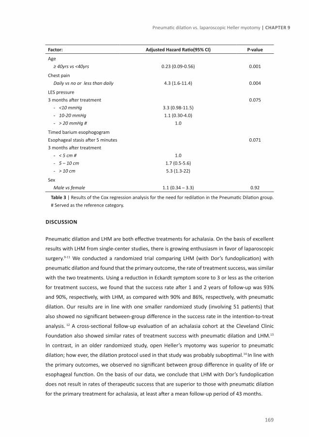
169
Pneumatic dilation vs. laparoscopic Heller myotomy | Chapter 9
Factor: Adjusted hazard Ratio(95% cI) P-value
Age ≥ 40yrs vs <40yrs 0.23 (0.09-0.56) 0.001
Chest pain Daily vs no or less than daily 4.3 (1.6-11.4) 0.004
LES pressure 3 months after treatment
- <10 mmHg - 10-20 mmHg- > 20 mmHg #
3.3 (0.98-11.5)1.1 (0.30-4.0)
1.0
0.075
Timed barium esophogogramEsophageal stasis after 5 minutes3 months after treatment
- < 5 cm #- 5 – 10 cm- > 10 cm
1.01.7 (0.5-5.6)5.3 (1.3-22)
0.071
SexMale vs female 1.1 (0.34 – 3.3) 0.92
Table 3 | Results of the Cox regression analysis for the need for redilation in the Pneumatic Dilation group. # Served as the reference category.
dIscussIon
Pneumatic dilation and LHM are both effective treatments for achalasia. On the basis of excellent
results with LHM from single-center studies, there is growing enthusiasm in favor of laparoscopic
surgery.9-11 We conducted a randomized trial comparing LHM (with Dor’s fundoplication) with
pneumatic dilation and found that the primary outcome, the rate of treatment success, was similar
with the two treatments. Using a reduction in Eckardt symptom score to 3 or less as the criterion
for treatment success, we found that the success rate after 1 and 2 years of follow-up was 93%
and 90%, respectively, with LHM, as compared with 90% and 86%, respectively, with pneumatic
dilation. Our results are in line with one smaller randomized study (involving 51 patients) that
also showed no significant between-group difference in the success rate in the intention-to-treat
analysis. 12 A cross-sectional follow-up evaluation of an achalasia cohort at the Cleveland Clinic
Foundation also showed similar rates of treatment success with pneumatic dilation and LHM.13
In contrast, in an older randomized study, open Heller’s myotomy was superior to pneumatic
dilation; how ever, the dilation protocol used in that study was probably suboptimal.14 In line with
the primary outcomes, we observed no significant between group difference in quality of life or
esophageal function. On the basis of our data, we conclude that LHM with Dor’s fundoplication
does not result in rates of therapeutic success that are superior to those with pneumatic dilation
for the primary treatment for achalasia, at least after a mean follow-up period of 43 months.

170
Part II | Achalasia
Figure 3 | Percentage of PD patients requiring redilation after the initial PD series for recurrent symptoms and percentage of LHM patients requiring PD after failure of LHM.
Success rates reported in the literature vary widely depending on the criteria used to define
success. In particular, if efficacy is defined as the lack of need for any subsequent intervention,15
the success rate with pneumatic dilation is much lower than that with surgery. The use of repeated
dilations to treat recurrent symptoms is, however, a generally accepted strategy in clinical practice
and leads to excellent control of symptoms, even during long-term follow-up.16-18 In line with
these studies, we allowed patients who were randomly assigned to pneumatic dilation to undergo
additional pneumatic dilations if symptoms recurred. The number of pneumatic dilations was
limited to a maximum of three series of dilations, each comprising up to two or three dilation
procedures, but the third and final series was allowed only if it took place more than 2 years
after the second series. One might argue, therefore, that with longer follow-up, more than three
series of dilations may be required to control symptoms, thus leading to lower success rates with
pneumatic dilation. On the other hand, differences in the dilation protocol between our study and
the study at the Cleveland Clinic Foundation (in which the balloon pressure was increased up to 10
to 12 PSI to obliterate the balloon waist)13 and differences in the length of the cut in the myotomy
between our study and a 2003 study (in which the length of the cut extended up to 3 cm in the
stomach) 19 may have led to differences in the rates of therapeutic success. Nevertheless, the
treatment protocols included in our study are internationally accepted and widely used in clinical
practice.
Previous studies have identified baseline chest pain (as assessed on a scale of 0 to 5, with 0
indicating no pain and 5 indicating daily pain), basal pressure at the lower esophageal sphincter
above 30 mm Hg, a sigmoid esophagus, and a long duration of symptoms as predictors of a
negative outcome with laparoscopic surgery in multivariate analyses.20,21 On the other hand,
younger age (<40 years) and higher pressure at the lower esophageal sphincter after treatment
have been reported as predictors of a negative outcome with pneumatic dilation.17,22 In our study,
preexisting daily chest pain, the height of the bariumcontrast column 5 minutes after ingestion
171
Pneumatic dilation vs. laparoscopic Heller myotomy | Chapter 9
of barium, and a width of the esophagus of less than 4 cm before treatment were identified as
predictors of treatment failure in a Cox regression analysis. These data confirm that monitoring
esophageal emptying after treatment is a helpful tool for predicting recurrence23 and for deciding
whether further dilation is required. The reason that a diameter of the esophagus less than 4 cm
before treatment is associated with treatment failure is unclear, unless it may be indicative of
type III achalasia, which is known to have a worse outcome.24 Finally, our data indicate that chest
pain is a difficult symptom to resolve with either treatment and contributes substantially to the
need for retreatment and to patient dissatisfaction.25 Although age was not a predictive factor
for clinical success with either treatment, we did observe a greater need for redilation in patients
younger than 40 years of age in the pneumatic-dilation group. This finding seems to support the
recommendation26 that younger patients (especially men) should be treated preferentially with
LHM.
In the first 13 patients randomly assigned to pneumatic dilation, the initial pneumatic dilation
was performed with the use of a 35-mm balloon, with the result that perforations occurred in 4
of the patients. As a consequence, the distension protocol was amended to specify that the first
pneumatic dilation should be performed with a 30-mm balloon; this amendment led to a substantial
reduction in the rate of perforation, to 4%. No other risk factor for perforation other than balloon
size and older age could be identified. The significantly higher perforation rate associated with
the use of a 35-mm balloon for the first dilation argues in favor of a graded distention protocol
in which a 30-mm balloon is used during the initial pneumatic dilation, with a larger balloon for
subsequent dilations. In the surgery group, mucosal tears, which were repaired immediately
during surgery, occurred in 12% of the patients, a rate that is similar to that previously reported.27
The clinical outcome was not affected by this complication. The most frequent complication of
both treatments was gastroesophageal reflux.15,27-29 Abnormal exposure to esophageal acid was
observed in 15% of the patients treated with pneumatic dilation and 23% of the patients treated
with LHD. These data raise the question of whether proper screening and treatment of increased
acid exposure are required to avoid long-term complications such as Barrett’s esophagus, stenosis,
or even esophageal carcinoma.29,30
The strengths of our study include the large number of patients enrolled; the fact that the study
was performed at 14 study centers in five European countries, making our conclusions widely
applicable (probably in the United States as well); the fact that objective measures were used for
the assessment of clinical success and functional improvement in the case of both treatments;
and the fact that randomization was stratified according to center. Our data showed that LHM
was not associated with rates of therapeutic success that were superior to those with pneumatic
dilation and suggest that graded dilation starting with a 30-mm balloon is a reasonable protocol
for pneumatic dilation.

172
Part II | Achalasia
ReFeRence lIsT
1. Boeckxstaens GE. Achalasia. Best Pract Res Clin Gastroenterol 2007;21:595-608.
2. Ferguson MK. Achalasia: current evaluation and therapy. Ann Thorac Surg 1991;52:336-342.
3. Costantini M, Zaninotto G, Guirroli E, Rizzetto C, Portale G, Ruol A, Nicoletti L, Ancona E. The laparoscopic Heller-Dor operation remains an effective treatment for esophageal achalasia at a minimum 6-year follow-up. Surg Endosc 2005;19:345-351.
4. Eckardt VF. Clinical presentations and complications of achalasia. Gastrointest Endosc Clin N Am 2001;11:281-92, vi.
5. Brazier JE, Harper R, Jones NM, O’Cathain A, Thomas KJ, Usherwood T, Westlake L. Validating the SF-36 health survey questionnaire: new outcome measure for primary care. BMJ 1992;305:160-164.
6. Blazeby JM, Alderson D, Winstone K, Steyn R, Hammerlid E, Arraras J, Farndon JR. Development of an EORTC questionnaire module to be used in quality of life assessment for patients with oesophageal cancer. The EORTC Quality of Life Study Group. Eur J Cancer 1996;32A:1912-1917.
7. de Oliveira JM, Birgisson S, Doinoff C, Einstein D, Herts B, Davros W, Obuchowski N, Koehler RE, Richter J, Baker ME. Timed barium swallow: a simple technique for evaluating esophageal emptying in patients with achalasia. AJR Am J Roentgenol 1997;169:473-479.
8. Lundell LR, Dent J, Bennett JR, Blum AL, Armstrong D, Galmiche JP, Johnson F, Hongo M, Richter JE, Spechler SJ, Tytgat GN, Wallin L. Endoscopic assessment of oesophagitis: clinical and functional correlates and further validation of the Los Angeles classification. Gut 1999;45:172-180.
9. Zaninotto G, Costantini M, Molena D, Buin F, Carta A, Nicoletti L, Ancona E. Treatment of esophageal achalasia with laparoscopic Heller myotomy and Dor partial anterior fundoplication: prospective evaluation of 100 consecutive patients. J Gastrointest Surg 2000;4:282-289.
10. Sharp KW, Khaitan L, Scholz S, Holzman MD, Richards WO. 100 consecutive minimally invasive Heller myotomies: lessons learned. Ann Surg 2002;235:631-638.
11. Patti MG, Molena D, Fisichella PM, Whang K, Yamada H, Perretta S, Way LW. Laparoscopic Heller myotomy and Dor fundoplication for achalasia: analysis of successes and failures. Arch Surg 2001;136:870-877.
12. Kostic S, Kjellin A, Ruth M, Lonroth H, Johnsson E, Andersson M, Lundell L. Pneumatic dilatation or laparoscopic cardiomyotomy in the management of newly diagnosed idiopathic achalasia. Results of a randomized controlled trial. World J Surg 2007;31:470-478.
13. Vela MF, Richter JE, Khandwala F, Blackstone EH, Wachsberger D, Baker ME, Rice TW. The long-term efficacy of pneumatic dilatation and Heller myotomy for the treatment of achalasia. Clin Gastroenterol Hepatol 2006;4:580-587.
14. Csendes A, Velasco N, Braghetto I, Henriquez A. A prospective randomized study comparing forceful dilatation and esophagomyotomy in patients with achalasia of the esophagus. Gastroenterology 1981;80:789-795.
15. Lopushinsky SR, Urbach DR. Pneumatic dilatation and surgical myotomy for achalasia. JAMA 2006;296:2227-2233.
16. Zerbib F, Thetiot V, Richy F, Benajah DA, Message L, Lamouliatte H. Repeated pneumatic dilations as long-term maintenance therapy for esophageal achalasia. Am J Gastroenterol 2006;101:692-697.

173
Pneumatic dilation vs. laparoscopic Heller myotomy | Chapter 9
17. Hulselmans M, Vanuytsel T, Degreef T, Sifrim D, Coosemans W, Lerut T, Tack J. Long-term outcome of pneumatic dilation in the treatment of achalasia. Clin Gastroenterol Hepatol 2010;8:30-35.
18. Bravi I, Nicita MT, Duca P, Grigolon A, Cantu P, Caparello C, Penagini R. A pneumatic dilation strategy in achalasia: prospective outcome and effects on oesophageal motor function in the long term. Aliment Pharmacol Ther 2010;31:658-665.
19. Oelschlager BK, Chang L, Pellegrini CA. Improved outcome after extended gastric myotomy for achalasia. Arch Surg 2003;138:490-495.
20. Zaninotto G, Costantini M, Rizzetto C, Zanatta L, Guirroli E, Portale G, Nicoletti L, Cavallin F, Battaglia G, Ruol A, Ancona E. Four hundred laparoscopic myotomies for esophageal achalasia: a single centre experience. Ann Surg 2008;248:986-993.
21. Schuchert MJ, Luketich JD, Landreneau RJ, Kilic A, Gooding WE, velo-Rivera M, Christie NA, Gilbert S, Pennathur A. Minimally-invasive esophagomyotomy in 200 consecutive patients: factors influencing postoperative outcomes. Ann Thorac Surg 2008;85:1729-1734.
22. Eckardt VF, Gockel I, Bernhard G. Pneumatic dilation for achalasia: late results of a prospective follow up investigation. Gut 2004;53:629-633.
23. Vaezi MF, Baker ME, Achkar E, Richter JE. Timed barium oesophagram: better predictor of long term success after pneumatic dilation in achalasia than symptom assessment. Gut 2002;50:765-770.
24. Pandolfino JE, Kwiatek MA, Nealis T, Bulsiewicz W, Post J, Kahrilas PJ. Achalasia: a new clinically relevant classification by high-resolution manometry. Gastroenterology 2008;135:1526-1533.
25. Eckardt VF, Stauf B, Bernhard G. Chest pain in achalasia: patient characteristics and clinical course. Gastroenterology 1999;116:1300-1304.
26. Richter JE. A young man with a new diagnosis of achalasia. Clin Gastroenterol Hepatol 2008;6:859-863.
27. Campos GM, Vittinghoff E, Rabl C, Takata M, Gadenstatter M, Lin F, Ciovica R. Endoscopic and surgical treatments for achalasia: a systematic review and meta-analysis. Ann Surg 2009;249:45-57.
28. Leeuwenburgh I, van DH, Scholten P, Hansen BE, Haringsma J, Siersema PD, Kuipers EJ. Oesophagitis is common in patients with achalasia after pneumatic dilatation. Aliment Pharmacol Ther 2006;23:1197-1203.
29. Csendes A, Braghetto I, Burdiles P, Korn O, Csendes P, Henriquez A. Very late results of esophagomyotomy for patients with achalasia: clinical, endoscopic, histologic, manometric, and acid reflux studies in 67 patients for a mean follow-up of 190 months. Ann Surg 2006;243:196-203.
30. Leeuwenburgh I, Haringsma J, van DH, Scholten P, Siersema PD, Kuipers EJ. Long-term risk of oesophagitis, Barrett’s oesophagus and oesophageal cancer in achalasia patients. Scand J Gastroenterol Suppl 2006;7-10.


Chapter 10Outcomes of Treatment for Achalasia
depend on Manometric SubtypeWout Rohof and Renato Salvador , Vito Annese,
Stanislas Bruley des Varannes, Stanislas Chaussade, Mario Costantini, Ignasi Elizalde, Marianne Gaudric, André Smout, Jan Tack, Olivier Busch,
Giovanni Zaninotto, Guy Boeckxstaens
Gastroenterology, e-pub ahead of print
176
Part II | Achalasia
AbsTRAcT
Background and aims:
Achalasia is currently treated by pneumatic dilation (PD) or laparoscopic Heller myotomy (LHM)
with comparable success rates. Recently, a new subtype classification for achalasia has been
described associated with different response to treatment. In this study we evaluated the impact
of the manometric subtype on therapeutic success in a large study population randomized to PD
or LHM (i.e. the European achalasia trial).
Methods:
Esophageal pre-treatment manometries were collected from patients included in the European
achalasia trial. Symptoms (weight loss, dysphagia, retrosternal pain and regurgitation) were
assessed using the Eckardt score, and treatment was considered successful if the Eckardt score <
3. Manometric tracings were classified into the three types according to the Chicago classification.
Results:
Manometric tracings of 176 of the 201 randomised patients could be collected and analyzed. Type
I, II and III achalasia were observed in 44 (25%), 114 (65%) and 18 (10%) patients respectively. After
a minimum follow-up of two years, success rates were significantly higher in patients with type II
(96%), compared to type I (81%, p<0.01 log-rank) and type III achalasia (66%, p<0.001, log-rank).
Treatment success of PD was significantly higher in type II achalasia compared to LHM (100% vs.
93%, p<0.05). In type III patients the success rate after LHM was higher than after PD (86% vs.
40%), however, due to the small patient number, statistical significance was not reached (p=0.12).
For type I achalasia, LHM and PD had similar success rates (81% vs. 85%, p=0.84).
Conclusion:
Achalasia types I and III are important predictors of treatment failure compared to type II. Success
rates in type II are high for both treatment groups, but significantly higher in the PD group. Patients
with type III can probably best be treated by LHM.
177
Treatment outcome depends on manometric subtype | Chapter 10
InTRoducTIon
Achalasia is a rare motility disorder of the esophagus characterized by the absence of peristalsis
and a defective relaxation of the lower esophageal sphincter (LES) resulting in an impaired bolus
transport and stasis of food in the esophagus.1 As its exact aetiology is still unknown, current
treatment options of achalasia are only directed at relieving the functional obstruction at the
level of the LES and consist mainly of endoscopic pneumatic dilation (PD) and laparoscopic Heller
myotomy (LHM).2-6
It is generally accepted that the loss of enteric neurons, in particular nitric oxide releasing neurons,
is responsible for the lack of peristalsis and impaired relaxation of the LES upon swallowing.1, 7,
8 Recently, three manometric subtypes were identified based on the residual esophageal wave
pattern: Type I, in which the esophageal body exhibits minimal contractility; type II, in which there
is no peristalsis but intermittent periods of compartmentalized esophageal pressurization; and
type III, in which there are spastic contractions in the distal esophagus. (Figure 1.) 9 Importantly, this
study suggested that the efficacy of treatment, mainly consisting of PD, strongly varies depending
on the manometric type. Success rates were indeed significantly higher for type II achalasia (96%)
compared to type I (56%) and type III (29%) achalasia. Also in patients treated by LHM, differences
in treatment success between the subtypes were confirmed, with success rates of 85%, 95%
and 70% for type I, II and III respectively. 9-11 However, these studies had a different definition of
treatment success and patients were not followed up prospectively, making comparison between
PD and LHM impossible.
Figure 1 | Based on the residual wave type on high resolution manometry, 3 subtypes of achalasia can be determined. No distal pressurisation is observed in type I (AI), whereas panesophageal pressurisations and spastic contractions are observed in type II (AII) and type III (AIII) respectively. As demonstrated in figure 1B a similar classification can be made when conventional manometry is used. Note that pressure recordings in type II achalasia are similar in every line tracing, compatible with panesophageal pressurisation.
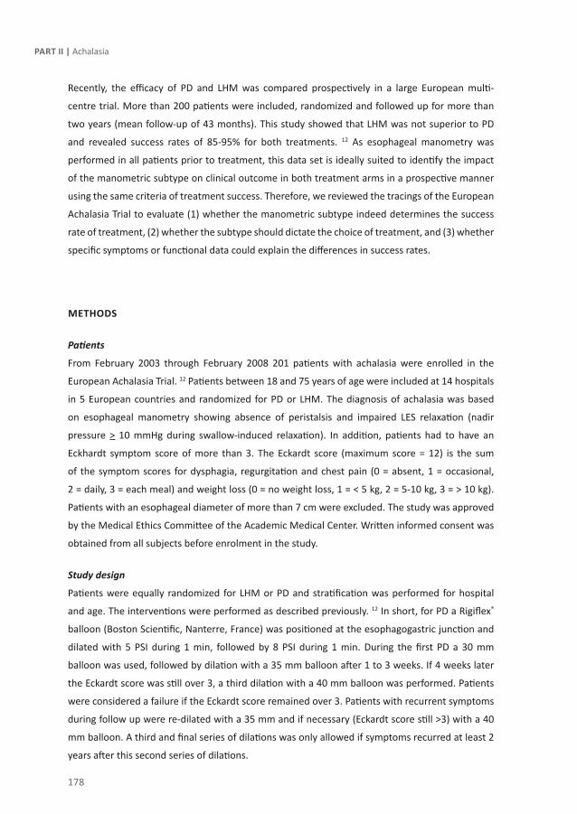
178
Part II | Achalasia
Recently, the efficacy of PD and LHM was compared prospectively in a large European multi-
centre trial. More than 200 patients were included, randomized and followed up for more than
two years (mean follow-up of 43 months). This study showed that LHM was not superior to PD
and revealed success rates of 85-95% for both treatments. 12 As esophageal manometry was
performed in all patients prior to treatment, this data set is ideally suited to identify the impact
of the manometric subtype on clinical outcome in both treatment arms in a prospective manner
using the same criteria of treatment success. Therefore, we reviewed the tracings of the European
Achalasia Trial to evaluate (1) whether the manometric subtype indeed determines the success
rate of treatment, (2) whether the subtype should dictate the choice of treatment, and (3) whether
specific symptoms or functional data could explain the differences in success rates.
MeThods
Patients
From February 2003 through February 2008 201 patients with achalasia were enrolled in the
European Achalasia Trial. 12 Patients between 18 and 75 years of age were included at 14 hospitals
in 5 European countries and randomized for PD or LHM. The diagnosis of achalasia was based
on esophageal manometry showing absence of peristalsis and impaired LES relaxation (nadir
pressure > 10 mmHg during swallow-induced relaxation). In addition, patients had to have an
Eckhardt symptom score of more than 3. The Eckardt score (maximum score = 12) is the sum
of the symptom scores for dysphagia, regurgitation and chest pain (0 = absent, 1 = occasional,
2 = daily, 3 = each meal) and weight loss (0 = no weight loss, 1 = < 5 kg, 2 = 5-10 kg, 3 = > 10 kg).
Patients with an esophageal diameter of more than 7 cm were excluded. The study was approved
by the Medical Ethics Committee of the Academic Medical Center. Written informed consent was
obtained from all subjects before enrolment in the study.
Study design
Patients were equally randomized for LHM or PD and stratification was performed for hospital
and age. The interventions were performed as described previously. 12 In short, for PD a Rigiflex®
balloon (Boston Scientific, Nanterre, France) was positioned at the esophagogastric junction and
dilated with 5 PSI during 1 min, followed by 8 PSI during 1 min. During the first PD a 30 mm
balloon was used, followed by dilation with a 35 mm balloon after 1 to 3 weeks. If 4 weeks later
the Eckardt score was still over 3, a third dilation with a 40 mm balloon was performed. Patients
were considered a failure if the Eckardt score remained over 3. Patients with recurrent symptoms
during follow up were re-dilated with a 35 mm and if necessary (Eckardt score still >3) with a 40
mm balloon. A third and final series of dilations was only allowed if symptoms recurred at least 2
years after this second series of dilations.

179
Treatment outcome depends on manometric subtype | Chapter 10
In patients randomized for LHM, a myotomy was performed extending at least 6 cm over the
esophagus above the junction and at least 1-1.5 cm over the stomach. Thereafter, an anterior 180
degree fundoplication according to Dor was performed to reduce postoperative gastroesophageal
reflux. If symptoms recurred after surgery with an Eckardt score > 3, the patient was considered
a failure.
Prior to treatment, esophageal manometry was performed for the diagnosis of achalasia, and to
determine LES pressure. Furthermore, a timed barium esophagogram was conducted to quantify
esophageal stasis. After treatment, symptom scores were assessed and esophageal manometry
and timed barium esophagogram were performed after 1 month and on a yearly basis. Esophageal
manometries were retrospectively collected to determine the type of achalasia. All authors had
access to the study data and reviewed and approved the final manuscript.
Manometry
Esophageal manometry was performed using a pneumohydraulic perfusion system and a 6-10
channel esophageal manometry catheter with a sleeve sensor incorporated at the distal end.
After introduction and equilibration, basal pressure was monitored during at least 5 minutes. LES
pressure was determined end-expiratory. The esophageal pressure wave amplitude was analysed
in the two channels above the LES. These sensors were located at 3 or 4 cm and at 6 or 8 cm above
the LES respectively, depending on the design of the manometric catheter used in the participating
centres. Swallow-induced relaxation of the sphincter and esophageal pressure wave amplitude are
assessed on 10 consecutive 5 ml wet swallows, at least 30 s apart.
Timed barium esophagogram
Esophageal stasis was determined on a timed barium esophagogram 1, 2 and 5 minutes after
ingestion of the maximal tolerable amount of low density barium sulphate over 30-45 s without
regurgitation or aspiration, with the patient upright in a slight left posterior position. 13 The distance
from the tapered distal esophagus to the top of the barium column and the maximal diameter of
the esophagus were measured.
Data analysis
Manometric tracings were reviewed using MMS (Medical Measurements Systems, Enschede,
The Netherlands), Medtronic (Medtronic, Minneapolis, USA) or Dynosystem (Memphis, Bologna,
Italy) software by two reviewers (WR and RS). We classified the study patients according to their
dominant distal esophageal pressurization pattern using modified definitions from Pandolfino et
al. 9: Type I achalasia when 9/10 swallows elicited contractions with an amplitude < 30 mm Hg;
Type 2 when 2 or more contractions had an amplitude > 30 mm Hg; and Type 3 when at least
two spastic waves were detected (lasting > 6.0 sec with an amplitude > 70 mm Hg). These criteria
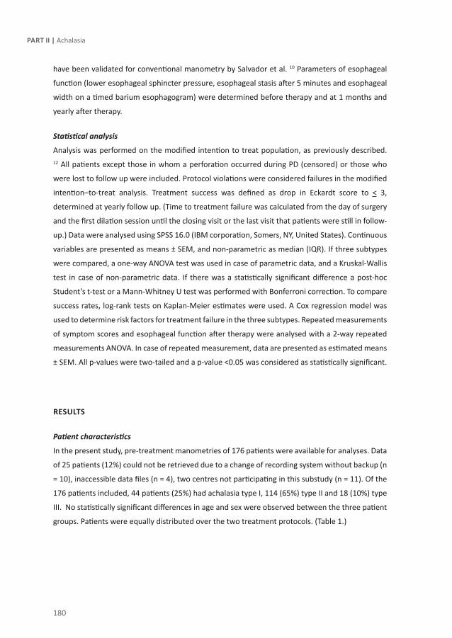
180
Part II | Achalasia
have been validated for conventional manometry by Salvador et al. 10 Parameters of esophageal
function (lower esophageal sphincter pressure, esophageal stasis after 5 minutes and esophageal
width on a timed barium esophagogram) were determined before therapy and at 1 months and
yearly after therapy.
Statistical analysis
Analysis was performed on the modified intention to treat population, as previously described. 12 All patients except those in whom a perforation occurred during PD (censored) or those who
were lost to follow up were included. Protocol violations were considered failures in the modified
intention–to-treat analysis. Treatment success was defined as drop in Eckardt score to < 3,
determined at yearly follow up. (Time to treatment failure was calculated from the day of surgery
and the first dilation session until the closing visit or the last visit that patients were still in follow-
up.) Data were analysed using SPSS 16.0 (IBM corporation, Somers, NY, United States). Continuous
variables are presented as means ± SEM, and non-parametric as median (IQR). If three subtypes
were compared, a one-way ANOVA test was used in case of parametric data, and a Kruskal-Wallis
test in case of non-parametric data. If there was a statistically significant difference a post-hoc
Student’s t-test or a Mann-Whitney U test was performed with Bonferroni correction. To compare
success rates, log-rank tests on Kaplan-Meier estimates were used. A Cox regression model was
used to determine risk factors for treatment failure in the three subtypes. Repeated measurements
of symptom scores and esophageal function after therapy were analysed with a 2-way repeated
measurements ANOVA. In case of repeated measurement, data are presented as estimated means
± SEM. All p-values were two-tailed and a p-value <0.05 was considered as statistically significant.
ResulTs
Patient characteristics
In the present study, pre-treatment manometries of 176 patients were available for analyses. Data
of 25 patients (12%) could not be retrieved due to a change of recording system without backup (n
= 10), inaccessible data files (n = 4), two centres not participating in this substudy (n = 11). Of the
176 patients included, 44 patients (25%) had achalasia type I, 114 (65%) type II and 18 (10%) type
III. No statistically significant differences in age and sex were observed between the three patient
groups. Patients were equally distributed over the two treatment protocols. (Table 1.)

181
Treatment outcome depends on manometric subtype | Chapter 10
Type I Type II Type III P-value
Number of patients (no.(%)) 44 (25) 114 (65) 18 (10)
Age 44 ± 2.4 46 ± 1.4 49 ± 3.4 0.43
Sex (no.)MaleFemale
2519
6549
810
0.58
Treatment protocol (no.)PDLHM
2222
5361
108
0.76
Symptoms
Total Eckardt score 7.3 ± 0.28 7.1 ± 0.19 7.4 ± 0.49 0.68
Chest painDysphagiaRegurgitationWeight loss
1 (1-1)3 (3-3)2 (1-2)2 (1-3)
1 (0-1)3 (3-3)2 (1-3)1 (0-2)
2 (1-2)#
3 (3-3)1 (1-3)1 (0-2)
<0.010.810.740.46
Esophageal function
LES pressure (mmHg) 28 ± 2.3 31 ± 1.4 34 ± 5.3 0.17
Mean distal esophageal pressure wave amplitude (mmHg)
17 ± 1.0 38 ± 1.7 81 ± 8.3 <0.0001
Timed barium esophagogram – Height of stasis (cm)
1 min2 min5 min
16 ± 1.116 ± 1.114 ± 1.1
14 ± 0.713 ± 0.712 ± 0.7
14 ± 1.612 ± 1.610 ± 1.7
0.390.170.15
Esophageal width 4.5 ± 0.23 4.0 ± 0.12 3.1 ± 0.30 <0.01
Table 1 | Table 1 demonstrates patient demographics, symptom scores and parameters of esophageal function, all before treatment. # p<0.05 vs type I and p<0.01 vs type II.
Pre-treatment Eckhardt symptom scores were similar for all types, with mean values of 7.3 ± 0.3,
7.1 ± 0.2 and 7.4 ± 0.5 for type I, II and II respectively. Type III achalasia patients had a significantly
higher median chest pain score of 2 (IQR 1-2) compared to type I (median 1 (IQR 1-1) p=0.03) and
type II patients (median 1 (IQR 0-1) p<0.01). Other symptoms (dysphagia, regurgitation and weight
loss) were similar in the 3 treatment groups. Prior to treatment, LES pressure and the height of
the barium column (assessed after 5 minutes) were comparable in the three treatment groups.
(Figure 2.) The mean amplitude measured by the pressure sensors in the distal esophagus during
wet swallows was higher in type III achalasia, compared to type I (p<0.001) and type II (p<0.001).
Lastly, esophageal width was significantly larger in type I patients (4.5 ± 0.23 cm) compared to type
II (4.0 ± 0.12 cm, p=0.02) and type III (3.1 ± 0.30 cm, p<0.001).
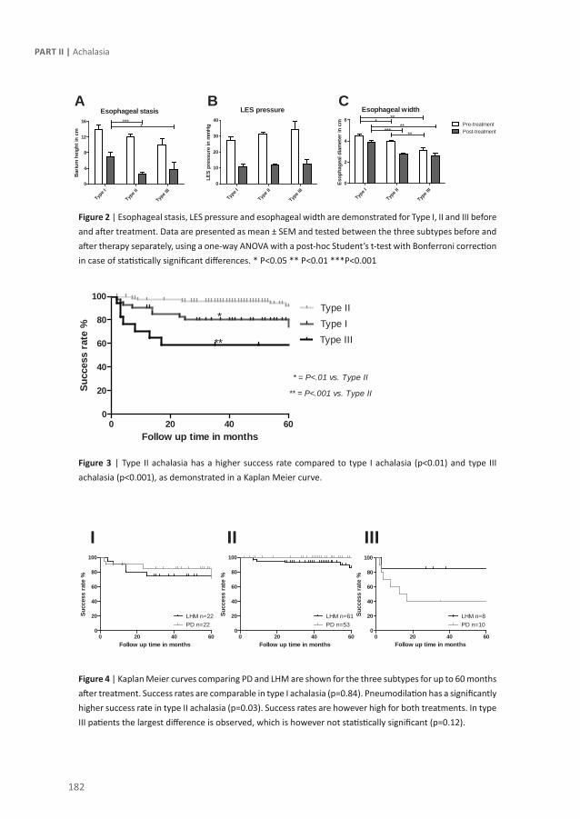
182
Part II | Achalasia
Figure 2 | Esophageal stasis, LES pressure and esophageal width are demonstrated for Type I, II and III before and after treatment. Data are presented as mean ± SEM and tested between the three subtypes before and after therapy separately, using a one-way ANOVA with a post-hoc Student’s t-test with Bonferroni correction in case of statistically significant differences. * P<0.05 ** P<0.01 ***P<0.001
Figure 3 | Type II achalasia has a higher success rate compared to type I achalasia (p<0.01) and type III achalasia (p<0.001), as demonstrated in a Kaplan Meier curve.
Figure 4 | Kaplan Meier curves comparing PD and LHM are shown for the three subtypes for up to 60 months after treatment. Success rates are comparable in type I achalasia (p=0.84). Pneumodilation has a significantly higher success rate in type II achalasia (p=0.03). Success rates are however high for both treatments. In type III patients the largest difference is observed, which is however not statistically significant (p=0.12).
Esophageal stasis
Type I
Type I
I
Type I
II0
4
8
12
16 ****
Bar
ium
hei
ght i
n cm
LES pressure
Type I
Type I
I
Type I
II0
10
20
30
40
LES
pres
sure
in m
mH
g
Esophageal width
Type I
Type I
I
Type I
II0
2
4
6Pre-treatmentPost-treatment
*****
*** **
Esop
hage
al d
iam
eter
in c
m
A B C
0 20 40 600
20
40
60
80
100
PD n=22LHM n=22
Follow up time in months
Succ
ess
rate
%
0 20 40 600
20
40
60
80
100
LHM n=61PD n=53
Follow up time in months
Succ
ess
rate
%
0 20 40 600
20
40
60
80
100
LHM n=8PD n=10
Follow up time in months
Succ
ess
rate
%
I II III
0 20 40 600
20
40
60
80
100
Type IType II
Type III
* = P<.01 vs. Type II
** = P<.001 vs. Type II
*
**
Follow up time in months
Succ
ess
rate
%
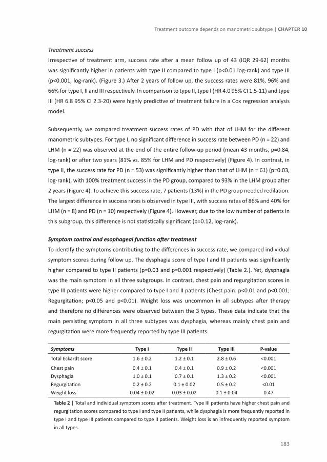
183
Treatment outcome depends on manometric subtype | Chapter 10
Treatment success
Irrespective of treatment arm, success rate after a mean follow up of 43 (IQR 29-62) months
was significantly higher in patients with type II compared to type I (p<0.01 log-rank) and type III
(p<0.001, log-rank). (Figure 3.) After 2 years of follow up, the success rates were 81%, 96% and
66% for type I, II and III respectively. In comparison to type II, type I (HR 4.0 95% CI 1.5-11) and type
III (HR 6.8 95% CI 2.3-20) were highly predictive of treatment failure in a Cox regression analysis
model.
Subsequently, we compared treatment success rates of PD with that of LHM for the different
manometric subtypes. For type I, no significant difference in success rate between PD (n = 22) and
LHM (n = 22) was observed at the end of the entire follow-up period (mean 43 months, p=0.84,
log-rank) or after two years (81% vs. 85% for LHM and PD respectively) (Figure 4). In contrast, in
type II, the success rate for PD (n = 53) was significantly higher than that of LHM (n = 61) (p=0.03,
log-rank), with 100% treatment success in the PD group, compared to 93% in the LHM group after
2 years (Figure 4). To achieve this success rate, 7 patients (13%) in the PD group needed redilation.
The largest difference in success rates is observed in type III, with success rates of 86% and 40% for
LHM (n = 8) and PD (n = 10) respectively (Figure 4). However, due to the low number of patients in
this subgroup, this difference is not statistically significant (p=0.12, log-rank).
Symptom control and esophageal function after treatment
To identify the symptoms contributing to the differences in success rate, we compared individual
symptom scores during follow up. The dysphagia score of type I and III patients was significantly
higher compared to type II patients (p=0.03 and p=0.001 respectively) (Table 2.). Yet, dysphagia
was the main symptom in all three subgroups. In contrast, chest pain and regurgitation scores in
type III patients were higher compared to type I and II patients (Chest pain: p<0.01 and p<0.001;
Regurgitation; p<0.05 and p<0.01). Weight loss was uncommon in all subtypes after therapy
and therefore no differences were observed between the 3 types. These data indicate that the
main persisting symptom in all three subtypes was dysphagia, whereas mainly chest pain and
regurgitation were more frequently reported by type III patients.
Symptoms Type I Type II Type III P-value
Total Eckardt score 1.6 ± 0.2 1.2 ± 0.1 2.8 ± 0.6 <0.001
Chest painDysphagiaRegurgitationWeight loss
0.4 ± 0.11.0 ± 0.10.2 ± 0.2
0.04 ± 0.02
0.4 ± 0.10.7 ± 0.1
0.1 ± 0.020.03 ± 0.02
0.9 ± 0.21.3 ± 0.2 0.5 ± 0.2
0.1 ± 0.04
<0.001<0.001<0.010.47
Table 2 | Total and individual symptom scores after treatment. Type III patients have higher chest pain and regurgitation scores compared to type I and type II patients, while dysphagia is more frequently reported in type I and type III patients compared to type II patients. Weight loss is an infrequently reported symptom in all types.
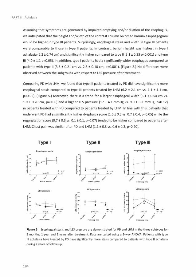
184
Part II | Achalasia
Assuming that symptoms are generated by impaired emptying and/or dilation of the esophagus,
we anticipated that the height and/width of the contrast column on timed barium esophagogram
would be higher in type III patients. Surprisingly, esophageal stasis and width in type III patients
were comparable to those in type II patients. In contrast, barium height was highest in type I
achalasia (6.2 ± 0.74 cm) and significantly higher compared to type II (3.1 ± 0.33 p<0.001) and type
III (4.0 ± 1.1 p<0.05). In addition, type I patients had a significantly wider esophagus compared to
patients with type II (3.6 ± 0.21 cm vs. 2.8 ± 0.10 cm, p<0.001). (Figure 2.) No differences were
observed between the subgroups with respect to LES pressure after treatment.
Comparing PD with LHM, we found that type III patients treated by PD did have significantly more
esophageal stasis compared to type III patients treated by LHM (6.2 ± 2.1 cm vs. 1.1 ± 1.1 cm,
p<0.05). (Figure 5.) Moreover, there is a trend for a larger esophageal width (3.1 ± 0.54 cm vs.
1.9 ± 0.20 cm, p=0.06) and a higher LES pressure (17 ± 4.1 mmHg vs. 9.0 ± 3.2 mmHg, p=0.12)
in patients treated with PD compared to patients treated by LHM. In line with this, patients that
underwent PD had a significantly higher dysphagia score (1.6 ± 0.3 vs. 0.7 ± 0.4, p<0.05) while the
regurgitation score (0.7 ± 0.3 vs. 0.1 ± 0.1, p=0.07) tended to be higher compared to patients after
LHM. Chest pain was similar after PD and LHM (1.1 ± 0.3 vs. 0.6 ± 0.2, p=0.20).
Figure 5 | Esophageal stasis and LES pressure are demonstrated for PD and LHM in the three subtypes for 3 months, 1 year and 2 years after treatment. Data are tested using a 2-way ANOVA. Patients with type III achalasia have treated by PD have significantly more stasis compared to patients with type II achalasia during 2 years of follow up.
Esophageal stasis
0
2
4
6
8
10
3 months
1 year
2 years
p=0.91 PDLHM
Follow up time
Hei
ght o
f bar
ium
(in
cm)
Esophageal stasis
0
2
4
6
8
10
p=0.48
3 months
1 year
2 years
LHMPD
Follow up time
Hei
ght o
f bar
ium
(in
cm)
Esophageal stasis
0
2
4
6
8
10
LHMPDp=0.007
3 mon
ths1 y
ear
2 yea
rs
Follow up time
Hei
ght o
f bar
ium
(in
cm)
LES pressure
0
5
10
15
20
25
p=0.28
3 months
1 year
2 years
PDLHM
Follow up time
LES
pres
sure
in m
mH
g
LES pressure
0
5
10
15
20
25
p=0.0001
3 mon
ths1 y
ear
2 yea
rs
LHMPD
Follow up time
LES
pres
sure
in m
mH
g
LES pressure
0
5
10
15
20
25
LHMPDp=0.12
3 months
1 year
2 years
Follow up time
LES
pres
sure
in m
mH
g
Type I Type IIIType II

185
Treatment outcome depends on manometric subtype | Chapter 10
Evolution of subtypes after treatment
It is hypothesized that the three types represent a different stage of achalasia. 9, 14 To define
the course of the subtypes after treatment, we collected 397 post-treatment manometries of
107 patients (median 2 post-treatment manometries [range 1-8]). During follow up, the mean
contraction amplitude remained the lowest in type I patients (type I: 17 ± 0.9 mmHg, type II: 37
± 1.5 mmHg and type III: 78 ± 7.6 mmHg, p<0.0001, one way anova). After therapy, most patients
with type I achalasia were classified as having absent peristalsis (n=16, 89%), while simultaneous
contractions (with an amplitude of >30mmHg) were observed in 11% of patients (n=2), classified
as having esophageal spasm in the context of treated achalasia. In contrast, in patients with type
II achalasia, only 7% of patients were classified as having absent peristalsis. In these patients this
pattern was observed starting after a mean of 2.6 years after treatment. Spastic or simultaneous
contractions were observed in follow up manometries in 83% of type II patients, and weak
peristalsis in 10% of patients. All type III patients were classified as having esophageal spasms
during the entire follow up period.
dIscussIon
Different success rates have been reported for the three manometric subtypes of achalasia
suggesting that classification by manometry may be useful to determine the treatment of choice. 9-11 In the present study we confirmed that the manometric subtype is indeed an important
determinant of clinical success with type I and especially type III achalasia having an increased
odds rate for treatment failure compared to type II. The main symptoms for treatment failure are
dysphagia in type I and dysphagia, chest pain and regurgitation in type III. Of note, type I and II
patients, representing the majority (90%) of achalasia patients, respond excellent to both LHM
and PD during a mean follow-up of 43 months with only a small difference in success rate between
PD and LHM in type II patients (100% vs 93% respectively). Mainly patients with type III have an
impaired response rate to PD, but due to small patient number, there was no significant difference
compared to LHM. Based on our current data, we conclude that achalasia subtyping is clinically
helpful to estimate the success rate irrespective of the treatment (LHM or PD) used, but may only
prove relevant to determine the most optimal treatment option in type III patients.
In achalasia, substantial variability in residual esophageal pressure patterns and dynamics is
observed using high resolution manometry. Based on the residual dominant distal esophageal
pressurization pattern, Pandolfino et al. classified achalasia into 3 types (type I: No pressurization,
type II: panesophageal pressurization and type III: rapidly propagating contractions). 9 Interestingly,
after a follow period of at least one year, the treatment success rate of type II patients (96%) was

186
Part II | Achalasia
significantly higher compared to type I (56%) and type III (29%) patients. Moreover, the number
of interventions (PD, LHM and Botox combined) was twice as high in type III compared to type
II, suggesting that patients with type II respond better to treatment. Due to the low number, no
comparison could be made between the different treatments used. In the same line, Salvador et
al reported better clinical response to LHM (n=246) for type II patients (success rates type I=85%
(82/96), type II= 95% (121/127), and type III=70% (16/23; p=0.0007) 10. Although the follow-up
period in our study was longer (median of 43 months) compared to the previous two studies
(ranging between 6 and 31 months), our results are largely comparable, confirming that the
highest success rate was observed in type II achalasia (96% after 2 years) compared to type I (81%,
p<0.01) and type III (66%, p<0.001). Also in a Cox regression analysis model, type I and type III were
identified as risk factor for treatment failure with a hazard ratio of 4.0 and 6.8 respectively. Taken
together we confirmed that type I and in particular type III achalasia are important predictors of
treatment failure.
In this study we provide more insight in the functional differences and the symptoms underlying
treatment failure in the different subtypes. Chest pain scores before as well as after treatment
are higher in patients with type III achalasia compared to type I and II. Although chest pain has
repeatedly been shown to be an independent predictor of therapeutic failure, the mechanisms
leading to this symptom are incompletely understood. 3, 12 It is hypothesized that chest pain is
mainly evoked by high amplitude esophageal contractions rather than esophageal widening. 9 Our
data are in line with this hypothesis as type III patients have the narrowest esophagus and the
highest contraction amplitude, associated with the highest chest pain score. It should also be
noted though that patients with type III achalasia reported more symptoms of dysphagia and
regurgitation than patients with type I and II. Patients with type I achalasia had a higher dysphagia
score after treatment compared to patients with type II achalasia. The higher dysphagia score was
associated with impaired esophageal emptying on timed barium esophagogram in type I patients.
Moreover, type I patients had a significantly wider esophagus. Esophageal stasis and a wide
esophagus have been identified as risk factors for treatment failure in multiple earlier studies. 3,
15, 16 For instance, Vaezi et al demonstrated that 90% of patients with persistent stasis but without
symptoms failed therapy within one year after initial treatment. Furthermore, Zaninotto et al
demonstrated that a wider esophagus is an important risk factor for treatment failure in a large
study with patients treated by LHM. Combined high resolution manometry (HRM) and impedance
measurements have demonstrated that esophageal emptying in achalasia mainly occurs during
panesophageal pressurizations. 14 The absence of esophageal pressurization in type I achalasia
might therefore explain differences in esophageal stasis levels and thereby contribute to higher
dysphagia scores and impaired success rates.
187
Treatment outcome depends on manometric subtype | Chapter 10
Accepting that the manometric subtype is indeed an important predictor of clinical success, the
main clinical question that remains is to what extent the choice of treatment, i.e. PD or LHM, can be
guided by the manometric subtype. For this purpose, we compared success rates for PD and LHM
in the three subtypes. For type I achalasia LHM and PD had similar success rates (81% vs. 85%),
whereas only a small difference was observed for type II achalasia (93% vs. 100% for LHM and PD
respectively). Of note, our success rates for LHM were comparable to those reported previously,
however we obtained higher success rates in patients with type I undergoing PD (56-63% vs 85%) 9, 11 One explanation could be the fact that patients with mega-esophagus (esophageal diameter
of >7cm), known to be more difficult to treat and to present with type I achalasia, were excluded
from our study. 9, 10, 17 It should be emphasized though that only one patient was excluded in our
series based on this criterion. Alternatively, this difference may be explained by the more rigorous
distension protocol we used, i.e. our protocol allowed redilation during the first years of follow up,
which was performed in 7 type I patient (23%) and 5 type II patients (13%). Based on our findings,
we conclude that when a graded distension protocol allowing redilation is used, PD and LHM are
both appropriate treatment options for type I and type II achalasia, at least until longer follow up
data are available. 18, 19
The largest difference was observed in type III patients, in whom the success rate after LHM was
higher than after PD (86% vs. 40%). Due to the small patient number (n= 18), this difference
was however not statistically significant (p=0.12). The success rates are in line with the available
literature (LHM 70% vs. PD 33-38%), though the number of patients with type III achalasia in
previous studies is rather small as well (47 in total (14 PD, 24 LHM and 9 Botox)). 9-11 9-11 It was
interesting to note that dysphagia was especially high in type III patients treated with PD, a
finding that was associated with impaired emptying and a wider esophagus. As demonstrated
by Pandolfino et al, type III patients have a functional obstruction not only encompassing the
esophagogastric junction (EGJ) but also the distal smooth muscle segment of the esophagus. 9, 10 In
the European Achalasia Trial, the myotomy was extended 6 cm above the EGJ, which may account
for the differences in esophageal emptying, dysphagia and regurgitation, and thereby to the higher
success rates compared to PD. Therefore, in combination with the large difference in success rates
both in our study and in literature, we suggest that LHM may be the preferred treatment option
in type III patients. Still, an additional study focussing on patients with type III achalasia seems
indicated. Due to the very limited incidence of type III achalasia, additional studies probably need
to be performed in multi-center studies to provide sufficient patient numbers.
The strength of our study is the prospective randomized trial design and the relatively large number
of patients. In 12 centres in 5 European countries, patients were randomized and evaluated in a
regular follow up protocol with validated and objective outcome measures, both symptoms as

188
Part II | Achalasia
well as functional outcome measures. As a result, we were able to compare the effect of the
two standard treatments in three different manometric subtypes. In addition, this study provided
more insight in functional differences and symptoms of treatment failure in the different subtypes.
A possible limitation of our study is the use of conventional manometry instead of HRM, for which
the classification was designed. However, during the initiation of the study in 2003, HRM was not
available in the participating centres, and diagnosis was based on conventional manometry with
a sleeve sensor. Of note, Salvador et al. reported 100% agreement between the classification of
subtypes based on conventional pressure line tracings versus HRM plots.10 Although classification
of subtypes based on conventional might be less precise than the sophisticated HRM-based
classification, our three patient groups were similar to those reported by Pandolfino, in terms of
both clinical features and outcome after therapy.9 Therefore, we are confident that our conclusions
are of clinical significance.
In conclusion, achalasia types I and III are important predictors of treatment failure compared
to type II, and therefore achalasia subtyping is useful to determine the risk for treatment failure.
Success rates in types I and II are high for both treatment groups, whereas patients with type III
have an impaired treatment response, primarily after PD. This implies that patients with type III
achalasia may better be treated by LHM, or be included in a more rigorous follow up protocol.
189
Treatment outcome depends on manometric subtype | Chapter 10
ReFeRence lIsT
1. Boeckxstaens GE. The lower oesophageal sphincter. Neurogastroenterol Motil 2005;17 Suppl 1:13-21.
2. Richter JE. Modern management of achalasia. Curr Treat Options Gastroenterol 2005;8:275-283.
3. Zaninotto G, Costantini M, Rizzetto C, Zanatta L, Guirroli E, Portale G, Nicoletti L, Cavallin F, Battaglia G, Ruol A, Ancona E. Four hundred laparoscopic myotomies for esophageal achalasia: a single centre experience. Ann Surg 2008;248:986-993.
4. Patti MG, Pellegrini CA, Horgan S, Arcerito M, Omelanczuk P, Tamburini A, Diener U, Eubanks TR, Way LW. Minimally invasive surgery for achalasia: an 8-year experience with 168 patients. Ann Surg 1999;230:587-593.
5. Vaezi MF, Richter JE, Wilcox CM, Schroeder PL, Birgisson S, Slaughter RL, Koehler RE, Baker ME. Botulinum toxin versus pneumatic dilatation in the treatment of achalasia: a randomised trial. Gut 1999;44:231-239.
6. Richter JE, Boeckxstaens GE. Management of achalasia: surgery or pneumatic dilation. Gut 2011;60:869-876.
7. Goyal RK, Hirano I. The enteric nervous system. N Engl J Med 1996;334:1106-1115.
8. Mearin F, Mourelle M, Guarner F, Salas A, Riveros-Moreno V, Moncada S, Malagelada JR. Patients with achalasia lack nitric oxide synthase in the gastro-oesophageal junction. Eur J Clin Invest 1993;23:724-728.
9. Pandolfino JE, Kwiatek MA, Nealis T, Bulsiewicz W, Post J, Kahrilas PJ. Achalasia: a new clinically relevant classification by high-resolution manometry. Gastroenterology 2008;135:1526-1533.
10. Salvador R, Costantini M, Zaninotto G, Morbin T, Rizzetto C, Zanatta L, Ceolin M, Finotti E, Nicoletti L, Da DG, Cavallin F, Ancona E. The preoperative manometric pattern predicts the outcome of surgical treatment for esophageal achalasia. J Gastrointest Surg 2010;14:1635-1645.
11. Pratap N, Kalapala R, Darisetty S, Joshi N, Ramchandani M, Banerjee R, Lakhtakia S, Gupta R, Tandan M, Rao GV, Reddy DN. Achalasia cardia subtyping by high-resolution manometry predicts the therapeutic outcome of pneumatic balloon dilatation. J Neurogastroenterol Motil 2011;17:48-53.
12. Boeckxstaens GE, Annese V, des Varannes SB, Chaussade S, Costantini M, Cuttitta A, Elizalde JI, Fumagalli U, Gaudric M, Rohof WO, Smout AJ, Tack J, Zwinderman AH, Zaninotto G, Busch OR. Pneumatic dilation versus laparoscopic Heller’s myotomy for idiopathic achalasia. N Engl J Med 2011;364:1807-1816.
13. de Oliveira JM, Birgisson S, Doinoff C, Einstein D, Herts B, Davros W, Obuchowski N, Koehler RE, Richter J, Baker ME. Timed barium swallow: a simple technique for evaluating esophageal emptying in patients with achalasia. AJR Am J Roentgenol 1997;169:473-479.
14. Hong SJ, Bhargava V, Jiang Y, Denboer D, Mittal RK. A unique esophageal motor pattern that involves longitudinal muscles is responsible for emptying in achalasia esophagus. Gastroenterology 2010;139:102-111.
15. Vaezi MF, Baker ME, Achkar E, Richter JE. Timed barium oesophagram: better predictor of long term success after pneumatic dilation in achalasia than symptom assessment. Gut 2002;50:765-770.
190
Part II | Achalasia
16. Pechlivanides G, Chrysos E, Athanasakis E, Tsiaoussis J, Vassilakis JS, Xynos E. Laparoscopic Heller cardiomyotomy and Dor fundoplication for esophageal achalasia: possible factors predicting outcome. Arch Surg 2001;136:1240-1243.
17. Zaninotto G, Costantini M, Rizzetto C, Zanatta L, Guirroli E, Portale G, Nicoletti L, Cavallin F, Battaglia G, Ruol A, Ancona E. Four hundred laparoscopic myotomies for esophageal achalasia: a single centre experience. Ann Surg 2008;248:986-993.
18. Zerbib F, Thetiot V, Richy F, Benajah DA, Message L, Lamouliatte H. Repeated pneumatic dilations as long-term maintenance therapy for esophageal achalasia. Am J Gastroenterol 2006;101:692-697.
19. Katzka DA, Castell DO. Review article: an analysis of the efficacy, perforation rates and methods used in pneumatic dilation for achalasia. Aliment Pharmacol Ther 2011;34:832-839.


Chapter 11Esophageal stasis on a timed barium
esophagogram predicts recurrent symptoms in patients with long-standing
achalasiaWout Rohof, Aaltje Lei, Guy Boeckxstaens
Am J Gastroenterol. 2012 Sep 25 epub ahead of print.

194
Part II | Achalasia
AbsTRAcT
Objective:
In achalasia, early recognition of the need for retreatment is of crucial importance to reduce
morbidity and long-term complications such as esophageal decompensation. In clinical practice,
symptoms and parameters of esophageal function including lower esophageal sphincter (LES)
pressure and esophageal emptying are used to decide whether additional treatment is required.
However, which of these tests performs best remains unclear.
Methods:
A cohort of 41 patients with longstanding achalasia (median 17 yrs), underwent esophageal
manometry, timed barium esophagogram and symptom evaluation. Patients were followed up for
ten years, and were regarded as a therapeutic failure if Eckardt score was > 3 or when retreatment
was needed. Predictors of therapeutic failure were evaluated.
Results:
Of the 41 included patients, 7 patients had an elevated LES pressure (>10mmHg) and 26 had
esophageal stasis >5cm on timed barium esophagogram. During follow up, 25 patients had
recurrence of symptoms and were considered therapeutic failures. Five of these 25 patients had
an elevated LES pressure, whereas 22 had esophageal stasis on barium esophagogram. Hence,
the sensitivity to predict the need of retreatment is higher for esophageal stasis (88%) compared
to LES pressure (20%). Sixteen patients (39%) were in long-term remission, of which 12 patients
(75%) did not have stasis at their initial visit.
conclusion:
In contrast to LES pressure, esophageal stasis is a good predictor of treatment failure in patients with
longstanding achalasia. Based on these findings, we propose to use timed barium esophagogram
rather than esophageal manometry as test to decide on retreatment.

195
Esophageal stasis predicts recurrent symptoms | Chapter 11
InTRoducTIon
Achalasia is a primary esophageal motility disorder characterized by the absence of peristalsis
in the distal ⅔ of the esophagus and a defective relaxation of the lower esophageal sphincter
(LES). This leads to impaired bolus propulsion and stasis of food in the esophagus and thereby
to symptoms of dysphagia, regurgitation of undigested food, retrosternal pain and weight loss.1
Treatment is aimed at reducing LES pressure and thereby at improving bolus transport. Although
initial treatment success rates are as high as 90% after 2 years, success rates decline in time, leading
to retreatment in many patients.2-5 Identification of patients in need of retreatment is challenging
and often delayed, as symptoms are underreported or absent due to decompensation of the
esophagus or impaired esophageal sensitivity. Moreover, patients get used to symptoms or adapt
their diet.6,7 However, timely recognition of functional obstruction and subsequent stasis of food
in the esophagus is of major importance for at least 2 reasons: First, continuous obstruction and
stasis can result in esophageal decompensation and mega-esophagus or sigmoid-like esophagus
associated with higher morbity and refractory symptoms.8-10 Second, esophageal stasis has been
suggested to be a risk factor for the development of dysplasia and neoplasia, especially in patients
with longstanding disease.11,12 Therefore, objective quantification of esophageal and LES function
during the follow up and management of patients with achalasia, especially with longstanding
disease, is of imminent importance to prevent these complications.
Manometry has been repeatedly proposed as a useful test to determine whether patients should
be retreated. Indeed, several studies have shown that LES pressure ≥ 10 mmHg is a predictor of
treatment failure,5,13 suggesting that patients with LES pressure ≥ 10 mmHg should be considered
for additional therapy. However, a significant proportion of patients with persistent symptoms
has a low or even absent LES pressure, questioning the validity of this parameter. Alternatively,
assessment of esophageal emptying has been proposed to better detect the need for additional
treatment in naive patients within one year after initial therapy with pneumatic dilation.6 To what
extent this test performs better than esophageal manometry to predict the need for retreatment
during longer follow up, and might be the test of choice to routinely monitor patients with
longstanding disease has not been evaluated. Therefore, we compared esophageal manometry
and timed barium esophagogram to predict the need for retreatment during a prolonged period
of follow up in a cohort of patients with longstanding achalasia.
196
Part II | Achalasia
MeThods
Patients
Patients diagnosed with idiopathic achalasia were eligible for inclusion. Diagnosis was based
on the absence of peristalsis and impaired LES relaxation (nadir pressure ≥10 mmHg during
swallow-induced relaxation) at esophageal manometry. Forty-one patients with known achalasia
participated in the study. Each subject gave written informed consent to participate in the study.
The study was approved by the Medical Ethical Committee of the Academic Medical Center
(Amsterdam, the Netherlands).
Study design
At inclusion (between 2000-2002), symptoms were evaluated and patients underwent esophageal
manometry and timed barium esophagogram. Patients were followed up yearly during an
outpatient clinic visit or by telephone by their treating physician. In 2011 new symptom scores
were obtained by telephone and patient record files were evaluated for recurrent treatment and
complications related to achalasia.
Symptom evaluation
Symptoms were scored using the Eckardt score, which is the sum of the scores for dysphagia,
regurgitation and chest pain on a scale from 0 to 3 (0 = absent, 1 = occasional, 2 = daily, 3 = each
meal) and weight loss (0 = no weight loss, 1 = < 5 kg, 2 = 5-10 kg, 3 = > 10 kg). The total score ranges
from 0 to 12 points. Clinical remission was defined as an Eckardt score of ≤ 3. With an Eckardt score
of ≥3 retreatment was proposed.
Esophageal manometry
Esophageal manometry was performed using a 10 lumen assembly that incorporated a sleeve
sensor at its distal end to monitor LES pressure (Dentsleeve Int, Mississauga, ON, Canada). Side
holes monitored pressure in the stomach (1 cm below the distal margin of the sleeve) and at 2, 5,
8, 11, 14, 17 and 20 cm above the LES. A side hole in the pharynx monitored swallows. The side
holes and sleeve were perfused at 0.6 ml/min with degassed distilled water by a pneumohydraulic
capillary perfusion pump (Dentsleeve Int, Mississauga, ON, Canada). Pressures were sensed by
external transducers connected to a polygraph (Synectics Medical, Sweden). LES pressure was
determined as the visual mean over a 1 minute period, at end-expiration and referred to the end-
expiration intragastric pressure.
Timed barium esophagogram
Esophageal emptying was assessed with the patient in an upright, slightly left posterior oblique
position. The patient ingested a low density barium sulphate suspension. Patients were instructed

197
Esophageal stasis predicts recurrent symptoms | Chapter 11
to drink the amount of barium they could tolerate without regurgitation or aspiration (100-250
ml). Radiographs were taken at 1, 2 and 5 min after barium intake as described by de Oliveira
et al. (14) The distance in centimetres from the distal oesophagus (identified by the bird’s beak
appearance of the esophagogastric junction) to the top of a distinct barium column (barium
height) was measured. The barium height at five minutes was used to determine completeness
of emptying. In the study by Vaezi et al, emptying was considered incomplete when the height of
stasis after 5 minutes was over 50% of the pre-treatment height of stasis.6 As we did not perform
pre-treatment measurements in our cohort, emptying was considered incomplete if more than
5 cm of stasis was present after 5 minutes, which is comparable to 30-50% of the pre-treatment
level of stasis in earlier studies.2,15 The maximum width of the barium column after ingestion was
measured as the esophageal width.
Statistical analysis
At first visit, patients with an Eckardt score ≤ 3 were regarded as in remission. In 2011, patients
were considered in long-term remission if no additional treatment was performed and Eckardt
score was still ≤ 3. Continuous variables are presented as mean ± SEM and tested using a Student’s
t-test. Categorical data are tested using a Pearson’s chi-square, and correlation determined using
Pearson’s correlation test. Log-rank tests on Kaplan-Meier estimates were used to determine if
patients with LES pressure ≥10 mmHg and patients with esophageal stasis ≥ 5 cm had a higher risk
of becoming a treatment failure. Patients were censored if they deceased. Cox proportional hazard
models were used to estimate hazard ratios for treatment failure. All reported p-values were 2
tailed, and a value of 0.05 was regarded as statistically significant.
ResulTs
Patients
The cohort of patients consisted of 26 (63%) males and 15 (37%) females with a mean age of
54 years at the time of the initial visit (range 32-86 years). All patients were included between
February 2000 and December 2002. At that time, all patients had been diagnosed with and
treated for achalasia. Ten patients (24%) were diagnosed and treated 5-10 years, 10 patients (24%)
between 10-15 years and 21 patients (51 %) > 15 years before the first study visit. The median
time after diagnosis was 17 years (range 5-63 years). Thirty-nine patients were initially treated by
graded pneumatic dilations and 2 patients by Heller myotomy. In 2011, 34 of the 41 patients (83%)
were available for follow up, the 7 remaining patients had died; 2 from a complication of achalasia:
one patient died of esophageal squamous cell carcinoma and one of aspiration pneumonia. The
flowchart of the study is shown in Figure 1.
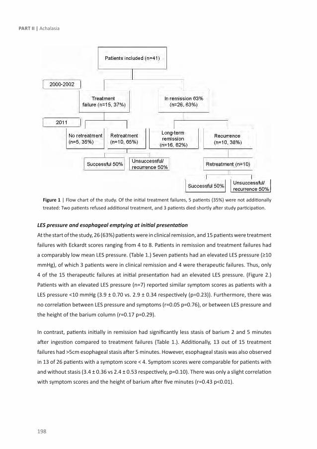
198
Part II | Achalasia
Figure 1 | Flow chart of the study. Of the initial treatment failures, 5 patients (35%) were not additionally treated: Two patients refused additional treatment, and 3 patients died shortly after study participation.
LES pressure and esophageal emptying at initial presentation
At the start of the study, 26 (63%) patients were in clinical remission, and 15 patients were treatment
failures with Eckardt scores ranging from 4 to 8. Patients in remission and treatment failures had
a comparably low mean LES pressure. (Table 1.) Seven patients had an elevated LES pressure (≥10
mmHg), of which 3 patients were in clinical remission and 4 were therapeutic failures. Thus, only
4 of the 15 therapeutic failures at initial presentation had an elevated LES pressure. (Figure 2.)
Patients with an elevated LES pressure (n=7) reported similar symptom scores as patients with a
LES pressure <10 mmHg (3.9 ± 0.70 vs. 2.9 ± 0.34 respectively (p=0.23)). Furthermore, there was
no correlation between LES pressure and symptoms (r=0.05 p=0.76), or between LES pressure and
the height of the barium column (r=0.17 p=0.29).
In contrast, patients initially in remission had significantly less stasis of barium 2 and 5 minutes
after ingestion compared to treatment failures (Table 1.). Additionally, 13 out of 15 treatment
failures had >5cm esophageal stasis after 5 minutes. However, esophageal stasis was also observed
in 13 of 26 patients with a symptom score < 4. Symptom scores were comparable for patients with
and without stasis (3.4 ± 0.36 vs 2.4 ± 0.53 respectively, p=0.10). There was only a slight correlation
with symptom scores and the height of barium after five minutes (r=0.43 p<0.01).

199
Esophageal stasis predicts recurrent symptoms | Chapter 11
Initial presentation In remission
(n=26)
Therapeutic failure
(n=15)
P-value
Age (yrs) 53 ± 3.2 55 ± 3.5 0.84
Time after diagnosis ( yrs) 17 ± 2.3 18 ± 2.2 0.78
- 0-10 yrs
- 10-15 yrs
- > 15 yrs
8
6
12
2
4
9
0.45
Eckardt score: 1.9 ± 0.2 5.1 ± 0.4 <0.0001
Esophageal manometry:
- LES pressure (mmHg) 6.2 ± 1.4 8.6 ± 1.8 0.29
Timed Barium Esophagram :
- Barium height at 1 min (cm)
- Barium height at 2 min (cm)
- Barium height at 5 min (cm)
- Max. barium width (cm)
11 ± 0.9
8.8 ± 0.9
5.5 ± 1.0
3.7 ± 0.3
14 ± 1.9
13 ± 1.9
11 ± 2.0
4.7 ± 0.6
0.11
<0.05
<0.01
0.09
Initial presentation, divided for the
need for retreatment during follow up
In long term
remission (n=16)
Recurrence
(n=10)
P-value
- Eckardt score: 1.9 ± 0.2 1.9 ± 0.4 0.96
Esophageal manometry:
- LES pressure (mmHg) 6.7 ± 2.1 5.4 ± 1.3 0.29
Timed Barium Esophagram :
- Barium height at 1 min (cm)
- Barium height at 2 min (cm)
- Barium height at 5 min (cm)
- Max. barium width (cm)
9.9 ± 1.3
6.6 ± 1.1
3.4 ± 1.1
3.6 ± 0.3
14 ± 0.9
12 ± 0.7
8.9 ± 1.5
4.0 ± 0.4
0.05
<0.05
<0.01
0.44
Table 1 | Patient characteristics at initial presentation
Recurrence rate during follow up
Of the 26 patients in remission, 16 patients (61%) remained in clinical remission until the end
of the study. (Figure 1.) Retreatment was performed in the other 20 patients; 10 patients were
initially in remission and 10 were initial treatment failures. Five initial treatment failures (33%)
were not additionally treated: 2 patients refused additional treatment, and 3 patients died shortly
after study participation. These patients were regarded as treatment failure. Of the 20 patients
requiring retreatment, 17 patients received PD, 2 patients underwent LHM and in 1 patient an
esophagectomy was performed because of refractory symptoms. During the follow up period, 30
pneumatic dilations were performed in the 17 patients after a median of 3.0 years (range 2-8), 5
with a balloon size of 30 mm, 24 with a size of 35 mm and 1 with a size of 40 mm. Twenty-one
200
Part II | Achalasia
of 34 (62%) patients had sufficient symptom control (=Eckardt score ≤3) in 2011, regardless of
additional treatment or initial treatment failure, but only 16 of 41 patients (39%) were in long-
term remission. (Figure 1.)
Esophageal stasis and recurrence rate
At the initial visit, 15 patients had normal esophageal emptying (<5 cm) whereas 26 patients
presented with stasis. Importantly, 12 of 15 patients (80%) without stasis (<5 cm) at initial
presentation remained in remission, even up to 10 years after the timed barium esophagogram,
compared to only 5 of 26 patients (19%) with esophageal stasis (p<0.01, log-rank). (Figure 3.) Of
the 26 patients with initial esophageal stasis, 14 patients were initially in remission (symptom score
≤3). However, 9 of these patients (64%) had recurrent symptoms requiring additional treatment,
after a median of 4 years (range 1-8 years). In contrast, 11 of 12 patients who were initially in
remission and had no esophageal stasis did not need additional treatment. This was significantly
higher than in patients with esophageal stasis (p=0.01, log-rank). (Figure 3.)
Lower esophageal sphincter pressure and recurrence rate
Additionally, we evaluated LES pressure as a marker to predict recurrence during follow up. Seven
patients had an elevated LES pressure (≥10 mmHg), whereas 34 patients had a LES pressure <10
mmHg. Five of 7 (71%) patients with an elevated LES pressure needed additional treatment, which
is comparable to 20 of 34 (59%) patients with LES pressure <10 mmHg (p=0.40, log-rank). (Figure
3.) Of the 7 patients with an elevated LES pressure, 3 patients were initially in remission. During
follow up, 1 of these patients needed retreatment (33%), which was comparable to 9 of the 23
(39%) patients with LES pressure <10 mmHg (p=0.72 log-rank). (Figure 3.)
Predictive values of esophageal stasis and LES pressure
Subsequently, we determined sensitivity, specificity and predictive values of esophageal stasis and
LES pressure to detect patients requiring retreatment. The sensitivity and specificity of esophageal
stasis ≥ 5 cm to predict treatment failure, either immediately or during follow up, were 88%
and 75%. Positive and negative predictive values are 85% and 80% respectively. These findings
indicate that esophageal stasis ≥5 cm is a good predictor of the need for retreatment, with a fair
specificity. LES pressure ≥ 10 mm Hg has a sensitivity and specificity of 20% and 88% respectively
to predict the need for retreatment, whereas positive and negative predictive values are 71% and
41%. Thus, if elevated, LES pressure is a fairly specific predictor for treatment failure, however
it identifies less than half of the patients that will require retreatment during follow up. Finally,
using a proportional hazard Cox regression model, esophageal stasis ≥ 5 cm was identified as a risk
factor for recurrent disease in patients in remission with a hazard ratio of 8.0 (95%CI 1.5-20). In
contrast, initial symptom scores and a LES pressure of ≥10 mmHg were no risk factors for the need
of retreatment. (Table 2)
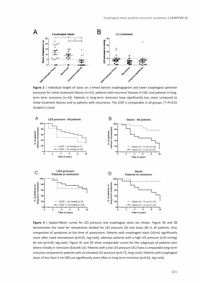
201
Esophageal stasis predicts recurrent symptoms | Chapter 11
Figure 2 | Individual height of stasis on a timed barium esophagogram and lower esophageal sphincter pressures for initial treatment failures (n=15), patients with recurrent disease (n=10), and patients in long-term term remission (n=16). Patients in long-term remission have significantly less stasis compared to initial treatment failures and to patients with recurrence. The LESP is comparable in all groups. (*=P<0.01 Student’s t-test)
Figure 3 | Kaplan-Meier curves for LES pressure and esophageal stasis are shown. Figure 3A and 3B demonstrate the need for retreatment divided for LES pressure (A) and stasis (B) in all patients, thus irrespective of symptoms at the time of assessment. Patients with esophageal stasis (≥5cm) significantly more often need retreatment (p<0.01, log-rank), whereas patients with a high LES pressure (≥10 mmHg) do not (p=0.40, log-rank). Figure 3C and 3D show comparable curves for the subgroups of patients who where initially in remission (Eckardt ≤3). Patients with a low LES pressure (3C) have a comparable long-term outcome compared to patients with an elevated LES pressure (p=0.72, long-rank). Patients with esophageal stasis of less than 5 cm (3D) are significantly more often in long term remission (p=0.01, log-rank).
LES pressure - All patients
0 2 4 6 8 100
20
40
60
80
100
LESP ≥ 10 mmHg (n=7)LESP < 10 mmHg (n=34)
A
Time in years
% o
f pat
ient
sw
ithou
t ret
reat
men
t
Stasis - All patients
0 2 4 6 8 100
20
40
60
80
100
Stasis of ≥ 5 cm (n=27)Stasis of < 5 cm (n=14)
B
Time in years
% o
f pat
ient
sw
ithou
t ret
reat
men
t
LES pressurePatients in remission
0 2 4 6 8 100
20
40
60
80
100
LESP < 10 mmHg (n=23)LESP ≥ 10 mmHg (n=3)
C
Time in years
% o
f pat
ient
s in
long
-ter
m r
emis
sion
StasisPatients in remission
0 2 4 6 8 100
20
40
60
80
100
Stasis of < 5 cm (n=12)Stasis of ≥ 5 cm (n=14)
D
Time in years
% o
f pat
ient
s in
long
-ter
m r
emis
sion

202
Part II | Achalasia
Factor: Adjusted hazard ratio(95% cI)
p-value
Initial symptom score 0.81 (0.3 - 2.1) 0.42
Barium height ≥5 cm vs <5 cm
8.0 (1.5 – 21) 0.01
LES pressure<10 mmHg vs ≥10 mmHg
1.7 (0.4 – 7.3) 0.40
Table 2 | Adjusted hazard ratios for recurrence are given for patients with longstanding achalasia who where initially in remission.
dIscussIon
This study shows that stasis on a timed barium esophagogram but not basal LES pressure is
an important predictor of recurrent symptoms and the need for retreatment in patients with
longstanding achalasia. Therefore, when significant stasis on a timed barium esophagogram is
observed, even in the presence of a LES pressure < 10 mmHg, additional therapy may be warranted.
Currently, the most commonly used treatment modalities for achalasia are laparoscopic Heller
myotomy (LHM) and PD. Both treatments have high short-term success rates of 80-100%,2
gradually decreasing in patients with longstanding disease (≥10 yr) to 40-60%. (3;5;13) The cohort
presented in the current study is unique with a mean follow up of 26 years after diagnosis. Of the
41 patients, 62% had adequate symptom control (= Eckardt score ≤3). However, to reach this rate
of symptom control, 49% of patients needed retreatment during the 10 year follow up period.
Most long follow up studies demonstrate similar retreatment rates, of up to 87% of patients after
a follow up of 12 years after initial PD.3,5,16 Notably, not only after PD but also after LHM symptom
control decreases to around 60% with a longer follow up period, as recently demonstrated by
several studies.8,16-18 Early recognition of incomplete therapeutic response or the need for
retreatment is of crucial importance to reduce morbidity and long-term complications such as
esophageal decompensation.8,10 Furthermore, inadequately treated patients have a higher risk
to develop dysplasia and eventually esophageal squamous carcinoma.11,12 Importantly, additional
treatment both after initial PD or LHM has satisfactory results with success rates of 60-80% and
improvement of esophageal emptying.5,8,16,19,20 Therefore regular follow up visits to determine the
need for retreatment are indicated.
To determine if retreatment is indeed indicated, symptoms are currently most commonly used
in clinical practice. Symptom scores including the Eckardt score use typical symptoms such as
dysphagia, regurgitation and chest pain.4,8 However, since symptoms are not always reported or even

203
Esophageal stasis predicts recurrent symptoms | Chapter 11
absent due to decompensation of the esophagus or impaired esophageal sensitivity, identification
of patients in need of retreatment may be difficult and is often delayed. In our study, 13 of 26 (50%)
patients initially in “remission” had over 5 cm of stasis on their timed barium esophagogram, of
which 9 eventually needed retreatment due to recurrent symptoms. Additionally, symptoms only
weakly correlate with esophageal stasis (correlation coefficient is 0.43), and do no correlate with
LES pressure. This underlines that symptoms and objective parameters of esophageal function
often do not match, as repeatedly demonstrated before, emphasizing that symptoms are not
reliable to determine therapeutic strategy in patients with achalasia.6,21,22
An alternative for symptoms as predictor for retreatment may be basal LES pressure.13,23 Several
studies indeed demonstrated that patients with a basal pressure above 10 mmHg are at risk to
develop recurrent symptoms.4,5 Hence, the decision to repeat PD is often based on the presence
of an increased basal LES pressure (>10 mm Hg). Also in our study, the specificity of this criterion
to predict the need for retreatment was acceptable, i.e. 71%. Based on these findings, graded
pneumatic dilation is often repeated aiming at reducing basal LES pressure to less than 10mmHg.
However, we here show that a low LES pressure is not necessarily indicative of clinical success.
In most of our patients (83%), basal LES pressure was <10 mmHg, even in case of high symptom
scores or persistent stasis. Of these, 59% required retreatment during follow up. Hence, in our
series, basal LES pressure > 10 mmHg only identifies 20% of patients that will require retreatment,
resulting in sensitivity of 20% to detect treatment failures. Other studies with large numbers of
patients also failed to identify LES pressure a predictor of therapeutic failure, both after LHM or
PD.2,8,24 One possible explanation could be that esophageal emptying and clearance (and most
likely symptoms) depend on other factors than LES pressure. Indeed, 19 of 26 patients with a low
LES pressure had esophageal stasis while we found no correlation between LES pressure and the
height of stasis. The ability of the esophagogastric junction to open, largely determined by its
distensibility, may be a more important determinant of flow across this region.25-27 Indeed, we
recently demonstrated that impaired distensibility of the esophagogastric junction rather than low
LES pressure was associated with poor esophageal emptying and treatment failure.27
Based on these findings, assessment of esophageal emptying could be a more integrated and
clinically more useful parameter. Indeed, our data of the current study demonstrate that patients
with stasis of ≥ 5cm have an 8-fold increased risk for retreatment compared to patients with < 5cm
stasis, irrespective of initial symptoms or LES pressure. Our data support a study of Vaezi et al, in
which 90% of patients with persistent stasis but without symptoms failed therapy within one year
after initial treatment.6 Of our patients with persistent stasis but without symptoms, 69% needed
retreatment during the follow up period, although only 10% within the first year. In contrast, in
patients without esophageal stasis failure rates are only 20% after an additional follow up of 10
204
Part II | Achalasia
years. This confirms the study of Vaezi et al. reporting a failure rate in patients without stasis of
23% after 6 years of follow up. The current study shows that esophageal stasis on a timed barium
swallow has a sensitivity of 88% and a specificity of 75% to detect patients in need for retreatment.
In addition, the timed barium is a simple and widely available test with very limited radiation
exposure (0.08 mSv).28 Therefore, assessment of esophageal stasis is a safe and easy to perform
test that is not only helpful directly after treatment as demonstrated by multiple studies,2,6,19 but
also during long-term follow up, up to at least 26 years as demonstrated by this study.
In conclusion, stasis on a timed barium esophagogram is an important functional predictor of
treatment failure in patients with longstanding achalasia, in contrast to LES pressure. Although
several studies have advised to perform regular follow up,30,32 current guidelines do not advice
on functional testing in the follow up of achalasia.1,33 Based on our data, however, we argue that
regular follow up visits may be indicated, not only to inform about symptoms, but also to perform
a timed barium esophagogram in order to assess esophageal emptying. Clearly, prospective trials
are required to confirm the benefit of (serial) functional testing and early additional treatment for
the prevention of sigmoid-like esophagus and the development of dysplasia.

205
Esophageal stasis predicts recurrent symptoms | Chapter 11
ReFeRence lIsT
1. Vaezi MF, Richter JE. Diagnosis and management of achalasia. American College of Gastroenterology Practice Parameter Committee. Am J Gastroenterol 1999;94(12):3406-12.
2. Boeckxstaens GE, Annese V, des Varannes SB et al. Pneumatic dilation versus laparoscopic Heller’s myotomy for idiopathic achalasia. N Engl J Med 2011;364(19):1807-16.
3. West RL, Hirsch DP, Bartelsman JF et al. Long term results of pneumatic dilation in achalasia followed for more than 5 years. Am J Gastroenterol 2002;97(6):1346-51.
4. Eckardt VF, Aignherr C, Bernhard G. Predictors of outcome in patients with achalasia treated by pneumatic dilation. Gastroenterology 1992;103(6):1732-8.
5. Hulselmans M, Vanuytsel T, Degreef T et al. Long-term outcome of pneumatic dilation in the treatment of achalasia. Clin Gastroenterol Hepatol 2010;8(1):30-5.
6. Vaezi MF, Baker ME, Achkar E et al. Timed barium oesophagram: better predictor of long term success after pneumatic dilation in achalasia than symptom assessment. Gut 2002;50(6):765-70.
7. Ponce J, Ortiz V, Maroto N et al. High Prevalence of Heartburn and Low Acid Sensitivity in Patients with Idiopathic Achalasia. Dig Dis Sci 2010.
8. Zaninotto G, Costantini M, Rizzetto C et al. Four hundred laparoscopic myotomies for esophageal achalasia: a single centre experience. Ann Surg 2008;248(6):986-93.
9. Richter JE. Modern management of achalasia. Curr Treat Options Gastroenterol 2005;8(4):275-83.
10. Richter JE, Boeckxstaens GE. Management of achalasia: surgery or pneumatic dilation. Gut 2011.
11. Brossard, E, Ollyo JB, and Fontolliet CH et al. Achalasia and squamous cell carcinoma of the esophagus: is an endoscopic surveillance justified? Gastroenterology 1992;102(suppl)[A4]. Ref Type: Abstract
12. Rohof, W. O, Bergman, J. J., Bartelsman, J. F., Hirsch, D. P., and Boeckxstaens, G. E. Screening for Dysplasia in Idiopathic Achalasia Using Lugol Staining. Gastroenterology 2011;140(suppl):S-227 Ref Type: Abstract
13. Eckardt VF, Gockel I, Bernhard G. Pneumatic dilation for achalasia: late results of a prospective follow up investigation. Gut 2004;53(5):629-33.
14. de Oliveira JM, Birgisson S, Doinoff C et al. Timed barium swallow: a simple technique for evaluating esophageal emptying in patients with achalasia. AJR Am J Roentgenol 1997;169(2):473-9.
15. Vaezi MF, Baker ME, Richter JE. Assessment of esophageal emptying post-pneumatic dilation: use of the timed barium esophagram. Am J Gastroenterol 1999;94(7):1802-7.
16. Vela MF, Richter JE, Khandwala F et al. The long-term efficacy of pneumatic dilatation and Heller myotomy for the treatment of achalasia. Clin Gastroenterol Hepatol 2006;4(5):580-7.
17. Lopushinsky SR, Urbach DR. Pneumatic dilatation and surgical myotomy for achalasia. JAMA 2006;296(18):2227-33.
18. Chen Z, Bessell JR, Chew A et al. Laparoscopic cardiomyotomy for achalasia: clinical outcomes beyond 5 years. J Gastrointest Surg 2010;14(4):594-600.
19. Guardino JM, Vela MF, Connor JT et al. Pneumatic dilation for the treatment of achalasia in untreated patients and patients with failed Heller myotomy. J Clin Gastroenterol 2004;38(10):855-60.

206
Part II | Achalasia
20. Finley RJ, Clifton JC, Stewart KC et al. Laparoscopic Heller myotomy improves esophageal emptying and the symptoms of achalasia. Arch Surg 2001;136(8):892-6.
21. Tsiaoussis J, Athanasakis E, Pechlivanides G et al. Long-term functional results after laparoscopic surgery for esophageal achalasia. Am J Surg 2007;193(1):26-31.
22. Kim CH, Cameron AJ, Hsu JJ et al. Achalasia: prospective evaluation of relationship between lower esophageal sphincter pressure, esophageal transit, and esophageal diameter and symptoms in response to pneumatic dilation. Mayo Clin Proc 1993;68(11):1067-73.
23. Alderliesten J, Conchillo JM, Leeuwenburgh I et al. Predictors for outcome of failure of balloon dilatation in patients with achalasia. Gut 2011;60(1):10-6.
24. Kadakia SC, Wong RK. Graded pneumatic dilation using Rigiflex achalasia dilators in patients with primary esophageal achalasia. Am J Gastroenterol 1993;88(1):34-8.
25. Pandolfino JE, Shi G, Trueworthy B et al. Esophagogastric junction opening during relaxation distinguishes nonhernia reflux patients, hernia patients, and normal subjects. Gastroenterology 2003;125(4):1018-24.
26. Ghosh SK, Kahrilas PJ, Lodhia N et al. Utilizing intraluminal pressure differences to predict esophageal bolus flow dynamics. Am J Physiol Gastrointest Liver Physiol 2007;293(5):G1023-G1028.
27. Rohof WO, Hirsch DP, Kessing BF et al. Efficacy of Treatment for Patients With Achalasia Depends on the Distensibility of the Esophagogastric Junction. Gastroenterology 2012. epub ahead of print
28. Mettler FA, Jr., Huda W, Yoshizumi TT et al. Effective doses in radiology and diagnostic nuclear medicine: a catalog. Radiology 2008;248(1):254-63.
29. Andersson M, Lundell L, Kostic S et al. Evaluation of the response to treatment in patients with idiopathic achalasia by the timed barium esophagogram: results from a randomized clinical trial. Dis Esophagus 2009;22(3):264-73.
30. Zerbib F, Thetiot V, Richy F et al. Repeated pneumatic dilations as long-term maintenance therapy for esophageal achalasia. Am J Gastroenterol 2006;101(4):692-7.
31. Gerson LB. Pneumatic dilation or myotomy for achalasia? Gastroenterology 2007;132(2):811-3.
32. Wong RK. Achalasia: should we or should we not follow the bag? Am J Gastroenterol 2006;101(4):698-700.
33. Spechler SJ. AGA technical review on treatment of patients with dysphagia caused by benign disorders of the distal esophagus. Gastroenterology 1999;117(1):233-54.


Chapter 12Efficacy of Treatment for Patients with
Achalasia Depends on the Distensibility of the Esophagogastric Junction
Wout Rohof, David Hirsch, Boudewijn Kessing, Guy Boeckxstaens
Gastroenterology. 2012;143(2):328-35

210
Part II | Achalasia
AbsTRAcT
Background & aims:
Many patients with persistent dysphagia and regurgitation after therapy have low or no lower
esophageal sphincter (LES) pressure. Distensibility of the esophagogastric junction (EGJ) largely
determines esophageal emptying. We investigated whether assessment of the distensibility of
the EGJ is a better and more integrated parameter than LES pressure for determining efficacy of
treatment for patients with achalasia.
Methods:
We measured distensibility of the EGJ using an endo functional luminal imaging probe (EndoFLIP)
in 15 healthy volunteers (controls; 8 male, 40±4.1 y) and 30 patients with achalasia (16 male,
age 51±3.1 y). Patients were also assessed by esophageal manometry and a timed barium
esophagogram. Symptom scores were assessed using the Eckardt score, with a score below 4
indicating treatment success. The effect of initial and additional treatment on distensibility and
symptoms were evaluated in 7 and 5 patients respectively.
Results:
EGJ distensibility was significantly reduced in untreated patients with achalasia, compared
with controls (0.7±0.9 mm2/mmHg vs 6.3±0.7 mm2/mmHg; P<.001). In patients with achalasia,
EGJ distensibility correlated with esophageal emptying (r=–0.72; P<.01) and symptoms (r=0.61;
P<.01), and was significantly increased with treatment. EGJ distensibility was significantly higher in
patients successfully treated (Eckardt score<3) compared to those with an Eckardt score >3 (1.6 ±
0.3 vs 4.4 ± 0.5 mm2/mmHg; P=.001). Even when LES pressure was low, EGJ distensibility could be
reduced, which was associated with impaired emptying and recurrent symptoms.
Conclusions:
EGJ distensibility is impaired in patients with achalasia, and in contrast to LES pressure, is associated
with esophageal emptying and clinical response. Assessment of EGJ distensibility by EndoFLIP is a
better parameter than LES pressure for evaluating efficacy of treatment for achalasia.

211
Distensibility of the EGJ | Chapter 12
InTRoducTIon
Achalasia is a rare motility disorder of the esophagus, with an incidence of 1 per 100.000. 1, 2
Disappearance of neurons in the esophagus leads to loss of peristalsis and a defective relaxation
of the lower esophageal sphincter (LES).3 The subsequent retention of food and saliva in the
esophagus results in the typical symptoms of achalasia, i.e. dysphagia, regurgitation of undigested
food, respiratory complications, chest pain and weight loss.3, 4 As the neuronal loss is irreversible,
treatment of achalasia is limited to disruption of the LES by pneumatic dilation or Heller myotomy,
aiming to reduce LES tone and improve bolus transport across the esophagogastric junction (EGJ).5
The clinical management of achalasia is challenging, especially as treatment success declines in
the long term, leading to an increasing risk for retreatment.6-8 Identification of patients in need of
retreatment may be difficult and is often delayed as symptoms are underreported or absent due
to decompensation of the esophagus or impaired esophageal sensitivity. Moreover, patients get
used to symptoms or adapt their diet. It should be emphasized though that timely recognition of
functional obstruction and subsequent stasis of food in the esophagus is of importance for at least
two reasons: first, decompensation of the esophagus must be prevented, as this can lead to mega-
esophagus or sigmoid-like esophagus associated with higher morbidity and refractory symptoms. 5,
9 Second, esophageal stasis has been suggested to be a risk factor for the development of dysplasia
and neoplasia, especially in patients with longstanding achalasia. 10-12
Several studies have proposed manometry to be a useful test to determine whether patients
should be retreated. Reduction of LES pressure to less than 10 mmHg has been reported to be a
good predictor for long-term treatment success. 6, 13, 14 Based on these reports, patients with a LES
pressure ≥ 10 mm Hg are therefore considered for additional therapy. However, several studies
and clinical experience demonstrate that a significant proportion of patients with persistent
symptoms has a low or even absent LES pressure.15-17 Interestingly, these patients often have
incomplete esophageal emptying on a timed barium esophagogram.17 Moreover, despite absence
of LES pressure, a significant proportion of these patients benefit from additional treatment with
pneumodilation. These observations argue against LES pressure as a useful test to assess the need
for treatment.
As shown by Pandolfino et al. flow across the esophagogastric junction (EGJ) is mainly determined
by its distensibility in response to increased intraluminal pressure. 18 Although LES pressure will
definitely contribute, other factors such as fibrosis due to previous treatment or natural history of
the disease may impair distensibility and subsequently EGJ opening. Reduced distensibility even
in the absence of LES pressure may therefore explain why esophageal emptying on timed barium
esophagogram may be impaired. In the present study therefore, we determined EGJ distensibility

212
Part II | Achalasia
in patients with achalasia using the commercially available Endo functional luminal imaging probe
(EndoFLIP) during volume controlled distensions. 19 We compared its ability to predict clinical
success with that of LES pressure and esophageal emptying.
MeThods
Subjects
Fifteen healthy volunteers (8 males, 40 ± 4.1 years) and 34 patients with achalasia were included
in the study. In 4 patients, the EGJ could not be passed, leaving 30 achalasia patients in which
distensibility measurements could be performed (16 males, 51 ± 3.1 years, P=0.05 vs healthy
volunteers). The diagnosis of achalasia was based on the absence of peristalsis and impaired
LES relaxation (nadir pressure > 10 mmHg during swallow-induced relaxation) assessed during
esophageal manometry. Patients known with a sigmoid like esophagus (>6cm diameter) were
excluded from the study. One patient was excluded for this reason.
Seven of the 30 patients were newly diagnosed and were measured before and 3 months after
treatment. (Figure 1.) The rest of the patients (n=23) were diagnosed 5.4 ± 1.4 years earlier.
Thirteen patients were initially treated with graded pneumodilation (PD), 7 patients with
laparoscopic Heller myotomy (LHM) and 3 patients had undergone both treatments. In patients
with newly diagnosed achalasia, PD was performed in 6 patients and LHM in 1. Treatment
procedures were performed according to the protocol described earlier. 20 Briefly, PD is performed
with Rigiflex balloons (Boston Scientific, Nanterre, France) positioned at the EGJ and dilated with 5
PSI during 1 min, followed by 8 PSI during 1 min. Two dilations are performed with 30 and 35 mm
balloons respectively separated by 1-3 weeks. For LHM, a myotomy was performed extending 6
cm above the EGJ and 1.5 cm over the stomach. Subsequently a fundoplication according to Dor
was performed. Each subject gave written informed consent, and the study was approved by the
Medical Ethics Committee of the Academic Medical Centre.
Study design
Healthy volunteers underwent EndoFLIP measurement. Patients underwent esophageal
manometry, EndoFLIP measurement of the EGJ and timed barium esophagogram. In addition,
symptoms were assessed using the Eckardt score. Studies were performed after at least a 4 hr
fast. First, esophageal manometry was performed, followed by the EndoFLIP measurement and
timed barium esophagogram. Patients with additional treatment after the procedure were invited
to undergo the study protocol 3 to 6 months after treatment.

213
Distensibility of the EGJ | Chapter 12
FIGuRe 1 | A flowchart describes the patients groups in the study. Seven patients were studied before and after initial treatment. Thirty patients were studied after therapy, of which 18 were treated successfully (Eckardt score <4) and 12 patients were treated unsuccessfully. Eight of 12 patients unsuccessfully treated patients were additionally treated, of which 5 patients underwent an additional EndoFLIP measurement.
EndoFLIP measurement
Distensibility of the EGJ was determined using the commercially available EndoFLIP system (MMS,
Enschede, the Netherlands). In this technique, a probe is inserted into the esophagus and placed at
the level of the EGJ. The probe consists of a 240cm catheter with a 14cm bag attached to its distal
end, which is compliant to a maximal diameter of 25 mm. Inside the bag 17 electrodes are placed
at 4 mm intervals. An excitation current of 100 μA is generated between two adjacent electrodes
at a frequency of 5 kHz. Using impedance-planimetry, cross sectional areas (CSAs) are determined
for the 16 balloon cross sections during volume controlled distensions. Additionally two pressure
sensors are located on the probe to determine intrabag pressure allowing assessment of EGJ
distensibility.
The distension probe and the pressure transducers were calibrated by the manufacturer. The
pressure sensor was zeroed prior to insertion of the catheter, and subsequently the deflated
catheter was inserted through an anaesthesized nostril. Manometry readings were used to
position the balloon. In our distension protocol, the balloon was inflated to a 20, 30, 40 and 50 mL
volume. Pressures and CSAs are collected at a rate of 10 Hz. Distensibility was assessed using the
median value over a 30s dynamic measurement of the narrowest CSA, which corresponds to the
EGJ, and the median intrabag pressure. 21
Symptom scores
Symptom scores were assessed using the Eckardt score, which is the sum of the symptom scores
for dysphagia, regurgitation and chest pain (0 = absent, 1 = occasional, 2 = daily, 3 = each meal)
completed with the score of weight loss (0 = no weight loss, 1 = < 5 kg, 2 = 5-10 kg, 3 = > 10 kg),
and thus ranges from 0 to 12. 13 Treatment was considered as successful if the Eckardt score was
below 4.

214
Part II | Achalasia
Esophageal manometry
Esophageal manometry was performed using a 10 lumen assembly (Dentsleeve, Missisauga,
Ontario, Canada) with a sleeve incorporated at its distal end. The sideholes and the sleeve sensor
were perfused at a rate of 0.6 mL.min-1. The sleeve was positioned as such that it incorporated the
EGJ. Data were stored on an MMS-solar system (MMS, Enschede, the Netherlands). End-expiratory
LES resting pressure was determined after wet swallows and relative to intragastric pressure. A LES
pressure ≥ 10 mm Hg was considered as elevated. Residual LES pressure was determined as the
mean residual pressure after 10 water swallows of 5 mL.
Timed barium esophagogram
Esophageal stasis was determined on a timed barium esophagogram immediately after, 1, 2 and 5
minutes after ingestion of the maximal tolerable amount of low density barium sulphate over 30-
45 s without regurgitation or aspiration, with the patient upright in a slight left posterior position.
The distance from the tapered distal esophagus to the top of the barium column and the maximal
diameter of the esophagus were measured. A maximal diameter of >3cm was considered enlarged.
Data analysis and statistics
Basal LES pressure was measured at end-expiration. Distensibility of the EGJ was measured as the
median cross sectional area at the narrowest point divided by the balloon pressure. Statistical
analysis was performed using SPSS 16.0 (IBM corporation, Somers, NY, United States). Data
are presented as mean ± SEM when parametric, and as median (IQR) when not parametric.
Parametric data were tested using a Student’s t-test, with Bonferroni correction in case of multiple
comparisons. Paired unparametric data were tested using the Wilcoxon signed ranks test. Chi-
square was used to determine sensitivity and specificity. All reported P-values are 2 tailed, and a
value of 0.05 is regarded as statistically significant.
ResulTs
Patients
To determine normal values, 15 healthy volunteers were invited to undergo an EndoFLIP
procedure. Of the 30 patients that completed the protocol, 18 had an Eckardt symptom score of
3 or less, and were therefore regarded as successfully treated. (Figure 1.) Esophageal manometry,
symptom evaluation and EndoFLIP procedure were performed in all patients. A timed barium
esophagogram was performed in 24 patients. After inclusion, 8 patients with recurrent symptoms
underwent additional treatment, of which 5 patients agreed to undergo additional EndoFLIP and
manometry 3 to 6 months after additional treatment. The EndoFLIP procedure was well tolerated.

215
Distensibility of the EGJ | Chapter 12
An hourglass shape of the bag was observed in all measurements, with the narrowest CSA at the
level of the LES, as determined during esophageal manometry.
EGJ distensibility
Healthy volunteers had a mean EGJ distensibility of 6.3 ± 0.7 mm2 / mmHg using a 50 mL
volume distension. Healthy volunteers had a significantly higher EGJ distensibility compared to
untreated patients (0.7±0.9 mm2 / mmHg P<0.001), unsuccessfully treated patients (1.6 ± 0.3
mm2 / mmHg P<0.01) and successfully treated patients (4.4 ± 0.5 mm2 / mmHg P=0.02) in a 50
mL volume distension. (Figures 2 and 3.) Unsuccessfully treated patients had a significantly lower
EGJ distensibility compared to successfully treated patients, both at 40 mL (P<0.01) and 50 mL
distension (P<0.01).
Next, we determined the cut-off value using the lower 90th percentile of EGJ distensibility in healthy
volunteers. The cut-off for normality was 2.9 mm2 / mmHg. As demonstrated in Figure 2B, 4 of 18
(22%) successfully treated patients have an EGJ distensibility value lower than the 90th percentile
value of healthy volunteers. This is in contrast to 11 of 12 (92%) unsuccessfully treated patients
and 7 of 7 (100%) newly diagnosed patients. Using this cut-off value, the sensitivity and specificity
of EGJ distensibility (50 mL) for treatment failure is 92% and 72% respectively (chi-square).
Effect of treatment
a) Newly diagnosed patients
Seven patients with newly diagnosed achalasia were included in the study, to verify the effect
of initial treatment on EGJ distensibility. Before treatment, distensibility was significantly
impaired compared to HV with a median distensibility of 0.60 (IQR 0.37-0.94) mm2 / mmHg in
a 50 mL volume distension. Treatment significantly improved distensibility to 2.9 (IQR 1.2-7.4)
mm2 / mmHg after treatment (P=0.02 compared to pre-treatment, Wilcoxon signed rank test).
(Figure 4.) Interestingly, the increase in distensibility after treatment had an excellent correlation
with the decrease in symptom score (r=0.89 P=0.005, Spearman’s rank). Distensibility did not
increase above the 2.9 mm2/mmHg cut-off value in three patients and was associated with
persistent symptoms, as indicated by an Eckardt score above 3. These patients all underwent
additional treatment: 2 patients underwent PD with a 40 mm balloon, whereas 1 patient underwent
LHM. This led to a decrease in symptoms in all patients. In the single patient that underwent an
additional EndoFLIP measurement, LHM resulted in an increase in distensibility to 6.0 mm2/mmHg.
216
Part II | Achalasia
FIGuRe 2 | The EGJ opening is plotted against the intrabag pressure in figure 2A for healthy volunteers (n=15), patients with successful treatment (n=18), treatment failures (n=12) and newly diagnosed patients (n=7). Starting from a 40 mL volumetric distension, treatment failures have a clearly smaller EGJ opening at higher intrabag pressure, corresponding with a significantly lower distensibility compared to successfully treated patients and healthy volunteers. Newly diagnosed patients clearly have the most impaired EGJ distensibility. Figure 2B shows the separate measurements of the included groups at the maximal volumetric distension of 50 mL. The 90th percentile of healthy volunteers EGJ distensibility is marked. Open symbols are used in case values are below the 90th percentile. Of the successfully treated patients 78% is above the 90th percentile of healthy volunteers, in contrast to only 8% and 0% of treatment failures and newly diagnosed patients respectively. ***P<0.001 **P<0.01 *P<0.05 Test are performed with a Student’s T-test with Bonferroni correction.

217
Distensibility of the EGJ | Chapter 12
FIGuRe 3 | Examples of EndoFLIP measurement during volumetric distensions in a healthy volunteer (A), a patient with successful treatment (B), a patient with recurrent symptoms (C) and a patient before initial treatment (D) are depicted. The bag has a cylindrical shape, of which the varying diameter corresponds with the opening at that point, and the exact values can be observed in the right panel of each measurement. The narrowest diameter corresponds with the EGJ opening. The EGJ opening is clearly wider in the healthy volunteer and the patient with treatment success compared to the patient with treatment failure and the newly diagnosed patient. Additionally, bag pressures are lower in treatment success, indicating that less pressure is needed to open the EGJ, corresponding with a higher distensibility.

218
Part II | Achalasia
b) Previously treated patients
Five of the 12 patients with treatment failure/ recurrent symptoms were evaluated by EndoFLIP
after additional treatment. Before treatment, patients had a median distensibility of 0.79 mm2/
mmHg (IQR 0.58-1.2). Three patients were treated by PD (35 mm) and 2 by LHM. After a median of
6 months, distensibility increased significantly to 5.6 mm2/mmHg (IQR 3.7-6.6) (P=0.03 vs. before
treatment, Wilcoxon signed rank test) and was above 2.9 mm2/mmHg in all patients. This was
associated with clinical success and a reduction of the Eckardt score < 3 (median 1 (range 0-2)).
EGJ distensibility, esophageal emptying and LES pressure in relation to treatment success
As shown in Figure 2A, treatment failures had a significantly lower EGJ distensibility compared
to successfully treated patients. Moreover, treatment failures had significantly more esophageal
stasis after five minutes (4.5 ± 1.1 cm vs. 1.2 ± 0.7 cm, P=0.01), and a higher LES pressure (4.4 ± 1.1
mmHg vs 10 ± 3.0 mmHg, P=0.03) compared to successfully treated patients. (Figure 5.)
Although LES pressure > 10 mmHg is accepted to indicate inadequate treatment 6, 13, 14, LES
pressure was < 10 mmHg in 7 of the 12 (58%) patients with treatment failure (Figure 5 panel B).
Residual LES pressure after swallowing was >10 mmHg in only 5 of 12 in patients with treatment
failure. In contrast, impaired distensibility as assessed with the EndoFLIP and residual stasis on
timed barium esophagogram were present in 92% and 89% of patients with treatment failure
respectively. (Figure 5, panels A and C) To define why patients are treatment failures even though
LES pressure is < 10 mmHg, we compared LES pressure to EGJ distensibility and esophageal stasis.
LES pressure in relation to esophageal emptying and EGJ distensibility
Of the 22 patients with a low LES pressure, 8 patients had impaired distensibility on EndoFLIP.
These patients had significantly more stasis on their timed barium esophagogram compared to
patients with a normal distensibility (6.5 ± 0.6 cm vs. 1.2 ± 0.5 cm, P<0.01). Interestingly, 5 of these
patients reported a symptom score of 4 or higher. Thus, even if LES pressure is low, distensibility
can be impaired leading to impaired esophageal emptying and recurrent symptoms. In line with
this, 7 of 14 (50%) patients with impaired esophageal emptying had a LES pressure <10 mmHg,
whilst there was no significant relationship between LES pressure and the height of stasis (r=0.25,
P=0.26 Spearman’s rank).
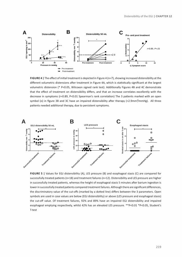
219
Distensibility of the EGJ | Chapter 12
FIGuRe 4 | The effect of initial treatment is depicted in Figure 4 (n=7), showing increased distensibility at the different volumetric distensions after treatment in Figure 4A, which is statistically significant at the largest volumetric distension (* P<0.05, Wilcoxon signed rank test). Additionally Figures 4B and 4C demonstrate that the effect of treatment on distensibility differs, and that an increase correlates excellently with the decrease in symptoms (r=0.89, P<0.01 Spearman’s rank correlation) The 3 patients marked with an open symbol (o) in figure 3B and 3C have an impaired distensibility after therapy (<2.9mm²/mmHg). All three patients needed additional therapy, due to persistent symptoms.
FIGuRe 5 | Values for EGJ distensibility (A), LES pressure (B) and esophageal stasis (C) are compared for successfully treated patients (n=18) and treatment failures (n=12). Distensibility and LES pressure are higher in successfully treated patients, whereas the height of esophageal stasis 5 minutes after barium ingestion is lower in successfully treated patients compared treatment failures. Although there are significant differences, the discriminatory value of the cut-offs (marked by a dotted line) differs between the 3 parameters. Open symbols are used in case values are below (EGJ distensibility) or above (LES pressure and esophageal stasis) the cut-off value. Of treatment failures, 92% and 89% have an impaired EGJ distensibility and impaired esophageal emptying respectively, whilst 42% has an elevated LES pressure. **P<0.01 *P<0.05, Student’s T-test
Distensibility
0 20 40 600
50
100
150
200
Pre-treatment Post-treatment
20 mL30 mL 40 mL50 mL
20 mL
30 mL
40 mL
50 mL
*
Pressure in mmHg
EGJ
open
ing
in m
m2
Distensibility 50 mL
Pre-treatment Post-treatment0
2
4
6
8
10
2.9
*
Dis
tens
ibili
ty m
m2 / m
mHg
Pre- and post treatment
-5 0 5 10 15
2
4
6
8
r=0.89, P<.01
-∆ Symptom score
∆ D
iste
nsib
ility
A B C
EGJ distensibility 50 mL
Succes
sful tr
eatm
ent
Treatm
ent fa
ilures
0
2
4
6
8 **
Dis
tens
ibilit
y m
m2 / m
mHg
LES pressure
Succes
sful tr
eatm
ent
Treatm
ent fa
ilures
0
10
20
30
40
*
LES
pres
sure
in m
mH
g
Esophageal stasis
Succes
sful tr
eatm
ent
Treatm
ent fa
ilures
0
4
8
12 *
Res
idua
l sta
sis
in c
m a
fter
5 m
inA B C

220
Part II | Achalasia
Esophageal emptying and EGJ distensibility
In contrast to LES pressure, EGJ distensibility significantly correlated with the height of stasis
(r=0.72, P<0.001 Spearman’s rank). Patients with complete emptying indeed had a higher
distensibility of the EGJ compared to patients with residual stasis (50 mL: 2.2 ± 0.40 vs. 4.7 ± 0.76
mm2 / mmHg P=0.006). Furthermore, in patients with impaired EGJ distensibility after treatment
(<2.9 mm2 / mmHg) the height of the barium column was significantly higher after 1, 2 and 5
minutes, compared to patients with a normal distensibility. (Figure 6.) Lastly, in 9 of 10 patients
(90%) with a normal EGJ distensibility, emptying was complete after 5 minutes.
FIGuRe 6 | The height of the barium column is plotted at the different measurement times, for patients with a normal distensibility (n=10) and for patients with an impaired distensibility (n=14). Right after ingestion, there is no difference in column height. The height of the barium column is significantly lower after 1, 2 and 5 minutes in patients with a normal distensibility, corresponding with a significantly better esophageal emptying. * P<0.05 **P<0.01 Student’s t-test.
EGJ distensibility and esophageal width
To assess the effect of esophageal width on EGJ distensibility, we used the maximal esophageal
diameter of the barium column on the timed barium esophagogram. No correlation of esophageal
width and EGJ distensibility was found (r=-0,35 P=0.14, Pearson’s correlation). Furthermore,
patients with a widened esophagus (>3cm) have comparable EGJ distensibility compared to
patients with a normal esophageal width (2.6 ± 0.3 vs. 4.0 ± 0.7 mm2 / mmH, P=0.08). Therefore,
the effect of esophageal width on EGJ distensibility is probably limited.
dIscussIon
In the present study we showed that EGJ distensibilty, but not LES pressure, strongly correlates
with clinical response to treatment: impaired EGJ distensibility is observed in 92% of patients with
0 1 2 3 4 50
2
4
6
8
10
Normal distensibility (≥2.9 mm2/mmHg)Impaired distensibility (<2.9 mm2/mmHg)
***
**
Time in minutes
Bar
ium
hei
ght i
n cm

221
Distensibility of the EGJ | Chapter 12
recurrent symptoms, whilst only 42% of these patients have an elevated LES pressure. Moreover,
patients with impaired EGJ distensibility have recurrent symptoms and impaired esophageal
emptying, even if LES pressure is low. Based on these findings, we conclude that EGJ distensibility
is a better parameter to evaluate the efficacy of treatment in patients with achalasia compared to
LES pressure.
Treatment of patients with achalasia aims at relieving functional obstruction at the level of the
EGJ. This is mostly achieved by disruption of the LES circular muscle fibres, either by endoscopic
pneumodilaton or surgical myotomy. 5, 20 Although initial treatment success rates are high,
recurrences may occur in up to 87% of patients especially when follow up is as long as 12 years. 6-8, 20 Since previous studies have suggested that basal LES pressure ≥ 10 mmHg is a risk factor to
develop recurrent symptoms, LES pressure is often used in current clinical practice to decide if
patients require further treatment. 6, 13, 14 In our study however, LES pressure did not discriminate
patients with clinical success from those with persistent or recurrent symptoms: in 7 of 12 (58%)
patients with recurrent symptoms, LES pressure was lower than 10 mmHg. Alternatively, one could
reason that the residual LES pressure during swallowing is a more relevant parameter determining
LES opening and subsequent passage of the bolus and esophageal emptying. Unfortunately, no
data are available in literature evaluating the relationship between this parameter and clinical
success. Nevertheless, we checked whether the residual LES residual pressure after swallowing
would be an alternative parameter to discriminate patients with recurrent symptoms during
manometry. However, comparable to basal LES pressure, only 5 of 12 [42%] treatment failures
had a residual LES pressure > 10mm Hg. Similarly, Zaninotto et al. reported that following LHM
LES pressure was comparable in treatment failures and successfully treated patients.9 Moreover,
multiple studies have failed to confirm LES pressure as a risk factor for additional treatment. 9, 20, 22,
23 Taken together, these findings would argue against LES pressure as a clinically useful parameter
to determine clinical management of patients with achalasia.
Impaired emptying of the esophagus is a major hallmark of achalasia and largely determines
severity of clinical symptoms. Obviously, increased LES pressure and impaired LES relaxation
upon swallowing result in increased resistance to flow across the EGJ. Conversely, however, it
should be emphasized that flow across the EGJ, even when LES pressure is low or absent, is mainly
determined by the distensibility of the EGJ. In a study evaluating the dynamics of gastroesophageal
reflux, Pandolfino et al. indeed elegantly demonstrated that increase of EGJ opening leads to an
increase of flow across the EGJ to the fourth power. 18 In line with these findings, we observed
that EGJ distensibility significantly correlated with esophageal emptying (correlation coefficient
-0.72) and most importantly with clinical symptoms and treatment success. In 78% of successfully
treated patients, EGJ distensibility was found to be within the normal range, compared to 8% of

222
Part II | Achalasia
patients with recurrent symptoms and none of the newly diagnosed patients. In a recent case
report, clinical response to LHM was also associated with increased EGJ distensibility in a patient
with achalasia. 24 Moreover, we showed that failure of PD or LHM to increase distensibility led to
incomplete symptom improvement and resulted in the need for additional treatment, even if LES
pressure was reduced to less than 10 mmHg. Similar to our findings, previous studies reported
that low LES pressure does not necessarily lead to complete esophageal emptying and prolonged
clinical success. 22, 25-28 For instance, in a study by Guardino et al., patients who failed to respond
to LHM had a significant reduction in LES pressure but no improvement in esophageal emptying. 27 Our finding that esophageal emptying and EGJ distensibility can be abnormal even though LES
pressure is low, suggests that other factors contribute to the impaired distensibility observed in
achalasia. One potential explanation could be fibrosis of the LES or the surrounding tissue resulting
from previous treatment(s) or progression of the disease. Histopathological evaluation of LES
specimens from achalasia patients indeed reveals extensive fibrosis, especially after disappearance
of myenteric neurons. 29-31 In contrast to EGJ distensibility, we did not find any correlation between
LES pressure and esophageal emptying or treatment success. Taken together, these data indicate
that EGJ may be a better parameter than LES pressure to evaluate the efficacy of treatment or the
need for retreatment.
Previously, Vaezi et al. elegantly demonstrated that patients with incomplete esophageal emptying
have a four-fold increased risk for retreatment, even in the absence of symptoms at the time
of assessment. 4, 25, 28 Since then, multiple studies have confirmed that incomplete esophageal
emptying is an important predictor of the need for retreatment. 20, 25, 28, 32 Based on our results, we
hypothesize that assessment of EGJ distensibility can also timely diagnose incomplete therapeutic
response. As demonstrated, EGJ distensibility correlates well with esophageal emptying, and
has a high sensitivity to detect patients with current treatment failure. Therefore, impaired EGJ
distensibility might eventually be helpful to detect patients with incomplete therapeutic response,
but yet without symptoms, comparably to incomplete emptying as demonstrated by Vaezi et
al.25 Early additional therapy in these patients might reduce morbidity and avoid esophageal
decompensation in the long term. Further studies are needed however to confirm this hypothesis.
Based on our data, we propose to use either EndoFLIP or timed barium esophagogram instead of
esophageal manometry as clinical useful tools in the management of achalasia patients. Obviously,
esophageal manometry, preferentially high resolution manometry, remains the most important
tool to diagnose achalasia. However, measurement of esophageal emptying using timed barium
esophagram or assessment of EGJ distensibility by EndoFLIP are superior to manometry when
it comes to evaluate the effect of therapy and to assess the need for retreatment. The choice
between these two techniques is determined by several factors. EndoFLIP has the advantage

223
Distensibility of the EGJ | Chapter 12
that it can be performed intra-operative.24 However, the test is invasive and requires expertise.
Hence, this technique will probably be reserved for research purposes in tertiary care referral
centers. In contrast, a barium esophagogram is a non-invasive, simple and widely available tool
and is repeatedly demonstrated to be predictive for treatment failure, and only involves a low
dose of radiation. 4, 20, 25 Our data confirm that this technique is indeed an important tool to assess
treatment success, and in view of its simplicity very well suited for routine clinical practice.
A possible limitation of our study is the small number of patients in each category and the relatively
short follow up. Potentially, a longer follow up period could demonstrate that patients with an
impaired distensibility but a symptom score of 3 or lower will indeed need additional therapy
with prolonged follow-up. Additional larger studies with longer follow up period are therefore
warranted.
In conclusion, improved esophageal emptying and EGJ distensibility rather than low LES pressure
is associated with clinical success. In patients with low LES pressure, delayed esophageal emptying
results from impaired distensibility, probably resulting from fibrosis due to earlier treatment or
progression of the disease. From these data, we conclude that a timed barium esophagogram or
EndoFLIP are better tools to evaluate treatment in achalasia than LES manometry.
224
Part II | Achalasia
ReFeRence lIsT
1. Podas T, Eaden J, Mayberry M, Mayberry J. Achalasia: a critical review of epidemiological studies. Am J Gastroenterol 1998;93:2345-2347.
2. Gennaro N, Portale G, Gallo C, Rocchietto S, Caruso V, Costantini M, Salvador R, Ruol A, Zaninotto G. Esophageal Achalasia in the Veneto Region: Epidemiology and Treatment : Epidemiology and Treatment of Achalasia. J Gastrointest Surg 2010.
3. Boeckxstaens GE. The lower oesophageal sphincter. Neurogastroenterol Motil 2005;17 Suppl 1:13-21.
4. Vaezi MF, Richter JE. Diagnosis and management of achalasia. American College of Gastroenterology Practice Parameter Committee. Am J Gastroenterol 1999;94:3406-3412.
5. Richter JE, Boeckxstaens GE. Management of achalasia: surgery or pneumatic dilation. Gut 2011;60:869-876.
6. Hulselmans M, Vanuytsel T, Degreef T, Sifrim D, Coosemans W, Lerut T, Tack J. Long-term outcome of pneumatic dilation in the treatment of achalasia. Clin Gastroenterol Hepatol 2010;8:30-35.
7. West RL, Hirsch DP, Bartelsman JF, de BJ, Ferwerda G, Tytgat GN, Boeckxstaens GE. Long term results of pneumatic dilation in achalasia followed for more than 5 years. Am J Gastroenterol 2002;97:1346-1351.
8. Vela MF, Richter JE, Khandwala F, Blackstone EH, Wachsberger D, Baker ME, Rice TW. The long-term efficacy of pneumatic dilatation and Heller myotomy for the treatment of achalasia. Clin Gastroenterol Hepatol 2006;4:580-587.
9. Zaninotto G, Costantini M, Rizzetto C, Zanatta L, Guirroli E, Portale G, Nicoletti L, Cavallin F, Battaglia G, Ruol A, Ancona E. Four hundred laparoscopic myotomies for esophageal achalasia: a single centre experience. Ann Surg
2008;248:986-993.
10. Brossard E, Ollyo JB, Fontolliet CH et al. Achalasia and squamous cell carcinoma of the esophagus: is an endoscopic surveillance justified? Gastroenterology 1992; 102 (suppl)
11. Rohof WO, Bergman JJ, Bartelsman JF, Hirsch DP, Boeckxstaens GE. Screening for dysplasia in idiopathic achalasia using lugol staining. Gastroenterology 2011;140(suppl):S-227.
12. Leeuwenburgh I, Scholten P, Alderliesten J, Tilanus HW, Looman CW, Steijerberg EW, Kuipers EJ. Long-term esophageal cancer risk in patients with primary achalasia: a prospective study. Am J Gastroenterol 2010;105:2144-2149.
13. Eckardt VF, Aignherr C, Bernhard G. Predictors of outcome in patients with achalasia treated by pneumatic dilation. Gastroenterology 1992;103:1732-1738.
14. Eckardt VF, Gockel I, Bernhard G. Pneumatic dilation for achalasia: late results of a prospective follow up investigation. Gut 2004;53:629-633.
15. Katz PO, Richter JE, Cowan R, Castell DO. Apparent complete lower esophageal sphincter relaxation in achalasia. Gastroenterology 1986;90:978-983.
16. Mearin F, Malagelada JR. Complete lower esophageal sphincter relaxation observed in some achalasia patients is functionally inadequate. Am J Physiol Gastrointest Liver Physiol 2000;278:G376-G383.
17. Amaravadi R, Levine MS, Rubesin SE, Laufer I, Redfern RO, Katzka DA. Achalasia with complete relaxation of lower esophageal sphincter: radiographic-manometric correlation. Radiology 2005;235:886-891.
18. Ghosh SK, Kahrilas PJ, Lodhia N, Pandolfino JE. Utilizing intraluminal pressure differences to predict esophageal bolus flow dynamics. Am J Physiol Gastrointest Liver Physiol 2007;293:G1023-G1028.

225
Distensibility of the EGJ | Chapter 12
19. Beaumont H, Gondrie JJ, McMahon BP, Pouw RE, Gregersen H, Bergman JJ, Boeckxstaens GE. Stepwise radiofrequency ablation of Barrett’s esophagus preserves esophageal inner diameter, compliance, and motility. Endoscopy 2009;41:2-8.
20. Boeckxstaens GE, Annese V, des Varannes SB, Chaussade S, Costantini M, Cuttitta A, Elizalde JI, Fumagalli U, Gaudric M, Rohof WO, Smout AJ, Tack J, Zwinderman AH, Zaninotto G, Busch OR. Pneumatic dilation versus laparoscopic Heller’s myotomy for idiopathic achalasia. N Engl J Med 2011;364:1807-1816.
21. Kwiatek MA, Pandolfino JE, Hirano I, Kahrilas PJ. Esophagogastric junction distensibility assessed with an endoscopic functional luminal imaging probe (EndoFLIP). Gastrointest Endosc 2010;72:272-278.
22. Kim CH, Cameron AJ, Hsu JJ, Talley NJ, Trastek VF, Pairolero PC, O’Connor MK, Colwell LJ, Zinsmeister AR. Achalasia: prospective evaluation of relationship between lower esophageal sphincter pressure, esophageal transit, and esophageal diameter and symptoms in response to pneumatic dilation. Mayo Clin Proc 1993;68:1067-1073.
23. Kadakia SC, Wong RK. Graded pneumatic dilation using Rigiflex achalasia dilators in patients with primary esophageal achalasia. Am J Gastroenterol 1993;88:34-38.
24. Perretta S, Dallemagne B, McMahon B, D’Agostino J, Marescaux J. Improving functional esophageal surgery with a “smart” bougie: endoflip. Surg Endosc 2011;25:3109.
25. Vaezi MF, Baker ME, Achkar E, Richter JE. Timed barium oesophagram: better predictor of long term success after pneumatic dilation in achalasia than symptom assessment. Gut 2002;50:765-770.
26. Tsiaoussis J, Athanasakis E, Pechlivanides G, Tzortzinis A, Gouvas N, Mantides A, Xynos E. Long-term functional results after laparoscopic surgery for esophageal achalasia. Am J Surg 2007;193:26-31.
27. Guardino JM, Vela MF, Connor JT, Richter JE. Pneumatic dilation for the treatment of achalasia in untreated patients and patients with failed Heller myotomy. J Clin Gastroenterol 2004;38:855-860.
28. Rohof WO, Lei.A, Boeckxstaens GE. Esophageal stasis on a timed barium esophagogram predicts recurrent symptoms in patients with long-standing achalasia. Gastroenterology, 140 ed. 2011:S-227.
29. Goldblum JR, Rice TW, Richter JE. Histopathologic features in esophagomyotomy specimens from patients with achalasia. Gastroenterology 1996;111:648-654.
30. Goldblum JR, Whyte RI, Orringer MB, Appelman HD. Achalasia. A morphologic study of 42 resected specimens. Am J Surg Pathol 1994;18:327-337.
31. Zarate N, Wang XY, Tougas G, Anvari M, Birch D, Mearin F, Malagelada JR, Huizinga JD. Intramuscular interstitial cells of Cajal associated with mast cells survive nitrergic nerves in achalasia. Neurogastroenterol Motil 2006;18:556-568.
32. Andersson M, Lundell L, Kostic S, Ruth M, Lonroth H, Kjellin A, Hellstrom M. Evaluation of the response to treatment in patients with idiopathic achalasia by the timed barium esophagogram: results from a randomized clinical trial. Dis Esophagus 2009;22:264-273.


Chapter 13Screening for dysplasia in idiopathic
achalasia using Lugol stainingWout Rohof, Jacques Bergman, Joep Bartelsman, David Hirsch,
Mike Visser, Guy Boeckxstaens
Submitted

228
Part II | Achalasia
AbsTRAcT
Background and aims:
Patient with achalasia have a 10-50 fold increased risk to develop esophageal squamous cell
carcinoma (ESCC). Early diagnosis of ESCC is essential, and detection of an earlier dysplastic stage
is preferred. Endoscopic detection is however difficult and often delayed. Chromoendoscopy with
Lugol dye increases detection rates dysplasia and ESCC to 91-100%. The aim of this study was
therefore to evaluate a screening program using chromoendoscopy with Lugol to detect dysplasia
in patients with idiopathic achalasia.
Methods:
A cohort of 138 patients with achalasia (86 males, mean age 50.4yr, range 20-87) underwent
3-annually upper endoscopy with Lugol dye staining. Suspected regions for dysplasia with white
light endoscopy and unstained lesions after Lugol staining were biopsied. Patients with low grade
intraepithelial neoplasia (LGIN) underwent yearly screening, while patients with high grade
intraepithelial neoplasia or ESCC were treated endoscopically, surgically or by radiotherapy.
Results:
On a per patient basis, 2 of 138 patients (1.4%) developed ESCC in our cohort, leading to an
incidence of 122/100.000 person-years. The hazard rate of esophageal cancer was 10 compared
to the matched population. Dysplasia/ESCC was detected only after a median of 23.5 yrs (IQR 22-
29yr) after achalasia was diagnosed. On a per-endoscopy basis, 56 and 76 suspicious lesions were
identified during white light endoscopies (n=273) and after Lugol staining respectively. Biopsies
taken from these lesions were positive for dysplasia and ESCC in 13 and 2 cases respectively. Lugol
staining detected significantly more histologically proven cases of LGIN (n=13, 100%) compared
to white light endoscopy (n=8, 62%, p<0.01). Patients with LGIN underwent annual control
endoscopies: no progression to high grade dysplasia or ESCC was observed during a period of 4.6
yrs of follow-up. LGIN was confirmed in 9 control endoscopies of 4 patients.
Conclusions:
Patients with long-term disease (>15-20yr) have an increased risk to develop dysplasia and
carcinoma. In these patients, the use of a screening program may be indicated, preferably using
Lugol chromoendoscopy to detect early lesions.
229
Screening for dyplasia using Lugol’s staining | Chapter 13
InTRoducTIon
In achalasia, dysplastic changes are likely due to esophageal stasis of food and fluids caused by the
functional obstruction of the distal esophagus.1,2 Increased bacterial growth and chemical irritation
from the continuous decomposition of food and saliva can induce chronic hyperplastic esophagitis
and eventually malignant transformation of esophageal epithelial cells.1 In addition, following
treatment, reduced lower esophageal sphincter (LES) pressure combined with absence of peristalsis
may promote prolonged acid exposure and thereby lead to esophagitis, Barrett’s esophagus and
eventually adenocarcinoma.3,4 As a result, the risk of developing esophageal carcinoma is highly
increased in patients with achalasia, ranging from 10- to 50-fold.2,5-11 In line, a recent large long-
term prospective trial demonstrated a hazard ratio of 28 for developing esophageal squamous
cell carcinoma (ESCC) in patients with achalasia compared to matched controls.11 In addition, we
previously observed in a retrospective study of patients with longstanding achalasia that 19% of
the deceased patients had died due to esophageal carcinoma.12
As one of the main symptoms of esophageal carcinoma, i.e. dysphagia, is frequently attributed
to recurrence or exacerbation of achalasia, the diagnosis of this long-term complication is often
delayed. Since the prognosis of advanced esophageal carcinoma is very poor, early detection of
dysplastic lesions, similar to Barrett’s oesophagus, is essential.13,14 However, dysplastic lesions in
the esophagus are difficult to detect using conventional endoscopy, especially in patients with
achalasia due to hyperplastic esophagitis. Therefore, alternative screening tools should be applied.
Chromoendoscopy with Lugol dye has proven efficacious in patients with a high prevalence of ESCC
to detect precursor lesions such as high and low grade intraepithelial neoplasia with a sensitivity
of 91-100%.15-19 Hence, we evaluated the presence of intraepithelial neoplasia and intestinal
metaplasia in patients with longstanding achalasia using this technique.
MeThods
Patients
Following approval by the Institutional Review Board of the Academic Medical Center (Amsterdam,
The Netherlands) and after obtaining patient written informed consent, 138 patients were
invited to participate in the study. Achalasia was diagnosed based on the absence of peristalsis
and relaxation of the LES (nadir relaxation pressure >10mmHg) on esophageal manometry. As
we anticipated that mainly patients with longstanding achalasia would be at risk, patients were
categorised based on the time since diagnosis of achalasia. The diagnosis of achalasia was made
<10 years before study participation in 79 patients, 10-20 years in 31 patients and ≥20 years in 28
patients.
230
Part II | Achalasia
Treatment
Patients had been treated by pneumodilation (n= 92, 64%) or Heller myotomy (n= 28, 19%), or
both (n=18, 13%). Pneumodilation was performed using a graded pneumodilation protocol as
described previously (20). In short, dilations were performed with a Rigiflex balloon (Rigiflex,
Nanterre, France) of 30 and 35 mm with at least 2 weeks between the two dilation sessions. In case
of recurrent symptoms, pneumatic dilation was repeated with a 35 mm balloon or patients were
referred to the surgeon. For Heller myotomy, the esophagogastric junction (EGJ) was approached
laparoscopically and a myotomy was performed extending 6 cm above the EGJ and 1.5 cm over the
stomach followed by a fundoplication according to Dor.
Design
Patients underwent upper endoscopy combined with Lugol staining. Endoscopies were performed
after a liquid diet for 3 days and an overnight fast. When no abnormalities were found, the
endoscopy was repeated after 3 years. Upper endoscopy was repeated one year later in case
of low-grade intraepithelial neoplasia (LGIN). Patients with high-grade intraepithelial neoplasia
(HGIN) or ESCC underwent endoscopic resection and/or ablation, surgery or radiotherapy.
Endoscopy
Patients were given local pharyngeal anaesthesia with lidocaine (Xylocaine). When requested,
patients were sedated with midazolam (Dormicum). Upper endoscopy was performed using a
flexible GIF160Q or GIF180Q gastroscope (Olympus Europe, Hamburg, Germany). After thorough
inspection with white light suspicious lesions were noted. Subsequently, a spray catheter was
inserted through the instrumentation channel. The esophagus was sprayed with acetylcysteine
followed by distilled water. The esophagus was then stained from the upper esophageal sphincter
to the Z-line with 20 ml Lugol’s iodine solution (2.5%) under direct vision. Excess dye was aspirated,
the esophageal wall was rinsed with distilled water and the esophagus was re-inspected for poorly
or unstained areas. (Figure 1.)
Subsequently biopsies were taken. In the first 30 endoscopies, random biopsies were taken
Subsequently only areas suspicious for dysplasia, as indicated by white light endoscopy, Lugol
staining or both were biopsied. Biopsies were fixed in formaldehyde and sent for pathological
evaluation.

231
Screening for dyplasia using Lugol’s staining | Chapter 13
Figure 1 | White light images (A and C) and the corresponding Lugol chromoendoscopy images (B and D) are demonstrated for 2 patients. In Figure A, a widened and elongated esophagus with hyperplastic squamous epithelium is observed. After Lugol staining (B), no unstained lesions are observed. In the second example, a comparably elongated and widened esophagus is observed (C), which is typically observed in patients with achalasia. In contrast, 2 unstained lesions are observed with Lugol chromoendoscopy (D), 1 in which histopathological evaluation revealed LGIN.
Histopathological evaluation
Formalin-fixed biopsies were processed to hematoxylin and eosin stained slides for routine
histological evaluation by a junior and senior GI-pathologists. Biopsies were evaluated for the
presence of dysplasia according to the criteria of the World Health Organisation, defined as: no
intraepithelial neoplasia, LGIN, HGIN, or ESCC. (21). Cases of LGIN were re-evaluated by an expert
GI-pathologist (MV).

232
Part II | Achalasia
Timed barium esophagogram
Dysplastic changes are likely due to esophageal stasis of food and fluids. To determine the level
of esophageal stasis, timed barium esophagograms were obtained. According to de Oliveira et
al.,22 patients ingested the maximum tolerable amount of low density barium sulphate, without
regurgitation or aspiration. With the patient upright in a slight left posterior position, a radiograph
was taken at time = 0 and after 1, 2 and 5 min. The distance from the tapered distal esophagus to
the top of the barium column was measured. The five min barium height is used as measure of
esophageal emptying. The height is measured relative to the height of the vertebrae (level of the
diaphragm).
Statistical analysis
Statistical analysis was performed using SPSS 16.0 (IBM Corporation, Somers, NY, United States).
Data are presented as mean ± SEM when parametric, and as median (IQR) when not parametric.
Data on the incidence of esophageal carcinoma in the general population of the Netherlands
were obtained from the study of Leeuwenburgh et al. (23) Parametric data were compared using a
Student’s t-test. Comparisons of proportions were made using a Fisher’s exact test. All reported
p-values are 2 tailed, and a value of 0.05 is regarded as statistically significant.
ResulTs
Patients and follow up
The cohort of patients consisted of 138 patients with achalasia (86 males (60 %)) with a mean age
of 50 ± 1.2 years at the time of the first endoscopy (range 20-87 years). During the inclusion period
(from 2000 to 2011), 5 patients were excluded due to pseudo-achalasia. The first endoscopy was
performed 9.7 ± 0.8 years after diagnosis of achalasia (range 0-37 years). The diagnosis was made
after a median of 3 (IQR 2-8) years after onset of complaints. A total of 273 endoscopies were
performed with a median of 2 (range 1-8) endoscopies per patient. The median follow up time
during the study was 32 months. Patients had a total of 1642 years of achalasia, and 2124 years
of symptoms.
Lesions
Overall, carcinoma or dysplasia was detected in 6 patients (4.3%), of which 2 cases had ESCC
(1.4%) and 4 were diagnosed with LGIN (2.8%). The incidence of ESCC therefore is 122/100.000
person-years. Compared to the incidence of 12/100.000 in the normal population23, the hazard
ratio is 10 for ESCC in our total cohort of patients with achalasia.

233
Screening for dyplasia using Lugol’s staining | Chapter 13
Figure 2 | Incidence curves for dysplasia and ESCC demonstrate that incidences only start rising after at least 20 years of diagnosis. Patients numbers for the patients of follow up at 0, 10, 20 and 30 years after diagnosis are shown at the bottom of the curve.
Esophageal carcinoma developed a median of 23.5 yrs (IQR 22-29yr) after diagnosis, and mainly
occurred >20 years after diagnosis. In the group from 0-10 years after diagnosis (n=73), 1 case of
LGIN was detected during follow up, whereas no lesions were detected in the 10-20 yrs (n=31). In
contrast, most lesions were detected in the > 20 years group (n=34) with 2 ESCCs and 3 cases of
LGIN. This results in a prevalence of cancer or dysplasia of 14.7% in this subgroup. (Figure 2) The
incidence for ESCC in the >15 years category is 519/100.000 person-years, which is 43 times higher
than in the overall population.
Both ESCCs were detected during the initial screening endoscopy, 22 and 33 years after diagnosis.
Both patients had no alarm signs such as worsening of dysphagia, haematemesis or weight loss.
One patient was considered to have a superficially spreading tumour and underwent curative
surgery (esophagectomy). Eight years later, this patient is still disease free. The other patient with
ESCC had an invasive tumour and underwent palliative radiotherapy. This patient died 9 months
after diagnosis. In both patients HGIN was detected in biopsies obtained during endoscopy
surrounding the squamous cell carcinoma.
Low grade intestinal neoplasia was detected by routine endoscopy in 2 patients and by Lugol
staining in 2 additional patients (4 in total). During a mean follow up of 55 months, LGIN was
confirmed in 9 of 16 follow up endoscopies, at least once in each patient. No progression to HGIN
or ESCC was observed.
Dysplasia and ESCC
0 10 20 300
10
20
30
40
50
DysplasiaESCC
n=138 n=34n=65 n=11Time after diagnosis in years
Perc
ent o
f pat
ient
s
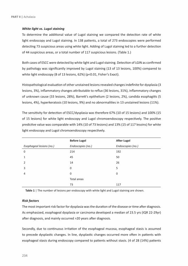
234
Part II | Achalasia
White light vs. Lugol staining
To determine the additional value of Lugol staining we compared the detection rate of white
light endoscopy and Lugol staining. In 138 patients, a total of 273 endoscopies were performed
detecting 73 suspicious areas using white light. Adding of Lugol staining led to a further detection
of 44 suspicious areas, or a total number of 117 suspicious lesions. (Table 1.)
Both cases of ESCC were detected by white light and Lugol staining. Detection of LGIN as confirmed
by pathology was significantly improved by Lugol staining (13 of 13 lesions, 100%) compared to
white light endoscopy (8 of 13 lesions, 62%) (p<0.01, Fisher’s Exact).
Histopathological evaluation of other unstained lesions revealed changes indefinite for dysplasia (3
lesions, 3%), inflammatory changes attributable to reflux (36 lesions, 31%), inflammatory changes
of unknown cause (33 lesions, 28%), Barrett’s epithelium (2 lesions, 2%), candida esophagitis (5
lesions, 4%), hyperkeratosis (10 lesions, 9%) and no abnormalities in 13 unstained lesions (11%).
The sensitivity for detection of ESCC/dysplasia was therefore 67% (10 of 15 lesions) and 100% (15
of 15 lesions) for white light endoscopy and Lugol chromoendoscopy respectively. The positive
predictive value was comparable with 14% (10 of 73 lesions) and 13% (15 of 117 lesoins) for white
light endoscopy and Lugol chromoendoscopy respectively.
before lugol After lugol
Esophageal lesions (no.) Endoscopies (no.) Endoscopies (no.)
0 214 192
1 45 50
2 14 26
3 0 5
4 0 0
Total areas
73 117
Table 1 | The number of lesions per endoscopy with white light and Lugol staining are shown.
Risk factors
The most important risk factor for dysplasia was the duration of the disease or time after diagnosis.
As emphasized, esophageal dysplasia or carcinoma developed a median of 23.5 yrs (IQR 22-29yr)
after diagnosis, and mainly occurred >20 years after diagnosis.
Secondly, due to continuous irritation of the esophageal mucosa, esophageal stasis is assumed
to precede dysplastic changes. In line, dysplastic changes occurred more often in patients with
esophageal stasis during endoscopy compared to patients without stasis. (4 of 28 (14%) patients

235
Screening for dyplasia using Lugol’s staining | Chapter 13
vs. 2 of 97 (2.1%) patients, Fisher’s exact p=0.02). To determine the validity of stasis observed
during endoscopy we compared it to the level of stasis on a timed barium esophagogram. As
expected, patients with esophageal stasis during endoscopy also had significantly higher levels of
esophageal stasis on timed barium esophagogram after 5 minutes (8.3 ± 1.3 cm vs. 3.0 ± 0.5 cm,
p<0.0001).
Although alcohol consumption and tobacco use was not prospectively assessed in detail in all
patients, only 1 patient with dysplasia or cancer had a history of tobacco use whereas none had a
history of alcohol abuse.
dIscussIon
In the present study, we evaluated the risk of esophageal dysplasia and carcinoma in patients
with achalasia using Lugol chromoendoscopy. Dysplastic changes and ESCC occurred primarily in
patients with longstanding disease (> 20 years), i.e. in 4.3 and 1.4% of this subgroup respectively.
Using Lugol staining, the detection rate of LGIN significantly increased compared to white light
endoscopy. Screening for dysplasia using Lugol staining might therefore be advocated in patients
with longstanding disease.
Continuous irritation of the esophageal mucosa due to esophageal stasis or reflux of gastric
contents is assumed to precede dysplastic changes.24,25 Subsequent hyperplastic or metaplastic
changes are frequently observed in patients with achalasia, and eventually these epithelial
changes can lead to malignant transformation. According to a recent meta-analysis by Eckardt et
al, patients with achalasia have an increased risk for ESCC, with a hazard ratio of 10-50 compared
to the normal population.9 We confirmed the increased prevalence of esophageal squamous
cell carcinoma in our total cohort, demonstrating a hazard ratio of 10. Interestingly, ESCC only
occurred after a median time of 26 years after the diagnosis of achalasia in our cohort. This time
after diagnosis is in line with five earlier studies, in which the time between symptoms of achalasia
and the diagnosis of ESCC varied from 24 to 32 years.7,9,11,26-28 The only study that failed to find
an increased risk for squamous cell carcinoma had a mean follow up after diagnosis of only 6
years.29 The relatively low hazard ratio10 in our study may also be attributable to the large number
of patients with 0-10 years of follow up after diagnosis (n=73, 53%). When only patients with
>15 years of achalasia were analysed, the relative risk increases to 43 compared to the general
population. Therefore, screening of patients with achalasia for dysplastic changes seems only
indicated in patients with longstanding disease

236
Part II | Achalasia
The prognosis of ESCC is mainly determined by the stage of disease at the time of detection.13,30
In achalasia, the prognosis of ESCC is considered to be even worse than in the general population,
mainly attributed to the delayed diagnosis. Three main reasons can be proposed: first, most
patients are used to having some residual degree of dysphagia even after treatment, masking the
increase in dysphagia caused by the development of ESCC. Secondly, patients adapt to dysphagia
and seek medical care at a much later stage. Finally, as the esophagus is often dilated, a malignant
lesion must reach a substantial size before causing additional dysphagia.23,31 In line, both patients
with ESCC in the current study were diagnosed in the screening program but did not report
aggravating dysphagia or alarm symptoms.
Based on the delayed diagnosis and the bad prognosis of ESSC, early detection of dysplastic lesions
becomes of crucial importance. This study demonstrated for the first time that staining with Lugol
significantly improves the detection of LGIN in achalasia with a sensitivity of up to 100%. Previous
studies have clearly demonstrated that the risk of ESCC is strongly dependent on the grade of
dysplasia. For squamous LGIN, the risk to develop ESCC is 5% within 3.5 years and 24% within
13.5 years in a high-risk population in northern China, which is 2.9 times higher compared to
the local general population.32,33 Although none of the patients with LGIN in our study showed
progression to HGIN or ESCC, it should be emphasized that our follow-up period of 55 months
is rather short. For HGIN, the risk to develop ESCC was 50-74% during a follow up period of 13.5
years in a prospective study in the same high risk population in China. Taken together, we conclude
that Lugol staining improves detection of dysplastic lesions, which are important precursor lesions
of ESCC.
Although the sensitivity of Lugol staining as screening tool is excellent (100%), a main disadvantage
is the high false positive lesion rate.36 The positive predictive value of lesions observed after Lugol
staining was only 13% for dysplasia or carcinoma in our study, mainly due to a high rate of reflux-
and aspecific inflammatory changes causing unstained lesions. Moreover, the optimal screening
interval remains to be determined. To avoid numerous screening endoscopies, ideally, patients
should be stratified according to their risk to develop dysplasia and cancer. One potential approach
could be histopathological evaluation of p53 immunoreactivity in the squamous mucosa. In
patients with achalasia, overexpression of p53 was observed in 82% of patients with ESCC and 67%
of patients with LGIN, and interestingly, even 6 years (range 1-11) prior to the diagnosis of ESCC
or LGIN.23 Further studies involving p53 and other biomarkers such as Ki67 or p16 might therefore
possibly be useful to stratify patients according to their risk to obtain for ESCC.37 However, for now
we would recommend to only screen patients with long-term achalasia on a 3-year basis.
Current guidelines do not propose regular screening for dysplasia or ESCC in patients with
achalasia. A screening program is only indicated if early detection leads to adequate therapy and

237
Screening for dyplasia using Lugol’s staining | Chapter 13
a reduction in mortality or morbidity. Large studies in a high-risk population for ESCC in China
demonstrated that screening and timely treatment reduced mortality of ESCC to the level that
survival of patients is comparable to healthy controls.38 Based on our data, we propose to screen
patients with > 15 years of achalasia. However, as our study is small and only has a limited follow
up period of 32 months, further studies are definitely needed to prove that screening in patients
with achalasia can indeed increase survival.
In conclusion, patients with longstanding disease and esophageal stasis, the risk to develop
esophageal dsplasia and carcinoma is significantly increased. As Lugol staining is more sensitive
than standard endoscopy to detect dysplastic lesions, a screening program may including Lugol
staining may be indicated. Further long-term follow-up studies are however needed to demonstrate
its impact on survival, preferably in a multi-center setting.

238
Part II | Achalasia
ReFeRence lIsT
1. Chino O, Kijima H, Shimada H et al. Clinicopathological studies of esophageal carcinoma in achalasia: analyses of carcinogenesis using histological and immunohistochemical procedures. Anticancer Res 2000;20(5C):3717-22.
2. Just-Viera JO, Haight C. Achalasia and carcinoma of the esophagus. Surg Gynecol Obstet 1969;128(5):1081-95.
3. Csendes A, Braghetto I, Burdiles P et al. Very late results of esophagomyotomy for patients with achalasia: clinical, endoscopic, histologic, manometric, and acid reflux studies in 67 patients for a mean follow-up of 190 months. Ann Surg 2006;243(2):196-203.
4. Leeuwenburgh I, Haringsma J, van DH et al. Long-term risk of oesophagitis, Barrett’s oesophagus and oesophageal cancer in achalasia patients. Scand J Gastroenterol Suppl 2006;(243):7-10.
5. Streitz JM, Jr., Ellis FH, Jr., Gibb SP et al. Achalasia and squamous cell carcinoma of the esophagus: analysis of 241 patients. Ann Thorac Surg 1995;59(6):1604-9.
6. Meijssen MA, Tilanus HW, van BM et al. Achalasia complicated by oesophageal squamous cell carcinoma: a prospective study in 195 patients. Gut 1992;33(2):155-8.
7. Wychulis AR, Woolam GL, Andersen HA et al. Achalasia and carcinoma of the esophagus. JAMA 1971;215(10):1638-41.
8. Dunaway PM, Wong RK. Risk and surveillance intervals for squamous cell carcinoma in achalasia. Gastrointest Endosc Clin N Am 2001;11(2):425-34, ix.
9. Eckardt AJ, Eckardt VF. Editorial: Cancer surveillance in achalasia: better late than never? Am J Gastroenterol 2010;105(10):2150-2.
10. Zaninotto G, Rizzetto C, Zambon P et al. Long-term outcome and risk of oesophageal cancer after surgery for achalasia. Br J Surg 2008;95(12):1488-94.
11. Leeuwenburgh I, Scholten P, Alderliesten J et al. Long-term esophageal cancer risk in patients with primary achalasia: a prospective study. Am J Gastroenterol 2010;105(10):2144-9.
12. West RL, Hirsch DP, Bartelsman JF et al. Long term results of pneumatic dilation in achalasia followed for more than 5 years. Am J Gastroenterol 2002;97(6):1346-51.
13. Westerterp M, Koppert LB, Buskens CJ et al. Outcome of surgical treatment for early adenocarcinoma of the esophagus or gastro-esophageal junction. Virchows Arch 2005;446(5):497-504.
14. Wang KK, Wongkeesong M, Buttar NS. American Gastroenterological Association technical review on the role of the gastroenterologist in the management of esophageal carcinoma. Gastroenterology 2005;128(5):1471-505.
15. Dawsey SM, Fleischer DE, Wang GQ et al. Mucosal iodine staining improves endoscopic visualization of squamous dysplasia and squamous cell carcinoma of the esophagus in Linxian, China. Cancer 1998;83(2):220-31.
16. Meyer V, Burtin P, Bour B et al. Endoscopic detection of early esophageal cancer in a high-risk population: does Lugol staining improve videoendoscopy? Gastrointest Endosc 1997;45(6):480-4.
17. Fagundes RB, de Barros SG, Putten AC et al. Occult dysplasia is disclosed by Lugol chromoendoscopy in alcoholics at high risk for squamous cell carcinoma of the esophagus. Endoscopy 1999;31(4):281-5.
18. Mori M, Adachi Y, Matsushima T et al. Lugol staining pattern and histology of esophageal lesions. Am J Gastroenterol 1993;88(5):701-5.
239
Screening for dyplasia using Lugol’s staining | Chapter 13
19. Toriie S, Akasaka Y, Yamaguchi K et al. New trial for endoscopical observation of esophagus by dye spraying method. G E N 1976;30(3-4):159-65.
20. Boeckxstaens GE, Annese V, des Varannes SB et al. Pneumatic dilation versus laparoscopic Heller’s myotomy for idiopathic achalasia. N Engl J Med 2011;364(19):1807-16.
21. Hamilton SR, Aaltonen LA. World Health Organization Classification of Tumours. Pathology and genetics of tumours of the digestive system. IARC Press, 2000.
22. de Oliveira JM, Birgisson S, Doinoff C et al. Timed barium swallow: a simple technique for evaluating esophageal emptying in patients with achalasia. AJR Am J Roentgenol 1997;169(2):473-9.
23. Leeuwenburgh I, Scholten P, Alderliesten J et al. Long-term esophageal cancer risk in patients with primary achalasia: a prospective study. Am J Gastroenterol 2010;105(10):2144-9.
24. Lehman MB, Clark SB, Ormsby AH et al. Squamous mucosal alterations in esophagectomy specimens from patients with end-stage achalasia. Am J Surg Pathol 2001;25(11):1413-8.
25. Porschen R, Molsberger G, Kuhn A et al. Achalasia-associated squamous cell carcinoma of the esophagus: flow-cytometric and histological evaluation. Gastroenterology 1995;108(2):545-9.
26. Aggestrup S, Holm JC, Sorensen HR. Does achalasia predispose to cancer of the esophagus? Chest 1992;102(4):1013-6.
27. Brucher BL, Stein HJ, Bartels H et al. Achalasia and esophageal cancer: incidence, prevalence, and prognosis. World J Surg 2001;25(6):745-9.
28. Eckardt VF, Hoischen T, Bernhard G. Life expectancy, complications, and causes of death in patients with achalasia: results of a 33-year follow-up investigation. Eur J Gastroenterol Hepatol 2008;20(10):956-60.
29. Chuong JJ, DuBovik S, McCallum RW. Achalasia as a risk factor for esophageal carcinoma. A reappraisal. Dig Dis Sci 1984;29(12):1105-8.
30. Wang GQ, Jiao GG, Chang FB et al. Long-term results of operation for 420 patients with early squamous cell esophageal carcinoma discovered by screening. Ann Thorac Surg 2004;77(5):1740-4.
31. West RL, Hirsch DP, Bartelsman JF et al. Long term results of pneumatic dilation in achalasia followed for more than 5 years. Am J Gastroenterol 2002;97(6):1346-51.
32. Wang GQ, Abnet CC, Shen Q et al. Histological precursors of oesophageal squamous cell carcinoma: results from a 13 year prospective follow up study in a high risk population. Gut 2005;54(2):187-92.
33. Dawsey SM, Fleischer DE, Wang GQ et al. Mucosal iodine staining improves endoscopic visualization of squamous dysplasia and squamous cell carcinoma of the esophagus in Linxian, China. Cancer 1998;83(2):220-31.
34. Porschen R, Molsberger G, Kuhn A et al. Achalasia-associated squamous cell carcinoma of the esophagus: flow-cytometric and histological evaluation. Gastroenterology 1995;108(2):545-9.
35. Gockel I, Bohl JR, Doostkam S et al. Spectrum of histopathologic findings in patients with achalasia reflects different etiologies. J Gastroenterol Hepatol 2006;21(4):727-33.
36. Dubuc J, Legoux JL, Winnock M et al. Endoscopic screening for esophageal squamous-cell carcinoma in high-risk patients: a prospective study conducted in 62 French endoscopy centers. Endoscopy 2006;38(7):690-5.

240
Part II | Achalasia
37. Wang WC, Wu TT, Chandan VS et al. Ki-67 and ProExC are useful immunohistochemical markers in esophageal squamous intraepithelial neoplasia. Hum Pathol 2011;42(10):1430-7.
38. Song PI, Liang H, Fan JH et al. Long-term survival after esophagectomy for early esophageal squamous cell carcinoma in Linxian, China. J
Surg Oncol 2011;104(2):176-80.


Chapter 14Summary, discussion and future
perspectives

244
suMMARY, dIscussIon And FuTuRe PeRsPecTIVes
The complicated task of the esophagogastric junction is to prevent gastroesophageal reflux while
passage through the sphincter has to be permitted and stasis of food, fluids and saliva avoided.
Dysfunction of this “gatekeeper” may result in the two typical examples of esophageal motility
disorders, i.e. gastroesophageal reflux disease (GERD) and achalasia. The pathophysiology and
management of these disorders represented the topic of the two parts presented in this thesis.
PART I: GAsTRoesoPhAGeAl ReFlux dIseAse
Gastroesophageal reflux is a physiological phenomenon but becomes pathological if troublesome
symptoms and/or complications occur. Gastroesophageal reflux disease is one of the most common
gastro-intestinal disorders and can present with typical symptoms such as regurgitation and
heartburn, or extra-esophageal symptoms such as cough and laryngitis. The different phenotypes
range from non-erosive reflux disease, through reflux esophagitis and Barrett’s esophagus.
Increased esophageal acid exposure is the most important hallmark of the disease. The mainstay
of GERD treatment therefore is acid suppression, most commonly achieved with the use of proton
pump inhibitors (PPIs). The efficacy of PPIs is very high for the treatment of erosive esophagitis,
especially for the healing of the esophageal injury.1 However, there is mounting evidence that
reflux symptoms persist in a high proportion (30%) of patients despite PPI therapy,2,3 and this
is associated with decreased psychological and physical well-being.4 These are the patients that
are seen in daily clinical practice by the gastroenterologist, and confront us with diagnostic and
therapeutic challenges.
To study the factors that influence persistent GERD symptoms we studied patients with persistent
symptoms on PPI treatment in chapter 2 and compared them to GERD patients without symptoms
during PPI use. We observed no significant differences in mucosal permeability, the position of
the acid pocket, the rate of acid reflux and the pH of the acid pocket in responders and partial
responders. In contrast, esophageal sensitivity to distension, the number of reflux episodes and
proximal extent were significantly increased in partial responders. Based on these results, we
concluded that an increased number of proximal reflux events in a hypersensitive esophagus are
associated with persistent symptoms and thereby lead to partial PPI failure in GERD.
From a therapeutic point of view, this implies that further acid suppression will not lead to a
reduction in persistent symptoms. Instead, insight into the multifactorial pathophysiology of GERD
is needed to develop new anti-reflux therapies. Most commonly gastroesophageal reflux disease

245
Summary, discussion and future perspectives | Chapter 14
is the result of incompetence of the gatekeeper between the stomach and the esophagus. The
two most important factors contributing to this incompetence are the physiological occurrence
of transient lower esophageal sphincter relaxations (TLESRs) and the anatomical distortion
of the LES and crural diaphragm, i.e. a hiatal hernia. Pharmacological inhibition of TLESRs is a
promising therapeutic approach because it has the potential to reduce most types of reflux. This is
particularly relevant for patients who have troublesome reflux symptoms despite acid suppression
from PPI use, because weakly acidic and weakly alkaline reflux play a significant role in symptom
generation in this group.5-7
Inhibition of TLESRs via interaction with the receptors involved in the underlying neural pathway
has been identified as a novel therapeutic target, and new pharmacological agents inhibiting
TLESRs are evaluated in clinical trials.8,9 For most of these studies the number of TLESRs is used
as the primary outcome variable, which emphasizes the importance of accurate detection of
TLESRs. Recently, high resolution esophageal pressure topography has been introduced and is
now considered the new standard to study esophageal and lower esophageal sphincter (LES)
function.10 In chapter 3 we performed a head-to-head comparison between high resolution
esophageal pressure topography and conventional sleeve manometry for the detection of TLESRs.
We demonstrated that high resolution esophageal pressure topography detected significantly
more TLESRs associated with a reflux event compared to sleeve manometry. This resulted in an
increase in sensitivity for detection of TLESRs with reflux from 86% to 96% for sleeve manometry
and high resolution esophageal pressure topography respectively. Therefore we concluded that
high resolution esophageal pressure topography is superior to sleeve manometry for the detection
of TLESRs associated with reflux, and that this technique should preferentially be used in clinical
trials evaluating the number of TLESRs.
Reflux inhibitors target receptors along the neural pathway involved in the triggering of TLESRS
such as mGluR5, GABA and cannabinoid receptors. Numerous studies in animals and humans
have demonstrated that (ant)agonism of these receptors can lead to a reduction in TLESRs
and reflux episodes. The distribution of receptors along the neural pathway involved in the
triggering of TLESRs, had however only been studied in animals. To optimise bench to bedside
translation in the development of new reflux inhibitors, we analyzed the distribution of mGluR5,
GABA and cannabinoid receptors along the vago-vagal neural pathway in chapter 4. Using
immunohistocheminal stainings, we evaluated the distribution of mGluR5, GABAA, GABAB, CB1
and CB2 receptors in human nodose ganglion, the brain stem and the myenteric plexus of the
esophagus. Similar to previous animal studies, all receptors were abundantly expressed in the
brain stem, and all but GABAAα1 were present in the nodose ganglion and myenteric plexus of
the LES. These findings are in line with the central side effects reported during treatment with

246
reflux inhibitors such as GABAB receptor agonists and mGluR5 antagonists, and underscore that
peripheral acting compounds may still be effective as reflux inhibitors.
In chapter 5 we performed a randomized, double-blind, crossover phase 1 study in healthy
volunteers, to study the effect of a novel mGluR5 receptor antagonist (AZD2066) on TLESRs and
reflux episodes. A single oral 13 mg dose of AZD2066 was found to reduce the numbers of TLESRs
and reflux episodes by 27% and 55% respectively compared with placebo. In addition, AZD2066
had an acceptable safety and tolerability profile. Based on these results we concluded that mGluR5
antagonism represents an interesting new avenue for reflux inhibitor treatment of GERD. It should
be noted however that in a similar designed study the GABAB-receptor agonist lesogaberan was
previously shown to reduce the number of reflux episodes in healthy volunteers to a greater
extent than AZD2066 did in the current study.8 Lesogaberan has since been discontinued owing
to a lack of evidence for a clinically significant effect as an add-on to PPI therapy in patients with
reflux symptoms despite PPI treatment. Currently, there is no reflux inhibitor available on the
market with an acceptable efficacy and side effect profile. Nevertheless, the concept of direct
inhibition of the most important mechanism underlying reflux episodes (i.e. TLESRs) remains very
promising. In the upcoming years, additional research has to sort out whether a safe and effective
reflux inhibitor can be developed that significantly reduces symptoms refractory to PPIs.
Most (acid) reflux episodes occur after a meal, when the stomach is filled with ingested food. In
contrast to the believe that meal ingestion buffers gastric acid, acid reflux episodes occur even in
the early postprandial period.11 Fletcher et al elegantly showed that gastric acid floats on top of
the meal acting as a reservoir from which acid can enter the esophagus during episodes of opening
of the esophagogastric junction. This unbuffered pool of gastric acid is referred to as the gastric
acid pocket.12 The position of the acid pocket relative to the crural diaphragm is an important
determinant of the acidity of the refluxate. In GERD, proximal extension of the acid pocket above
the diaphragm increases the risk for acid reflux.13 The acid pocket is therefore an important source
of postprandial acid in GERD and, as such, represents a unique therapeutic target. In the next
chapters of this thesis we tested the hypothesis that the acid pocket is a therapeutic target in
patients with GERD for several medical treatments using concurrent scintigraphy, high resolution
manometry and pH-impedance monitoring in a postprandial research protocol.
In chapter 6 we studied to what extent changes in position, size and acidity of the acid pocket
contributed to the therapeutic effect of PPIs. We demonstrated that also during PPI treatment
gastric secretions accumulate on top of the ingested food and form a pocket. Proton pump
inhibitors do not alter the number of reflux events, but increase the pH of the acid pocket which
in its turn leads to the increased pH of reflux events. Therefore we concluded that the acid pocket
is still the source of refluxate during treatment with PPI, and as such represents a potential target
for additional therapy.

247
Summary, discussion and future perspectives | Chapter 14
In chapter 7 we studied the effect of Azithromycin, a macrolide antibiotic with gastroprokinetic
properties, on acid pocket position and acid exposure in GERD patients off PPI. Azithromycin
reduced the number of acid reflux episodes and esophageal acid exposure in GERD patients,
especially in patients with a small hiatal hernia. This effect was mainly caused by a more distal
position of the acid pocket, probably resulting from a reduction of the hiatal hernia size. These
data for the first time indicated that modulation of the position of the acid pocket had an impact
on the acidity of the refluxate.
Alginate-antacid formulations are frequently used to treat GERD and are proposed to be efficient
through formation of a floating raft after reaction with the acid pocket. To evaluate this hypothesis,
we visualized the alginate raft relative to the position of the acid pocket in chapter 8, and compared
its effect on gastroesophageal reflux to a commonly used antacid. The alginate formulation and
the acid pocket were visualized using scintigraphy with radioactive indium and technetium-
pertechnetate respectively. Reflux episodes were detected using concurrent high-resolution
manometry and pH-impedance monitoring. We demonstrated that the alginate-antacid labeling
persisted in the proximal stomach for the entire study period co-localizing with the acid pocket
and displacing it distally. In addition, the alginate-antacid reduced acid reflux events by more
than 75% compared to an antacid alone. These findings demonstrate that alginate formulations
directly target the acid pocket with a non-systemic, alginate-antacid formulation for the treatment
of postprandial acid reflux in GERD.
Future perspectives
Gastroesophageal reflux disease remains a complex disease with multiple factors involved. In
this thesis we mainly focussed on the pathophysiology and additional treatment options of GERD
patients with refractory symptoms during PPI use. Most patients with true GERD have at least a
partial response to PPIs.3 Moreover, PPIs have a good safety profile, are widely available and are
very effective in the treatment of reflux esophagitis.14,15 Therefore, acid suppression by means
of PPIs will remain the mainstay of GERD therapy, and additional studies should mainly focus on
the treatment of refractory symptoms during PPI use.8,16 The studies in this thesis illustrate how
insight in pathophysiology can lead to new treatment options in these patients. We demonstrated
that visceral hypersensitivity and frequent reflux episodes with a high proximal extent contribute
to residual symptoms in patients with recurrent symptoms on PPI therapy. Moreover, the
acid pocket is still the source of refluxate during treatment with PPI, and as such represents a
potential target for additional therapy. Potential additional treatment options therefore include
pain modulators, reflux inhibitors and treatment aimed at the acid pocket. Recent exploring
studies already demonstrated the potential benefit of these approaches: For instance, Viazis et al
compared citalopram 20 mg as a pain modulator to placebo in 75 patients with refractory GERD
248
symptoms on twice daily PPI. Interestingly, success rate of treatment was significantly higher in the
patients receiving citalopram (62%, n=39) compared to patients receiving placebo (33%, n=36).17
Second, in a large clinical trial, we have previously demonstrated that treatment with the reflux
inhibitor lesogaberan resulted in a significantly larger proportion of responders to treatment
compared with placebo.8 It has to be emphasized however that the proportion of responders
was small (16% vs. 8% of patients), and therefore the development of lesogaberan has since been
discontinued. Finally, a recent study in patients with non-erosive reflux disease demonstrated
that supplementing PPI therapy with alginates more than doubled the number of patients who
experienced complete reflux symptom relief (26% vs. 57% respectively).18 Treatment with alginates
is of particular interest as it interacts directly with the acid pocket, is not systemically absorbed
and therefore has very limited side effects. In the near future, more studies are awaited in which
the benefits of these drugs as add-on treatment to PPIs are evaluated. Moreover, studies have to
address which patients benefit most from which add-on treatment. Eventually, guidelines have
to be established how patients with refractory PPI symptoms should be diagnosed and treated to
ultimately improve quality of life of our patients.
PART II: AchAlAsIA
In the second part of this thesis we studied the treatment and follow up of achalasia. Achalasia is
a primary esophageal motor disorder characterized by the absence of peristalsis and a defective
relaxation of the LES, resulting in impaired bolus transport and stasis of food in the esophagus.19
The characteristic feature of achalasia is the loss of enteric neurons in the myenteric plexus,
leading to severely disturbed esophageal motility and typical symptoms such as dysphagia,
regurgitation and weight loss.20 Mainly due to a lack in pathophysiological insight, treatment of
achalasia aims at relieving functional obstruction at the level of the esophagogastric junction. For
many years, repeated endoscopic pneumodilation (PD) had been the treatment of choice leading
to therapeutic success in 70-80%. (21;22) There was a growing enthusiasm in favor of laparoscopic
surgery, based on recent publications showing excellent results from single-center series.23-25
In chapter 9 we described a large randomized controlled trial comparing laparoscopic Heller
myotomy (LHM) with Dor fundoplication and PD. We demonstrated that the success rate, defined
as the reduction in Eckardt symptom score below 4, was comparable for both treatments. Using
this criterion, the success rate for surgery was 93% and 90% after 1 and 2 years of follow-up
respectively compared to 90% and 86% for PD. Redilation, allowed once during the first 2 years
of follow up, was performed in 23 of 95 patients after PD (25%). No difference in the level of
esophageal stasis or in the quality of life measured by SF-36 was observed. To what extent failure
rates may increase in time as more than 3 dilation series are required to maintain patients in
249
Summary, discussion and future perspectives | Chapter 14
clinical remission remains to be seen. Nevertheless, with the data currently available after a mean
follow up of 43 months, both pneumatic dilation and LHM can be offered as effective treatment
to patients with achalasia.
By defining risk factors for failure/success it might perhaps be possible to design an individualized
therapy for the patient with achalasia. Pandolfino et al recently described three manometric
subtypes based on the residual esophageal manometric pattern: In type I, no pressure waves can
be recorded in the distal esophagus, type II is characterized by panesophageal pressurisation,
whereas in type III at least 20% of swallows reveals rapidly propagating or spastic contractions.26
Importantly, recent studies have suggested that treatment efficacy of LHM and PD strongly varies
depending on the manometric subtype.26,27 In chapter 10, we performed an ad hoc analysis to the
randomized controlled trial described in chapter 9 to define whether the subtype classification of
achalasia could predict outcome. Moreover, we aimed to determine whether the subtype should
dictate the choice of treatment, i.e. LHM or PD. In short, in the randomized European trial, both
type I and II patients had similar, high success rates after either LHM or PD. Patients with type
III, however, had a lower response rate, in particular those who underwent PD. Based on these
results, one could argue that LHM might be the preferred treatment option in patients with type
III achalasia, though the number of patients studied so far is rather small.
Since we cannot cure the motor abnormalities of achalasia, recurrences after any form of
treatment will occur with longer follow-up periods. Treatment success gradually decreases in
patients with longstanding disease (≥10 yr) to 40-60%.22,28-30 Importantly, however, additional
treatment, both after initial PD or LHM, has satisfactory results with success rates of 60-80% and
improvement of esophageal emptying.22,23,31,32 Follow up of patients with achalasia is not only
important to ensure optimal symptom control, but also to prevent possible complications such
as esophageal decompensation. Timely detection of patients in need for additional treatment
is indicated to avoid these long-term complications. The decision to retreat patients may be a
difficult one, and objective quantification of esophageal and LES function seems indicated by
means of measurement of LES pressure or detection of esophageal stasis on a timed barium
esophagogram. In chapter 11 we studied a cohort of patients with achalasia with a mean follow
up of 26 years after diagnosis. This study shows that stasis on a timed barium esophagogram
but not basal LES pressure is an important predictor of recurrent symptoms and the need for
retreatment in patients with longstanding achalasia. Therefore, patients should preferentially
be followed up using a timed barium esophagogram. When significant stasis on a timed barium
esophagogram is observed, even in the presence of a LES pressure < 10 mmHg, additional therapy
may be warranted.

250
A significant proportion of patients with persistent symptoms has a low or even absent LES
pressure. Interestingly, these patients often have incomplete esophageal emptying on a timed
barium esophagogram. Reduced distensibility even in the absence of LES pressure may therefore
explain why esophageal emptying on timed barium esophagogram may be impaired. We tested
this hypothesis in chapter 12 by measuring EGJ distensibility, esophageal stasis and LES pressure.
We demonstrated that EGJ distensibility, but not LES pressure, strongly correlated with clinical
response to treatment: impaired EGJ distensibility is observed in 92% of patients with recurrent
symptoms, whilst only 42% of these patients had an elevated LES pressure. Moreover, patients
with impaired EGJ distensibility had recurrent symptoms and impaired esophageal emptying,
even if LES pressure was low. Based on these findings, we concluded that EGJ distensibility is a
better parameter to evaluate the efficacy of treatment in patients with achalasia compared to LES
pressure.
Chronic irritation of the esophageal mucosa, due to poor emptying or excessive reflux of
gastric contents, is a risk factor to develop esophageal carcinoma. Patients with achalasia have
an increased risk for esophageal carcinoma, i.e. adenocarcinoma as well as squamous cell
carcinoma.33-35 In chapter 13 we confirmed that patients with achalasia have an increased risk for
esophageal carcinoma. In addition, we demonstrated that Lugol staining, significantly increased
the detection rate of dysplasia compared to white light endoscopy. To increase probability for
detection during screening and thereby cost-effectiveness, risk factors for dysplasia need to be
recognized. Esophageal squamous cell carcinoma only occurred after a median time of 26 years
after the diagnosis of achalasia in our cohort. This time after diagnosis is in line with five earlier
studies, in which the time between symptoms of achalasia and the diagnosis of ESCC varied from 24
to 32 years.33,35-37 These data suggest that screening for dysplasia should be performed in patients
with a long history of dysphagia (>15-20 years), especially those with long-term esophageal stasis.
Future perspectives
In summary, due to the lack of understanding of the pathogenesis of achalasia, initial treatment
still focuses on mechanical disruption of the LES, rather than on restoring esophageal motility.
We demonstrated that a graded pneumodilation protocol with the possibility for redilation
in case of recurrent symptoms is as successful as LHM, and leads to comparable drop in LES
pressure, reduction in esophageal stasis and improved quality of life. The long-term results of
the randomized achalasia trial, with at least five years of follow up, are expected in the upcoming
year. These results will be of imminent importance to determine whether pneumodilation also has
comparable success to LHM with longer follow up.
Recently, a new endoscopic technique termed peroral endoscopic myotomy (POEM) was described
to treat achalasia. In this technique, the endoscopist creates a submucosal tunnel to reach the LES

251
Summary, discussion and future perspectives | Chapter 14
and to dissect the circular muscle.38 Initial studies reported very high success rates of 94-100%,
even following multiple previous dilations.38,39 Although POEM has generated excellent initial
results and great enthusiasm among endoscopists, it should be emphasized though, that follow
up of these studies is still short and thus longer follow up is absolutely needed before this new
technique can be accepted. Moreover, randomized studies comparing POEM to pneumodilation
or LHM should be performed and such studies are presently in progress. Ideally, the choice of
treatment should be based on risk factors such as achalasia subtype and age in order to generate
an individualized therapeutic strategy.40 The current randomized trials comparing POEM to
pneumatic dilation and LHM are ideally suited to further determine risk factors for treatment
failure. Using these risk factors, the best available option for a specific patient can be sought.
An interesting alternative approach would be to transplant neuronal stem cells. In mice, neuronal
stem cells injected in the pylorus survived and even expressed nitric oxide synthase.41 Such
approach could lead to a completely restored sphincter function and peristalsis. Recently, it was
even shown that neural stem cells can be isolated from mucosal biopsies, creating possibilities to
treat achalasia.42 Clearly, a lot of research remains to be done further exploring this approach. Until
more insight in pathophysiology leads to a better targeted therapy, possibly thus even restoring
esophageal motility, treatment will be aimed at mechanical disruption of the LES.
With adequate follow up and timely retreatment significant morbidity and possibly late
complications such as esophageal decompensation and esophageal carcinoma can be avoided.
Based on the studies in this thesis we concluded that regular follow up visits may be indicated, not
only to inform about symptoms, but also to perform a timed barium esophagogram in order to
assess esophageal emptying. Clearly, prospective trials are required to confirm the benefit of (serial)
functional testing and early additional treatment for the prevention of long-term complications.
One can hypothesize that risk factors can also be determined for esophageal decompensation and
for esophageal dysplasia or carcinoma. For instance, patients with type I achalasia already have
a wide and elongated esophagus at diagnosis, and as indicated by our data are prone to develop
further esophageal widening during follow-up. Especially for these patients repetitive functional
testing and early additional treatment might be indicated to avoid this complication. On the
other hand, we showed that patients with type II achalasia respond best to treatment with better
symptom control, reduced need for retreatment and diminished stasis. These data would suggest
that type II patients may be less likely to develop complications. Clearly, prospective studies are
required to prove this hypothesis, which in view of the low incidence of achalasia will be almost
impossible to achieve.
Early detection of curable, dysplastic lesions in patients with achalasia is essential to reduce
mortality related to esophageal carcinoma. Therefore, alternative screening tools should be applied.

252
To avoid numerous screening endoscopies, ideally, patients should be stratified according to their
risk to develop dysplasia and cancer. Patients who develop esophageal cancer had a long history
of dysphagia in combination with long-term esophageal stasis.33,35-37 For now we would therefore
recommend to only screen patients with long-term achalasia on a 3-year basis. Another potential
approach could be the use histopathological markers such as p53 or Ki67 immunoreactivity. In
patients with achalasia, overexpression of p53 is observed in 82% of patients with squamous cell
carcinoma and 67% of patients with dysplasia, even 6 years (range 1-11) prior to the diagnosis
of carcinoma or dysplasia.43 Further studies involving biomarkers might therefore be extremely
useful to stratify patients according to their risk to obtain for squamous cell carcinoma.
Clearly, after diagnosis, achalasia is a lifelong disease for which adequate treatment and follow up
is indispensable. With the right approach most patients can have good quality of life, and serious
morbidity and complications can be avoided.

253
Summary, discussion and future perspectives | Chapter 14
ReFeRence lIsT
1. Chiba N, De Gara CJ, Wilkinson JM et al. Speed of healing and symptom relief in grade II to IV gastroesophageal reflux disease: a meta-analysis. Gastroenterology 1997;112(6):1798-810.
2. Armstrong D, Pare P, Pericak D et al. Symptom relief in gastroesophageal reflux disease: a randomized, controlled comparison of pantoprazole and nizatidine in a mixed patient population with erosive esophagitis or endoscopy-negative reflux disease. Am J Gastroenterol 2001;96(10):2849-57.
3. Fass R, Shapiro M, Dekel R et al. Systematic review: proton-pump inhibitor failure in gastro-oesophageal reflux disease--where next? Aliment Pharmacol Ther 2005;22(2):79-94.
4. El-Serag H, Becher A, Jones R. Systematic review: persistent reflux symptoms on proton pump inhibitor therapy in primary care and community studies. Aliment Pharmacol Ther 2010;32(6):720-37.
5. Boeckxstaens GE, Smout A. Systematic review: role of acid, weakly acidic and weakly alkaline reflux in gastro-oesophageal reflux disease. Aliment Pharmacol Ther 2010;32(3):334-43.
6. Zerbib F, Duriez A, Roman S et al. Determinants of gastro-oesophageal reflux perception in patients with persistent symptoms despite proton pump inhibitors. Gut 2008;57(2):156-60.
7. Tutuian R, Vela MF, Hill EG et al. Characteristics of symptomatic reflux episodes on Acid suppressive therapy. Am J Gastroenterol 2008;103(5):1090-6.
8. Boeckxstaens GE, Beaumont H, Hatlebakk JG et al. A novel reflux inhibitor lesogaberan (AZD3355) as add-on treatment in patients with GORD with persistent reflux symptoms despite proton pump inhibitor therapy: a randomised placebo-controlled trial. Gut 2011;60(9):1182-8.
9. Boeckxstaens GE, Beaumont H, Mertens V et al. Effects of lesogaberan on reflux and lower esophageal sphincter function in patients with gastroesophageal reflux disease. Gastroenterology 2010;139(2):409-17.
10. Fox MR, Bredenoord AJ. Oesophageal high-resolution manometry: moving from research into clinical practice. Gut 2008;57(3):405-23.
11. Beaumont H, Bennink RJ, de JJ et al. The position of the acid pocket as a major risk factor for acidic reflux in healthy subjects and patients with GORD. Gut 2010;59(4):441-51.
12. Fletcher J, Wirz A, Young J et al. Unbuffered highly acidic gastric juice exists at the gastroesophageal junction after a meal. Gastroenterology 2001;121(4):775-83.
13. Beaumont H, Bennink RJ, de JJ et al. The position of the acid pocket as a major risk factor for acidic reflux in healthy subjects and patients with GORD. Gut 2010;59(4):441-51.
14. Chiba N, De Gara CJ, Wilkinson JM et al. Speed of healing and symptom relief in grade II to IV gastroesophageal reflux disease: a meta-analysis. Gastroenterology 1997;112(6):1798-810.
15. Massoomi F, Savage J, Destache CJ. Omeprazole: a comprehensive review. Pharmacotherapy 1993;13(1):46-59.
16. Fass R, Sifrim D. Management of heartburn not responding to proton pump inhibitors. Gut 2009;58(2):295-309.
17. Viazis N, Keyoglou A, Kanellopoulos AK et al. Selective serotonin reuptake inhibitors for the treatment of hypersensitive esophagus: a randomized, double-blind, placebo-controlled study. Am J Gastroenterol 2012;107(11):1662-7.

254
18. Manabe N, Haruma K, Ito M et al. Efficacy of adding sodium alginate to omeprazole in patients with nonerosive reflux disease: a randomized clinical trial. Dis Esophagus 2012;25(5):373-80.
19. Boeckxstaens GE. The lower oesophageal sphincter. Neurogastroenterol Motil 2005;17 suppl 1:13-21.
20. Goldblum JR, Rice TW, Richter JE. Histopathologic features in esophagomyotomy specimens from patients with achalasia. Gastroenterology 1996;111(3):648-54.
21. Zerbib F, Thetiot V, Richy F et al. Repeated pneumatic dilations as long-term maintenance therapy for esophageal achalasia. Am J Gastroenterol 2006;101(4):692-7.
22. Hulselmans M, Vanuytsel T, Degreef T et al. Long-term outcome of pneumatic dilation in the treatment of achalasia. Clin Gastroenterol Hepatol 2010;8(1):30-5.
23. Zaninotto G, Costantini M, Rizzetto C et al. Four hundred laparoscopic myotomies for esophageal achalasia: a single centre experience. Ann Surg 2008;248(6):986-93.
24. Sharp KW, Khaitan L, Scholz S et al. 100 consecutive minimally invasive Heller myotomies: lessons learned. Ann Surg 2002;235(5):631-8.
25. Patti MG, Molena D, Fisichella PM et al. Laparoscopic Heller myotomy and Dor fundoplication for achalasia: analysis of successes and failures. Arch Surg 2001;136(8):870-7.
26. Pandolfino JE, Kwiatek MA, Nealis T et al. Achalasia: a new clinically relevant classification by high-resolution manometry. Gastroenterology 2008;135(5):1526-33.
27. Salvador R, Costantini M, Zaninotto G et al. The preoperative manometric pattern predicts the outcome of surgical treatment for esophageal achalasia. J Gastrointest Surg 2010;14(11):1635-45.
28. West RL, Hirsch DP, Bartelsman JF et al. Long term results of pneumatic dilation in achalasia followed for more than 5 years. Am J Gastroenterol 2002;97(6):1346-51.
29. Eckardt VF, Aignherr C, Bernhard G. Predictors of outcome in patients with achalasia treated by pneumatic dilation. Gastroenterology 1992;103(6):1732-8.
30. Lopushinsky SR, Urbach DR. Pneumatic dilatation and surgical myotomy for achalasia. JAMA 2006;296(18):2227-33.
31. Guardino JM, Vela MF, Connor JT et al. Pneumatic dilation for the treatment of achalasia in untreated patients and patients with failed Heller myotomy. J Clin Gastroenterol 2004;38(10):855-60.
32. Vela MF, Richter JE, Khandwala F et al. The long-term efficacy of pneumatic dilatation and Heller myotomy for the treatment of achalasia. Clin Gastroenterol Hepatol 2006;4(5):580-7.
33. Eckardt AJ, Eckardt VF. Editorial: Cancer surveillance in achalasia: better late than never? Am J Gastroenterol 2010;105(10):2150-2.
34. Zendehdel K, Nyren O, Edberg A et al. Risk of esophageal adenocarcinoma in achalasia patients, a retrospective cohort study in Sweden. Am J Gastroenterol 2011;106(1):57-61.
35. Leeuwenburgh I, Scholten P, Alderliesten J et al. Long-term esophageal cancer risk in patients with primary achalasia: a prospective study. Am J Gastroenterol 2010;105(10):2144-9.
36. Wychulis AR, Woolam GL, Andersen HA et al. Achalasia and carcinoma of the esophagus. JAMA 1971;215(10):1638-41.
37. Brucher BL, Stein HJ, Bartels H et al. Achalasia and esophageal cancer: incidence, prevalence, and prognosis. World J Surg 2001;25(6):745-9.
255
Summary, discussion and future perspectives | Chapter 14
38. Inoue H, Minami H, Kobayashi Y et al. Peroral endoscopic myotomy (POEM) for esophageal achalasia. Endoscopy 2010;42(4):265-71.
39. von RD, Inoue H, Minami H et al. Peroral Endoscopic Myotomy for the Treatment of Achalasia: A Prospective Single Center Study. Am J Gastroenterol 2011.
40. Richter JE, Boeckxstaens GE. Management of achalasia: surgery or pneumatic dilation. Gut 2011.
41. Micci MA, Learish RD, Li H et al. Neural stem cells express RET, produce nitric oxide, and survive transplantation in the gastrointestinal tract. Gastroenterology 2001;121(4):757-66.
42. Metzger M, Caldwell C, Barlow AJ et al. Enteric nervous system stem cells derived from human gut mucosa for the treatment of aganglionic gut disorders. Gastroenterology 2009;136(7):2214-25.
43. Leeuwenburgh I, Gerrits MM, Capello A et al. Expression of p53 as predictor for the development of esophageal cancer in achalasia patients. Dis Esophagus 2010;23(6):506-11.


Nederlandse samenvatting
258
nedeRlAndse sAMenVATTInG
De overgang tussen de slokdarm en de maag is een speciaal overgangsgebied bestaande uit de
onderste slokdarmsfincter en het crurale deel van het diafragma. Samen hebben deze structuren
de belangrijke taak om ervoor te zorgen dat voedsel de maag in kan, terwijl terugvloed van
maaginhoud (reflux) vermeden dient te worden. In principe is er dus sprake van eenrichtingsverkeer,
alleen in het geval van boeren of braken moet de terugvloed vanuit de maag in de slokdarm wel
mogelijk zijn. Wanneer deze poortwachterfunctie niet goed wordt uitgevoerd kunnen de twee
typische slokdarmmotiliteitsaandoeningen ontstaan: gastro-oesofageale refluxziekte en achalasie.
De pathofysiologie en behandeling van deze twee aandoeningen vormen de twee delen van dit
proefschrift.
deel I: GAsTRo-oesoFAGeAle ReFluxZIekTe
Wanneer reflux van maaginhoud in de slokdarm aanleiding geeft tot klachten of complicaties wordt
het gastro-oesofageale refluxziekte genoemd. Gastro-oesofageale refluxziekte is een van de meest
voorkomende gastro-intestinale aandoeningen, met ongeveer 15-20% van de bevolking die op
zijn minst wekelijks last heeft van zuurbranden of zure oprispingen (regurgitatie). De meerderheid
van patiënten heeft milde tot matige symptomen, maar in een minderheid van patiënten leidt
voortdurende expositie aan schadelijke maaginhoud tot complicaties zoals erosieve oesofagitis,
Barrett slokdarm, peptische stricturen of zelfs slokdarmcarcinoom.
De toegenomen expositie van maagzuur aan de slokdarm is een belangrijk kenmerk van de ziekte.
De belangrijkste pijler van behandeling is dan ook onderdrukking van zuurproductie in de maag,
meestal met protonpompremmers. Protonpompremmers zijn zeer effectief voor de genezing van
oesofagitis. Echter, symptomen van refluxziekte persisteren in een relatief hoog percentage (30%)
van de patiënten, en leiden tot een verminderde kwaliteit van leven. Juist deze patiënten worden
doorgestuurd door de huisarts naar de maag-darm-leverarts, en stellen ons voor een diagnostische
en therapeutische uitdaging.
Om te bepalen wat de oorzaak is van symptomen die blijven bestaan tijdens behandeling met
protonpompremmers hebben we in hoofdstuk 2 patiënten geanalyseerd met persisterende
klachten, en deze vergeleken met patiënten zonder persisterende klachten tijdens het gebruik
van protonpompremmers. We toonden aan dat de gevoeligheid van de slokdarm voor distensie
veel hoger was in patiënten met persisterende klachten. Daarnaast hadden patiënten met
persisterende klachten meer, en hoger opgaande reflux episoden vergeleken met patiënten

259
Nederlandse samenvatting | Chapter 12
zonder persisterende klachten. In tegenstelling tot wat eerdere studies suggereerden vonden we
geen verschil in de permeabiliteit van het slokdarmepitheel. Ook was de mate van zuurremming
niet verschillend. Op basis van deze resultaten concluderen we dat persisterende symptomen het
gevolg zijn van hoogopgaande reflux episoden in een overgevoelige slokdarm, en daarmee leiden
tot het falen van protonpompremmers in patiënten met refluxziekte.
Vanuit een therapeutisch oogpunt impliceert dit dat verdere zuurremming niet zal leiden tot een
betere behandeling van persisterende symptomen. In plaats daarvan zou aanvullende behandeling
een andere route moeten kiezen.
Het belangrijkste onderliggende mechanisme van reflux episoden zijn de zogenaamde transiente
relaxaties van de onderste slokdarmsfincter (TLESRs), zowel in gezonde vrijwilligers als in
refluxpatiënten. Een TLESR is een vago-vagaal gemedieerde reflex die wordt gekenmerkt door een
verlengde relaxatie van de onderste slokdarmsfincter met tegelijkertijd inhibitie van het crurale
diafragma. Deze TLESRs worden voornamelijk getriggerd door uitrekking van de maag, en zijn het
fysiologische mechanisme waarmee de maag gas ventileert. De farmacologische inhibitie van deze
TLESRs is een veelbelovende therapeutische benadering omdat niet alleen zure, maar ook niet
zure reflux episodes kunnen worden geremd. Dit is vooral van belang voor patiënten die klachten
houden ondanks zuurremmende medicatie, omdat niet zure reflux episodes een belangrijke
veroorzaker zijn van symptomen in deze patiënten.
Vele receptoren die betrokken zijn in de signaaltransductie in de onderliggende neurale pathway
zijn geïdentificeerd en nieuwe geneesmiddelen die op deze receptoren aangrijpen om TLESRs te
onderdrukken worden getest in klinische studies. Deze nieuwe geneesmiddelen worden ook wel
‘refluxremmers’ genoemd. Voor veel van deze studies wordt het aantal TLESRs aangehouden als
primaire uitkomstmaat. Dit toont aan dat een adequate registratie Van TLESRs van het grootste
belang is. Recentelijk is hoogresolutie manometrie geïntroduceerd, en dit wordt nu gezien als de
nieuwe standaard om de motiliteit van de slokdarm te bestuderen. In hoofdstuk 3 hebben we
aangetoond dat de nieuwe hoogresolutie manometrie ook beter TLESRs detecteert vergeleken
met conventionele manometrie. Daarom adviseren we dat in het vervolg TLESRs in studies
gedetecteerd moeten worden met hoogresulotie manometrie.
Refluxremmers richten zich op de receptoren die betrokken zijn in de signaaltransductie van de
TLESR-reflex, zoals de mGluR5, GABA en cannabinoid receptoren. Verschillende studies in zowel
proefdieren als mensen hebben aangetoond dat het agoneren van deze receptoren (of antagoneren,
afhankelijk van de betrokken receptor) het aantal TLESRs en daarmee het aantal reflux episodes
kan reduceren. De distributie van de receptoren die betrokken zijn bij de inhibitie van TLESRs was
tot nu toe alleen bestuurd in proefdieren. Om een betere vertaling van het laboratorium naar de

260
klinische situatie te kunnen maken hebben we in hoofdstuk 4 de distributie van mGluR5, GABA
en cannabinoid receptoren onderzocht in het humane ganglion nodosum, de hersenstam en de
plexus myentericus van de oesofagus. Vergelijkbaar met eerdere studies in proefdieren, waren
deze receptoren overvloedig aanwezig in de hersenstam, het ganglion nodosum en in de plexus
myentericus. Deze bevindingen verklaren de centrale bijwerkingen die gerapporteerd worden in
studies met reflux remmers zoals GABAB-receptor agonisten en mGluR5 antagonisten. Bovendien
onderstreept de bevinding dat receptoren in humaan weefsel zowel centraal als perifeer aanwezig
zijn, dat alleen perifeer werkende verbindingen nog steeds effectief kunnen zijn als refluxremmers,
maar dan zonder centrale bijwerkingen.
In hoofdstuk 5 verrichtten we een gerandomiseerde, dubbelblinde, cross-over fase 1 studie in
gezonde vrijwilligers, om het effect van een nieuwe mGluR5 receptor antagonist (AZD2066) op
TLESRs en reflux episodes te bestuderen. Een enkele dosis van 13 mg AZD2066 verminderde
het aantal TLESRs en reflux episodes met respectievelijk 27% en 55% vergeleken met placebo.
Bovendien was het aantal en ernst van de bijwerkingen die vrijwilligers rapporteerden laag. Op
basis van deze resultaten concludeerden we dat het antagoneren van mGluR5 receptoren een
interessante nieuwe methode van reflux remming is. Hierbij moet echter wel worden opgemerkt
dat in een studie met een vergelijkbare opzet de refluxremmer Lesogaberan het aantal TLESRs
en reflux episodes beduidend meer verminderde dan AZD2066 in deze studie. De ontwikkeling
van Lesogaberan is inmiddels gestopt omdat het effect op symptomen in patiënten die al
protonpompremmers gebruikten in klinische studies te gering was. Op dit moment is er dan
ook geen refluxremmer op de markt die acceptabel is qua effectiviteit en bijwerkingenprofiel.
Desalniettemin blijft het concept van directe remming van het belangrijkste mechanisme dat
reflux episodes veroorzaakt, namelijk TLESRs, erg veelbelovend. In de komende jaren moet uit
nieuwe studies gaan blijken of er een veilige en effectieve refluxremmer ontwikkeld kan worden.
De meeste reflux episodes treden na de maaltijd op, als de maag gevuld is met voedsel. Hoewel
men zou verwachten dat de ingenomen maaltijd het zuur in de maag buffert, vinden ook zure
reflux episodes vrijwel meteen na de maaltijd plaats. Recente studies hebben laten zien dat bijna al
het zuur in de maag wordt gebufferd door het ingenomen voedsel, maar dat bovenop het voedsel
in de proximale maag een hoeveelheid maagzuur drijft die niet gebufferd wordt. Dit maagzuur
fungeert daar als een reservoir van waaruit zuur terug kan stromen in de slokdarm, wanneer de
onderste slokdarmsfincter zich opent. Deze hoeveelheid zeer geconcentreerd maagzuur wordt
ook wel de ‘acid pocket’ genoemd.
In eerdere studies hebben we laten zien dat de positie van de acid pocket voor een groot deel
bepaalt of het refluxaat zuur is. In patiënten met refluxziekte is de acid pocket vaak in de opening
van de slokdarm gesitueerd, waardoor er in het geval van reflux gelijk het geconcentreerde

261
Nederlandse samenvatting | Chapter 12
zuur terugvloeit. Hierdoor is de acid pocket bij patiënten met refluxziekte vaker de bron van het
refluxaat dan bij gezonde mensen en zij hebben dan ook vaker zure reflux episoden. Bij patiënten
met een hiatus hernia zijn de onderste slokdarmsfincter en het diafragma anatomisch van elkaar
gescheiden waardoor een deel van de maag zich boven het diafragma bevindt. In deze patiënten
is het zelfs zo dat de acid pocket zich voor een deel in de breukzak boven het diafragma bevindt,
dus in de borstholte. Indien de onderste slokdarmsfincter zich opent in deze patiënten is er meer
dan 90% kans dat de reflux episode zuur is. Patiënten met een hiatus hernia hebben dan ook
een bewezen verhoogde blootstelling van de slokdarm aan maagzuur en daardoor ook veel vaker
erosieve oesofagitis of Barrett oesofagitis.
Omdat de acid pocket de belangrijkste bron van refluxaat is in patiënten met refluxziekte, biedt
het ook meteen unieke therapeutische mogelijkheden. In de volgende hoofdstukken van dit
proefschrift keken we wat er gebeurt met de acid pocket tijdens behandeling, en we testten de
hypothese dat de acid pocket een belangrijk doelwit kan zijn in de behandeling van patiënten met
refluxziekte. In hoofdstuk 6 bestudeerden we in hoeverre veranderingen in de positie, grootte en
zuurgraad van de acid pocket bijdroegen aan het therapeutische effect van protonpompremmers.
We toonden allereerst aan dat maagsecretie ook tijdens behandeling met protonpompremmers
accumuleert bovenop het voedsel, en hier een pocket vormt. Deze pocket is kleiner dan de
pocket zonder het gebruik van protonpompremmers, maar is nog altijd een belangrijke bron van
refluxaat. Protonpompremmers verhogen de pH van de pocket, waardoor ook de pH van het
refluxaat hoger wordt. Omdat de acid pocket nog altijd de belangrijkste bron is van het refluxaat
tijdens zuurremming, kan het ook voor patiënten die slechts een partiële respons hebben op het
gebruik van protonpompremmers een belangrijk doelwit zijn voor aanvullende therapie.
In hoofdstuk 7 onderzochten we het effect van Azitromycine, een macrolide antibioticum met
gastroprokinetische eigenschappen op de positie van de acid pocket en zuurexpositie in patiënten
met refluxziekte. Dit onderzochten we terwijl patiënten geen protonpompremmers gebruikten.
Azitromycine verminderde het aantal zure reflux episodes en de oesofageale zuurexpositie,
voornamelijk in patiënten met een kleine hiatus hernia. Dit effect werd veroorzaakt omdat de acid
pocket verplaatst werd naar distaal, wat waarschijnlijk het gevolg was van een verkleining van de
hiatus hernia grootte. Deze data lieten voor de eerste keer zien dat een verplaatsing van de acid
pocket leidt tot een vermindering van zure reflux episoden.
Een andere benadering dan met prokinetica is het gebruik van alginaten. Alginaten bestaan
uit polysacharide polymeren die geïsoleerd worden uit bruin zeewier, en bij contact met zuur
binnen enkele seconden veranderen in een drijvende viskeuze gel. Alginaten worden veelvuldig
gebruikt voor de behandeling van refluxziekte. Men veronderstelt tegenwoordig dat de alginaten
een drijvend vlot vormen bovenin de maag na reactie met de acid pocket. Om deze hypothese
262
te onderzoeken hebben we in hoofdstuk 8 met behulp van scintigrafische technieken het
alginaat en de acid pocket gevisualiseerd. Het effect van alginaten op reflux episoden hebben we
vergeleken met dat van een veelgebruikt antacidum in patiënten met een grote hiatus hernia.
Met deze studieopzet konden we aantonen dat het alginaten tot uren na inname persisteerden
in de proximale maag, bovenop of samen met de acid pocket. De acid pocket is hierbij meer naar
distaal gesitueerd dan bij het gebruik van een antacidum. Bovendien verminderde het gebruik van
alginaten het aantal zure reflux episodes met meer dan 75% vergeleken met een antacidum. Dit
toont aan dat alginaten direct aangrijpen op de acid pocket en zo het aantal zure reflux episodes
verminderen op een niet-systemische manier.
deel II: AchAlAsIe
In het tweede deel van dit proefschrift bestudeerden we de behandeling en nazorg van patiënten
met achalasie. Achalasie is een aandoening van de neuronen in de oesofagus die gekenmerkt wordt
door de afwezigheid van peristaltiek en een afwezige relaxatie van de onderste slokdarmsfincter.
Dit resulteert in een bemoeilijkte slokdarmpassage en uiteindelijk in stagnatie van voedsel en
vocht. Typische symptomen van achalasie zijn het gevoel dat voedsel blijft hangen achter het
borstbeen, het opgeven van onverteerd voedsel (wat ‘s nachts kan leiden tot hoestaanvallen),
pijnkrampen achter het borstbeen en gewichtsverlies.
Omdat we de oorzaak van achalasie onvoldoende begrijpen richt de behandeling zich op het
verminderen van klachten door het opheffen van de obstructie ter plaatse van de onderste
slokdarmsfincter. Gedurende vele jaren gebeurde dit bij voorkeur met een endoscopische
pneumodilatatie, waarbij door het opblazen van een ballon de sfincter wordt opgerekt, om
zo de passage van voedsel te bevorderen. Door deze behandeling verminderden klachten van
de meeste patiënten gedurende meerdere jaren, met succes percentages van 70-80%. In de
afgelopen jaren werd de chirurgische benadering steeds populairder, waarbij laparoscopisch de
onderste slokdarmsfincter werd gekliefd, gevolgd door een anti-reflux procedure. Dit wordt een
laparoscopische Heller myotomie (LHM) genoemd, waarvoor succespercentages van 90-100%
werden gerapporteerd. Echter, directe vergelijking op basis van deze genoemde succespercentages
is onmogelijk omdat in de verschillende studies andere criteria voor succesvolle behandeling
werden gebruikt. Nog belangrijker is dat eerdere studies niet in een gerandomiseerde opzet
zijn verricht. In hoofdstuk 9 beschrijven we een grote prospectieve gerandomiseerde studie
die deze twee behandelingsstrategieën vergelijkt. Er werden 201 patiënten gerandomiseerd, en
deze werden tot gemiddeld 43 maanden na behandeling vervolgd. Op basis van de symptomen
op vaste momenten tijdens de follow-up werd bepaald of de behandeling succesvol was. We
263
Nederlandse samenvatting | Chapter 12
toonden aan dat het succespercentage van de behandelingen ongeveer gelijk was, met na 2 jaar
een succespercentage van 90% voor LHM en 86% voor pneumodilatatie. Redilatatie, eenmalig
toegestaan in de eerste 2 jaar in de pneumodilatatie groep, werd in 25% van de patiënten
verricht. We vonden geen verschil in de hoogte van stase van bariumcontrast in de slokdarm op
slokdarmfoto’s na inname van bariumcontrast, en in de druk van de onderste slokdarmsfincter
gemeten met manometrie. Ook was de kwaliteit van leven als gemeten met de SF-36 vragenlijst
niet verschillend. In hoeverre succespercentages vergelijkbaar blijven met een langere follow-up
zal nog moet blijken. Desalniettemin kunnen we op basis van de huidige data met een follow-up
duur van gemiddeld 43 maanden concluderen dat zowel LHM als pneumodilatatie kan worden
aangeboden als initiële behandeling aan patiënten met achalasie.
Door het bepalen van risicofactoren voor het falen van behandeling zou het mogelijk kunnen zijn
om de behandeling verder te individualiseren. Recentelijk zijn er voor het eerst drie subtypen van
achalasie beschreven, die onderscheiden kunnen worden met behulp van manometrie: In type
I is er geen enkele contractie meer in de distale slokdarm, type II wordt gekarakteriseerd door
drukverhogingen in de gehele slokdarm, terwijl er in type III sprake is van spastische contracties
van hoge amplitude. Opmerkelijk hierbij is dat het succespercentage van de behandeling verschilde
voor het achalasie subtype. In hoofdstuk 10 hebben we daarom geanalyseerd of er inderdaad
een verschil was van succespercentage in de verschillende subtypes in de gerandomiseerde
studiepopulatie uit hoofdstuk 9. Bovendien wilden we bepalen of we de subtypen van achalasie
de keuze van behandeling zouden moeten bepalen, hetgeen je kunt concluderen als LHM of
pneumodilatatie een hoger succespercentage heeft in een van de drie subtypen. Voor type I en
type II achalasie was dit niet het geval, omdat beide behandelingen leidden tot vergelijkbare, hoge
succespercentages in deze subtypes. Patiënten met type III deden het echter beduidend slechter
na behandeling, voornamelijk als ze behandeld waren met een pneumodilatatie. Op basis hiervan
zou men ervoor kunnen pleiten dat behandeling middels LHM de voorkeur heeft voor dit subtype.
Echter, omdat type III erg zeldzaam is (ongeveer 10% van de patiënten met achalasie) kunnen we
dit nog niet met zekerheid stellen.
De behandeling van achalasie richt zich op het opheffen van de obstructie ter plaatse van
de onderste slokdarmsfincter. Omdat hierbij de onderliggende motiliteitsafwijkingen die ten
grondslag liggen aan achalasie niet worden hersteld, keren symptomen tijdens follow-up geregeld
terug. Als gevolg hiervan dalen de succespercentages van initiële behandeling in patiënten met
langbestaande achalasie (meer dan 10 jaar) tot 40-60%, zowel na pneumodilatatie als na LHM.
Belangrijk om hierbij te vermelden is dat aanvullende behandeling in deze patiënten opnieuw leidt
tot goede resultaten, waarbij bij 60-80% van de patiënten de symptomen verminderen en ook de
passage door de oesofagus objectief verbetert. De follow-up van patiënten is echter niet alleen

264
noodzakelijk omdat patiënten zo minder symptomen ervaren, ook is bekend dat bij patiënten
die niet tijdig herkend worden ernstige complicaties zoals decompensatie van de slokdarm
en slokdarmcarcinoom kunnen ontstaan. Een tijdige herkenning van patiënten die opnieuw
behandeld moeten worden is daarom van groot belang. Na verloop van jaren kunnen patiënten
echter gewend raken aan een bepaalde mate van symptomen, of ze passen hun eetgedrag en
dieet zo aan dat ze minder last hebben. Subjectieve waarnemingen van passage laten ons daarom
vaak in de steek. Er zijn ook objectieve parameters die veelvuldig worden bepaald in patiënten
met achalasie, zoals het meten van de druk in de onderste slokdarmsfincter of het bepalen van
de hoeveelheid stase in de slokdarm op een röntgenfoto na inname van contrastvloeistof. De
resultaten van deze 2 onderzoeken laten alleen vaak tegenstrijdige resultaten zien. In hoofdstuk 11
hebben we daarom in een cohort van patiënten met langbestaande achalasie (gemiddeld 26 jaar)
onderzocht welke objectieve meting het beste aangeeft wanneer patiënten opnieuw behandeling
nodig gaan hebben. Deze studie met een follow-up duur van 10 jaar toonde overduidelijk aan
dat significante stase op een contrastfoto en niet de druk in de onderste slokdarmsfincter
voorspelde of patiënten weer behandeling nodig hadden. Daaruit concluderen we dat de follow-
up van patiënten met achalasie het beste kan plaatsvinden met behulp van deze röntgenfoto’s na
inname van contrastvloeistof. Als er significante stase van contrast wordt gezien, lijkt aanvullende
behandeling mogelijk al geïndiceerd, ook als de druk in de onderste slokdarmsfincter laag is en
op basis van symptomen aanvullende behandeling nog niet noodzakelijk is. Van de andere kant,
patiënten zonder enige stase hadden een zeer lage kans om opnieuw symptomen te krijgen en
behandeld te moeten worden. Deze patiënten zouden dan ook voor langere tijd uit controle
ontslagen kunnen worden.
Vele patiënten met behandelde achalasie hebben persisterende passageklachten terwijl er een
lage tot afwezige druk in de onderste slokdarmsfincter wordt gemeten. Zoals ook beschreven
in hoofdstuk 11 wordt bij deze patiënten toch vaak significante stase op contrastfoto’s gezien.
Een verklaring voor deze stase en symptomen in afwezigheid van druk in de sfincter zou kunnen
liggen in een verminderde distensibiliteit van de overgang tussen de maag en de slokdarm. In
hoofdstuk 12 testten we deze hypothese door het meten van de distensibiliteit, stase, druk in
de onderste slokdarmsfincter en sfincterdruk in patiënten met achalasie. We toonden aan dat
de distensibiliteit van de overgang tussen de maag en de slokdarm, en niet de sfincterdruk sterk
overeenkwam met symptomen na behandeling van achalasie. Verder lieten we zien dat 92% van
de patiënten met teruggekeerde symptomen na behandeling een gestoorde distensibiliteit had,
terwijl maar 42% van deze patiënten een verhoogde sfincterdruk had. Bovendien toonden we
aan dat patiënten met een gestoorde distensibiliteit ook vaak stase van contrast in de slokdarm
hadden, ook als de sfincterdruk laag of afwezig was. Aan de hand van deze gegevens concluderen
we dat distensibiliteit een betere objectieve parameter is dan sfincterdruk in patiënten met
265
Nederlandse samenvatting | Chapter 12
behandelde achalasie.
Chronische irritatie van de mucosa van de slokdarm, hetzij door slechte lediging, hetzij door
excessieve reflux van maaginhoud, is een belangrijke risicofactor voor het ontwikkelen van
slokdarmcarcinoom. Het is bekend dat patiënten met achalasie een 10 tot 50-maal verhoogde
kans hebben op de ontwikkeling van slokdarmcarcinoom. De symptomen van slokdarmcarcinoom
(passageklachten, gewichtsverlies) lijken op de symptomen van achalasie, en daarom wordt de
diagnose slokdarmcarcinoom bij patiënten met achalasie vaak pas in een laat stadium gesteld. Een
vroege detectie van premaligne afwijkingen is echter essentieel voor behandelingsmogelijkheden
en een goede prognose. Voor vroege detectie is mogelijk screening noodzakelijk in deze patiënten.
Echter, screening middels conventionele endoscopie is niet sensitief genoeg om premaligne
afwijkingen op te sporen, en lijkt daarom niet zinvol. In hoofdstuk 13 demonstreren we dat
endoscopische detectie met behulp van Lugolkleuringen de detectie van premaligne afwijkingen
verbetert ten opzichte van detectie met conventionele endoscopie. Bovendien bevestigen we dat
patiënten met achalasie een verhoogde kans op slokdarmcarcinoom hebben. Verder toonden we
aan dat alleen bij patiënten die al langer achalasie hebben het risico op slokdarmkanker toeneemt,
gemiddeld 26 jaar na diagnose van achalasie in ons cohort. Vijf eerdere studies vonden ook een
tijd van 24 tot 32 jaar tussen diagnose van achalasie en slokdarmkanker. Daarom zouden we
screening op premaligne afwijkingen alleen adviseren in patiënten met langbestaande achalasie,
dus vanaf 15-20 jaar na aanvang van klachten. In het geval screening wordt verricht kan dit het
beste gebeuren met behulp van Lugolkleuringen, totdat er betere technieken beschikbaar komen.


Contributing authors
268
conTRIbuTInG AuThoRs
V. Annese, Md Phd Department of GastroenterologyOspedale Casa Sollievo della Sofferenza, San Giovanni Rotondo, Italy
e. Aronica, Md Phd Department of NeuropathologyAcademic Medical Center, Amsterdam
M. Astrand, PhdAstraZeneca R&D, Mölndal, Sweden
J.F. bartelsman, Md PhdDepartment of Gastroenterology and Hepatology Academic Medical Center, Amsterdam
h. beaumont, Md Phd Department of Gastroenterology and Hepatology Academic Medical Center, Amsterdam
R.J. bennink, Md Phd Department of Nuclear Medicine Academic Medical Center, Amsterdam
J.J. bergman, Md Phd Department of Gastroenterology and Hepatology Academic Medical Center, Amsterdam
G.e. boeckxstaens Md Phd Department of GastroenterologyUniversity Hospital of Leuven, Leuven, Belgium
s. bruley des Varannes, Md Phd Department of GastroenterologyCentre Hospitalier Universitaire, Nantes, France
o.R. busch Md PhdDepartment of Surgery Academic Medical Center, Amsterdam
s. chaussade, Md Phd Department of GastroenterologyCochin University Hospital, Paris, France
M. costantini, Md Phd Department of SurgeryPadova University Hospital, Padova, Italy
A. cuttitta, Md PhdDepartment of GastroenterologyOspedale Casa Sollievo della Sofferenza, San Giovanni Rotondo, Italy
J.I. elizalde, Md PhdDepartment of GastroenterologyHospital Clinic, Barcelona, Spain
u. Fumagalli, Md Phd Department of GastroenterologyIstituto Clinico Humanitas, Milan, Italy
M. Gaudric, Md PhdDepartment of GastroenterologyCochin University Hospital, Paris, France
M.b. hansen, Md Phd AstraZeneca R&D, Mölndal, Sweden
d.P. hirsch, Md PhdDepartment of GastroenterologyRijnstate Hospital, Arnhem
b.F. kessing, Md Department of Gastroenterology and Hepatology Academic Medical Center, Amsterdam

269
Contributing authors | Chapter 12
hugo de Jonge, Md PhdDepartment of Gastroenterology and Hepatology, Erasmus University Medical Center, Rotterdam
A. leiDepartment of Gastroenterology and Hepatology Academic Medical Center, Amsterdam
l. ny, Md Phd AstraZeneca R&D, Mölndal, Sweden
A.A. de Ruigh Department of Gastroenterology and Hepatology Academic Medical Center, Amsterdam
R. salvador, Md Department of SurgeryPadova University Hospital, Padova, Italy
A.J. smout Md PhdDepartment of Gastroenterology and Hepatology Academic Medical Center, Amsterdam
J. Tack Md Phd Department of GastroenterologyUniversity Hospital of Leuven, Leuven, Belgium
d. Troost Md PhdDepartment of NeuropathologyAcademic Medical Center, Amsterdam
M. Visser, Md Phd Department of PathologyAcademic Medical Center, Amsterdam
Giovanni Zaninotto Md Phd Department of SurgeryGiovanni e Paolo Hospital, Venice, Italy
A.h. Zwinderman, PhdDepartment of Clinical Epidemiology, Biostatistics and Bioinformatics
Academic Medical Center, Amsterdam


List of publications

272
lIsT oF PublIcATIons
efficacy of treatment for patients with achalasia depends on the distensibility of the esophagogastric junction.W.O. Rohof, D.P. Hirsch, B.F Kessing,. G.E. BoeckxstaensGastroenterology. 2012 Aug;143(2):328-35
effect of azithromycin on acid reflux, hiatus hernia and proximal acid pocket in the postprandial period.W.O. Rohof, R.J. Bennink, A.A. de Ruigh, D.P. Hirsch, A.H. Zwinderman, G.E. Boeckxstaens.Gut. 2012 Dec;61(12):1670-7
Post-operative problems 2011: Fundoplication and obesity surgeryW.O. Rohof, R. Bisschops, J. Tack, G.E. Boeckxstaens,Gastroenterol Clin North Am. 2011 Dec;40(4):809-21.
Treatment of the patient with achalasia.W.O. Rohof, G.E. BoeckxstaensCurr Opin Gastroenterol. 2012 Jul;28(4):389-94.
new insights in pathophysiology and management of achalasiaW.O.Rohof, G.E. Boeckxstaens,J Pediatr Gastroenterol Nutr 2011, 2011 Dec;53 Suppl 2:S17-9.
endoFlIP Assessment of Achalasia Therapy: Interpreting the distensibility data is a bit of a stretchW.O. Rohof, A.Moonen, G.E. BoeckxstaensGastroenterology. 2013
Pneumodilation versus laparoscopic heller myotomy for idiopathic achalasia.G.E. Boeckxstaens, V. Annese, S. Bruley des Varannes, S. Chaussade, M. Costantini, A. Cuttita, I. Elizalde, U. Fumagalli, M. Gaudric, W.O. Rohof, A.J. Smout, J. Tack, A.H. Zwinderman, G. Zaninotto and O.R. BuschN Engl J Med 2011 May 12;364(19):1807-16.
Pneumodilation versus laparoscopic heller myotomy for idiopathic achalasia, author reply G.E. Boeckxstaens, G. Zaninotto, W.O. RohofN Engl J Med. 2011 Aug 4;365(5):467;
The effects of a novel metabotropic glutamate receptor 5 antagonist (AZd 2066) on transient lower esophageal sphincter relaxations and reflux episodes in healthy volunteers.W.O. Rohof, A. Lei, D.P. Hirsch, L. Ny, M. Astrand, M.B. Hansen, G.E.BoeckxstaensAliment Pharmacol Ther. 2012 May;35(10):1231-42.

273
List of publications | Chapter 12
outcomes of Treatment for Achalasia depend on Manometric subtype.W.O. Rohof, R. Salvador, V Annese, S Bruley des Varannes, S. Chaussade, M. Costantini, I. Elizalde, M. Gaudric, A.J. Smout, J. Tack, G. Zaninotto, O.R. Busch, G.E. Boeckxstaens.Gastroenterology. 2012 Dec 28. Epub ahead of print
localisation of mGluR5, GAbAb, GAbAA and cannabinoid receptors on the vago-vagal reflex pathway responsible for transient lower esophageal sphincter relaxations : An immunohistochemical study.W.O. Rohof, E. Aronica, H. Beaumont, D. Troost, G.E. BoeckxstaensNeurogastroenterol Motil. 2012 Apr;24(4):383-e173
esophageal stasis on a timed barium esophagogram predicts recurrent symptoms in patients with long-standing achalasia.W.O. Rohof, A.Lei, G.E. BoeckxstaensAm J Gastroenterol. 2013 Jan;108(1):49-55
high resolution esophageal pressure topography is superior to conventional sleeve manometry for the detection of transient lower esophageal sphincter relaxations associated with a reflux event.W.O. Rohof, G.E. Boeckxstaens, D.P. HirschNeurogastroenterol Motil. 2011 May;23(5):427-32, e173
Pathofysiology and management of gastroesophageal reflux disease.W.O. Rohof, D.P. Hirsch, G.E. BoeckxstaensMinerva Gastroenterologica and Dietologica 2009 Sep ;55(3) :289-300.
Misselijkheid en braken W.O. Rohof, G.E. BoeckxstaensHandboek Maag-, Darm en Leveraandoeningen
diagnosis and management of patients with esophageal disease: AchalasiaG.E. Boeckxstaens en W.O. RohofHigh Resolution Manometry Manual
effect of Peroral endoscopic Myotomy (PoeM) on esophagogastric junction physiology in patients with achalasiaT. Verlaan, W.O. Rohof, A.J. Bredenoord, S. Eberl, P. FockensGastrointest Endosc. 2013, accepted
electrical Tissue Impedance spectroscopy; a novel device to measure esophageal mucosal integrity changes during endoscopy.P.W. Weijenborg, W.O. Rohof, L.M. Akkermans, P.D. Lundin, A.J. Smout, A.J. BredenoordNeurogastroenterol Motil. 2013 accepted


Dankwoord

276
dAnkwooRd
Dit proefschrift is de afsluiting en bekroning van 4 mooie jaren als arts-onderzoeker. Zonder de
inspiratie, ondersteuning en kritische blik van velen was het nooit geworden wat het nu is. Graag
wil ik iedereen bedanken die heeft bijgedragen aan deze mooie tijd en het tot stand komen van
dit proefschrift.
Zonder de vaak onbaatzuchtige inbreng van vele patiënten en vrijwilligers was de uitvoer van de
studies in dit proefschrift onmogelijk geweest. Patiënten en vrijwilligers hebben vaak nagenoeg
belangeloos deelgenomen aan onderzoeken met allerlei invasieve metingen en ander ongemak.
Ik, maar ook vele patiënten met vergelijkbare aandoeningen zijn jullie hiervoor zeer erkentelijk.
Prof. Dr. G.E. Boeckxstaens, beste Guy, Ik wil je hartelijk bedanken voor het enorme vertrouwen en
de autonomie die je mij gegund hebt. Ondanks de afstand was je vanuit Leuven toch altijd tot in
de puntjes op de hoogte van mijn studies en voortgang. Ik bewonder je bijzonder groot analytisch
en kritisch vermogen, het gemak waarmee je schrijft en je nooit aflatende enthousiasme. Onze
samenwerking was zeer prettig en heeft tot erg mooie resultaten geleid. Ik hoop dat we in de
toekomst nog veelvuldig kunnen samenwerken en samen het glas kunnen heffen.
Dr. D.P. Hirsch, beste David, Ik weet nog goed dat jij me benaderde voor een functie van arts-
onderzoeker bij het motiliteitscentrum. Hierin toestemmen was de beste keuze die ik gemaakt
heb in mijn nog jonge carrière. Na een goede start van mijn promotie waarin we vele studies uit
mijn proefschrift hebben opgezet, vertrok je uit het AMC. Gelukkig bleef je ook na je vertrek nog
geruime tijd actief betrokken bij mijn promotie. Ik ben blij dat je het nu helemaal naar je zin hebt
in Arnhem.
Prof Dr. A.J. Smout en Dr. A.J. Bredenoord, Beste André en Arjen, hartelijk dank voor de gastvrijheid
die ik na jullie komst naar het AMC heb ervaren. Ik heb altijd het gevoel gehad helemaal in de
nieuwe groep te worden opgenomen. André, ik vind het een eer dat jij in mijn promotiecommissie
plaatsneemt.
Prof. Dr. P.J.Kahrilas, It is an immense honour that you are here for the opposition of my thesis, as
the international esophageal motility expert.
Ook de overige leden van mijn promotiecommissie wil ik bedanken voor het kritisch doornemen
van mijn proefschrift en hun bereidheid zitting te nemen in mijn promotiecommissie
Dr. R.J. Bennink, beste Roel, Door de flexibiliteit, behulpzaamheid en creativiteit van jou en
anderen van de afdeling nucleaire geneeskunde hebben we in korte tijd een aantal unieke ‘acid
277
Dankwoord | Chapter 12
pocket’ projecten kunnen doen. Elke vrijdag kon ik terecht en was alles tot in de puntjes geregeld.
Ik wil je heel erg bedanken voor deze mogelijkheden en voor de zeer prettige samenwerking.
Prof A.H. Zwinderman, beste Koos, jouw statistische bijdrage aan een aantal studies mag gerust
essentieel genoemd worden. Ik hoop in de toekomst nog vaker bij je aan te mogen kloppen.
Dr T. Omari, dear Taher, I want to thank you for a great and inspiring time on a fabulous continent.
I thoroughly enjoyed your enthusiasm and our discussions on esophageal motility.
Wouter de Jonge en René van den Wijngaard, jullie zijn het verlengstuk van de motiliteit in het
Tytgat laboratorium. Mede door de door jullie geboden mogelijkheden en begeleiding heb ik in
het lab een aantal basale onderzoeken kunnen doen, die uiteindelijk echt een verrijking van mijn
proefschrift blijken.
Aaltje, vele studies hebben wij als een hecht team samengewerkt, en altijd was jij mijn steun
en toeverlaat. Je bent de rots in de branding van het motiliteitscentrum, waar niet voor niets
iedereen altijd binnen loopt voor een praatje. Met veel plezier denk ik terug aan onze reizen door
heel Europa voor de follow up van de achalasie studie, en alle andere leuke momenten die we
hebben beleefd.
Altijd kon ik rekenen op hulp en gezelligheid van Ramona, Jac en Sem. Ramona, talloze manometriën
hebben we samen gedaan. Altijd stond jij voor me klaar om van en alles nog wat te regelen. Jac,
jouw komst naar de motiliteit heeft voor een grote technische impuls gezorgd. Ik kan erg genieten
van je droge humor en nuchtere blik. Sem, dank voor al je hulp en succes als jonge vader.
Kamergenoten van C2-310. Ontelbare uren hebben we samen doorgebracht in de donkere
krochten van het AMC, op elkaar gepakt in een gehorige en stoffige ruimte. Toch ging ik hier elke
dag met verbazingwekkend veel plezier naartoe. Ik mis nu al de (vaak) slechte kantoorhumor,
de stipte lunch en de dagelijkse bakkies, maar bovenal de goede band die je opbouwt door de
ontelbare uren samen. In goed gezelschap zijn de mindere kanten van promoveren toch een stuk
minder zwaar.
Ook alle andere arts-onderzoekers: Ik kijk met enorm veel plezier terug op mijn tijd als arts-
onderzoeker mede door het plezier en de collegialiteit van alle onderzoekers. Alle feestjes, borrels,
sportevenementen en uiteraard de buitenlandse congressen maakten de afgelopen jaren tot een
waar feest. Ik ben erg blij dat ik vele van jullie in de kliniek weer tegenkom, en ook dan zal het weer
als vanouds worden!

278
Annemijn en Sabine, de studenten die me met veel inzet hebben geholpen bij studies uit dit
proefschrift. Veel dank hiervoor en succes met jullie verdere carrière.
David, amice, met veel bravoure meldde jij je als nieuwe kamergenoot, en vanaf dat moment was
je een sfeermaker en trouwe vriend. Paul, vriend sinds ik-weet-niet-hoe-lang, jij was altijd oprecht
geïnteresseerd in mijn wetenschappelijke besognes. Het is een grote eer dat jullie mijn paranimfen
zijn, en mij terzijde zullen staan bij mijn verdediging.
Gelukkig zijn er ook vele mooie dingen buiten werk. Ik ben blij dat ik er nu zelf ook weer wat meer
van kan genieten! Alle vrienden en familie heel erg bedankt voor jullie support.
Stijn, Geertje, Teun, Jits, Thijs en Isabelle, een hechte familieband is goud waard. Jullie zijn daarom
onbetaalbaar. Er gaat nu een hele nieuwe periode aankomen nu een nieuwe generatie zich meldt,
erg mooi!
Papa en mama, jullie steun is zo vanzelfsprekend, maar toch zo belangrijk. In mindere tijden staan
jullie altijd meteen voor ons klaar. Gelukkig zijn jullie er nu ook om samen de hoogtepunten te
vieren!
Allerliefste Myrte, zonder jou had ik hier niet gestaan… Ik ben erg blij dat mijn proefschrift nu af is,
nu jij nog! Ik heb zin in de toekomst met jou, jij en ik voor altijd!


Curriculum vitae
282
cuRRIculuM VITAe
Wout Olav Anne Rohof werd op 16 juli 1983 geboren in het Brabantse Oss. In de zomer van 2001
behaalde hij cum laude zijn gymnasiumdiploma aan het Lorentz Casimir Lyceum te Eindhoven.
Hierna startte hij de studie geneeskunde aan de Universiteit van Amsterdam. Tijdens zijn
studententijd was hij lid van het vrij dispuutsgezelschap H.E.R.A. en speelde fanatiek hockey bij
Pinoké in het Amsterdamse bos. Voordat Wout aan zijn co-schappen begon studeerde hij een
half jaar Spaans aan de Universidad Buenos Aires in Argentinië. Tijdens zijn co-schapppen kwam
hij in aanraking met het vakgebied Maag-Darm- Leverziekten en raakte hij hierin geïnteresseerd.
In de zomer van 2008 behaalde hij zijn artsexamen aan de Universiteit van Amsterdam, en begon
als arts-onderzoeker aan een promotietraject onder leiding van Prof. dr. Guy Boeckxstaens en dr.
David Hirsch in het Academisch Medisch Centrum te Amsterdam. Tijdens deze periode verrichtte
hij klinisch en basaal onderzoek naar de twee best omschreven motiliteitsafwijkingen van de
slokdarm: gastro-oesofageale refluxziekte en achalasie. Sinds oktober 2012 is hij begonnen aan zijn
vooropleiding Interne Geneeskunde in het Onze Lieve Vrouwe Gasthuis in Amsterdam (opleider dr.
Y.F.C. Smets). Hierna zal hij verder gaan met zijn opleiding tot Maag-Darm- Leverarts in het Onze
Lieve Vrouwe Gasthuis (opleider dr. L.C. Baak) en Academisch Medisch Centrum (opleider Prof. dr.
U.H.W. Beuers). Wout woont samen met Myrte Brands in de Pijp in Amsterdam.