Embed Size (px)
Citation preview
UvA-DARE is a service provided by the library of the University of Amsterdam (http://dare.uva.nl)
UvA-DARE (Digital Academic Repository)
Constipation in infancy and childhood : New insights into pathophysiological aspects andtreatment
Bekkali, N.L.H.
Link to publication
Citation for published version (APA):Bekkali, N. L. H. (2010). Constipation in infancy and childhood : New insights into pathophysiological aspectsand treatment.
General rightsIt is not permitted to download or to forward/distribute the text or part of it without the consent of the author(s) and/or copyright holder(s),other than for strictly personal, individual use, unless the work is under an open content license (like Creative Commons).
Disclaimer/Complaints regulationsIf you believe that digital publication of certain material infringes any of your rights or (privacy) interests, please let the Library know, statingyour reasons. In case of a legitimate complaint, the Library will make the material inaccessible and/or remove it from the website. Please Askthe Library: https://uba.uva.nl/en/contact, or a letter to: Library of the University of Amsterdam, Secretariat, Singel 425, 1012 WP Amsterdam,The Netherlands. You will be contacted as soon as possible.
Download date: 27 Dec 2019
5CHAPTER
Muscularis mucosae of the rectum in children with functional constipation
and Hirschsprung’s disease
Noor BekkaliMerit TabbersMarc BenningaJan Hindrik RaveslootMatthijs OomenJim WildeFiebo Ten Kate
Submitted
94
Chapter 5
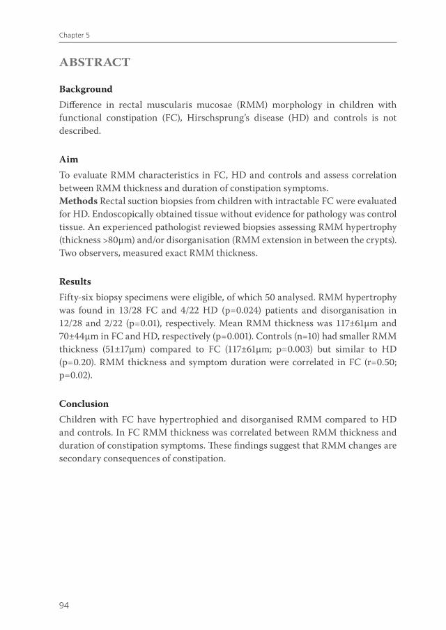
ABSTRACT
Background Difference in rectal muscularis mucosae (RMM) morphology in children with functional constipation (FC), Hirschsprung’s disease (HD) and controls is not described.
AimTo evaluate RMM characteristics in FC, HD and controls and assess correlation between RMM thickness and duration of constipation symptoms. Methods Rectal suction biopsies from children with intractable FC were evaluated for HD. Endoscopically obtained tissue without evidence for pathology was control tissue. An experienced pathologist reviewed biopsies assessing RMM hypertrophy (thickness >80µm) and/or disorganisation (RMM extension in between the crypts). Two observers, measured exact RMM thickness.
ResultsFifty-six biopsy specimens were eligible, of which 50 analysed. RMM hypertrophy was found in 13/28 FC and 4/22 HD (p=0.024) patients and disorganisation in 12/28 and 2/22 (p=0.01), respectively. Mean RMM thickness was 117±61µm and 70±44µm in FC and HD, respectively (p=0.001). Controls (n=10) had smaller RMM thickness (51±17µm) compared to FC (117±61µm; p=0.003) but similar to HD (p=0.20). RMM thickness and symptom duration were correlated in FC (r=0.50; p=0.02).
ConclusionChildren with FC have hypertrophied and disorganised RMM compared to HD and controls. In FC RMM thickness was correlated between RMM thickness and duration of constipation symptoms. These findings suggest that RMM changes are secondary consequences of constipation.

95
Rectal muscularis mucosae in constipation
CHAPTER
5
INTRODUCTION
Constipation is common during childhood with a median prevalence of 8.9% in the western world and is characterised by infrequent painful defecation in combination with involuntary faecal incontinence.1 Organic causes of constipation are only found in 5-10% of cases.2 Withholding behaviour is suggested to be the major cause of functional constipation in children.3 Defecation is a complex interplay between the autonomic and somatic nervous system and the group of muscles controlling the anal sphincters and the pelvic floor muscles. Consequent faecal stasis might result in higher intraluminal pressure and may cause histological changes in rectal colonic musculature. Moreover, it is suggested that changes in muscularis mucosae such as hypertrophy can be expected in gastrointestinal disease.4 For example, the rectal muscularis mucosae (RMM) in solitary rectal ulcer syndrome in adults, is characterized by hypertrophy and disorganisation.5, 6 In this condition, ischemia, atherosclerosis as well as colonic stasis are suspected factors in the pathogenesis.7 While paediatric patients with intractable constipation are still considered to suffer from a disease of functional nature, recent studies showed structural abnormalities of the colonic enteric nervous system (ENS) in those patients. These include, morphological changes or reduced numbers of ganglia cells and/or glial cells 8-11, abnormal nerve fibre density in the circular muscle layer 12 and a reduced or abnormal number of interstitial cells of Cajal 8, 9, 13-15 have been observed. In addition, colonic smooth muscle defects of patients with slow transit constipation have been reported.16, 17 These structural changes may play a crucial role in the pathophysiology of colorectal motility disorders such as constipation. But consequential histological changes as a result of constipation symptoms have yet to be evaluated in children. Therefore we hypothesized that paediatric patients with constipation have different rectal mucosal musculature and that these changes are associated with duration of constipation symptoms.
METHODS
SpecimensRectal suction biopsy specimensSixty-five rectal suction biopsy specimens, taken between November 2000 and June 2008, were retrospectively evaluated from patients (aged 0-18 years) presenting to the department of paediatric gastroenterology or paediatric surgery with intractable constipation to diagnose or to exclude Hirschsprung’s disease (HD). For HD diagnostics, rectal suction biopsies between 2 and 4 cm
96
Chapter 5
from the dentate line were taken and directly snap frozen. Slides from the frozen biopsies were stained by hematoxylin-eosin and assayed for acethylcholinesterase activity. The latter was determined as previously described by Karnovsky and Roots. 18 Nonspecific acethylcholinesterase was inhibited by 2.5x 10⁶ mol/L tetra-isopropylpyrophosphoramide (ISO-OMPA). 19 Rectal suction biopsies were considered positive for HD when acethylcholinesterase activity was elevated in combination with absence of ganglion cells. If ganglion cells were present, HD was excluded. Patients excluded for HD were diagnosed with functional constipation (FC).
Resection specimens & endoscopically obtained control tissueIn HD patients with poor quality rectal suction biopsies, resection specimens obtained by the paediatric surgeon were used for RMM thickness evaluation. In patients suspected for inflammatory bowel disease colonoscopy was performed. When no macro- and/or microscopical evidence were found for colonic pathology, obtained rectal tissue was analysed as control samples (n=10). The resection and endoscopic specimens were fixed in 4% formaldehyde buffered with phosphate-buffered saline (PBS), dehydrated, and embedded in paraffin. Tissue sections were made at 5 µm, stretched and dried at 57°C overnight. The sections were rehydrated in an alcohol series, endogenous peroxidase activity was blocked by 1% H2O2 in methanol for 1 hour.
ProtocolSemi-quantitative evaluationAn experienced pathologist, blinded for the patient’s diagnosis, first screened the biopsy specimens for muscle thickness measurement suitability. Rectal suction biopsies were considered suitable for measurement when the entire thickness of the RMM was visible. Biopsies were discarded when the limits of the muscularis mucosae were not clear due to absence of the border of the submucosa. Furthermore, semi-quantitative evaluation of the RMM was performed by the pathologist. We defined RMM hypertrophy when its thickness exceeded 80 µm. The pathologist determined RMM thickness with a light microscope (Olympus U-SD03) equipped with a x20 objective. Muscle architecture was also evaluated and disorganisation of the RMM was defined by the pathologist as extension of the RMM in between the crypts.
Quantitative evaluationAfter the first screening of the biopsies, two medical doctors (NB and MMT), blinded for the patient’s diagnosis, measured independently of each other the exact thickness (in µm) of the RMM of all suitable specimens (20x magnification; Leica DM 5000B). These observers measured the maximal and minimal muscle thickness
97
Rectal muscularis mucosae in constipation
CHAPTER
5
of each specimen while taking photographs (Leica Microsystems). The mean of the maximal and minimal measurements of both observers was used for analysis.
Statistical analysisStatistical analysis of the data was conducted using the statistical software package SPSS (version 14.0; Inc, Chicago, IL). Baseline characteristics of the group were analysed in a descriptive way. Differences in evaluation of muscle hypertrophy and organisation, as described by the pathologist (yes/no), between FC, HD and control patients were analyzed by Fisher’s exact χ2 statistics. Agreement between the two observers was determined using the Pearson correlation test. Difference in exact muscle thickness between FC, HD and control patients was calculated using the student t-test. Correlation between muscle thickness (µm) and duration of symptoms (months) or age (months) was calculated using the Pearson correlation test. A p-value <0.05 was considered significant.
RESULTS
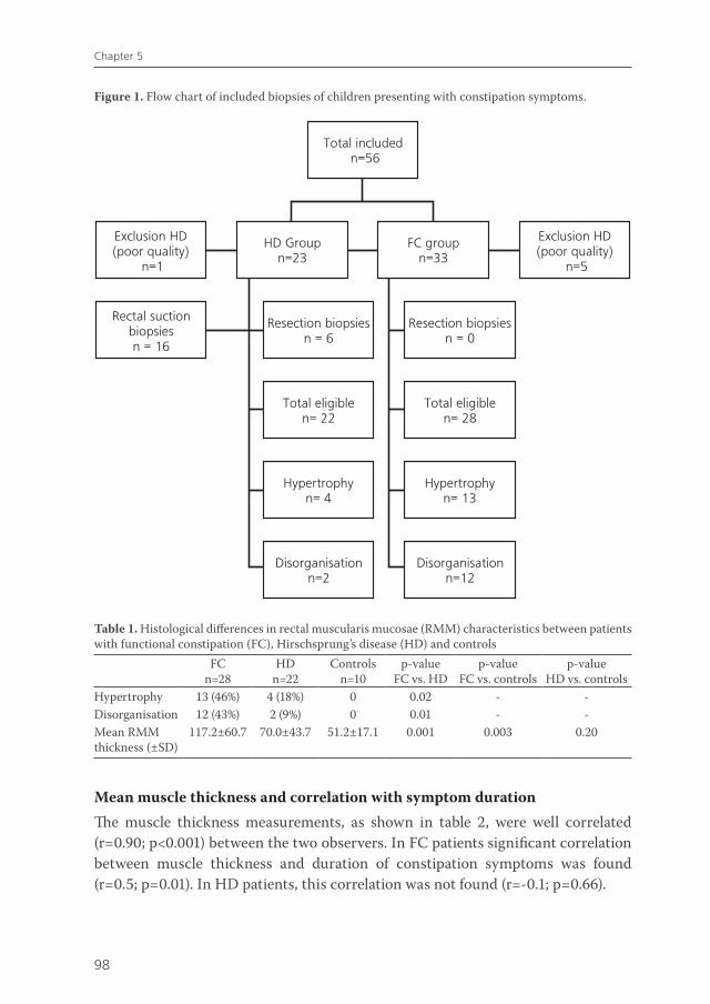
BaselineFifty six rectal suction biopsy specimens were included (fig. 1). Six specimens were excluded by the pathologist because of poor quality, these included five rectal suction biopsies from the FC group and one from the HD group. Children with FC (n=28) and HD (n=22) had a median age (range) of 8.0 (0.2-288) months and 5.4 (0.2-22.0) months (p=0.003), respectively. Control patients (n= 10) had a median age of 12.4 (7.2-18.2) years. Mean duration of constipation symptoms was 33.1±54.0 months and 6.5±9.2 months in respectively FC and HD patients, respectively.
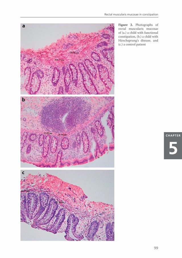
Hypertrophy and disorganisation of the RMMRectal muscularis mucosae was significantly thicker in FC patients compared to HD patients and controls as shown in table 1 and illustrated in figure 2. When resection specimens were excluded from analysis, difference in hypertrophy of the RMM remained statistically significant between HD and FC patients (p=0.004). Both hypertrophy as well as disorganisation of the RMM was found in 9 and 2 patients of FC and HD patients, respectively. In three FC patients RMM disorganisation was found without hypertrophy.A total of 10 patients from the HD group compared to 18 FC patients had no hypertrophy and nor RMM disorganisation in the same biopsy specimen (p=0.002).
98
Chapter 5
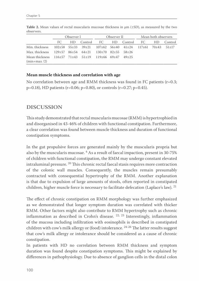
Mean muscle thickness and correlation with symptom durationThe muscle thickness measurements, as shown in table 2, were well correlated (r=0.90; p<0.001) between the two observers. In FC patients significant correlation between muscle thickness and duration of constipation symptoms was found (r=0.5; p=0.01). In HD patients, this correlation was not found (r=-0.1; p=0.66).
Table 1. Histological differences in rectal muscularis mucosae (RMM) characteristics between patients with functional constipation (FC), Hirschsprung’s disease (HD) and controls
FCn=28
HDn=22
Controlsn=10
p-valueFC vs. HD
p-valueFC vs. controls
p-valueHD vs. controls
Hypertrophy 13 (46%) 4 (18%) 0 0.02 - -Disorganisation 12 (43%) 2 (9%) 0 0.01 - -Mean RMM thickness (±SD)
117.2±60.7 70.0±43.7 51.2±17.1 0.001 0.003 0.20
Figure 1. Flow chart of included biopsies of children presenting with constipation symptoms.
99
Rectal muscularis mucosae in constipation
CHAPTER
5
Figure 2. Photographs of rectal muscularis mucosae of (a.) a child with functional constipation, (b.) a child with Hirschsprung’s disease, and (c.) a control patient
100
Chapter 5
Mean muscle thickness and correlation with ageNo correlation between age and RMM thickness was found in FC patients (r=0.3; p=0.18), HD patients (r=0.06; p=0.80), or controls (r=0.27; p=0.45).
DISCUSSION
This study demonstrated that rectal muscularis mucosae (RMM) is hypertrophied in and disorganised in 43-46% of children with functional constipation. Furthermore, a clear correlation was found between muscle thickness and duration of functional constipation symptoms.
In the gut propulsive forces are generated mainly by the muscularis propria but also by the muscularis mucosae. 4 As a result of faecal impaction, present in 30-75% of children with functional constipation, the RMM may undergo constant elevated intraluminal pressure. 20 This chronic rectal faecal stasis requires more contraction of the colonic wall muscles. Consequently, the muscles remain presumably contracted with consequential hypertrophy of the RMM. Another explanation is that due to expulsion of large amounts of stools, often reported in constipated children, higher muscle force is necessary to facilitate defecation (Laplace’s law). 21
The effect of chronic constipation on RMM morphology was further emphasized as we demonstrated that longer symptom duration was correlated with thicker RMM. Other factors might also contribute to RMM hypertrophy such as chronic inflammation as described in Crohn’s disease. 22, 23 Interestingly, inflammation of the mucosa including infiltration with eosinophils is described in constipated children with cow’s milk allergy or (food) intolerance. 24-26 The latter results suggest that cow’s milk allergy or intolerance should be considered as a cause of chronic constipation. In patients with HD no correlation between RMM thickness and symptom duration was found despite constipation symptoms. This might be explained by differences in pathophysiology. Due to absence of ganglion cells in the distal colon
Table 2. Mean values of rectal muscularis mucosae thickness in µm (±SD), as measured by the two observers.
Observer I Observer II Mean both observersFC HD Control FC HD Control FC HD Control
Min. thickness 102±58 55±33 39±21 107±62 56±40 41±24 117±61 70±43 51±17Max. thickness 129±57 86±54 64±21 130±70 82±55 58±26Mean thickness (min+max /2)
116±57 71±43 51±19 119±66 69±47 49±25
101
Rectal muscularis mucosae in constipation
CHAPTER
5
(the rectum), faeces do not reach the level at which biopsies are taken. Faeces remain impacted above the biopsied level and therefore no changes are found in histological characteristics of the RMM. The latter further emphasizes that the histological changes found in FC children such as hypertrophy and disorganisation, might well be the consequence of faecal impaction.
Hirschsprung’s disease is a developmental disorder of the enteric nervous system characterized by the absence of ganglion cells along a variable portion of the distal intestine. This results in a functional obstruction caused by dysmotility of the diseased segment. 27, 28 Delayed passage of meconium is the cardinal symptom in neonates with HD which is in contrast with healthy term born neonates. 28, 29 Approximately 80-90% of all HD cases present with symptoms of constipation, abdominal distension and vomiting during the neonatal period. 30 In accordance with earlier studies, in 80% of the children in this study, HD was diagnosed before the age of 6 months.30 In contrast, children with functional constipation usually present with symptoms at the time of toilet training. 31-33 Consequently, children with HD in this study were younger compared to the children with functional constipation. Therefore, we evaluated correlation between age and RMM thickness. No correlation however was found between age and RMM thickness in any group, indicating that the correlation between constipation symptom duration and RMM thickness was not due to age.
Interestingly, the histochemical characteristics might be helpful additional tools to distinguish functional constipation from HD in inconclusive rectal suction biopsies. The latter problem is found when rectal suction biopsies contain too superficial specimen and do not contain muscularis mucosae.34, 35 Rectal suction biopsies are frequently repeated due to inconclusive findings which may increase the risk of complications. 30 Some clinicians even choose for full thickness biopsies, if rectal suction biopsies are inconclusive. Therefore, more histological characteristics such as disorganisation and hypertrophy of the rectal muscularis mucosae might help prevent repeated testing.
In solitary rectal ulcer syndrome in children, RMM can be hypertrophied and disorganised as well.6 Disorganisation of the RMM in the latter syndrome includes distorsion of the crypt architecture as one of the features which we did not find in our group of patients. This could be explained by the more severe presentation of patients with solitary ulcus syndrome compared to our constipated patients. Although the syndrome is believed to be associated with a disorder of evacuation, the pathophysiology remains uncertain.6, 36, 37
This study had also shortcomings. Resection specimens of patients with HD are processed differently as they are embedded in paraffin which may shrink
102
Chapter 5
the tissue. Consequently the RMM measured from paraffin embedded tissue may appear thinner. In contrast, the rectal suction specimens are snap frozen at -80°C, a process which may swell the tissue. This implies that frozen tissue may appear more distended compared to the tissue embedded in paraffin. This could be a confounder in our study. However, when we analysed the rectal suction biopsies only, by excluding resection specimens, difference in muscle thickness remained between the constipated and HD patients. Another shortcoming was the retrospective character of our study which made evaluation between symptoms of constipation, such as faecal impaction and RMM thickness difficult to assess.
In conclusion, in this study we demonstrated that rectal muscularis mucosae is hypertrophied and disorganised in patients with functional constipation. In addition, we found a correlation between muscle thickness and symptom duration of functional constipation. Further prospective studies are needed to confirm our findings and the effect of constipation on the rectal muscularis mucosae. Interestingly, these findings contribute to more insight into the pathophysiology of functional constipation and might also be useful additional diagnostic criteria for pathologists distinguishing constipation from Hirschsprung’s disease in inconclusive rectal suction biopsies.
103
Rectal muscularis mucosae in constipation
CHAPTER
5
REFERENCE LIST
1. Van den Berg MM, Benninga MA, Di Lorenzo C. Epidemiology of childhood constipation: a systematic review. Am J Gastroenterol 2006;101:2401-2409.
2. Loening-Baucke V. Chronic constipation in children. Gastroenterology 1993;105:1557-1564.
3. Loening-Baucke V. Prevalence, symptoms and outcome of constipation in infants and toddlers. J Pediatr 2005;146:359-363.
4. Alan Stevens, James Lowe. Human Histology. Mosby, 1997.
5. Crespo PL, Moreira V, V, Redondo VC, Lopez San RA, Milicua Salamero JM. [“The three-lies disease”: solitary rectal ulcer syndrome]. Rev Esp Enferm Dig 2007;99:663-666.
6. Dehghani SM, Haghighat M, Imanieh MH, Geramizadeh B. Solitary rectal ulcer syndrome in children: a prospective study of cases from southern Iran. Eur J Gastroenterol Hepatol 2008;20:93-95.
7. Ong J, Lim JF, Lim HK, Eu KW. Benign Solitary Cecal Ulcer Syndrome: Series of Eight Cases and Literature Review. Dis Colon Rectum 2008.
8. Wedel T, Spiegler J, Soellner S, Roblick UJ, Schiedeck TH, Bruch HP, Krammer HJ. Enteric nerves and interstitial cells of Cajal are altered in patients with slow-transit constipation and megacolon. Gastroenterology 2002;123:1459-1467.
9. Bassotti G, Villanacci V, Maurer CA, Fisogni S, Di FF, Cadei M, Morelli A, Panagiotis T, Cathomas G, Salerni B. The role of glial cells and apoptosis of enteric neurones in the neuropathology of intractable slow transit constipation. Gut 2006;55:41-46.
10. Schouten WR, Ten Kate FJ, de Graaf EJ, Gilberts EC, Simons JL, Kluck P. Visceral neuropathy in slow transit constipation: an immunohistochemical investigation with monoclonal antibodies against neurofilament. Dis Colon Rectum 1993;36:1112-1117.
11. Wedel T, Roblick UJ, Ott V, Eggers R, Schiedeck TH, Krammer HJ, Bruch HP. Oligoneuronal hypoganglionosis in patients with idiopathic slow-transit constipation. Dis Colon Rectum 2002;45:54-62.
12. Hutson JM, Catto-Smith T, Gibb S, Chase J, Shin YM, Stanton M, King S, Sutcliffe J, Ong SY, Djaja S, Farmer P, Southwell B. Chronic constipation: no longer stuck! Characterization of colonic dysmotility as a new disorder in children. J Pediatr Surg 2004;39:795-799.
13. He CL, Burgart L, Wang L, Pemberton J, Young-Fadok T, Szurszewski J, Farrugia G. Decreased interstitial cell of cajal volume in patients with slow-transit constipation. Gastroenterology 2000;118:14-21.
14. Lyford GL, He CL, Soffer E, Hull TL, Strong SA, Senagore AJ, Burgart LJ, Young-Fadok T, Szurszewski JH, Farrugia G. Pan-colonic decrease in interstitial cells of Cajal in patients with slow transit constipation. Gut 2002;51:496-501.
15. Van den Berg MM, Di LC, Mousa HM, Benninga MA, Boeckxstaens GE, Luquette M. Morphological changes of the enteric nervous system, interstitial cells of cajal, and smooth muscle in children with colonic motility disorders. J Pediatr Gastroenterol Nutr 2009;48:22-29.
16. Wedel T, Van Eys GJ, Waltregny D, Glenisson W, Castronovo V, Vanderwinden JM. Novel smooth muscle markers reveal abnormalities of the intestinal musculature in severe colorectal motility disorders. Neurogastroenterol Motil 2006;18:526-538.
17. Knowles CH, Nickols CD, Scott SM, Bennett NI, de Oliveira RB, Chimelli L, Feakins R, Williams NS, Martin JE. Smooth muscle inclusion bodies in slow transit constipation. J Pathol 2001;193:390-397.
18. Karnovsky MJ, Roots L. A “direct-coloring” thiocholine method for cholinesterases. 12 ed. 1964:219-221.
19. Luider TM, van Dommelen MW, Tibboel D, Meijers JH, Ten Kate FJ, Trojanowski JQ, Molenaar JC. Differences in phosphorylation state of neurofilament proteins in ganglionic and aganglionic bowel segments of children with Hirschsprung’s disease. J Pediatr Surg 1992;27:815-819.
20. van Ginkel R, Reitsma JB, Buller HA, van Wijk MP, Taminiau JA, Benninga MA. Childhood constipation: Longitudinal follow-up beyond puberty. Gastroenterology 2003;125:357-363.
104
Chapter 5
21. Voskuijl WP, van GR, Benninga MA, Hart GA, Taminiau JA, Boeckxstaens GE. New insight into rectal function in pediatric defecation disorders: disturbed rectal compliance is an essential mechanism in pediatric constipation. J Pediatr 2006;148:62-67.
22. Lee EY, Stenson WF, Schryver-Kecskemeti K. Thickening of muscularis mucosae in Crohn’s disease. Mod Pathol 1991;4:87-90.
23. Burke JP, Mulsow JJ, O’Keane C, Docherty NG, Watson RW, O’Connell PR. Fibrogenesis in Crohn’s disease. Am J Gastroenterol 2007;102:439-448.
24. Iacono G, Cavataio F, Montalto G, Florena A, Tumminello M, Soresi M, Notarbartolo A, Carroccio A. Intolerance of cow’s milk and chronic constipation in children [see comments]. N Engl J Med 1998;339:1100-1104.
25. Carroccio A, Scalici C, Maresi E, Di PL, Cavataio F, Noto D, Porcasi R, Averna MR, Iacono G. Chronic constipation and food intolerance: a model of proctitis causing constipation. Scand J Gastroenterol 2005;40:33-42.
26. Daher S, Tahan S, Sole D, Naspitz CK, Da Silva Patricio FR, Neto UF, De Morais MB. Cow’s milk protein intolerance and chronic constipation in children. Pediatr Allergy Immunol 2001;12:339-342.
27. Scharli AF, Kiesewetter WB. Defecation and continence: some new concepts. Dis Colon Rectum 1970;13:81-107.
28. Puri P. Hirschsprung’s disease: Clinical Generalities. In: Holschneider AM and Puri P, eds. Hirschsprung’s disease and allied disorders. 2 ed. 2000:129-135.
29. Bekkali N, Hamers SL, Schipperus MR, Reitsma JB, Valerio PG, Van TL, Benninga MA. Duration of meconium passage in preterm and term infants. Arch Dis Child Fetal Neonatal Ed 2008;93:F376-F379.
30. de Lorijn F, Kremer LC, Reitsma JB, Benninga MA. Diagnostic tests in Hirschsprung disease: a systematic review. J Pediatr Gastroenterol Nutr 2006;42:496-505.
31. Iacono G, Merolla R, D’Amico D, Bonci E, Cavataio F, Di PL, Scalici C, Indinnimeo L, Averna MR, Carroccio A. Gastrointestinal symptoms in infancy: a population-based prospective study. Dig Liver Dis 2005;37:432-438.
32. Loening-Baucke V. Constipation in early childhood: patient characteristics, treatment, and longterm follow up. Gut 1993;34:1400-1404.
33. Davidson M, Kugler MM, Bauer CH. Diagnosis and management in children with severe and protracted constiption. Medical Progress 1963;62:261-275.
34. Martucciello G. Hirschsprung’s disease, one of the most difficult diagnoses in pediatric surgery: a review of the problems from clinical practice to the bench. Eur J Pediatr Surg 2008;18:140-149.
35. Huntley CC, Shaffner LD, Challa VR, Lyerly AD. Histochemical diagnosis of Hirschsprung disease. Pediatrics 1982;69:755-761.
36. Tjandra JJ, Fazio VW, Petras RE, Lavery IC, Oakley JR, Milsom JW, Church JM. Clinical and pathologic factors associated with delayed diagnosis in solitary rectal ulcer syndrome. Dis Colon Rectum 1993;36:146-153.
37. Chiang JM, Changchien CR, Chen JR. Solitary rectal ulcer syndrome: an endoscopic and histological presentation and literature review. Int J Colorectal Dis 2006;21:348-356.