Embed Size (px)
Citation preview

UV-Triggered Polymerization, Deposition, and Patterning of Plant Phenolic Compounds
Farid Behboodi-Sadabad, Huijie Zhang, Vanessa Trouillet, Alexander Welle, Nicolas Plumeré, and Pavel A. Levkin*
Plant-derived phenolic compounds, rich in catechol and pyrogallol moieties, can form multifunctional coatings on various substrates following polymeri-zation under mildly alkaline conditions. Despite many appealing features of such coatings, the difficulty to control polymerization of phenolic compounds spatially and temporally limits their number of potential applications. In this study, it is demonstrated that UV irradiation can trigger oxidative polymeriza-tion and deposition of plant-derived phenolic compounds, which opens the possibility to create 2D gradients and patterns of polyphenol coatings and control this polymerization temporally. UV–vis spectroscopy, electrospray ionization mass spectrometry, and cyclic voltammetry analyses are used to investigate the UV-induced polymerization of several plant-derived phenolic compounds including pyrogallol, tannic acid, caffeic acid, and gallic acid. Formation of polyphenol coatings on polar and nonpolar substrates after UV irradiation has been studied using water contact angle measurements, atomic force microscopy, time of flight secondary ion mass spectrometry, and X-ray photoelectron spectroscopy (XPS). The possibility to use UV-light to accel-erate polymerization of phenolic compounds and perform micropatterning can extend the scope of potential applications of the large class of structurally diverse plant-derived phenolic compounds.
materials by simple immersion of objects into a dopamine solution under basic con-ditions,[7] in the presence of oxidants,[4a,8] and under UV irradiation.[9]
Recently it was shown that different plant-derived phenolic compounds iso-lated from various plants, and rich in 1,2-dihydroxybenzene (catechol) and 1,2,3-trihydroxybenzene (pyrogallol, PG) moieties, could also form nanocoat-ings following polymerization using enzymes,[10] coordination complexes,[11] or using mildly alkaline solutions in the presence of dissolved oxygen.[7b,12] In com-parison to polydopamine, coatings based on plant phenolic compounds were found to possess more rapid adhesion rate, lower cost, excellent availability, and good struc-tural diversity.[11a,13] Phenolic compounds, found in various plant-derived foods (for example, fruits, vegetables, cereals, choc-olate) and beverages (for example, tea, coffee, beer, wine) are known to be an important part of the defense system of plants.[14] Plant phenolics show antioxi-
dant and anticancer effect.[14,15] Long-term use of plant pheno-lics can eliminate the destructive effect of undesired reactive oxygen or nitrogen species in the body.[15] Furthermore, plant phenolics are able to chelate metal ions,[16] interact with sur-faces and materials via charge–charge, charge–dipole, and cova-lent bonds,[16] quench reactive radical species, and interact with oxidizing agents.[17] Oxidized catechol and pyrogallol moieties enable antimicrobial activity of plant phenolics.[18]
Various lithographic techniques such as soft lithography and dip-pen lithography have been developed for surface patterning,[19] yet most of the patterning methods are not applicable for the formation of gradients or for the fabrication
1. Introduction
The ability to control surface properties via functional coatings is fundamentally important in various applications.[1] Recently, mussel adhesive proteins and their analogs containing multiple 3,4-dihydroxyphenylalanine (DOPA) moieties inspired many groups to use catechol (1,2-dihydroxyphenyl)-containing mol-ecules for surface coating and functionalization,[2] nanoparticles fabrication and modification,[3] surfaces with special wetta-bility,[4] and development of new hydrogels[5] and membrane.[6] Dopamine, which is structurally similar to DOPA, has been found to form versatile polydopamine coatings on different
F. Behboodi-Sadabad, Dr. P. A. LevkinInstitute of Toxicology and GeneticsInstitute of Organic ChemistryKarlsruhe Institute of Technology76021 Karlsruhe, GermanyE-mail: [email protected]. Zhang, Dr. N. PlumeréCenter for Electrochemical SciencesRuhr-Universität BochumD-44780 Bochum, Germany
V. TrouilletInstitute for Applied Materialsand Karlsruhe Nano Micro FacilityKarlsruhe Institute of Technology76021 Karlsruhe, GermanyDr. A. WelleInstitute of Functional Interfacesand Karlsruhe Nano Micro FacilityKarlsruhe Institute of Technology76021 Karlsruhe, Germany

of patterns inside closed microfluidic channels or on curved surfaces. Creating patterns of functional structures within microfluidic channels or capillaries is crucial in research areas ranging from micro- and nanofluidics,[20] analytical chem-istry,[21] drug delivery,[22] clinical and diagnostics devices,[23] to separation science.[24]
Coatings based on polyphenols can potentially lead to a lot of useful applications. However, the ability to control poly-phenol nanocoatings spatially and temporally would signifi-cantly extend the number of potential applications. Until now, to the best of our knowledge, no methods for patterning plant-derived phenolic compounds have been reported. Recently, we showed that dopamine polymerization and deposition could be accelerated by UV irradiation.[9,25] In this paper, we dem-onstrate for the first time that the UV irradiation could induce oxidation and polymerization of various plant-derived phenolic compounds. The effect of UV on plant phenolic compounds was investigated using UV–vis spectroscopy, electrospray ioni-zation mass spectrometry (ESI-MS), and cyclic voltammetry (CV). Various plant-derived phenolic compounds including PG, tannic acid (TA), caffeic acid (CA), and gallic acid (GA) could be polymerized under UV irradiation. We used UV-assisted polym-erization and deposition of plant phenolic compounds to make polyphenol nanocoatings on polar and nonpolar substrates as well as to create gradients of polyphenol coatings. The ability to use UV-l ght to trigger polymerization and deposition of phenolic compounds opens the possibility to create polyphenol patterns and gradients within microfluidic channels, which was demonstrated by creating a pattern of PG inside a fused silica capillary. The use of UV-l ght to accelerate formation of poly-phenol nanocoatings opens the possibility for the photolitho-graphic patterning of functional polyphenolic nanocoatings on
various substrates, extending the scope of potential applications of plant polyphenols.
2. Results and Discussion
In order to realize the oxidative effect of UV irradiation, UV–vis spectra of 1,2,3-trihydroxybenzene (PG), TA, 3,4,5-trihydroxy-benzoic acid (GA), and 3-(3,4-dihydroxyphenyl)-2-propenoic acid (CA) (Figure 1), representing some of the most common phenolic compounds derived from plants,[26] in variety of acidic and basic solutions were measured. Kinetics and pH depend-ence of oxidation both in the dark and under UV irradiation (260 nm, 10 mW cm−2) were investigated for each compound at 30 min time intervals for 2 h (Figure 2). Our results show that UV irradiation of PG, TA, GA, and CA even under acidic pH leads to a change of color of solutions and increase of UV absorbance. Figure 2A demonstrates the corresponding color change of the PG and TA solutions irradiated with UV light in comparison with those stored in the dark. UV–vis spectra of irradiated PG (0.2 mg mL−1, acetate buffer at pH 5.0) and TA (0.2 mg mL−1, phosphate buffer at pH 7.0) solutions shifted to higher absorbance at 350 nm along with increasing UV irradiation time, while absorbance of non-irradiated samples remained unchanged (Figure 2B,C). Increased absorbance of solutions after UV irradiation was also observed for PG, TA, GA, and CA at different pH conditions (both acidic and basic) (Figures S1–S9, Supporting Information). The increase of absorbance at 350 nm as a function of time for each sample is plotted in Figure 2D. For example, normalized UV absorb-ance at 350 nm of PG (pH 5.0), TA (pH 7.0), GA (pH 6.0), and CA (pH 6.0) solutions after 2 h of UV irradiation increased to
Figure 1. Schematic representation of the UV-induced A) polymerization, B) deposition, and C) patterning of plant-derived phenolics: tannic acid (TA), pyrogallol (PG), gallic acid (GA), and caffeic acid (CA).

19.3, 3.6, 13.5, and 4.9, respectively (Figure 2E). However, UV absorbance of these samples stored in the dark for 120 min remained the same (Figure 2D,E). An increase in the kinetics of phenolics oxidation was observed by increasing the pH from 5.0 to 10.0 (Figures S1–S8, Supporting Information). Although increasing the pH of solutions speeds up the oxidation and associated increase of absorbance of the phenolic solutions even in the dark, UV irradiation accelerates this process even more (Figure 2D,E; Figure S9, Supporting Information, for other phenolics). Darkening of the solution and increase of UV absorbance at 350 nm in oxidative condition is usually associ-ated with higher molecular weight species and polymerization of phenolics.[10d,12c]
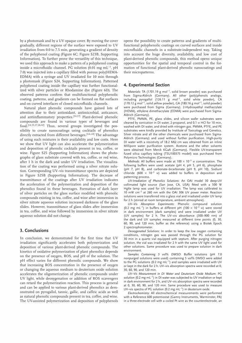
In order to prove that UV irradiation triggers polymerization of phenolic compounds, we performed ESI-MS analysis of PG solutions (pH 5.0) either subjected to UV or kept in the dark for 2 h (Figure 3). Increase in UV–vis absorbance at 350 nm of plant phenolics under basic conditions was attributed to qui-none formation.[8,10d] However, formation of higher molecular weight species through oxidation of catechol and gallol moieties in plant phenolics was also reported.[27] Our ESI-MS analysis clearly shows the presence of higher molecular-weight species following UV irradiation of PG solution, while no oligomers or polymers were detected in the case of nonirradiated sam-ples (Figure 3A). The repeating unit of the observed oligomer is 105.96 m/z, which corresponds to the monomeric unit and
Figure 2. UV irradiation of plant-derived phenolics leads to a change of color and increase in the absorbance of solutions. The concentration of plant-derived phenolic compounds in each sample is 0.2 mg mL−1 in a corresponding buffer (100 × 10−3 M). A) Photographs of the corresponding solutions after UV irradiation and in the dark. UV–vis spectra of B) PG solution in acetate buffer at pH 5.0 and C) TA in phosphate buffer at pH 7.0 stored in the dark (left) and after UV irradiation (right) measured at different time intervals. D) Absorbance of the PG (left) and TA (right) solutions at 350 nm as a function of time and pH. E) Normalized UV absorbance of PG, TA, GA, and CA solutions at 350 nm at 0 h, before and after 2 h of UV irradiation. A significant increase in UV absorbance is observed for phenolics solution after 2 h UV irradiation.

oligomer structures depicted in Figure 3B. The same repeating unit was previously proposed for the oligomerization of PG under alkaline conditions.[27a]
Having shown that UV irradiation can accelerate polymeriza-tion of the phenolic compounds, additional experiments were performed in order to investigate the mechanistic aspects of the UV-induced transformation. It is known that reactive oxygen species (ROS), including singlet oxygen (1O2), superoxide radi-cals (O2
−•), or highly reactive hydroxyl radicals (•OH) can be generated under UV irradiation even in the presence of traces of O2.[28] Previously, we reported the importance of ROS in the case of UV-induced dopamine polymerization.[9] On the other hand, plant phenolics have also been known for their antioxi-dant characteristics and ability to react with ROS.[14,15,17,29]
In order to investigate the role of oxygen and ROS in the UV-induced polymerization of plant phenolic compounds, several experiments were performed (Figure 3C,D; Figures S10–S13, Supporting Information). First, PG solution at pH 7.0 was
deoxygenated by passing nitrogen for 30 min, followed by UV irradiation. As shown in Figure 3C, the UV-absorbance of the deoxygenated solution at 350 nm did not increase even after 2 h of continuous UV irradiation, contrary to the same sample in the presence of oxygen (Figure 3C). Interestingly, the same inhibition of the polymerization was observed when the PG solution at pH 7.0 stored in the dark was deoxygenated (Figure 3C). In another experiment, 5 vol% of dimethyl sulfoxide (DMSO) was added to the reaction solution. DMSO is known to be a hydroxyl radical scavenger.[30] UV irradiation of the DMSO-containing PG solution did result in a smaller increase of the absorption at 350 nm in comparison to the control without DMSO (Figure 3C). The same slight inhibition of polymeri-zation in the presence of DMSO was observed in the case of solutions stored in the dark (Figure 3C). On the other hand, deuterium oxide is known to be a singlet oxygen half-life pro-longer.[31] Changing the medium from deionized (DI) water to deuterium oxide increased the UV absorption of PG solution
Figure 3. A) ESI-MS spectra (positive mode, data acquired for 30 s) of PG solution in acetate buffer at pH 5.0 in dark and after UV irradiation for 2 h. ESI-MS spectra of UV-irradiated PG polymerization solution clearly shows presence of higher molecular weight species. B) Schematic representation of UV-induced polymerization of PG. C) UV absorbance at 350 nm of PG solution in phosphate buffer at pH 7.0 (blue), with addition of 5 vol% DMSO (green), and after deoxygenation with N2 (red) for solutions kept in dark (left) and under UV irradiation (right). D) UV absorbance of PG solution at 350 nm in deionized water (blue) and deuterium oxide (red) media stored in dark (left) and under UV irradiation (right).

stored in dark and UV-irradiated PG solution (Figure 3D). These observations suggest that UV-triggered polymerization of plant phenolics requires ROS and oxygen. Increasing ROS con-centration can increase the rate of the UV-induced polymeriza-tion of phenolics, while quenching ROS and deoxygenation of the solution lead to the inhibition of polymerization.
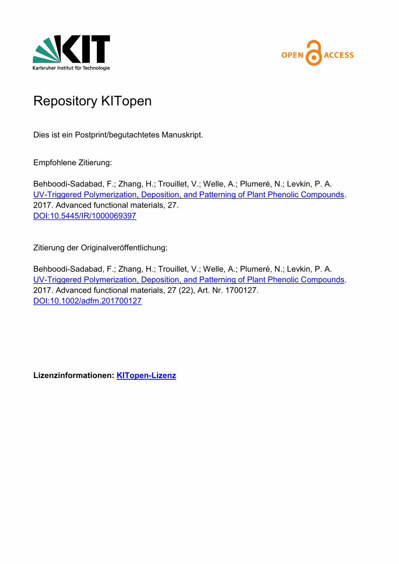
Antioxidant properties and electrochemical behavior of plant phenolics depend on their chemical structure and experimental conditions.[14,17,32] We investigated the effect of UV irradiation on electrochemical oxidation of plant phenolic compounds using cyclic voltammetry (Figure 4; Figures S14–S21, Sup-porting Information). PG as well as GA display two anodic peaks corresponding to the oxidation to the semi-quinone and the subsequent oxidation to the quinone form.[33] Moni-toring of PG in acetate buffer at pH 5.0 incubated for 2 h in dark environment indicates a slight change in peak current values (Figure 4A). However, continuous UV irradiation for 2 h enhanced the oxidation rate of PG and decreased the peak current values by 20% (Figure 4B). Significant decrease in peak current is due to consumption of PG after UV irradiation. UV irradiation also increased the rate of oxidation of PG in basic pH as indicated by more reduction of peak currents after UV irradiation compared to solutions stored in dark (Figures S14 and S15, Supporting Information).
The CV also confirmed irreversible oxidation of TA (Figures S18 and S19, Supporting Information). However, in contrast to PG and GA, the anodic peak currents for TA remain mostly con-stant over time independently of illumination and pH value. TA is composed of 10 quinone moieties, which may predominantly undergo intramolecular reactions. In this case, the total concen-tration and molecular weights (or diffusion coefficient) of TA and its reaction products would not change significantly, thereby explaining the mostly unchanged oxidation peak current. Nev-ertheless, although CV is not sensitive enough to quantitatively monitor the minor fluctuation in peak currents, intermolecular reaction leading to polymeric compounds in analogy to the behavior of GA and PG are also expected to take place.
Caffeic acid in contrast to the other three phenolic compounds displays a reductive peak in addition to the oxidation peak in the CV at low pH values (Figures S20 and S21, Supporting Information). The electrochemical response is attributed to a one-electron oxidation to the semiquinone form followed by a
second oxidation to the quinone. Both oxidized forms undergo follow up irreversible chemical reactions[34] whereby the semi-quinone reaction involves dimerization.[35] In more basic media (pH > 8), an increasingly irreversible behavior suggests that the oxidation of CA is followed by a polymerization process.[36] This is confirmed by the decrease in peak current over time indi-cating the consumption of the monomeric compounds. The π conjugated systems in CA also induces a different behavior under illumination. While PG, GA, and TA undergo light accel-erated oxidative polymerization, caffeic acid first undergoes a light-induced trans–cis-isomerization followed by intramolecular cyclization.[37] The CVs confirm this behavior with the appear-ance of a new oxidation wave upon illumination in acidic media. The decrease of the reduction waves reveals the consumption of the caffeic acid (both cis and trans forms) over prolonged illumination times. Observations from cyclic volta mmetry for all phenolic compounds are in agreement with UV–vis spectroscopic data.
Plant-derived phenolic compounds are able to form func-tional nanocoatings on various substrates.[7b,10c,d,11a,d,f,12,38] Messersmith and co-workers[12c] showed that even phenolic crude extracts from red wine, cacao bean, and green tea could form coatings on different materials.[7b] Jeon et al.[10d] reported the use of laccase enzyme to catalyze polymerization of dopamine, cat-echin/catechol, ferulic acid/catechol, catechin/syringic acid, and tannic acid/catechol to form functional coatings. Ejima et al.[11f ] introduced the use of multivalent coordination of TA and Fe(III) for surface coatings of particulate and planar substrates. All men-tioned examples utilized the ability of plant phenolic compounds to spontaneously polymerize either under basic conditions,[7b,12] in the presence of metal ions[11] or enzymes.[10c,d,38a,39]
In order to investigate the ability to create functional phenolic nanocoatings using UV light as a trigger, polar and nonpolar polymeric substrates (poly(tetrafluoroethylene) (PTFE), poly(ethylene) (PE), poly(methyl methacrylate) (PMMA)) were immersed into a PG solution (10 × 10−3 M, pH 5.0) and irradi-ated with UV light for 2 h. Formation of a PG layer onto the surface was visualized by immersing the substrate in AgNO3 aqueous solution (Figure 5). Substrates modified by PG under UV irradiation turned darker due to the reductive effect of the polyphenol nanocoating and the formation of metallic silver nanoparticles from silver nitrate (Figure 5A).[12c] The color of
Figure 4. Monitoring of PG by cyclic voltammetry (CV). Cyclic voltammograms of PG in acetate buffer at pH 5.0 A) stored for 2 h in dark (left) and under UV irradiation (right) versus time. B) Peak currents over time for PG in acetate buffer pH 5.0 stored in dark or under UV irradiation for 2 h. All CVs were measured in 100 × 10−3 M buffer at a scan rate of 100 mV s−1 at glassy carbon electrodes with 1.58 × 10⁻³ M concentration of PG.

unmodified substrates and substrates dipped in PG solution stored in dark remained unchanged (Figure 5A).
The acceleration of deposition of plant phenolics under UV irradiation is also confirmed by water contact angle (WCA) measurements (Figure 5B). Thus, the static WCA of PE sub-strate immersed in PG solution dropped from 96.2° to 68.1° after 2 h of UV irradiation at pH 5.0, while the static WCA on the same substrate kept in dark did not change after 2 h and decreased only to 92.3° after 48 h. It is known that basic pH accelerates polymerization and deposition of PG. However, even at pH 7.0 static WCA of PE decreased only to 82.4° after 2 and 24 h of incubation was required to reach the lowest static WCA 63.1° achievable at pH 7.0. Similar static WCAs were measured on PTFE and PMMA substrates after 48 h of incubation of the surfaces with PG solution at pH 7.0 in the dark (Figure 5B), confirming homogeneous deposition of a
PG layer independent of the substrate. UV irradiation of the PE surface for 2 h at pH 5.0 without addition of PG did not result in a decrease of the static WCA (Figure S22A, Supporting Information). A Aimilar trend was observed for other plant phenolics studied (Figure S22B, Supporting Information).
Messersmith and co-workers reported a thickness of 19 and 71.6 nm for a PG-based layer on TiO2 and polycarbonate surfaces, respectively. This measurement was performed after 24 h of incubation in the solution at pH 7.[7b] Jeon et al. reported thickness of dual-monomer systems of plant pheno-lics on PET substrates ranging between 90 and 200 nm after 15 h of dipping in polymerization media.[10d] Ejima et al. reported a thickness of Fe(III)-TA films on polystyrene tem-plates and gold substrates around 2 and 10 nm, respectively, within one deposition cycle.[11f ] We demonstrate that no poly mer deposition happens after 2 h incubation of a silicon
Figure 5. Deposition of a plant-derived phenolic coating on PE, PMMA, and PFTE substrates. A) Photographs of unmodified, PG-modified, and AgNO3-treated substrates. B) Decrease of the static WCA over time for PE substrate after immersion in 10 × 10−3 M PG solution (acetate buffer at pH 5.0, phosphate buffer at pH 7.0) stored in dark environment and UV-irradiated substrates for 2 h (top, Figure S22C with error bar, Supporting Informa-tion). Static WCA reaches to a substrates independent value after immersion of PTFE, PE, and PMMA substrates in 10 × 10−3 M PG solution (bottom, phosphate buffer at pH 7.0) for 48 h. C) AFM image of bare silicon (left), silicone immersed in PG solution in dark (acetate buffer at pH 5.0) (middle), and PG-modified silicon wafer (right) gently scratched by tweezers. Corresponding diagram of line scan of each sample through the red line is shown below the image. D) C 1s XP spectra of polyethylene before and after modification with a coating based on PG (acetate buffer at pH 5.0, 2 h UV irradia-tion) and TA (phosphate buffer at pH 7.0, 2 h UV irradiation). All spectra are normalized to the highest intensity.

wafer in PG solution in acetate buffer at pH 5.0, while a 10 ± 2 nm thick polymer layer as measured by atomic force micros-copy (AFM) is formed after 2 h UV irra-diation (Figure 5C). The root mean square deviation of the roughness profile of the silicon wafer incubated for 2 h in the dark (acetate buffer, pH 5.0) changed from 0.128 ± 0.020 to 1.195 ± 0.288 nm for the silicon substrate immersed in PG solution (acetate buffer, pH 5.0) and irradiated with UV for 2 h.
In order to investigate the surface chem-istry of plant-derived phenolics coated substrates, X-ray photoelectron spectros-copy (XPS) measurements of coated PE substrates immersed in PG and TA solu-tions (acetate buffer at pH 5.0 and phos-phate buffer at pH 7.0, respectively) after 2 h of UV irradiation were conducted. PE substrate immersed in the same buffer after 2 h of UV irradiation was used as the ref-erence substrate. As expected, the C 1s XP spectrum from PE shows one main peak at 285.0 eV attributed to CC, CH groups (Figure 5D). A further peak at 286.6 eV in the C 1s XP spectra of substrates coated with PG and TA appears, corresponding to the presence of CO groups and proving clearly the deposition of a thin layer of phe-nolics on the surface.[40] Furthermore, the carboxyl group present in TA can be clearly detected at 289.0 eV. In addition, the higher intensity ratio CO/(CC, CH) for the TA coating (0.7) as for PG deposition (less than 0.2) leads to conclude that the TA film is thicker than that of the PG film.
The ability to use UV light to induce poly-merization of various plant-derived phenolic compounds opens the possibility for both spatial and temporal control of the deposition of phenolic nanocoatings on different sur-faces. In order to demonstrate this, we irradi-ated a PTFE substrate covered either with a 125 µm layer of 10 × 10−3 M PG solution (ace-tate buffer, pH 5.0) or TA solution (phosphate buffer, pH 7.0) with UV light for 1 h through a quartz photomask (Figure S23A, Supporting Information). A clear pattern of silver parti-cles or fluorescence was formed on the sur-face after immersing the substrates either in a silver nitrate aqueous solution or rhodamine-thiol solution,[41] respectively (Figure 6A). The formation of a pattern based on PG on PTFE followed by modification with silver particles was confirmed by time-of-flight secondary ion mass spectrometry (ToF-SIMS) (Figure 6B).
To demonstrate the unique advantages of UV-assisted poly-merization and deposition of plant phenolic compounds, a gradient pattern of PG was formed on the poly(2-hydroxyethyl
methacrylate)-co-(ethylene dimethacrylate) (poly(HEMA-EDMA) surface. In order to create a gradient pattern, PG solu-tion (0.01 mg mL−1, pH 7.0) was added to the surface, covered
Figure 6. A) Bright-field microscopy image of a silver particle pattern produced on surface of phenolic pattern based on PG (UV, acetate buffer at pH 5.0) or TA (UV, phosphate buffer at pH 7.0) after immersing the substrate in silver nitrate solution for 48 h. Red fluorescence pattern formed by treatment of the phenolic patterns with a rhodamine-thiol solution. B) ToF-SIMS ion intensity map of a phenolic square pattern (PG, UV, acetate buffer at pH 5.0) on PTFE substrate produced by photopatterning (top), and silver ion intensity map of modified pattern with silver nitrate aqueous solution for 48 h (bottom). C) Polyphenolic gradient pattern formed by UV irradiation of a poly(HEMA-EDMA) surface in the presence of a PG solution (0.01 mg mL−1, pH 7.0). The phenolic pattern was incubated for 48 h in a silver nitrate aqueous solution to obtain a pattern of silver particles. Corresponding gray value versus UV exposure time along the blue line is shown in the graph (right). D) Phenolic pattern inside a microfluidic capillary (inner diameter 100 µm). Fused silica capillary filled with a porous polymethacrylate was filled with a pyrogallol solution (0.01 g mL−1, pH 7.0) and irradiated with UV light for 10 min through a photomask. A pattern of silver particles (top) and rhodamine dye (bottom) formed inside the capillary by corresponding postmodification of the polyphenol pattern.
by a photomask and by a UV opaque cover. By moving the cover gradually, different regions of the surface were exposed to UV irradiation from 0.0 to 7.5 min, generating a gradient of density of the polyphenol coating (Figure 6C; Figure S23B, Supporting Information). To further prove the versatility of this technique, we used this approach to make a pattern of a polyphenol coating inside a microfluidic channel. PG solution (0.01 mg mL−1, pH 7.0) was injected into a capillary filled with porous poly(HEMA-EDMA) with a syringe and UV irradiated for 10 min through a photomask (Figure S24, Supporting Information). Patterned polyphenol coating inside the capillary was further functional-ized with silver particles or Rhodamine dye (Figure 6D). The observed patterns confirm that multifunctional polyphenolic coating, patterns, and gradients can be formed on flat surfaces and on curved interfaces of closed microfluidic channels.
Natural plant phenolic compounds have gained lots of attention due to their antioxidant, antimicrobial, anticancer, and antiinflammatory properties.[14,17] Plant-derived phenolic compounds are found in various types of beverages and food.[14,15,17,26,42] Thus, several groups investigated the pos-sibility to create nanocoatings using cocktails of phenolics directly extracted from different beverages.[12c,43] The advantage of using such mixtures is their availability and low costs. Here we show that UV light can also accelerate the polymerization and deposition of phenolic cocktails present in tea, coffee, or wine. Figure S25 (Supporting Information) shows the photo-graphs of glass substrate covered with tea, coffee, or red wine, after 1 h in the dark and under UV irradiation. The visualiza-tion of the coating was performed using a silver nitrate solu-tion. Corresponding UV–vis transmittance spectra are depicted in Figure S25B (Supporting Information). The decrease of transmittance of the coatings after UV irradiation indicates the acceleration of the polymerization and deposition of the phenolics found in these beverages. Formation of dark layer of silver particles on the glass surface modified with phenolic compounds existing in tea, coffee, and wine after immersion in silver nitrate aqueous solution increased darkness of the glass slides. However, transmittance of glass slides after immersion in tea, coffee, and wine followed by immersion in silver nitrate aqueous solution did not change.
3. Conclusions
In conclusion, we demonstrated for the first time that UV irradiation significantly accelerates both polymerization and deposition of various plant-derived phenolic compounds. The kinetics of oxidative polymerization of plant phenolics depends on the presence of oxygen, ROS, and pH of the solution. The pH effect varies for different phenolic compounds. We show that increasing ROS concentration in the presence of oxygen or changing the aqueous medium to deuterium oxide solution accelerates the oligomerization of phenolic compounds under UV light, while deoxygenation or addition of ROS scavengers can retard the polymerization reaction. This process is general and can be applied to various plant-derived phenolics as dem-onstrated on pyrogallol, tannic, gallic, and caffeic acids as well as natural phenolic compounds present in tea, coffee, and wine. The UV-assisted polymerization and deposition of polyphenols
opens the possibility to create patterns and gradients of multi-functional polyphenolic coatings on curved surfaces and inside microfluidic channels in a substrate-independent way. Taking into account the huge diversity, availability, and low cost of plant-derived phenolic compounds, this method opens unique opportunities for the spatial and temporal control in the for-mation of functional plant-derived phenolic nanocoatings and their micropatterns.
4. Experimental SectionMaterials: TA (1701.19 g mol−1, solid brown powder) was purchased
from Sigma-Aldrich (Germany). All other (poly)phenols analogs, including pyrogallol (126.11 g mol−1, solid white powder), CA (170.12 g mol−1, solid yellow powder), GA (180.16 g mol−1, solid powder) were purchased from Sigma (Germany). 2-Hydroxyethyl methacrylate (HEMA), ethylene dimethacrylate (EDMA) were purchased from Sigma-Aldrich (Germany).
PTFE, PMMA, PE, glass slides, and silicon wafer substrates were cleaned by sonication in DI water, 2-propanol, and 0.1 M HCl for 10 min, washed with DI water, and dried with nitrogen gas. PMMA, PTFE, and PE substrates were kindly provided by Institute of Toxicology and Genetics. Silver nitrate and all the other chemicals were purchased from Sigma-Aldrich (Germany) and used without further purification. High-purity DI water with a resistivity of 18.2 MΩ cm was obtained from an inline Millipore water purification system. Acetone and the other solvents were obtained from Merck KGaA (Germany). Flexible UV-transparent fused silica capillary tubing (TSU100375 model) was purchased from Polymicro Technologies (Germany).
Methods: All buffers were made at 100 × 10−3 M concentration. The following buffers were used: acetate (pH 4, pH 5, pH 6), phosphate (pH 7, pH 8), and carbonate–bicarbonate (pH 9, pH 10). Sodium chloride (600 × 10−3 M) was added to buffers in deposition and patterning process.
UV-Irradiation of Phenolics Solutions: An OAI model 30 deep-UV collimated light source (San Jose, CA, USA) fitted with a 500 W HgXe lamp was used for UV irradiation. The lamp was calibrated to 10 mW cm−2 at 260 nm with the OAI 306 UV power meter. Reaction solutions were transferred into a glass vial and irradiated under UV lamp for 2 h (stirred at room temperature, ambient atmosphere).
UV–Vis Absorption Experiments: Phenolic compound solution (0.2 mg mL−1) in buffers at different pH (100 × 10−3 M) were stored in dark environment (dark samples) and were irradiated under UV (UV samples) for 2 h. The UV–vis absorbance (300–900 nm) of the dark and UV samples measured at different time points (0, 30, 60, 90, and 120 min, buffer as the reference) using a Biotek Epoch 2 spectrophotometer.
Deoxygenated Solutions: In order to keep the low oxygen containing conditions, nitrogen gas was passed through the PG solution for 30 min in a quartz vial equipped with septum. After purging nitrogen solution, the vial was irradiated for 2 h with the same UV light used for other solutions. Same procedure was used to prepare solution in dark environment.
Samples Containing 5 vol% DMSO: Buffer solutions (pH 7.0 nonpurged solutions were used) containing 5 vol% DMSO were added to the PG solutions (0.2 mg mL−1) and samples were irradiated with UV or kept in the dark for 2 h. UV–vis absorption spectra were recorded at 0, 30, 60, 90, and 120 min.
UV–Vis Measurement in DI Water and Deuterium Oxide Medium: PG solution (0.2 mg mL−1) in DI water was subjected to UV irradiation or kept in dark environment for 2 h, and UV–vis absorption spectra were recorded at 0, 30, 60, 90, and 120 min. Same procedure was used to measure UV–vis spectra of PG solution (0.2 mg mL−1) in deuterium oxide.
Electrochemistry: All electrochemical measurements were performed with a Reference 600 potentiostat (Gamry Instruments, Warminster, PA) in a three-electrode cell with a coiled Pt wire as the counterelectrode, an

Ag/AgCl (3 M KCl) reference electrode and a glassy carbon disk electrode (3 mm in diameter) was employed as a working electrode. The phenolic compounds (1.58 × 10⁻³ M) were dissolved in buffer (100 × 10−3 M) at different pH values (vide supra). Cyclic voltammograms were scanned from −0.2 to 1 V for gallic acid and −0.2 to 0.8 V for the other phenolic compounds at a scan rate of 100 mV s−1. The glassy carbon electrode was polished before each cycle with 0.3 µm alumina slurry and rinsed thoroughly with DI water to remove adsorbed polymeric species resulting from the previous measurements. A neoLab-UV Inspection Lamp Type 6 with 14 mW cm−2 was used for UV irradiation and positioned at a distance of 0.5 cm of a quartz cuvette containing the solution of the phenolic compound. The cyclic voltammograms were recorded at different time points (0, 30, 60, 90, and 120 min).
Deposition of Phenolic Layer on Substrate: Clean substrates were immersed in buffered solutions of 10 × 10−3 M precursor for 2, 6, 12, 24, and 48 h in dark room at room temperature. Modified samples were then rinsed thoroughly with DI water and dried with nitrogen gas. Same conditions used to modify substrate with precursor under UV irradiation for 2 h. Coatings were visualized by immersing samples in 10 × 10−3 M AgNO3 for 48 h, followed by rinsing thoroughly with DI water, and drying with N2 gas.
Photopatterning of Phenolics: For patterning on substrate, a photomask was fixed on top of the substrate. After filling the 10 × 10−3 M phenolic solution (pH 5.0 for PG and pH 7.0 for TA), the sample was UV irradiated for 1 h. Then photomask was removed and the sample was rinsed with DI water and dried with N2. For the secondary modification by AgNO3, the patterned substrates were immersed into a 10 × 10−3 M AgNO3 aqueous solution for 48 h, followed by washing with DI water and drying with N2. For the secondary modification by Rhodamine-SH (Rhodamine B was modified with cysteamine as described in our previous report[41] to yield a rhodamine-thiol shown in Scheme S1), the patterned substrates were immersed in a mixture containing 3 mL of DI water, 10 mg of the dye, and 70 µL of triethylamine for 24 h, then the substrate was carefully washed with DI water and dried with N2.
Gradient Pattern: In order to make a polyphenolic gradient pattern, poly(HEMA-EDMA)-modified substrate (details of nanoporous poly(HEMA-EDMA) could be found in our previous report)[44] was fed into the patterning setup described before, and filled with PG solution (0.01 mg mL−1, pH 7.0, phosphate buffer). A black cardboard cover was used to cover the photomask. To make a gradient pattern of polyphenol, different regions of the surface were exposed to UV light from 0.0 to 7.5 min by moving the cardboard gradually.
Patterning Inside a Capillary: First, capillaries were modified with porous poly(HEMA-EDMA). Briefly, capillaries were filled with a sodium hydroxide solution (1 mol L−1) for 1 h, followed by rinsing with DI water, then filling with an HCl solution (1 mol L−1) for 30 min, then washing with DI water and drying with pumping air inside the capillary. The activated glass surface was functionalized with 20 vol% 3-(trimethoxysilyl)propyl methacrylate in ethanol for 30 min followed by washing with ethanol. The polymerization mixture (HEMA 24 wt%, EDMA 16 wt%, 1-decanol 45.5 wt%, cyclohexanol 14.5 wt%, 2,2-dimethoxy-2-phenylacetophenone 1 wt% with respect to monomers) was injected into the modified capillary using a syringe. Capillary filled with the polymerization mixture was placed under the UV lamp and irradiated with UV light for 15 min (the lamp was calibrated to 10 mW cm−2 at 260 nm with the OAI 306 UV power meter) followed by washing with ethanol. Porous polymer was formed inside the capillary. In order to make a polyphenolic pattern inside the capillary, PG solution (0.01 mg mL−1, pH 7.0, phosphate) was injected into the capillary using a syringe. Capillary filled with the PG solution was placed under a photomask and irradiated with UV light (10 mW cm−2 at 260 nm) for 10 min followed by washing with DI water and acetone. For secondary modification with silver particles and fluorescent dye, aqueous solution of silver nitrate (10 × 10−3 M) or Rhodamine 110 chloride solution (0.2 mg mL−1 in 10 × 10−3 M phosphate buffer at pH 8.0) were injected into the capillaries and reacted overnight, followed by washing with DI water and acetone and drying with air.
Polymerization and Deposition of Phenolic Compounds Present in Tea, Coffee, and Wine: Green tea bags (TEEKANNE Grüner TEE) and 20 g coffee powder (Bellarom espresso coffee) were steeped for 10 min in
100 mL DI water at 80 °C and left to be cooled to room temperature following by filtration with paper filter. 50 mL of tea infusion, coffee infusion, wine (VIÑA DEL ASADOR Rioja DOCa) transferred to petri dish, and a cleaned glass slide were dipped into beverages solution. After 1 h in dark environment or 1 h UV irradiation with the described setup, glass slides were rinsed with DI water. In order to visualize the phenolic coating, glass slides were placed in 10 × 10−3 M AgNO3 aqueous solution for 48 h followed by rinsing with DI. Photographing and UV–vis spectroscopy of glass slides were performed before and after each step.
Characterization: UV–vis spectroscopy was performed with an Epoch 2 microplate spectrophotometer (BioTech). UV–vis transmittance of glass slides was measured with a Lambda 35 UV–vis Spectrometer (PerkinElmer). A UK 1115 digital camera from EHD imaging (Germany) was used to take images of the water droplet on the surface under ambient conditions. ImageJ software with a Dropsnake plugin was used to measure the static water contact angle. The bright-field and fluorescence images were taken using a Leica DFC360 microscope (Germany). Mass analysis was performed using an ESI-MS (Bruker ESI-TOF in INT, KIT).
XPS measurements were performed using a K-Alpha+ XPS spectrometer (ThermoFisher Scientific, East Grinstead, UK). Data acquisition and processing using the Thermo Avantage software is described elsewhere.[45] All coatings were analyzed using a microfocused, monochromated Al Kα X-ray source (400 µm spot size). The K-Alpha charge compensation system was employed during analysis, using electrons of 8 eV energy and low-energy argon ions to prevent any localized charge buildup. The spectra were fitted with one or more Voigt profiles (BE uncertainty: ±0.2 eV) and Scofield sensitivity factors were applied for quantification.[46] All spectra were referenced to the C 1s peak (CC, CH) at 285.0 eV binding energy controlled by means of the well-known photoelectron peaks of metallic Cu, Ag, and Au, respectively.
The distributions of phenolic mass fragments and silver ions on the surface were investigated with ToF-SIMS (ION TOF Inc , Münster, Germany), IFG, KIT. Atomic force microscopy was performed on a Dimension Icon AFM (Bruker) in standard tapping mode in air, INT, KIT. Cantilevers used where of type HQ:NSC15/AI BS (MikroMasch) with a nominal force constant of 40 N m−1 and a resonance frequency of 325 kHz.
AcknowledgementsThe research was supported by the ERC Starting Grant (ID: 337077-DropCellArray) and the Helmholtz Association’s Initiative and Networking Fund (Grant No. VH-NG-621). The K-Alpha+ instrument was financially supported by the Federal Ministry of Economics and Technology on the basis of a decision by the German Bundestag.
Keywordsphotopatterning, plant phenolics, polyphenol nanocoating, surface modification, UV-induced polymerization
[1] Q. Ye, F. Zhou, W. M. Liu, Chem. Soc. Rev. 2011, 40, 4244.[2] a) C. Zhang, Y. Ou, W. X. Lei, L. S. Wan, J. Ji, Z. K. Xu, Angew. Chem.,
Int. Ed. Engl. 2016, 55, 3054; b) D. Perrot, C. Croutxe-Barghorn,

X. Allonas, Polym. Chem. 2016, 7, 2635; c) D. B. Knorr, N. T. Tran, K. J. Gaskell, J. A. Orlicki, J. C. Woicik, C. Jaye, D. A. Fischer, J. L. Lenhart, Langmuir 2016, 32, 4370; d) Q. Wei, K. Achazi, H. Liebe, A. Schulz, P. L. M. Noeske, I. Grunwald, R. Haag, Angew. Chem., Int. Ed. Engl. 2014, 53, 11650; e) A. Rai, C. C. Perry, J. Mater. Chem. 2012, 22, 4790.
[3] a) X. Y. Zheng, F. Chen, J. X. Zhang, K. Y. Cai, J. Mater. Chem. B 2016, 4, 2435; b) Y. Song, G. Ye, Y. X. Lu, J. Chen, J. C. Wang, K. Matyjaszewski, ACS Macro Lett. 2016, 5, 382; c) J. Saiz-Poseu, J. Sedo, B. Garcia, C. Benaiges, T. Parella, R. Alibes, J. Hernando, F. Busque, D. Ruiz-Molina, Adv. Mater. 2013, 25, 2066; d) D. Ling, W. Park, Y. I. Park, N. Lee, F. Li, C. Song, S. G. Yang, S. H. Choi, K. Na, T. Hyeon, Angew. Chem., Int. Ed. Engl. 2011, 50, 11360.
[4] a) F. Ponzio, J. Barthès, J. Bour, M. Michel, P. Bertani, J. Hemmerlé, M. d’Ischia, V. Ball, Chem. Mater. 2016, 28, 4697; b) Z. X. Wang, Y. C. Xu, Y. Y. Liu, L. Shao, J. Mater. Chem. A 2015, 3, 12171; c) B. Garcia, J. Saiz-Poseu, R. Gras-Charles, J. Hernando, R. Alibes, F. Novio, J. Sedo, F. Busque, D. Ruiz-Molina, ACS Appl. Mater. Interfaces 2014, 6, 17616.
[5] a) Q. M. Zhang, M. J. Serpe, ACS Appl. Mater. Interfaces 2015, 7, 27547; b) M. Krogsgaard, M. A. Behrens, J. S. Pedersen, H. Birkedal, Biomacromolecules 2013, 14, 297; c) N. Holten-Andersen, M. J. Harrington, H. Birkedal, B. P. Lee, P. B. Messersmith, K. Y. C. Lee, J. H. Waite, Proc. Natl. Acad. Sci. USA 2011, 108, 2651.
[6] H.-C. Yang, J. Luo, Y. Lv, P. Shen, Z.-K. Xu, J. Membr. Sci. 2015, 483, 42.
[7] a) E. Faure, C. Falentin-Daudre, C. Jerome, J. Lyskawa, D. Fournier, P. Woisel, C. Detrembleur, Prog. Polym. Sci. 2013, 38, 236; b) D. G. Barrett, T. S. Sileika, P. B. Messersmith, Chem. Commun. 2014, 50, 7265.
[8] Q. Wei, F. L. Zhang, J. Li, B. J. Li, C. S. Zhao, Polym. Chem. 2010, 1, 1430.
[9] X. Du, L. X. Li, J. S. Li, C. W. Yang, N. Frenkel, A. Welle, S. Heissler, A. Nefedov, M. Grunze, P. A. Levkin, Adv. Mater. 2014, 26, 8029.
[10] a) W.-Z. Qiu, Q.-Z. Zhong, Y. Du, Y. Lv, Z.-K. Xu, Green Chem. 2016, 18, 6205; b) J. R. Jeon, T. T. Le, Y. S. Chang, Microb. Biotechnol. 2016, 9, 305; c) C. Dhand, S. Harini, M. Venkatesh, N. Dwivedi, A. Ng, S. P. Liu, N. K. Verma, S. Ramakrishna, R. W. Beuerman, X. J. Loh, R. Lakshminarayanan, ACS Appl. Mater. Interfaces 2016, 8, 1220; d) J. R. Jeon, J. H. Kim, Y. S. Chang, J. Mater. Chem. B 2013, 1, 6501.
[11] a) S. Y. Huang, Y. Zhang, J. F. Shi, W. P. Huang, ACS Sustain-able Chem. Eng. 2016, 4, 676; b) L. W. Yang, L. L. Han, J. Ren, H. L. Wei, L. Y. Jia, Colloids Surf., A 2015, 484, 197; c) C. Tang, D. Amin, P. B. Messersmith, J. E. Anthony, R. K. Prad’homme, Langmuir 2015, 31, 3612; d) M. A. Rahim, H. Ejima, K. L. Cho, K. Kempe, M. Mullner, J. P. Best, F. Caruso, Chem. Mater. 2014, 26, 1645; e) J. Guo, Y. Ping, H. Ejima, K. Alt, M. Meissner, J. J. Richardson, Y. Yan, K. Peter, D. von Elverfeldt, C. E. Hagemeyer, F. Caruso, Angew. Chem., Int. Ed. Engl. 2014, 53, 5546; f) H. Ejima, J. J. Richardson, K. Liang, J. P. Best, M. P. van Koeverden, G. K. Such, J. W. Cui, F. Caruso, Science 2013, 341, 154.
[12] a) S. H. Zhang, Z. Y. Jiang, X. L. Wang, C. Yang, J. F. Shi, ACS Appl. Mater. Interfaces 2015, 7, 19570; b) D. Pranantyo, L. Q. Xu, K. G. Neoh, E. T. Kang, Y. X. Ng, S. L. M. Teo, Biomacromolecules 2015, 16, 723; c) T. S. Sileika, D. G. Barrett, R. Zhang, K. H. A. Lau, P. B. Messersmith, Angew. Chem., Int. Ed. Engl. 2013, 52, 10766.
[13] Q. Wei, R. Haag, Mater. Horiz. 2015, 2, 567.[14] J. Dai, R. J. Mumper, Molecules 2010, 15, 7313.[15] K. B. Pandey, S. I. Rizvi, Oxid. Med. Cell. Longevity 2009, 2, 270.[16] J.-R. Jeon, J.-H. Kim, Y.-S. Chang, J. Mater. Chem. B 2013, 1, 6501.[17] S. Quideau, D. Deffieux, C. Douat-Casassus, L. Pouysegu, Angew.
Chem., Int. Ed. Engl. 2011, 50, 586.[18] M. M. Cowan, Clin. Microbiol. Rev. 1999, 12, 564.[19] J. Genzer, R. R. Bhat, Langmuir 2008, 24, 2294.
[20] D. Mark, S. Haeberle, G. Roth, F. von Stetten, R. Zengerle, Chem. Soc. Rev. 2010, 39, 1153.
[21] a) C. Cruces-Blanco, L. Gamiz-Gracia, A. M. Garcia-Campana, TrAC, Trends Anal. Chem. 2007, 26, 215; b) K. Kawamura, T. Yasuda, T. Hatanaka, K. Hamahiga, N. Matsuda, M. Ueshima, K. Nakai, Chem. Eng. J. 2017, 307, 1066.
[22] a) M. Hitzbleck, L. Gervais, E. Delamarche, Lab Chip 2011, 11, 2680; b) P. C. Warnke, J. Timmer, C. B. Ostertag, K. Kopitzki, Ann. Neurol. 2005, 57, 136.
[23] a) F. R. de Souza, G. L. Alves, W. K. Coltro, Anal. Chem. 2012, 84, 9002; b) J. L. Krleza, A. Dorotic, A. Grzunov, M. Maradin, Biochem. Med. 2015, 25, 335.
[24] Z. Zhu, J. J. Lu, S. Liu, Anal. Chim. Acta 2012, 709, 21.[25] X. Du, L. Li, F. Behboodi-Sadabad, A. Welle, J. Li, S. Heissler,
H. Zhang, N. Plumeré, P. A. Levkin, Polym. Chem. 2017 DOI: 10.1039/C7PY00051K.
[26] N. Balasundram, K. Sundram, S. Samman, Food Chem. 2006, 99, 191.
[27] a) E. Haslam, Phytochemistry 2003, 64, 61; b) J. W. Drynan, M. N. Clifford, J. Obuchowicz, N. Kuhnert, Nat. Prod. Rep. 2010, 27, 417; c) S. Kobayashi, H. Higashimura, Prog. Polym. Sci. 2003, 28, 1015; d) S. Kobayashi, A. Makino, Chem. Rev. 2009, 109, 5288.
[28] a) A. Q. Hou, G. C. Feng, J. Y. Zhuo, G. Sun, ACS Appl. Mater. Inter-faces 2015, 7, 27918; b) U. Wolfle, P. R. Esser, B. Simon-Haarhaus, S. F. Martin, J. Lademann, C. M. Schempp, Free Radical Biol. Med. 2011, 50, 1081; c) T. Herrling, K. Jung, J. Fuchs, Spectrochim. Acta, Part A 2006, 63, 840.
[29] a) S. Hong, J. Yeom, I. T. Song, S. M. Kang, H. Lee, H. Lee, Adv. Mater. Interfaces 2014, 1; b) J. S. Wright, E. R. Johnson, G. A. DiLabio, J. Am. Chem. Soc. 2001, 123, 1173.
[30] a) Z. H. Zhang, B. D. Goldstein, G. Witz, Biochem. Pharmacol. 1995, 50, 1607; b) L. G. Littlefield, E. E. Joiner, S. P. Colyer, A. M. Sayer, E. L. Frome, Int. J. Radiat Biol. 1988, 53, 875; c) M. Wasil, B. Halliwell, M. Grootveld, C. P. Moorhouse, D. C. S. Hutchison, H. Baum, Biochem. J. 1987, 243, 867.
[31] a) J. L. Yang, L. C. Wang, C. Y. Chang, T. Y. Liu, Environ. Mol. Mutagen. 1999, 33, 194; b) P. B. Merkel, D. R. Kearns, R. Nilsson, J. Am. Chem. Soc. 1972, 94, 1030; c) T. Kajiwara, D. R. Kearns, J. Am. Chem. Soc. 1973, 95, 5886; d) U. Takahama, Plant Cell Physiol. 1983, 24, 495.
[32] K. Halake, M. Birajdar, J. Lee, J. Ind. Eng. Chem. 2016, 35, 1.[33] R. Abdel-Hamid, E. F. Newair, J. Electroanal. Chem. 2011, 657,
107.[34] H. Hotta, S. Nagano, M. Ueda, Y. Tsujino, J. Koyama, T. Osakai, Bio-
chim. Biophys. Acta, Gen. Subj. 2002, 1572, 123.[35] a) R. Arakawa, M. Yamaguchi, H. Hotta, T. Osakai, T. Kimoto, J. Am.
Soc. Mass Spectrom. 2004, 15, 1228; b) H. Fulcrand, A. Cheminat, R. Brouillard, V. Cheynier, Phytochemistry 1994, 35, 499.
[36] H. Hotta, H. Sakamoto, S. Nagano, T. Osakai, Y. Tsujino, Biochim. Biophys. Acta, Gen. Subj. 2001, 1526, 159.
[37] F. Parrino, A. Di Paola, V. Loddo, I. Pibiri, M. Bellardita, L. Palmisano, Appl. Catal., B 2016, 182, 347.
[38] a) K. Zheng, L. Zhang, Y. H. Gao, Y. F. Wu, W. S. Zhao, Y. C. Cui, J. Appl. Polym. Sci. 2015, 132, 9; b) M. Krogsgaard, A. Andersen, H. Birkedal, Chem. Commun. 2014, 50, 13278.
[39] J. R. Jeon, P. Baldrian, K. Murugesan, Y. S. Chang, Microb. Biotechnol. 2012, 5, 318.
[40] C. M. Preuss, M. M. Zieger, C. Rodriguez-Emmenegger, N. Zydziak, V. Trouillet, A. S. Goldmann, C. Barner-Kowollik, ACS Macro Lett. 2014, 3, 1169.
[41] L. X. Li, J. S. Li, X. Du, A. Welle, M. Grunze, O. Trapp, P. A. Levkin, Angew. Chem., Int. Ed. Engl. 2014, 53, 3835.
[42] C. Manach, A. Scalbert, C. Morand, C. Remesy, L. Jimenez, Am. J. Clin. Nutr. 2004, 79, 727.

[43] a) M. Cazzola, I. Corazzari, E. Prenesti, E. Bertone, E. Verne, S. Ferraris, Appl. Surf. Sci. 2016, 367, 237; b) Z. M. Wang, C. L. Xu, X. Li, Z. H. Liu, Colloids Surf., A 2015, 485, 102; c) C. Das, B. Jain, K. Krishnamoorthy, Chem. Commun. 2015, 51, 11662; d) H. Wang, L. Chen, L. L. Weng, M. Y. Zhang, Q. Shen, J. Adhes. Sci. Technol. 2014, 28, 2416.
[44] F. L. Geyer, E. Ueda, U. Liebel, N. Grau, P. A. Levkin, Angew. Chem., Int. Ed. Engl. 2011, 50, 8424.
[45] K. L. Parry, A. G. Shard, R. D. Short, R. G. White, J. D. Whittle, A. Wright, Surf. Interface Anal. 2006, 38, 1497.
[46] J. H. Scofield, J. Electron Spectrosc. Relat. Phenom. 1976, 8, 129.
Repository KITopen
Dies ist ein Postprint/begutachtetes Manuskript.
Empfohlene Zitierung:
Behboodi-Sadabad, F.; Zhang, H.; Trouillet, V.; Welle, A.; Plumeré, N.; Levkin, P. A.
UV-Triggered Polymerization, Deposition, and Patterning of Plant Phenolic Compounds.
2017. Advanced functional materials, 27.
DOI:10.5445/IR/1000069397
Zitierung der Originalveröffentlichung:
Behboodi-Sadabad, F.; Zhang, H.; Trouillet, V.; Welle, A.; Plumeré, N.; Levkin, P. A.
UV-Triggered Polymerization, Deposition, and Patterning of Plant Phenolic Compounds.
2017. Advanced functional materials, 27 (22), Art. Nr. 1700127.
DOI:10.1002/adfm.201700127
Lizenzinformationen: KITopen-Lizenz