Embed Size (px)
Citation preview

Louisiana State UniversityLSU Digital Commons
LSU Historical Dissertations and Theses Graduate School
1990
Using the Domestic Chicken Egg for CulturingPreimplantation Mammalian Embryos.Eldred Griffin BlakewoodLouisiana State University and Agricultural & Mechanical College
Follow this and additional works at: https://digitalcommons.lsu.edu/gradschool_disstheses
This Dissertation is brought to you for free and open access by the Graduate School at LSU Digital Commons. It has been accepted for inclusion inLSU Historical Dissertations and Theses by an authorized administrator of LSU Digital Commons. For more information, please [email protected].
Recommended CitationBlakewood, Eldred Griffin, "Using the Domestic Chicken Egg for Culturing Preimplantation Mammalian Embryos." (1990). LSUHistorical Dissertations and Theses. 4972.https://digitalcommons.lsu.edu/gradschool_disstheses/4972

O rder N um ber 0112219
U sing the dom estic chicken egg fo r c u ltu rin g p re im p lan ta tio n m am m alian embryos
Blakewood, E ldred G riffin , Ph.D.
The Louisiana State University and Agricultural and Mechanical Col., 1990
U M I300 N. Zeeb Rd.Ann Arbor, MI 48106

Using the Domestic Chicken Egg for Culturing
Preimplantation Mammalian Embryos
a Dissertation
Submitted to the Graduate Faculty of the Louisiana State University and
Agricultural and Mechanical College in partial fulfillment of the
requirements for the degree of Doctor of Philosophy
in the
Department of Animal Science
by
Eldred Griffin Blakewood B.S., Louisiana State University, 1983
August 1990

ACKNOWLEDGMENTS
I would like to thank quite a number of people for attempting to make the
rigors of graduate school a little more pleasant for me. Special thanks is
extended to each member of my graduate committee. Dr. Robert Godke served
as my major professor and make sure research materials, equipment and
animals were always available. Through his diligence and persistence he
taught me to pay attention to details and to complete projects, enabling me to
earn my graduate degree. Additional thanks to Dr. Godke for keeping a three
year assistantship alive for an amazing six years. Thanks to Dr. Jesse Jaynes
for lab space, biochemical training and encouragement. Thanks also to Dr.
Paul Humes and Dr. Glen Hembry for encouragement, support and
administrative assistance. Gratitude is also extended to Dr. Ken White and Dr.
Dennis Ingram for their time and input as members of my committee.
Several persons outside of the L.S.U. system also made meaningful
contributions to my graduate career. Thanks to Dr. Brenda Bordson for her
considerable help and training in the area of human IVF, and thanks to Dr.
Richard Truman for the giving me the experience of working with an exotic
species of mammals, the nine-banded armadillo.
Thanks to former associate Dr. Steve Voelkel for valuable counsel and
for spending the time to impart a little of his considerable knowledge and
experience. Thank also to former associates Drs. Rick Rorie, Steve Pool and
Danny Ryan for technical assistance and help in completing several projects.
Thanks to former associate Dr. Klaus Wiemer for indirect encouragement ("if he
can make it, I can make it"). Also thanks to former associated Chris Evans for
spending generous amounts of time assisting with biochemical training, and for
converting the author from an ignorant PC user to a Macintosh aficionado.
A rigato -gozaim asu and Xie Xie to Yutaka Kajihara and Li Zhang,

respectively, for bringing bovine IVF to the St. Gabriel laboratory. Additional
thanks are extended to: the Big Chief for lots of laughs, Ms. Taylor for chicken
eggs and hens, Pat DeRouen and his staff (Par, John, Arlen, Titus and Chili) for
catching cows, Elvete for rodent care and the rest of my fellow students and co-
workers for their friendship and assistance. Thanks also to Charlie, Rusty and
Ms. Barnes at Raucher's Meat Market, and to Allen, Calvin, Brian and Ray at
Bobby Hyde's Slaughter Emporium for help in obtaining ovaries for IVF.
Finally, I would like to extend special appreciation to my precious wife
Alice, who was willing to put her plans for a family on hold and work for five
years to support this endeavor. She did so without complaint and I can only
hope to be capable of repaying the favor one day.

T a b le o f C o n t e n t s
Page
a c k n o w led g em en ts .................................................................................................. ii
List of Ta b l e s ................................................................................................................vii
list o f Fig u r e s ............................................................................................................. viii
Ab s tr a c t ....................................................................................................................... ix
Chapter I. Literature Re v ie w ............................................................................ 1
Introduction...................................................................................................... 1
I. Development of Embryo Culture Techniques......................................... 4
Biological Fluids................................................................................. 4
Attempts at Defining Culture Conditions....................................... 6
In Vitro Blocks to Normal Embryo Development......................... 8
II. Embryo Co-Culture System s...................................................................12
In Vivo Oviductal Culture..................................................................12
Cell Co-Culture................................................................................... 16
Trophoblastic Vesicles........................................................................20
III. In Vitro Fertilization.................................................................................... 22
Background........................................................................................... 22
Potential Applications of IVF Procedures.......................................27
Culture of IVF-Derived Embryos....................................................... 29
IV. Chick Embryo Co-Culture........................................................................34
Biochemistry of the Avian Embryo................................................... 34
Early Use of Chick Embryo Extractsin Mammalian Cell Culture................................................................ 36
In Vitro Culture of Chick Embryos.................................................... 38
Amniotic Fluids in Embryo Culture..................... 39
iv

Table of Contents (cont'd) Page
Chapter II. Developing a Method using the Chick em bryo amnionfor Mammalian Embryo Cu l t u r e ..................................................42
Introduction...................................................................................................... 42
Experimental Procedure................................................................................43
Discussion........................................................................................................ 47
Chapter III. C ulture of Pronuclear Murine Embryos in the C hickEmbryo Am n io n ...................................................................................49
Introduction....................................................................................................... 49
Materials and Methods...................................................................................50
Results............................................................................................................... 56
Discussion........................................................................................................57
Chapter IV. C ulture of Tw o to Eight-Cell Caprine Embryos in theChick embryo Am n io n ....................................................................... 60
Introduction.......................................................................................................60
Materials and Methods............................... ................................................... 61
Results............................................................................................................... 72
Discussion........................................................................................................73
Chapter V. C ulture of Early Stage Bovine m o rulae in the C hickEmbryo a m n io n ...................................................................................78
Introduction....................................................................................................... 78
Materials and Methods.................................................................................. 80
Results............................................................................................................... 88
Discussion........................................................................................................ 90
Chapter VI. Culture of iv f -derived Bovine Embryos in the ChickEmbryo Am n io n ...................................................................................93
Introduction....................................................................................................... 93
Materials and Methods...................................................................................97
Results................................................................................................................106
v

Table of Contents (cont'd) Page
Discussion......................................................................................................... 109
Chapter vil. The Use of Chick Embryo am niotic Fluids as a SupplementFOR MAMMALIAN EMBRYO CULTURE MEDIUM.................................... 112
Introduction........................................................................................................ 112
Materials and Methods....................................................................................114
Results.................................................................................................................120
Discussion..........................................................................................................122
Summary and Co n c l u s io n s ...................................................................................... 126
Literature C it e d ................................................................. 131
APPENDIX.........................................................................................................................144
VITA.................................................................................................................................. 145
vi

LIST OF TABLES
Table Page
1 C hemical composition of th e fresh hen 's egg (excluding shell) ........ 35
2 The num ber and percent o f m ur in e blasto cysts th at develo pedFOLLOWING CULTURE IN THE CHICK AMNION OR WHITTEN'S CONTROL MEDIUM................................................................................................................ 56
3 IN VITRO DEVELOPMENT OF EARLY-STAGE CAPRINE EMBRYOS IN FOURCULTURE SYSTEMS............................................................................................ 72
4 IN VITRO DEVELOPMENT OF EARLY-STAGE CAPRINE EMBRYOS IN THREECULTURE SYSTEMS............................................................................................ 73
5 Co -culture of precompaction stage bovine morulae in the chickEMBRYO OR MONOLAYER CO-CULTURE SYSTEMS......................................... 89
6 CO-CULTURE OF BOVINE MORULAE IN THE CHICK EMBRYO OR MONOLAYERCO-CULTURE SYSTEMS PRIOR TO FREEZING IN LN2 ...................................... 90
7 Co -culture of ivf-derived bovine zygotes w ith cumulus cells orCHICK EMBRYO CULTURE SYSTEMS ................................................................106
8 CO-CULTURE OF ONE CELL BOVINE IVF-DERIVED EMBRYOS FOR TWO DAYSIN THE CHICK EMBRYO CULTURE SYSTEM ........................................................108
9 CO-CULTURE OF EARLY STAGE BOVINE IVF-DERIVED EMBRYOS FOR THREEDAYS IN THE CHICK EMBRYO CULTURE SYSTEM..................................................109
10 CULTURE OF TWO-CELL MOUSE EMBRYOS IN THE CHICK EMBRYO AMNION ORIN MEDIUM SUPPLEMENTED WITH CHICK AMNIOTIC FLUIDS..............................121
11 IN VITRO MATURATION OF BOVINE OOCYTES AND SUBSEQUENT CULTURE OFIVF-DERIVED BOVINE EMBRYOS IN MEDIUM SUPPLEMENTED WITH CHICK AMNIOTIC FLUIDS.................................................................................................122
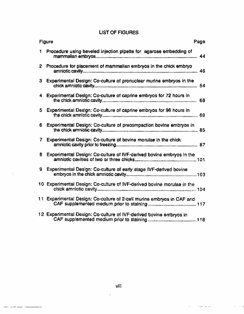
LIST OF FIGURES
Figure Page
1 Procedure using beveled injection pipette for agarose embedding ofmammalian embryos........................................................................................ 44
2 Procedure for placement of mammalian embryos in the chick embryoamniotic cavity......................................................................................................46
3 Experimental Design: Co-culture of pronuclear murine embryos in thechick amniotic cavity......................................................................................... 54
4 Experimental Design: Co-culture of caprine embryos for 72 hours inthe chick amniotic cavity.................................................................................. 68
5 Experimental Design: Co-culture of caprine embryos for 96 hours inthe chick amniotic cavity.....................................................................................69
6 Experimental Design: Co-culture of precompaction bovine embryos inthe chick amniotic cavity.....................................................................................85
7 Experimental Design: Co-culture of bovine morulae in the chickamniotic cavity prior to freezing...................................................................... 87
8 Experimental Design: Co-culture of IVF-derived bovine embryos in theamniotic cavities of two or three chicks...................................................... 101
9 Experimental Design: Co-culture of early stage IVF-derived bovineembryos in the chick amniotic cavity..............................................................103
10 Experimental Design: Co-culture of IVF-derived bovine morulae in thechick amniotic cavity.......................................................................................104
11 Experimental Design: Co-culture of 2-cell murine embryos in CAF andCAF supplemented medium prior to staining...........................................117
12 Experimental Design: Co-culture of IVF-derived bovine embryos inCAF supplemented medium prior to staining...........................................118

ABSTRACT
A novel embryo culture system has been developed using 96-hour chick
embryos. One to four mammalian embryos can be injected into the chick
embryo amnion (CEA) and allowed to develop for 72 to 96 hours. Pronuclear-
stage mouse embryos from two different strains were cultured in the CEA or in
Whitten's medium. There were more expanded blastocysts from one strain of
embryos when cultured in the CEA. More hatched blastocysts resulted from
embryos of both strains when cultured in the CEA.
Two to eight-cell goat embryos cultured in the CEA for 72 hours or on cell
monolayers reached the blastocyst stage at higher rates than when cultured
with trophoblastic vesicles or in medium alone. When culture in the CEA was
extended to 96 hours, more blastocyst were obtained than when embryos were
co-cultured on monolayers for 96 hours or in medium alone. More expanded
blastocysts were observed following the culture of precompaction stage bovine
morulae in the CEA than when embryos were cultured on monolayers or
cultured in medium alone. Culture of bovine morulae on monolayers or in the
CEA prior to freezing improved post-thaw viability when compared with culture
in medium alone.
When in vitro fertilization (IVF)-derived bovine embryos were cultured
sequentially in two or three CEA, development was not improved over culture
with cumulus cells and unacceptable loss of embryos occurred. The culture of
IVF-derived embryos in the chick embryos during the first 48 hours of
development resulted in less four to six-cell embryos than culture with cumulus
cells, however, culture of later-stage IVF-derived embryos in the CEA appears
to be as effective as cumulus cell co-culture.
Extracted chick amniotic fluids (CAF) were used to supplement the
culture medium for mouse and cow embryos. Two-cell mouse embryos

developed at similar rates when cultured in CAF or fetal bovine serum (FBS)
supplemented medium, however, embryos placed in the CEA cleaved at higher
rates. The use of CAF as a supplement in in vitro maturation and culture
medium for bovine IVF procedures appears to be as effective as
supplementation with FBS.
x

CHAPTER I REVIEW OF LITERATURE
Introduction
The ability to foster continued development of the mammalian conceptus
in vitro represents an invaluable resource for the disciplines of both basic and
applied science. In terms of increasing our understanding of developmental
biology, the refinement of functional embryo culture systems is a prerequisite to
future avenues of scientific exploration. These include the determination of the
precise metabolic and physical requirements of the embryo at various stages of
development, as well as defining the role of the embryo in the maternal
recognition of pregnancy. In addition, elucidation of the complex developmen
tal control mechanisms of the activated mammalian zygote will only be possible
if "normal" patterns of development can occur in an artificially controlled
environment.
The availability of effective embryo culture techniques will also play an
important role in the practical application of many experimental methods now
being developed. Although the procedures for nonsurgical collection and
transfer of bovine embryos are still widely used by commercial cattle breeders,
research laboratories no longer rely on these somewhat expensive and time
consuming techniques for the production of bovine embryos. The now routine
techniques of in vitro oocyte maturation and in vitro fertilization (IVF) are
currently providing many investigators with previously unattainable quantities of
early stage embryos from abattoir ovaries.
The availability of viable gametes produced by IVF will likely expedite the
development of procedures for producing genetically engineered and cloned
embryos. Although attempts to introduce foreign genes into the genome of
domestically important species have been disappointing to date, the eventual
1

2
production of transgenic lines of farm animals will necessitate genetic
manipulation at embryonic stages. Emerging methodologies for the production
of mammalian "clones" also require the use of embryos and embryonic cells at
very early stages of development.
In order for genetic manipulation, cloning or any other technique
involving early stage embryos to result in live offspring, the viability of the
embryo must be maintained until it can be transferred to a recipient female. In
the case of IVF-derived bovine embryos, in vitro embryo maintenance for a
period of 6 or 7 days is required if the embryo is to be transferred via
nonsurgical techniques.
Previously mentioned IVF techniques also have the potential of aiding in
the captive reproduction of endangered species of mammals. The union of
selected gametes in vitro could enable the production of offspring unattainable
by natural matings between specific individuals from an exotic species. Like
domestic embryos, these IVF-derived exotic embryos would require effective in
vitro culture systems for maintenance of viability prior to transfer.
Improved embryo culture systems are currently needed in the clinical
field of assisted human reproduction. Although human embryos can readily be
produced using in vitro fertilization techniques, pregnancy rates following
transfer remain less than 20% overall. These low pregnancy rates are likely
due to several inadequacies in the human IVF system, one of which is the lack
of an effective in vitro culture system. IVF-derived human embryos are normally
transplanted to the uterus of the oocyte donor within 36 hours of fertilization,
while at the six- to eight-cell stage. Although the embryonic stage at this time is
very early for continued development in the uterus, the loss of viability that
occurs using current in vitro culture techniques is too great to justify waiting to
transfer the embryos at later, more advanced stages.

3
Human embryos are also routinely frozen at these early stages, despite
the very low pregnancy rates obtained following thawing and transfer. It has
been reported that the later embryonic stages of morula and blastocyst survive
the freezing and thawing process at higher rates, but effective systems for
culturing human embryos to these stages are not presently available.
The following discussion reviews the development of methods and
systems designed to promote growth and development in mammalian embryos
during periods of in vitro culture. Although none of the procedures discussed
represent a definitive solution, considerable progress has been made during
the past decade.

I. Development of Embryo Culture Techniques
Biological Fluids
Mammalian embryo culture has been an important area of biological
research for over half a century. At this time, relatively little is known about the
specific growth factors necessary for maintaining the normal development of
most mammalian embryos in an in vitro environment. It is known that an
effective in vitro culture requires the presence of yet undefined biological
components in order for embryonic development to proceed at a normal rate.
The pioneering efforts in maintaining embryonic development outside of
the female reproductive tract were conducted primarily using rabbit embryos.
By culturing rabbit blastocysts in glass dishes that contained plasma clots
(Brachet, 1912), the development of the primitive groove and rudimentary
placental structures were observed, although embryonic survival was less than
40 hours. In later studies, Lewis and Gregory (1929) used blood plasma for the
culture of one cell rabbit embryos and observed development to the eight-cell
stage within 48 hours.
The development of embryo culture medium that followed the early use
of undiluted blood plasma involved the addition of biological fluids to balanced
salt solutions. Among these in vitro growth-promoting fluids were chick embryo
extracts (CEE). Carrel (1913) noted that extracts of chick embryos increased
the growth of mammalian tissues in vitro, and CEE were used in some of the
embryo culture experiments that followed. Pincus (1930) used hanging-drop
cultures which contained various mixtures of rabbit plasma, chick plasma, rabbit
embryo extract and CEE to study embryonic development. Cleavage of early
stage embryos was observed, as was the development of two and four-cell
rabbit embryos to the morula stage.
In 1933, the development of later-stage rabbit embryos was evaluated in
4

5
medium containing chicken plasma and CEE (Waddington and Waterman,
1933). Embryonic cell differentiation was reported in embryos that had reached
the primitive streak stage.
The first successful culture of early-stage, pre-implantation mouse
embryos in a saline solution required supplementation of physiological saline
with egg white and yolk from hen's eggs (Hammond, 1949). Development to
the blastocyst stage was noted when eight-cell embryos were cultured in this
medium, however, little cleavage of two-cell embryos was observed. During
this same period, Dowling (1949) cultured bovine embryos in egg-white or yolk
supplemented saline. Only one of 14 eight-cell bovine embryos developed to
the 16-cell stage in this medium.
Chang (1949) demonstrated that heat-inactivated serum could be used
as a supplement in culture medium for two-cell rabbit embryos. Edwards (1964)
obtained acceptable rates of development in Waymouth's medium
supplemented with 10% rabbit serum when culturing one cell rabbit embryos
which had been removed from the zona pellucida. In this study, eight of 10 one
cell embryos developed to at least the 16-cell stage, and three of 10 developed
to at least the 32-cell stage.
These results using rabbit embryos led to the development of bovine
embryo culture systems using bovine serum. Brock and Rowson (1952)
attempted to culture bovine embryos in bovine serum, then in 1963 Hafez etal.
cultured single cell bovine embryos in serum supplemented saline. Both of
these groups failed to achieve high rates of embryonic development, however,
the bovine serum used was not heat treated to inactivate complement. When
Onuma and Foote (1969) used heat treated bovine and rabbit sera for the
culture of one-cell bovine embryos, they obtained cleavage in 45% of 184 ova
cultured in vitro.
6
Gordon (1975) used serum supplemented, phosphate buffered saline for
the temporary storage of bovine embryos at various stages and obtained
normal development in 30 of 50 embryos. Wright et a!. (1976) used
bicarbonate-buffered medium (HF-10) supplemented with 10% heat-treated
fetal bovine serum (FBS) for the culture of bovine embryos in a 5% C 0 2
atmosphere. These culture conditions resulted in improved in vitro embryo
development and became the standard for bovine embryo culture.
Unfortunately, the culture conditions defined by Wright et al. do not
represent an ideal substitute for embryo development in vivo. The necessary
presence of undefined biological fluids in the culture milieu can produce
inconsistent results. Sirard and Lambert (1985) have shown that identically
prepared batches of bovine serum from different animals give different results in
their ability to promote cleavage of four-cell bovine embryos. Production of a
repeatable and consistent in vitro environment is an important consideration for
developing embryos.
Attempts to Define Embryo Culture Conditions
Whitten (1956) modified Hammond's procedure by using bicarbonate-
buffered Kreb's medium instead of physiological saline to stabilize the pH of the
culture medium. No development of eight-cell mouse embryos was noted in
Kreb's medium alone, however, the supplementation of Kreb’s with 1% egg
white resulted in development to the blastocyst stage. More importantly,
Whitten showed that crystalline bovine serum albumin (BSA) could be
substituted for egg whites. This allowed work to continue with a more definable
medium, and led to the discovery that embryos from some strains of mice could
develop from the pronuclear to the blastocyst stage in a defined in vitro
environment (Whitten and Biggers, 1968). The use of BSA has become a

7
standard component of defined embryo culture media.
Following Whitten's discovery that murine embryos could undergo
complete in vitro development in a defined medium, attempts at culturing
embryos of domestic species in defined medium were attempted. One of the
first effective defined media for the culture of embryos from domestic species
was based on the biochemical analysis of sheep oviductal fluid (Restall and
Wales, 1966).
The first long term culture of early-stage bovine embryos was conducted
by Tervit etai. (1972) using this synthetic oviductal fluid (SOF) medium. These
workers obtained development from the one-cell to the 16-cell stage (3/6) using
SOF. They also obtained blastocyst-stage embryos when eight-cell embryos
were cultured in SOF.
Later studies comparing SOF with another defined medium, Brinster's
modified ova culture medium (BMOC-3) resulted in development to the morula
stage at rates of 26% and 57% when 8 to 12-cell bovine embryos were cultured
in SOF and BMOC-3, respectively (Shea et at., 1974). Two pregnancies
resulted when morulae were transferred to 17 bovine recipients.
Bowen et at., (1975) compared SOF with defined Ham's F-10 medium
(HF-10) and obtained 48% and 80% morulae when two to eight-cell embryos
were cultured for 48 hours in SOF and modified HF-10, respectively.
Development of later stage pre-implantation embryos in SOF and BMOC-2 was
observed by Kanagawa et al., (1975). When eight to 32-cell bovine embryos
were cultured for 120 hours 65 to 80% developed to the blastocyst stage in both
culture media.
Although BSA is considered a component of all defined embryo culture
media, individual batches of BSA are themselves poorly defined. Different
batches of BSA from the same supplier have been shown to have different

8
growth promoting effects on mammalian embryos in vitro. Kane (1987) has
reported that rabbit morulae cultured to the blastocyst stage in medium
supplemented with 1.5% BSA from one batch had more than twice as many
cells as morulae cultured in the same medium supplemented with a different
batch of BSA.
The use of defined media has contributed to the study of the specific in
vitro requirements of cells in culture (Rizzino eta!., 1979). By using serum-free
culture conditions, specific hormones and growth factors can be added to the
culture medium individually and their effects on cell growth and differentiation
evaluated (see review by Barnes and Sato, 1980). In addition, the use of
defined medium allows for the analysis of growth factors and hormones that are
produced by the cultured cells themselves.
A more complete understanding of embryonic growth factors might also
be possible if embryos could be cultured in medium with defined components.
Unfortunately, attempts at culturing mammalian embryos from the single cell to
the blastocyst stage in a completely defined in vitro environment have been
successful only with certain strains of mouse embryos (Whitten, 1968). Supple
mentation of the culture medium with complex, undefined biological fluids (e.g.
serum, BSA) are required to obtain in vitro development in the embryos of
domestic mammalian species (see review by Wright and Bondioli, 1981).
Even with the addition of undefined biological fluids, little development is
obtained by culturing early stage domestic animal embryos in medium alone.
Betterbed and Wright (1985) cultured one-cell ovine embryos in several media
with different gas mixtures, and obtained only two blastocysts from 104 embryos
cultured. In earlier studies using two to eight-cell sheep embryos, Wright et al.
(1976) obtained £50% blastocyst development using medium-only culture
conditions.

9
In Vitro Blocks to Normal Development
One common finding in the pioneering attempts at culturing early stage
mammalian embryos was the apparent block to development at a species-
specific stage. In many experiments involving bovine embryos, development of
very early-stage embryos (one to four cells) proceeds to the eight- to 16-cell
stage in vitro and then ceases. However, embryos collected at the eight- to 16-
cell stage readily develop to morulae and blastocyst stages (Thlbault, 1966).
This indicates an apparent inadequacy of the in vitro systems at this stage,
resulting in an in vitro "block" to development.
Additional studies (Eyestone and First, 1986) have indicated that bovine
embryos which have become "blocked" at the eight- to 16-cell stage usually
cannot be rescued (i.e. further development is not possible even if the embryo is
returned to an in vivo system). Unpublished data by W .H. Eyestone is
mentioned in the latter study indicating that the embryonic cells are "alive”
during the block, however they are incapable of dividing.
The in vitro developmental block was first described in murine embryos.
Cole and Paul (1965) observed development of one-cell embryos to the two
cell stage in vitro, however, these two-cell embryos failed to undergo further
cleavage and subsequently degenerated. This degeneration occurred in spite
of the fact that embryos collected from mice at the two-cell stage were capable
of normal development to the blastocyst stage in vitro. Whittingham and
Biggers (1967) transferred in vitro cultured, developmental^ blocked embryos
at the two-cell stage to the ampulla of oviduct organ cultures and were able to
"rescue" embryos from the in vitro block state. They obtained blastocyst from
these previously blocked embryos, and pregnancies resulted following the
transfer of these blastocyst.
The species-specific timing of the developmental block in mouse embryo

10
culture is coincident with an important biochemical transition occurring in the
embryonic cells. At the two-cell stage, the murine embryonic genome is
activated and protein synthesis is no longer dependent on pre-existing maternal
mRNA (Braude et al., 1979).
Recent evidence indicates that the transition from maternal to embryonic
mRNA dependence in the bovine embryo is at the same stage as the bovine
embryonic block. Frei et al., (1989) cultured oocytes and early stage embryos
with radiolabelled methionine and analyzed proteins synthesized with one
dimensional electrophoresis and fluorography. It was noted that a progressive
decrease in protein synthesis occurred from the oocyte to the eight-cell stage,
with protein synthesis increasing from the eight-cell to the blastocyst stage.
This decrease and subsequent increase in protein synthesis indicates
the transition from translation of maternal mRNA accumulated during oogenesis
and the translation of newly transcribed mRNA from the activated embryonic
genome. In addition to these quantitative changes, there were definite
qualitative changes in the patterns of proteins produced after the 16-cell stage
in bovine embryos.
Although there is currently no evidence that the in vitro block is directly
linked to a breakdown in the maternal to zygotic transition (MZT) in vitro, there
are several pieces of information that suggest that this may be the case. Bovine
embryos begin to synthesize ribosomal RNA (rRNA) at the time of the MZT, or at
the 8-cell stage (King et al., 1989). The activation of rRNA synthesis is
detectable by staining embryos for nucleolar organizing regions (NOR). When
IVF-derived bovine embryos which had blocked in vitro were stained in this
fashion, NOR did not appear (Barnes and Eyestone, 1990). However, when
these IVF-derived embryos were cultured in vivo in the ligated oviducts of ewes
(where the block is not seen), normal NORs were present following staining.

11
These workers suggest that the in vitro block to growth may be caused by a
breakdown in the transition from maternal to zygotic control of development due
to inadequacies in the in vitro culture system.
The effect of in vitro culture on protein synthesis by rabbit embryos has
been investigated by Jung (1989). In this study, culturing rabbit embryos in an
in vitro environment resulted in decreased protein half lives (i.e. accelerated
protein degradation) when compared with similar blastocysts that had
developed in vivo. This trend towards rapid protein degradation in vitro was
partially reversed by supplementing the in vitro culture medium with uterine
secretions.
Successful duplication of the uterine environment in vitro is proving to be
a difficult task, due to the complexity of the in vivo embryonic environment.
Brigstock et al. (1989) have detected the presence of a number of growth factors
with potential embryotropic effects in uterine tissues and fluids. In their study,
the synthesis of these growth factors appeared to be regulated by the female
sex steroids estrogen and progesterone. Among these growth factors are
epidermal growth factor, transforming growth factor, insulin-like growth factor,
colony-stimulating factor I, oestromedins, uterine luminal fluid mitogens and
fibroblast growth factors. Since little is known about the precise function of any
of these factors in stimulating uterine or embryonic growth, the synthesis of a
defined in vitro system will be a formidable task.
Adding to the the difficulties in developing optimal embryo culture
environments are the trace contaminants that can occur in tissue culture
reagents and water. It is well known that water purity is critical for the
maintenance of embryo viability in vitro. Whittingham (1977) reported that
three-times glass distilled water was necessary for a high rate of blastocyst
formation from two-cell murine embryos. Abramczuk et al. (1977) showed a

12
beneficial effect on the culture of one-cell murine embryos when the chelating
agent ethylenediaminetetraacetic acid (EDTA) was added to the culture
medium. These results suggest that even trace impurities can greatly impede
the normal development of mammalian embryos in culture.
II. Embryo Co-Culture Systems
In Vivo Oviductal Culture
Although defining the in vitro culture requirements of mammalian
embryos remains an important basic science question, there remains a need for
applicable embryo culture systems for use in other areas of embryo research.
In an effort to overcome the apparent inadequacies of in vitro embryo culture in
medium alone, embryo co-culture systems are now being developed. One of
the more successful methods for promoting the development of early-stage
mammalian embryos requires the in vivo culture in the oviducts of an
intermediate recipient.
In 1955 Averill and co-workers demonstrated that interspecies embryo
transfer to the oviducts of a surrogate recipient could be used to maintain
mammalian embryo development. These researchers transferred early-stage
sheep embryos to the ligated oviducts of psuedopregnant rabbits and
subsequently recovered them 4 or 5 days later. Of the original 18 embryos (two
to 12-cells) transferred to the rabbit oviducts, a total of nine embryos were
recovered, all at the morula or blastocyst-stage. When three of these embryos
were transferred to recipient ewes, two were capable of implantation in utero
and further development.
This technique was used by Hunter and co-workers (1961) to transport
sheep embryos collected in Cambridge, England to recipient ewes in
Pietermaritzburg, South Africa. Storage periods in the rabbit oviduct ranged

13
from 101 to 128 hours, and four lambs were born following the transfer of 16
embryos recovered from the rabbit oviducts.
Lawson et al., (1972a) confirmed these results by using recipient rabbits
with ligated oviducts. The ligation of rabbit oviducts at the uterotubal junction
was performed at the time of transfer to prevent passage of the ovine embryos
into the uterus. A total of 456 early-stage sheep embryos (2 to 16-cell stage)
were transferred to the oviducts of pseudopregnant rabbit females. By ligating
the recipient oviducts, a recovery rate of 87% was achieved. Of the 397
embryos recovered, 368 (93%) had progressed to later developmental stages
in the rabbit oviduct. Up to 69% survival rates were observed when embryos
were transferred to synchronized recipient ewes.
In 1968, Sreenan and co-workers first demonstrated that the rabbit
oviduct could also promote development in bovine embryos. These workers
collected 32 fertilized embryos from donor heifers three days after breeding.
These embryos were transferred to the ligated oviducts of pseudo-pregnant
rabbits, and 19 embryos were recovered after at least 94 hours in the rabbit
oviduct. Of the 19 recovered embryos, 17 had developed to later embryonic
stages and 15 had >80 cells.
Lawson et al. (1972b) also transferred 48 early-stage bovine embryos
(one- to eight-cell) to the ligated oviducts of psuedopregnant rabbits. Of the 41
embryos recovered two to four days later, 34 (83%) had advanced to later
developmental stages and appeared normal. These workers performed a
second experiment using bovine embryos to determine the post-culture viability
of recovered embryos. Following a three or four day culture period in the
ligated rabbit oviduct, embryos were transferred to synchronized recipient
heifers. Of the 15 embryos transferred to heifers within one day of donor
synchrony, 11 (73%) calves were born.

14
Boland (1984) conducted extensive tests to determine the feasibility of
using rabbit oviducts for the viability screening of bovine embryos prior to
transfer. Despite the inherent loss of embryos involved in using the rabbit
oviduct as a culture system, it was determined that potential for further
development in bovine embryos could be assessed by using the rabbit oviduct.
Culture of agarose embedded embryos in the ligated oviducts of sheep
has become an important technique when micromanipulated embryos must be
cultured prior to nonsurgical transfer. This technique was developed by
Wiiladsen (1979) to facilitate the development of micromanipulated sheep
embryos.
Following the microsurgical separation of the blastomeres of a two-cell
embryo, individual blastomeres were replaced in a zona pellucida and
embedded in agarose. The agarose embedded blastomeres were then
transferred to the oviducts of ewes on days 1 to 2 of the recipient estrous cycle.
The oviducts were then ligated at the uterotubal junction to maintain placement
of the embryos. The embryos remained in the ligated oviducts of the ewe for a
period of 3.5 to 4.5 days. A total of 31 agarose-embedded pairs of embryos
were transferred to ewes, and 20 of these pairs were subsequently recovered.
Of the total of 40 embryos recovered, 35 had developed to the late morula or
early blastocyst stage in the intermediate host.
Eyestone and co-workers used agarose embedding on 35 one- and two-
cell bovine embryos transferred to the ligated oviducts of ewes synchronized to
the donor estrus cycle, and observed 42% normal development after 74%
embryo recovery. Additionally, these workers transferred 39 one- and two-cell
bovine embryos to the ligated oviducts of ovariectomized ewes, and observed
27% development following 67% embryo recovery.
Agarose embedding has recently been used in an attempt to increase

15
recovery rates of bovine embryos cultured in the rabbit oviduct. Westhusin and
co-workers (1989) recovered 77% of 69 one- and two-cell bovine embryos from
the ligated rabbit oviduct, with 43% of the recovered embryos developing to the
morula and blastocyst stages. When these same workers performed agarose
embedding prior to culture in the ligated oviducts of ewes, 100% of 52 embryos
were recovered after 6 days, with 52% at the morula or blastocyst stage.
This approach has, in recent years, been used extensively to promote
development in "cloned" one-cell embryos at a commercial transplant station
(Bondioli et al., 1990). In this case, electrofusion of oocytes and single
blastomeres was performed, then the fusion embryos were agarose-embedded
and cultured in the ligated sheep oviduct for up to 6 days. Recovery rates of 93
to 97% were reported by the cattle transplant unit using this technique.
In spite of the effectiveness of this in vivo technique, it has not proven to
be practical in most embryo culture applications. The primary disadvantage to
using the ligated sheep oviduct for embryo culture is the difficulty and time
involved in performing multiple surgical procedures on the sheep. In addition, a
herd of recipient ewes must be maintained and this often proves to be costly.
For these reasons, rabbit oviducts are still used for in vivo culture of mammalian
embryos.
Recently mouse oviducts have been evaluated for culturing embryos
from large domestic species. Ebert and Papaioannou (1989) transferred
porcine embryos (at various stages of development) to the oviducts of immature
mice, and compared the resulting development to in vitro culture in Ham's F-12
with 10% FBS in an atmosphere of 5% CO 2 . When early blastocysts were
cultured for two days in the mouse oviduct, the embryos recovered had twice as
many embryonic cells compared with similar embryos which had been cultured
in vitro (104 vs. 57.3 cells). When four- to six-cell porcine embryos were

16
cultured in the mouse oviduct 10 of 13 (77%) reached the blastocyst stage
compared with only one of 10 cultured in the in vitro control group.
Cell Co-Culture
The first demonstration of murine in vitro embryonic development from
the one cell through to the blastocyst stage involved culture in explanted murine
oviducts. Biggers et al. (1962) cultured oviducts from eight to nine-week-old
mice on 2 cm x 2 cm stainless steel grids in 6 cm petri dishes. The oviducts
were removed from females which had been superovulated and mated 12 to 14
hours previously. The organ culture system was maintained in a defined
medium and incubated at 37° C in a 5% CO2 atmosphere for three or four days.
Of the total of 106 embryos recovered following four days of organ culture, 14
(13%) morulae and 69 (65%) blastocysts were present.
Early-stage hamster embryos have been reported to be difficult to culture
in vitro. Although one-cell hamster embryos will readily cleave to the two-cell
stage, the block at the two-cell stage has been shown to be near "absolute"
(Whittingham and Bavister, 1974). Bavister and Minami (1986), however, were
able to overcome this block in 11% of 830 one-cell hamster embryos by
culturing them in explanted mouse oviducts.
Explanted mouse oviducts have also been shown to support the
development of early stage embryos from domestic species. Krisher et al.
(1989) cultured one-cell porcine embryos in explanted mouse oviducts taken
from recently mated females. Following six days of in vitro culture, 25 of 32
(78.1%) one-cell porcine embryos developed to the blastocyst stage when
cultured in the explanted mouse oviduct compared with only 10 of 28 (35.7%)
developing to the blastocyst stage in medium alone.
Cole and Paul (1965) have used irradiated HeLa cells for co-culture of

17
two-cell murine embryos through the hatching blastocyst stage and improved
culture rates when compared with culture medium alone. This finding led to the
development of embryo co-culture systems using fibroblast monolayers. Kuzan
and Wright (1981) first reported co-culture using farm animal embryos. They
observed increased rates of porcine blastocyst hatching during co-culture on
bovine fibroblasts when compared with culture in medium alone.
Kuzan and Wright (1982) subsequently demonstrated that co-culture with
fibroblast monolayers was beneficial for the development of bovine embryos in
vitro. The in vitro development of bovine morulae co-cultured on monolayers of
either bovine uterine or testicular fibroblasts was improved when compared with
bovine morulae cultured either in conditioned or fresh medium. Additionally,
blastocyst hatching was significantly increased when embryos were co-cultured
with monolayers. These workers postulated that "helper cells" may secrete an
embryotropic substance into the culture medium and/or they may remove toxic
by-products of embryo metabolism from the culture medium.
O ther workers have demonstrated that the in vitro viability of
micromanipulated bovine embryos was enhanced by co-culture on bovine
uterine fibroblasts (Voelkel et al., 1985). At the same laboratory Wiemer et al.
(1988, 1989) used a fibroblast monolayer system derived from the reproductive
tract of a female bovine fetus. This fetal bovine uterine fibroblast (FBUF)
monolayer has given successful co-culture results with embryos from several
mammalian species. Pregnancies have been obtained in horses (Wiemer et
al.,1988) and humans (Wiemer et al., 1989) following co-culture of embryos on
the FBUF monolayer. This system has also proven beneficial for the in vitro
culture of early stage bovine morulae (Wiemer et al., 1990).
More recent studies have revealed that co-culture of embryos with cells
of endometrial origin result in improved developmental rates when compared

18
with co-culture with cells of fibroblastic origin. Gandolfi and Moor (1987)
cultured a total of 511 pronuclear sheep embryos on fibroblast or on oviduct
epithelial cells or in medium alone. Only 13% of the embryos cultured in
medium alone reached the morula stage after three days of culture compared
with 95% morulae on both feeder cell monolayers. Following transfer to
recipient ewes, however, only 33% of the embryos cultured on fibroblast were
capable of continued development compared with 80% that continued to
develop for embryos cultured on epithelial cells.
When these pronuclear sheep embryos were allowed to develop for six
days in vitro, 42% of the embryos on epithelial cell monolayers developed to the
expanded blastocyst stage compared with only 4.5% developing to expanded
blastocysts on the fibroblast monolayer.
White et a l (1989) obtained similar results when culturing early stage
porcine embryos on porcine oviduct epithelial cells or porcine fetal endometrial
fibroblasts. When two- to 16-cell porcine embryos were cultured on epithelial
cells 44 of 63 (70%) reached the expanded blastocyst stage, significantly more
than the 16 of 60 (27%) expanded blastocyst observed on fibroblast
monolayers.
Prichard et al. (1990) attempted to mimic embryo descent from the
oviduct to the uterus in vitro by transferring early stage caprine embryos from
oviductal cells to uterine cells after 36 hours of culture. Surprisingly, two to
four-cell caprine embryos which remained on oviductal cells developed to the
blastocyst stage at higher rates than did embryos which were transferred from
oviductal to uterine cells during co-culture. Of 30 embryos co-cultured on
oviductal cells, 27 (90%) reached the blastocyst stage compared with only 20 of
30 (67%) blastocysts when embryos were sequentially cultured on oviductal the
uterine cells. These results suggest that cells obtained from the oviductal

19
epithelium provide a superior embryo co-culture environment.
Eyestone and First (1989) collected one- to eight-cell embryos from cattle
and co-cultured them with either suspended or monolayered oviductal cells.
When embryos were cultured for four days with suspensions of oviductal cells,
46% (38/82) developed to the morula or blastocyst stage compared with only
4% (1/27) reaching those stages in medium alone. Co-culture with
monolayered oviductal cells gave similar results, with 43% (15/34) reaching the
morula or blastocyst stage on the oviductal monolayers compared with 3%
(1/37) reaching those stages in medium alone.
Ellington et al. (1990a) surgically collected one- and two-cell bovine
oocytes then cultured them with fresh bovine oviductal epithelial cells, frozen-
thawed bovine oviductal epithelial cells or in medium conditioned with oviductal
cells. No difference was noted with regard to development past the eight to 16-
cell block, however, the embryos cultured on fresh oviductal epithelial cells had
significantly higher cell counts than embryos cultured on frozen-thawed cells or
in conditioned medium alone. The embryos cultured on fresh cells also had
better embryo viability scores than embryos in the other two treatments.
Ellington et al. (1990b) also compared co-culture on monolayers of
bovine oviductal epithelial cells to in vivo culture in the ligated oviducts of
rabbits using one- to two-cell bovine embryos. Embryos co-cultured on these in
vitro monolayers for 5 days had similar numbers of cells and resulted in similar
pregnancy rates following transfer when compared with embryos cultured in
vivo using the rabbit oviduct system. Most embryo co-culture research is now
conducted using cells of epithelial or granulosa cell origin rather than fibroblast
origin.
Gandoifi and co-workers (1989) recently isolated two oviductal proteins
which are secreted only during the period corresponding to embryonic passage

20
through the oviduct. Both of these proteins showed an affinity for the zona
pellucida, and studies using a monoclonal antibody to one of the proteins
reveal that it crosses the zona pellucida and associates with the developing
blastomeres. Little is Known about the exact nature of the embryotropic effect of
cell co-culture, however, this recent report (Gandolfi et al., 1990) suggests a
direct effect of oviductal cells on the developing embryo.
Trophoblastic Vesicles
Although early stage mammalian embryos have proved difficult to culture
in medium alone, later stages of the same embryos will readily develop in
culture following their hatching from the zona pellucida. Heyman et al. (1984)
developed an embryo co-culture method using tissue from these later-stage
embryos
Embryos collected at days 12 to 14 following estrus are at the elongating
blastocyst stage. These embryos can be microsurgically sectioned and the
resulting pieces will form spherical vesicles that survive £3 weeks in culture.
These spheres of embryonic tissue are now called trophoblastic vesicles (TV).
Camous et al. (1984) were able to overcome the eight- to 16-cell block to
bovine embryo development in vitro by co-culture with TV prepared from day-13
or 14 bovine blastocysts. When one- to eight-cell embryos were cultured with
these TV 46% developed to the morula stage compared with 18% morulae
when embryos were cultured in medium alone.
Heyman etal. (1987) cultured early stage bovine and ovine embryos with
TV and compared the development of these embryos with similar embryos
cultured in medium alone. When one- to eight-cell bovine embryos were co-
cultured with TV prepared from day-14 bovine blastocysts, 46% reached the
morula stage compared with only 18% reaching the morula stage in medium

21
alone. When 55 one-cell bovine embryos were cultured with TV, 23 (44%)
cleaved beyond the eight-cell stage, compared with only nine of 67 (13%) one
cell embryos cultured in medium alone. When one-cell sheep embryos were
co-cultured with TV prepared from day-12 ovine blastocyst, 75% reached the
morula stage compared with onty 35% of the one-cell embryos cultured in
medium alone.
These workers were able to obtain high rates of development by using
medium conditioned with bovine TV. When one- to two-cell bovine embryos
were cultured in TV-conditioned medium, 30 of 36 (83%) cleaved past the eight
cell stage compared with 38 of 55 (69%) embryos co-cultured with bovine TV.
Pool et al. (1988) used microsurgical techniques to place early-stage
bovine morula inside of bovine TV for co-culture. Following 60 hours of co
culture 69% (25/36) of the embryos co-cultured with TV were evaluated at good
or excellent quality grades compared with 36% (13/36) of the embryos co-
cultured inside of TV and 22% (8/36) of the embryos cultured in medium alone.
The placement of embryos inside of TV did not prove to be as effective as
simple co-culture with TV.
Maciulis et al. (1987) used dispersed cells from day-12 to 14 sheep
embryos and found that these cells enhanced the embryotropic effects of
epithelial cell co-culture. When one- and two-cell ovine embryos were co-
cultured on ovine oviductal epithelial cells for four days only 15% morulae were
observed on oviductal cell co-cultures compared with 54% morulae on
combined oviductal and embryonic cell co-cultures.
Bunch et al. (1987) used day-12 to 14 sheep embryos to produce both
TV and dispersed embryonic cells for embryo co-culture. When two to eight-cell
embryos were co-cultured with dispersed embryonic cells 64% reached the
hatched blastocyst stage compared with only 6% hatched blastocysts when

22
embryos were co-cultured with TV. The results of this experiment are
inconclusive, however, since different culture media were used in the different
treatment groups.
The timing of embryo collection for the production of TV has been shown
to be critical. Rexroad and Powell (1988) used day-14 ovine blastocyst to
produce TV for embryo co-culture. When one-cell ovine embryos were cultured
with these TV for 24 or 72 hours, the cleavage rates were slightly less than
when one-cell embryos were cultured in medium alone.
III. In Vitro Fertilization
B ackground
Prior to 1981, in vitro fertilization (IVF) resulting in the birth of live young
had only been successfully performed in rabbits, rats, mice, hamsters and
humans (See review by Wright and Bondioli, 1981). Attempts at producing
calves by IVF of bovine oocytes were numerous, but unsuccessful. The
development of repeatable IVF procedures for the production of early-stage
bovine embryos has been an important goal for researchers working with
bovine gametes. Apart from basic science, there are several obvious reasons
for the development of practical IVF techniques. The collection of multiple
embryos from superovulated cattle first requires the injection of donor cattle with
expensive hormone treatments. In addition, the actual collection of early stage-
bovine embryos requires labor intensive and time consuming surgical
procedures that could be avoided by using IVF. Another desirable aspect of
producing embryos via IVF is the large numbers of oocytes which can be
obtained from inexpensive slaughterhouse materials, allowing experiments to
be completed in shorter periods of time.
Some of the early researchers working with bovine IVF did achieve

23
fertilization of oocytes, but little development subsequently occurred. Brackett et
al. (1978) reported some success using oocytes collected from oviducts or
follicles following gonadotropin-treated donor cattle. They achieved 56%
fertilization (14/25) using these in wVo-matured oocytes. Of the 14 fertilized
oocytes, 10 (71%) underwent cleavage to the two and four-cell stage.
Successful production of a live calf by IVF procedures was accomplished
in 1981 (Brackett et al., 1982). Oocyte donors were prepared by injection with
1500 III of pregnant mare serum gonadotropin followed at 72 hours by a 40 mg
injection of prostaglandin F2 tt- In wVo-matured oocytes were surgically
recovered from oviducts and ovarian follicles and then placed with sperm cells
for 18 to 24 hours.
Fresh semen was collected from five bulls for use in IVF procedures.
Data from one bull used on the oocytes recovered from 7 cows resulted in the
fertilization of 22 oocytes when 35 tubal oocytes were exposed to sperm in vitro
(62.9%). When oocytes were aspirated from follicles nine of 39 (23.1%) were
fertilized. A total of seven embryo were surgically transferred to the oviducts of
synchronized recipients resulting in the birth of the first live calf from IVF in
cattle.
Brackett et al. (1984) later reported the birth of twin calves following the in
vitro fertilization of bovine oocytes matured in vivo. Oocytes were surgically
recovered from donor cows following hormonal stimulation with either pregnant
mare serum gonadotropin (PMSG) or follicle stimulation hormone (FSH).
Following fertilization, embryos were surgically transferred to the oviducts of five
recipient cows. Two of the recipients carried twin pregnancies to term, following
the transfer of two and three embryos to their oviducts. One set of twins died
during parturition, the second set resulted in the birth of two normal bull calves.
Sirard and Lambert (1985) recovered in vivo matured oocytes from

24
PMSG and FSH stimulated cattle using laparoscopic techniques. These
workers found that the highest rates of cleavage following fertilization occurred
when oocytes had expanded cumulus complexes rather than compact cumulus
complexes. Cleavage rates of 60% were obtained when Brackett's defined
medium was used for in vitro final maturation, fertilization, and growth.
Development to the 16-celi stage was observed in vitro. Lambert eta l. (1986)
also used laparoscopic techniques to recover 1618 oocytes from donor cattle
stimulated with FSH. The highest rates of cleavage following fertilization
occurred when oocytes were surrounded by expanded cumulus cell complexes.
These workers also obtained 16-cell bovine embryos in vitro following
fertilization.
The oocytes used by the researchers in the latter four studies were
matured in vivo following hormone treatment of donor cattle. Oocytes obtained
in this manner do not represent a practical resource for domestic animal embryo
research, since the oocytes must be collected using the same surgical
procedures as early-stage embryos.
In order for IVF to be a practical tool for the production of cattle embryos,
it is necessary to develop in vitro maturation (IVM) procedures. The ability to
promote maturation in oocytes aspirated from small follicles allows oocytes
obtained from the abattoir to be used, increasing the numbers of oocytes
available and decreasing the difficulty their collection.
Newcomb e ta l. (1978) obtained pregnancies following the in vivo
fertilization of oocytes which had been matured in vitro. Oocytes were obtained
by aspirating 2 to 5 mm follicles on abattoir ovaries. Oocytes were matured for
22 hours at 37° C in Ham’s F-10 medium supplemented with estrus-cow serum,
estradiol-178 and HCG. Fertilization of these in v/fro-matured oocytes was
executed in vivo by transferring the oocytes to the oviducts of previously

25
inseminated heifers. Overall, 23% of the embryos recovered seven days after
transfer were at the 28-cell stage. Twin calves were born following the transfer
of two blastocyst to a synchronized recipient.
Development in IVM-IVF-derived bovine embryos appears to be at least
partially dependent on the presence of cumulus cells during IVM culture. Ball et
al. (1983) investigated some of the factors affecting IVM of oocytes aspirated
from abattoir ovaries. These workers obtained increased numbers of
pronuclear-stage embryos (i.e. presence of male and female pronuclei)
following fertilization when cumulus cells were present during IVM. The
addition of either cAMP or FSH to the IVM medium (modified Tyrode's medium)
also increased the number of oocytes which were fertilized. When Parrish etal.
(1985) coupled this IVM technique with a 6 hour pre-incubation of sperm cells in
medium containing 10jig/ml of heparin, fertilization rates reached 81%.
The latter two reports seem to indicate that supplementation of culture
medium with hormones is necessary for successful IVM. Additionally, Fukui et
al. (1983) obtained fertilization rates as high as 46% following IVM of oocytes in
Ham's F-12 medium supplemented with FBS, LH, and estradiol. However,
Iritani et al. (1984) used defined Kreb’s medium supplemented only with BSA
for IVM of oocytes aspirated from abattoir ovaries and obtained fertilization rates
as high as 58%.
Although the need for hormone supplementation of IVM medium is
unclear, the presence of cumulus cells appears to be critical. Critser et al.
(1986) matured oocytes from 1 to 5 mm follicles in vitro by culturing them with or
without granulosa celts. Although no differences in nuclear maturation,
fertilization or formation of mate pronuclei were noticed, there were differences
in the subsequent embryo development. Following transfer to sheep oviducts,
no development (0/44) was observed when oocytes were matured without

26
granulosa cells. However, 36% (8/22) of the fertilized oocytes that had been
matured with granulosa cells developed to the morula and blastocyst stage.
Faundez et al. (1988) have compared the IVM of bovine oocyte following
co-culture of oocytes on monolayers of granulosa cells from different sized
follicles. Co-culture with granulosa cells obtained from preovulatory follicles
tended to give higher maturation rates than did co-culture with granulosa cells
obtained from small follicles. However, fertilization rates for oocytes cultured on
granulosa cell monolayers were higher than when oocytes were cultured in
medium alone. The highest rates of fertilization were achieved when oocytes
with intact cumulus cell masses were co-cultured on granulosa cell monolayers.
The ultimate test of an IVM-IVF system is not, however, fertilization rates
but the birth of live young. Cheng et at. (1986) aspirated oocytes from the
ovaries of non-stimulated ewes either surgically of following slaughter and
matured the oocytes in vitro with additional cumulus cells in medium 199
supplemented with LH, FSH, prolactin and estradiol-178. Following fertilization,
pronuclear-stage embryos were surgically transferred to the oviducts of
synchronized recipient ewes. Pregnancies were obtained in seven of 16 (44%)
recipient ewes and 10 normal lambs were born. The birth of live young from
IVM-IVF-derived ovine embryos in the latter study set a standard for IVF in
domestic species, however, the need for hormone supplementation in bovine
IVM-IVF systems remains unclear.
Lu et al. (1987) established pregnancies in cattle following the transfer of
IVM -IVF derived bovine embryos which were matured in medium 199
supplemented with estrus cow serum (ECS) and no additional hormones. ECS
likely contains higher hormone (FSH, LH and estradiol) levels than FBS,
however, Goto et al. (1988) subsequently reported pregnancy rates of 50%
following the transfer of blastocyst developed from oocytes matured in medium

27
199 supplemented with FBS and fertilized in vitro.
To date, studies directly comparing the supplementation of IVM medium
with either FBS or ECS have yielded conflicting results. In a recent review,
Gordon (1990) states that ECS is more effective than FBS for the IVM of
immature bovine oocytes. However, Fukui and Ono (1989) found no difference
in the rate of blastocyst formation in IVF-derived embryos following IVM in
medium supplemented with either FBS or ECS. These workers do state that the
presence of cumulus cells during IVM culture is critical, and this appears to be
consistent in the current literature concerning the IVM of bovine oocytes.
Potential Applications of IVF Procedures
Abattoir ovaries are currently the most available source of bovine ovaries
(and consequently the most widely used in the research laboratory), however,
they are of little use for commercial exploitation of IVF procedures. Gametes
obtained from abattoir material do not represent genetically superior cattle, and
therefore are of marginal use to the feedstock industry.
New techniques for obtaining oocytes from high quality commercial cows
are being developed (Ryan etal., 1990; Kruip eta l., 1990). Kruip etal., (1990)
has recently demonstrated that oocytes can be successfully collected from 4 to
8 mm follicles on the ovaries of nonstimulated cycling dairy cows using
sonographic guided transvaginal collection procedures. Pregnancies resulted
from the transfer of IVF produced bovine zygotes that were cultured for six days
in the ligated oviducts of ewes following these transvaginal collection
procedures.
Commercial application of IVF procedures may also be practical in high
producing dairy cows using a new technique developed by Ryan eta l. (1990).
Development of IVF oocytes to the morula stage was observed following

28
aspiration of follicles from the ovaries of pregnant cows that were stimulated
with follicle stimulating hormone. The use of this technique would allow
embryos to be obtained from valuable, high producing dairy cows while they
were gestating. The commercial exploitation of IVF procedures could become
practical by combining this technique with the transvaginal collection
procedures reported by Kruip et al.
Another application of IVM-IVF procedures is the potential ability to
decrease the length of time between successive generations of animals.
Kajihara (personal communication) has obtained pregnancies following the
transfer of embryos derived from IVM-IMF of oocytes from 8-week-old heifer
calves. Repeated use of such techniques would result in successive
generations of bovine offspring less than one year apart, greatly accelerating
the selective breeding process.
In spite of the current limitations of IVF procedures, the research
implications of such techniques are considered a major breakthrough.
Techniques for producing embryonic clones (Robl, e ta l., 1987) may cause a
more immediate need for oocytes obtained from the abattoir. Embryonic clones
are produced by electrically fusing single blastomeres from 16 to 32-cell
embryos with half of a fertilized oocyte. Although the commercial application of
this technique currently uses oocytes obtained by surgically collecting
stimulated donors (Bondioli et al., 1990), limited development has been
reported using oocytes aspirated from the ovaries of slaughtered animals
(Prather e ta l., 1987). As oocyte maturation techniques improve, the use of
oocytes obtained following slaughter for nuclear transplantation will most likely
become more efficacious due to the decreased cost and effort involved.
Current techniques for introducing genes into the genome of farm
animals also require the use of early-stage embryos. These techniques were

29
developed using mouse embryos, and require that embryos be microinjected
while at the pronuclear stage (Gordon e ta l., 1980, 1981). Such techniques
have been attempted with very limited success in the farm species. Pursel et al.
(1989) recently reviewed the progress in applying murine techniques to
livestock embryos, and reported that only 8% of 7000 microinjected pig
embryos developed to live young following transfer. Of this 8%, only 7%
expressed the exogenous genes, giving an integration efficiency of .56% for pig
embryos.
Clearly there is much work to be done in the area of farm animal genetic
engineering, but the large numbers of early stage embryos that can be
produced by IVF techniques may assist in the development of these techniques.
Culture of IVF-Derived Embryos
The technologies of mammalian IVF, nuclear transplantation and gene
transfer require the manipulation of very early-stage embryos. The commercial
application of these techniques, depends on the ability of these embryos to
develop following transfer to recipient animals. It is highly desirable to perform
such transfers to recipient cattle using nonsurgical techniques, however, the
embryos produced by means of IVF and nuclear transplantation are not at the
proper embryonic stage for nonsurgical transfer. A minimum of six days of
embryonic development must occur for the IVF-derived embryo to be at the
developmental stage of morula or blastocyst. Embryos must be at this stage to
survive in the recipient uterus, the site of nonsurgical embryo transfer
(Schneider et al., 1980; Massey and Oden, 1984; Hasler et al., 1987). In a
recent review comparing the efficiency rates for the various stages of the bovine
IVF process (including oocyte maturation, fertilization rate, pregnancy rate and
offspring born) the lowest rate given was for the development of one-cell

30
embryos to blastocyst stage (First and Parrish, 1987).
The techniques developed for the culture of early stage embryos have
been used with varying success on embryos produced by IVF. Much of the
early successful co-culture done with IVF-produced bovine embryos used the
ligated oviducts of sheep or rabbits, or co-culture with oviductal epithelial cells.
These are the techniques which had proven to be the most effective for the co
culture of surgically collected early stage bovine embryos.
Sirard et al. (1985) transferred IVF-derived embryos at the one to eight
cell stage to the ligated oviducts of pseudopregnant rabbit females, and
obtained 41% morulae when embryos were incubated in vivo for £99 hours.
Several embryos reached the blastocyst stage in the rabbit oviduct. When
these in vivo cultured embryos were transferred to 14 synchronized recipient
heifers, six (43%) pregnancies resulted. Lambert et al. (1986) obtained 46%
pregnancies (6/13) following the transfer of IVF-derived embryos cultured in the
rabbit oviduct.
Xu et al. (1987) aspirated follicular oocytes from abattoir ovaries and then
matured and fertilized the oocytes in vitro. The embryos were then surgically
transferred to the oviducts of synchronized heifers and nonsurgically recovered
by flushing the uterine horns six days later. Of the 40 embryos recovered, 15
(38%) had reached the morula or blastocyst stage. One pregnancy resulted
when two blastocysts were transferred to a single recipient.
Interspecies embryo transfer to the ligated oviducts of ewes is being
successfully used in commercial applications of IVF techniques in Europe. One
of the first reports of significant blastocyst development from in vitro fertilized
bovine oocytes was achieved by transferred bovine zygote to ligated sheep
oviducts immediately following fertilization procedures (Lu et al., 1987). After
six or seven days of culture, blastocyst development was as high as 74% in
31
several treatments. Following the transfer of these embryos, 67% of the
recipients (12/18) were diagnosed pregnant at £69 days gestation.
Ectors and co-workers (1989) used synchronized rabbits and ewes for
the culture of IVF derived bovine embryos at the two-cell stage. Embryos
placed in ligated rabbit oviducts were cultured for four days, and only 36 of 180
(15%) were recovered. Of these 36 recovered embryos, 9 (25%) underwent
normal development. When these workers transferred 50 IVF-derived two-cell
embryos to sheep oviducts, 36 (72%) were recovered. Normal embryonic
development was observed in 9 (25%) of these embryos.
Lu et al. (1988) reported the birth of live calves following the transfer of
morula that resulted from the in vitro culture of IVM-IVF produced embryos.
These embryos were cultured on oviductal epithelial cells prepared by stripping
bovine oviducts obtained at slaughter. The first twin IVF-calves were born
following the transfer of two morulae to a single recipient.
In their 1989 report on co-culture of surgically collected early stage
bovine embryos with oviductal cells and conditioned medium, Eyestone and
First also tested these culture conditions with embryos obtained from IVF. The
proportion of embryos which developed to the morula and blastocyst stage was
greater when embryos were co-cultured with oviductal cells or in conditioned
medium than when embryos were cultured in medium alone. IVF-derived
embryos cultured in medium alone cleaved at similar rates, but only 3% (7/203)
reached the morula or blastocyst stage compared with 22% (44/203) in co
culture and 22% (46/205) in conditioned medium. Pregnancies resulted in 6 of
11 (55%) recipient cows when these embryos were nonsurgically transferred.
Kim eta l. (1990) recently obtained development of IVF-derived bovine
embryos to the blastocyst stage using co-culture with bovine oviductal epithelial
cells and simple, defined medium. When 138 fertilized oocytes were placed on

32
bovine oviductal epithelial cells in serum-free Chatot-Ziomek-Bavister medium,
104 (75%) underwent cleavage and 14 (10%) reached the blastocyst stage.
Amniotic sac cells have also been used in co-culture systems for IVF-
derived bovine embryos. Aoyagi et al. (1989) compared four in vitro co-culture
systems (bovine oviductal epithelial cells, cumulus cells, trophoblastic vesicles
and amniotic sac cells) to culture in medium alone and in vivo culture in the
rabbit oviduct. Eight-celi embryos produced by IVF went to the blastocyst stage
at higher rates with oviductal cell, trophoblastic vesicle and amniotic sac cell co
culture (39.0, 50.7 and 48.6 % ) when compared with cumulus cell co-culture
(19.5 % ) and in vivo culture in the rabbit oviduct (29.3 %). In vitro culture in
medium alone resulted in only 1.9 % of the embryos reaching blastocysts.
However, recent reports indicates that the granulosa cells obtained
during aspiration of cumulus oocyte complexes do provide an effective co
culture system for IVF-derived bovine zygotes (Kajihara etal., 1987; Goto etal.,
1988a, 1988b). Kajihara et al., (1987) reported development of IVF-derived
embryos to the hatched blastocyst stage during in vitro culture on granulosa
cells obtained during follicular aspiration. Goto et al. (1988) subsequently
reported that pregnancies could be obtained following long term co-culture of in
vitro-fertilized oocytes (6 to 7 days) with these granulosa cells. In this report, 84
of 562 (15%) IVM-IVF embryos reached the blastocyst stage when co-cultured
with cumulus cells.
In a subsequent report using this co-culture procedure on 684 IVM-IVF
embryos, Goto et ai. (1988b) reported that 171 (25%) of the bovine embryos
reached the eight-cell stage after three to four days of culture. At five to six days
of culture, 144 (21%) embryos reached the morula and blastocyst stages. In a
more recent study, Berg and Brem (1990) obtained significantly higher rates of
development to the morula and blastocyst stages when embryos were co

33
cultured with granulosa cells (32%) compared with co-culture on monolayers of
oviductal epithelial cells (17%).
Nakao and Nakatsuji (1990) compared bovine TV co-culture with
cumulus cell co-culture for the development of IVF derived bovine embryos and
determined that both were able to promote development past the in vitro
developmental block. The IVF-derived embryos were removed from their
attached cumulus cells at the two to eight-cell stages and then cultured in
medium alone or co-cultured with bovine TV, cumulus cells, bovine TV and
cumulus cells or with bovine fibroblasts. Development to the morula stage was
higher when embryos were co-cultured with TV (17.2%), cumulus cells (19.2%),
or TV and cumulus cells (16.2%) when compared with culture on fibroblast
monolayers (6%). In this study, there appeared to be no additional benefit
when co-culture treatments were combined.
Despite acceptable rates of development to the morula and blastocyst
stages using cumulus cell co-culture, some studies have indicated that the
viability of such embryos is not optimal. Analysis of bovine embryos recovered
in vivo has revealed that the number of blastomeres present was also an
important parameter for assaying embryonic development (Wurth etal., 1988).
Iwasaki and Nakahara (1990) have recently reported that IVF-derived bovine
blastocysts resulting from co-culture with cumulus cells have only half as many
cells as IVF-derived blastocyst that were cultured in the ligated oviducts of
rabbits.
The effectiveness of co-culturing IVF-derived embryos on cell
monolayers has recently been confirmed in several human IVF laboratories.
Wiemer and co-workers (1989) uses bovine uterine fibroblast for the co-culture
of IVF-derived human embryos for periods of 26 to 32 hours. When 288 zygotes

34
were cultured on monolayers or in medium alone, significantly more embryos
had "good" morphology when cultured on monolayers (52%) compared with
culture in medium alone (30%). Higher rates of implantation and ongoing
pregnancies were also observed when embryos were co-cultured.
Bongso and co-workers (1989) observed improved in vitro development
of IVF-derived human zygotes by co-culturing them with human ampullary cells.
Formation of a blastocoel cavity was observed in 78% of 23 human embryos in
co-culture, compared with only 33% blastocyst forming from 18 embryos
cultured in medium alone. Menezo and co-workers (1990) used vero (green
monkey kidney) cells for the co-culture of poor quality human embryos which
were not transferred and judged to be unsuitable for freezing. When these poor
quality embryos were placed on monolayer co-culture for five days, 61% of 41
embryos reached the blastocyst stage, compared with only 3% of 31 similar
quality embryos cultured in medium alone.
IV. Chick Embryo Co-Culture
Biochemistry of the Avian Embryo
The avian egg represents a complete environment for the development
of the avian embryo from the blastoderm to the hatchling stage. The chemical
composition of the egg and its yolk and albumen components are shown in
Table 1.
At the time of laying the albumen contains almost 90% of the water in the
egg in addition to two-thirds of the carbohydrates and half of the proteins. The
components of the egg albumen are consumed very slowly prior to day-13,
when the albumin pours through the newly opened sero-amniotic connection
and is ingested by the chick embryo.

35
Table 1. Chemical composition of the fresh hen's egg (excluding shell)
Nutrient Whole egg Albumen Yolk
Water (%) 73.7 87.77 49.0Protein (%) 13.4 10.00 16.7
Lipid (%) 10.5 .05 31.6Ash (%) 1.0 0.82 1.5
Runge (1982)
The egg yolk supplies most of the nutritional requirements of the developing
embryo during the early stages of development (Romanoff, 1967).
Egg yolk is a rich source of energy, 64% of the dry weight of the egg yolk
is made up of high energy lipids. These include lecithin, cephalin, cholesterol
and other sterols, carotenoids and ergosterol. Egg yolk contains the
carbohydrates glucose, glycogen, lactic acid, as well as bound sugars and
molecules of high energy adenosine triphosphate (ATP). Egg yolk contains all
20 essential amino acids, as well as the vitamin A, ascorbic acid, vitamin B1 2 ,
choline and thiamine. Among the minerals found in egg yolk are phosphorus,
sulfur, chloride, calcium, sodium, potassium, magnesium, silicon and iron. The
yolk also contains the proteins ovovittellin, ovolivetin, and vitellomucoid
(Romanoff, 1967).
During early development, these yolk components are transported to the
developing chick embryo via the vitelline circulation. The embryo then uses
these basic compounds for the synthesis of specific embryonic proteins and
structures. The amniotic fluids which constantly bathe the chick embryo during
development are 99% water up until day-11. This water is thought to be the by
product of embryonic metabolism and utilization of foodstuffs (Runge, 1982).
These watery fluids are isoosmotic and contain many of the soluble

36
components of the egg yolk and the chick embryo blood plasma.
The amniotic fluid contains ions of chloride, sodium, potassium,
phosphorus, magnesium, calcium, iron and sulfur. The ionic strength of sodium,
potassium, calcium and chloride on day 11 of development have been
measured at 134, 4.5, 1.6 and 131 mM (Smoczkiewiczowa, 1959). Low levels
of protein and carbohydrates have also been detected in the amniotic fluids of
the early chick embryo.
Early Use of Chick Embryo Extracts in Mammalian Cell Culture
Chick embryo extract (CEE) was among the first factors used to stimulate
the growth of mammalian cells in culture. Carrel (1913) reported preparing
CEE by grinding the tissues of six to 20-day-old chicks, then centrifuging the
ground tissues in Ringer's solution and removing the supernatant. By adding
these extracts to in vitro cultures of canine connective tissue a three fold
increase was noted in the rate of in vitro growth. In a second experiment, Carrel
(1913) added Ringer's solution to the ground chick embryos and placed them in
cold storage for 20 days prior to centrifugation. When the supernatant from
these chick embryo preparations was used for in vitro culture of canine
connective tissues, a thirty fold increase in cell growth resulted.
In a later study Willmer and Jacoby (1936) prepared CEE from seven-
day-old chick embryos and found that this extract had the ability to stimulate the
development of avian cells that had ceased to grow in culture. In this study the
rate of cell proliferation was found to be proportional to the concentration of
CEE in the culture medium. This growth promoting effects were not detected
when male serum (rooster) was added to the culture medium.
Endocrine activity in chick embryos is known to occur at relatively early
stages of development (Scanes e ta l., 1987). The hypothalamic hormones

37
thyrotropin-releasing hormone (Thommes etal., 1985) and lutenizing hormone-
releasing hormone (Woods et al., 1985) have been detected at days 4.5 and 5.5
of development, respectively. The pituitary hormones adrenocorticotropic
hormone, growth hormone, prolactin (Jozsa et al., 1979), lutenizing hormone,
follicle stimulating hormone (Woods et al., 1985) and thyrotropin (Thommes et
al., 1985) have also been detected at days 7, 12, 6, 4.5, 4.5 and 8.5,
respectively. Additionally, estrone and 178-estradiol have been detected in the
amniotic fluids of day-10 chick embryos of both sexes (Ozon, 1969). The
presence of these hormones may account for some of the ability of CEE to
promote the proliferation of mammalian cells.
New and Stein (1964) first reported the use of CEE in mammalian
embryo culture. An attempt to improve the techniques for culturing post
implantation stage mouse and rat embryos was made by placing day-seven to
10 mouse embryos in plasma clots which contained 15 drops of fowl plasma
and five drops of CEE prepared from day-13 chick embryos. The mouse
embryos were at the one to seven somite stage and had a yolk sac with a 1 to
1.5 mm diameter when taken from the uteri of pregnant mice and placed in
culture. When 32 mouse embryos were placed in plasma clots containing CEE,
50% (16/32) developed blood circulation in vitro. After 36 hours of in vitro
culture, four of the 32 mouse embryos (12.5%) contained 24 to 32 somites, had
a yolk sac diameter of 3.5 to 4.5 mm and some development of tail and posterior
limb buds. It was also noted that CEE was capable of promoting the in vitro
growth of mouse embryos at a rate similar to that of mouse embryo extract
prepared from day-17 to day-18 mouse embryos.
In Vitro Culture of Chick Embryos
The chorioallantoic membrane (CAM) of the chick embryos has proven to
38
be an effective site for the transfer and study of tumors and other rapidly
proliferating tissues, due to the rapid angiogenesis this membrane allows (Vu et
al., 1985). However, transfer and subsequent analysis of foreign tissues on the
CAM chick embryos is difficult to complete when the chick remains inside the
intact eggshell. For this reason, techniques have been developed for culturing
chick embryos without the eggshell.
Aurbach e ta l. (1975) reported a technique using petri dishes which
allowed shell-less growth of chick embryos from day-3 to day-20 of incubation.
After 3 days of incubation in the shell, the eggs were cracked and egg contents
deposited in a 20 x 100 mm plastic petri dish. This petri dish was placed in a
larger petri dish containing a small volume of water and the system was capped
and incubated at 37° C in a humidified 2% CO2 atmosphere. Although some
chicks developed to day-20 of incubation using this procedure, 50% died during
the first three days of culture.
A more effective in vitro technique for the shell-less culture of avian
embryos was subsequently reported by Dunn (1974), and later modified by
Dunn and Boone (1976). In this technique, the chick embryo was suspended in
a piece of plastic wrap which was placed over a 5 x 7.8 cm tube. This system
was also incubated at 37° C in a humidified 2% CO2 atmosphere. Using this
system, 75% of the chick embryos survived through 17 days of incubation.
Although chick embryos cultured in vitro commonly develop through 21
days of total incubation, successful hatching does not occur (Rowlett and
Simkiss, 1987). The CAM itself does not develop normally if it is not in contact
with the shell membrane (Dunn and Fitzharris, 1979). Perhaps more
importantly, developing shell-less chicks are hypocalcaemic since the eggshell
normally supplies 80% of the embryos calcium requirements (Crooks and
Simkiss, 1975).

39
Recently, Rowlett and Simkiss (1987) have successfully "hatched" chicks
cultured in vitro by replacing the plastic bowl with a surrogate eggshell. The top
third of shell was removed from either a turkey egg or a larger chicken egg and
the contents discarded. Day-3 chick embryos suspended in plastic kitchen
wrap were carefully lowered into the surrogate eggshell and the plastic wrap
slipped away. Incubation of these surrogate eggshell systems resulted in 20%
of the embryos surviving to hatching.
Amniotic Fluids In Embryo Culture
Human amniotic fluids have recently been used as an alternative to
balanced salt solutions for in vitro fertilization and culture of human embryos
(Gianaroli et al., 1986). The amniotic fluids used in this study were collected
from women during the 16th to the 21st weeks of gestation. The fluids were
derived by amniocentesis and the supernatant was used following
centrifugation. When amniotic fluids were used for the culture of two-cell mouse
embryos, 91% developed to the blastocyst stage compared with 85% cultured
in Whittingham’s T6 medium supplemented with FBS.
When amniotic fluids from different stages of gestation (weeks 16 to 21)
were used for the culture of two-cell mouse embryos, no differences in
embryotropic activity were observed in fluids from different stages of pregnancy.
These fluids were also used for the fertilization, culture, and transfer of human
embryos. When amniotic fluids were used in these procedures for nine
patients, four (44%) pregnancies resulted. This compared with only two
pregnancies in 12 patients when T6 medium supplemented with maternal
serum was used in fertilization, culture and transfer procedures.
In a study using fresh and frozen-thawed amniotic fluids of human origin,
Fugger e ta l. (1987) noted no differences in the development of early-stage

40
murine embryos when amniotic fluids were frozen prior to culture. In this study,
human amniotic fluids from weeks 14 to 23 of pregnancy were used for the in
vitro culture of two-cell murine embryos. When non-frozen amniotic fluids were
used, 95.6% blastocyst development occurred, compared with 97.4% blastocyst
development in frozen-thawed amniotic fluids. The results from both amniotic
fluid cultures were greater than the 77.8% blastocyst development in control
medium (Ham's F-10 + 10% human serum).
Recently, embryotropic activity has been verified in human amniotic fluids
using early-stage murine embryos cultured with amniotic fluids obtained from
women during early pregnancy (Ball ef a/., 1988). In this study, amniotic fluids
from later stages of pregnancy were also used for the culture of two-cell mouse
embryos. The use of amniotic fluids from women at 35 to 39 weeks of
pregnancy resulted in significantly less development to the hatching blastocyst
stage than when amniotic fluids from women at 15 to 16 weeks of gestation
were used.
In another study Oettleg and Wiswedel (1990) used human amniotic fluid
for the culture of murine embryos. When 1000 mouse embryos were cultured for
72 hours in human amniotic fluid extracted during the 16th week of pregnancy
or in Earle's medium there were significantly more expanded blastocyst in the
amniotic fluid culture. In another study, 92 % of the IVF-derived murine embryos
cultured in human amniotic fluids underwent cleavage compared with 86%
undergoing cleavage in Ham's F-10 medium (Coetzee et a l., 1990).
Bovine amniotic fluids have recently been used for the culture of murine
embryos (Javed et al., 1990). Two-cell murine embryos were successfully
cultured to the hatched blastocyst stage in frozen-thawed bovine amniotic fluids
obtained at less than 70 days of gestation. Rates of development to the hatched
blastocyst stage in these frozen-thawed amniotic fluids were less than when
41
two-cell mouse embryos were cultured in Whitten's medium (17 vs. 59.6%).
However, when the fluids were not frozen prior to culture, amniotic fluids from
<70 days of gestation resulted in developmental rates equal to culture in
Whitten's medium (66.6 vs. 63.9%).
The successful reports of in vitro mammalian embryo development in
amniotic fluids from several mammalian species suggest that these fluids may
offer an alternative to serum-supplemented culture media. Additionally, if avian
amniotic fluids share these embryotropic properties the in vivo characteristics of
the chick embryo amniotic cavity may make it suitable for the culture of
mammalian embryos.

CHAPTER IIDEVELOPING A METHOD USING THE CHICK EMBRYO AMNION
FOR MAMMALIAN EMBRYO CULTURE
Introduction
The harvesting of oocytes, pronuclear-stage and early-stage farm animal
embryos for research applications previously required the use of stimulatory
gonadotropins for donor females and time consuming surgical procedures. The
recent advent of bovine in vitro fertilization (IVF) has increased the availability of
these eariy-stage embryos by allowing oocytes to be harvested from slaugh
terhouse tissues. In order for these embryos to be incorporated into micro
manipulation and successful transfer studies, they must develop to later
morphological stages in an effective culture system (Leibfried-Rutledge et al.,
1989). Development of embryos produced by IVF has recently been achieved
by transferring to the oviducts of surrogate animals (Lu et al., 1987), however,
these procedures are labor intensive and require multiple surgeries. The
application of tissue culture techniques to mammalian embryos have proven
inadequate due to the in vitro culture "block" in early-stage mammalian
embryos. Improved development of preimplantation mammalian embryos
outside of a female reproductive tract apparently requires co-culture with cells
derived from embryonic origin or from the reproductive tract (Rexroad, 1989).
The use of avian eggs for the culture of mammalian embryos was
envisioned by R.A. Godke as early as 1975 (personal communication).
However, attempts at using the unfertilized hen's egg for the culture of
mammalian embryos repeatedly yielded poor results (Blakewood et al.,
unpublished), therefore, later experiments were conducted using fertilized hen's
eggs. After several months of trial and error using fertile hen's eggs at various
stages of incubation, a technique was developed that used shell-less chick
42

43
embryos for the co-culture of mammalian embryos. This unique co-culture
system allows the mammalian embryos to be introduced into the amniotic cavity
of a developing 4-day chick embryo and subsequently recovered after up to 96
hours of co-incubation. This approach was used for culturing embryos from
mice, goats and cattle in a series of ten experiments described in this
dissertation.
Experimental Procedure
The necessary supplies and materials required for the chick embryo co-culture method described below are listed in Appendix I.
A. Preparation of in vitro chick embryos1. Rinse fertile hen's eggs with a 70% ethanol solution.2. Incubate the fertile eggs 72 hours in a humidified commercial egg
incubator at 37.5° C with periodic rotation of the eggs.3. After 72 hours of incubation, rinse the eggs a second time with
70% ethanol and allow them to air dry.4. Coat the eggs with Betadine solution and allow them to dry
horizontally for appropriate positioning of the chick embryo.5. Place a 100 ml plastic drying dish under a laminar flow hood and
cover it with a 30 x 30 cm piece of cellophane wrap.6. Affix the egg to two pieces of medical adhesive tape stretched
across the jaws of a pair of 200 mm surgical retractors.7. Crack the area of shell between the pieces of tape by striking the
shell against the rim of a sterile 500 ml beaker.8. Hold the cracked egg above the cellophane wrap and release the
egg contents by opening the retractors.9. Fold the cellophane around the plastic drying dish and trim away
any excess.10. Place the 100 ml drying dish containing the chick-embryo into a
second dish to secure the cellophane and loosely cap the system with a plastic lid.
11. Incubate shell-less chick embryo system for 24 hours in a 37° incubator with a 2% CO2 in air atmosphere prior to the introduction of the mammalian embryos.
B. Preparation of injection pipettes1. Heat a 1 mm O.D. borosilicate glass capillary tube over a gas
microburner and draw it out to an O.D. of 250 urn (200 pm I.D.).

Low melting Agarose
Cool to 37 C
Add Aspirate embryos Allow agaroseembryos into injection to solidify at
pipette 20-23 C
Figure 1. Procedure using beveled injection pipette for agarose embedding of mammalian embryos.

45
2. Bevel the pipette at a 45° angle on a rotary microgrinder.
C. Injection of agarose-embedded embryos into the chick amnion1. Add 15 mg of low-melting point, electrophoresis-grade agarose to
1 ml of sterile PBS and dissolve it by warming the PBS to 75° C for 10 minutes.
2. Filter the dissolved agarose through a 0.2 pm acrodisc and allow it to cool to 50° C.
3. Add 10 pi of Ab-Am solution to the agarose and maintain it at 50°C in a water bath.
4. Pipet 50 pi of agarose solution into a sterile 65 mm petri dish and place under a warming hood at 30 to 35° C.
5. Transfer one to four mammalian embryos to the agarose droplet with stirring to dilute the holding medium in the agarose.
6. Aspirate the embryos in agarose into a beveled glass pipette (200 pm I.D.) and allow the pipette to cool to 25° C for 1 minute to permit setting up of the agarose (Figure 1).
7. Remove a 96-hour shell-less chick embryo from incubation and place it under a stereomicroscope.
8. Remove the lid from the dish and visualize the chick embryo amniotic membrane by using an overhead light source.
9. Manually pierce the amniotic membrane using the beveled pipette containing the agarose-embedded embryos.
10. Expel the embedded embryos from the pipette by means of positive pressure and carefully withdraw the pipette as shown in Figure 2.
11. Return the chick embryo to 37° C incubation for 24 to 96 hours in a humidified atmosphere of 2% CO 2 in air.
D. Mammalian Embryo Recovery.1. Remove the shell-less chick embryo from incubation and remove
the plastic lid.2. Carefully lift the posterior portion of the chick chorioallantoic
membrane with a pair of small, sterile forceps.3. Gently lift the chick and amnion from the rest of the egg contents
using a sterile plastic spoon .4. Trim away the extra-embryonic membranes (other than the
amnion) with a pair of surgical scissors.5. Place the amnion containing the chick embryo in a sterile 65 mm
petri dish and rinse 2x with 5 ml of PBS with 1% FBS and 1% Ab- Am (PBS medium).
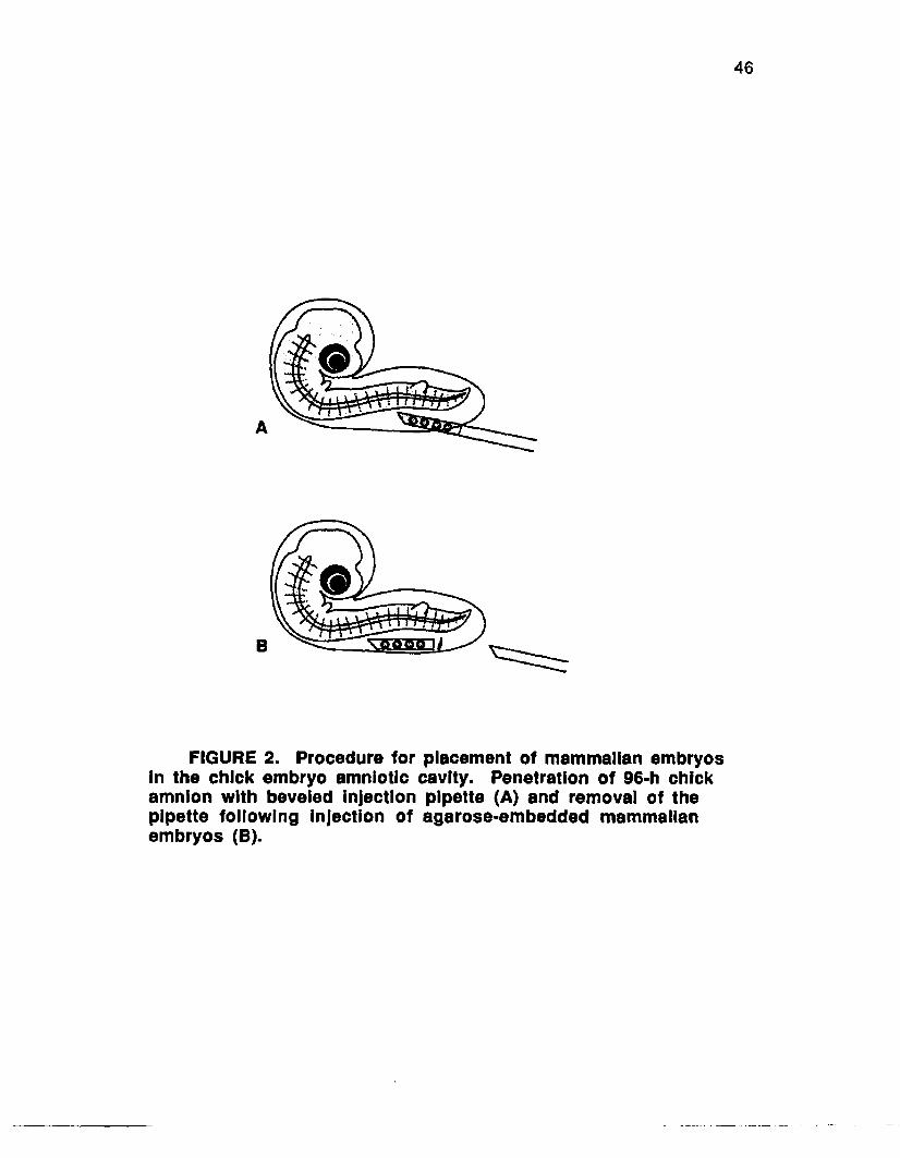
46
©
B
FIGURE 2. Procedure for placement of mammalian embryos in the chick embryo amniotic cavity. Penetration of 96-h chick amnion with beveled injection pipette (A) and removal of the pipette following injection of agarose-embedded mammalian embryos (B).
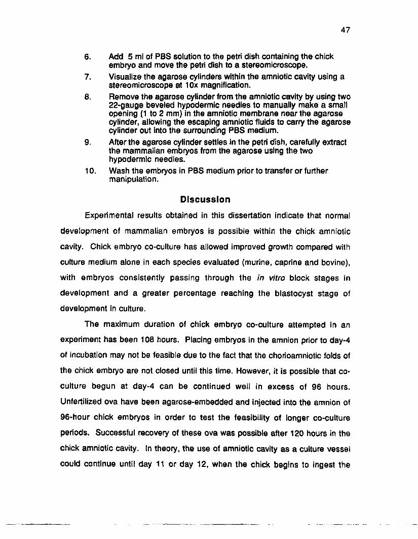
47
6. Add 5 ml of PBS solution to the petri dish containing the chick embryo and move the petri dish to a stereomicroscope.
7. Visualize the agarose cylinders within the amniotic cavity using a stereomicroscope at 10x magnification.
8. Remove the agarose cylinder from the amniotic cavity by using two 22-gauge beveled hypodermic needles to manually make a small opening (1 to 2 mm) in the amniotic membrane near the agarose cylinder, allowing the escaping amniotic fluids to carry the agarose cylinder out into the surrounding PBS medium.
9. After the agarose cylinder settles in the petri dish, carefully extract the mammalian embryos from the agarose using the two hypodermic needles.
10. Wash the embryos in PBS medium prior to transfer or further manipulation.
Discussion
Experimental results obtained in this dissertation indicate that normal
development of mammalian embryos is possible within the chick amniotic
cavity. Chick embryo co-culture has allowed improved growth compared with
culture medium alone in each species evaluated (murine, caprine and bovine),
with embryos consistently passing through the in vitro block stages in
development and a greater percentage reaching the blastocyst stage of
development in culture.
The maximum duration of chick embryo co-culture attempted in an
experiment has been 108 hours. Placing embryos in the amnion prior to day-4
of incubation may not be feasible due to the fact that the chorioamniotic folds of
the chick embryo are not closed until this time. However, it is possible that co
culture begun at day-4 can be continued well in excess of 96 hours.
Unfertilized ova have been agarose-embedded and injected into the amnion of
96-hour chick embryos in order to test the feasibility of longer co-culture
periods. Successful recovery of these ova was possible after 120 hours in the
chick amniotic cavity. In theory, the use of amniotic cavity as a culture vessel
could continue until day 11 or day 12, when the chick begins to ingest the

48
amniotic fluid (Romanoff, 1960).
Since the amniotic cavity must be isolated from the remainder of the egg
contents intact, the increased size of the chick embryo and amniotic cavity at
this stage may result in increased pressure on the amniotic membrane during
isolation. Any resultant rupture of the amniotic membrane would decrease
mammalian embryo recovery rates. Additionally, the amniotic fluids of later-
stage chick embryos may not prove to be as beneficial as those from earlier
stages. Furthermore, recovery methods and properties of the amniotic
environment using these later stages require further evaluation.
To date, the only real drawback to using chick-embryo co-culture is the
inherent mortality rate of in vitro chick embryos. It was noted that a rapid
degeneration of mammalian embryos occurs following chick embryo death,
although the exact cause of this degeneration is not known. Chick embryo
survival rates of 90 to 95% during the co-culture period appear to be as high as
can be expected (Dunn and Boone, 1976). Hatchability for intact chicken eggs
during incubation is typically reported to be 90%, with a peak of mortality
occurring around day 3 (Guilbert, 1974). In the case of the large numbers of
gametes afforded by IVF techniques, the potential benefits of chick-embryo co
culture may offset this loss. Nonetheless, it is clear that a fresh source of quality
fertile eggs is a must.

CHAPTER III
CULTURE OF PRONUCLEAR MURINE EMBRYOS IN THE CHICKEMBRYO AMNION
Introduction
The apparent inability of defined culture media to support the growth of
early mammalian embryos to the blastocyst stage has resulted in the develop*
ment of several alternative culture techniques. These include in vitro co-culture
with "feeder" cells (Cole and Paul, 1965), fibroblast monolayers (Kuzan and
Wright, 1982; Voelkel et al., 1985} or trophoblastic vesicles (Camous et a l,
1984; Heyman et a l, 1987; Pool et a l, 1988) and in vivo culture in the ligated
oviducts of sheep (Willadsen, 1979). Recent successes in producing in vitro
fertilized bovine embryos (Lu et a l, 1987), pronuclear gene injection (Hammer
© fa /.,1985) and nuclear transplantation (Willadsen, 1986; Robl et a l, 1987)
have increased the importance of early embryo culture. A technique has been
developed that allows preimplantation mammalian embryos to be introduced
into the amniotic cavity of a developing chick embryo and subsequently
recovered after 72 to 96 hours of culture.
Chick embryo extract (CEE) was among the first factors used to stimulate
the growth of mammalian cells in culture. Carrel (1913) found that extract of
chick embryos increased the growth of canine connective tissue at least three
fold in vitro. In later studies with avian tissues, Willmer and Jacoby (1936)
showed that CEE had the ability to stimulate development of cells that had
ceased to grow in culture, with the rate of proliferation proportional to the con
centration of CEE. New and Stein (1964) cultured post-implantation mouse
embryos on plasma clots containing either mouse embryo extract or CEE and
noted that CEE was capable of promoting the in vitro growth of mouse embryos
at a rate similar to that of mouse embryo extract.
49

50
Materials and Methods
Preparation of In Vitro Chick Embryos
Fertilized chicken eggs were collected from domestic White Leghorn
hens on the day of oviposition and stored at 10° C until incubation. Four days
prior to the collection of mammalian embryos, eggs were removed from cold
storage, rinsed with a 70% ethanol solution and placed in a 37.5° C commercial
egg incubator (75 cm x 75 cm), which automatically rotated the eggs 60° every
20 min to prevent adhesion of egg contents to the shell membrane. After 72
hours of incubation, the eggs were again rinsed with a 70% ethanol solution
and allowed to air dry. The eggs were then coated with a commercial Betadine
solution (Purdue Fredrick Company, Norwald, CT) and allowed to dry in a
horizontal position to allow for appropriate positioning of the chick embryo. All
subsequent preparation of shell-less cultures was performed under a laminar
flow hood to minimize the potential for contamination.
The original procedure for shell-less incubation of developing chick
embryos (Dunn, 1974; Dunn and Boone, 1976) was modified as follows for use
as a mammalian embryo co-culture system. To remove the egg contents, two
pieces of medical adhesive tape stretched across the jaws of a pair of 200 mm
surgical retractors were used to affix the egg in place at each pole. A crack was
made by striking the area of shell between the pieces of tape against the rim of
a sterile beaker. The cracked egg was then held above a piece of cellophane
kitchen wrap (Saran®, Dow Chemical, Midland, Ml) covering a 100 ml plastic
embryo collection bowl (Veterinary Concepts, Spring Valley, W l), and the egg
contents were released by opening the spreaders. The cellophane wrap was
folded around the rim of the collection bowl and any excess was trimmed away.
The bowl containing the egg contents was then placed in a second bowl to
secure the cellophane wrap and loosely capped with a plastic lid (Veterinary

51
Concepts, Spring Valley, W l). This shell-less culture system was then
maintained in a 37° C incubator with a 2% CO 2 in air atmosphere for an
additional 24 hours prior to introduction of the mammalian embryos.
Injection of Agarose-Embedded Embryos Into the Chick Amnion
The procedure for embedding early-stage embryos in agarose was
modified from that used by Willadsen (1979) for micromanipulated ovine
embryos prior to culture in the ligated oviducts of sheep. Since recovery rates
of non-embedded embryos from the chick embryo amnion were typically less
than 50% during a preliminary study, agarose embedding was implemented in
an attempt to increase embryo recovery rates in the present experiment. Low-
melting point, electrophoresis-grade agarose (Catalog No. 5517UA; Bethesda
Research Laboratories, Gathersburg, MD) was added to 1 ml of sterile
Dulbecco's phosphate-buffered saline (PBS: Gibco, Grand Island, NY) (pH 7.2,
270 mOsm) at a concentration of 1.5% and dissolved by warming to 75° C for
10 minutes. Dissolved agarose was filtered through a .2 pm acrodisc and
cooled to 50° C, then 10 pi of an antibiotic-antimycotic solution containing 100
units of penicillin, 100 pg streptomycin, and .25 pg of amphotericin-B (Gibco,
Grand Island, NY) was added, and the agarose solution was maintained in a
50° C water bath.
Immediately prior to embedding, a 50 pi drop of agarose was pipetted
into a sterile petri dish and moved to a warming hood where the embryos were
held in either N-2-hydroxyethylpiperazine-N'-2-ethanesulfonic acid (HEPES)-
buffered Whitten’s medium (murine embryos) or a PBS holding medium (bovine
and caprine embryos) at 35° C. One to four embryos were transferred to each
50 pi agarose droplet. As the embryos were added, the droplets were stirred to
dilute the holding medium (with the embryos) in the agarose. Embryos in the

52
agarose solution were then aspirated into a beveled glass pipette (200 pm I.D.)
and cooled to 25° C for 1 minute to allow gelling of the agarose.
Pipettes were prepared by heating 1 mm o.d. borosilicate glass capillary
tubes over a microburner and drawing them out to an o.d. of 250 pm. Pipettes
were beveled on a microgrinder at an angle of 45° to enable controlled
puncture of the chick amniotic membrane. An in vitro chick embryo at the
appropriate developmental stage for injection (96 hours of incubation) was
removed from the incubator as the agarose began to congeal. The lid was
removed from the bowl containing the shell-less chick embryo under a warming
hood and the amniotic membrane visualized (10X) using a Zeiss stereo
microscope (Model M-3, Thornwood, NY) equipped with an overhead light
source. The amniotic membrane was manually pierced using the beveled
pipette containing agarose-embedded embryos. When puncture of the amniotic
membrane was accomplished, the embedded embryos were expelled from the
pipette, and the pipette was carefully withdrawn. After introduction of
embedded embryos into the amniotic cavity, the in vitro chick embryos were
incubated an additional 72 to 96 hours at 37° C in an atmosphere of 2% CO2 in
air.
Mammalian Embryo Recovery
In vitro chick embryos were individually removed from incubation after 72
hours of mammalian embryo (murine) co-culture, and the plastic lids were
removed. The posterior portion of the chorioallantoic membrane was carefully
lifted and drawn anteriorly over the chick embryo. A sterile plastic spoon was
then used to gently lift the chick and amnion from the rest of the egg contents.
Extra-embryonic membranes were trimmed away using a pair of surgical
scissors, and the amnion containing the chick embryo was placed in a sterile

53
petri dish and rinsed twice with 5 ml of PBS with 1% heat-treated fetal bovine
serum (FBS) and 1% Gibco antibiotic-anti mycotic solution (Ab-Am). After
rinsing, 5 ml of PBS solution was again added to the chick embryo, and the
petri dish was then evaluated under a stereomicroscope. Agarose cylinders
containing the mammalian embryos could be visualized within the amniotic
cavity using a stereomicroscope at 10X magnification.
The agarose cylinder was removed from the amniotic cavity by using two
22-gauge beveled hypodermic needles to manually make a small opening (1 to
2 mm) in the amniotic membrane near the agarose cylinder. The escaping
amniotic fluids then carried the agarose cylinder out into the surrounding PBS
medium. When the agarose cylinder settled in the petri dish, the mammalian
embryos were carefully extracted from the agarose using the same two
hypodermic needles. Embryos were then rinsed in PBS holding medium prior
to further culture.
Experimental Design
Donor mice (21-day C57/J and Fi SJL/JxC57 females) were super
ovulated with 5 IU of pregnant mare serum gonadotropin (PMSG) followed at 48
hours with 5 IU human chorionic gonadotropin (hCG) and placed with either
C57 or SJL/J males. The F-i females were crossed back to SJL/J males since
SJL/J embryos characteristically show a two-cell in vitro developmental block.
The cumulus ceils were removed from the embryos (collected 26 hours after
HCG) by treatment with 150 lU/ml hyaluronidase in HEPES-buffered Whitten's
medium (Whitten and Biggers, 1968).
Embryos were evaluated for the presence of pronuclei and then
randomly and equally allotted by parental strain to either treatment or control
groups (Figure 3). Embryos in Treatment A were embedded in agarose

Experimental DesignParental Strain
C57 x C57
F1 (SJL/J X C57) x SJL/J
Figure 3.
Treatment A
Ham's F-1024 48 72
Time, Hours
96 120
Treatment BWhitten's Medium (Control) Ham's F-10 Medium
24 48
Time, Hours
72 96 120
Treatment A
24 48 72
Time, Hours
96 120
Treatment BHam's M O r " \| Whitten's Medium (Control)
24 48
Time, Hours
72 96 120
Co-Culture of Murine Embryos in the Chick Embryo Amniotic Cavity

55
cylinders and injected into the amniotic cavity of a 96-hour incubated chick
embryo, as previously described. Following injection, the culture system was
placed in a 37° C incubator containing 2% CO 2 in air. Control embryos
(Treatment B) were cultured in Whitten's medium with 1% Ab-Am in a 5% CO2
incubator at 37° C. After 72 hours of culture, the agarose cylinders were
recovered from the chick amniotic cavity, and the late morula- to blastocyst-
stage embryos were carefully removed from each cylinder. Embryos from
Treatments A and B were then placed in Ham's F-10 medium (Gibco, Grand
Island, NY) with 10% FBS and 1% Ab-Am (HF-10) and cultured at 37° C in a 5%
C O 2 atmosphere in air for an additional 48 hours to evaluate in vitro
development.
Since mortality of shell-less chick embryos maintained in incubation
typically occurs (Rowlett and Simkiss, 1987), like-quality pronuclear mouse
embryos (collected from corresponding donor females) were used to replace
embryos lost if chick embryo mortality occurred in Treatment A during the co
culture period. Likewise, if contamination of individual culture wells used for the
medium-only group (Treatment B) occurred, like-quality embryos were also
used for replacement. This was done to evaluate the growth and development
of the mammalian embryos across functional culture systems.
Statistical Analysis
Chi-square analysis using a contingency table with continuity correction
(Pearson and Hartley, 1954) was used to compare the mean number of
mammalian embryos with complete blastocoel cavity development (expanded
blastocyst) and the mean number of embryos emerging from a rent in the zona
pellucida (hatching blastocyst) in treatment groups during and following culture
in Treatments A and B.

56
Results
Of the 100 pronuclear mouse embryos embedded and placed in the
amniotic cavities of chick embryos, 84% were successfully recovered for culture
in HF-10 medium. Loss of embryos occurred during recovery or was due to
chick embryo mortality. Chick embryo mortality was the primary reason for the
loss, 5 of the 35 chick embryos (14%) injected did not survive shell-less
incubation. Chick embryo mortality appeared to be evenly distributed over the
72-hour culture period.
Table 2. The Number and Percent of Murine Blastocysts that Developed Following Culture in the Chick Amnion or Whitten's Control Medium
Parentalstrain Treatment
No./group
Expanded blastocysts (%)
Hatching blastocysts (%)
C57/J A 42 38 (90) 24a (57)x C57/J B
(control)42 33 (78) 11b (26)
F i (SJL/JxC57) A 42 39a (93) 27a (64)x SJL/J B
(control)42 15b (36) 4b (10)
a,b Different superscripts in the same column within strain are different (P<.01)
Significantly more hatched blastocysts resulted from mouse embryos
cultured in the chick-embryo culture system both within strains of mice and
overall for treatments when compared with those from the control culture system
(P<.01) (Table 1). There was not a significant difference in the number of
expanded blastocysts obtained using the C57/J embryos, with 90% (38/42) of
the chick embryo treatment (Treatment A) expanding and 78% (33/42) of the
Whitten's medium treatment (Treatment B) expanding . Among the Fi (S jlu xC 57 )
x SJL/J embryos, however, there were significantly more expanded blastocysts
obtained using chick embryo co-culture, with 93% (39/42) expanding

57
(Treatment A) compared with 36% (15/42) in the Whitten's control medium
reaching expanded blastocysts in culture (Treatment B).
In terms of hatching blastocysts, there were significantly more hatched
blastocysts obtained from both strains of embryos when cultured in the chick
embryo amnion (Treatment A), with 57% (24/42) and 64% (27/42) hatching
among C57 x C57 and Fi (s jl /J x c s 7 ) x SJL/J embryos, respectively, when
compared with 26% (11/42) and 10% (4/42) hatching among the control
embryos (Treatment B).
Discussion
These results indicate that factors present in the amniotic cavity of
developing chick embryos are capable of supporting the growth of early murine
embryos. Since the living chick embryo actively regulates the pH, osmolarity,
and O 2 content of its own environment, this may make it a more stable
physiological system for mammalian embryo development than existing in vitro
embryo culture systems. In addition, the active synthesis of growth factors
associated with the chick embryo may provide a stimulus to growth and
development not obtainable in conventional culture systems that require
supplementation with active serum or other growth factors. The growth factors
associated with the highly regulated mechanisms of early chick embryo
development may also prove to be more consistent in their growth promoting
effects than those found in processed mammalian serum. Sirard and Lambert
(1985) have shown that identically prepared batches of bovine serum from
different animals gave different results in their ability to promote cleavage of
four-cell bovine embryos. Different batches of bovine serum albumin (BSA)
from the same supplier have also been shown to have different growth
promoting effects on mammalian embryos in vitro. Kane (1983) has reported

58
that rabbit morulae cultured to the blastocyst stage in medium supplemented
with 1.5% BSA from one batch had more than twice as many cells as morulae
cultured in the same medium supplemented with a different batch of BSA.
Water purity has been shown to be critical for the maintenance of embryo
viability in vitro. Whitting ham (1977) reported that three-times glass distilled
water was necessary for a high rate of blastocyst formation from two-cell murine
embryos. Abramczuk et al. (1977) showed a beneficial effect on the culture of
one-cell murine embryos when the chelating agent ethylenediaminetetraacetic
acid (EDTA) was added to the culture medium. These results suggest that even
trace impurities can greatly impede the normal development of mammalian
embryos in culture. Gianaroli et a l (1986) considered human amniotic fluid to
be an appropriate embryo culture medium since it is an ultrafiltrate produced in
contact with the developing human fetus. Likewise, chick-embryo amniotic fluid
may be an ultrafiltrate of the avian embryo. In this case, embryotoxic impurities
may be absorbed and filtered by the egg membranes and chick tissues prior to
reaching the amniotic fluid.
Shell-less chick embryo cultures in this study were maintained in the 2%
C O 2 atmosphere described by Dunn and Boone (1976). More recent work
involving the in vitro culture of shell-less chick embryos in this laboratory
indicates that normal development is possible in lower CO 2 concentrations in
atmospheric air. Rowlett and Simkiss (1987) have successfully "hatched"
chicks cultured in atmospheric air by replacing the plastic bowl with a surrogate
eggshell. The apparent ability of the in vitro chick embryo to grow in a warm,
moist atmosphere that does not require careful monitoring of C O 2
concentrations may make chick embryo co-culture a viable option in situations
where C0 2 incubators are not available.
In this experiment, embryos were not recovered from chicks that died

59
during the co-culture interval. In previous trials, it was noted that a rapid
degeneration of mammalian embryos occurred following chick embryo death.
The inherent mortality of developing chick embryos appears to be one
drawback in using this procedure. The hatchability of intact fertile chicken eggs
during incubation is reported to be 90%, with a peak in embryo mortality
occurring around day 3 of incubation (Guilbert, 1974). Correspondingly,
Rowlett and Simkiss (1987) have previously reported a 27% loss of chick
embryos between days 4 and 7 when 362 shell-less chick embryos were
maintained in incubation.
In the present study, chick embryo mortality was 14%. This death loss
may be due, in part, to those manipulations associated with shell-less chick
embryo culture. Although the exact cause of mammalian embryo death in the
present experiment is not known, the observed decline in mammalian embryo
viability following chick embryo death does suggest that there is a high rate of
transfer across the agarose cylinder containing the mammalian embryos. The
frequent amniotic contractions observed in the healthy chick embryo (Romanoff,
1952) may have promoted an even distribution of nutrients, growth factors, and
dissolved oxygen within the amniotic fluids surrounding the agarose cylinder.
Further experiments involving the culture and transfer of embryos from
larger mammals must therefore be conducted in order to determine the efficacy
of the chick embryo co-culture system. If this unique chick-embryo culture
system was to prove at least as effective as in vivo culture in the ligated oviducts
of sheep, the relative ease of chick embryo co-culture would make it an
invaluable tool for both research and commercial application.
CHAPTER IVCULTURING TWO- TO EIGHT-CELL CAPRINE EMBRYOS
USING DOMESTIC CHICKEN EGGS
Introduction
Repeatable techniques for producing in vitro-fertilized bovine embryos
have made the early-stage bovine conceptus more accessible to researchers.
This increased accessibility has also increased the need for culture systems
capable of promoting the in vitro growth of early-stage bovine embryos.
Unfortunately, attempts at culturing early-stage mammalian embryos in a
completely defined in vitro environment have been successful only with specific
strains of mouse embryos (Whitten, 1968). Supplementation of the culture
medium with biological fluids (usually serum) is most often required to obtain in
vitro development in the embryos of other mammalian species (see review by
Wright and Bondioli, 1981).
In most cases, optimal in vitro viability in long-term cultures of farm
animal embryos (>48 hours) requires the addition of other living cells to the
culture system (i.e. co-culture). The most frequently used co-culture systems
are monolayers of feeder cells (Cole and Paul, 1965), uterine fibroblasts (Kuzan
and Wright, 1982; Voelkel et al., 1985) or more recently fetal uterine fibroblasts
(Wiemer et al., 1988a,1989a) and oviductal cell preparations (Gandolfi and
Moor, 1987; Rexroad and Powell, 1988; Prichard et al., 1990). Another
procedure involves the microsurgical sectioning of later-stage conceptus
trophectoderm to produce trophoblastic vesicles (Camous et al., 1984; Heyman
et al., 1987; Pool etal., 1988) for co-culture with earlier-stage embryos. In spite
of the uncharacterized nature of such culture systems, they represent the most
effective means of obtaining development of farm animal embryos in vitro.
The need for such culture systems has recently increased with the advent
60

61
of successful in vitro fertilization (IVF) procedures for bovine oocytes. The use
of oocytes obtained from slaughterhouse materials to produce pronuclear-stage
bovine embryos (Lu et al., 1987) has recently reached the level of commercial
application in Ireland. The lack of an effective in vitro culture system for these
early-stage IVF embryos is evidenced by the fact that they were not cultured in
vitro , but rather in vivo in the ligated oviducts of sheep.
A procedure has been developed that allows preimplantation
mammalian embryos to be introduced into the amniotic cavity of a developing
chick embryo and subsequently recovered after 72 to 96 hours of culture. The
amniotic cavity of the living chick embryo has been shown to be a suitable
environment for growth of pronuclear murine embryos through to the blastocyst
stage (Blakewood, et ah, 1988). Although the embryotropic properties of avian
amniotic fluids remain largely undefined, the current need for a practical in vitro
culture system may make shell-less chick embryo co-culture a viable alternative
to those presently in use.
Materials and Methods
Preparation of Shell-less Chick Embryo Cultures
Preparation of shell-less chick embryos was modified from the basic
procedures reported by Dunn (1974) and Dunn and Boone (1976). Fertilized
eggs of domestic White Leghorn hens were collected on the day of oviposition
and stored at 10° C until incubation. Four days prior to mammalian embryo
collection the eggs were rinsed with a 70% ethanol solution and placed in a
Marsh Roll-X automatic incubator (Garden Grove, CA) at 37.5° C that
automatically rotated the eggs 60° at 20-minute intervals. After 72 hours of
incubation, the eggs were prepared for shell removal by rinsing with a 70%
ethanol solution which was allowed to dry before coating the eggs with a

62
Betadine solution (Purdue Fredrick Company, Norwalk, CT). The Betadine
solution was allowed to dry with the eggs in a horizontal position to achieve
correct positioning of the chick embryo prior to shell removal. Eggs were then
individually moved to a laminar flow hood in order to maintain sterility while
removing the shell.
The procedure and cumbersome shell removal apparatus used by Dunn
(1974) were eliminated by simply affixing the egg to two pieces of surgical tape
stretched across the jaws of a pair of 200 mm surgical retractors. By cracking
the area of shell between the pieces of tape against the rim of a sterile glass
beaker the egg contents were released by opening the retractors. Egg contents
were deposited into a 100 ml plastic embryo collection bowl (Veterinary
Concepts, Spring Valley, Wl) that was covered with cellophane kitchen wrap
(Saran®, Dow Chemical, Midland, Ml). The excess plastic wrap was trimmed
away and the bowl containing the egg contents was placed in a second bowl
and loosely capped with a sterile plastic lid (Veterinary Concepts). This culture
system was maintained in a 37° C incubator with a 2% CO2 atmosphere for an
additional 24 hours prior to introduction of the mammalian embryos.
Preparation of Fetal Monolayers
Monolayers of fetal bovine uterine fibroblasts were prepared as
previously described by W iemer et al. (1989b). Immediately after adult
slaughter, the uterus of a 270-day-old bovine fetus was removed and
transported to the laboratory in cooled Hank's Balanced Salt solution
supplemented with 500 units penicillin and 500 pg streptomycin per ml (HBSS).
At the laboratory, the fetal uterus was placed under a laminar flow hood
and the lumen exposed via a longitudinal incision. Endometrial tissue samples
were aseptically dissected away from the myometrium and placed in a sterile

63
petri dish with HBSS. Samples were washed with HBSS and sectioned into »2
mm cubes. Three to four of these cubes were then placed into each 25 cm2
plastic tissue culture flask containing Ham's F-10 medium (Gibco, Grand Island,
NY) with 10% Gibco heat treated fetal bovine serum and 1% Gibco antibiotic-
antimycotic (containing 100 units of penicillin, 100 pg of streptomycin and .25 g
of amphotericin-B per ml of flushing medium) (HF-10). During the period of
explant culture, the HF-10 medium was supplemented with an additional 4%
antibiotic-antimycotic (Ab-Am).
Flasks were incubated at 37° C in a humidified 5% CO2 in air atmo
sphere for 9 days. At 72-hour intervals during this period, 50% of the HF-10
medium was replaced in each flask. After 9 days of incubation, explants were
removed and fibroblasts which had attached to the plastic strata were allowed
to proliferate for an additional 5 to 7 days to establish confluent monolayers.
Monolayered flasks were then trypsinized (.025%), and cells passed to new
flasks at a 1:2 dilution ratio. Confluent monolayers which resulted after 4 to 7
days of incubation in each new flask were again trypsinized (.025%) and
passed in this manner. Following seven passages of the uterine fibroblast
cultures, all cells were frozen and stored in liquid nitrogen (-196° C) prior to use
in the embryo co-culture system.
Between 24 and 48 hours before caprine embryo collection, frozen fetal
uterine fibroblasts were thawed, and 1 x 10s viable cells were plated in each
well of a 24-well tissue culture plate (Gibco). The cells were maintained in HF-
10 at 37° C in a humidified atmosphere of 5% C 0 2 in air. Fifty percent of the
HF-10 medium was changed 1 to 2 hours prior to placing the experimental
embryos on the monolayer culture.

64
Preparation of Trophoblastic Vesicles
Conceptus trophoblastic tissue was obtained by surgically flushing the
uterus of a superovulated caprine female 15 days after mating. The doe was
placed in dorsal recumbancy after administration of general anesthesia.
Anesthesia was achieved by administering intramuscularly .11 mg/kg of
xylazine (Rompun®: Miles Laboratories, Shawance, KS) followed 10 minutes
later by 5.5 mg/kg of ketamine hydrochloride (Ketaset®: Bristol Laboratories,
Syracuse, NY). The uterus was exteriorized via a mid-ventral incision and a 10-
gauge Foley catheter was passed through a small incision in the uterine body
and the cuff inflated near the uterine bifurcation. An 18-gauge hypodermic
needle was used to make a small opening 1 cm from the utero-tubal junction.
Dulbecco's phosphate-buffered saline (PBS: Gibco, Grand Island, NY) with 2%
heat treated fetal bovine serum (FBS) and 1% Ab-Am flushing medium was
passed through the uterus and recovered in a glass collection bowl.
Elongated embryos were washed and placed in PBS holding medium in
a 60 mm petri dish. A fragment of a double-edge razor blade held in the jaws of
a small pair of hemostats was used to cut the elongated embryos into 1 to 2
mm2 sections as previously described by Rorie et al. (1987a). Sections were
cultured in Minimum Essential Medium (MEM) with 10% FBS and 1% Ab-Am for
48 hours to allow for vesicle formation. Vesicles were then frozen (8 to 10
vesicles per .25 ml straw) in 1.5 M glycerol using a standard bovine embryo
freezing curve used in this laboratory (Rorie et al., 1987b) and then stored in
liquid nitrogen. Vesicles were thawed 24 to 48 hours prior to use in co-culture
and allowed to re-expand in HF-10 medium. Only those vesicles that re
expanded were placed in 24-well plates (one vesicle/well) with HF-10 for co
culture.

65
Experimental Animals
Embryos used in this study were obtained from 30 mixed-breed dairy
goats that were superovulated and collected during the fall breeding season
(October and November). All does were in good body condition and had an
mean body weight of 38.9 kg (range = 24.9 to 59 kg). The wide range of body
weights was due to differing body sizes of the Nubian, Toggenberg, Saanen
and French Alpine genetics represented in these crossbred females. The does
were maintained on mixed pastures of native and bermudagrass during the day
and fed .22 kg/head/day each evening of a total mixed ration containing 12.5%
crude protein, 3.1% fat, 12.7% fiber and 5.1% ash.
Superovulation
Donor does were synchronized with a 6 mg norgestomet implant
( S y n c r o m a t e - B ® : CEVA Laboratories, Overland Park, KS) placed
subcutaneously on the dorsal surface of the ear for 14 days as previously
described by Pendelton et at. (1986a). Beginning 2 days prior to implant
removal, follicle stimulating hormone (FSH-P®: Schering, Omaha, NE) was
administered intramuscularly twice daily in descending doses (4, 3, 2 and 1 mg)
to stimulate follicular growth (total dose = 20 mg/doe).
Treated does were placed with a fertile male and observed for signs of
behavioral estrus twice daily beginning 12 hours after implant removal. Estrual
females were mated with two fertile males at 12-hour intervals until they were
no longer in standing estrus.
Following implant removal, 27 of 30 does (90%) exhibited standing
estrus when placed with fertile bucks. The mean (±SE) duration of standing
estrus was 38.9 ± 1.6 hours (range = 24.9 to 59 hours). All does exhibiting
signs of behavioral estrus were mated by one or more fertile males.

66
Embryo Collection
Embryos were surgically collected from donor animals 36 hours after
they would no longer stand to be mated (day 3 of the estrous cycle; onset of
estrus « day 0). Prior to surgery, feed and water were withheld from each doe
for 24 hours. Does were anesthetized by administering intramuscularly .11
mg/kg of xylazine (Rompun®: Miles Laboratories) followed 10 minutes later by
5.5 mg/kg of ketamine hydrochloride (Ketaset®: Bristol Laboratories). Females
were then placed in dorsal recumbancy and the reproductive tract exteriorized
via a mid-ventral incision just anterior to the mammary tissue. The number of
follicles and corpora lutea on each ovary were then counted and recorded.
A laboratory animal intubation needle was then inserted into the
fimbriated end of the oviduct and embryos were gently flushed through the
oviduct towards the utero-tubal junction by applying positive pressure to an
attached 20 ml syringe. Embryos were then recovered from the lumen of the
distal end of the uterine horn where a small diameter (3-5 mm) beveled-end
glass canula had been inserted to harvest embryos using a glass collection
bowl.
The flushing medium consisted of Dulbecco’s PBS (Gibco) with 2% FBS
and 1% Ab-Am. Following harvesting of the embryos, the glass collection bowls
were carefully searched at 20x magnification under a Zeiss stereomicroscope
(Thornwood, NY). A total of 211 fertilized and unfertilized ova were
successfully collected from 21 of 27 mated does (77.7%) resulting in a mean of
10 ova per donor. Of the 211 fertilized and unfertilized ova collected, 162
(76.8%) were at the two- to eight-cell developmental stage and were assessed
as being of good quality based on caprine embryo morphology and quality
grade scores (1 = excellent, 2 = good, 3 - fair and 4 = poor) as previously
outlined by Pendleton etal. (1986b). The harvested embryos were then placed

67
into a modified PBS holding medium with 10% FBS as described by Pool et at.
(1988) and held at 37° C until allotment to treatments.
Experimental Design
Experiment I
Embryos (quality grades 1 = excellent and 2 = good) from donors were
randomly assigned to four treatments in the first experiment using caprine
embryos (EXP-I) (n = 80). In EXP-i, embryos allotted to Treatment A (control)
were cultured in HF-10, embryos in Treatment B were placed on a bovine fetal
uterine fibroblast monolayer in HF-10 and embryos in Treatment C were
agarose embedded and injected into the amniotic cavity of a day-4 chick
embryo. Embryos allotted to Treatment D were co-cultured with frozen-thawed
caprine trophoblastic vesicles in HF-10 (Figure 4).
Experiment II
Embryos (quality grades 1 = excellent and 2 * good) from donors were
randomly assigned to three treatments in the second experiment (EXP-II) (n =
66). In EXP-II, embryos allotted to Treatment A (control) were cultured in HF-10,
embryos in Treatment B were placed on a bovine fetal uterine fibroblast
monolayer in HF-10 and embryos in Treatment C were agarose embedded and
injected into the amniotic cavity of a day-4 chick embryo (Figure 5). Co-culture
with caprine trophoblastic vesicles was not included in EXP-II.
Experimental Procedure
EXP-I and II differed in that chick embryo co-culture was maintained for
72 hours in EXP-I, but was extended to 96 hours in EXP-II. Also, the monolayer
co-culture was limited to 96 hours in EXP-II; whereas, embryos in EXP-I
remained on the monolayer for the duration of the experiment. Control,

Experimental Design
Treatment AHF-10 Medium (Control)
24 48 72 96 120 144
Time, Hours
Treatment B
48 72 96
Time, Hours
120 144
Treatment CHam’s F-10 Medium
24 48 72 96
Time, Hours
120 144
Treatment D
) 24 48 72 96 120 144
Time, Hours
Figure 4. Co-Culture of Caprine Embryos for 72 Hours in the Chick Embryo Amniotic Cavity

Experimental DesignTreatment AHam's F-10 Medium (Control)
24 48 72 96 120 144
Time, Hours
Treatment BHam's F-10 Medium""]
120 144
Time, Hours
Treatment C
S9S144
Time, Hours
Figure 5. Co-Culture of Caprine Embryos for 96 Hours in the Chick Embryo Amniotic Cavity

70
monolayer and trophoblastic vesicle cultures were maintained in 24-well tissue
culture plates incubated at 37° C in a humidified atmosphere of 5% CO2 in air
and chick embryo co-cultures were incubated (37° C) in a 2% CO2 atmosphere.
After removal from the monolayer (EXP-II 96 hours) or the chick amnion (EXP-I
72 hours, EXP-II 96 hours), the embryos were placed in HF-10 for further
incubation at 37° C in a 5% CO2 atmosphere in air. During this culture interval,
the rate of expanded and hatched blastocyst formation was evaluated at 12-
hour intervals for an additional 72 hours. If embryos in Treatment C were lost
during co-culture (due to chick embryo mortality) or during the embryo recovery
process, like quality two- to eight-cell embryos were replaced in the chick
amnion culture system from embryo collections of subsequent donor does in
order to maintain an equal number of embryos across treatment groups. Eleven
embryos were replaced in this fashion during EXP-I, however, only four
embryos had to be replaced in EXP-II. This decrease in embryo loss was
attributed to improved injection and recovery techniques as the experiments
progressed.
Agarose Embedding
Two to four embryos were placed in a liquid solution of low-melting
agarose (1.5% agarose in Dulbecco's PBS with 1% Ab-Am) at 37° C. The
embryos in the agarose solution were then aspirated into a beveled injection
pipette and the agarose within the pipette was allowed to gel at room
temperature, thereby embedding the embryos. The beveled tip of this pipette
was then used to penetrate the amniotic membrane of the shell-less 96-hour
chick embryo, and the agarose cylinder containing the embedded caprine
embryos was expelled into the chick amniotic cavity. The chick embryo co
culture system was placed in an incubator at 37° C with 2% CO2 in an

71
atmosphere of humidified air.
After 72 or 96 hours of co-culture (Treatment C), the amniotic cavity
containing both mammalian and chick embryos was dissected away from the
remainder of the egg contents within the shell-less system and placed in
Dulbecco's PBS with 1% FBS and 1% Ab-Am. By renting the amnion with a
pair of 22-gauge hypodermic needles, the agarose cylinder containing the
caprine embryos was allowed to escape into the surrounding PBS medium.
While still in the PBS medium, the caprine embryos were carefully dissected
from the agarose cylinder using the same pair of 22-gauge hypodermic needles
under a Zeiss stereomicroscope (20X).
Embryo Transfer
Embryos from the first two experiments were not transferred due to the
lack of available recipient does. Donor collections were subsequently
performed in a similar manner from three contemporary does to obtain embryos
for transfer following chick embryo co-culture. Fourteen four-cell embryos were
agarose embedded and similarly incubated in the chick embryo amnion. After
72 hours of co-culture, 12 good quality morulae (quality grades 1 and 2) were
recovered from chick embryo co-culture. Two to four morulae were surgically
transferred to the uterine horn ipsilateral to the corpus luteum of each of four
synchronized (± 24 hours) recipient does.
Statistical Analysis
Chi-square analysis using a contingency table and continuity correction
(Pearson and Hartley, 1954) was used to compare the number of two- to eight
cell goat embryos reaching the expanded blastocyst stage and the number
hatching when cultured in vitro using treatment and control culture methods.

72
Results
Experiment I
In EXP-I, none of the oviductal-stage goat embryos developed into
blastocysts when cultured in the control medium alone. Co-culture with caprine
trophoblastic vesicles also failed to produce blastocysts. However, in EXP-I
both chick embryo and monolayer co-culture treatments resulted in significantly
more blastocysts than did the control or trophoblastic vesicle treatments (Table
1). Development of early-stage goat embryos to the expanded blastocyst stage
at rates of 70% and 50% were obtained using chick embryo and monolayer co
culture, respectively.
Table 3. In Vitro Development of Early-Stage Caprine Embryos in Four Culture Systems (Experiment I)
Treatment No./groupExpanded
blastocyst (%)Hatched
blastocyst (%)
A (control) 20 03 (0) 0a (0)B (monolayer) 20 10b (50) 9b (45)C (chick embryo) 20 14b (70) 11b (55)D (trophoblastic vesicles) 20 0a (0) 0a (0)
a'b Different superscripts in the same column are significantly different (P<.01)
Experiment II
In EXP-II, culture of two- to eight-cell caprine embryos in control medium
again failed to produce blastocysts. When the duration of chick embryo co
culture was increased to 96 hours (EXP-II), an even greater percentage of the
caprine embryos reached the expanded and hatched blastocyst stage than in
EXP-I. Development to the expanded blastocyst stage in chick embryo co
culture reached 86% in this experiment.
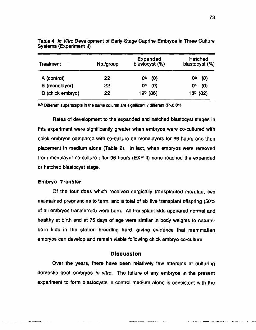
73
Table 4. In Vitro Development of Early-Stage Caprine Embryos in Three Culture Systems (Experiment II)
Treatment No./groupExpanded
blastocyst (%)Hatched
blastocyst (%)
A (control) 22 0® (0) 0® (0)B (monolayer) 22 0® (0) 0® (0)C (chick embryo) 22 19b (86) 18b (82)
a-b Different superscripts in the same column are significantly different (P<0.01)
Rates of development to the expanded and hatched blastocyst stages in
this experiment were significantly greater when embryos were co-cultured with
chick embryos compared with co-cutture on monolayers for 96 hours and then
placement in medium alone (Table 2). In fact, when embryos were removed
from monolayer co-culture after 96 hours (EXP-II) none reached the expanded
or hatched blastocyst stage.
Embryo Transfer
Of the four does which received surgically transplanted morulae, two
maintained pregnancies to term, and a total of six live transplant offspring (50%
of all embryos transferred) were born. All transplant kids appeared normal and
healthy at birth and at 75 days of age were similar in body weights to natural-
born kids in the station breeding herd, giving evidence that mammalian
embryos can develop and remain viable following chick embryo co-culture.
Discussion
Over the years, there have been relatively few attempts at culturing
domestic goat embryos in vitro. The failure of any embryos in the present
experiment to form blastocysts in control medium alone is consistent with the

74
unpublished results mentioned by Wright and Bondioli in their 1981 review
article, in which none of 89 one- to eight-cell caprine embryos developed
beyond the morula stage when cultured in Ham's F-10 with 10% FBS in a 5%
C 0 2 atmosphere. Rabbit oviducts have been used to successfully store goat
embryos in vivo for short periods prior to transfer (Agrawal et al., 1983). It
appears that goat embryos can develop in medium alone under more defined
conditions. McLaughlin et al. (1989) have recently obtained blastocysts when
one- to four-cell caprine embryos were cultured in vitro using a modified
synthetic oviduct fluid medium supplemented with either human or fetal calf
serum.
The embryos of the domestic sheep have been used more frequently
than goat embryos in attempts to develop in vitro embryo culture systems.
Rexroad and Powell (1988) have reported that co-culture of one- to eight-cell
sheep embryos with oviductal cells resulted in higher rates of cleavage than
with medium alone, and concluded that Ham's F-10 culture medium was
inadequate for the development of early-stage ovine embryos. In addition,
Rexroad and Powell noted that trophoblastic vesicles produced from day-14
ovine embryos were not beneficial for early-stage ovine embryos when placed
in co-culture. The frozen-thawed caprine trophoblastic vesicles used in EXP-I of
the present study were produced from day-15 embryos, and were apparently
unable to stimulate blastocyst development in early-stage caprine embryos. It
has been proposed by Y. Menezo (personnel communication) that the
embryotropic effects of bovine trophoblastic vesicles occur only when vesicles
are produced from trophoblastic tissues originating from embryos ranging
between 12 and 14 days of age. The stage of conceptus development may
account for the lack of success with the caprine trophoblastic-vesicle co-culture
in our experiment.

75
The bovine fetal uterine fibroblast (BFUF) cultures used in the
present experiment have been successfully used to co-culture bovine morulae
to the hatching blastocyst stage in this laboratory (Wiemer et at., 1 9 8 7 ,1989a).
In addition, co-culture on BFUF cells has proven beneficial to both equine
embryos (Wiemer et al., 1988b) and one-cell human zygotes (Wiemer et al.,
1989b) cultured in vitro, with pregnancies obtained in both species.
Numbers of expanded and hatched blastocysts which developed in vitro
following the removal of embryos from the chick embryo amnion were used for
comparing culture treatments in the present studies. Development to earlier
stages could not be assessed due to the fact that the caprine embryos placed in
the chick embryo amnion were not visible during chick embryo co-culture. The
co-culture of early-stage caprine embryos on monolayers of BFUF cells in HF-
10 did allow for successful development to the expanded and hatched
blastocyst stages, however, the rate of development was somewhat behind
those reported for caprine embryos developing in vivo (Amoroso et al., 1942).
Embryos which had been cultured in the chick embryo amnion in this study
generally reached the expanded blastocyst stage 24 hours earlier than those
co-cultured on a BFUF monolayer; thus approaching a more normal rate of
embryonic development.
Perhaps the most surprising finding was that embryos co-cultured on
monolayers for 96 hours in EXP-II were not capable of continued development
when removed from the monolayer for culture in HF-10 medium alone. This is
in contrast to the continued rate of development noted when embryos were
cultured in HF-10 medium following either 72 or 96 hours of culture in the chick
embryo amnion. Although blastocyst development occurred on the monolayer
co-cultures in EXP-I, the dramatic loss of in vitro viability observed when
embryos were removed from monolayers in EXP-II suggests less than optimal

76
development using this particular co-culture procedure. Although more
beneficial than culture in medium alone, the BFUF cells may not be the best
choice for co-culture of oviductal-stage embryos. Gandolfi and Moor (1987)
obtained 42% expanded blastocysts when pronuclear ovine embryos were
cultured for 6 days on oviductal cells, but only 4.5% resulted in expanded
blastocysts when these early-stage embryos were cultured on fibroblast
monolayers.
Given the poor development rates noted in the HF-10 control and in HF-
10 culture following monolayer co-culture (EXP-II), the high rates of blastocyst
formation obtained in HF-10 following culture in the chick embryo amnion are
promising. The ability of the chick embryo co-culture system to stimulate the
development of early-stage mammalian embryos warrants investigation into
potential benefits for bovine in vitro fertilization (IVF). Recent live births resulting
from the immediate transfer of caprine embryos cultured in the chick embryo
amnion for 72 hours at this station are also encouraging.
The use of agarose embedding to facilitate the recovery of mammalian
embryos following their incubation in the chick embryo amnion was carried over
from previous experiments using murine embryos in this laboratory (Blakewood
e ta /., 1988). In a pilot study using unfertilized bovine oocytes, high recovery
rates were obtained without the use of agarose embedding, presumably due to
the larger size of the bovine ova compared with that of the mouse ova. Injecting
unembedded embryos would make this technique more amenable to culture of
embryos obtained from IVF, due to the lengthy culture time needed to produce
morulae and blastocysts. Although it is theoretically possible to maintain chick
embryo co-culture for up to 6 days, the increased size of the chick embryo and
the amniotic cavity may actually decrease mammalian embryo recovery rates.
Therefore, sequential culture periods in two different chick embryo amnions

77
may be necessary for long-term culture of early-stage farm animal embryos.
Sequential culture of farm animal embryos in the amniotic cavities of two
different chick embryos has proven successful in our laboratory. The transfer of
bovine embryos recovered from one chick amnion to a second chick amnion
would be simplified if embryo extraction from agarose and re-embedding in
agarose cylinders could be omitted.
There is currently no definitive culture technique for use with bovine
embryos produced by IVF. In a recent review of successful production of bovine
zygotes by IVF {Leibfried-Rutledge e ta i, 1989), a number of in vivo and in vitro
culture techniques were reviewed. These include in situ culture in the oviducts
of sheep, cows and rabbits, as well as in vitro culture with bovine oviductal or
cumulus cells. The chick embryo amnion should be compared with these
culture systems, particularly the more practical in vitro culture methods. If chick
embryo co-culture proves to be superior to existing in vitro co-culture systems, it
would represent a practical alternative to intermediate culturing of early-stage
embryos in the oviducts of a host female.
CHAPTER VCULTURE OF EARLY STAGE BOVINE MORULAE USING CHICK
EMBRYO AND MONOLAYER CO-CULTURE SYSTEMS
Introduction
With increasingly successful reports of bovine in vitro fertilization (IVF)
procedures in the literature, the development of methodologies for in vitro
culturing early-stage bovine embryos have become important for practical
application of these procedures. The in vitro development of bovine morulae
has been improved by co-culture on monolayers of either bovine uterine
fibroblasts or testicular fibroblasts when compared with culture in conditioned or
fresh medium (Kuzan and Wright, 1982).
Other workers have demonstrated that the in vitro viability of
micromanipulated bovine embryos could be enhanced by co-culture on uterine
fibroblasts (Voelkel et al., 1985). Fetal bovine uterine fibroblast (FBUF)
monolayers have also given successful co-culture results with embryos from
several mammalian species (Wiemer et a/.,1988) including humans (Wiemer et
al., 1989a,b). This system has also proven beneficial for the in vitro culture of
early-stage bovine morulae (Wiemer et al., 1990).
Chick embryo co-culture has also been shown to promote development
in early stage embryos from several mammalian species, including mice
(Blakewood et al., 1988) and goats (Blakewood et al., 1989a). The purpose of
the first experiment in this series was to compare the chick embryo system with
FBUF monolayers to determine if chick embryo co-culture would have beneficial
effects for the culture of early-stage bovine embryos.
The increased success in producing bovine embryos by IVF will also
increase the need to freeze embryos that have developed with in vitro systems.
in vitro culture prior to freezing in liquid nitrogen (LN2 ) has been reported to
78

79
decrease the viability of embryos after thawing (Massip et al., 1984). These
workers reported an additive effect on fetal wastage when mouse embryos were
cultured in vitro prior to freezing. Techakumpu eta l. (1986) have confirmed the
additive detrimental effects of freezing procedures following the in vitro culture
of early-stage rabbit embryos. In this study, 65% of the rabbit embryos
transferred to a recipient female immediately following collection developed into
viable fetuses. This compared with 48% viable fetuses developing in utero
following transfer of embryos that had been cultured in vitro prior to transfer, and
and 43% viable fetuses when embryos were frozen prior to transfer. When
embryos were both in vitro cultured and then frozen prior to transfer, only 19%
developed into viable fetuses in the recipient females.
Chesne et al., (1987) attempted to use a period of in vitro culture to
enhance the viability of bisected bovine embryos prior to freezing. Pregnancy
rates were higher when bisected embryos were cultured in vitro for 4 to 6 hours
prior to freezing (71%) compared with pregnancy rates obtained from embryos
that were frozen immediately following splitting procedures (13%). However,
when the period of pre-freeze in vitro culture was increased to 20 to 24 hours,
pregnancy rates dropped to 25%. These results indicate that the culture of
bovine embryos prior to freezing also results in an additive effect on fetal
wastage.
It has been suggested that a decrease in the post-thaw viability of in vitro
cultured embryos may be due to the spontaneous peroxidation of lipids that can
occur in an in vitro environment (Techakumpu et al.,1986). Such peroxidation
has been measured in mammalian sperm cells after in vitro incubation (Alvarez
and Storey, 1985). Techakumpu e ta l. (1986) have proposed that the low
concentrations of lipids found in in vitro systems may contribute to the peroxida
tion of membrane lipids, which may modify their affinity for cryoprotectants, or

60
cause other biomechanical constraints to freezing.
The second experiment using bovine embryos was carried out to
determine if the co-culture environment of the chick embryo amnion would
enhance freeze-thaw viability of bovine embryos that had developed in co
culture prior to freezing. The in vivo nature of the intact chick embryo amnion
may provide a more suitable lipid environment than in vitro culture systems,
such as monolayer co-culture. The second experiment in this series was
conducted to determine if early stage bovine embryos co-cultured in the chick
embryo amnion could survive the freeze-thaw process at higher rates when
compared to other in vitro culture systems.
Materials and Methods
Collection of Bovine Embryos
Crossbred beef donor cows between days 9 and 14 of their estrous cycle
(estrus = day 0) were superovulated using descending doses of 5,4,3 and 3 mg
of FSH-P® (Schering) injected intramuscularly (IM) twice daily. A single IM
injection of 25 mg of prostaglandin Faa was given to all donor cows 48 hours
before the last FSH injection to promote luteolysis and return to estrus. Donors
were penmated with one or two fertile bulls at standing estrus and again 12
hours later, if still exhibiting estrus. If donor cows would not stand to be mated a
second time, they were inseminated with one unit of frozen-thawed dairy bull
semen.
Precompaction-stage morulae (8- to 16-cell) were nonsurgically
recovered on day 5 to 5.5 post-estrus (estrus s day 0) using Dulbecco's
phosphate-buffered saline with 1% fetal calf serum (FCS) and 1% Gibco
antibiotic-antimycotic (Ab-Am) as a collection medium. Late morula and
blastocyst stage embryos used in Experiment II were collected on day 7

81
following estrus using the same collection medium. All viable appearing
embryos were washed in PBS holding medium (PBS flushing medium
supplemented with 4 g BSA, 1 g glucose and .036 g Na pyruvate/liter) and held
in this medium at 20° C until allotment to culture treatments.
Shell-less Chick Embryo Culture
Preparation of shell-less chick embryos were as previously reported
(Blakewood and Godke, 1989; Blakewood et al., 1989). The procedure for
maintaining the avian embryo in shell-less incubation was modified from the
basic procedures reported by Dunn (1974) and Dunn and Boone (1976). After
the 72-hour incubation of fertile chicken eggs (37° C turning the eggs at 1-hour
intervals) the egg contents were removed from the shell and returned to
incubation in an in vitro system. This culture system was maintained in a 37° C
incubator with a 2% CO 2 atmosphere for an additional 24 hours prior to
introduction of the bovine embryos.
Bovine embryos were agarose-embedded in 250 pm x 6 mm cylinders of
low melting agarose (Bethesda Research Laboratories) prior to injection in both
of these experiments. Embryo recovery from the chick embryo amnion following
co-culture was as previously described.
Preparation of Monolayers for Co-culture
Monolayers of fetal bovine uterine fibroblasts used in EXP I were
prepared as previously described by Wiemer eta l. (1989). Immediately after
slaughter, the uterus of a 270-day-old bovine fetus placed on ice and brought to
the laboratory. Small samples of endometrial tissue (« 2mm) were dissected
away from the myometrium and placed in a 25 cm2 plastic tissue culture flasks
containing Ham's F-10 medium (Gibco, Grand Island, NY) with 10% Gibco heat-

82
treated fetal bovine serum and 1% Gibco antibiotic-antimycotic (containing 100
units of penicillin, 100 ng of streptomycin and .25 g of amphotericin-B per ml of
flushing medium) (HF-10). Flasks were incubated at 37° C in a humidified 5%
C 0 2 in air atmosphere for 14 days to establish confluent monolayers.
Flasks with confluent monolayers were then trypsinized (.025%) and
cells were passed to new flasks at a 1:2 dilution ratio. Confluent monolayers
which resulted after 4 to 7 days of incubation in each new flask were again
trypsinized (.025%) and passed in this manner. Following seven passages of
the uterine fibroblast cultures, all cells were frozen and stored in liquid nitrogen
(-196°C) prior to use in the embryo co-culture system (Wiemer e ta l, 1989a).
Frozen fetal uterine fibroblasts were thawed and returned to culture in 25
cm 2 tissue culture flasks containing Ham's F-10 7 to 14 days prior to the
beginning of this experiment. Re-established monolayers in these flasks were
trypsinized (.025%) 24 to 48 hours prior to bovine embryo collection and 1 x 10⁵
viable cells were plated in each well of a 24-well tissue culture plate (Gibco).
The cells were maintained in HF-10 at 37° C in a humidified atmosphere of 5%
C 0 2 in air. Fifty percent of the HF-10 medium was changed 1 to 2 hours prior to
placing the experimental embryos on the monolayer culture.
The monolayers of caprine uterine epithelial cells used in EXP II were
prepared by trypsinizing the luminal surface of uteri removed from mature,
cycling goat females. Following general anesthesia does were placed in dorsal
recumbancy (Pendleton e ta l, 1986) and the uterus was excised through a mid-
ventral incision . The uterus from each female was washed 3x in Hank's
balanced salt solution containing 5x Ab-Am. The uterine lumen was then rinsed
with .05% trypsin (Trypsin-EDTA; Sigma Co.) in PBS. After rinsing, the distal
ends of the uterine horns were clamped using two pairs of hemostats and the
uterus was inflated with the trypsin-PBS solution. The inflated uterus was

83
incubated for 10 minutes at 37° C, then the uterine horns were drained into a 15
ml conical plastic centrifuge tube (Prichard et at., 1990).
Tissue fragments were resuspended in PBS containing Ab-Am and
centrifuged at 100 x g for 10 minutes. Red blood cells in the pellet were lysed
by resuspending the pellet in 10 ml of sterile deionized water followed 10
seconds later by 10 ml of 1.8% saline. Cells were then washed twice in Ham's
F-12 medium (Gibco, Grand Island, NY) with 10% heat-treated fetal bovine
serum and 1% Gibco antibiotic-anti mycotic (containing 100 units of penicillin,
100 tig of streptomycin and .25 g of amphotericin-B per ml of medium) (HF-12)
and plated in 25 cm2 plastic tissue culture flasks with HF-12. Cell populations
were subpassed five times prior to use for embryo co-culture.
Embryo Freezing and Thawing
In preparation for freezing, embryos were placed in a 10% solution of
glycerol in PBS supplemented with 4 g/liter bovine serum albumin and allowed
to equilibrate for 10 minutes. Embryos were then drawn into .25 ml semen
straws, with one to four embryos occupying the middle column of three columns
of medium drawn in the straw. When the first column of medium reached the
cotton plug at the end of the straw, the opposite end was sealed using a heat
sealer.
Straws were placed in the methanol bath of a cell freezing machine (FTS
BioCool II™) and cooled at a rate of -1.0° C/minute to -7.0° C. At -7.0° C, straws
were seeded, then cooled at a rate of -.3° C/minute to a temperature of -33° C.
After reaching this temperature, straws were plunged in LN2 at -196°C for
storage. Frozen embryos were thawed by removing the straws from LN2 and
allowing the cryoprotective medium to thaw in air at 20° C. After compiete
thawing, the straw contents were released into a sterile 35 mm petri dish and

84
the embryos recovered under a stereomicroscope (20x).
The cryoprotectant was removed from the embryos using a three step
dilution procedure. The embryos were removed from the freezing solution and
passed through decreasing concentrations of glycerol in PBS medium
supplemented with 10.3% sucrose. Embryos were allowed to equilibrate for 5
minutes in each solution, with the concentration of glycerol sequentially
decreasing from 6% to 3% to 0%. Embryos were then washed in PBS holding
medium and returned to in vitro culture for evaluation of post-thaw viability.
Experimental Design
Experiment I
Early stage embryos (day 5 to 5.5) from individual donor cows (n=25)
were randomly assigned across three treatment groups in two experiments . In
Experiment I, embryos allotted to Treatment A were cultured in Ham's F-10
(Gibco) with 10% FCS and 1% AbAm (HF-10); those assigned to Treatment B
were placed on a bovine fetal uterine fibroblast monolayer in HF-10 and
embryos in Treatment C were agarose-embedded and injected into the
amniotic cavity of a day-4 chick embryo (Figure 7). Embryos in Treatments A
and B were incubated at 37° C in a humidified atmosphere of 5% CO2 in air.
Chick-embryos containing embedded embryos (Treatment C) were incubated at
37° C in a 2% CO 2 atmosphere. After 72 hours of culture, embryos in
Treatment C were recovered from the chick amnion and placed in Ham’s F-10
and incubated at 37° C in an atmosphere of 5% CO 2 in air. All embryos were
evaluated (100x) at 12-hour intervals during the final 72-hour culture period.
Experiment II
In Experiment II, early-stage embryos within donors were equally and

Experimental Design
Treatment AHam's F-10 Medium (Control)
0 24 48 72 96 120 144
Time, Hours
Treatment B
Time, Hours
Treatment C
24 48
Ham's F-10 Medium72 96 120 144
Time, Hours
Figure 6. Co-Culture of Bovine Embryos in the Chick Embryo Amniotic Cavity

86
randomly assigned to three treatment Treatment groups. Embryos in Treatment
A were cultured in HF-12 medium, those assigned to Treatment B were placed
on a caprine uterine epithelial cell monolayer in HF-12, and those in Treatment
C were agarose-embedded and injected into the amniotic cavity of a day-4
chick embryo (Figure 7). All embryos were maintained for 48 hours in culture,
then embryos in Treatment C were recovered from the chick embryo amnion
and the bovine embryos from all three culture treatments were frozen and
stored in LN2 at -196° C (Rorie etal., 1987b).
Experimental Procedure
In order to more completely assess the effects of in vitro culture on post
thaw embryo viability, the donor cows used in the second experiment were
subjected to a second superovulatory schedule two or three estrous cycles after
the first stimulation. Non-surgical embryo collection procedures were
performed on day 7, 2 days later than the initial embryo collection. This was
done to obtain embryos that had developed entirely in vivo to equivalent stages
of day-5 embryos frozen following 48 hours of in vitro culture. These donor
cows were mated with the bulls used at the first mating to obtain embryos with a
minimum of genetic difference. These late morula and blastocyst stage
embryos were frozen within 1 to 2 hours following collection procedures.
After £7 days of storage in LN2 all frozen embryos were thawed in air at
20° C. Thawed embryos were then cultured in HF-12 in a humidified
atmosphere of 5% C 0 2 in air at 37° C. Post-thaw cultures were maintained for
72 hours to evaluate the post-thaw culture viability of embryos frozen with or
without previous culture. Evaluation of post-thaw cultures was performed at 12
hour intervals to determine the number of embryos developing blastoceol
cavities following freezing and thawing.

Treatment A
Experimental Design
[P If ©§©110 Ham's F-12 Medium24
Treatment B
24
48 Thaw 24
Time, Hours
^ F If © 3 ©(TO | Ham's F-12 Medium
48
48 Thaw
Time, Hours
24 48
72
72
Treatment CTrT7777777777777777777777rr7777777777J7r
I?if © 2 ©(TO | Ham's F-12 Medium24 48 Thaw 24
Time, Hours
48
Treatment D _______________________________________•In Vivo (No Culture) j Fr©8©(R) j Ham's F-12 Medium0 24 48 Thaw 24 48
72
72
Time, Hours
Figure 7. Co Culture of Bovine Embryo in the Chick Embryo Amnion Prior to Freezing in Liquid Nitrogen

88
In Experiment I, bovine embryos with embryo quality grades 1,2, and 3
were placed randomly and equally across culture treatment groups (Treatments
A and B). In Experiment II only those embryos which would normally be
considered prime candidates for freezing procedures (embryo quality grades of
1 and 2) were placed in culture treatment groups (Treatments A, B and C).
Similarly, when late morulae and blastocysts were collected from designated
donors on day 7, only embryos with quality grades 1 and 2 were subjected to
freezing.
Statistical Analysis
A Chi-square analysis using a contingency table with continuity
correction (Pearson and Hartley, 1954) was used to compare the number of
embryos developing to the expanded and hatching blastocyst stages across
treatment groups in Experiment I, and to compare the number of post-thaw
embryos that had blastocyst morphology among treatment groups in Experiment
II.
Results
Experiment I
When 24 early-stage bovine morulae were placed in each culture
treatment, significantly more expanded blastocysts were noted following co
culture in the chick embryo amnion than when embryos were co-cultured on
FBUF monolayers or cultured in medium alone (Table 5). The percent
developing to the expanded blastocyst stage was 80% for precompaction-stage
morulae injected into the chick embryo amnion compared with 50% expanded
blastocyst for embryos co-cultured on monolayers and 33% expanded
blastocyst for embryos cultured in medium alone.
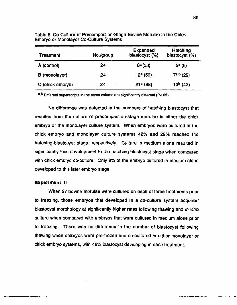
89
Table 5. Co-Culture of Precompaction-Stage Bovine Morulae in the Chick Embryo or Monolayer Co-Culture Systems
Treatment No./groupExpanded
blastocyst (%)Hatching
blastocyst (%)
A (control) 24 8a(33) 2a (8)
B (monolayer) 24 12a (50) 7a,b (29)
C (chick embryo) 24 21b (88) 10b (42)
a-b Different superscripts in the same column are significantly different (P<.05)
No difference was detected in the numbers of hatching blastocyst that
resulted from the culture of precompaction-stage morulae in either the chick
embryo or the monolayer culture system. When embryos were cultured in the
chick embryo and monolayer culture systems 42% and 29% reached the
hatching-blastocyst stage, respectively. Culture in medium alone resulted in
significantly less development to the hatching-blastocyst stage when compared
with chick embryo co-culture. Only 8% of the embryo cultured in medium alone
developed to this later embryo stage.
Experiment II
When 27 bovine morulae were cultured on each of three treatments prior
to freezing, those embryos that developed in a co-culture system acquired
blastocyst morphology at significantly higher rates following thawing and in vitro
culture when compared with embryos that were cultured in medium alone prior
to freezing. There was no difference in the number of blastocyst following
thawing when embryos were pre-frozen and co-cultured in either monolayer or
chick embryo systems, with 48% blastocyst developing in each treatment.

90
Table 6. Co-Culture of Bovine Morulae in the Chick Embryo or Monolayer Co- Cuiture Systems Prior to Freezing in LN2
No. with blastocyst morphology(%)
Treatment No./group Pre-freeze Post-thaw
A (control) 27 18b (67) 5b (19)
B (monolayer) 27 20b (74) 13c (48)
C (chick embryo) 27 19b (70) 13c (48)
D (in vivo) 27a NA 20c (77)
a One embryo lost during thawing proceduresb'c Values in the same column with different superscripts are significantly different (P<.05)
Although embryos cultured in medium alone developed to the blastocyst
stage at similar rates to co-cultured embryos during the pre-freeze culture
interval, only 19% blastocyst morphology was observed when these embryos
were thawed and returned to culture following freezing. The highest number of
blastocysts developing in vitro following freezing and thawing resulted from the
embryos collected on day 7 after mating and frozen without prior culture. These
embryos acquired blastocyst morphology at a rate of 77%.
Discussion
In light of recent progress in producing bovine zygotes using IVF
techniques (Leibfried-Rutledge et a t, 1989), co-culture systems capable of
supporting the in vitro development of early stage bovine embryos are
becoming essential laboratory techniques. Culture of IVF-derived embryos in
the ligated oviducts of ewes or rabbits (Lu et al., 1987; Ectors et a t, 1989) is
currently an effective, albeit impractical embryo culture approach. The
development of early bovine morulae to the expanded and hatched blastocyst

91
stage using the chick embryo in this study suggests the potential application of
this novel embryo co-culture technique.
Due to the lengthy culture periods («7 days) necessary for the
development of blastocyst from IVF-derived zygotes, complete development in
the chick embryo amnion may not be feasible using the techniques employed in
the present experiment. The amniotic cavity of the developing chick embryo
appears to be useful for embryo co-culture only during the interval from day 4 to
day 9 of development. Additionally, the embryotropic characteristics of the
amniotic cavity beyond 8 days of development have not been verified. The use
of this technique with IVF-derived embryos may require chick embryo co-culture
for only a portion of the total culture interval or sequential co-culture in two or
more chick embryos.
Although in vitro culture on cell monolayers in Experiment I resulted in
fewer expanded blastocyst than when embryos were co-cultured in the chick
embryo amnion, the monolayers used in the first experiment consisted of
fibroblast cells. Recent studies indicate that embryo co-culture with cells of
epithelial origin result in improved embryonic development when compared
with co-culture with fibroblast cells (Gandolfi and Moor, 1987; White et al.,
1989). When epithelial cells were used in Experiment II, post-thaw blastocyst
development in embryos co-cultured with epithelial cells was equal to post-thaw
blastocyst development in embryos co-cultured in the chick embryo amnion.
Although post thaw development in embryos co-cultured prior to freezing
was not equal to that of embryos which were not cultured prior to freezing, co-
cultured embryos did develop into blastocyst at higher rates than when embryos
were cultured in medium alone prior to freezing. These results suggest that the
use of a co-culture system for the short-term storage of bovine embryos may be
advantageous if there is a possibility of freezing any of the embryos.

92
The results of Experiment II indicate that in vitro culture may have a
detrimental effect on the ability of bovine embryos to survive the freezing
procedures used routinely on in vivo developed embryos of the same stage.
This additive effect of pre-freeze culture is in agreement with results reported for
mouse embryos (Massip etal., 1984) and those of rabbit embryos (Techakumpu
et a i , 1986) that were cultured for 48 hours prior to freezing. Although there
was an apparent increase in post-thaw development when embryos were
maintained on either of the co-culture systems prior to freezing, the use of the
chick embryo amnion as a culture vessel did not enhance post-thaw embryo
over the use of monolayer co-culture.
The bovine embryos co-cultured in the chick embryo system were
agarose-embedded in the present experiment. In previous experiments, the
use of agarose appeared to allow for the free diffusion of amniotic fluids to the
embedded embryos. However, the ability of lipids to permeate the agarose
matrix has not been determined. If in vitro culture affects the lipid components
of the embryo as has been suggested by Techakumpu etal. (1986), the use of
agarose embedding may have negated any positive effects of the chick
amniotic environment. Future experiments using chick embryo co-culture on
bovine embryos prior to freezing may prove more successful if the bovine
embryos are injected without the use of agarose.
CHAPTER VI
CULTURE OF IN V/7RO-FERTILIZED BOVINE EMBRYOS IN THE CHICK EMBRYO AMNION
Introduction
Successful production of a live transplant calf by in vitro fertilization (IVF)
procedures did not occur until 1981 (Brackett e ta l.., 1982). In more recent
years, in vitro fertilization has become a realistic method for the laboratory
production of bovine zygotes. Although abattoir ovaries are currently the most
available source of bovine oocytes for use in the research laboratory, the
practical application of IVF using this approach has a major drawback. Abattoir
ovaries generally do not provide genetically superior gametes for fertilization.
Therefore, bovine IVF procedures have not been accepted for use by the
seedstock industry.
New approaches for obtaining oocytes from high genetic quality cattle
are presently being developed (Kruip et al., 1990; Ryan et al., 1990). Using
sonographic-guided transvaginal collection procedures, Kruip et al. (1990) has
recently demonstrated that oocytes can be successfully collected from the
ovaries (4 to 8 mm follicles) of cycling dairy cows. In this study, pregnancies
have resulted from the transfer of IVF-derived bovine zygotes that were
transferred to recipients following their culture for 6 days in the ligated oviducts
of ewes. Ryan eta l. (1990) has recently developed a new approach to IVF by
collecting gametes from pregnant cattle. With this method, cumulus-intact
oocytes were successfully collected from the ovaries of FSH-treated pregnant
cows. IVF-derived embryos were then cultured to morulae in vitro. The use of
this method would allow embryos to be obtained from valuable, high producing
donor cows while they were carrying a fetus in utero.
Methods developed for producing embryonic clones in farm animals
93

94
(Willadsen, 1986: Robl, etal., 1987) will increase the need for abattoir oocytes.
In this basic cloning procedure, a single blastomere from a 16 or 32-cell embryo
is joined with half of an unfertilized oocyte by electrofusion. Although this
procedure often uses oocytes obtained from the surgical collection of stimulated
donor cattle (Bondioli et al., 1990), it is possible to use oocytes aspirated from
the ovaries of slaughtered animals (Prather etal., 1987). As oocyte maturation
methodology improves, the use of abattoir oocytes for embryonic cloning will
likely become more commonplace. Although gene transfer techniques for farm
animals remain in the developmental stages, future breakthroughs in the
production of transgenic embryos by gene injection into the pronucleus
(Hammer et al.,1985) or other methods will likely necessitate the use of very
early stage embryos from genetically superior animals.
The commercial application of these new embryo production techniques
will depend on the ability of very early stage embryos to develop into
pregnancies and produce live calves. The embryos produced by IVF and
embryo cloning procedures are not at the stage of embryo development that is
optimal for nonsurgical transplantation. Approximately 6 days of embryonic
development has been shown to be needed for the IVF-derived zygotes to be at
the appropriate stage of development to survive in utero following nonsurgical
embryo transfer, since acceptable pregnancy rates following embryo transfer in
cattle have been shown to occur only when embryos have reached the morula
and blastocyst stages of development (Schneider e ta l., 1980; Massey and
Oden, 1984; Hasler et al., 1987). If early-stage embryos could then be cultured
to a stage which allowed them to be transferred nonsurgically to recipient
females, significant gains could be made in these research areas.
In a recent review, First and Parrish (1987) compared the overall
efficiency rates reported for the various stages of the bovine IVF process
95
(including oocyte maturation, fertilization rate, pregnancy rate and calves born)
and found that techniques for obtaining blastocysts from one-cell embryos were
less successful than any other IVF-related procedure. One- to four-cell embryos
cultured in vitro rarely develop past the eight- to 16-cell stage, however, when in
vivo-derived eight- to 16-cell embryos are cultured in vitro they are able to
develop to the morula and blastocyst stages (Thibault, 1966).
One of the first approaches to overcome the in vitro block in farm animal
embryos incorporated synchronized sheep as an intermediate host (Willadsen,
1979). The oviducts of the ewe were ligated and embryos were allowed to
develop in vivo for up to 4.5 days. Variations of this in vivo technique have
been reported using the oviducts of rabbits (Boland, 1984) and more recently
mice (Ebert etal., 1989). The use of intermediate host oviduct techniques labor
intensive, time consuming and often expensive.
Consequently, in vitro systems capable of fostering embryonic growth
through the in vitro block stage are being developed. In recent years the co
culture of embryos with other mammalian cells has become a popular in vitro
embryo culture technique. Kuzan and Wright (1981) reported the first use of
uterine fibroblast monolayers for the co-culture of embryos from farm animals.
In this and other early co-culture studies, the cell type used was primarily
fibroblast monolayers (Kuzan and Wright, 1982; Allen and Wright, 1984).
Subsequent studies using fetal uterine fibroblast monolayers for the co-culture
of mammalian embryos have resulted in pregnancies in cows (Wiemer,
personal communication), horses (Wiemer etal., 1989c) and humans (Wiemer
etal., 1989a).
Rexroad et al. (1986) evaluated the development of ovine embryos
transferred following 24 hours of co-culture with ovine epithelial cells and noted
similar cleavage rates when these embryos were compared with embryos
96
transferred immediately following collection. Later studies revealed that co
culture of ovine embryos with cells of endometrial origin resulted in improved
developmental rates when compared with co-culture with fibroblast cells
(Gandolfi and Moor, 1967). Eyestone and First (1989) have noted an increase
in in vitro development to morulae and blastocysts when early stage bovine
embryos were cultured with oviductal cell monolayers, oviductal cell
suspensions or medium conditioned in oviductal cell suspensions. In this study,
pregnancies were obtained when IVF-derived embryos were transferred
following culture with cells or conditioned medium.
Ellington et al. (1990a) surgically collected one- and two-cell bovine
oocytes then cultured them with fresh bovine oviductal epithelial cells, frozen-
thawed bovine oviductal epithelial cells, or in medium conditioned with
oviductal cells. No differences were noted with regard to development past the
eight to 16-cell in vitro block stage with these culture systems. However, the
embryos cultured on fresh oviductal cells had significantly higher number of
blastomeres per embryo than embryos cultured on frozen thawed celts or in
conditioned medium. The embryos cultured on fresh cells also had better
embryo viability scores than embryos in the other two treatments. Ellington et
al. (1990b) also compared co-culture on monolayers of bovine oviductal
epithelial cells to in vivo culture in the ligated oviducts of rabbits using one- to
two-cell bovine embryos. Embryos co-cultured on these in vitro monolayers for
5 days had a similar number of cells and resulted in similar transfer pregnancy
rates when compared with embryos cultured in vivo using the rabbit oviduct
system.
Much of the early successful co-culture experiments with IVF-derived
bovine embryos used oviductal epithelial cells, however, recent results indicate
that granulosa cells also provide an effective co-culture system for IVF produced
97
bovine zygotes. Kajihara et al., (1987) reported development of IVF-derived
embryos to the hatched blastocyst stage during in vitro culture on follicular
granulosa cells obtained during the aspiration of the cumulus oocyte
complexes. Goto et al. (1988) subsequently reported that pregnancies could be
obtained following long term co-culture of in wfro-fertilized oocytes (6 to 7 days)
with these granulosa cells.
An alternative technique for promoting the growth and development of
early stage mammalian embryos uses the amniotic cavity of the 4- to 7-day-old
chick embryo as an in vivo culture system (Blakewood etal., 1988). This system
has proven successful with embryos from mice (Blakewood eta l., 1988), goats
(Blakewood etal., 1989a) and cows (Blakewood eta l., 1989b) when compared
with in vitro culture in medium alone. Live births have resulted when goat
embryos were transferred to recipient animals following 72 hours of co-culture
in the chick embryo amnion (Blakewood etal., 1990).
The objective of this series of experiments was to determine if the chick
embryo amnion was suitable for the development of one-cell IVF-derived
bovine embryos to a stage that would allow transfer to recipient cows.
Secondly, to compare the developmental rates of IVF-produced bovine
embryos cultured in the chick embryo amnion with the developmental rates of
similar embryos co-cultured with granulosa cells.
Materials and Methods
A recently published procedure for the in vitro maturation and fertilization
of oocytes was used in the first of these experiments (Goto et al., 1988). A
second IVF procedure was then evaluated, which also resulted in acceptable
rates of cleavage and in vitro development (Zhang et al., 1990a, 1990b). This
procedure was subsequently used in Experiments (EXP) II and III.

98
Ovaries from mature dairy and beef cattle were collected at slaughter and
placed in a 500 ml wide-mouth glass bottle containing Dulbecco's phosphate-
buffered saline (PBS). Ovaries were then transported (22 to 24°C) to the
laboratory within 5 hours from the time of slaughter. At the laboratory, ovaries
were rinsed twice in PBS, then £3 mm follicles were aspirated by inserting a 20-
gauge hypodermic needle through the ovarian stroma and into the follicles from
the base of the antral cavity. The oocytes with cumulus cells (cumulus oocyte
complexes, COC) were then gently aspirated into a 6 ml syringe. Follicular
fluids and COC were pooled in a 15 ml centrifuge tube until the follicles of 15 to
20 ovaries were aspirated. COC were allowed to settle to the bottom of the
centrifuge tube (2 to 3 minutes), then the top 12 to 13 ml of follicular fluid was
carefully withdrawn and discarded.
COC were then resuspended in a washing medium that consisted of
either HEPES-buffered TCM-199 (EXP I) or PBS + .1% polyvinyl alcohol (EXP II
and III). COC were evaluated under a stereomicroscope (80x) and rinsed twice
with washing medium prior to oocyte in vitro maturation (IVM) procedures.
In EXP I, 160 COC were aspirated from bovine ovaries and incubated in
hormone-supplemented, HEPES-buffered TCM-199 with 5% fetal bovine serum
for 26 hours. In EXP II and III, 195 and 440 COC, respectively, were incubated
in TCM-199 with 5% FCS for 22 to 24 hours (Zhang et a i, 1990a, 1990b). All
incubations were conducted in a humidified atmosphere of 5% CO2 in air at 39°
C. Following incubation, COC were placed in drops of Brackett-Oliphant (B-O)
medium containing sperm celts for in vitro fertilization.
Straws of frozen sperm from two different dairy bulls were thawed and
pooled in 15 ml plastic centrifuge and washed twice using B -0 medium. In EXP
I, sperm cell capacitation was enhanced by incubating «4 x 10⁵ sperm cells in

99
100 ill drops of B -0 medium under mineral oil. B -0 medium in EXP I was
supplemented with 5 pM heparin and 2 mg/ml fatty-acid free BSA (Sigma).
Following a 4-hour capacitation period, 12 to 15 COC were added to each drop
and incubation was continued for an additional 4 hours. Sperm capacitation
was enhanced in EXP II and III by a 1 minute exposure of sperm cells to a .1 (iM
solution of calcium ionophore A23187. Sperm cells in Experiments II and III
were then added to 50 pi drops of B -0 medium under mineral oil at a
concentration of -1 .5 x 1 0 6 cells/ml and incubated for 5 hours with 15 to 25
COC per drop. Heparin was not used in EXP II and III, however B-O was
supplemented with 2 mg/ml fatty acid-free BSA and 10 mM caffeine. All
incubation of sperm and oocytes was completed in a humidified atmosphere of
5 % CO 2 in air at 39° C. Following in vitro fertilization, ova were placed in
designated in vitro culture systems to assess subsequent development.
Experimental Design
Experiment I
This experiment was designed to determine if IVF-derived embryos could
develop to morulae and blastocyst at similar rates to IVF-derived embryos co-
cultured with cumulus cells for 6 or 7 days. Following IVF procedures, ova
were randomly allotted to one of three treatments for in vitro culture. Fertilized
oocytes allotted to Treatment A were co-cultured with cumulus cells in vitro by
returning the oocytes and their attached cumulus cells to the same tissue
culture wells previously used for in vitro maturation (Figure 8 ). During the in
vitro maturation period, cumulus cells from COC began attachment to these
culture wells. Following the removal of COC from the maturation wells for
fertilization, fresh HEPES-buffered TCM-199 was added to the cumulus cells
and they were returned to incubation. Immediately after fertilization in

100
microdrops, ova allotted to Treatment A were placed back into the wells
containing cumulus cells for subsequent co-culture. Co-culture wells were
incubated at 39° C in a humidified atmosphere of 5% CO 2 in air. Fresh
HEPES-buffered TCM -199 was added to the co-culture wells at 72-hour
intervals. This in vitro culture technique was compared with two in vivo methods
for maintaining mammalian embryos inside the chick amniotic cavity for 6 or 7
days (Treatments B and C).
Due to the developmental patterns of the chick embryo, successful use of
its amniotic cavity for mammalian embryo co-culture appears to be confined to
the period between days 4 and 8 of incubation. In order to test this system over
a 6- or 7-day culture period, the sequential use of multiple chick embryos was
necessary. Recovery of bovine ova from one chick embryo amnion followed by
immediate injection into another chick embryo amnion was used in Treatments
B and C to allow chick embryo co-culture for this extended period. In Treatment
B, fertilized oocytes were injected into the amniotic cavities of a total of three
chick embryos without the use of agarose embedding. Incubation periods in the
first, second and third chick amnions were 2 days, 2 days and 2.5 days,
respectively.
Fertilized oocytes allotted to Treatment C were injected into the amniotic
cavities of a total of two chick embryos. Ova placed the first chick amnion were
injected without the use of agarose embedding and were co-cultured for 2 days.
Following recovery from the first amnion, bovine ova were agarose-embedded
and injected into a second amnion for a co-culture period of 4.5 days. In both
Treatments B and C, agarose-embedding was not used immediately after
fertilization to allow attached cumulus cells to also be injected into the first chick
amnion. All injections into the chick embryo amnion occurred at day 4 of chick

Experimental Design
Treatment A| IVM-IVF 1 Cumulus Cells" Cumulus Cells
24 48 72 96 120 144
Time, Hours
Treatment BIVM-IVF
24 48 72 96 120 144
Time, Hours
Treatment CIVM-IVF
24rAmmmmf///////A
48 72 96 120 144
Time, Hours
Figure 8. Co-Culture of IVF-Derlved Bovine Embryo in Two or Three Chick Embryo Amniotic Cavities

102
embiyo development. Shell-less chick embryos were incubated at 39° C in a
humidified atmosphere of 2% COg in air.
Experiment II
This experiment was designed to compare chick embryo co-culture to
granulosa cell co-culture during the first 2 days of culture following IVF. COC
were matured and fertilized as described previously, then randomly allotted to
either cumulus coll or chick embryo co-culture treatments (Figure 9). Ova
allotted to Treatment A were returned to in vitro culture with granulosa cells as
described in Treatment A of Experiment I. Fertilized oocytes allotted to
Treatment B were injected into the amniotic cavities of 4-day-old chick embryos
without the use of agarose in order to inject the attached cumulus cells into the
amniotic cavity. After 48 hours of incubation, ova and cumulus cells were
recovered from chick embryo co-culture and placed into culture wells with
cumulus cells maintained from the in vitro maturation procedures. All in vitro co
culture wells (Treatments A and B) received fresh HEPES-buffered TCM-199
medium at 72-hour intervals and were incubated at 39° C in a humidified
atmosphere of 5% CO 2 in air. Shell-less chick embryos were incubated at 39°
C in a humidified atmosphere of 2% CO 2 in air.
Experiment III
In this experiment, the developmental effect of chick embryo co-culture
on early cleavage stage embryos derived from IVF was compared with that of
granulosa cell co-culture. Following maturation and IVF procedures, all oocytes
were allowed to develop for 3 days in cumulus cell co-culture as described in
Treatment A of Experiment I (Figure 10). After 3 days cumulus cell co-culture,
one half of the embryos undergoing cleavage were randomly allotted to

Experimental Design
Treatment A| IVM-IVF | Cumulus Cells Cumulus Cells I
0 24 48 72 96
Time, Hours
120 144 168
Treatment BIVM-IVF Cumulus Cells I
0 24 48 72 96 120 144 168
Time, Hours
Figure 9. Co-culture of Early-Stage, IVF-Derlved Bovine Embryos in the Chick Embryo Amniotic Cavity

Experimental Design
Treatment AIVM-IVF | Cumulus Cells | Cumulus Cells
Treatment
0 24
B
48 72 96
Time, Hours
120 144
IVM-IVF [ Cumulus Cells0 24 48 72 96 120 144
Time, Hours
Figure 10. Co-Culture of IVF-Derived Early Bovine Morulae in the Chick Embryo Amniotic Cavity

105
Treatment A and allowed to remain in vitro with cumulus cells. The other half of
the cleaving embryos were allotted to Treatment B and removed from cumulus
cell co-culture and injected into the amniotic cavities of 4-day-old chick embryos
without the use of agarose embedding. After an additional 3 days of incubation
these embryos were recovered and compared with embryos which remained
with cumulus cells in vitro.
All in vitro co-culture wells (Treatments A and B) received fresh HEPES-
buffered TCM-199 medium at 72 hour intervals and were incubated at 39° C in
a humidified atmosphere of 5% CO 2 in air. Shell-less chick embryos in this
experiment were incubated as in the previous experiments.
Embryo Evaluation
In all Treatments of Experiments I and II, the morphology of cultured ova
was evaluated 2 days after fertilization procedures using a stereomicroscope
(80x). Single-cell ova and two-cell embryos that were present at this time were
considered nonviable and were removed from the culture system. Only those
ova that had undergone cleavage to the 4-cell stage were considered viable
embryos and were used for further testing of culture conditions. In Experiment
III, evaluation of embryo morphology was performed 3 days after fertilization
procedures. Only those embryos which had reached the eight-cell stage were
considered viable and remained in culture for the duration of the experiment.
The numbers of embryos undergoing normal morphological development at the
initial evaluation was used to compare different culture treatments.
In all three Experiments, final morphological evaluation of embryos was
performed after 6 or 7 days of in vitro culture. Development of embryos to the
morula and blastocyst stages during this time period was used as the final
comparison of culture treatments.

106
Statistical Analysis
A Chi-square analysis using a contingency table with continuity
correction (Pearson and Hartley, 1954) was used to compare the number of
embryos developing during culture across treatment groups.
Results
Experiment I
After 2 days of incubation, 41 and 38 ova were recovered from the
amniotic cavities of nine chicks. Of the total 79 ova recovered from the chick
embryo amnion, 48 (61%) appeared to be viable four- to six-cell embryos. This
compared with 26 (51%) four- to six-cell embryos which developed from 51
fertilized oocytes cultured in the in vitro cumulus cell co-culture system (Table
7). At this time, the 4- to 6-cell embryos recovered from the chick embryo
amnion were injected into a second shell-less chick embryo amnion using the
two techniques described previously. Treatment B embryos were injected
without the use of agarose (n=25) and Treatment C embryos were agarose-
embedded prior to injection (n=23).
Table 7. Co-culture of IVF-Derived Bovine Zygotes with Cumulus Cells or Chick Embryo Culture Systems
Dav 2 of culture Dav 6.5 of cultureCo-cultureTreatment
No./group
Recovered no. (%)
4-6 cell no. (%)
Recovered no. (%)
Morulae no. {%)
A CUM -*■ CUM 51 51 (100) 26a (51) 26 (100) 7a (27)
B CHK-» CHK CHK 51 41 (80) 25a (61) 9 (36) 5a (55)
C CHK -* CHK 51 38 (75) 23a (61) 10 (43) 1a (10)
CUM - cumulus cell co-culture, CHK » chick embryo co-culture a No significant difference between treatments

107
Treatment B embryos were recovered after 48 hours and injected into the
amniotic cavity of a third chick embryo for the final 2.5 days of incubation. The
agarose embedded embryos in Treatment C were incubated an additional 4.5
days in a single chick embryo amnion (Figure 8). Of the 26 cleaving embryos
that were maintained in vitro with cumulus cells, seven (27% ) continued
development to the morula or blastocyst stage after 6.5 days of co-culture. At
this time, nine of the 25 embryos allotted to Treatment B were recovered, and
55% (5/9) of these embryos were judged to be transferable quality morulae or
blastocysts. Of the 23 embryos allotted to Treatment C, 10 were recovered and
only one of these (10%) was a transferable quality morula. There were no
significant differences in the number of four- to six-cell embryos after 2 days of
culture between the three treatments. Neither were there any significant
differences in the number of morulae between treatments.
The 6 morulae and blastocysts recovered from chick embryo amnions in
Treatments B and C were transferred to day-6 recipient cattle (two morulae in
one recipient, four single-embryo transfers). At the time of this writing, three of
five (60%) recipients remain pregnant.
Experiment II
During the first 2 days of culture following fertilization, 47 of the original
95 oocytes cultured in vitro with cumulus cells (Treatment A) developed to the
four- to six-cell stage (49%) (Table 8). At this time, 96 of the original 100 (95%)
ova were recovered from the amniotic cavities of chick embryos (Treatment B).
Of these 96 recovered ova, 33 were at the four- to six-cell stage (34%) which
was significantly less than the number of four- to six-cell embryos obtained from
cumulus cell co-culture.

108
Table 8. Co-culture of One-Cell Bovine IVF-Derived Embryos for Two Days in the Chick Embryo Culture System
Day 2 of culture ___ Day.Z-Qf cultureCo-culture No./ Recovered 4-6 cell Recovered Ma and B treatment group no. (%) no. (%) no. (%) no. (%)
A CU M -»CUM 95 95 (100) 47b (49%) 47 (100) 33b (70%)
B CHK - * CUM 100 96 (96) 33c (34%) 33 (100) 20b (61%)
CUM - cumulus cell co-culture, CHK - chick embryo co-culture a M - morula, B - blastocystb,c Different superscripts In the same column are significantly different (P<.05)
When cumulus cell co-culture of embryos from both treatments was
continued for 5 additional days, 33 of the 47 (70%) four- to six-cell embryos
recovered from initial co-culture with cumulus cells (Treatment A) continued
development to the morula and blastocyst stages. This was not different from
the 20 morula and blastocyst (61%) developing in vitro from the 33 four- to six
cell embryos recovered from the chick embryo amnion (Treatment B) (Table 8).
Experiment III
After the initial 3 days of in vitro co-culture with cumulus cells, 190 of 440
fertilized oocytes (43%) developed to the eight- to 16-cell stage. When 95 of
these embryos remained in cumulus cell co-culture (Treatment A), 51 (53%)
developed to the morula and blastocyst stages after 3 additional days in culture
(Table 9).
Of the 95 embryos allotted to Treatment B and injected into the amniotic
cavities of chick embryos, 43 of the 70 embryos recovered after three days of
chick embryo co-culture developed to the morula and blastocyst stage (61%).
Neither the number of eight to 16-cell embryos nor the number of morulae and
blastocyst were significantly different between treatments.

109
Table 9. Co-culture of Early-Stage Bovine IVF-Derived Embryos for Three Days in the Chick Embryo Culture System
Day 3 of culture ________ Day,6 Qf.culture___Co-culture No./ Recovered 8-16 cell Recovered Ma andB treatment group no. {%) no. (%) no. (%) no. (%)
A CUM -» CUM 220 220 (100) 9 5 (4 3 ) 95 (100) 51b (53)
B CUM -* CHK 220 220 (100) 9 5 (4 3 ) 70 (74) 43b (61)
CUM » cumulus cell co-culture, CHK - chick embryo co-culture a M - morula, B - blastocysts b No significant difference between treatments
Discussion
In addition to confirming the effectiveness of granulosa cell co-culture,
which has previously been shown to be an effective technique for promoting the
growth of IVM-IVF embryos through the block stage (Goto et al.t 1988), the
results of this series of experiments indicate that chick embryo co-culture is also
an effective means of culturing these embryos. The use of chick embryo co
culture for the entire 6 or 7 days needed for embryos to reach the blastocyst
stage, however, does not appear to have an advantage over the cumulus cell
co-culture system. Co-culture in the chick embryo amnion for periods longer
than 5 days has been attempted in our laboratory, but without success.
Injection of the chick amniotic cavity cannot occur prior to day 4 of incubation,
and by day 10 or day 11 the amnion has become too large and fragile to be
efficiently isolated from the remainder of the egg contents. Additionally, the
chick begins to ingest amniotic fluids on day 11 of development (Romanoff,
1960), increasing the risk of losing any mammalian embryos within the amniotic
cavity.
Attempts at using sequential culture in the amniotic cavities of multiple
chick embryos does not appear to be a viable alternative to 6 or 7 days of

1 1 0
culture using a single chick embryo. In the case of EXP I, the compound losses
of embryos noted when embryos were recovered from one chick amnion and
injected into another appear to outweigh any potential increase in viability. The
use of either two or three chick embryos both resulted in unacceptable loss of
bovine embryos. With regard to the sequential use of two chick embryos, only
one bovine embryo which was agarose embedded and cultured for 5 days in a
single chick amnion reached the morula stage. This may be due to a less than
favorable amniotic environment at later stages of chick embryo development.
Previous studies at this laboratory have only evaluated chick embryo co-culture
for a period of 4 days.
Experiment II and III were subsequently conducted in order to determine
if chick embryo co-culture could be effectively used along with cumulus cell co
culture to enhance the development of IVF embryos to the morula and
blastocyst stages. Results from EXP II indicate that the the injection of oocytes
in the chick embryo amnion immediately after in vitro fertilization does not
enhance cleavage rates during the first 2 days of development when compared
with co-culture of similar embryos on cumulus cells. Subsequent in vitro
development of embryos following recovery from the chick embryo amnion was
also no different than that of embryos which remained with cumulus cells for the
duration of the experiment.
In Experiment III, eight to 16-cell, IVF-derived embryos co-cultured in the
chick embryo amnion for 3 days did develop to the morula and blastocyst stage.
However, the numbers of morulae and blastocysts were not increased when
compared with cumulus cell co-culture. Although results from all three
experiments indicate that the chick embryo amnion is an effective embryotropic
environment, its use with embryos derived from IVF procedures does not
111
appear to be advantageous over that of cumulus cell co-culture. Due to the
increased difficulty and decreased embryo recovery involved with using chick
embryo co-culture as an alternative to in vitro culture systems, it may not be an
realistic alternative to cumulus cell co-culture. However, there may be some
element of the chick embryo co-culture system that can be incorporated into an
effective in vitro system.
Attempts to incorporate the beneficial effects of chick embryo amniotic
fluid with those of cumulus cell co-culture are currently underway. Up to 1 ml of
amniotic fluid can easily be extracted from the amniotic cavity of a 7-day-old
chick embryo and used as a medium for in vitro culture. A combination of chick
embryo and cumulus cell culture procedures may have more potential for
success than that of either technique alone. This approach could prevent the
losses of embryos associated with injection into and recovery from the intact
amniotic cavity.
CHAPTER VIITHE USE OF CHICK EMBRYO AMNIOTIC FLUIDS FOR THE IN VITRO
CULTURE OF EARLY-STAGE MAMMALIAN EMBRYOS
Introduction
The successful maintenance of mammalian embryos in vitro is
apparently dependent on the presence of some type of biological fluid. From
the pioneering works involving the culture of rodent embryos in undiluted blood
plasma (Brachet, 1912; Lewis and Gregory, 1929), biological extracts have
remained an essential component of the in vitro milieu. Balanced salt solutions
currently provide the basis for commercially available embryo culture media,
however, successful embryonic development in these media requires the
addition of serum or bovine serum albumin (BSA) (Wright and Bondioli, 1981).
Heat treated bovine serum is most often used in the culture of domestic
embryos, and human serum is currently being used for studies involving the
culture of human embryos.
Despite the apparent standardization of heat-treated serum and BSA as
supplements for embryo culture media, inconsistencies in their effectiveness
have been reported. Sirard and Lambert (1985) have shown that identically
prepared batches of bovine serum from different animals gave different results
in their ability to promote cleavage of four-cell bovine embryos. Additionally,
Kane (1987) has reported that rabbit morulae cultured to the blastocyst stage in
medium supplemented with BSA had more than twice as many cells when a
different batch of BSA was used for culture.
Human amniotic fluids have recently been used as an alternative to
balanced salt solutions for in vitro fertilization and culture of human embryos
(Gianaroli et al., 1986). The amniotic fluids used in this study were taken from
women during the 16th to the 21st weeks of gestation, and supported the
112
113
development of two-cell mouse embryos to the blastocyst stage as effectively as
medium supplemented with fetal bovine serum (FBS). These fluids were also
used for the fertilization, culture and transfer of human embryos. When amniotic
fluids were used in the procedures of nine patients, four (44%) pregnancies
resulted. Recently, embryotropic activity has been verified in human amniotic
fluids using early-stage murine embryos cultured with amniotic fluids obtained
from women during early pregnancy (Ball et al., 1988).
In another study using human amniotic fluid for the culture of murine
embryos, 1000 mouse embryos were cultured in human amniotic fluid extracted
during the 16th week of pregnancy. After 72 hours of culture, there were
significantly more expanded blastocyst in amniotic fluid culture than in Earle's
medium culture (Oettleg and Wiswedel, 1990). In a study using IVF produced
murine embryos, 92% of the embryos cultured in human amniotic fluids
underwent cleavage compared with 86% undergoing cleavage in Ham's F-10
medium (Coetzee et al., 1990). Other studies have indicated that frozen-thawed
human amniotic fluids retain their ability to promote in vitro development of
early-stage murine embryos (Fugger et al., 1987).
Bovine amniotic fluids have also been used for the culture of murine
embryos (Javed et al., 1990). Murine two-cell embryos were successfully
cultured to the hatched blastocyst stage in frozen-thawed bovine amniotic fluids
obtained at less than 70 days of gestation. These researchers did, however,
note a decrease in embryonic developmental rates when amniotic fluids were
frozen prior to use in embryo culture.
The purpose of this series of experiments was to evaluate if the amniotic
fluids of the developing chick embryos could be used with in vitro culture
systems, either undiluted or as a supplement in standard culture media. The in
situ chick amnion has been shown to allow development of early stage embryos

114
from mice (Blakewood et al., 1988), goats (Blakewood et al., 1989a) and cattle
(Blakewood et al., 1989b). If the extracted amniotic fluids of the developing
chick embryo proved to have embryotropic properties in vitro, these fluids may
represent an alternative supplement for embryo culture media. Due to the
highly inbred nature of domestic fowl, it is likely that amniotic fluids taken from
the same stage chick embryos of a particular breed would likely be uniform in
their constituents, and therefore their embryotropic properties.
Materials and Methods
Amniotic fluid extraction
Fertilized chicken eggs were obtained from naturally-mated white
leghorn hens and incubated at 37° C in a commercial chicken egg incubator
which rotated the eggs at 1-hour intervals. After 7 days of incubation, eggs
were wiped with a 70% ethanol solution and held in an upright position using a
small ring stand placed in a laminar flow hood. The portion of the egg shell
above the air space was carefully cracked and shell fragments were removed,
exposing a circular region (25 mm diameter) of the shell membrane directly
above the chick embryo. The shell membrane was carefully peeled away using
a small pair forceps, exposing the day-7 chick embryo. A 25-gauge hypodermic
needle attached to a 3 ml syringe was used to pierce the amniotic cavity, and .5
to 1 ml of amniotic fluid was aspirated from the amniotic cavity. Extracted
amniotic fluids from three to five chick embryos were pooled in sterile, 5 ml
glass tubes prior to the in vitro culture of embryos. If storage for longer than 7
days was required, chick amniotic fluids (CAF) were frozen and held at -20° C.
For storage periods of less than 7 days, CAF were held at 4° C.

115
Collection of Mouse Embryos
Female mice (ICR) at 21 to 28 days of age were injected with 5 IU of
PMSG, followed at 48 hours by 5 IU of hCG and placed with mature ICR males.
The donor mice were sacrificed 36 to 42 hours after hCG injection the distal
ends of the uterine horns, oviducts and ovaries were excised and placed under
a stereomicroscope. The ovaries and most of the uterine horns were carefully
trimmed away from the oviducts, which were then flushed by inserting a fine
glass tube into the infundibulum and forcing «.1 ml of flushing medium through
the oviduct. The flushing medium consisted of PBS + .1% polyvinyl alcohol and
1% antibiotic (Gibco), which was also used to wash the recovered two-cell
embryos prior to placement in culture wells.
fn Vitro Fertilization of Bovine Embryos
Procedures for the production of in vitro fertilized bovine embryos were
performed as described for Experiments II and III of Chapter VI. Ovaries from
mature dairy and beef cattle were brought to the laboratory within 5 hours from
the time of slaughter. At the laboratory, ovaries were rinsed, then follicles 23
mm were aspirated. The follicular fluids and oocytes with cumulus cells
(cumulus oocyte complexes, COC) were pooled in a 15 ml centrifuge and COC
were allowed to settle to the bottom of the tube. COC were evaluated for intact
cumulus complexes and washed prior to oocyte in vitro maturation (IVM)
procedures.
For in vitro maturation of oocytes, COC were incubated in TCM -199
supplemented with either 10% FCS or 10% CAF for 22 to 24 hours. Incubation
was conducted in a humidified atmosphere of 5 % C O 2 in air at 39° C.
Following incubation COC were placed 15 to 25 per drop in 50 pi drops of
Brackett-Oliphant (B-O) medium under mineral oil for in vitro fertilization

116
procedures. B -0 medium was supplemented with 2 mg/mi fatty acid-free BSA
and 10 mM caffeine.
Frozen bull sperm was thawed and capacitation was enhanced by a 1
minute exposure of sperm cells to a .1 pM solution of calcium ionophore
A23187. Sperm cells were then added to the 50 pi drops of B-O medium
containing matured oocytes at a concentration of *1 .5 x 106 cells/ml and
incubated for 5 hours in a humidified atmosphere of 5 % CO 2 in air at 39° C.
Following in vitro fertilization, ova were placed in designated in vitro culture
systems for comparison.
Cell Staining
At the conclusion of each experiment, embryonic nuclei were stained
using Hoescht-33342 stain and visualized using UV fluorescence microscopy
(Crister and First, 1986). Embryos were placed into 10 pi drops of working stain
solution (1 pg/ml Hoescht 33342 in 25% ethanol, 75% 2.3 M Na citrate) on
siliconized slides. After 5 minutes at 20° C, the stain solution was removed and
coverslips were mounted using Permount® (Sigma) solution. Embryonic nuclei
were observed at 200x using a Zeiss fluorescent microscope equipped with a
10x Neofluar objective and a filter system consisting of a 340 to 360 nm exciter
and a 430 nm barrier filters. Nuclei that fluoresced were counted by a trained
observer.
Experimental Design
Experiment I
Murine embryos were randomly and equally allotted to one of five
treatment groups (Figure 11). In Treatment A, murine embryos were cultured in
Ham's F-10 without supplements. Treatment B consisted of Ham's F-10

Experimental Design
Treatment AHF-10
0 24
Time, Hours
48
Treatment B| HF-10 + Fetal Bovine Serum 30 24
Time, Hours
48
Treatment CHam's F-10 + CAF n
0 24
Time, Hours
48
Treatment D| Chick Amnfotic Fluid0 24 48
Time, Hours
Treatment E
0 24 48
Time, Hours
Figure 11. Co-Culture of 2-Cell Murine Embryos In the Chick Embryo Amnlotic Cavity and Culture in Extracted Chick-EmbryoAmnfotic Fluids
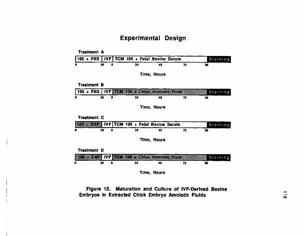
Experimental Design
Treatment A199 + FBS | IVF | TCM 199 ♦ Fetal Bovine Serum
24 0 24 48
Time, Hours
72
Treatment B199 ♦ FBS | IVF
24 0 24 48
Time, Hours
Treatment CIVF | TCM 199 + Fetal Bovine Serum"
24 0 24 48
Time, Hours
72
Treatment D
24 48
Time, Hours
S t a i n i n g96
St ai ni ng
S t a i n i n g96
St ai ni ng
Figure 12. Maturation and Culture of IVF-Derived Bovine Embryos in Extracted Chick Embryo Amniotic Fluids

119
medium supplemented with 10% FBS and Treatment C consisted of Ham's F-
10 medium supplemented with 10% CAF. Embryos cultured in Treatment D
were cultured in vitro in undiluted CAF. Embryos allotted to Treatment E were
agarose embedded and injected into the amniotic cavities of 4-day-old in vitro
chick embryos, as described previously in Chapter III.
Experiment II
The effects of CAF supplementation on both the maturation of bovine
oocytes and their subsequent in vitro development were evaluated using four
different combinations of maturation and culture media (Figure 12). Following
the evaluation of aspirated oocytes, COC were randomly and equally allotted to
one of the four treatment groups. In Treatments A and B, oocytes were matured
in TCM-199 supplemented with 10% FBS. In Treatment C and D, oocytes were
matured in TCM -199 supplemented with 10% CAF. Following in vitro
fertilization procedures, zygotes from Treatments A and C were cultured in
TCM-199 supplemented with 10% FBS and zygotes from Treatments B and D
were cultured in TCM-199 supplemented with 10% CAF.
Experimental Procedures
Experiment I
CAF used for murine embryo culture were extracted from day-7 chick
embryos at the beginning of the experiment and were not frozen. Culture
medium (HF-10) and amniotic fluids used in Treatments A, B, C, and D
contained 1% antibiotic and was filtered through a .2 p.m syringe filter into four-
well tissue culture plates which were preincubated for at least 1 hour before the
introduction of mammalian embryos. Culture plates were maintained in a
humidified 5% CO 2 atmosphere at 37° C. in vitro chick embryos used for
120
mouse embryo co-culture were maintained in a humidified atmosphere of 2%
C O 2 at 37° C. Viable appearing two-cell mouse embryos were randomly and
equally placed on treatments immediately following collection from donor mice.
All mouse embryos were cultured for 48 hours, then stained with Hoescht
33342 for assessment of cleavage and morphological development.
Experiment II
The CAF used for supplementation of TCM-199 in this experiment were
collected from day-7 chick embryos and were frozen prior to use in maturation
and culture media. Cumulus cells which had attached to the tissue culture wells
during in vitro maturation procedures were used for the co-culture of zygotes
following fertilization procedures by returning the zygotes to their original
maturation wells with fresh medium. During maturation and culture, oocytes
and embryos were incubated in a humidified 5% CO2 atmosphere at 39° C.
After 96 hours of culture (post-fertilization), cleaving embryos were stained with
Hoescht 33342 and nuclei were counted using fluorescence microscopy.
Statistical Analysis
The mean number of cells between treatments within experiments were
compared using a one-way analysis of variance (Steele and Torrie, 1980). The
mean number of cleaving bovine embryos in treatments for Experiment II were
compared by Chi-squared analysis using a contingency table with continuity
correction (Pearson and Hartley, 1954).
Results
Experiment I
No significant differences in cell numbers were detected among embryos
cultured in HF-10, FBS supplemented HF-10, CAF supplemented HF-10 or

121
undiluted CAF with mean cell numbers of 6 .9 ,6 .4 , 7.0 and 6.5, respectively.
Table 10. Culture of Two-Cell Mouse Embryos in the Chick Embryo Amnion or in Medium Supplemented with Chick Amniotic Fluids
Treatmentgroup
No./group
No.stained
No. cells8 at 48 hours
A HF-10 40 40 6.9 ± .65b
B HF-10 + FBS 40 40 6.4 ± .43b
C HF-10 + CAF 40 38 7.0 ± .66b
D CAF 40 38 6.5 ± .50b
E Chick Embryo 40 31d 11.4 ± .86°
FBS - fetal bovine serum, CAF - chick amniotic fluid a Mean plus or minus Standard Errorb,c Different superscripts In the same column are significantly different (P<.05) d 9 embryos lost during chick embryo co-culture
However, those embryos that were agarose embedded and injected into the
amniotic cavities of day-4 chick embryos did have significantly higher numbers
of cells, with a mean cell count of 11.4. Of the original 40 embryos injected into
the chick embryo amnion, 9 (23%) were lost during culture due to chick embryo
death or during recovery procedures.
Experiment II
When bovine oocytes were both matured in medium supplemented with
FBS and the fertilized zygotes subsequently cultured in medium supplemented
with FBS, significantly less cleavage occurred than in any other combination of
CAF and FBS supplemented maturation and culture media. Cleavage occurred
in only 68% of the embryos in Treatment A (FB S-»FB S) compared with
cleavage rates of 78, 78 and 77%, respectively in Treatments B (FBS-»CAF), C
(CAF->FBS) and D (CAF-»CAF).

122
Table 11 . I n Vitro Maturation of Bovine Oocytes and Subsequent Culture of IVF- Derived Bovine Embryos in Medium Supplemented with Chick Amniotic Fluids
TreatmentNo./
groupNo.
Cleaved (%)No.
StainedNo. Cellsa at 96 hours post-IVF
A FBS->FBS 193 131b (68) 125 8.3 ± .42b
B FBS->CAF 193 151° (78) 71 d 8.7 ± .55b
C CAF-»FBS 193 151® (78) 142 6.4 ± .30®
D CAF—»CAF 193 150° (77) 130 8.3 ± .37b
FBS - fetal bovine serum, CAF - chick amniotic fluid a Mean plus or minus Standard Errorb<® Different superscripts in the same column are significantly different (P<.05) d 50% of the embryos in this treatment were lost to contamination prior to staining
Although the total number of cleaving embryos was less in Treatment A,
the mean number of cells in cleaving embryos from Treatment A (FBS->FBS)
was not different from the mean number of cells in embryos from Treatments B
(F B S ^ C A F ) and D (CAF-»CAF). The mean number of cells in embryos from
Treatment C (CAF-»FBS) was, however, less than the mean cell counts for the
other three treatments, with only 6.4 cells/embryo in Treatment C compared with
8 .3 ,8 .7 and 8.3 cells/embryo in Treatments A, B and D, respectively.
Discussion
Although previous experiments involving chick embryo co-culture have
used the amniotic environment of shell-less chicks maintained in static (non-
rotating) incubation systems, the amniotic fluids used in these experiments were
taken from chick embryos maintained inside their shells and incubated in a
commercial system with egg rotation at 1-hour intervals. Observations by New
(1957) show that the critical period for egg turning is between days 4 and 7 of
incubation. In fact, normal hatchability can be obtained from eggs which are
turned on days 4 through 7 and then left unturned for the remainder of the

123
incubation period.
Although the need for egg turning has long been realized, it was
commonly held that embryonic death in unturned eggs was due to adhesion of
embryonic membranes to the shell membrane. Deeming ef a/. (1987) have
recently suggested that embryonic death in unturned chick embryos may
instead be due to a settling and non-distribution of proteins necessary for
normal embryonic growth. The critical period of egg turning (day 4 to day 7 of
incubation) coincides with the time of subembryonic fluid formation (day 3 to
day 7) in the chick embryo. These subembryonic fluids are a source of yolk
proteins for the chick embryo.
Rowlett and Simkiss (1987) have shown that embryonic growth in chick
embryos maintained in vitro is enhanced by rocking the shell-less culture
systems during the first half of the incubation period. The prospect of increasing
the potential growth promoting characteristics of the chick embryo environment
by rocking the eggs during the first 7 days of incubation was a consideration in
not using amniotic fluids extracted from static, shell-less chicks.
The use of undiluted CAF for the culture of murine embryos does not
appear to be an effective alternative to serum supplemented medium in the
present experiment, however, more thorough evaluation of several aspects of
this technique are needed. Culture wells containing undiluted CAF were
incubated in a humidified atmosphere of 5% C 0 2 in air. Previous reports using
human amniotic fluids for the culture of mammalian embryos (Gianaroli et a t,
1986; Fugger e ta l , 1987) also used a 5% C 0 2 in air atmosphere. Although the
pH of CAF was measured and found to be 7.2 immediately following extraction
from the chick amnion, pH measurements were not taken at the conclusion of
the experiment. Evaluation of pH stability in the culture system should be
assessed prior to further investigations.

124
Additionally, the human amniotic fluids used in the previously mentioned
reports were heat treated at 56° C for 30 minutes. Heat inactivation was not
performed on the CAF, since positive co-culture results had previously been
obtained following the injection of mammalian embryos into the intact chick
embryo amnion. Nonetheless, the potential for embryotoxic immunological
activity in CAF should be investigated in the future.
Although the use of CAF did not result in higher cleavage rates than
supplementation with FBS, the injection of agarose-embedded murine embryos
into the chick embryo amnion on day 4 of chick embryo incubation did result in
significantly higher cell counts. The use of amniotic fluids extracted from chicks
at day 7 of incubation may not provide the same embryotropic qualities found in
the in situ amniotic fluids during days 4, 5 and 6 of development. Although the
volume of amniotic fluids at day 4 may be too small for successful extraction, the
fluids from chicks at day 5 and 6 of development should also be tested.
With regard to the effects of CAF on the in vitro maturation of bovine
oocytes and their subsequent in vitro development following fertilization, the
results of the second experiment offer no definitive conclusions. Although less
cleavage occurred when oocytes were matured in medium supplemented with
FBS and subsequently cultured in medium supplemented with FBS, it cannot
be concluded that the use of FBS for in vitro maturation was responsible, since
oocytes matured in medium supplemented with FBS and subsequently cultured
in medium supplemented with CAF had higher rates of cleavage.
Similarly, the lower cell counts noted when embryos were cultured in
medium supplemented with FBS following oocyte maturation in medium
supplemented with CAF does not appear to be due to FBS in the culture
medium, since higher cell counts were obtained from embryos cultured in FBS
supplemented medium following oocyte maturation in FBS supplemented

125
medium.
It appears that CAF would be a viable alternative to FBS in bovine IVF
and embryo culture applications, however, further testing is warranted. Perhaps
CAF extracted from chick embryos of different breeds and at different stages of
development may provide better and more conclusive results. If an optimal
breed and stage of chick embryo for amniotic fluid extraction were found, a
more consistent culture media supplement than serum would be easily within
the reach of most laboratories.

SUMMARY AND CONCLUSIONS
A total of ten experiments were conducted to evaluate if the amniotic
cavity of the developing chick embryo could serve as an alternative culture
system for the in vitro development of preimplantation mammalian embryos.
Chick embryos were maintained in shell-less culture rather than inside the
eggshells in order to facilitate the repeatable injection and subsequent recovery
of mammalian embryos from the chick amniotic cavity.
Pronuclear-stage murine embryos were used in the initial experiment to
evaluate the potential benefits of culturing mammalian embryos in the chick
embryo amnion. Agarose embedding of mouse embryos was performed in
order to minimize the loss of embryos during culture and recovery. When two
strains of mouse embryos were cultured for 72 hours in either the chick embryo
amnion or in a Whitten's control medium, more hatched blastocyst-stage
embryos resulted from both strains when cultured in the chick embryo amnion.
These results suggest that the amniotic cavity of the developing chick embryo is
a suitable environment for growth and development of early-stage mammalian
embryos.
In order to determine the effectiveness of using this procedure on the
embryos from a larger species, two experiments were performed using early-
stage embryos from the domestic goat. Co-culture of goat embryos in the chick
embryo amnion for 72 hours resulted in blastocyst development at rates equal
to co-culture of similar embryos on uterine fibroblast monolayers. Both co
culture treatments resulted in higher rates of blastocyst formation when
compared with co-culture with caprine trophoblastic vesicles or culture in
medium alone. When chick embryo co-culture was extended to 96 hours and
compared with co-culture on uterine monolayers for 96 hours, more blastocyst
126

127
development occurred when embryos were cultured in the chick embryo
amnion. These results suggest that the chick embryo amniotic cavity might
serve as an alternative culture vessel for embryos from domestic species.
To determine if the chick embryo amnion could promote development in
embryos obtained from cattle, precompaction-stage bovine morulae were co
cultured for 72 hours in the chick embryo amnion or cultured on uterine
fibroblasts or in medium alone. More expanded blastocysts resulted from
embryos co-cultured in the chick embryo amnion than resulted from monolayer
co-culture or culture in medium alone, indicating that chick embryo co-culture
was also an effective procedure for promoting the in vitro development of
bovine embryos.
In order to determine the effects of in vitro culturing bovine embryos in the
chick embryo amnion prior to freezing, precompaction-stage bovine morulae
were cultured for 48 hours in each of three treatments. Bovine embryos were
cultured in the chick embryo amnion, on monolayers of oviductal epithelial cells
or in medium alone. At the end of a 48 hour in vitro culture interval, all embryos
were frozen in liquid nitrogen. Following thawing, blastocyst development in
vitro was higher for embryos which were co-cuttured with either the chick
embryo or cell monolayers than for embryos cultured in medium alone.
However, when embryos were allowed to develop in vivo, blastocyst
development following thawing tended to be higher than when embryos were
co-cultured prior to freezing. Although co-culture prior to freezing did improve
post-thaw viability when compared with pre-freeze culture in medium alone,
these results suggest that neither in vitro culture system mimics in vivo embryo
development adequately enough to allow for acceptable freeze-thaw survival
rates.
There currently exist a need to culture bovine embryos derived from in

128
vitro fertilization (IVF) procedures. However, 6 or 7 days of culture are required
for development of IVF-derived bovine embryos to the morula and blastocyst
stages. In order to determine if co-culture in the chick embryo amnion would be
efficacious for a period of 6 or 7 days, two methods for culturing IVF-derived
bovine embryos in multiple chick embryo amnions were evaluated. Sequential
co-culture in either two or three chick embryo amnions resulted in an
unacceptable high loss of bovine embryos. Additionally, co-culture of agarose-
embedded bovine embryos for a period of 108 hours in a single chick embryo
amnion resulted in low rates (10%) of embryonic development. These results
suggested that using chick embryo co-cutture for the entire 6 or 7 day period
required for blastocyst formation in IVF-derived bovine embryos may not be
practical.
Two experiments were conducted to determine if chick embryo co-culture
for a 2 or 3 day portion of the total culture period would improve the rate of
morula and blastocyst development from IVF-derived bovine embryos. The use
of chick embryo co-culture during the first 2 days of development following IVF
resulted in less morula and blastocyst development at the end of 7 days of total
in vitro culture. When IVF-derived eight- to 16-cell bovine embryos were
injected into the chick embryo amnion following 3 days of in vitro culture with
cumulus cells and cultured an additional 3 days, rates of morula and blastocyst
development were not different from morulae and blastocysts developing in
cumulus cell co-culture. These results suggest that the chick embryo amnion
can support the development of early-stage IVF-derived bovine embryos,
however, cumulus cell co-culture appears to be more beneficial during the first
2 days of development.
Amniotic fluids were extracted from day-7 chick embryos to determine if
these fluids could be used for the in vitro culture of mammalian embryos. When

129
two-cell mouse embryos were cultured in undiluted amniotic fluids or amniotic
fluid-supplemented culture medium for 48 hours, the resulting numbers of
blastomeres were not different from the number of blastomeres in embryos
cultured in medium supplemented with fetal bovine serum (FBS). However,
when two-cell mouse embryos were agarose embedded and injected into the 4-
day chick embryo amnion, cell numbers after 48 hours of culture were higher
than the cell counts from embryos cultured in amniotic fluids or in medium sup
plemented with either amniotic fluids or FBS. These results indicate that in vitro
culture in extracted avian amniotic fluids did not duplicate the embryotropic
effect of culture in the living chick embryo amnion in this study. Possibly more
refinement of this technique is needed before a final conclusion can be made.
Chick amniotic fluids were used as a supplement in medium for the in
vitro maturation of bovine oocytes and their subsequent in vitro culture in
another study. Results suggest that chick amniotic fluids supported cleavage
and development in bovine IVF-derived embryos at similar rates to FBS-
supplemented medium. No overt differences in the effectiveness of
supplementation with either chick amniotic fluids or FBS were detected in this
study.
The novel technique of culturing mammalian embryos in the chick
embryo amniotic cavity as designed and tested in this series of experiments is
still in an early stage of development. Although chick embryo co-culture does
not appear to provide a definitive alternative to in vivo embryonic growth, the
ability of the chick embryo amniotic cavity to maintain mammalian embryo
development at rates equal to other currently used in vitro systems is
encouraging. This system has the potential of contributing to more effective,
integrated culture systems for promoting the development of preimplantation
mammalian embryos in vitro.

130
The amniotic fluids of the developing chick embryo have not been
defined, particularly during the first week of development. More complete
characterization of the chick embryo amniotic fluid coupled with more precise
examination of its embryotropic properties could allow for more effective chick
embryo co-culture systems in the future. Such "fine tuning" of the culture system
could include the use of avian embryo from different breeds or species, the use
of dynamic incubation systems (i.e. rocking), or the use of eggs with intact
shells. Further culture studies using extracted amniotic fluids are also
warranted. Amniotic fluids extracted on different days of development or used
in different concentrations with different media may yield more definite results.
Combining the use of these amniotic fluids with other supplements and co
culture techniques may also prove effective.
The chicken egg is certainly a marvel of vertebrate developmental
potential. The ability of this compact, self contained package to produce a
precocious hatchling after only 21 days of incubation remains one of the
wonders of biology. In spite of volumes of written material describing the events
that take place during incubation, the precise control of development in the
fertile avian egg is poorly understood. As these developmental mechanisms
are elucidated, they may represent an important resource which can be applied
to assisting in the in vitro development of mammalian embryos.
LITERATURE CITED
Abramczuk, J., D. Softer and H. Koprowski. 1977. The beneficial effect ofEDTA on development of mouse one-cell embryos in chemically defined medium. Dev. Biol. 61 :378-383.
Agrawal, K.P., I.V. Mongha and N.K. Bhattacharyya. 1983. Survival of goat embryos in rabbit oviducts. Vet. Rec. 112:200.
Allen, W.R. and R.W. Wright, Jr., 1984. In vitro development of porcine embryos in co-culture with endometrial cell monolayers or culture supernatants. J. Anim. Sci. 59:1657-1661.
Alvarez, J.G. and B.T. Storey. 1985. Spontaneous lipid peroxidation in rabbit and mouse epididymal spermatozoa: Dependence of rate on temperature and oxygen concentration. Biol. Reprod. 32:342-351.
Amoroso, E.C., W.F.B. Griffiths and W.J. Hamilton. 1942. The early development of the goat. J. Anat. 76:377-405.
Aoyagi, Y., Y. Fukui, Y. Iwazumi, M. Urakawa, Y. Minegishi and H. Ono. 1989. Effects of culture system on development of in vitro fertilized bovine ova into blastocysts. Theriogenology 31 :168 (Abstr.).
Auerbach, R., L. Kubai, D. Knighton and J. Folkman. 1974. A simple procedure for the long term cultivation of chicken embryos. Dev. Biol. 41 :391-394.
Averill, R.L.W., C.E. Adams and L.E.A. Rowson. 1955. Transfer of mammalian ova between species. Nature (London) 176:167-168.
Ball, G.D., M.L. Leibfried, R.W. Lenz, R.L. Ax, B.D. Bavister and N.L. First. 1983. Factors affecting successful in vitro fertilization of bovine follicular oocytes. Biol. Reprod. 28:717-725.
Ball, G.D., B.W. Arneson, D.V. Aaker and A.R. Benda. 1988. Human amniotic fluid as a culture medium for 2-cell murine embryos. Biol. Reprod.(Suppl. 1) 38:83 (Abstr.).
Bavister, B.D. and N. Minami. 1 986. Use of cultured mouse oviducts to bypass in vitro develpment block in cleavage stage hamster embryos. Biol. Reprod 34 (Suppl. 1 )119 (Abstr.).
Barnes, D. and G. Sato. 1980. Serum-free cell culture: A unifying approach.Cell 22:649-655.
Barnes, F.L., and W.H. Eyestone. 1990. Early cleavage and the maternal zygotic transition in bovine embryos. Theriogenology 33:141-152.
131

132
Berg, U., and G. Brem. 1990. Developmental rates of in vitro produced IVM-IVF bovine oocytes in different cell culture systems. Theriogenology 33:195 (Abstr.).
Biggers, J.D., R.B.L. Gwatkin and R.L. Brinster. 1962. Development of mouse embryos in organ cultures of fallopian tubes on a chemically defined medium. Nature (London) 194: 747-749.
Blakewood E.G., J.M. Jaynes and R.A. Godke. 1988. Culture of pronuclear mammalian embryos using domestic chicken eggs. Theriogenology 29:226 (Abstr.).
Blakewood E.G., S.H. Pool, J.F. Prichard and R.A. Godke. 1989a. Culturing two to eight-cell caprine embryos using domestic chicken eggs. Theriogenology 31:175 (Abstr.).
Blakewood E.G., S.H. Pool, K.E. Wiemer and R.A. Godke. 1989b. Culturing early stage bovine morulae using domestic chicken eggs.Theriogenology 31 :176 (Abstr.).
Blakewood E.G., J.F. Prichard, S.H. Pool and R.A. Godke. 1990. Live births following transfer of caprine embryos cultured for 72 hours in domestic chicken eggs. Theriogenology 33:197 (Abstr.).
Boland, M.P. 1984. Use of the rabbit oviduct as a screening tool for the viability of mammalian eggs. Theriogenology 21:126-137.
Bondioli, K.R., M.W. Westhusin and C.R. Looney. 1990. Production of identical bovine offspring by nuclear transfer. Theriogenology 33:165-174.
Bongso, A., N. Soon-Chye, H. Sathananthan, N. P. Lian, M. Rayff and S.Ratnam. 1989. Improved quality of human embryos when co-cuitured with human ampullary cells. Human Reprod. 4:706-713.
Bowen, R.A., J.F. Hasler and G.E. Seidel, Jr. 1975. In vitro development ofbovine embryos in chemically defined media. Proc. 8th Ann. Res. Conf., Colorado State University, Fort Collins, p. 171 (Abstr.).
Brachet, A. 1912. Development in vitro de blastodermes et de jeunes embryons de mammiferes. C.R. Hebd. Seanc. Acad. Sci., Paris. 55:1191-1193
Brackett, B.G., Y.K. Oh, J.F. Evans and W.J. Donawick. 1978. In vitro fertilization of cow ova. Theriogenology 9:89 (Abstr.).
Brackett, B.G., D. Bosquet, M.L. Boice, W.J. Donawick, J.F. Evans and M.A Dressel. 1982. Normal development following in vitro fertilization in the cow. Biol. Reprod. 27:147-158.

133
Brackett, B.G., C.L. Keefer, C.G. Troop, W.A. Donawick and K.A. Bennett. 1984. Bovine twins resulting from in vitro fertilization. Theriogenology 21 :224 (Abstr.).
Brigstock, D.R., R.B. Heap and K.D. Brown. 1989. Polypeptide growth factors in uterine tissues and secretions. J. Reprod. Fertil. 85:747-758.
Brock, H. and L.E.A. Rowson. 1952. The production of viable bovine ova. J. Agric. Sci. (Camb.) 42:479-488.
Bunch, T.D., W.C. Foote, J.W. Call, R.W. Wright, Jr., J.P. Selgrath and W.D. Foote. 1987. Long term culture of two to eight-cell ovine embryos in various co-culture systems. Encyclia 64:66-72.
Camous, S., Y. Hey man, W. Meziou and Y. Menezo. 1984. Cleavage beyond the block stage and survival after transfer of early bovine embryos cultured with trophoblastic vesicles. J. Reprod. Fertil. 72:479-485.
Chang, M.C. 1949. Effects of heterologous sera on fertilized rabbit ova. J. Gen. Physiol. 32:291-300.
Cheng, W.T.K., R.M. Moor and C. Polge. 1986. In vitro fertilization of pig and sheep oocytes matured in vitro and in vivo. Theriogenology 25:146 (Abstr.)
Chesne, P., Y. Heyman, D. Chupin, R. Procureur and Y. Menezo. 1987.Freezing cattle demi-embryos: Influence of a period of culture between splitting and freezing on survival. Therioglenology 27:218 (Abstr.).
Coetzee, J.C., C.B.V.O. Sevenster, F. Le R. Fourie, J.V. van der Merwe and L.A. Heiberg. 1990. in vitro culture of mouse embryos in human amniotic fluid. So. Afric. Med. J. 76:62-63.
Cole R.J., Paul J., 1965. Properties of cultured preimplantation mouse andrabbit embryos and cell strains developed from them. In: Wolstenhoume, G.E.W. and M. O'Conner (Eds.). Preimplantation Stages of Pregnancy. Boston: Little, Brown & Co., pp. 82-155.
Critser, E.S., and N.L. First. 1986. Use of a fluorescent stain for visualization of nuclear material in living oocytes and early embryos. Stain Technology 6 1 :1-5.
Critser, E.S., M.L. Leibfried-Rutiedge, W.E. Eyestone, D.L. Northey and N.L.First. 1986. Acquisition of developmental competence during maturation in vitro. Theriogenology 25 :150 (Abstr.).
Crooks, R.J. and K. Simkiss. 1975. Calcium transport by the chick chorio- allantois in vivo. Quart. J. Exp. Physiol. 60:55-63.

134
Deeming, D.C., K. Rowlett and K. Simkiss. 1987. Physical influences on embryo development. J. Exp. Zool. (Suppl.) 1:341-345.
Dowling, D.F. 1949. Problems of the transplantation of fetilized ova. J. Agric.Sci. (Camb.) 39:374-396.
Dunn, B.E. 1974. Technique for shell-less culture of the 72-hour avian embryo. Poult. Sci. 53:409-412.
Dunn, B.E. and M.A. Boone. 1976. Growth of the chick embryo in vitro. Poult. Sci. 55:1067-1071.
Dunn, B.E. and T.P. Fitzharris. 1979. Differentiation of the chorionic epithelium of chick embryos maintained in shell-less culture. Develop. Biol. 71:216-227.
Ebert, K.M. and V.E. Papaioannou. 1989. in vivo culture of embryos in the immature mouse oviduct. Theriogenology 31:299-308.
Ectors F.J., P. Van der Zwalmen, K. Touati, J.F. Beckers and F. Ectors. 1989. In vitro maturation and fertilization of bovine oocytes. Theriogenology 31:188 (Abstr.).
Edwards, R.G. 1964. Cleavage of one and two-celled rabbit eggs in vitro after removal of the zona pellucida. J. Reprod. Fertil. 7:413-415.
Ellington, J.E., P.B. Farrell, E.W. Carney, M.E. Simkin and R.H. Foote. 1990a. In vitro developmental potential of bovine zygotes in oviduct epithelial cell co-culture systems. Theriogenology 33:223 (Abstr.).
Ellington, J.E., P.B. Farrell, M.E. Simkin, E.E. Goldman and R.H. Foote. 1990b. Bovine zygote development in oviduct cell co-culture versus rabbit oviduct. Theriogenology 33:224 (Abstr.).
Eyestone, W.H. and N.L. First. 1986. A study of the 8 to 16-celi developmental block in bovine embryos cultured in vitro. Theriogenology 25:152 (Abstr.)
Eyestone, W.H., M.L. Leibfried-Rutledge, D.L. Northey, B.G. Gilligan and N.L. First. 1987. Culture of one and two-cell bovine embryos to the blastocyst stage in the ovine oviduct. Theriogenology 28:1-7.
Eyestone, W.H. and N.L. First. 1989. Co-culture of early cattle embryos to the blastocyst stage with oviductal tissue or in conditioned medium. J. Reprod. Fertil. 86:715-720.
Faundez R., I. Spohr and Z. Boryczko. 1988. Effect of follicle cells on maturation and in vitro fertilization of cattle oocytes. Proc. 11th Inti. Congr. Anim. Reprod. Artif.lnsem. Dublin, Ireland 5:325 (Abstr.).

135
First, N.L. and J.J. Parrish. 1987. In vitro fertilization of ruminants. J. Reprod. Fertil. (Suppl.) 34:151-165.
Frei, R.E., G.A. Schultz and R.B. Church. 1989. Qualitative and quantitative changes in protein synthesis occur at the 8-16-cell stage of embryo- genesis in the cow. J. Reprod. Fert. 86:637-641.
Fugger M.B., A.Dorfmann and J.D. Schulman.1987. The effect of humanamniotic fluid on in vitro development of mouse embryos. Biol. Reprod. (Suppl. 1) 36:391 (Abstr.).
Fukui, Y., M. Fukushima and H. Ono. 1983. Fertilization in vitro of bovineoocytes after various sperm procedures. Theriogenology 20:651-660.
Gianaroli, L., R. Seracchioli, A.P. Ferraretti, A. Trounson, C. Flamigni and L. Bovicelli.1986. The successful use of human amniotic fluid for mouse embryo culture and human in vitro fertilization, embryo culture, and transfer. Fertil. Steril. 46:907-913.
Gordon, J.W., G.A. Scangos, D.J. Plotkin, J.A. Barbos and F.H. Ruddle. 1980. Genetic transformation of mouse embryos by microinjection of purified DNA. Proc. Natl. Acad. Sci. (USA) 77:7380-7384.
Gordon, J.W. and F.H. Ruddle. 1981. Integration and stable germ line transmission of genes injected into mouse pronuclei. Science 214:1244-1246.
Goto, K., Y. Kajihara, S. Kosaka, M. Koba, Y. Nakanishi and K. Ogawa. 1988a. Pregnancies after in vitro fertilization of cow follicular oocytes, their incubation in vitro and their transfer to the cow uterus. Theriogenology 29:251 (Abstr.).
Goto, K., Y. Kajihara, S. Kosaka, M. Koba, Y. Nakanishi and K. Ogawa. 1988b. Pregnancies after co-culture of cumulus cells with bovine embryos derived from in vitro fertilization of in vitro matured follicular oocytes. J. Reprod. Fertil. 83:753-758.
Gandolfi, F. and R.M. Moor. 1987. Stimulation of early embryonic development in the sheep by co-culture with oviduct epithelial cells. J. Reprod. Fertil. 81:23-28.
Gandolfi, F., T.A. Brevini, L. Richardson, C.R. Brown and R.M. Moor. 1989.Characterization of proteins secreted by sheep oviduct epithelial cells and their function in embryonic development. Development 106:303-312.
Gordon, 1.1975. Cattle twinning by the egg transfer approach. In: L.E.A. Rowson (Ed.) Egg Transfer in Cattle, pp. 305-319. Commission of European Communities, Luxembourg.

136
Gordon, I. and K.H. Lu. 1990. Production of embros in vitro and its impact on livestock production. Theriogenology 33:77-87.
Guilbert, A.B., 1974. Reproductive cycles: Poultry. In: E.S.E. Hafez (Ed.) Reproduction in Farm Animals. pp. 301-323. Lea&Febiger. Philadelphia, PA.
Hafez, E.S.E., T. Sugie and I. Gordon. 1963. Superovulation and relatedphenomena in the beef cow. I. Superovulatory response following PMS and HCG injections. J. Reprod. Fertil. 5:359-379.
Hammer, R.E., V.G. Pursel, C.E. Rexroad Jr., R.J. Wall, K.J. Bolt, K.M. Ebert, R.D. Palmiter, and R.L. Brinster, 1985. Production of transgenic rabbits, sheep and pigs by microinjection. Nature 315:680-683.
Hasler, J.F., A.D. McCauley, W.F. Lathrop and R.H. Foote. 1987. Effect ofdonor-embryo-recipient interactions on pregnancy rate in a large-scale bovine embryo transfer program. Theriogenology 27:139-168.
Heyman, Y., S. Camous, J. Fevre, W. Meziou and J. Martal. 1984. Maintenance of the corpus luteum after uterine transfer of trophoblastic vesicles to cyclic cows and sheep. J. Reprod. Fertil. 70:533-540.
Heyman, Y., Y. Menezo, P. Chesne, S. Camous and V. Gamier. 1987. In vitro cleavage of bovine and ovine early embryos: Improved development using co-culture with trophoblastic vesicles. Theriogenology 27:59-68.
Hunter, G.L., G.P. Bishop, C.E. Adams and L.E. Rowson. 1962. Successful longdistance aerial transport of fertilized sheep ova. J. Reprod. Fertil. 3:33-40.
Iritani, A., M. Kasai, K. Niwa and H.B. Song. 1984. Fertilization in vitro of cattle follicular oocytes with ejaculated spermatozoa capacitated in a chemically defined medium. J. Reprod. Fertil. 70:487-492.
Iwasaki, S. and T. Nakahara. 1990. Cell number and incidence ofchromosomal anomalies in bovine blastocysts fertilized in vitro followed by culture in vitro or in vivo in rabbit oviducts. Theriogenology 33:669-675.
Javed, M.H. and R.W. Wright, Jr. 1990. Bovine amniotic and allantoic fluids for the culture of murine embros. Theriogenology 33:257 (Abstr.).
Jozsa, R., C.G. Scanes, S. Vigh and B. Mess. 1979. Functional differentiation of the embryonic chick pituitary gland by immunochemical approach. Gen. Comp. Endocrinol. 39:158-163.
Jung, T. 1989. Protein synthesis and degradation in non-cultured and in vitro cultured rabbit blastocyst. J. Reprod. Fertil. 86:507-512.

137
Kajihara, Y., K. Goto, S. Kosaka, Y. Nakanishi and K. Ogawa. 1987. In vitro fertilization of bovine follicular oocytes and their development up to hatched blastocysts in vitro. Japan J. Animal Reprod. 33:173-179.
Kanagawa, H„ K. Bedirian, C. Ringelberg and P.K. Basrur. 1975. In vitro culture of bovine ova. Proc. 8th Ann. Res. Conf., Colorado State University, Fort Collins, p. 74 (Abstr.).
Kane, M.T., 1983. Variability in different lots of commercial bovine serum albumin affects cell multiplication and hatching of rabbit blastocyst in culture. J. Reprod. Fertil. 69:555-558.
Kim, C.I., J.E. Ellington and R.H. Foote. 1990. Maturation, fertilization and development of bovine oocytes in vitro using TCM-199 and a simple defined medium with co-culture. Theriogenology 33:433-440.
King, W.A., I. Chart rain, V. Kopeony, K.J. Betteridge and H. Bergeron. 1989. Nucleolus orgainizer regions and nucleoli in mammalian embryos. J. Reprod. Fertil. (Suppl.) 38:63-71.
Krisher, R.L., R.M. Petters, B.H. Johnson, B.D. Bavister and A.E. Archibong.1989. Development of porcine embryos from the one-cell stage to blastocysts in mouse oviducts maintained in organ culture. J. Exp. Zool. 249:235-239.
Kruip, T.A.M, M.C. Pieterse, T.H. van Beneden, P. Vos, Y.A. Wurth and M.A.M Taverne. 1990. Increased success rate of IVM and IVF in the bovine after sonographic guided transvaginal collections of the oocytes. Theriogenology 33:269 (Abstr.).
Kuzan, F.B., and R.W. Wright, Jr. 1981. Attachment of porcine blastocysts to fibroblast monolayers in vitro. Theriogenology 16:651-658.
Kuzan, F.B. and R.W. Wright, Jr. 1982. Observations on the development of bovine morulae on various cellular and noncellular substrata. J Anim. Sci. 54:811-816.
Lambert, R.D., M.A. Sirard, C. Bernard, R. Beiand, J.E. Rioux, P. Leclerc, D.P. Menard and M. Bedoya. 1986. In vitro fertilization of bovine oocytes matured in vivo and collected at laparoscopy. Theriogenology 25:117- 133.
Lawson, R.A.S, L.E.A. Rowson and C.E. Adams. 1972a. The development of sheep eggs in the rabbit oviduct and their viabilty after re-transfer to ewes. J. Reprod. Fertil. 29:105-116.
Lawson, R.A.S, L.E.A. Rowson and C.E. Adams. 1972b. The development of cow eggs in the rabbit oviduct and their viabilty after re-transfer to heifers. J. Reprod. Fertil. 28:313-315.

138
Leibfried-Rutledge, M.L., E.S. Critser, J.J. Parrish and N .L First. 1989. In vitro maturation and fertilization of bovine oocytes. Theriogenology 31 :61-74.
Lewis, W.H. and P.W. Gregory. 1929. Cinematographs of living developing rabbit eggs. Science 69:226-229.
Lu, K.H., I. Gordon, M. Gallagher and H. McGovern. 1987. Pregnancy established in cattle by transfer of embryos derived from in vitro fertilization of oocytes matured in vitro. Vet. Rec. 121:259-260.
Lu, K.H., I. Gordon, H.B. Chen, M. Gallagher and H. McGovern. 1988a. Birth of twins after transfer of cattle embryos produced by in vitro techniques.Vet. Record 122:539-540.
Lu, K.H., I. Gordon, and H. McGovern, 1988b. Development in vitro of bovine morulae derived from in vitro fertilization (IVF) of follicular oocytes matured in vitro. Proc. 11th Int. Conf. Anim. Reprod. Artif. Insemin. 1:341-343.
Maciulis, A., T.D. Bunch, W.C. Foote and J.W. Call. 1987. The influence ofoviductal and embryo cell co-cultures on the development of one to twocell ovine embryos. Encyclia 64:73-78.
Massey, J.M., and A.J. Odem, 1984. No seasonal effect of embryo donor performance in the southwest region of the USA. Theriogenology 21:196-217.
Massip, A., P. Van der Zwalmen, F. Puissant, M. Camus and F. Leroy. 1984.Effects of in vitro fertilization, culture, freezing and transfer on the ability of mouse embryos to implant and survive. J. Reprod. Fertil. 71:199-204.
McLaughlin, K.J., S.K. Walker. L. Davies, B. Mawson and R.F. Seamark. 1989. Culture and manipulation of early goat embryos. Theriogenology 31:227 (Abstr.).
Menezo, Y.J., J.F. Guerin and J.C. Czyba. 1990. Improvement of human early development in vitro by co-culture on monolayers of vero cells. Biol. Reprod. 42:301-306.
Nakao, H. and N. Nakatsuji. 1990. Effects of co-culture, medium components and gas phase on in vitro culture of in vitro matured and in vitro fertilized bovine embryos. Theriogenology 33:591-600.
New, D.A.T. 1957. A critical period for the turning of hen’s eggs. J. Embryol.Exp. Morphol. 5:293-299.

139
Newcomb, R., W.B. Christie and L.E.A. Rowson. 1978. Birth of calves after in vivo fertilization of oocytes removed from follicles and matured in vitro. Vet. Rec. 102:461-462.
Oettle E.E. and K. Wiswedel. 1990. Mouse embryos cultured in amniotic fluid. So. Afric. Med. J. 76:63-64.
Onuma, H. and R.H. Foote. 1969. in vitro development of ova from prepuberal cattle. J. Dairy Sci. 52:1085-1087.
Ozon, R. Steroid biosynthesis in larval and embryonic gonads of lower vertebrates. Gen. Comp. Endocrinol. (Suppl.) 2:135-150.
Parrish, J.J., J.L. Susko-Parrish and N.L. First. 1985. Effect of heparin andchondroitin sulfate on the acrosome reaction and fertility of bovine sperm in vitro. Theriogenology 24:537-549.
Pearson, E.S. and H.O. Hartley. 1954. Biometrika tables for statisticians, vol. 1, Cambridge University Press, New York.
Pendleton, R.J., C.R. Youngs, R.W. Rorie, M.A. Memon and R.A. Godke. 1986a. The use of norgestomet implants and FSH for estrus synchronization and superovulation in goats. Theriogenology 25:180 (Abstr.).
Pendleton, R.J., C.R. Youngs, R.W. Rorie, S.H. Pool, M.A. Memon and R.A.Godke. 1986b. The use of glycerol and sucrose for frozen goat embryos. La. Anim. Sci. Res. Rep. 27:118-121.
Pool, S.H., R.W. Rorie, R.J. Pendleton, A.R. Menino and R.A. Godke. 1988. Culture of early-stage bovine embryos inside day-13 and day-14 precultured trophoblastic vesicles. Ann. NY Acad. Sci. 541:407-418.
Prather, R.S., F.L. Barnes, M.M. Sims, J.M. Robl, W.H. Eyestone and N .L First. 1990. Nuclear transplantation in the bovine embyro: assessment of donor nuclei and recipient oocyte. Biol. Reprod. 37:859-866.
Prichard, J.F., S.H. Pool, E.G. Blakewood and R.A. Godke. 1990. Culture of early-stage caprine embryos using goat oviductal and uterine cell monolayers. Theriogenology 33:300 (Abstr.).
Pursel, V.G., C.A. Pinkert, K.F. Miller, D.J. Bolt, R.G. Campbell, R.D. Palmiter,R.L. Brinster and R.E. Hammer. 1989. Genetic engineering of livestock. Science 244:1281-1288.
Restall, B.J. and R.G. Wales. 1966. The fallopian tube of the sheep. III. The chemical composition of the fluid from the fallopian tube. Aust. J. Biol.Sci. 19:687-698.

140
Rexroad C.E. Jr., and A.M. Powell. 1988. Co-culture of ovine eggs withoviducta! cells and trophoblastic vesicles. Theriogenology 29:387-397.
Rizzino, A., H. Rizzino and G. Sato. 1979. Defined media and the determination of nutritional and hormonal requirements of mammalian cells in culture. Nutr. Rev. 37:369-378.
Robl, J.M., R. Prather, F. Barnes, W. Eyestone, D. Northy, B. Gilligan and N.L. First, 1987. Nuclear transplantation in bovine embryos. J. Animal Sci. 64:642-647.
Romanoff, A.L. 1952. Membrane growth and function. Ann. N.Y. Acad. Sci. 55:288-301.
Romanoff, A.L. 1967. Biochemistry of the Avian Embryo: A quantitative analysis of prenatal development. New York, J. Wiley & Sons.
Romanoff, A.L. 1960. The avian embryo. New York, Macmillian.
Rorie, R.W., R.J. Pendleton, S.H. Pool, K.L. White and R.A. Godke. 1987a. Cryopreservation of bovine trophoblastic vesicles. Theriogenology 27:272 (Abstr.).
Rorie, R.W., R.J. Pendleton, S.H. Pool, C.R. Youngs and R.A. Godke. 1987b.The viability of bovine "half" embryos produced before or after liquid nitrogen freezing. In: W Feichtinger and P Kemeter (Eds). Future Aspects in Human In Vitro Fertilization. Berlin Heidelberg: Springer Verlag, pp. 26-35.
Rowlett, K. and K. Simkiss. 1987. Explanted embryo culture: In vitro and in ovo techniques for domestic fowl. Br. Poult. Sci. 28:91-101.
Runge, G. 1982. Incubation and hatching for poultry. Queensland Agric. J. 6:305-316.
Ryan, D.P., E.G Blakewood, W.F. Swanson, H. Rodriguez and R.A. Godke.1990. The use of follicle stimulating hormone (FSH) to stimulate follicle development for in vitro fertilization during the first trimester of pregnancy in cattle. Theriogenology 33:315 (Abstr.).
Scanes, C.G., L.E. Hart, E. Decuypere and E.R. Kuhn. 1987. Endocrinology of the avian embryo: An overview. J. Exp. Zool. (Suppl.) 1:253-264.
Schneider, Jr., H.J., R.S. Castleberry, and J.L. Griffin, 1980. Commercial aspects of bovine embryo transfer. Theriogenology 13:73-85.
Shea, B.F., D.J. Hines, D.E. Lightfoot, G.W. Ollis and S.M. Olsa. 1974. In: L.E.A. Rowson (Ed.). Egg transfer in cattle, pp. 145-152. Commission of European Communities, Luxembourg.

141
Sirard, M.A., and R.D. Lambert, 1985. In vitro fertilization of bovine follicular oocytes obtained by laproscopy. Biol. Reprod. 33:487-494.
Sirard, M.A., R.D. Lambert, D.P. Menard and M. Bedoya. 1985. Pregnancies after in vitro fertilization of cow follicular oocytes, their incubation in rabbit oviduct and their transfer to the cow uterus. J. Reprod. Fertil. 75:551 -556.
Smoczkiewiczowa, A. 1959. Sodium, potassium, calcium and chloride ion contents and protein fractions in the fluids of chick embryos. Nature 183:1260-1261.
Sreenan. J. and P. Scanlon. 1968. Continued cleavage of fertilized bovine ova in the rabbit. Nature 217:867.
Steele, R.G.D. and J.H. Torrie. 1980. Principles and procedures of statistics, 2ed., McGraw-Hill, New York. pp. 146-148.
Techakumpu, M., S. Torres, Y. Heyman and Y. Menezo. 1986. Effect of culture, freezing and culture associated with freezing on the viability of rabbit embryos controlled by synchronous and asynchronous transfer. In: Y. Menezo and C. Merieux (Ed.). Workshop on Embryos and Oocyte Freezing: Reports, pp. 123-134. Les Pensieres Annecy-France. June 28 1986.
Tervit, H.R., D.G. Whittingham and L.E.A. Rowson. 1972. Successful culture in vitro of sheep and cattle ova. J. Reprod. Fertil. 30:495-497.
Thibault, C. 1966. La culture in vitro die I'oeuf de vache. Ann. Biol. Anim. Bioch. Biophys. 15:159-164.
Thommes, R.C., J. Caliendo and J.E. Woods. 1985. Hypothalamo-adeno- hypophyseal-thyroid interrelationships in the developing chick embryo. VII. Immunocytochemical demonstration of thyrotropin-releasing hormone. Gen. Comp. Endocrinol. 57:1-9.
Vu, M.T., C.F. Smith, P.C. Burger and G.K. Klintworth. An evaluation of methods to quantitate the chick chorioallantoic membrane assay in angiogenesis. Lab. Invest. 53:499-508.
Voelkel, S.A., G.F. Amborski, K.G. Hill and R.A. Godke. 1985. Use of a uterinecell monolayer culture system for micromanipulated bovine embryos. Theriogenology 24:271-281.
Waddington, C.H. and A.J. Waterman. 1933. The development in vitro of young rabbit embryos. J. Anat. 67:356-370.

142
Westhusin, M.E., J.R. Slapak, D.T. Fuller and D.C. Kraemer. 1989. Culture of agar-embedded one and two cell bovine embryos and embryos produced by nuclear transfer in the sheep and rabbit oviduct. Theriogenology 31 :371.
White, K .L , K. Hehnke, L.F. Rickords, L.L. Southern, D.L. Thompson, Jr. and T.C. Wood. 1989. Early embryonic development in vitro by co-culture with oviductal epithelial cells in pigs. Biol. Reprod. 41 :425-430.
Whitten, W.K. 1956. Culture of tubal ova. Nature (London) 177:96.
Whitten, W.K. and J.D. Biggers. 1968. Complete development in vitro of the preimplantation stages of the mouse in a simple chemically defined medium. J. Reprod. Fertil. 17:399-401.
Whittingham D.G., and B.D. Bavister. 1974. Development of hamster eggs fertilized in vitro or in vivo. J. Reprod. Fertil. 38:489-492.
Whittingham, D.G., and J.D. Biggers. 1967. Fallopian tube and early cleavage in the mouse. Nature (London) 213:942-943.
Wiemer, K.W., P.L. Casey and R.A. Godke. 1988. Short term storage of equine embryos on a fetal bovine uterine monolayer followed by transfer to recipients. Proc. 11th Intl. Congr. Anim. Reprod. Artif. Insem. (Dublin, Ireland) 2:198.
Wiemer, K.W., J. Cohen, S.R. Wiker, H.E. Matter, G. Wright and R.A. Godke. 1989a. Co-culture of human zygotes on fetal bovine uterine fibroblasts: embryonic morphology and implantation. Fertil. Steril. 52:503-508.
Wiemer, K.W., J. Cohen, G.F. Amborski, G. Wright, S.R. Wiker, L. Munyakaziand R.A. Godke. 1989b. In vitro development and implantation of human embryos following culture on fetal bovine uterine fibroblast cells. Human Reprod. 4:595-600.
Wiemer, K.W., P.L. Casey, P.S. Mitchell and R.A. Godke. 1989c. Pregnancies following 24-hour co-culture of equine embryos on fetal bovine uterine monolayer cells. Equine Vet. J. (Suppl.) 8:117-122.
Wiemer, K.W., G.A. Amborski, R.S. Denniston, E.G. Blakewood and R.A. Godke.1990. Development of a fetal bovine uterine-cell monolayer culture system for bovine embryos. Theriogenology 33 (in press)
Willmer, E.N., and F. Jacoby,1936. Studies on the growth of tissues in vitro. J. Exp. Biol. 13:237-248.
Willadsen, S.M. 1979. A method for culture of micromanipulated sheepembryos and its use to produce monozygotic twins. Nature 227:296-300.

143
Willadsen, S.M., 1986. Nuclear transplantation in sheep embryos. Nature 320: 63-65.
Wittingham, D.G., 1977. Culture of mouse ova. J. Reprod. Fertil. (Suppl.) 14:7- 21.
Woods, J.E. and L.H. Erton. 1978. The synthesis of estrogens in the gonads of the chick embryo. Gen. Comp. Endocrinol. 36:360-370.
Woods, J.E., W.E. Hopkins, J. Caliendo, M.J. Sorrentino, J.B. Martens, and R.C. Thommes. 1985. Ontogenesis of LHRH in the hypothalamus and LH and FSH in the pars distalis of the chick embryo. In: Current Trends in Comparative Endocrinology. B. Lofts and W.N. Holmes (Eds.). Hong Kong University Press, Hong Kong, pp. 131-134.
Wright, R.W. Jr., G.B. Anderson, P.T. Cupps, M. Drost and G.E. Bradford. 1976.In vitro culture of embryos from adult and prepuberal ewes. J. Anim. Sci. 42:912-917.
Wright, R.W. Jr., and K.R. Bondioli. 1981. Aspects of in vitro fertilization and embryo culture in domestic animals. J Anim Sci 53:702-729.
Wurth, Y.A., W. van der Zee-Kotting, Th.A.M. Kruip, S.J. Dieleman and M.M. Bevers. Relation between macroscopic qualification of bovine embryos and number of blastomeres. Proc. 11th Int. Congr. Anim. Reprod. Artif. Insem. (Dublin, Ireland) 3:352 (Abstr.).
Xu, K.P., T. Greve, H. Callesen and P. Hyttel. 1987. Pregnancy resulting from cattle oocytes matured and fertilized in vitro. J. Reprod. Fertil. 81:501- 504.
Zhang, L., E.G. Blakewood, R.S. Denniston and R.A. Godke. 1990a. The effect of ovary temperature on oocyte maturation, in vitro fertilization and embryo development in cattle. Proc. S.S. Dairy Sci. 45 (Abstr.).
Zhang, L., E.G. Blakewood, R.S. Denniston and R.A. Godke. 1990b. The effect of insulin on the development of in vitro fertilized bovine embryos. Proc. S.S. Dairy Sci. 40 (Abstr.).
APPENDIX 1. Materials Required for Chick Embryo Co-culture
A. Equipment1. Temperature-controlled chicken egg incubator, Marsh Roll-X12. Laminar-flow hood, Nuaire23. Temperature-controlled incubator with 2% CO2 , 37° C4. Temperature-controlled water bath, 50° C5. Covered warming hood 30-35° C6. Gas Bunsen burner, microburner head7. Rotary microgrinder, model EG-3 Narishige38. Stereomicroscope (10-40X) with overhead light source, Zeiss4
B. Chemicals1. 70% ethanol solution2. Betadine solution53. Low-melting point, electrophoresis-grade agarose, no. 5517UA BRL64. Dulbecco's phosphate-buffered saline (PBS) GIBCO75. Antibiotic-antimycotic solution (Ab-Am), no. 600-5240 GIBCO76. Heat-treated fetal bovine serum (FBS) GIBCO7
C. Supplies1. Fertilized domestic chicken eggs2. Medical adhesive tape, 20 mm width3. Surgical retractors, 200 mm4. Sterile glass beaker, 500 ml5. Cellophane kitchen wrap, Saran® Dow86. Plastic ET/drying dish, 100 ml, no. A1-04010 Vet. Concepts97. Plastic lid for 100 ml ET/drying dish, no. A1-04009 Vet. Concepts98. Acrodisc syringe filter, 0.2 pm, no. 4192 Gelman109. Sterile plastic petri dish, 65 mm10. Borosilicate glass capillary tubes, 1 mm O.D.11. Stainless steel small forceps, 40 mm12. Sterile plastic spoon (standard)13. Stainless steel surgical scissors14. Beveled hypodermic needles, 22 gauge
1 Marsh, Garden Grove, CA2 Nuaire, Minneapolis, MN3 Narashige, Great Neck, NY4 Zeiss, Thomwood, NY5 Purdue Fredrick Company, Norwalk, CT6 Bethesda Research Laboratories, Gathersburg, MD7 GIBCO, Grand Island, NY8 Dow Chemical, Midland, Ml9 Veterinary Concepts, Spring Valley, Wl
18 Gelman Sciences, Ann Arbor, Ml
144

VITA
Eldred Griffin Blakewood IV was born on March 17, 1960 in Baton Rouge,
Louisiana, the son of Eldred Griffin Blakewood III and the former Judith Ann
McMillan.
He graduated from the Baton Rouge Magnet High School in 1978.
Following high school graduation he was awarded an academic scholarship by
the Louisiana State University Alumni Federation, and began his studies at
Louisiana State University. He received his Bachelor of Science degree in
Zoology with an unofficial minor in Music in May of 1983.
In June of 1984, he enrolled as a graduated student in the Department of
Animal Science at Louisiana State University on a Dean's Academic
Assistantship, where he is currently a candidate for the degree of Doctor of
Philosophy.
145

DOCTORAL EXAMINATION AND DISSERTATION REPORT
Candidate:
Major Field:
Title of Dissertation:
Eldred Griffin Blakewood
Animal Science
Using the Domestic Chicken Egg for Culturing Preimplantation Mammalian Embryos
Approved:
______________________C L .jb d tU lL ,_______Major Professor and Chairman
W •________ j j VM m v ______________
Dean of the Graduate School
E X A M IN IN G C O M M ITTEE:
< L l
Date of Examination:
June 22 . 1990