Embed Size (px)
Citation preview

Full Terms & Conditions of access and use can be found athttp://www.tandfonline.com/action/journalInformation?journalCode=ldis20
Journal of Dispersion Science and Technology
ISSN: 0193-2691 (Print) 1532-2351 (Online) Journal homepage: http://www.tandfonline.com/loi/ldis20
Using molecular dynamics simulations to identifythe key factors responsible for chiral recognitionby an amino acid-based molecular micelle
Kevin F. Morris, Eugene J. Billiot, Fereshteh H. Billiot, Jordan A. Ingle, Kevin B.Krause, Corbin R. Lewis, Kenny B. Lipkowitz, William M. Southerland & YayinFang
To cite this article: Kevin F. Morris, Eugene J. Billiot, Fereshteh H. Billiot, Jordan A. Ingle, KevinB. Krause, Corbin R. Lewis, Kenny B. Lipkowitz, William M. Southerland & Yayin Fang (2018):Using molecular dynamics simulations to identify the key factors responsible for chiral recognitionby an amino acid-based molecular micelle, Journal of Dispersion Science and Technology, DOI:10.1080/01932691.2018.1479267
To link to this article: https://doi.org/10.1080/01932691.2018.1479267
View supplementary material
Published online: 23 Jul 2018.
Submit your article to this journal
View Crossmark data

Using molecular dynamics simulations to identify the key factors responsible forchiral recognition by an amino acid-based molecular micelle
Kevin F. Morrisa, Eugene J. Billiotb, Fereshteh H. Billiotb, Jordan A. Inglea, Kevin B. Krausea, Corbin R. Lewisb,Kenny B. Lipkowitzc, William M. Southerlandd, and Yayin Fangd
aDepartment of Chemistry, Carthage College, Kenosha, WI, USA; bDepartment of Physical and Environmental Sciences, Texas A&MUniversity-Corpus Christi, Corpus Christi, TX, USA; cOffice of Naval Research, Arlington, VA, USA; dDepartment of Biochemistry andMolecular Biology, Howard University College of Medicine, Howard University, Washington, DC, USA
ABSTRACTMolecular dynamics (MD) simulations were used to investigate the binding of six chiral compoundsto the amino acid-based molecular micelle (MM) poly-(sodium undecyl-(L)-leucine-leucine) orpoly(SULL). The MM investigated is used as a chiral selector in capillary electrophoresis. The projectgoal was to characterize the chiral recognition mechanism in these separations and to movetoward predictive models to identify the best amino acid-based MM for a given separation.Poly(SULL) was found to contain six binding sites into which chiral compounds could insert. Foursites had similar sizes, shapes, and electrostatic properties. Enantiomers of alprenolol, propranolol,1,10-bi-2-naphthyl-2,20-diyl hydrogen phosphate, 1,10-bi-2-naphthol, chlorthalidone, or lorazepamwere separately docked into each binding pocket and MD simulations with the resulting intermo-lecular complexes were performed. Solvent-accessible surface area calculations showed the com-pounds preferentially associated with binding sites where they penetrated into the MM core andshielded their non-polar atoms from solvent. Furthermore, with five of the six compounds theenantiomer with the most favorable free energy of MM association also experienced the mostfavorable intermolecular hydrogen bonding interactions with the MM. This result suggests thatstereoselective intermolecular hydrogen bonds play an important role in chiral discrimination inseparations using amino acid-based MMs.
GRAPHICAL ABSTRACT
ARTICLE HISTORYReceived 2 April 2018Accepted 16 May 2018
KEYWORDSAmino acid-basedsurfactant;chiral separations; moleculardynamics simulation;molecular micelle
Introduction
The enantiomers of chiral drugs often have different poten-cies, toxicities, and biochemical properties. For example,the L-enantiomer of dopamine calms tremors, whereasthe D-enantiomer is toxic to nerve cells.[1,2] Therefore, the
FDA and other worldwide regulatory agencies require manu-factures to test and prove the enantiomeric purity of chiraldrugs.[3] This requirement has led to the development ofmany chiral chromatographic techniques using thin layer
CONTACT Yayin Fang [email protected] Department of Biochemistry and Molecular Biology, Howard University College of Medicine, Howard University,520 W Street NW, Washington, DC 20059, USA.
Supplemental data for this article can be accessed on the publisher’s website.
� 2018 Taylor & Francis
JOURNAL OF DISPERSION SCIENCE AND TECHNOLOGYhttps://doi.org/10.1080/01932691.2018.1479267

chromatography, gas chromatography, high-performanceliquid chromatography, and capillary electrophoresis (CE)-based methods.[4,5] In each of these techniques, (R) and (S)enantiomers in a racemic mixture are separated based uponthe often small differences in their interactions with otherchiral molecules making up the chromatographic stationaryor pseudostationary phases.
In chiral CE, the enantiomers in a racemic mixture and achiral selector are pulled down a capillary by an electric field.[6]
Examples of chiral selectors include micelles, crown ethers,polysaccharides, polymers, and cyclodextrins.[7] The enantiom-ers are separated when they interact differentially with thesechiral selectors. Recent developments in chiral CE separationshave been reviewed by Sciba.[7] CE-based chiral separations,when compared to gas and liquid chromatography, often havelower operating costs, smaller sample size requirements,shorter analysis times, and higher separation efficiencies.[8]
The class of CE selectors investigated here are aminoacid-based chiral molecular micelles (MM). These selectorswere first applied in CE separations in the 1990s[9,10] andhave since been used to separate enantiomers of a widerange of chiral compounds.[11–22] Amino acid-based MMsare polymeric materials containing surfactant monomerslike those shown in Figure 1a with a hydrocarbon tailattached to an amino acid or dipeptide headgroup. Also asshown in Figure 1a, MMs are formed when a polymerizationreaction is carried out and the monomers are covalentlylinked to one another with covalent bonds at the end ofeach surfactant’s hydrocarbon tail. MMs as depicted inFigure 1a, therefore have a single macromolecular structure
with a chiral, hydrophilic surface, and a non-polar hydrocar-bon core.[23] In CE separations, covalently linking thesurfactant molecules eliminates the surfactants’ criticalmicelle concentration, allows MMs to be used in lower con-centrations than conventional micelles, and often increaseschiral resolution.[11,12]
The amino acids in a MM’s dipeptide headgroup and theorder in which the amino acids are connected have a dra-matic effect on chiral resolution in CE separations.[8,12,13]
Therefore, analysts must often decide which headgroup islikely to be the most effective chiral selector for the separ-ation problem at hand. The long-term goal of this project isto build a set of molecular modeling-based predictive modelsthat will identify the most effective chiral selector for a givenseparation problem. Reliable predictive models, however,must be based upon criteria that come from an understand-ing of the chiral recognition mechanism. This mechanismhas been previously investigated by examining how chiralcompounds bind to a MM with two different amino acids,leucine and valine, in the dipeptide headgroup.[24–28] In thisinvestigation, molecular dynamics (MD) simulations wereused to further probe this mechanism by studying how sixchiral compounds interacted with the MM poly-(sodiumundecyl-(L)-leucine-leucine) or poly(SULL). The structure ofthis MM is shown in Figure 1a. Note that there are twoidentical leucine amino acids in the dipeptide headgroup.Poly(SULL) was also chosen because results from the MDsimulations could be compared to those from literatureCE experiments in which poly(SULL) was used as thechiral selector.[22]
Figure 1. Chemical structures of (a) poly-(sodium undecyl-(L)-leucine-leucine), (b) alprenolol, (c) propranolol, (d) 1,10-bi-2-naphthol, (e) 1,10-binaphthyl-2,20-diylhydrogen phosphate, (f) chlorthalidone, (g) lorazepam.
2 K. MORRIS ET AL.
The ligands investigated in this study were alprenolol,propranolol, 1,10-bi-2-naphthyl-2,20-diyl hydrogen phosphate(BNP), 1,10-bi-2-naphthol (BOH), chlorthalidone, and loraze-pam. The structures of these compounds are shown inFigure 1b–1g. The binding of alprenolol, propranolol, BOH,and BNP enantiomers to poly(SULL) have been investigatedexperimentally by Billiot et al.[22] Propranolol and alprenololare b-blocker drugs and chlorthalidone is a thiazide diureticused to treat fluid retention in patients with hypertension.[29]
BNP and BOH are used in chiral syntheses[30,31] and theirinteractions with amino acid-based MM have been studiedby a variety of experimental techniques.[32–37] Finally, loraze-pam is a benzodiazepine drug used to treat anxiety, insom-nia, and seizures.[38] By investigating the association of thesestructurally diverse chiral compounds with poly(SULL), theMM binding sites with the most favorable ligand: MM inter-molecular interactions were identified along with the factorsdetermining which of a chiral compound’s enantiomers hadthe lower MM binding-free energy. The insight gained willthen be used in subsequent work to build the quantitativepredictive models discussed above.
Experimental details
MD simulations and ligand docking
The molecular modeling and MD simulation methods usedin this project are described in detail in references.[26,27] TheSupplemental Information section of reference[27] also con-tains the input files used to carry out the MD simulations.The methods used are summarized as follows. First, apoly(SULL) micelle was built by connecting together 19SULL surfactant monomer chains with covalent bonds at theend of each monomer’s hydrocarbon tail. Fluorescencequenching experiments have shown that poly(SULL) con-tains on average 19 surfactant monomers.[23] The monomerswere connected in this fashion in a manner consistent withour previous work and because the CE chiral selectors underinvestigation contain covalent bonds connecting the individ-ual surfactant monomers.[24–28] AMBER 14 was then used tocarry out a 60.0 ns MD simulation on a system containingthe poly(SULL) molecule, 19 sodium counter-ions and�8000 TIP3P water molecules.[39] The average poly(SULL)structure was then calculated. Next, the root mean squareddeviation (RMSD) of each MD simulation structure withrespect to the average structure was determined. A represen-tative poly(SULL) structure having a low RMSD with respectto the average structure was then extracted from the MDsimulation. This method was employed to choose a specificstructure from the MD simulation that was most similar tothe average structure. This representative poly(SULL) struc-ture was then used in the ligand docking analyses.[26–28]
The MOE (Molecular Operating Environment, ChemicalComputing Group, Inc., Montreal, Canada) software packagewas used to identify ligand binding sites within thepoly(SULL) micelle and to dock ligand enantiomers intoeach binding pocket.[40] The ligands were docked into thebinding pockets identified by MOE because CE experimentshave shown that the ligands investigated and other
compounds with similar structures do in fact associate withthis MM. Since the association between the ligands andpoly(SULL) has been confirmed experimentally, site identifi-cation and docking were done to determine exactly whereand how the ligands bound to the MM in question. Thesemethods were also employed in our previous studies.[24–28]
In the site identification and docking analyses, the represen-tative poly(SULL) structure from above was first importedinto MOE and the molecule’s binding pockets were identi-fied using the Site Finder module. In this step of the ana-lysis, the alpha sphere and discrete-flow methods developedby Edelsbrunner and Mucke[41] and Edelsbrunner andShah[42] were used. The site finder step identified six differ-ent binding pockets at different locations within thepoly(SULL) molecule. MOE also placed alpha spheres withineach binding pocket to characterize its electrostatic proper-ties. An alpha sphere in MOE is a dummy atom with fourreceptor atoms on its boundary.[41,42] The alpha spheresfound within each binding site were colored white if theywere in a non-polar or poor hydrogen bonding region of thepocket. Alpha spheres were colored red if they were in apolar pocket region where hydrogen bonding could occur.The relative numbers of white and red alpha spheres withina given pocket were used to assess whether the pocket pro-vided ligand enantiomers with a primarily hydrophobic orhydrophilic environment.
After the binding pockets were identified, MOE was thenused to separately dock either the (R) or (S) enantiomer ofeach ligand investigated into each of the six differentpoly(SULL) binding pockets. The poly(SULL) receptor wasrigid and each ligand enantiomer was completely flexible dur-ing the docking analysis. Hundreds of ligand poseswere examined. Each pose was also given a score based uponthe free energy of the ligand:receptor complex. Scoring wasdone using dG scoring function developed by Dal Benet al.[43] The highest scoring pose or the pose with the lowestfree energy was used in the MD simulation analyses. Sinceeach chiral ligand had two enantiomers and poly(SULL) hadsix biding pockets, a total of 12 docked structures, that is,either (R) or (S) docked into each of the six MM pockets,was generated for each chiral compound studied.
MD simulations with all of these intermolecular com-plexes were carried out using AMBER 14 and the parm99force field.[39,44] The system used for each MD simulationcontained the poly(SULL):enantiomer complex of interest,19 sodium counterions, and �8000 TIP3P water molecules.An energy minimization step was performed first followedby a 20 picoseconds MD simulation to warm the system to300K. A 1 ns MD simulation was then used to equilibrate toa pressure of one atmosphere before the 60.0 ns MD simula-tion production run was carried out. In the production run,the time step was 2 fs, structures were stored every 0.2 ps,and cubic periodic boundary conditions were employed.
Binding free energy analyses
MM binding free energy calculations were performed withAMBER 14 using the mm-PBSA method developed by
JOURNAL OF DISPERSION SCIENCE AND TECHNOLOGY 3
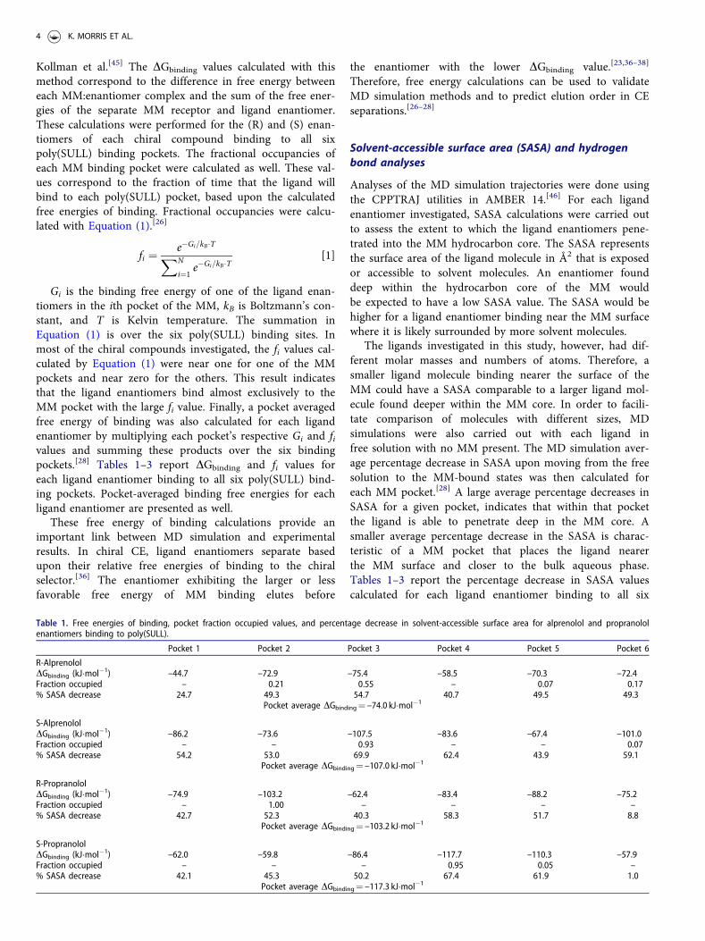
Kollman et al.[45] The DGbinding values calculated with thismethod correspond to the difference in free energy betweeneach MM:enantiomer complex and the sum of the free ener-gies of the separate MM receptor and ligand enantiomer.These calculations were performed for the (R) and (S) enan-tiomers of each chiral compound binding to all sixpoly(SULL) binding pockets. The fractional occupancies ofeach MM binding pocket were calculated as well. These val-ues correspond to the fraction of time that the ligand willbind to each poly(SULL) pocket, based upon the calculatedfree energies of binding. Fractional occupancies were calcu-lated with Equation (1).[26]
fi ¼ e�Gi=kB�TXN
i¼1e�Gi=kB�T
[1]
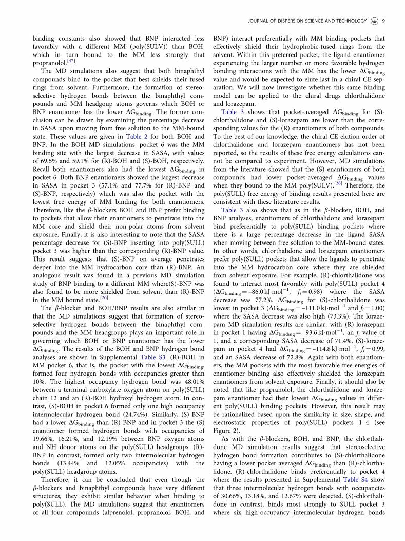
Gi is the binding free energy of one of the ligand enan-tiomers in the ith pocket of the MM, kB is Boltzmann’s con-stant, and T is Kelvin temperature. The summation inEquation (1) is over the six poly(SULL) binding sites. Inmost of the chiral compounds investigated, the fi values cal-culated by Equation (1) were near one for one of the MMpockets and near zero for the others. This result indicatesthat the ligand enantiomers bind almost exclusively to theMM pocket with the large fi value. Finally, a pocket averagedfree energy of binding was also calculated for each ligandenantiomer by multiplying each pocket’s respective Gi and fivalues and summing these products over the six bindingpockets.[28] Tables 1–3 report DGbinding and fi values foreach ligand enantiomer binding to all six poly(SULL) bind-ing pockets. Pocket-averaged binding free energies for eachligand enantiomer are presented as well.
These free energy of binding calculations provide animportant link between MD simulation and experimentalresults. In chiral CE, ligand enantiomers separate basedupon their relative free energies of binding to the chiralselector.[36] The enantiomer exhibiting the larger or lessfavorable free energy of MM binding elutes before
the enantiomer with the lower DGbinding value.[23,36–38]
Therefore, free energy calculations can be used to validateMD simulation methods and to predict elution order in CEseparations.[26–28]
Solvent-accessible surface area (SASA) and hydrogenbond analyses
Analyses of the MD simulation trajectories were done usingthe CPPTRAJ utilities in AMBER 14.[46] For each ligandenantiomer investigated, SASA calculations were carried outto assess the extent to which the ligand enantiomers pene-trated into the MM hydrocarbon core. The SASA representsthe surface area of the ligand molecule in Å2 that is exposedor accessible to solvent molecules. An enantiomer founddeep within the hydrocarbon core of the MM wouldbe expected to have a low SASA value. The SASA would behigher for a ligand enantiomer binding near the MM surfacewhere it is likely surrounded by more solvent molecules.
The ligands investigated in this study, however, had dif-ferent molar masses and numbers of atoms. Therefore, asmaller ligand molecule binding nearer the surface of theMM could have a SASA comparable to a larger ligand mol-ecule found deeper within the MM core. In order to facili-tate comparison of molecules with different sizes, MDsimulations were also carried out with each ligand infree solution with no MM present. The MD simulation aver-age percentage decrease in SASA upon moving from the freesolution to the MM-bound states was then calculated foreach MM pocket.[28] A large average percentage decreases inSASA for a given pocket, indicates that within that pocketthe ligand is able to penetrate deep in the MM core. Asmaller average percentage decrease in the SASA is charac-teristic of a MM pocket that places the ligand nearerthe MM surface and closer to the bulk aqueous phase.Tables 1–3 report the percentage decrease in SASA valuescalculated for each ligand enantiomer binding to all six
Table 1. Free energies of binding, pocket fraction occupied values, and percentage decrease in solvent-accessible surface area for alprenolol and propranololenantiomers binding to poly(SULL).
Pocket 1 Pocket 2 Pocket 3 Pocket 4 Pocket 5 Pocket 6
R-AlprenololDGbinding (kJ�mol�1) –44.7 –72.9 –75.4 –58.5 –70.3 –72.4Fraction occupied – 0.21 0.55 – 0.07 0.17% SASA decrease 24.7 49.3 54.7 40.7 49.5 49.3
Pocket average DGbinding¼ –74.0 kJ�mol�1
S-AlprenololDGbinding (kJ�mol�1) –86.2 –73.6 –107.5 –83.6 –67.4 –101.0Fraction occupied – – 0.93 – – 0.07% SASA decrease 54.2 53.0 69.9 62.4 43.9 59.1
Pocket average DGbinding¼ –107.0 kJ�mol�1
R-PropranololDGbinding (kJ�mol�1) –74.9 –103.2 –62.4 –83.4 –88.2 –75.2Fraction occupied – 1.00 – – – –% SASA decrease 42.7 52.3 40.3 58.3 51.7 8.8
Pocket average DGbinding¼ –103.2 kJ�mol�1
S-PropranololDGbinding (kJ�mol�1) –62.0 –59.8 –86.4 –117.7 –110.3 –57.9Fraction occupied – – – 0.95 0.05 –% SASA decrease 42.1 45.3 50.2 67.4 61.9 1.0
Pocket average DGbinding¼ –117.3 kJ�mol�1
4 K. MORRIS ET AL.

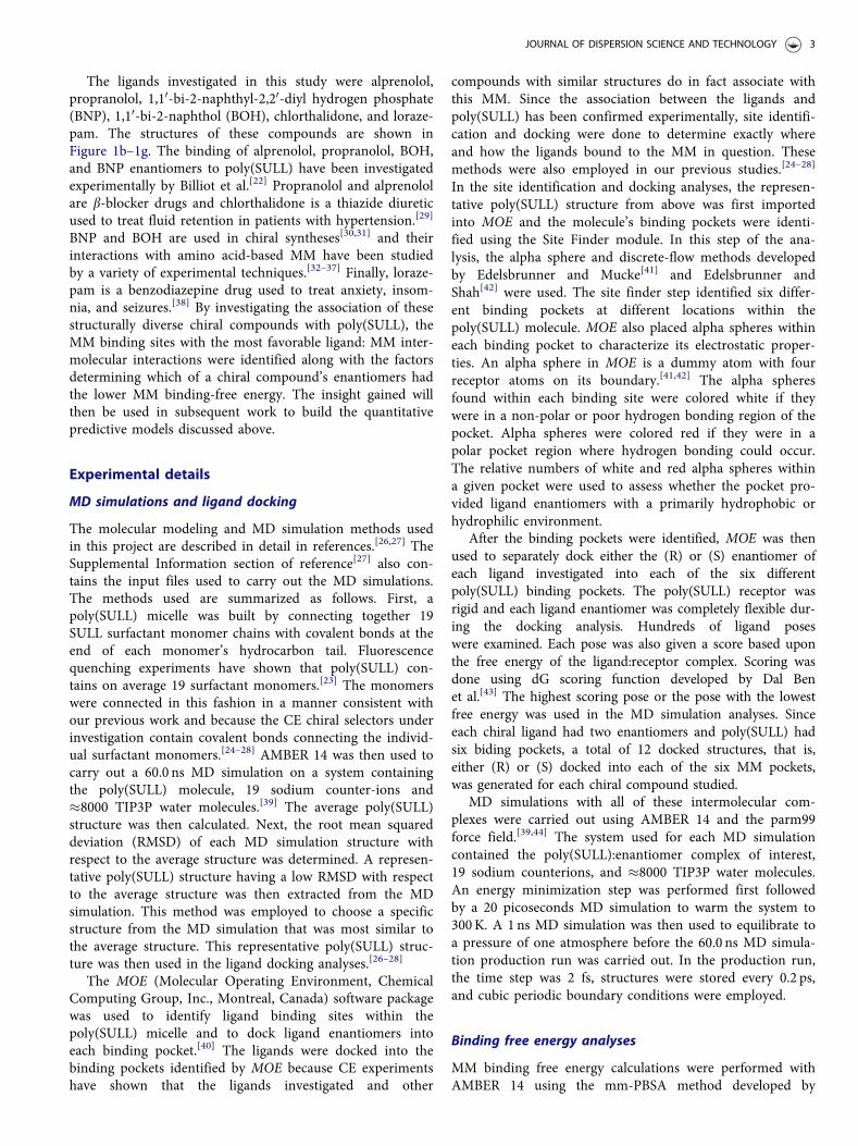
poly(SULL) binding pockets. Figure 2 shows a representativeplot of SASA versus simulation time for (R)-BOH bindingto poly(SULL) pocket 6. A plot of SASA versus simulationtime is also included for the MD simulation carried out withthe ligand enantiomer in free solution. The arrow on thegraph represents the percentage decrease in SASA.
The CPPTRAJ utilities in AMBER 14 were also used toinvestigate intermolecular hydrogen bond formation betweenligand enantiomers and the MM dipeptide headgroups.[46]
The criteria used to identify a hydrogen bond were as fol-lows. The distance cutoff between the heavy atoms makingup the hydrogen bond was 3.0 Å and the angle cutoffbetween the donor and acceptor atoms was ±30 �C.[46] TheseH-bond cutoffs are the default values in the AMBER 14ccptraj utility and were also used in our previous MD
simulation analyses.[24–28] Supplemental Tables S1–S5 pre-sent the hydrogen bond analyses carried out for each ligandenantiomer in the six poly(SULL) binding pockets. For eachH-bond, the donor atom, acceptor atom, and percentageoccupancy are reported. The later quantity corresponds tothe percentage of the 60.0 ns simulation time that a givenhydrogen bond was present.
Results and discussion
Poly(SULL) binding sites or pockets were identified asdescribed above by first importing a representativepoly(SULL) structure into the software package MOE. Thesoftware’s site finder feature then identified the six distinctbinding pockets shown in Figure 3a–3f.[40] In Figure 3, each
Table 2. Free energies of binding, pocket fraction occupied values, and percentage decrease in solvent-accessible surface area for BOH and BNP enantiomersbinding to poly(SULL).
Pocket 1 Pocket 2 Pocket 3 Pocket 4 Pocket 5 Pocket 6
R-BOHDGbinding (kJ.mol�1) –56.7 –53.1 –49.4 –58.6 –41.2 –88.1Fraction occupied – – – – – 1.00% SASA decrease 55.1 59.2 55.9 65.4 1.6 69.5
Pocket average DGbinding¼ –88.1 kJ.mol�1
S-BOHDGbinding (kJ.mol�1) –45.1 –39.6 –45.8 –39.8 –39.7 –61.7Fraction occupied – – – – – 1.00% SASA decrease 46.2 46.6 54.3 41.9 42.4 59.1
Pocket average DGbinding¼ –61.6 kJ.mol�1
R-BNPDGbinding (kJ.mol�1) –28.0 –36.8 –46.0 –28.4 –26.3 –25.6Fraction occupied – 0.02 0.97 – – –% SASA decrease 50.9 55.4 57.1 42.3 36.3 34.6
Pocket average DGbinding¼ –45.7 kJ.mol�1
S-BNPDGbinding (kJ.mol�1) –42.8 –56.8 –66.6 –28.0 –39.2 –51.6Fraction occupied – 0.02 0.98 – – –% SASA decrease 53.4 60.6 77.7 37.0 47.0 52.2
Pocket average DGbinding¼ –66.4 kJ.mol�1
Table 3. Free energies of binding, pocket fraction occupied values, and percentage decrease in solvent-accessible surface area for chlorthalidone and lorazepamenantiomers binding to poly(SULL).
Pocket 1 Pocket 2 Pocket 3 Pocket 4 Pocket 5 Pocket 6
R-ChlorthalidoneDGbinding (kJ.mol�1) –75.9 –59.5 –44.2 –86.0 –53.1 –42.1Fraction occupied 0.02 – – 0.98 – –% SASA decrease 71.1 66.3 48.9 77.2 51.9 48.7
Pocket average DGbinding¼ –85.8 kJ.mol�1
S-ChlorthalidoneDGbinding (kJ.mol�1) –96.0 –48.7 –111.0 –45.8 –41.3 –67.9Fraction occupied – – 1.00 – – –% SASA decrease 71.9 18.3 73.3 51.9 46.0 69.0
Pocket average DGbinding¼ –111.01 kJ.mol�1
R-LorazepamDGbinding (kJ.mol�1) –93.6 –52.4 –56.4 –62.1 –49.5 –58.3Fraction occupied 1.00 – – – – –% SASA decrease 71.4 42.2 44.8 46.4 46.5 40.9
Pocket average DGbinding¼ –93.6 kJ.mol�1
S-LorazepamDGbinding (kJ.mol�1) –71.6 –78.8 –70.9 –114.8 –103.1 –78.9Fraction occupied – – – 0.99 – –% SASA decrease 50.0 58.8 55.4 72.8 75.2 52.2
Pocket average DGbinding¼ –114.7 kJ.mol�1
JOURNAL OF DISPERSION SCIENCE AND TECHNOLOGY 5

pocket is displayed as a solid surface with green and red col-ors corresponding to pocket regions that are, respectivelyhydrophobic or hydrophilic. Alpha spheres are also shownwithin each binding site. Red and white alpha spheresidentify, respectively, good and poor hydrogen bonding loca-tions within the pocket.[40–42] In other words, a pocket col-ored green and containing a large number of white alphaspheres is very hydrophobic, whereas a pocket colored redcontaining a large number of red alpha spheres is insteadhydrophilic.
It should also be noted that the pockets shown inFigure 3a–3f are those found in a representative structureextracted from an MD simulation of the poly(SULL) MM.The monomer chains in the MM though are dynamic, so wewould expect the exact nature of these pockets to fluctuate
over time. However, the number of pockets and their diver-sity is governed by the number of monomers in the MMand the amino acids making up the dipeptide headgroup. Soalthough the pocket properties may fluctuate as the chainsmove, the number and diversity of binding sites will likelyremain largely the same. In other words, the pockets shownin Figure 3 represent a reasonable representation of thebinding sites available to chiral compounds when they bindto poly(SULL).
An examination of the poly(SULL) pockets in Figure 3illustrates interesting differences between the binding sitesformed by poly(SULL) and those detected in our previouswork with the MM poly-(sodium undecyl-(L)-leucine-valine) or poly(SULV). First, poly(SULL) was found toform six binding pockets, compared to only four pocketsfound in poly(SULV).[26–28] More significantly, though thepoly(SULL) analyses suggest that four of the MM’s six bind-ing sites, namely pockets 1–4 shown in Figure 3a–3d, aresimilar in both size and shape. This behavior contrasts previ-ously reported behavior for poly(SULV) where a deep nar-row pocket, two more non-polar dish-shaped pockets, and afourth non-polar pocket near the MM surface weredetected.[28] Figure 3a–3d suggest that poly(SULL) pockets1–4 have similar electrostatic properties as well. This similar-ity is evident if we examine the number of white and red(i.e. hydrophobic or hydrophilic) alpha spheres placed by theMOE software in poly(SULL) pockets 1 (21 white and4 red), 2 (27 white and 5 red), 3 (14 white and 6 red), and4 (15 white and 3 red). This behavior is somewhat expectedbecause poly(SULL) has two identical leucine amino acids inits dipeptide headgroup. Therefore, we might expect the bid-ing pockets formed in different regions of poly(SULL) to be
Figure 2. Plot of solvent-accessible surface area versus simulation time for (R)-BOH: pocket 6 MD simulation. The red and blue lines correspond to, respect-ively, a BOH MD simulation in free solution and bound to poly(SULL).
Figure 3. Poly(SULL) binding pockets. Green and red correspond to, respectively, hydrophobic and hydrophilic pocket regions.
6 K. MORRIS ET AL.

similar as well. Furthermore, the poly(SULL) pockets shownin Figure 3 when compared to the poly(SULV) pocketsreported in reference[28] suggest that changing the aminoacids making up the dipeptide headgroup also changes thecharacter of the MM’s binding pockets.
Finally, poly(SULL) pockets 5 and 6 appear to be some-what different than pockets 1–4. Figure 3e shows thatpoly(SULL) pocket 5 is primarily hydrophilic in nature withfive red and only one white alpha spheres. Pocket 5, though,appears to be approximately the same size as poly(SULL)pockets 1–4. Finally, poly(SULL) pocket 6 is somewhatsmaller than the other five pockets and is primarily hydro-phobic in nature, with 28 white and no red alpha spheres.Also note that all of pocket 6 in Figure 3f is colored greenfurther illustrating its non-polar nature.
We now move to an examination of how the chiral com-pounds listed above interact with these six poly(SULL) bind-ing pockets. Results from the MD simulations withalprenolol:poly(SULL) intermolecular complexes are sum-marized in Table 1 and Supplemental Table S1. Table 1 givesfree energies of MM binding, DGbinding, and fractional occu-pancies, fi, for (R) and (S)-alprenolol binding to poly(SULL)pockets 1 through 6. The pocket averaged DGbinding valuesin Table 1 for the (R) and (S) enantiomers of alprenololwere found to be –74.0 and –107.0 kJ�mol�1, respectively.Therefore, the MD simulation analyses predict that (S)-alprenolol interacts more favorably with poly(SULL) than(R)-alprenolol. This result is consistent with experiment, inthat a CE analysis of a racemic alprenolol mixture usingpoly(SULL) as the chiral selector showed that (S)-alprenololeluted after (R)-alprenolol.[22] Enantiomers separate in chiralCE based upon their relative free energies of binding to thechiral selector.[34–36] Therefore, the experimental CE resultsalso show that (S)-alprenolol interacts with poly(SULL)more favorably that (R)-alprenolol.
Table 1 also shows that the binding free energy for bothalprenolol enantiomers is lowest in poly(SULL) pocket 3. In(R)-alprenolol, the pocket 3 fi¼ 0.55. This fi value is thehighest of the six pockets, however, pockets 2 and 6 have fivalues of 0.21 and 0.17, respectively. This result suggests that(R)-alprenolol can interact favorably with these poly(SULL)pockets as well. In contrast, the (S)-alprenolol pocket 3 fivalue is 0.93 indicating that the (S) enantiomer binds moststrongly to this single MM pocket. Finally, for both alpreno-lol enantiomers, pocket 3 is likely preferred because alpreno-lol is a bulky, awkwardly shaped ligand and Figure 3 showsthat pocket 3 is the largest of the six binding sites.
SASA analyses can be used to further rationalize why thealprenolol enantiomers have a high affinity for poly(SULL)pocket 3. For example, in the (R)-alprenolol pocket 3 MDsimulation, the SASA percentage decrease in moving fromfree solution to the MM bound state was found to be 54.7%.This value represented the largest decrease among the sixpoly(SULL) binding pockets. In the (S)-alprenolol pocket 3MD simulation, the corresponding SASA decrease was foundto be 69.9%, which was also the largest decrease observedfor the (S)-enantiomer. Therefore, the results of Table 1show a correlation between a poly(SULL) pocket’s freeenergy of ligand binding and the SASA decrease experienced
by a ligand binding in that pocket, with the lowest DGbinding
corresponding to a high SASA decrease. This result suggeststhat the alprenolol enantiomers bind preferentially to thepoly(SULL) pocket that most effectively shields their hydro-phobic atoms from solvent.
Supplemental Table S1 presents an analysis of theintermolecular hydrogen bonds formed during thealprenolol:poly(SULL) MD simulations. For each MMpocket, the percentage occupancies are listed for intermo-lecular hydrogen bonds that were present for more than10% of the MD simulation time. The H-bond donor andacceptor atoms are given as well. In Supplemental Table S1,C-Leu and N-Leu are used to represent atoms on, respect-ively the C-terminal and N-terminal amino acids of thepoly(SULL) dipeptide headgroup. The symbol C¼O repre-sents the carbonyl oxygen connecting the dipeptide head-group to the surfactant monomer’s hydrocarbon chain.Finally, recall that the poly(SULL) MM contained 19 cova-lently bound surfactant monomers. The surfactant monomerchain containing the atoms forming each hydrogen bond(identified as chains 1–19) are listed in SupplementalTable S1 as well.
The Supplemental Table S1 results show that the MMpocket with the lowest binding free energy is not necessarilythe pocket where the most intermolecular hydrogen bondsformed between poly(SULL) and the alprenolol enantiomers.For example, (R)-alprenaolol had the lowest binding freeenergy in pocket 3, yet the intermolecular hydrogen bondpercentage occupancies were actually higher in poly(SULL)pockets 2 and 5. (S)-alprenolol also had the lowest DGbinding
in pocket 3, yet the percentage occupancies of the intermo-lecular H-bonds were higher in pocket 6. Therefore, the MDsimulation results suggest that alprenolol enantiomers bindto the pocket that best shields their hydrophobic atoms fromsolvent (thus the large SASA decrease discussed above), butnot necessarily to the pocket where ligand:MM hydrogenbonding is the strongest.
While hydrogen bonding interactions may not govern thepreferred ligand binding site, the Supplemental Table S1results suggest that the formation of intermolecular hydro-gen bonds is an important factor in determining whether(R) or (S)-alprenolol interacts more strongly with the MM.Table 1 results show that both alprenolol enantiomers inter-act most favorably with poly(SULL) pocket 3. DGbinding of(S)-alprenolol, however, in pocket 3 is lower than the corre-sponding value for (R)-alprenolol in the same pocket.Examination of the Supplemental Table S1 results showsthat in pocket 3, (S)-alprenolol forms seven intermolecularhydrogen bonds with poly(SULL) having percentage occu-pancies greater than 10%. The three highest occupancy (S)-alprenolol:poly(SULL) pocket 3 hydrogen bonds have per-centage occupancies of 34.65%, 24.30%, and 21.04%. In con-trast, (R)-alprenolol in pocket 3 forms only three hydrogenbonds with occupancies greater than 10%, the highest ofwhich is 13.29%. The (R)-alprenolol fi values in Table 1show that the (R) enantiomer also has affinity forpoly(SULL) pockets 2 and 6. However, the percentage occu-pancies of the (S)-alprenolol H-bonds in pocket 3 are gener-ally larger than the corresponding (R)-alprenolol pocket 2
JOURNAL OF DISPERSION SCIENCE AND TECHNOLOGY 7

and 6 occupancies. Therefore, the alprenolol results suggestthat once the ligand binds to the MM in its preferred lowfree energy pocket or pockets, the formation of stereoselect-ive hydrogen bonds, that is, H-bonds that are different forthe (R) and (S) enantiomers, plays an important role in gov-erning whether the (R) or (S) enantiomer has the overalllowest binding free energy. Formation of these stereoselect-ive hydrogen bonds would, therefore, be expected to deter-mine the enantiomer’s elution order in chiral CE separationsas well, since elution order is determined by the relative freeenergies of MM binding for each of the ligand enantiomers.
Conclusions similar to those discussed above can also bedrawn from the propranolol:poly(SULL) MD simulationanalyses. Table 1 shows that the pocket-averaged MM bind-ing free energy for (S)-propranolol is lower than the corre-sponding (R)-propranolol value. This result indicates thatthe (S) enantiomer experiences more favorable interactionswith the MM and is consistent with experiment in that (S)-propranolol was found to elute after (R)-propranolol in CEseparations with poly(SULL) as the chiral selector.[22]
Table 1 also shows that (R)-propranolol had the lowest bind-ing free energy in pocket 2, while DGbinding was lowest for(S)-propranolol in pocket 4. The fi values were 1.00 for (R)-propranolol in pocket 2 and 0.95 for (S)-propranolol inpocket 4, suggesting that both propranolol enantiomers bindpreferentially to a single poly(SULL) pocket. Furthermore,Figure 3 shows that poly(SULL) pockets 2 and 4 are verysimilar in size, shape, and electrostatic properties; therefore,it may not be surprising that the propranolol enantiomersdo not necessarily prefer the same binding site.
As with alprenolol, the propranolol MD simulation analy-ses show that the decrease in ligand SASA upon movingfrom free solution to the MM-bound state is a good pre-dictor of which MM pocket has the lowest DGbinding value.The SASA decrease of 52.3% for (R)-propranolol in pocket 2was the second highest of the six pockets investigated andthe SASA decrease of 67.4% for (S)-propranolol in pocket 4was the largest decrease observed in the (S) enantiomer anal-yses. Therefore, both b-blockers (alprenolol and propranolol)preferentially bind to a MM pocket that effectively shieldstheir hydrophobic rings from solvent.
In addition, once bound to the MM in the pocket with thelowest DGbinding and highest fi values, the propranolol MDsimulations suggest that hydrogen bond formation betweenthe MM and propranolol enantiomers plays an important rolein determining which enantiomer experiences the most favor-able interactions with poly(SULL). Supplemental Table S2shows that (S)-propranolol in pocket 4 forms seven hydrogenbonds with percentage occupancies greater than 10%; thehighest being 46.63% between a terminal carboxylate oxygenatom of poly(SULL) monomer chain six and the (S)-propran-olol hydroxyl hydrogen atom. In contrast, (R)-propranolol inpocket 2 forms five hydrogen bonds with occupancies greaterthan 10% with the highest value of only 21.53%. Therefore, asin the alprenolol MD simulation analysis, stereoselectivehydrogen bond formation between the propranolol enantiom-ers and poly(SULL) likely plays an important role in deter-mining chiral discrimination and elution order in CEexperiments.
We will now consider the BOH and BNP MD simulationanalyses. These compounds were investigated in part becausetheir interactions with poly(SULL) have been studied experi-mentally.[22] Furthermore, these two binaphthyl compoundshave very different structures than the b-blockers alprenololand propranolol. Therefore, comparing the b-blocker andBOH/BNP results will allow us to assess whether the conclu-sions drawn above hold only for b-blockers and moleculeswith similar structures or if they apply more generally to abroader range of structurally diverse compounds. Table 2shows that both enantiomers of BOH preferentially bind toMM pocket 6. The fi values are 1.00 for both enantiomers inthis pocket, indicating that the BOH enantiomers have avery high affinity for pocket 6 and a rather low affinity forthe other poly(SULL) binding sites. In contrast, both enan-tiomers of BNP preferentially bind to poly(SULL) pocket 3where the fi values were 0.97 and 0.98 for (R)-BNP and (S)-BNP, respectively. These results can be rationalized basedupon the shapes and electrostatic properties of thebinaphthyl compounds and their preferred poly(SULL) bind-ing pockets. Recall, pocket 6 is the most hydrophobic of thesix poly(SULL) binding sites, containing 28 white/non-polarand no red or polar alpha spheres. Therefore, it seems rea-sonable that enantiomers of a non-polar molecule like BOHwould have affinity for this pocket. BNP on the other handis anionic and more polar than BOH. Its enantiomers, there-fore, interact more favorably with pocket 3 which is morehydrophilic in nature than pocket 6. In fact, Figure 3 showsthat with 14 white and 8 red alpha spheres, MM pocket 3 isamong the most polar or hydrophilic poly(SULL) bind-ing site.
The pocket averaged MM binding free energies for(R)-BOH and (S)-BOH are –88.1 and –61.6 kJ�mol�1,respectively. This result shows that unlike the b-blockers, the(R) enantiomer of BOH interacts more favorably withpoly(SULL) than the (S) enantiomer. This result, however, isconsistent with experiment because a CE separation of aracemic BOH mixture with poly(SULL) as the chiral selectorshowed that (R)-BOH eluted after (S)-BOH.[22] The pocketaveraged DGbinding values for (R)-BNP and (S)-BNP are–45.7 and –66.4 kJ�mol�1, respectively. Again this resultis consistent with experiment because in a CE separationof racemic BNP using poly(SULL) as the chiral selector,(S)-BNP eluted after (R)-BNP.[22] It also interesting to notethat the association of the BOH enantiomers withpoly(SULL) (DGbinding¼ –88.1 and –61.6 kJ�mol�1 for thetwo enantiomers) is overall stronger than that of thecorresponding BNP enantiomers (DGbinding¼ –45.7 and–66.4 kJ�mol�1). This observation likely results from themore non-polar BOH enantiomers having a greater affinityfor poly(SULL) which also has two non-polar leucine aminoacids in its dipeptide headgroups. The binaphthyl com-pounds also interact less strongly with poly(SULL) than theb-blocker enantiomers. Again this result is expected giventhat the b-blockers are cationic and are thus attracted to theanionic MM. Alprenolol and propranolol also contain morehydrogen bond donor and acceptor atoms and thus likelyform more hydrogen bonds with the poly(SULL) headgroupsthan BNP and BOH. Finally, NMR measurements of MM
8 K. MORRIS ET AL.
binding constants also showed that BNP interacted lessfavorably with a different MM (poly(SULV)) than BOH,which in turn bound to the MM less strongly thatpropranolol.[47]
The MD simulations also suggest that both binaphthylcompounds bind to the pocket that best shields their fusedrings from solvent. Furthermore, the formation of stereo-selective hydrogen bonds between the binaphthyl com-pounds and MM headgoup atoms governs which BOH orBNP enantiomer has the lower DGbinding. The former con-clusion can be drawn by examining the percentage decreasein SASA upon moving from free solution to the MM-boundstate. These values are given in Table 2 for both BOH andBNP. In the BOH MD simulations, pocket 6 was the MMbinding site with the largest decrease in SASA, with valuesof 69.5% and 59.1% for (R)-BOH and (S)-BOH, respectively.Recall both enantiomers also had the lowest DGbinding inpocket 6. Both BNP enantiomers showed the largest decreasein SASA in pocket 3 (57.1% and 77.7% for (R)-BNP and(S)-BNP, respectively) which was also the pocket with thelowest free energy of MM binding for both enantiomers.Therefore, like the b-blockers BOH and BNP prefer bindingto pockets that allow their enantiomers to penetrate into theMM core and shield their non-polar atoms from solventexposure. Finally, it is also interesting to note that the SASApercentage decrease for (S)-BNP inserting into poly(SULL)pocket 3 was higher than the corresponding (R)-BNP value.This result suggests that (S)-BNP on average penetratesdeeper into the MM hydrocarbon core than (R)-BNP. Ananalogous result was found in a previous MD simulationstudy of BNP binding to a different MM where(S)-BNP wasalso found to be more shielded from solvent than (R)-BNPin the MM bound state.[26]
The b-blocker and BOH/BNP results are also similar inthat the MD simulations suggest that formation of stereo-selective hydrogen bonds between the binaphthyl com-pounds and the MM headgroups plays an important role ingoverning which BOH or BNP enantiomer has the lowerDGbinding. The results of the BOH and BNP hydrogen bondanalyses are shown in Supplemental Table S3. (R)-BOH inMM pocket 6, that is, the pocket with the lowest DGbinding,formed four hydrogen bonds with occupancies greater than10%. The highest occupancy hydrogen bond was 48.01%between a terminal carboxylate oxygen atom on poly(SULL)chain 12 and an (R)-BOH hydroxyl hydrogen atom. In con-trast, (S)-BOH in pocket 6 formed only one high occupancyintermolecular hydrogen bond (24.74%). Similarly, (S)-BNPhad a lower DGbinding than (R)-BNP and in pocket 3 the (S)enantiomer formed hydrogen bonds with occupancies of19.66%, 16.21%, and 12.19% between BNP oxygen atomsand NH donor atoms on the poly(SULL) headgroups. (R)-BNP in contrast, formed only two intermolecular hydrogenbonds (13.44% and 12.05% occupancies) with thepoly(SULL) headgroup atoms.
Therefore, it can be concluded that even though theb-blockers and binaphthyl compounds have very differentstructures, they exhibit similar behavior when binding topoly(SULL). The MD simulations suggest that enantiomersof all four compounds (alprenolol, propranolol, BOH, and
BNP) interact preferentially with MM binding pockets thateffectively shield their hydrophobic-fused rings from thesolvent. Within this preferred pocket, the ligand enantiomerexperiencing the larger number or more favorable hydrogenbonding interactions with the MM has the lower DGbinding
value and would be expected to elute last in a chiral CE sep-aration. We will now investigate whether this same bindingmodel can be applied to the chiral drugs chlorthalidoneand lorazepam.
Table 3 shows that pocket-averaged DGbinding for (S)-chlorthalidone and (S)-lorazepam are lower than the corre-sponding values for the (R) enantiomers of both compounds.To the best of our knowledge, the chiral CE elution order ofchlorthalidone and lorazepam enantiomers has not beenreported, so the results of these free energy calculations can-not be compared to experiment. However, MD simulationsfrom the literature showed that the (S) enantiomers of bothcompounds had lower pocket-averaged DGbinding valueswhen they bound to the MM poly(SULV).[28] Therefore, thepoly(SULL) free energy of binding results presented here areconsistent with these literature results.
Table 3 also shows that as in the b-blocker, BOH, andBNP analyses, enantiomers of chlorthalidone and lorazepambind preferentially to poly(SULL) binding pockets wherethere is a large percentage decrease in the ligand SASAwhen moving between free solution to the MM-bound states.In other words, chlorthalidone and lorazepam enantiomersprefer poly(SULL) pockets that allow the ligands to penetrateinto the MM hydrocarbon core where they are shieldedfrom solvent exposure. For example, (R)-chlorthalidone wasfound to interact most favorably with poly(SULL) pocket 4(DGbinding¼ –86.0 kJ�mol�1, fi¼ 0.98) where the SASAdecrease was 77.2%. DGbinding for (S)-chlorthalidone waslowest in pocket 3 (DGbinding¼ –111.0 kJ�mol�1 and fi¼ 1.00)where the SASA decrease was also high (73.3%). The loraze-pam MD simulation results are similar, with (R)-lorazepamin pocket 1 having DGbinding¼ –93.6 kJ�mol�1, an fi value of1, and a corresponding SASA decrease of 71.4%. (S)-loraze-pam in pocket 4 had DGbinding¼ –114.8 kJ�mol�1, fi¼ 0.99,and an SASA decrease of 72.8%. Again with both enantiom-ers, the MM pockets with the most favorable free energies ofenantiomer binding also effectively shielded the lorazepamenantiomers from solvent exposure. Finally, it should also benoted that like propranolol, the chlorthalidone and loraze-pam enantiomer had their lowest DGbinding values in differ-ent poly(SULL) binding pockets. However, this result maybe rationalized based upon the similarity in size, shape, andelectrostatic properties of poly(SULL) pockets 1–4 (seeFigure 2).
As with the b-blockers, BOH, and BNP, the chlorthali-done MD simulation results suggest that stereoselectivehydrogen bond formation contributes to (S)-chlorthalidonehaving a lower pocket averaged DGbinding than (R)-chlortha-lidone. (R)-chlorthalidone binds preferentially to pocket 4where the results presented in Supplemental Table S4 showthat three intermolecular hydrogen bonds with occupanciesof 30.66%, 13.18%, and 12.67% were detected. (S)-chlorthali-done in contrast, binds most strongly to SULL pocket 3where six high-occupancy intermolecular hydrogen bonds
JOURNAL OF DISPERSION SCIENCE AND TECHNOLOGY 9

were formed, three of which were over 30%. Therefore, withchlorthalidone we see that the enantiomer with the lowestDGbinding also experiences more favorable hydrogen bondinginteractions with the MM.
The results of the lorazepam:poly(SULL) hydrogen bond-ing analysis shown in Supplemental Table S5, however, areless straightforward. (R)-lorazepam was found to bind pref-erentially to poly(SULL) pocket 1 where Supplemental TableS5 shows that two intermolecular hydrogen bonds withoccupancies of 42.16% and 18.01% were detected. (S)-loraze-pam bound most strongly to poly(SULL) pocket 4 wherethere were more intermolecular hydrogen bonds (four versustwo for (R)-lorazepam), but the percentage occupancies ofthese hydrogen bonds were generally lower (21.82%, 18.08%,16.80%, and 14.01%). Therefore, we cannot conclude that(S)-lorazepam has the lower free energy of poly(SULL) bind-ing simply because the (S) enantiomer has more favorablehydrogen bonding interactions with the MM.
A previous investigation of lorazepam binding to a differ-ent MM (poly(SULV)) showed that ligand penetration intothe MM hydrocarbon core, intermolecular hydrogen bond-ing, and the orientation adopted by the ligand enantiomerswithin each pocket were all important factors in determininghow strongly each enantiomer interacted with the MM.[28]
The lorazepam enantiomers were found to interact mostfavorably with the MM pockets when the molecule’s seven-membered and two aromatic rings were all oriented towardthe MM hydrocarbon core. Less favorable orientations of therings with respect to the hydrocarbon core and solvent phaselead to less favorable DGbinding values.[28] Similar behaviormay be occurring in the lorazepam:poly(SULL) system.A more detailed analysis of all the factors governing thebinding of lorazepam enantiomers to poly(SULL) is cur-rently underway.
Finally, one conclusion drawn from all of the above MDsimulation results is that the ligands investigated bind pref-erentially to a poly(SULL) pocket that shielded their hydro-phobic rings from solvent exposure. Figure 4 shows threerepresentative structures extracted from the MD simulationsthat show ligand enantiomers inserted into the bindingpockets in this manner. Figure 4a shows a structureextracted from the (S)-propranolol: pocket 4 MD simulationat 21.8 ns. Figure 4b shows a structure extracted from the(R)-BOH: pocket 6 MD simulation at 41.0 ns, and Figure 4cis a structure extracted from the (S)-lorazepam: pocket 4MD simulation at 19.0 ns. The structures at each of thesetime steps were found to have a low RMSD value withrespect to the average structure. As shown in Figure 3, thepolar and non-polar regions of the Figure 4 binding pocketsare colored red and green, respectively. Each of the Figure 4structures shows the ligands’ hydrophobic fused rings point-ing into the non-polar pocket region and oriented towardthe poly(SULL) hydrocarbon core.
Conclusions
MD simulations have been used to investigate the stereo-selective binding of six different chiral compounds to theamino acid-based MM poly(SULL). Poly(SULL) was foundto have six pockets or binding sites into which chiral ligandenantiomers could insert. Four of these pockets were foundto have similar sizes, shapes, and electrostatic properties. Allsix chiral compounds bound most strongly to a poly(SULL)pocket that shielded their non-polar atoms from solvent. Infive of the six compounds studied, the formation of stereo-selective intermolecular hydrogen bonds determined whichenantiomer had the lower free energy of MM association.This enantiomer would be expected to elute last in a chiralCE separation. Subsequent studies will further test thismodel with single amino acid and other dipeptide MMbefore using the model to develop molecular modeling-basedtechniques that will identify the best MM for a given chiralseparation problem.
Acknowledgments
This work was supported by an NIH-NIMHD grant (#G12 MD007579)to the RCMI Program at Howard University, NSF-RUI grants(#1709680, #1709394, and #1708959), a Robert A. Welch ChemistryDepartmental Grant to the Chemistry Program at Texas A&MUniversity-Corpus Christi, an NSF CAREER grant (#0449742) toDr. Eugene Billiot and a HUMAA Endowed Founder’s Chair in BasicScience award and Office of Naval Research grants (#N00014-17-1-2105and #N00014-18-2145) grants to Dr. Yayin Fang. We also acknowledgethe generosity of the Ralph E. Klingenmeyer family.
References
[1] Li, B.; Haynie, D. T. Chiral Drug Separation. Encycl. Chem.Process. 2016, 1, 449–458.
[2] Lin, G.; You, Q.; Cheng, J. Chiral Drugs: Chemistry andBiological Action; Wiley & Sons: Hoboken, NJ, 2011.
Figure 4. Structures extracted from MD simulations showing ligands insertedinto micelle core. (a) (S)-propranolol:poly(SULL) complex at 21.8 ns, (b) (R)-BOH:poly(SULL) complex at 41.0 ns, and (c) (S)-lorazepam:poly(SULL) complexat 19.0 ns.
10 K. MORRIS ET AL.

[3] Announcement. FDA’s Policy Statement for the Development ofNew Stereoisomeric Drugs. Chirality. 1992, 4, 338.
[4] Subramanian, G. Chiral Separation Techniques, 3rd ed.; Wiley-VCH: New York, 2007.
[5] Ward, T. J.; Ward, K. D. Chiral Separations: FundamentalReview 2010. Anal. Chem. 2010, 82, 4712–4722.
[6] Valle, B. C.; Morris, K. F.; Fletcher, K. A.; Fernand, V.; Sword,D. M.; Eldridge, S.; Larive, C. K.; Warner, I. M. UnderstandingChiral Molecular Micelle Separations Using Steady-StateFluorescence Anisotropy, Capillary Electrophoresis, and NMR.Langmuir. 2007, 23, 425–435.
[7] Sciba, G. K. E. Chiral Recognition in Separation Science – AnUpdate. J. Chromatogr. A. 2016, 1467, 56–78.
[8] Billiot, E. J.; Macossay, J.; Thibodeaux, S.; Shamsi, S. A.;Warner, I. M. Chiral Separations Using Dipeptide PolymerizedSurfactants: Effect of Amino Acid Order. Anal. Chem. 1998, 70,1375–1381.
[9] Wang, J.; Warner, I. M. Chiral Separations Using MicellarElectrokinetic Chromatography and a Polymerized ChiralMicelle. Anal. Chem. 1994, 66, 3773–3776.
[10] Dobashi, A.; Hamada, M.; Dobashi, Y.; Yamaguchi, J.Enantiomeric Separation with Sodium Dodecanoyl-L-AminoAcidate Micelles and Poly(Sodium (10-Undecenoyl)-L-Valinate)by Electrokinetic Chromatography. Anal. Chem. 1995, 67,3011–3017.
[11] Palmer, C. P. Micelle Polymers, Polymer Surfactants, andDendrimers as Pseudostationary Phases in MicellarElectrokinetic Chromatography. J. Chromatogr. A. 1997, 780,75–92.
[12] Billiot, E. J.; Warner, I. M. Examination of Structural Changesof Polymeric Amino Acid-Based Surfactants onEnantioselectivity: Effect of Amino Acid Order, Steric Factors,and Number and Position of Chiral Centers. Anal. Chem. 2000,72, 1740–1748.
[13] Billiot, E. J.; Agbaria, R.; Thibodeaux, S.; Shamsi, S. A.; Warner,I. M. Amino Acid Order in Dipeptide Surfactants: Effect onPhysical Properties and Enantioselectivity. Anal. Chem. 1999,71, 1252–1256.
[14] Shamsi, S. A.; Warner, I. M. Monomeric and Polymeric ChiralSurfactants as Pseudostationary Phases for Chiral Separations.Electrophoresis 1997, 18, 853–872.
[15] Haddadian, F. H.; Shamsi, S. A.; Warner, I. M. ChiralElectrokinetic Chromatography Using Dipeptide PolymericSurfactants: State of the Art. Electrophoresis. 1999, 20,3011–3026.
[16] Hayes, J. L.; III.; Warner, I. M. Polymeric Surfactants asPseudostationary Phases for Separations in ElectrokineticChromatography (EKC): A Review. Rev. Anal. Chem. 1999, 18,317–382.
[17] Yarabe, H. H.; Billiot, E. J.; Warner, I. M. EnantiomericSeparations by Use of Polymeric Surfactant ElectrokineticChromatography. J. Chromatogr. A. 2000, 875, 179–206.
[18] Haynes, I. I. I.; J. L.; Billiot, E. J.; Yarabe, H. H.; Shamsi, S. A.;Warner, I. M. Chiral Separations with Dipeptide-TerminatedPolymeric Surfactants: The Effect of an Extra Hetroatom on thePolar Head Group. Electrophoresis. 2000, 21, 1597–1605.
[19] Thibodeaux, S. J.; Billiot, E. J.; Torres, E.; Valle, B. C.; Warner,I. M. Enantiomeric Separations Using Polymeric L-GlutamateSurfactant Derivatives: Effect of Increasing Steric Factors.Electrophoresis. 2003, 24, 1077–1082.
[20] Billiot, F. H.; Billiot, E. J.; Warner, I. M. Depth of Penetrationof Binaphthyl Derivatives into the Micellar Core of SodiumUndecenoyl Leucyl-Leucinate Surfactants. J. Chromatogr. A.2002, 950, 233–239.
[21] Shamsi, S. A.; Valle, B. C.; Billiot, F.; Warner, I. M. PolysodiumN-Undecanoyl-L-Leucylvalinate: A Versatile Chiral Selector forMicellar Electrokinetic Chromatography. Anal. Chem. 2003, 75,379–387.
[22] Billiot, E. J.; Thibodeaux, S.; Shamsi, S. A.; Warner, I. M.Evaluating Chiral Separation Interactions by Use of
Diastereometric Polymeric Dipeptide Surfactants. Anal. Chem.1999, 71, 4044–4049.
[23] Haddadian Billiot, F.; McCarroll, M. E.; Billiot, E. J.; Rugutt,J. K.; Morris, K. F.; Warner, I. M. Comparison of theAggregation Behavior of Fifteen Polymeric and MonomericDipeptide Surfactants in Aqueous Solution. Langmuir. 2002, 18,2993–2997.
[24] Morris, K. F.; Billiot, E. J.; Billiot, F. H.; Lipkowitz, K. B.;Southerland, W. M.; Fang, Y. Investigation of Chiral MolecularMicelles by NMR Spectroscopy and Molecular DynamicsSimulations. OJPC. 2012, 2, 2, 240–251.
[25] Morris, K. F.; Billiot, E. J.; Billiot, F. H.; Lipkowitz, K. B.;Southerland, W. M.; Fang, Y. A Molecular DynamicsSimulation Study of Two Dipeptide-Based Molecular Micelles:Effect of Amino Acid Order. OJPC. 2013, 3, 20–29. 3,
[26] Morris, K. F.; Billiot, E. J.; Billiot, F. H.; Lipkowitz, K. B.;Southerland, W. M.; Gladis, A. A.; Fang, Y. A MolecularDynamics Simulation Study of the Association of 1,10-Binaphthyl-2,20-Diyl Hydrogenphosphate Enantiomers with aChiral Molecular Micelle. Chem. Phys. 2014, 439, 36–43.
[27] Morris, K. F.; Billiot, E. J.; Billiot, F. H.; Hoffman, C. B.; Gladis,A. A.; Lipkowitz, K. B.; Southerland, W. H.; Fang, Y. MolecularDynamics Simulation and NMR Investigation of the Associationof the b-Blockers Atenolol and Propranolol with a ChiralMolecular Micelle. Chem. Phys. 2015, 457, 133–146.
[28] Morris, K. F.; Billiot, E. J.; Billiot, F. H.; Ingle, J. A.; Zack, S. R.;Krause, K. B.; Lipkowitz, K. B.; Southerland, W. H.; Fang, Y.Investigation of Chiral Recognition by Molecular Micelles withMolecular Dynamics Simulations. J. Dispersion Sci. Technol.2018, 39, 45–54.
[29] Chlorthalidone, U.S. National Library of Medicine: MedlinePlus, 2013.
[30] Marchand, A. P.; Chong, H. S.; Ganguly, B. Synthesis of aNovel Cage-Functionalized Chiral Binaphthol Host: A PotentialNew Agent for Enantioselective Recognition of ChiralAmmonium Salts. Tetrahedr. Asym. 1999, 10, 4695–4707.
[31] Jiehao, C.; Craig, J. F. Total Synthesis of Apratoxin A. J. Am.Chem. Soc. 2003, 125, 8734–8735.
[32] Billiot, F. H.; McCarroll, M. E.; Billiot, E. J.; Warner, I. M.Chiral Recognition of Binaphthyl Derivatives UsingElectrokinetic Chromatography and Steady-State FluorescenceAnisotropy: Effect of Temperature. Electrophoresis. 2004, 25,753–757.
[33] Rugutt, J. K.; Billiot, E. J.; Warner, I. M. NMR Study of theInteraction of Monomeric and Polymeric Chiral Surfactantswith (R)- and (S)-1,10-Binaphthyl-2,20-Diyl HydrogenPhosphate. Langmuir. 2000, 16, 3022–3029.
[34] Yarabe, H. H.; Rugutt, J. K.; McCarroll, M. E.; Warner, I. M.Capillary Electrophoretic Separation of Binaphthyl Enantiomerswith Two Polymeric Chiral Surfactants: 1H-Nuclear MagneticResonance and Fluorescence Spectroscopy Study.Electrophoresis. 2000, 21, 2025–2032.
[35] Kingsbury, S. A.; Ducommun, C. J.; Zahakaylo, B. M.;Dickinson, E. H.; Morris, K. F. NMR Characterization of 1,10-Binaphthyl-2,20-Diyl Hydrogen Phosphate Binding to ChiralMolecular Micelles. Magn. Reson. Chem. 2010, 48, 184–191.
[36] McCarroll, M. E.; Billiot, F. H.; Warner, I. M. FluorescenceAnisotropy as a Measure of Chiral Recognition. J. Am. Chem.Soc. 2001, 123, 3173–3174.
[37] Xu, Y.; McCarroll, M. E. Fluorescence Anisotropy as a Methodto Examine the Thermodynamics of Enantioselectivity. J Phys.Chem. B. 2005, 109, 8144–8152.
[38] Lorazepam, U.S. National Library of Medicine: Medline Plus,2010.
[39] Case, D. A.; Babin, V.; Berryman, J. T.; Betz, R. M.; Cai, Q.;Cerutti, D. S.; Cheatham III, T. E.; Darden, T. A.; Duke, R. E.;Gohlke.; et al. AMBER 14. San Francisco: University ofCalifornia, 2014.
[40] Molecular Operating Environment (MOE), ChemicalComputing Group, 2016.
JOURNAL OF DISPERSION SCIENCE AND TECHNOLOGY 11

[41] Edelsbrunner, H.; Mucke, E. P. Three Dimensional AlphaShapes. ACM Trans. Graph. 1994, 13, 43–72.
[42] Edelsbrunner, H.; Shah, N. R. Incremental Topological FlippingWorks for Regular Triangulations. Algorithmica. 1996, 15, 223–241.
[43] Dal Ben, D.; Buccioni, M.; Lambertucci, C.; Thomas, A.;Volpini, R. Simulation and Comparative Analysis of BindingModes of Nucleoside and Non-Nucleoside Agonists at the A2B
Adenosine Receptor. In Silico Pharmacol. 2013, 1, 24–38.[44] Wang, J.; Cieplak, P.; Kollman, P. A. How Well Does a
Restrained Electrostatic Potential (RESP) Model Perform inCalculating Conformational Energies of Organic and BiologicalMolecules? J. Comput. Chem. 2000, 21, 1049–1074.
[45] Kollman, P. A.; Massova, I.; Reyes, C.; Kuhn, B.; Huo, S.;Chong, L.; Lee, M.; Lee, T.; Duan, Y.; Wang, W.; et al.Calculating Structures and Free Energies of Complex Molecules:Combining Molecular Mechanics and Continuum Models. Acc.Chem. Res. 2000, 33, 889–897.
[46] Roe, D. R.; Cheatham, T. E. III, PTRAJ and CPPTRAJ: Softwarefor Processing and Analysis of Molecular Dynamics TrajectoryData. J. Chem. Theory Comput. 2013, 9, 3084–3095.
[47] Morris, K. F.; Becker, B. A.; Valle, B. C.; Warner, I. M.; Larive,C. K. Use of NMR Binding Interaction Mapping Techniques toExamine Interactions of Chiral Molecules with MolecularMicelles. J. Phys. Chem. B. 2006, 110, 17359–17369.
12 K. MORRIS ET AL.