Embed Size (px)
Citation preview

USG Cross Sectional Anatomy & US Pathology of Elbow & Wrist
Don-Kyu Kim M.D.
Chung-Ang University Hospital

Radiologic anatomy of elbow
Medial epicondyle
Coronoid process
Radial tuberosity
Lateral epicondyle
Head of radius
Olecranon
C RTR
O
AP Lateral
Anterior fat pad
Supinator fat line
Ligament of Elbow Lateral & Medial
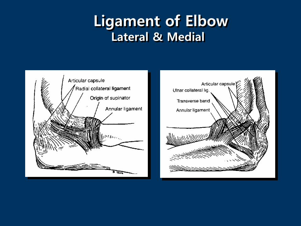
Ultrasound anatomy and scanning technique of the elbow
1. Anterior aspect
1) Elbow joint
a. Transverse scan (capitellum, trochlea)
b. Longitudinal scan (lateral radiocapital joint, medial trochlea ulnar joint)
2) Distal biceps tendon - longitudinal and transverse scan
2. Lateral aspect
1) Lateral epicondyle - longitudinal and transverse scan
2) Common extensor tendon - longitudinal and transverse scan
3. Medial aspect
1) Medial epicondyle - longitudinal and transverse scan
2) Common flexor tendon - longitudinal and transverse scan
3) Ulnar collateral ligament - longitudinal and transverse scan
4) Ulnar nerve - longitudinal and transverse scan
4. Posterior aspect
1) Triceps tendon and olecranon fossa - longitudinal and transverse scan
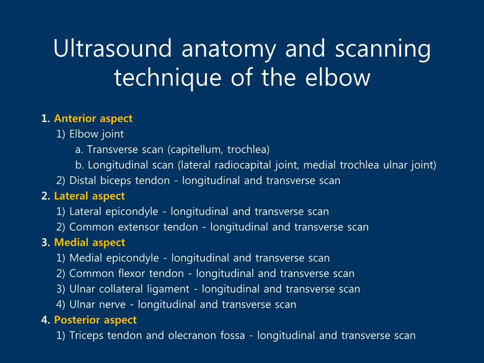
Transverse Scan of Anterior Elbow
Median N
Brachial a. & v.
Brachialis
Radial N
Brachioradialis
B: BrachialisC: capitellumT: Trochlear
B
C
T
Biceps tendon
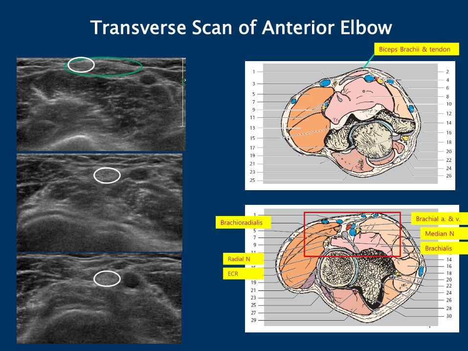
Transverse Scan of Anterior Elbow
Median N
Brachial a. & v.
Brachialis
Radial N
Brachioradialis
ECR
Biceps Brachii & tendon

Longitudinal Scan of Anterior Elbow: lateral side
RH: Radial head, C: Capitellum
Radial N Brachioradial m Brachialis
B
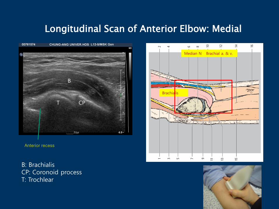
Longitudinal Scan of Anterior Elbow: Medial
Median N Brachial a. & v.
Brachialis
B
T CP
Anterior recess
B: BrachialisCP: Coronoid processT: Trochlear
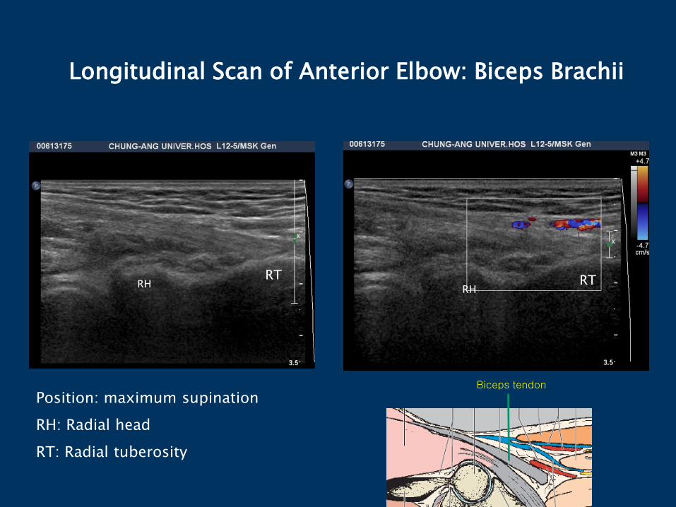
Longitudinal Scan of Anterior Elbow: Biceps Brachii
Position: maximum supination
RH: Radial head
RT: Radial tuberosity
RHRT RT
RH
Biceps tendon

Joint effusionM/53
(C.C) 팔꿈치 앞쪽 통증, 1년 전에 아령하고 난후
- 원위 이두근힘줄 부위의 통증
case
B
T
B: BrachialisT: Trochlear
Transverse image in the olecranon recess

Joint effusion
• F/75
• Pain ,elbow and wrist
• 5~6 month ago
• Fall down

Loose body in the joint
• M/50
• Pain on ROM in the elbow
case

Lateral elbow :Longitudinal & Transverse Scan of Common
Extensor Tendon
LE: Lateral Epicondyle
RH: Radial head
LERH
LE

Radial collateral ligament
-Teixeira et al, Eur radiol 2011 -

Lateral ligament complex-Radial collateral ligament-
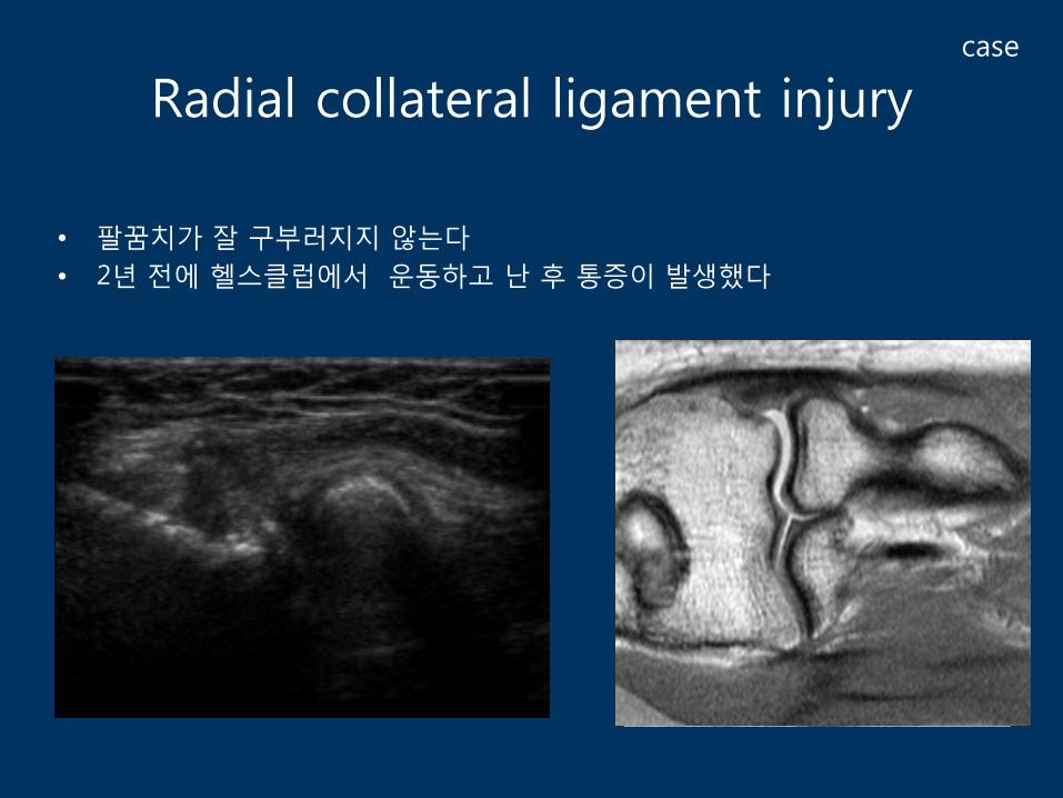
Radial collateral ligament injury
• 팔꿈치가 잘 구부러지지 않는다
• 2년 전에 헬스클럽에서 운동하고 난 후 통증이 발생했다
case

Lateral epicondylitis
- Tendinitis - Tendinosis
- partial tear - Complete tear
- Significant relationship between clinical symptoms and ultrasound findings
- Sensitivity: 72–88% - Specificity: 36–48.5%
-Levin et al, 2005-
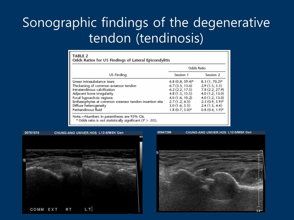
Sonographic findings of the degenerative tendon (tendinosis)

Tendinosis and hypervascularity
• Abnormally thickened tendons with altered echotexture (focal hypoechoic areas) and hypervascular pattern at color and power Doppler imaging (angiofibroblastic infiltration)
• Painful tendinosis : more hypervascular than of asymptomatic tendinosis,
• hypervascular pattern is not an unfavorable sign
• Part of reparative process ?
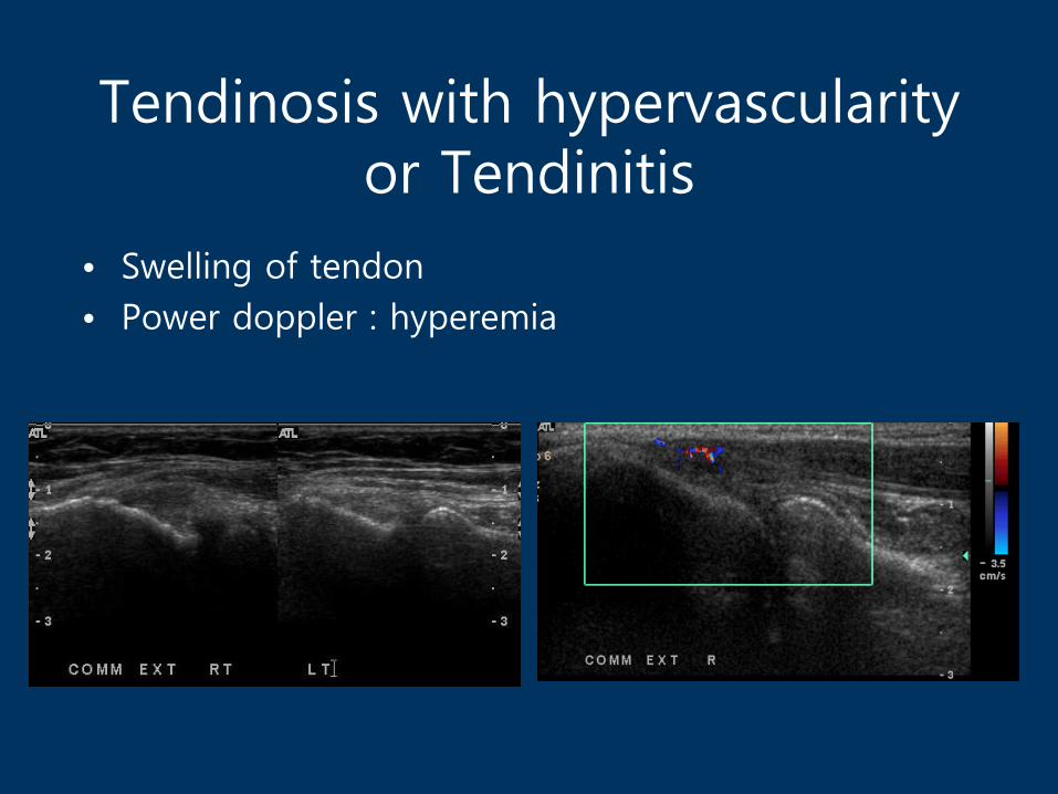
Tendinosis with hypervascularityor Tendinitis
• Swelling of tendon
• Power doppler : hyperemia
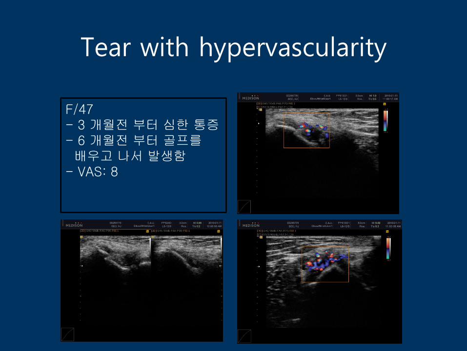
Tear with hypervascularity
F/47- 3 개월전 부터 심한 통증- 6 개월전 부터 골프를배우고 나서 발생함
- VAS: 8

Partial tear
• Macroscopically, produce discontinuities in individual portions of complex tendons
• loss of longitudinal fibrillar pattern
• Lack of retraction( DDX with complete tear)
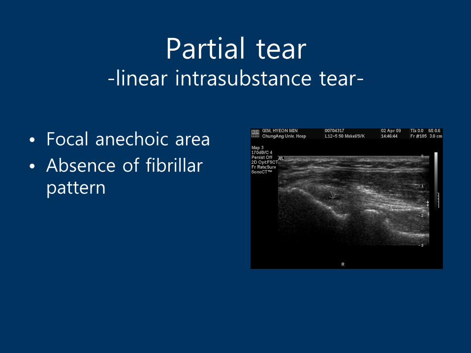
Partial tear-linear intrasubstance tear-
• Focal anechoic area
• Absence of fibrillar pattern
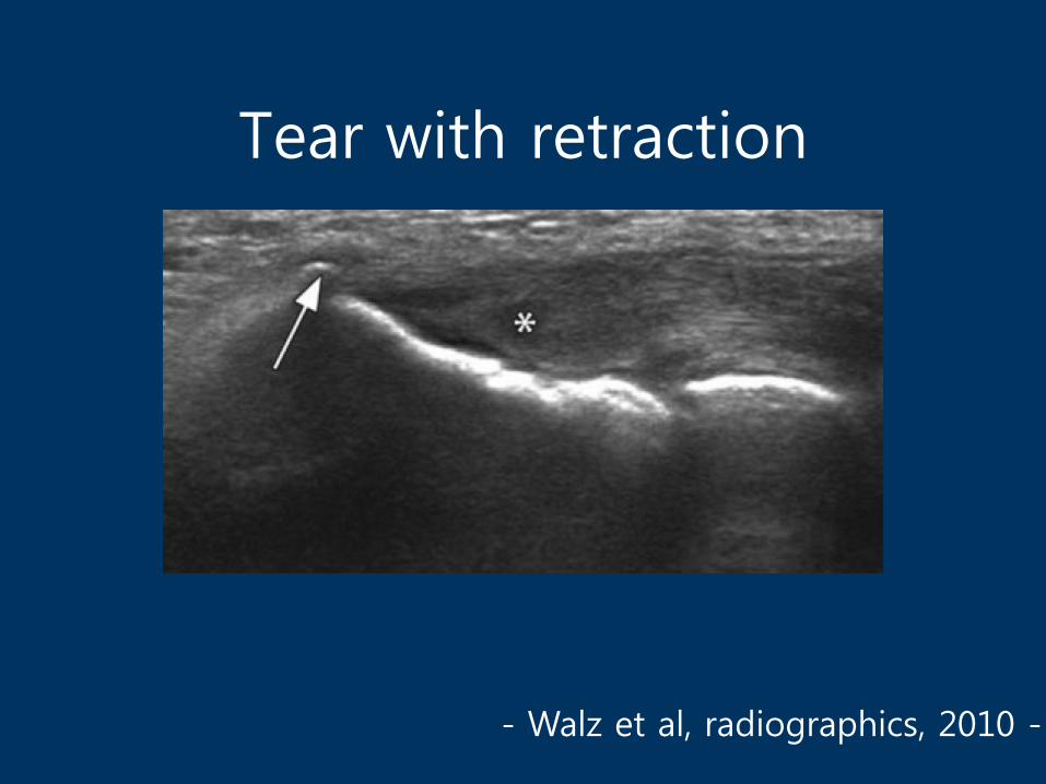
Tear with retraction
- Walz et al, radiographics, 2010 -
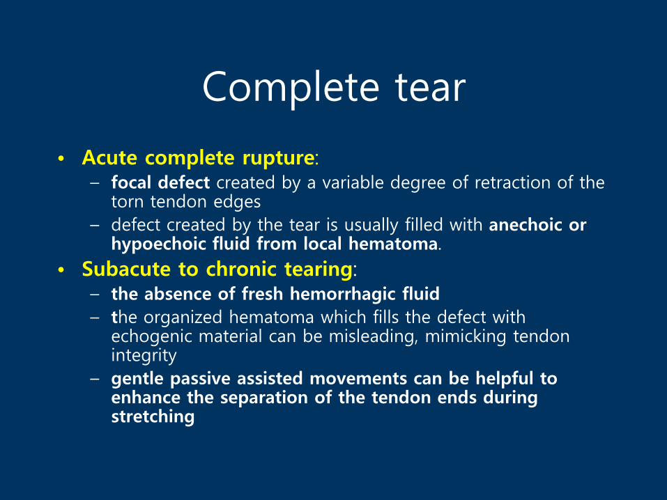
Complete tear
• Acute complete rupture: – focal defect created by a variable degree of retraction of the
torn tendon edges
– defect created by the tear is usually filled with anechoic or hypoechoic fluid from local hematoma.
• Subacute to chronic tearing: – the absence of fresh hemorrhagic fluid
– the organized hematoma which fills the defect with echogenic material can be misleading, mimicking tendon integrity
– gentle passive assisted movements can be helpful to enhance the separation of the tendon ends during stretching

Near full thickness tear
-Walz et al, radiographics, 2010 -
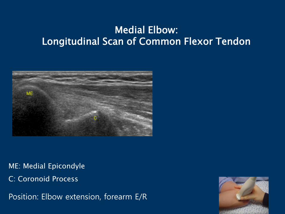
Medial Elbow:Longitudinal Scan of Common Flexor Tendon
ME: Medial Epicondyle
C: Coronoid Process
Position: Elbow extension, forearm E/R
C
ME
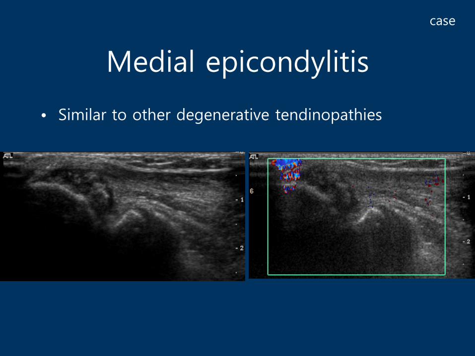
Medial epicondylitis
• Similar to other degenerative tendinopathies
case

Medial Collateral Ligament
• Thickness: 2.6~4 mm
ME
UT
90o
Full flexion

Medial Elbow :Transverse Scan of Cubital Tunnel
ME: Medial Epicondyle FCU: Flexor carpi ulnaris
O: Olecranon
U: Ulnar nerve
FCU
FCU
Ulnar nerve
Ulnar nerve
ME
O
Arcuate ligament

Medial Elbow:Longitudinal Scan of Cubital Tunnel
ME: Medial Epicondyle
UN: Ulnar nerve
Ulnar nerve
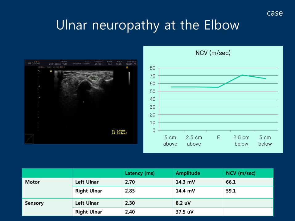
Ulnar neuropathy at the Elbow
Latency (ms) Amplitude NCV (m/sec)
Motor Left Ulnar 2.70 14.3 mV 66.1
Right Ulnar 2.85 14.4 mV 59.1
Sensory Left Ulnar 2.30 8.2 uV
Right Ulnar 2.40 37.5 uV
0
10
20
30
40
50
60
70
80
5 cm
above
2.5 cm
above
E 2.5 cm
below
5 cm
below
NCV (m/sec)
case

Ulnar neropathy at the elbow
- Beekman, Muscle & Nerve, 2011-

Posterior Elbow: Longitudinal Scan
H: Humerus
T: Triceps Brachii (arrow)
O: Olecranon
Olecranon fossa
O
TrochleaH
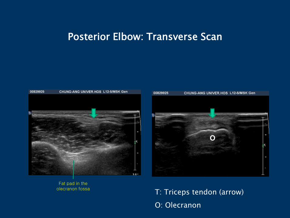
Posterior Elbow: Transverse Scan
Fat pad in the olecranon fossa
T: Triceps tendon (arrow)
O: Olecranon
O

Olecranon bursitis
case

Cystic lesion in the Olecranon
• Cystic lesion in the olecranon, from 2 years ago
• No trauma history
• Pain, swelling, redness
• 2*2 cm
Bx: Epidermal cyst
case
USG Cross Sectional Anatomy & US Pathology of Elbow & Wrist
Part II
Wrist

Radiologic anatomy
• 8 carpal bones
• Joint and its capsule
- Distal radio-ulnar joint
- Radio-carpal joint
- Midcarpal joint
- CMC joint
- MCP joint
- Distal & Proximal lP joint
L
SCP
HTm
Tq
TFCC
Tz
TFCC: stabilizing DRUJ & support compressive load
1) ulnar collateral ligament 2) ECU tendon sheath
3) articular disc 4) joint capsule
5) RU ligament, UL & Utq ligament

Wrist: volar aspect, transverse view
• Carpal tunnel: proximal (inlet)
- Scaphoid tubercle to pisiform
Median NerveUlnar a & nerve
FCR
FPL

Wrist: volar aspect, transverse view
• Distal carpal tunnel
– Tubercle of trapezium ~ hook of hamate
Hook of Hamate
Median Nerve
Ulnar a & nerve
Tubercle of trapezium

Wrist: volar, longitudinal view
Median NerveFDS
RadiusPQ
Radial a.
Median Nerve

• CSA measurement: the median nerve at the wrist (pisiform) and mid-forearm (approximately half the distance between the ulnar styloid and the elbow crease)
• Median nerve CSA at the carpal tunnel inlet (≥ 11 mm2) and wrist–forearm CSA difference (≥ 6 mm2) provides the best discrimination between patients with CTS and controls
- Muscle & Nerve, 2011-
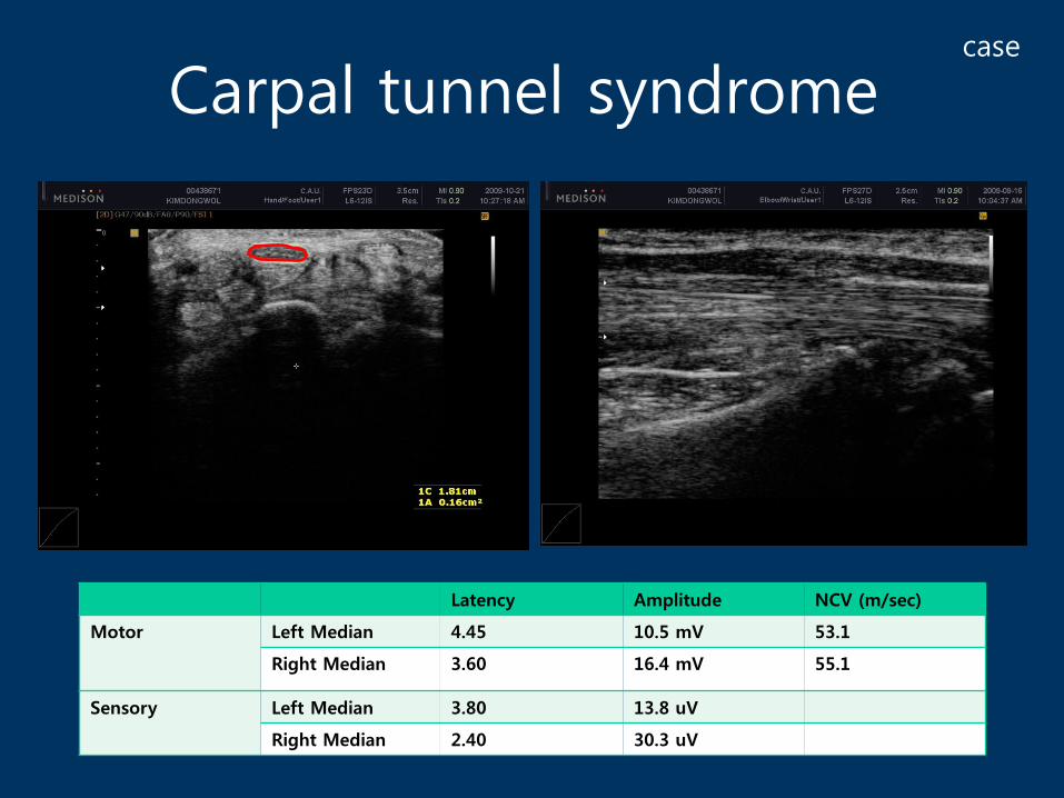
Carpal tunnel syndrome
Latency Amplitude NCV (m/sec)
Motor Left Median 4.45 10.5 mV 53.1
Right Median 3.60 16.4 mV 55.1
Sensory Left Median 3.80 13.8 uV
Right Median 2.40 30.3 uV
case
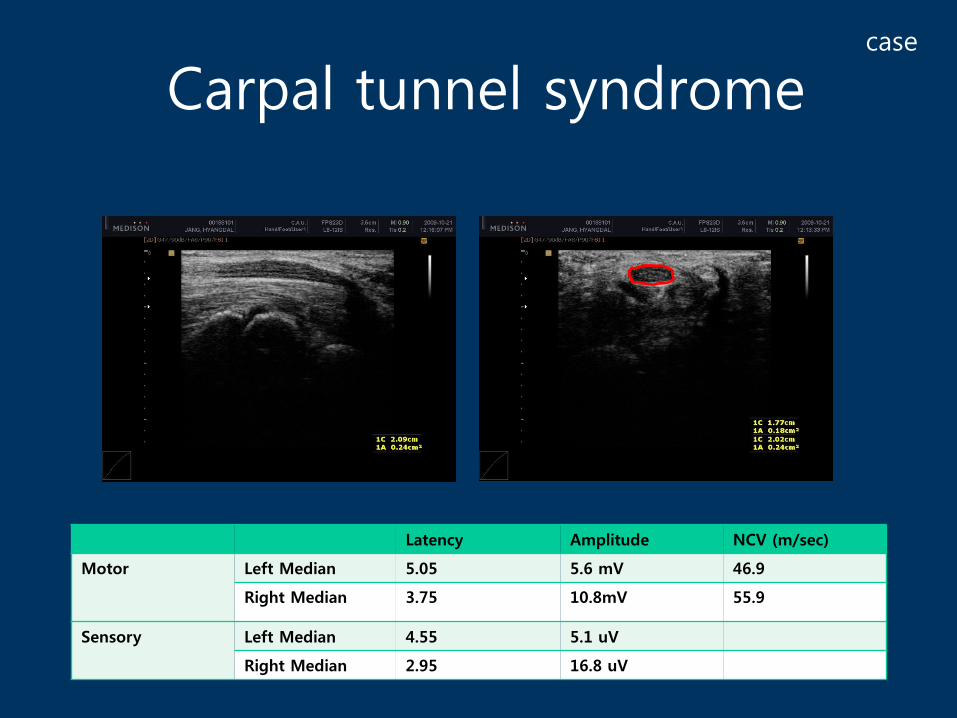
Carpal tunnel syndrome
Latency Amplitude NCV (m/sec)
Motor Left Median 5.05 5.6 mV 46.9
Right Median 3.75 10.8mV 55.9
Sensory Left Median 4.55 5.1 uV
Right Median 2.95 16.8 uV
case

Female / 45
EMG : carpal tunnel syndrome
case
1 month later : wrist swelling at volar side
with LOM, 2nd ~ 4th fingers(extension)
Wrist pain & Tingling sensationonset : 2 months ago

Ultrasonography: Transverse
right left
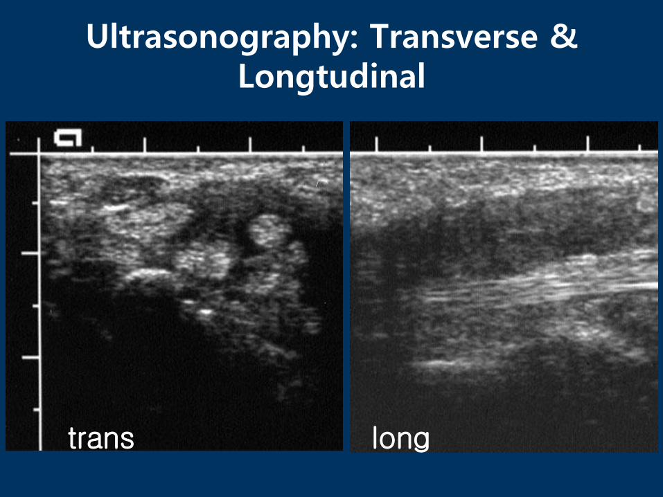
trans long
Ultrasonography: Transverse & Longtudinal

MRI T2
M
M: median nerve

Wrist: volar aspect, transverse view- Guyon’s canal -
Ulnar artery
Ulnar nerve
Ulnar nerve
Ulnar artery
Hook of Hamate

Wrist dorsal aspect
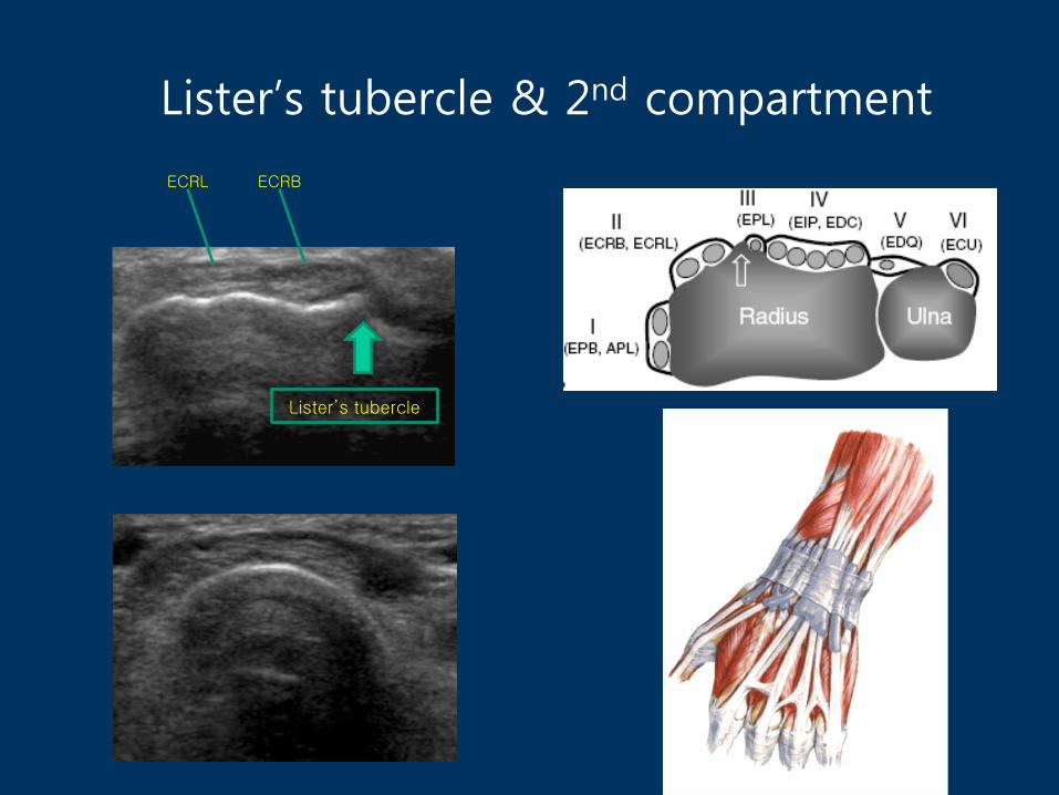
Lister’s tubercle & 2nd compartment
ECRBECRL
Lister’s tubercle
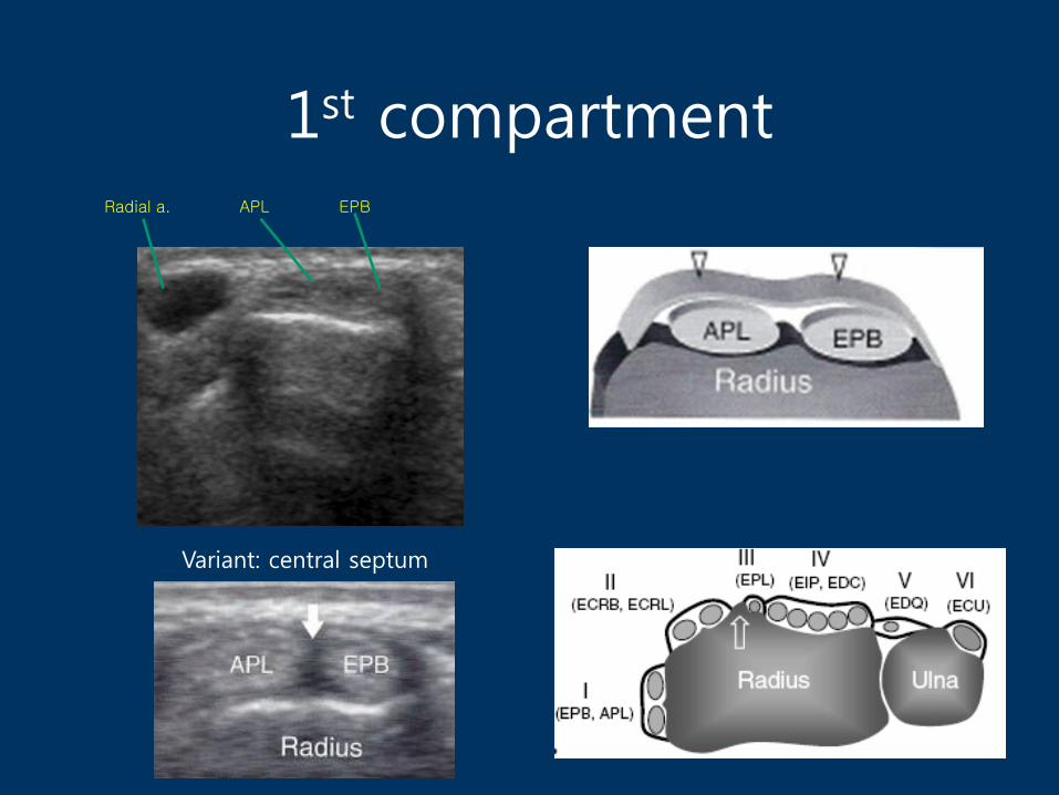
1st compartment APL EPBRadial a.
Variant: central septum

Wrist: dorsal aspect3,4,5th compartment
EPL EIP, EDC EDQ

Wrist: dorsal aspect6th compartment
UlnarTriquetrum
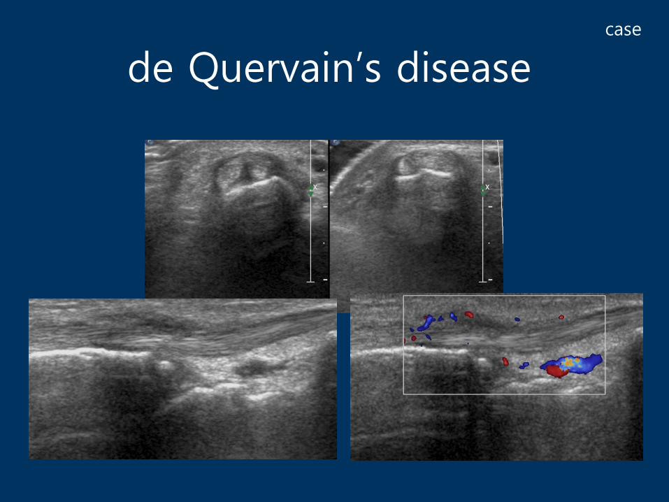
de Quervain’s diseasecase

EPL tendon rupture
• (C.C) 우측 엄지손가락 신전젗한
• F/62
• 한달 전에 의자에서 넘어지면서우측 손등으로 땅에 집으면서부종이 있었음
• 동네 정형외과에서는 엑스레이상 이상은 없었다고 들음.
• 실금이 의심되어 부목을 약 1주일 했고 부목 젗거 후에 우측 엄지손가락이 뒤로 젖혀지지 않아신경검사 및 재활치료 위하여내원함
case
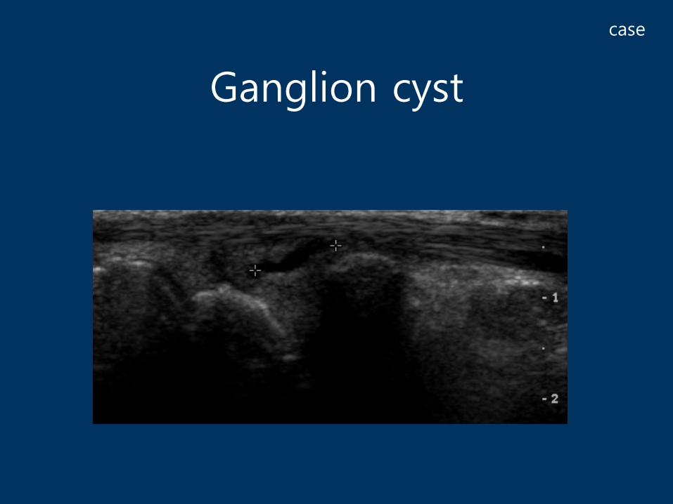
Ganglion cyst
case

Triangular Fibrocartilage Complex
• Triangular homogeneously hyperechoic area thicker than 2.5 mm
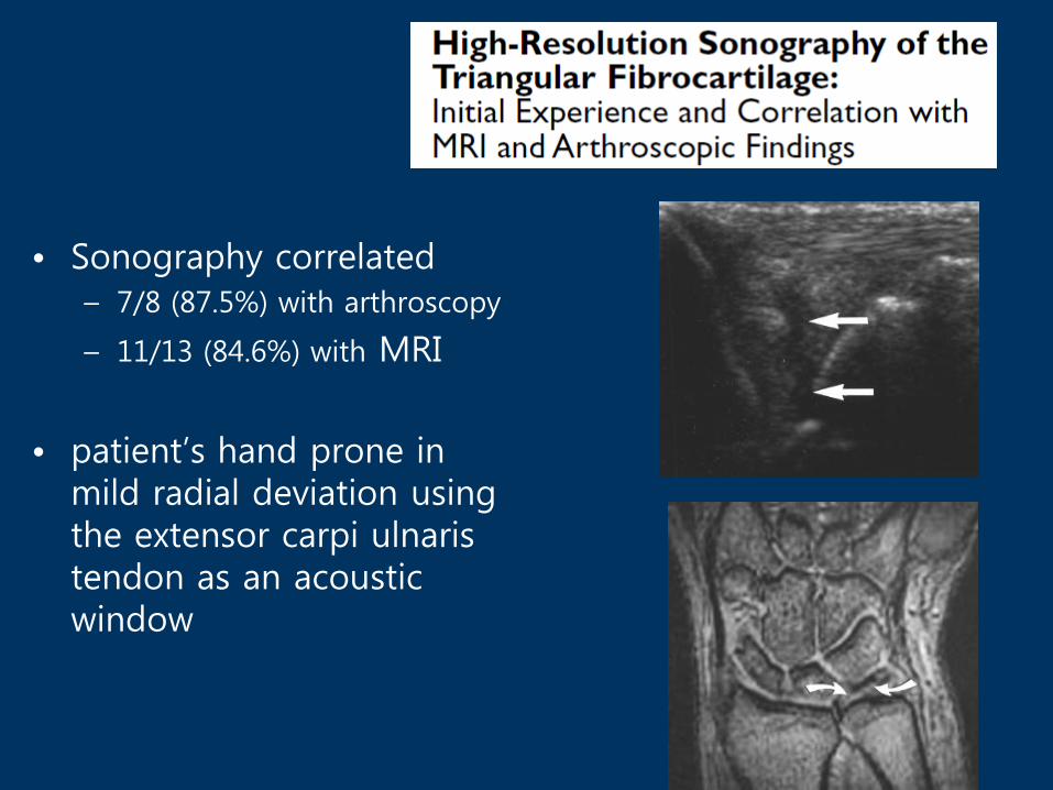
• Sonography correlated – 7/8 (87.5%) with arthroscopy
– 11/13 (84.6%) with MRI
• patient’s hand prone in mild radial deviation using the extensor carpi ulnaris tendon as an acoustic window

Triangular Fibrocartilage Complex(TFCC) tear
case

Triangular Fibrocartilage Complex(TFCC) tear
case
Thank you for listening !!