Embed Size (px)
Citation preview
CLINICAL ARTICLE - BRAIN TUMORS
Usefulness of three-dimensional navigable intraoperativeultrasound in resection of brain tumors with a special emphasison malignant gliomas
Aliasgar V. Moiyadi & Prakash M. Shetty &
Abhishek Mahajan & Amar Udare & Epari Sridhar
Received: 16 July 2013 /Accepted: 5 September 2013 /Published online: 14 September 2013# Springer-Verlag Wien 2013
AbstractBackground Intraoperative imaging is increasingly being usedin resection of brain tumors. Navigable three-dimensional (3D)-ultrasound is a novel tool for planning and guiding such resec-tions. We review our experience with this system and analyzeour initial results, especially with respect to malignant gliomas.Methods A prospective database for all patients undergoingsononavigation-guided surgery at our center since thissurgery’s introduction in June 2011 was queried to retrieveclinical data and technical parameters. Imaging was reviewedto categorize tumors based on enhancement and resectability.Extent of resection was also assessed.Results Ninety cases were operated and included in this anal-ysis, 75 % being gliomas. The 3D ultrasound mode was usedin 87 % cases (alone in 40, and combined in 38 cases). Use ofcombined mode function [ultrasound (US) with magneticresonance (MR) images] facilitated orientation of anatomicaldata. Intraoperative power Doppler angiography was used inone-third of the cases, and was extremely beneficial in delin-eating the vascular anatomy in real-time. Mean duration ofsurgery was 4.4 hours. Image resolution was good or
moderate in about 88 % cases. The use of the intraoperativeimaging prompted further resection in 59 % cases. In themalignant gliomas (51 cases), gross-total resection wasachieved in 47 % cases, increasing to 88 % in the “resectable”subgroup.Conclusions Navigable 3DUS is a versatile, useful and reliableintraoperative imaging tool in resection of brain tumors, espe-cially in resource-constrained settings where Intraoperative MR(IOMR) is not available. It has multiple functionalities that canbe tailored to suit the procedure and the experience of thesurgeon.
Keywords Intraoperative ultrasound .Navigable ultrasound .
Sononavigation . Brain tumors . Image-guided surgery
Abbreviations usedIOUS Intraoperative Ultrasound2D Two dimensional3D Three dimensionalIOMR Intraoperative Magnetic resonance ImagingCT Computed TomographyALA Aminolevulinc acidPD Power doppler
Introduction
The aim of surgery in brain tumors is safe maximal resection.Evidence is mounting that increasing the extent of resectionpositively impacts survivals and oncological outcomes [18].With improved technical adjuncts and equipped with a vary-ing therapeutic armamentarium, neurosurgeons are pushingfor more radical resections. Planning of the surgical procedureis therefore a very crucial step. Central to the plan is an
Electronic supplementary material The online version of this article(doi:10.1007/s00701-013-1881-z) contains supplementary material, whichis available to authorized users.
A. V. Moiyadi (*) : P. M. ShettyDivision of Neurosurgery, PS 245, ACTREC and TMH,Tata Memorial Centre, Kharghar, Navi Mumbai 410210, Indiae-mail: [email protected]
A. Mahajan :A. UdareDepartment of Radiodiagnosis,Tata Memorial Centre, Mumbai, India
E. SridharDepartment of Surgical Pathology,Tata Memorial Centre, Mumbai, India
Acta Neurochir (2013) 155:2217–2225DOI 10.1007/s00701-013-1881-z
accurate localization of the tumor, its extent, and its relation-ship to vital neurovascular structures. Navigation permitscustomized tailoring of small craniotomies. However, becauseof the phenomenon of brain shift once the dura is opened,navigation based on images acquired preoperatively alone haslimited use. A reliable intraoperative image update is increas-ingly needed in the operating room. Intraoperative magneticresonance imaging (IOMR), though ideal, is very costly andnot readily available. A more practical (and cost efficient)alternative is the intraoperative ultrasound (IOUS). IOUS[11, 25] is a very effective tool for localizing lesions. Thereare, however, certain limitations of conventional two-dimensional (2D) US [25]. Navigable three-dimensional(3D) US is a new technology that combines features of nav-igation with a high-resolution 3D US [27]. It surmounts mostof the limitations of conventional 2D US by essentially over-coming the orientation problem and providing more user-friendly multiplanar imaging capabilities. We recount ourexperience with one such 3D navigable US system specifical-ly for resection of brain tumors. Our aim was to assess itspractical utility and impact on intraoperative decisions (re-garding continuation of resection) with respect to tumor re-section at a tertiary care oncology center. One of the majorapplications of this technology is improving resections ofintraparenchymal tumors. To assess this, we also analyzedthe extent of resection achieved in malignant gliomas.
Materials and methods
This was a retrospective analysis. We have been using thenavigable 3D US system SonoWand [SONOWAND AS,Trondheim, Norway] since June 2011. The system has beendescribed in detail elsewhere [5]. Navigable 3DUS essentiallycombines navigation technology with a high-end dedicatedcranial insonation probe capable of generating 2D as well as3D images. The cranial probe is precalibrated and registered tothe navigation system. It can rapidly (30–40 seconds) acquirea series of 2D images (about 200–300) that are computedautomatically into a 3D volume that can then be displayedon the navigation system in either the traditional ACS (axial,coronal, sagittal) planes, or a more user-friendly and intuitive“dual-anyplane”mode. The US data can also be superimposedon preoperative MR (when available) to provide a betterorientation of the cross-sectional anatomy. Using this US dataset, the neurosurgeon is able to navigate. This data set can thenbe repeatedly updated (as and when necessary) during thecourse of the surgery. The system can be used as either astand-alone navigation system using preoperative images; asa stand-alone ultrasound machine providing real-timeintraoperative 2D images as well as a navigable 3D ultrasound(which allows navigation based solely on the IOUS without
requiring preoperative MR images); or in a combined modeusing both preoperative images and intraoperative US.
MRI protocol this was obtained a day or two prior to theplanned surgery in some of the cases. We used fiducial-based as well as anatomical registration. For the former,standard skin-adhering fiducials were placed on the scalp.Preoperative MRI was obtained with a 3-tesla system (3 THDXE, GE) . Post-contrast axial 3-DSPGR (BRAVO, slicethickness 1.6 mm, 0 spacing) and axial T2 sequences (FSE-XL, slice thickness 2 mm, 0 gap) were routinely use. Theimages were burnt on a disc and transferred to the navigationsystem prior to the surgery. Image registration was done on thesystem, and after positioning, patient-to-image registrationwas completed.
The operative procedure Patient positioning was alwaysplanned to ensure that the operative cavity would be as verticalas possible, so that it could be filled completely with saline inorder to optimize ultrasound image quality. Craniotomy wasplanned either using the preoperative images for navigation incertain cases, or using standard surface marking principles.Awake surgery with intraoperative clinical monitoring wasused whenever the tumor was close to the eloquent cortex.After craniotomy and before opening the dura, a baselineultrasound acquisition was carried out. Initially, a 2D acquisi-tion was performed and ultrasound parameters adjusted toobtain the best image resolution. Then, anatomical landmarkswere identified if possible and the lesion was characterized(delineation, size and extent, echo-characteristics). Wheneverpreoperative MR was available, the system automaticallysuperimposed the live 2D US images on the correspondingMR, thus allowing excellent orientation. Once the lesion wasidentified, a rapid 3D US acquisition was performed. Thisessentially involves scanning a sector of the volume of interestby sweeping the US probe over it in a slow and predeterminedpattern, so as to cover the entire region of interest. The phased-array probe was preferred for most deep lesions, with thelinear probe being used for superficial, subcortical lesions.The dura was then opened, and surgery proceeded adheringto routine microneurosurgical principles. Occasionally,thickened/calcified dura resulted in poor image resolution. Inthese cases, the baseline US was performed after opening thedura. Tumor resection proceeded, guided by the 3D US im-ages using a trackable pointer to navigate. Repeat 3D USimages were obtained as many times as required during thesurgery to update the information as tumor debulkingproceeded. The surgeons recorded their impression regardingthe completeness of resection at the end of the surgery. An USwas acquired after this, and if residual tumor was detected, thesurgeon either opted for proceeding with the resection orstopping (if it was close to eloquent areas). This was docu-mented prospectively in a proforma filled immediately after
2218 Acta Neurochir (2013) 155:2217–2225
the surgery. A final US was obtained at the end of the proce-dure and after dural closure (to look for any hematomacollection).
Data analysis During the procedure at various times,screenshots (Fig. 1) of the navigation display screen were cap-tured and recorded and subsequently compiled into case cap-sules. These were entered into a database. These case capsuleswere reviewed and the database queried to extract the informa-tion for this analysis (such as indication for use, mode of appli-cation, type of registration and error, type of probe used, andother subjective details). Descriptive analysis was performed.
Extent of resection (EOR) analysis This was performed in thesubset of malignant gliomas (n =51). These gliomas were clas-sified based on their resectability, as decided by the operatingsurgeons (AM, PS). Lesions were considered resectable whenthey were well circumscribed and localized with a possibility ofobtaining a complete radiological resection. Lesions were furtherclassified as “enhancing” (when there was complete or predom-inant contrast enhancement on the preoperative MRI) and “non-enhancing” (when there was minimal or no contrast enhance-ment). Postoperative contrast MR was obtained within 72 hoursin all cases. When this was not possible (logistical reasons), apostoperative computed tomography (CT) scan was obtainedand a delayed MR was performed subsequently. The MR scanswere analysed for extent of resection. Absence of all contrastenhancement was considered as gross total resection (GTR) forthe enhancing tumors. For the non-enhancing tumors, absence ofT2 and FLAIR changes on the postoperative MR scan wereconsidered as GTR. If there was any doubt, the delayed scans(at 3 months) were also reviewed to unequivocally confirm theabsence of T2/FLAIR changes. In seven cases, immediate post-operative MR could not be obtained (MR not working in fourinstances, and patient uncooperative in three cases). In each ofthese, a CT scan was obtained and a delayed MR performed as
soon as possible. Both CT and MR findings were reviewed toascertain the extent of resection in these cases.
Results
Between June 2011 and February 2013, the SonoWand systemwas used in 90 cases. The distribution of cases is shown inTable 1. A large majority of the cases (75 %) were gliomas.Malignant gliomas were the single most common group oftumors operated. Seven of these were recurrent tumors. Themean duration of surgery was 4.4 hours (range 2–9 hours).
The modes of use of the system and details about thetechnical parameters are depicted in Table 2. The navigableUS function was used in 87 % of the cases (stand-alone US in44 % of case, and combined with preoperative MR-basednavigation in 42 % of cases). Fiducial-based registration wasused in most cases when preoperative images were requiredfor navigation. We also used anatomical landmark-based reg-istration in a few cases. The mean accuracy for fiducial-basedregistration was better than for anatomical landmarks (2.8 mmv/s 7.8 mm).
The mini-craniotomy (MC) probe was used for the catheterplacement/ cyst drainage procedures employing a single 2.5centimeter burr hole. For resective surgeries, both linear andphased array probes were used (n =75). The image resolutionwas good in 44 (59 %), moderate in 22 (29 %), and poor in9(12 %) cases where US was used. When we reviewed thelatter, we realized that all were because of poor acoustic cou-pling (calcified or thick dura) or artifacts due to adjacent bone inthe skull-base cases. Power Doppler angiography is a veryuseful function of this system and (Fig. 2) was used in aboutone-third of the cases, especially in the skull base cases where itoften was the predominant intention of using the system.
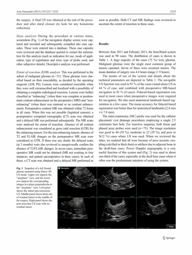
Fig. 1 Snapshot of a left frontalglioma operated using Direct 3DUS mode. Upper row depicts the“anyplane” view, and the lowerrow depicts the correspondingimages in a plane perpendicular tothe “anyplane” view. Left panelshows the initial (pre-resection)US.Middle panel shows thick rimof residual tumor in the middle ofthe surgery. Right panel shows thepost-resection US scan with noresidual tumor
Acta Neurochir (2013) 155:2217–2225 2219
The impact of the use of the intraoperative imaging systemon the course of the surgery is shown in Table 3. In 59% caseswhere the system was used with the intention of resectioncontrol, further resection was prompted by the newly acquiredintraoperative images. In another 21 %, the IOUS showedresidual tumor that was not resected because of proximity tovarious eloquent areas. Extent of resection of malignant glio-mas: a separate analysis was conducted in the 51 malignantgliomas. These accounted for 56 % of all the cases. Thelocation of these tumors and histological spectrum is shownin Table 4. Glioblastoma was the single most common tumor(52 %). Half of malignant gliomas were considered resectable(n =26). Based on the enhancement pattern, 36 of the 51(70 %) were classified as enhancing tumors (Figs. 3 and 4).GTRwas achieved in 29 of the cases overall (47 %). The GTRrate was 88 % for the resectable group (23 of 26) and muchlower at 24 % for the unresectable group (6 of 25). Resect-ability was similar in the enhancing and non-enhancinggroups (53 % and 47 % respectively). GTR rates were higher(61.1 %) for the enhancing group than for the non-enhancingones (47 %) (Table 5).
Perioperative outcomes: Neurological deterioration wasexperienced in seven cases (8 %). Seventeen patients im-proved (19 %) and the remaining 66 cases maintained theirneurological conditions postoperatively. There was no opera-tive mortality.
Discussion
History of 2D IOUS in Neurosurgery Intraoperative US hasbeen used in neurosurgery since the early 1970s [3]. Our ownprevious experience with 2D US has been extremely encour-aging [11]. However, many limitations with 2D US exist, andthese have been highlighted by Unsgård et al. [25]. Advancesin image processing and computational capabilities, coupledwith refined stereotactic principles, gave birth to navigation inthe late 1980s that has transformed the way neurosurgicalprocedures are performed [9, 30, 31]. There is no doubt thatnavigation based on preoperatively acquired images (CT orMR) enables the neurosurgeon to confidently plan accurateand small (“tailored”) craniotomies. However, soon after theintroduction of navigation, it was evident that once the dura isopened, there is a change in the intracranial anatomy obviatingthe usefulness of the preoperatively acquired images (thephenomenon called “brainshift”). Moreover, inaccuracies re-lated to image and patient registration add to the problem.Thus, purely navigation-based systems are not enough andintraoperative imaging update is essential. In this setting,intraoperative MR has emerged as the modality of choice.There is unequivocal evidence to support the benefit of IOMRin improving resections as well as in improving overall out-comes [8, 10, 20]. However, his tool still remains beyondreach for a majority of neurosurgical centers, and thus itsusefulness cannot be widely applied. This is especially truefor resource-constrained settings. The navigable 3D US fillsthis void, allowing navigation to proceed directly without theneed for preoperative images [27]. Admittedly, getting orient-ed to the US images takes a while. However, as is true of any
Table 2 Details about the modes of use, intended use and technicalparameters of the sononavigation system used
Mode of Application
Only Navigation 12 (13 %)
Only Ultrasound (3D Direct) 40 (45 %)
Combined (Navigation + US) 38 (42 %)
Intended use
Craniotomy Planning only 8 (9 %)
Tumor Localization only 6 (7 %)
Guiding extent of resection 39 (43 %)
Use for all of the above 31 (34 %)
Frameless targeted procedure (biopsy, etc.) 3 (3.5 %)
For intraoperative angiography only 3 (3.5 %)
Ultrasound probe used (n =78)
Linear (12FLA) probe 10 (11 %)
Phased array (8FPA) 65 (86 %)
Minicraniotomy (MC) probe 3 (3 %)
Intraoperative power Doppler angiography
Yes 28 (31 %)
Table 1 Pathological spectrum of cases operated (n =90)
Histological Classification
High Grade gliomas 51 (57 %)
Glioblastoma 26
Anaplastic Astrocytoma 7
Anaplastic Oligodendroglioma 6
Anaplastic Oligoastrocytoma 9
Anaplastic Ependymoma 3
Low grade gliomas 17 (9 %)
Pilocytic astrocytoma 3
Diffuse fibrillary astrocytoma 5
Oligodendroglioma 3
Oligoastrocytoma 6
Metastases 6 (7 %)
Meningiomas 3 (3 %)
Chondrosarcoma 3 (3 %)
Cavernoma/hemangioma 3 (3 %)
Schwannoma 2 (2 %)
Others (one each of lymphoma, PNET,craniopharyngioma,epidermoid, and cyst aspiration)
5 (6 %)
Type of Surgery
Non skull base 81 (90 %)
Skull Base 9 (10 %)
2220 Acta Neurochir (2013) 155:2217–2225
new technology, after a steep learning curve, the utility andconfidence in using it is enormous. In our experience, usingthe combined mode (preoperative MR plus intraoperative US)initially during the learning phase facilitates the orientationand makes the user more confident. Over a period of time, onecan graduate to using direct IOUS images. One big advantageof using US images directly is that it obviates the need foracquiring preoperative MR images. Often (as we see at ourreferral center), patients come with complete MRI done else-where. Repeating an MR for navigation specifically is notalways possible and certainly not in the emergency setting.Further, patients with large tumors often have raised intracra-nial pressure or cognitive deficits and may not always becooperative for a MR imaging. In such cases, the “3D Direct”mode is very useful. Besides, using the direct mode eliminatesthe inaccuracies due to image and patient registration. Indeed,in our hands while using the navigation mode of the samesystem, we had a minimum registration error of 2.8 mm. Itmay be theoretically possible to minimize this further bymeticulous registration techniques; however, it can never becompletely eliminated.
Proof of principle The 3D US has been extensively evaluatedby a few groups, the largest experience being from the group
at Trondheim that was actively involved in the development ofthe system, as well as many other centers [13, 14, 23, 27, 28].The Trondheim group showed (using meticulous histologicalcorrelation of biopsies with the US as well as MR images) thatnavigable 3D US was as good and reliable as navigated MRfor delineating high-grade and low-grade gliomas as well asmetastases [28]. They reported high specificity and positivepredictive values (PPV), indicating the safety of using thissystem for guiding resections. But they also found a lownegative predictive value, implying that when the IOUS wasnegative, there was a possibility of tumor still being leftbehind. Future improvements in image resolution capabilitiesare expected to resolve these issues. Interestingly, the samestudy also found a higher PPV for low-grade gliomas. In afollow-up study, the same group from Trondheim evaluatedthe accuracy of the system during the resection (in the subse-quent phase of the surgery) [16]. They found that due toimaging artifacts imparted by blood and other changes in theadjacent tissue due to handling, the specificity and PPV
Table 3 Impact of the intraoperative guidance on the course of theresection (n =70)
Impact
Re-resection after tumor seen on the Ultrasound images 41 (59 %)
No resection inspite of residual tumor 15 (21 %)
No residual tumor, resection stopped 14 (20 %)
Table 4 Location and proximity to eloquent regions of the malignantgliomas (n =51)
Tumor Characteristics Number Percent
Location Frontal 31 60.8
Gangliocapsular 2 3.9
Occipital 3 5.9
Parietal 3 5.9
Temporal 12 23.5
Eloquent location No 8 15.7
Close ( within 5–10 mm) 31 60.8
Involving (< 5 mm) 12 23.5
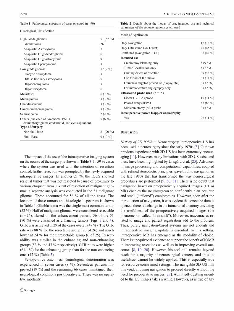
Fig. 2 Intraoperative powerDoppler angiography of a case oftemporal chondrosarcoma. Theentire circle of Willis can be wellappreciated in real-time
Acta Neurochir (2013) 155:2217–2225 2221
2222 Acta Neurochir (2013) 155:2217–2225
dropped. Careful attention during hemostasis and tissue han-dling, as well as employing strategies to overcome these arti-facts, can improve image quality [19]. Considering its potentialof use at a fraction of the cost of IOMR, this could be a cost-effective alternative, especially in resource-constrained set-tings. We were keen to evaluate the practical usefulness of theadjunct in guiding tumor-resections (primarily with respect tothe change it necessitated in our resection extent intraopera-tively). Not only was it a very reliable guide during resection,but we found that in about 59 % of the cases where it was usedwith the intention of controlling the resection, it made a positiveimpact, detecting residual tumors and prompting the surgeon togo ahead with surgery.
Navigable Ultrasound for resection of high-gradegliomas Solheim et al. have shown that the SonoWand systemwas effectively used in an unselected consecutive cohort ofhigh-grade gliomas [21]. In this study, they were able to
achieve acceptable results (37 % GTR with 13 % morbidity).They also showed that if the intent of surgery was to achieve aradical resection, a GTR was achieved in 55 % (versus only2 % GTR when the intent was only debulking). Our overallGTR rate of 47 % compares favorably to this. In the samestudy by Solheim et al., the GTR rate in the “resectable” groupwas 63 %. Interestingly, in that study, many neurosurgeons(including residents) were part of the operating team, anddespite the heterogeneity in surgeon experience and expertise,the rate of GTR of 63 % was fairly high. We were able toachieve higher GTR rates in our subgroup of “resectable”
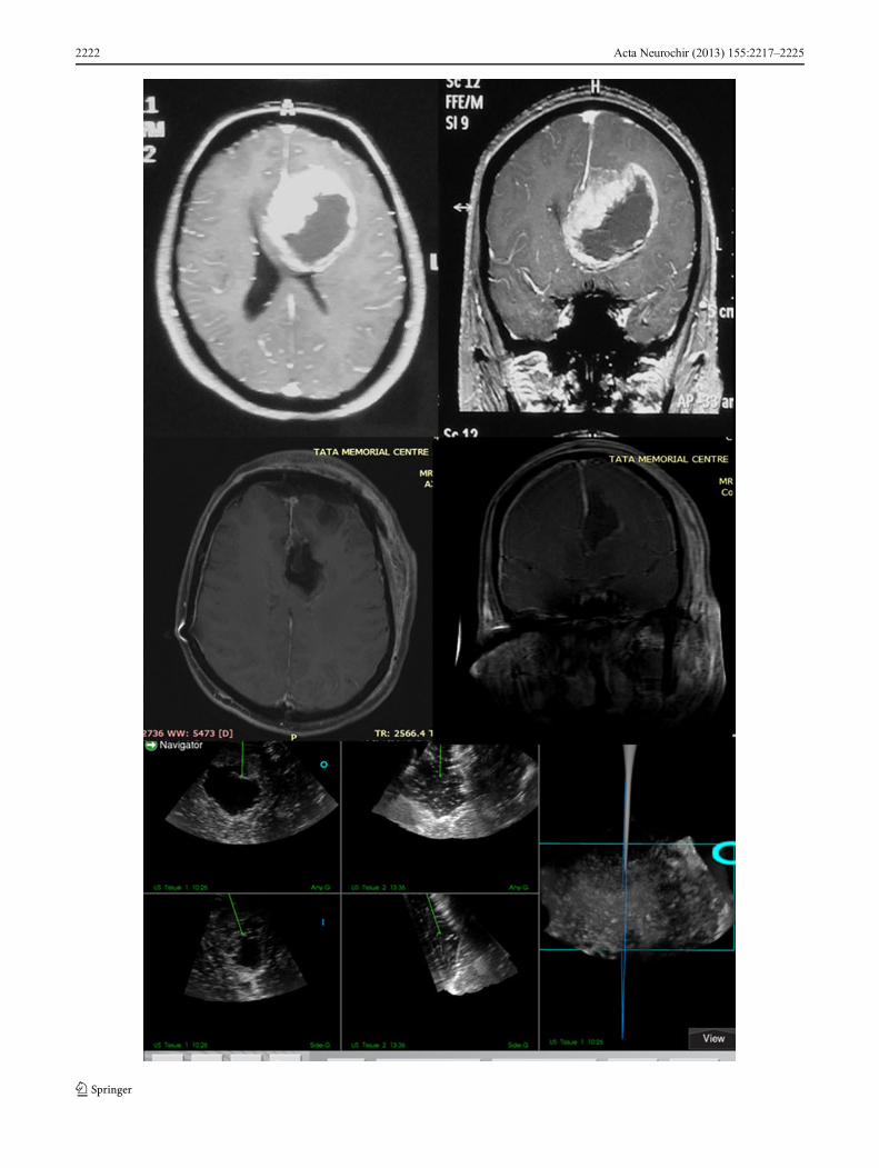
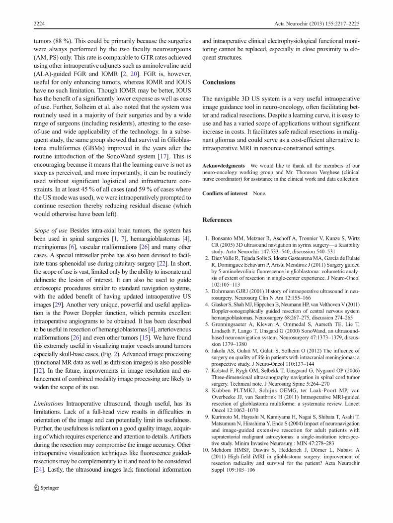
Fig. 4 Non-enhancing resectabletumor. Preoperative images (toprow): T2 weighted axial (left),post contrast T1 image (center)and pre-resection ultrasoundimage (right—note thehyperechoic texture and focalincreased echogenicity—whitearrow). Postoperative images(bottom row): T2 weightedimages—axial (right) and coronal(center) and post-resection USimage showing no residual tumor
Table 5 Extent of resection of the malignant gliomas as per pattern ofenhancement and resectability (n =51)
Tumor Type Extent of resection Total
GTR NTR STR PR
Enhancing Resectable 18 0 1 0 19
Unresectable 4 0 10 3 17
Non-enhancing Resectable 5 1 1 0 7
Unresectable 2 1 1 4 8
GTR Gross total resection (no residual contrast enhancement); NTRNear total resection (up to 90 %); STR Subtotal resection (50–90 %);PR Partial resection (< 50 %)
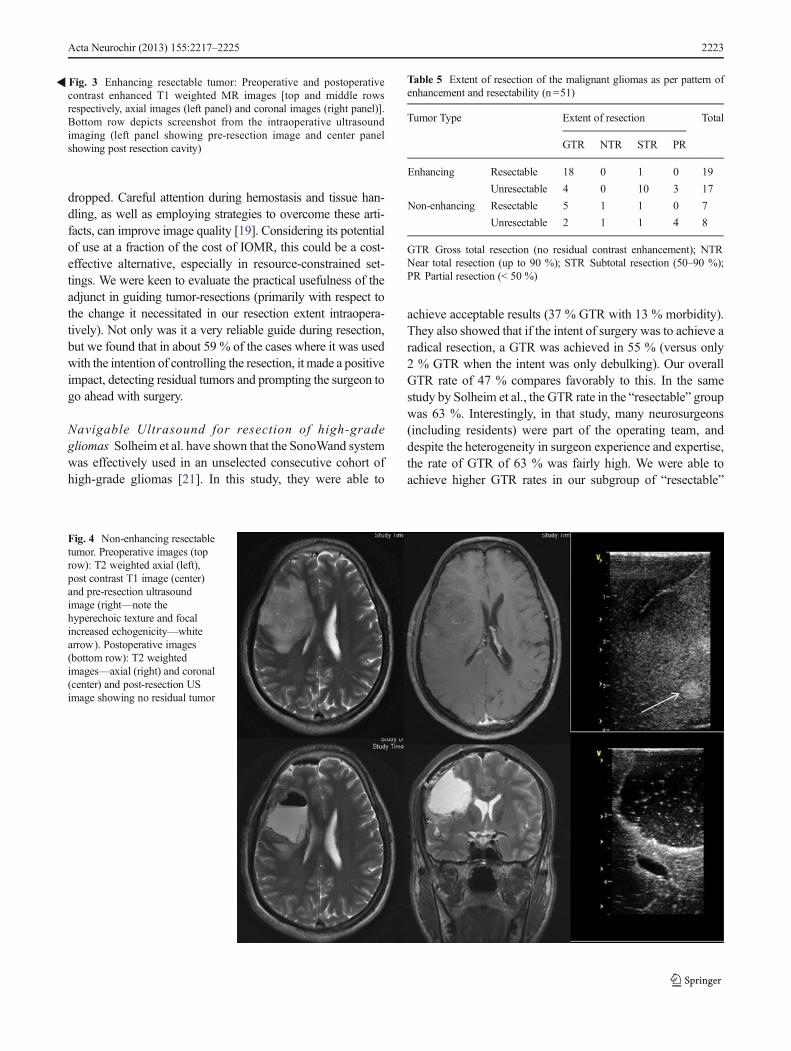
�Fig. 3 Enhancing resectable tumor: Preoperative and postoperativecontrast enhanced T1 weighted MR images [top and middle rowsrespectively, axial images (left panel) and coronal images (right panel)].Bottom row depicts screenshot from the intraoperative ultrasoundimaging (left panel showing pre-resection image and center panelshowing post resection cavity)
Acta Neurochir (2013) 155:2217–2225 2223
tumors (88 %). This could be primarily because the surgerieswere always performed by the two faculty neurosurgeons(AM, PS) only. This rate is comparable to GTR rates achievedusing other intraoperative adjuncts such as aminolevulinc acid(ALA)-guided FGR and IOMR [2, 20]. FGR is, however,useful for only enhancing tumors, whereas IOMR and IOUShave no such limitation. Though IOMR may be better, IOUShas the benefit of a significantly lower expense as well as easeof use. Further, Solheim et al. also noted that the system wasroutinely used in a majority of their surgeries and by a widerange of surgeons (including residents), attesting to the ease-of-use and wide applicability of the technology. In a subse-quent study, the same group showed that survival in Glioblas-toma multiformes (GBMs) improved in the years after theroutine introduction of the SonoWand system [17]. This isencouraging because it means that the learning curve is not assteep as perceived, and more importantly, it can be routinelyused without significant logistical and infrastructure con-straints. In at least 45 % of all cases (and 59 % of cases wherethe USmode was used), we were intraoperatively prompted tocontinue resection thereby reducing residual disease (whichwould otherwise have been left).
Scope of use Besides intra-axial brain tumors, the system hasbeen used in spinal surgeries [1, 7], hemangioblastomas [4],meningiomas [6], vascular malformations [26] and many othercases. A special intrasellar probe has also been devised to facil-itate trans-sphenoidal use during pituitary surgery [22]. In short,the scope of use is vast, limited only by the ability to insonate anddelineate the lesion of interest. It can also be used to guideendoscopic procedures similar to standard navigation systems,with the added benefit of having updated intraoperative USimages [29]. Another very unique, powerful and useful applica-tion is the Power Doppler function, which permits excellentintraoperative angiograms to be obtained. It has been describedto be useful in resection of hemangioblastomas [4], arteriovenousmalformations [26] and even other tumors [15]. We have foundthis extremely useful in visualizing major vessels around tumorsespecially skull-base cases, (Fig. 2). Advanced image processing(functional MR data as well as diffusion images) is also possible[12]. In the future, improvements in image resolution and en-hancement of combined modality image processing are likely towiden the scope of its use.
Limitations Intraoperative ultrasound, though useful, has itslimitations. Lack of a full-head view results in difficulties inorientation of the image and can potentially limit its usefulness.Further, the usefulness is reliant on a good quality image, acquir-ing ofwhich requires experience and attention to details. Artifactsduring the resection may compromise the image accuracy. Otherintraoperative visualization techniques like fluorescence guided-resectionsmay be complementary to it and need to be considered[24]. Lastly, the ultrasound images lack functional information
and intraoperative clinical electrophysiological functional moni-toring cannot be replaced, especially in close proximity to elo-quent structures.
Conclusions
The navigable 3D US system is a very useful intraoperativeimage guidance tool in neuro-oncology, often facilitating bet-ter and radical resections. Despite a learning curve, it is easy touse and has a varied scope of applications without significantincrease in costs. It facilitates safe radical resections in malig-nant gliomas and could serve as a cost-efficient alternative tointraoperative MRI in resource-constrained settings.
Acknowledgments We would like to thank all the members of ourneuro-oncology working group and Mr. Thomson Verghese (clinicalnurse coordinator) for assistance in the clinical work and data collection.
Conflicts of interest None.
References
1. Bonsanto MM, Metzner R, Aschoff A, Tronnier V, Kunze S, WirtzCR (2005) 3D ultrasound navigation in syrinx surgery—a feasibilitystudy. Acta Neurochir 147:533–540, discussion 540–531
2. Diez Valle R, Tejada Solis S, Idoate GastearenaMA, Garcia de EulateR, Dominguez Echavarri P, Aristu Mendiroz J (2011) Surgery guidedby 5-aminolevulinic fluorescence in glioblastoma: volumetric analy-sis of extent of resection in single-center experience. J Neuro-Oncol102:105–113
3. Dohrmann GJRJ (2001) History of intraoperative ultrasound in neu-rosurgery. Neurosurg Clin N Am 12:155–166
4. Glasker S, ShahMJ,HippchenB,NeumannHP, vanVelthovenV (2011)Doppler-sonographically guided resection of central nervous systemhemangioblastomas. Neurosurgery 68:267–275, discussion 274–265
5. Gronningsaeter A, Kleven A, Ommedal S, Aarseth TE, Lie T,Lindseth F, Lango T, Unsgard G (2000) SonoWand, an ultrasound-based neuronavigation system. Neurosurgery 47:1373–1379, discus-sion 1379–1380
6. Jakola AS, Gulati M, Gulati S, Solheim O (2012) The influence ofsurgery on quality of life in patients with intracranial meningiomas: aprospective study. J Neuro-Oncol 110:137–144
7. Kolstad F, Rygh OM, Selbekk T, Unsgaard G, Nygaard OP (2006)Three-dimensional ultrasonography navigation in spinal cord tumorsurgery. Technical note. J Neurosurg Spine 5:264–270
8. Kubben PLTMKJ, Schijns OEMG, ter Laak-Poort MP, vanOverbeeke JJ, van Santbrink H (2011) Intraoperative MRI-guidedresection of glioblastoma multiforme: a systematic review. LancetOncol 12:1062–1070
9. Kurimoto M, Hayashi N, Kamiyama H, Nagai S, Shibata T, Asahi T,Matsumura N, Hirashima Y, Endo S (2004) Impact of neuronavigationand image-guided extensive resection for adult patients withsupratentorial malignant astrocytomas: a single-institution retrospec-tive study. Minim Invasive Neurosurg : MIN 47:278–283
10. Mehdorn HMSF, Dawirs S, Hedderich J, Dörner L, Nabavi A(2011) High-field iMRI in glioblastoma surgery: improvement ofresection radicality and survival for the patient? Acta NeurochirSuppl 109:103–106
2224 Acta Neurochir (2013) 155:2217–2225
11. Moiyadi A, Shetty P (2011) Objective assessment of utility ofintraoperative ultrasound in resection of central nervous system tu-mors: a cost-effective tool for intraoperative navigation in neurosur-gery. J Neurosci Rural Pract 2:4–11
12. Rasmussen IA Jr, Lindseth F, Rygh OM, Berntsen EM, Selbekk T,Xu J, Nagelhus Hernes TA, Harg E, Haberg A, Unsgaard G (2007)Functional neuronavigation combined with intra-operative 3D ultra-sound: initial experiences during surgical resections close to eloquentbrain areas and future directions in automatic brain shift compensa-tion of preoperative data. Acta Neurochir 149:365–378
13. Rohde VCVA (2011) Intraoperative 3-dimensional ultrasound forresection control during brain tumour removal: preliminary results ofa prospective randomized study. Acta Neurochir Suppl 109:187–190
14. Roth J, Biyani N, Beni-Adani L, Constantini S (2007) Real-timeneuronavigation with high-quality 3D ultrasound SonoWand in pe-diatric neurosurgery. Pediatr Neurosurg 43:185–191
15. Rygh OM, Nagelhus Hernes TA, Lindseth F, Selbekk T, BrostrupMuller T, Unsgaard G (2006) Intraoperative navigated 3-dimensionalultrasound angiography in tumor surgery. Surg Neurol 66:581–592,discussion 592
16. Rygh OM, Selbekk T, Torp SH, Lydersen S, Hernes TA, Unsgaard G(2008) Comparison of navigated 3D ultrasound findings with histo-pathology in subsequent phases of glioblastoma resection. ActaNeurochir 150:1033–1041, discussion 1042
17. Saether CA, Torsteinsen M, Torp SH, Sundstrom S, Unsgard G,Solheim O (2012) Did survival improve after the implementation ofintraoperative neuronavigation and 3D ultrasound in glioblastomasurgery? A retrospective analysis of 192 primary operations. J NeurolSurg Part A Cent Eur Neurosurg 73:73–78
18. Sanai N, Polley MY, McDermott MW, Parsa AT, Berger MS (2011)An extent of resection threshold for newly diagnosed glioblastomas. JNeurosurg 115:3–8
19. Selbekk T, Jakola AS, Solheim O, Johansen TF, Lindseth F,Reinertsen I, Unsgard G (2013) Ultrasound imaging in neurosurgery:approaches to minimize surgically induced image artefacts for im-proved resection control. Acta Neurochir 155:973–980
20. Senft CBA, Franz K, Vatter H, Gasser T, Seifert V (2011)Intraoperative MRI guidance and extent of resection in glioma sur-gery: a randomised, controlled trial. Lancet Oncol 12:997–1003
21. Solheim O, Selbekk T, Jakola AS, Unsgard G (2010) Ultrasound-guided operations in unselected high-grade gliomas–overall results,
impact of image quality and patient selection. Acta Neurochir 152:1873–1886
22. Solheim O, Selbekk T, Lovstakken L, Tangen GA, Solberg OV,Johansen TF, Cappelen J, Unsgard G (2010) Intrasellar ultrasoundin transsphenoidal surgery: a novel technique. Neurosurgery 66:173–185, discussion 185–176
23. StenoA, KarlikM,Mendel P, CikM, Steno J (2012) Navigated three-dimensional intraoperative ultrasound-guided awake resection oflow-grade glioma partially infiltrating optic radiation. ActaNeurochir 154:1255–1262
24. StummerW, Pichlmeier U, Meinel T, Wiestler OD, Zanella F, ReulenH-J (2006) Fluorescence-guided surgery with 5-aminolevulinic acidfor resection of malignant glioma: a randomised controlled multicen-ter phase III trial. Lancet Oncol 7:392–401
25. Unsgaard G, Gronningsaeter A, Ommedal S, Nagelhus Hernes TA(2002) Brain operations guided by real-time two-dimensional ultra-sound: new possibilities as a result of improved image quality.Neurosurgery 51:402–411, discussion 411–402
26. Unsgaard G, Ommedal S, Rygh OM, Lindseth F (2005) Operation ofarteriovenous malformations assisted by stereoscopic navigation-controlled display of preoperative magnetic resonance angiographyand intraoperative ultrasound angiography. Neurosurgery 56:281–290
27. Unsgaard G, RyghOM, Selbekk T,Muller TB, Kolstad F, Lindseth F,Hernes TA (2006) Intra-operative 3D ultrasound in neurosurgery.Acta Neurochir 148:235–253, discussion 253
28. Unsgaard G, Selbekk T, Brostrup Muller T, Ommedal S, Torp SH,Myhr G, Bang J, Nagelhus Hernes TA (2005) Ability of navigated3D ultrasound to delineate gliomas and metastases–comparison ofimage interpretations with histopathology. Acta Neurochir 147:1259–1269, discussion 1269
29. Unsgard G, Solheim O, Lindseth F, Selbekk T (2011) Intra-operativeimaging with 3D ultrasound in neurosurgery. Acta Neurochir Suppl109:181–186
30. Willems PW, Taphoorn MJ, Burger H, Berkelbach van der SprenkelJW, Tulleken CA (2006) Effectiveness of neuronavigation inresecting solitary intracerebral contrast-enhancing tumors: a random-ized controlled trial. J Neurosurg 104:360–368
31. Wirtz CR, Albert FK, Schwaderer M, Heuer C, Staubert A, TronnierVM, Knauth M, Kunze S (2000) The benefit of neuronavigation forneurosurgery analyzed by its impact on glioblastoma surgery. NeurolRes 22:354–360
Acta Neurochir (2013) 155:2217–2225 2225


![NAVIGABLE WATERS RULES ANNOTATED - IN.govUpdated February 14, 2011) 1 NAVIGABLE WATERS RULES ANNOTATED AND INDEXED [Document Index: pp. 46 – 50] _____ The navigable waters rules](https://img.pdfslide.us/doc/110x75/5ac47a757f8b9a5c558cd870/navigable-waters-rules-annotated-in-updated-february-14-2011-1-navigable-waters.jpg)