Embed Size (px)
Citation preview

1534
AJNR Am J Neuroradiol 22:1534–1542, September 2001
Use of the Alberta Stroke Program Early CT Score(ASPECTS) for Assessing CT Scans in Patients with
Acute Stroke
J. H. Warwick Pexman, Philip A. Barber, Michael D. Hill, Robert J. Sevick, Andrew M. Demchuk, Mark E. Hudon,William Y. Hu, and Alastair M. Buchan
BACKGROUND AND PURPOSE: Clinicians are insecure reading CT scans by using the one-third rule for acute middle cerebral artery stroke (1/3 MCA rule) before treating patients withrecombinant tissue plasminogen activator. The 1/3 MCA rule is a poorly defined volumetricestimate of the size of cerebral infarction of the MCA. A 10-point quantitative topographic CTscan score, the Alberta Stroke Program Early CT Score (ASPECTS), is described and illus-trated. A sharp increase in dependence and death occurs with an ASPECTS of 7 or less. Wedescribe how to use ASPECTS and why it works with CT scans obtained on all commonlyused axial baselines. We also describe interobserver reliability among clinicians from differentspecialties and with different experience in reading CT scans in the context of acute stroke.
METHODS: The six physicians who developed ASPECTS answered a questionnaire on pre-cisely how they interpret and use ASPECTS. The ASPECTS areas as interpreted by thesephysicians were compared with one another and with standards in the literature. k statisticswere used to assess the interobserver reliability of ASPECTS versus the 1/3 MCA rule.
RESULTS: The exact methods of interpretation varied among the six individual observers,with either a 3:3 or 4:2 split on the specific questions. The overall interobserver agreement wasgood compared with that of the 1/3 MCA rule. Normal anatomic vascular and interobservervariations explain why ASPECTS can be applied with different CT axial baselines.
CONCLUSION: ASPECTS is a systematic, robust, and practical method that can be appliedto different axial baselines. Clinician agreement is superior to that of the 1/3 MCA rule.
Intravenous recombinant tissue plasminogen acti-vator (tPA) improves outcome after acute ischemicstroke (1). The European Cooperative Acute StrokeStudy (ECASS) trials pioneered the importance ofassessing early CT ischemic changes to predict thebenefit from intravenous thrombolysis (2, 3). Inboth trials, the randomization decision depended onwhether more or less than one third of the territoryof the middle cerebral artery (MCA) was involved.
Received November 2, 2000; accepted after revision March 27,2001.
From the Departments of Clinical Neurosciences (J.H.W.P.,P.A.B., M.D.H., A.M.D., A.M.B.) and Radiology (R.J.S.,M.E.H., W.Y.H.), Foothills Hospital, Calgary, Alberta,Canada.
Supported in part by grants from the Heart & Stroke Foun-dation of Canada (M.D.H., A.M.B.), the Canadian Institutes ofHealth Research (M.D.H., A.M.D., A.M.B.), and the AlbertaHeritage Foundation for Medical Research (M.D.H., A.M.D.,A.M.B.).
Address reprint requests to Professor J. H. Warwick Pex-man, Department of Clinical Neurosciences, Room MRG005,Foothills Hospital, 1403 29th St NW, Calgary, Alberta, CanadaT2N 2T9.
q American Society of Neuroradiology
Unfortunately, subsequent research has shown thateven experienced stroke physicians and radiologistshave difficulty recognizing and quantifying thesechanges (4–8).
The Alberta stroke program early CT score(ASPECTS) was developed to offer the reliabilityand utility of a standard CT examination with areproducible grading system to assess early ische-mic changes (,3 hours from symptom onset) onpretreatment CT studies in patients with acute is-chemic stroke of the anterior circulation (9). ThisCT score is simple and reliable and identifies strokepatients unlikely to make an independent recoverydespite thrombolytic treatment. Two hundred threeconsecutive patients with ischemic stroke weretreated with intravenous alteplase within 3 hours ofonset, and their pretreatment CT scans were scoredprospectively. The score divides the MCA territoryinto 10 regions of interest. ASPECTS is, therefore,a topographic scoring system applying a quantita-tive approach that does not ask physicians to esti-mate volumes from two-dimensional images. Is-chemic changes on the baseline CT study were seenin 117 (75%) of 156 treated patients (23 had is-

AJNR: 22, September 2001 CT IN ACUTE STROKE 1535
FIG 1. ASPECTS study form and MCA variants.A and B, Right hemisphere, observer variations: lower and up-
per ASPECTS slices show as shaded areas the minimal andmaximal variations in size of the cortical areas of the MCA (M1–M6) chosen by six expert observers. Left hemisphere, ASPECTSstudy form: A 5 anterior circulation; P 5 posterior circulation; C5 caudate head; L 5 lentiform nucleus; IC 5 internal capsule; I5 insular ribbon; MCA 5 middle cerebral artery; M1 5 anteriorMCA cortex; M2 5 MCA cortex lateral to insular ribbon; M3 5posterior MCA cortex; M4, M5, and M6 are anterior, lateral, andposterior MCA territories, respectively, approximately 2 cm su-perior to M1, M2, and M3, respectively, rostral to basal ganglia.
C and D, Cortical MCA area variations with change of baseline.In the right hemisphere, the baseline is parallel to the inferiorOML; in the left hemisphere, the baseline is the superior OML.
E and F, Normal vascular variations in MCA size on the twoASPECTS slices. The right hemisphere shows the larger normalvariations described by van der Zwan (18) (light shading). Theleft hemisphere of each shows the smaller, textbook (17), vari-ations (dark shading).
chemia in the posterior circulation and 24 weretreated outside the protocol). The baselineASPECTS value correlated inversely with the se-verity of the stroke on the National Institute ofHealth Stroke Scale (NIHSS) (Spearman’s r 52.56, P , .001). The baseline ASPECTS valuepredicted functional outcome and symptomatic in-tracerebral hemorrhage (P , .001 and P 5 .012,respectively). The sensitivity of ASPECTS forfunctional outcome was .78 and specificity was .96;the respective values for symptomatic intracerebralhemorrhage were .90 and .61. A sharp increase independent or fatal outcomes occurred withASPECTS of 7 of less. The inter- and intraobserverreliability of ASPECTS was good to excellent (k5 .71–.89) and consistently superior to the 1/3 rulebetween observer pairs from the same specialty.
In this article, we explain how ASPECTS is usedin practice, illustrate the method with examples, de-fine landmarks and definitions, show why it can beused on CT scans obtained on all commonly usedaxial baselines, and emphasize that clinicians fromdifferent specialties and with varying levels of ex-perience in assessing acute ischemic CT changescan agree on an acceptable level.
MethodsPatients were recruited from both Calgary and Houston, TX.
For the purposes of the current analysis, technical informationwas not available from CT scans obtained in Houston (n 570). CT scans obtained in Calgary (n 5 86) were performedon fourth-generation helical scanners. For better definition, in-dividual scans were acquired using contiguous axial 6-mm sec-tions from the foramen magnum to the suprasellar region and10-mm contiguous sections through the remainder of the brain.One sixth of the CT scans were obtained using the orbitomea-tal line (OML), one third using the superior OML, and halfusing the inferior OML. Scanner settings were kV 5 120, mAs5 400, and mA 5 200. The section time was 2 seconds, thematrix size was 512, and a small focal spot with an algorithmwas used to reduce bone artifacts and give finer granularity.The mA was reduced near the vertex to 150, 130, and 120 mAfor the upper three slices, respectively. Photography was doneat a window level of 30 H with a window width of 75 H. Allthe CT scans were interpreted from film. The areas studiedwere in the part of the brain cut with 10-mm sections. Imageswere read in a darkened room, and were studied from near andfar and even obliquely.
The interpreters used the ASPECTS method to read the CTscans. The ASPECTS was determined from two standardizedaxial CT cuts (Fig 1), one at the level of the thalamus andbasal ganglion and one adjacent to the most superior marginof the ganglionic structures, such that they were not seen. Onthese two sections, which were, by definition, not continuous,the MCA territory was allotted 10 points. A single point wassubtracted for an area of early ischemic change, such as focalswelling or parenchymal hypoattenuation, for each of the de-fined regions. A normal CT scan received an ASPECTS of 10points. A score of zero indicated diffuse ischemic involvementthroughout the MCA territory (Figs 2–4). Parenchymal hy-poattenuation was defined as a region of abnormally decreasedattenuation of brain structures relative to attenuation of otherparts of the same structures or of the contralateral hemisphere.Focal brain swelling or mass effect was defined as any focalnarrowing of the CSF space due to compression by adjacent
structures, such as effacement of cortical sulci or ventricularcompression (10).
Three neurologists specializing in stroke and three neuro-radiologists involved in ASPECTS were interviewed and com-pleted a questionnaire concerning their individual methods ofinterpretation (Table 1). They were also asked to draw the cor-tical areas M1 to M6 on the ASPECTS diagrammatic cuts (Fig1). In addition, they were asked how they differentiated oldfrom new infarcts and whether they used any special tech-niques for interpretation. The neurologists and the neuroradi-
AJNR: 22, September 20011536 PEXMAN
FIG 2. CT scans in a 65-year-old womanwith left-sided hemiplegia, hemianopia,and neglect less than 3 hours after symp-tom onset.
A and B, Baseline CT scans show hy-poattenuation with swelling and efface-ment in regions M1, M2, insula (I), M4, andM5 (ASPECTS 5 5). Intravenous throm-bolysis was administered.
C and D, Follow-up CT scans show alarge area of hypoattenuation involvingmuch of the MCA territory. The patient wasdependent at 3 months.
ologists had at least 2 years’ experience in assessing acuteischemic stroke with CT scans. Although radiology traineesparticipated in the validation of ASPECTS (9), they did notparticipate in the questionnaire.
The questionnaire, devised by a neuroradiologist who hadno prior experience with ASPECTS, was developed to assistphysicians who were inexperienced in the precise use of thistechnique. Because interobserver agreement in the originalASPECTS was good to excellent, it was hypothesized that allrespondents would give similar answers.
Recent textbooks state that ischemic stroke can be seen asearly as 6 hours after symptom onset (11, 12), quoting theabstracts from the classic articles of Tomura et al in 1988 (13)and Truwit et al in 1990 (14). However, the former group madea diagnosis in less than 3 hours in 13 of 15 patients withobscuration of the lentiform nucleus (from a total of 25 pa-tients), while the latter made it in 10 of 13 patients with lossof the insular ribbon (from a total of 27 patients). These de-scriptions formed the basis for diagnosis in two of the areasof ASPECTS. However, certain considerations remain; such as,did one lose a point if only a small part of the insular ribbonwas involved? The questionnaire also sought to answer howmany of the signs of ischemia (mass effect, abnormally lowattenuation in white matter, loss of demarcation of the gray/white junction) need to be present to lose a point in any area.
Furthermore, where were the boundaries of each M area, anddid the interpreters really only look at two CT sections?
The two extreme axial baselines, the superior OML and theinferior OML, were drawn on an outline traced from a lateralpilot film. The two ASPECTS cuts were then drawn in cor-respondence with each baseline, and these cut lines weremarked in thirds. It was then possible to measure the distancesbetween the two anterior third and the two posterior third junc-tions, respectively, by using the scale on the pilot scan (Fig5).
We hypothesized that ASPECTS would provide a more stan-dardized assessment of acute ischemic change than the 1/3rule. The CT scans were interpreted by neurologists special-izing in stroke, by neuroradiologists, and by radiology resi-dents individually and in isolation from the others, first withno clinical information and then only with knowledge of whichside was affected. A minimum of 3 weeks elapsed betweenthe readings. A sample of convenience of stroke patient CTscans from Calgary (n 5 68) was chosen to assess interspe-cialty reliability. Only the results given with knowledge of theaffected side are presented, since these are most relevant to theacute clinical scenario. Interobserver reliability for ASPECTSwas assessed according to divisions of greater than 7 and 7 orless, and for MCA territories of less than one third and greaterthan one third.
AJNR: 22, September 2001 CT IN ACUTE STROKE 1537
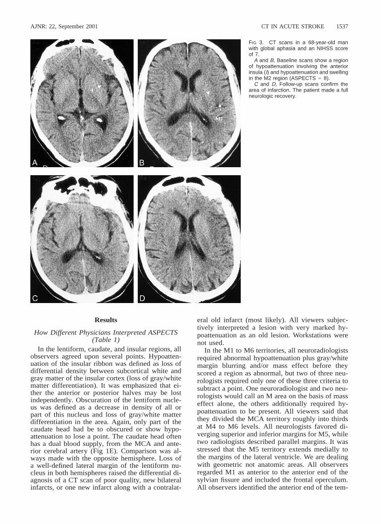
FIG 3. CT scans in a 68-year-old manwith global aphasia and an NIHSS scoreof 7.
A and B, Baseline scans show a regionof hypoattenuation involving the anteriorinsula (I) and hypoattenuation and swellingin the M2 region (ASPECTS 5 8).
C and D, Follow-up scans confirm thearea of infarction. The patient made a fullneurologic recovery.
Results
How Different Physicians Interpreted ASPECTS(Table 1)
In the lentiform, caudate, and insular regions, allobservers agreed upon several points. Hypoatten-uation of the insular ribbon was defined as loss ofdifferential density between subcortical white andgray matter of the insular cortex (loss of gray/whitematter differentiation). It was emphasized that ei-ther the anterior or posterior halves may be lostindependently. Obscuration of the lentiform nucle-us was defined as a decrease in density of all orpart of this nucleus and loss of gray/white matterdifferentiation in the area. Again, only part of thecaudate head had be to obscured or show hypo-attenuation to lose a point. The caudate head oftenhas a dual blood supply, from the MCA and ante-rior cerebral artery (Fig 1E). Comparison was al-ways made with the opposite hemisphere. Loss ofa well-defined lateral margin of the lentiform nu-cleus in both hemispheres raised the differential di-agnosis of a CT scan of poor quality, new bilateralinfarcts, or one new infarct along with a contralat-
eral old infarct (most likely). All viewers subjec-tively interpreted a lesion with very marked hy-poattenuation as an old lesion. Workstations werenot used.
In the M1 to M6 territories, all neuroradiologistsrequired abnormal hypoattenuation plus gray/whitemargin blurring and/or mass effect before theyscored a region as abnormal, but two of three neu-rologists required only one of these three criteria tosubtract a point. One neuroradiologist and two neu-rologists would call an M area on the basis of masseffect alone, the others additionally required hy-poattenuation to be present. All viewers said thatthey divided the MCA territory roughly into thirdsat M4 to M6 levels. All neurologists favored di-verging superior and inferior margins for M5, whiletwo radiologists described parallel margins. It wasstressed that the M5 territory extends medially tothe margins of the lateral ventricle. We are dealingwith geometric not anatomic areas. All observersregarded M1 as anterior to the anterior end of thesylvian fissure and included the frontal operculum.All observers identified the anterior end of the tem-
AJNR: 22, September 20011538 PEXMAN
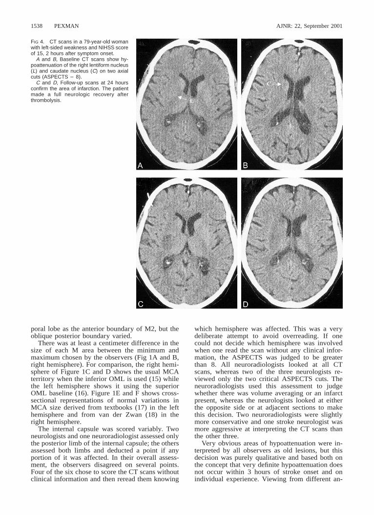
FIG 4. CT scans in a 79-year-old womanwith left-sided weakness and NIHSS scoreof 15, 2 hours after symptom onset.
A and B, Baseline CT scans show hy-poattenuation of the right lentiform nucleus(L) and caudate nucleus (C) on two axialcuts (ASPECTS 5 8).
C and D, Follow-up scans at 24 hoursconfirm the area of infarction. The patientmade a full neurologic recovery afterthrombolysis.
poral lobe as the anterior boundary of M2, but theoblique posterior boundary varied.
There was at least a centimeter difference in thesize of each M area between the minimum andmaximum chosen by the observers (Fig 1A and B,right hemisphere). For comparison, the right hemi-sphere of Figure 1C and D shows the usual MCAterritory when the inferior OML is used (15) whilethe left hemisphere shows it using the superiorOML baseline (16). Figure 1E and F shows cross-sectional representations of normal variations inMCA size derived from textbooks (17) in the lefthemisphere and from van der Zwan (18) in theright hemisphere.
The internal capsule was scored variably. Twoneurologists and one neuroradiologist assessed onlythe posterior limb of the internal capsule; the othersassessed both limbs and deducted a point if anyportion of it was affected. In their overall assess-ment, the observers disagreed on several points.Four of the six chose to score the CT scans withoutclinical information and then reread them knowing
which hemisphere was affected. This was a verydeliberate attempt to avoid overreading. If onecould not decide which hemisphere was involvedwhen one read the scan without any clinical infor-mation, the ASPECTS was judged to be greaterthan 8. All neuroradiologists looked at all CTscans, whereas two of the three neurologists re-viewed only the two critical ASPECTS cuts. Theneuroradiologists used this assessment to judgewhether there was volume averaging or an infarctpresent, whereas the neurologists looked at eitherthe opposite side or at adjacent sections to makethis decision. Two neuroradiologists were slightlymore conservative and one stroke neurologist wasmore aggressive at interpreting the CT scans thanthe other three.
Very obvious areas of hypoattenuation were in-terpreted by all observers as old lesions, but thisdecision was purely qualitative and based both onthe concept that very definite hypoattenuation doesnot occur within 3 hours of stroke onset and onindividual experience. Viewing from different an-

AJNR: 22, September 2001 CT IN ACUTE STROKE 1539
TABLE 1: Methods used to avoid overreading CT scans of infarcts less than 3 hours old
Question DO YOU. . . . . . N1 N2 N3 R1 R2 R3 Y* D*Comments Concerning
Dissenters
1. Read CT first without, and then with clinical in-formation?
Y D Y Y Y D 4 2 N2 and R3 read CT knowingsymptoms
2. Look at all CT sections? D D Y Y Y Y 4 2 N1 and N2 used the twoASPECTS sections in Fig 1
3. Differentiate volume averaging versus infarct byreviewing all CT sections?
D D D Y Y Y 3 3 N1 and N2 compared oppositeside; N3 looked at adjacentsections
4. In M1–M6 areas, make diagnosis if hypoatten-uation is present with mass effect and/or gray/white blurring?
Y D D Y Y Y 4 2 N2 and N3 made diagnosis onthe basis of hypoattenuationalone
5. In M1–M6 areas, make diagnosis with mass ef-fect plus hypoattenuation in
(A) one M area? Y D Y D D Y 3 3 N2, R1, and R2 made diagnosison the basis of mass effectalone
(B) several M areas? Y Y D Y D Y 4 2 N2 and R2 made diagnosis onthe basis of mass effect alone
6. Diagnose an internal capsular infarct only if theposterior limb is hypoattenuated?
D Y Y Y D D 3 3 N1, R2, and R3 would havemade the diagnosis if onlythe anterior limb had been af-fected
Total in agreement4 2 4 6 4 5
Note.—N indicates stroke neurologist, R 5 neuroradiologist, Y 5 agree with question, D 5 disagree with question, M 5 cortical middle cerebralartery.
*Average split among observers 5 3 : 3 and 4 : 2
FIG 5. Maximal variation of ASPECTS sections with baselinealteration. The two ASPECTS sections with two different base-lines: superior OML (solid line) and parallel two slices, and in-ferior OML (dashed line) and parallel two slices. The respectiveupper and lower slices are divided into thirds. Cuts are throughthe basal ganglia and roof of the lateral ventricle to show thatdisagreement is not more than 2 cm.
gles and distances helped in the recognition of sub-tle changes in hypoattenuation. In particular, itproved useful to view a crucial slice independently.This was done by using either the cupped hand ora roll of paper (telescoping). Localized atrophy, de-fined as sulcal widening or ventricular enlargement,also suggested an old infarct. When old infarctswere present in the basal ganglia of the asymptom-atic hemisphere, it was sometimes difficult (and oc-casionally impossible) to say whether areas of ab-normal hypoattenuation or obscuration in the basal
ganglia of the affected hemisphere represented newor old infarcts.
When a CT scan showed that a patient’s headwas not symmetrically situated in the scanner, ow-ing to either tilt or rotation in either direction, orboth, all observers dealt with this as best they couldby trying to compare corresponding areas in thetwo hemispheres, even though these were on dif-ferent CT sections.
Because of varying baselines, as much as a 1-cmdifference in the rostrocaudal-anteroposterior loca-tion of a given point occurred when different cutswere used through the basal ganglia; and as muchas a 2-cm difference occurred on the cuts at theupper edge of the lateral ventricles above the basalganglia (Fig 5).
Interobserver Agreement
Table 2 gives the balanced k scores for interob-server agreement among all the independent ob-servers when they used the 1/3 MCA rule andASPECTS dichotomized between 7 or greater andless than 7. The interobserver agreement betweenpairs of stroke neurologists (k 5 .61 for the 1/3MCA rule and .85 for ASPECTS), neuroradiolo-gists (k 5 .52 and .89), and radiology residents (k5 .64 and .71) was reported previously (9). Theremainder of the table shows that interobserveragreement across specialties was clearly superiorfor ASPECTS (k 5 .56–.83) over the 1/3 MCArule (k 5 .20–.51). Only one of the 12 results

AJNR: 22, September 20011540 PEXMAN
TABLE 2: Pairwise interobserver agreement between independentobservers for the 1/3 MCA territory rule and ASPECTS, withknowledge of the affected side
Observer Pairs
Balanced k*
1/3 MCATerritory(,1/3 vs
.1/3)
ASPECTS(,7 vs
.7)
Two stroke neurologistsTwo neuroradiologistsTwo radiology residentsA stroke neurologist and a neuroradiologistA stroke neurologist and a neuroradiologist
.61
.52
.64
.44
.51
.85
.89
.71
.61
.63A stroke neurologist and a radiology residentA stroke neurologist and a radiology residentA neuroradiologist and a radiology residentA neuroradiologist and a radiology resident
.54
.46
.43
.45
.75
.56
.75
.67A neuroradiologist and a stroke neurologistA radiology resident and a stroke neurologistA radiology resident and a stroke neurologistA stroke neurologist and a neuroradiologistA neuroradiologist and a radiology residentA neuroradiologist and a radiology resident
.43
.38
.20
.47
.51
.39
.67
.83
.60
.71
.83
.63
* Balanced kappa: k . .80 implies excellent reliability; .61 # k #
.80 implies good reliability; .41 # k # .60 implies moderate reli-ability; .21 # k # .40 implies fair reliability; k # .20 implies poorreliability (26).
showed less than good reliability for ASPECTS,while all 11 of the 1/3 MCA rule results showedjust fair reliability, with the remaining one imply-ing poor reliability.
DiscussionEarly ischemic changes seen on CT scans ob-
tained in the first few hours after stroke onset rep-resent early cytotoxic edema and perhaps the de-velopment of irreversible injury (19). Many authorshave cited the potential superiority of diffusion-weighted MR imaging over CT, but to date MRimaging has not been able to discriminate salvage-able brain tissue from that which is irretrievablyinjured (20). Although diffusion-weighted imagingmay become the method of choice, most physicianstreating stroke will remain dependent on CT be-cause of its accessibility. However, the ability ofphysicians to correctly interpret early radiologicsigns of acute stroke on CT scans is fraught withreservations and controversy (7). We believe thatASPECTS provides a solution to this problem.
ASPECTS is a robust clinical tool for severalreasons. First, it has excellent reliability in the clin-ical setting, much superior to the 1/3 MCA territoryrule. When the clinical situation is known,ASPECTS has proved reliable among physicians ofdifferent clinical backgrounds and experience. Theagreement among physicians using ASPECTS wasconsiderably better than when they applied the 1/3MCA rule (the range of k improved from .20–.64to .56–.89). Acute stroke therapy requires that the
treating clinician be comfortable in making an as-sessment of the severity of the CT findings at thebedside and that communication between col-leagues is consistent. We have shown that agree-ment between neuroradiologists and stroke neurol-ogists was good (k 5 .61–.71). This is essentialboth in facilitating the treatment process and inconducting clinical trials. Analysis of ECASS-1 CTscans found that 52 (8.4%) of 620 were misreadlocally. When three expert neuroradiologists reas-sessed the ECASS-1 CT scans, scoring them ac-cording to the 1/3 MCA rule, the chance adjustedpair-wise agreement was surprisingly low (k 5.23–.51) despite 90% to 91% agreement (4). In areview of 50 CT scans from the Atlantis study(which used the 1/3 MCA rule) (21), agreementamong the three neuroradiologist reviewers wasmoderate to good (pair-wise k coefficients of .44–.65) but consensus among reviewers could beachieved in 72% of cases. However, only neuro-radiologists interpreted the CT scans in the ECASSand Atlantis studies, and the scans were obtainedwithin 6 hours of ictus. Second, ASPECTS is asystematic method. Wardlaw and Seller (22)showed that a systematic approach to assessing ce-rebral infarcts on CT scans produces excellent re-sults. In that study, the k statistics between twoexperienced neuroradiologists reading 119 brainCT scans for site and size without clinical infor-mation were good to excellent (k 5 .69–.87) (22).Our analysis showed that good to excellent reli-ability can be achieved with ASPECTS when thestroke symptom side is known, despite variationsin the exact interpretation of the signs of early is-chemia and in the use of the two ASPECTS dia-grams (Fig 1A and B). One must conclude, there-fore, that the improvement stems from carefulstudy of 10 specific areas on the initial CT scan, inwhich each area is compared with the oppositeside.
It is interesting that the instructions forASPECTS were interpreted differently by the neu-rologists and the neuroradiologists. Why doesASPECTS work when there is disparity concerningits exact interpretation? First, the two neurologists,who used only two ASPECTS sections to assess ascore, missed only isolated infarcts near the vertex,and these by themselves are relatively rare. Thiswould not have much impact on the statistics of156 CT scans. The fact that one neurologist andone neuroradiologist initially read the CT scansblinded to clinical information reflects the speedand confidence with which these individualsworked. None of the three neurologists distin-guished between infarcts and volume averaging byreviewing the whole scan, as did the neuroradiol-ogists, but the neurologists still made limited com-parisons, which apparently helped them.
Second, within the narrow concept of usingASPECTS to assess the extent of MCA ischemia,the two dissenting stroke neurologists used onlytwo ASPECTS slices. However, in clinical practice,

AJNR: 22, September 2001 CT IN ACUTE STROKE 1541
they would be contemplating giving a powerfulthrombolytic drug, tPA, and they had already, inthe research protocol, assessed the CT scan usingthe 1/3 MCA rule. They must, therefore, have readthe whole CT scan, if only as a gestalt. The initialuse of a CT scan is to exclude hemorrhage andtumor, and, in this context, to look for venousthrombosis.
Six observers were equally divided on the inter-pretation of what constituted the internal capsulefor the purpose of ASPECTS. Surprisingly, this didnot make as big an impact on the score as onewould have expected. A possible explanation re-lates to the dichotomization at greater than 7 and 7or less. Often, the caudate head, lentiform nucleus,and insula are infarcted at the same time, and so amaximum ASPECTS score would be 7, dependingon how many, if any, cortical M areas were af-fected. Half our observers would give it anASPECTS of 7 or less. Those who deducted a pointfor the involvement of the anterior half of the in-ternal capsule would score it 6. Because of the levelof dichotomy, this would not affect the results.Questions 4 and 5 seem to indicate that in assessingthe cortical M areas the precise combination ofsigns of ischemia is not significant. The importantthing is that one look carefully at all areas. Eye-balling, or the gestalt method, of reading a CT scandoes not work for the more subtle changes seen inacute infarcts less than 3 hours from onset.
Third, ASPECTS retains its utility with differentCT techniques. Techniques have been fully dis-cussed by Graeb (23) and Russell (24), and optimalones suggested, particularly the use of a large mAs.Lev et al (25) have discussed the benefit of work-stations, showing that the detection of ischemicbrain parenchyma is facilitated with variable win-dow widths and center settings to accentuate thecontrast between normal and edematous tissue.These authors initially used a center of 32 H witha width of 8 H. Reviewers then changed the set-tings to accentuate differences. Use of a narrowwindow width to review the CT scan on the work-station should improve the detection of early acuteinfarcts, just as it facilitated the diagnosis of isoat-tenuating subdural hematomas over a decade ago.The design of the original ASPECTS study (9) didnot incorporate the use of workstations.
The ASPECTS system was used with three dif-ferent CT scan baselines. Early CT scans of thehead were obtained with a superior OML baseline,but some centers have now changed to an OML oran inferior OML. There was at least a 1-cm differ-ence in the size of each M area between the min-imum and maximum chosen by each of our ob-servers; normal variations in the size of the MCAterritory are probably greater than this (18). On theganglionic level, the anatomic division of sectionsby the observers was always based on the positionof the ends of the sylvian fissure. Therefore, achange of baseline would have no significant effecton the interpretation here. At the level of the roof
of the ventricles, the baseline changes made a ros-trocaudal-anteroposterior difference of 2 cm in thelocation of the M4 to M6 regions. The three vari-able factors are variations in observer interpreta-tions, baseline variations, and vascular anatomicvariations. As the latter is the most variable, wepostulate this is the reason that ASPECTS can beused successfully on scans obtained on all axialbaselines. This may not apply to the 1/3 MCA ter-ritory rule.
ConclusionThe availability and speed of CT scanners make
them the instrument of choice for assessing acuteischemic stroke in many hospitals. While acute MRimaging provides fantastic pathophysiological in-formation, the utility and widespread applicabilityof diffusion-weighted MR imaging has yet to beproved within the first 3 hours of stroke ictus.ASPECTS is a CT-based system that provides amore accurate, robust, and practical method for as-sessing acute ischemic stroke than the 1/3 MCArule. We encourage clinicians and radiologists toapply it in practice.
References1. The National Institute of Neurological Disorders and Stroke rt-PA
Stroke Study Group. Tissue plasminogen activator for acutehemisphere stroke. N Engl J Med 1995;333:1581–1587
2. Hacke W, Kaste M, Fieschi,et al. The European CooperativeAcute Stroke Study (ECASS): safety and efficacy of intrave-nous thrombolysis with a recombinant tissue plasminogen ac-tivator in the treatment of acute hemisphere stroke. JAMA1995;274:1017–1025
3. Hacke W, Kaste M, Fieschi C, for the ECASS II Group. Random-ized double blind placebo-controlled trial of thrombolytic ther-apy with intravenous alteplase in acute ischemic stroke. Lancet1998;352:1245–1251
4. von Kummer R, Allen KL, Holle R, et al. Acute stroke: useful-ness of early CT findings before thrombolytic therapy. Radi-ology 1997;205:327–333
5. Dippel DW, Du Ry van Beest Holle M, van Kooten F, KoudstaalPJ. The validity and reliability of signs of early infarction onCT in acute ischaemic stroke. Neuroradiology 2000;42:629–633
6. Schriger D, Kalafut M, Starkman S, et al. Cranial computed to-mography interpretation in acute stroke: physicians’ accuracyin determining eligibility for thrombolytic therapy. JAMA1998;279:1293–1297
7. Grotta J, Chiu D, Lu M, et al. Agreement and variability in theinterpretation of early CT changes in stroke patients qualify-ing for intravenous rtPA. Stroke 1999;30:1528–1533
8. Wardlaw JM, Dorman PJ, Lewis SC, Sandercock PAG. Canstroke physicians and neurologists identify signs of early ce-rebral infarction on CT? J Neurol Neurosurg Psychiatry 1999;67:651–653
9. Barber PA, Demchuk AM, Zhang J, Buchan AM, for theASPECTS Study Group. The validity and reliability of a novelquantitative CT score in predicting outcome in hyperacutestroke prior to thrombolytic therapy. Lancet 2000;355:1670–1674
10. Scott JN, Buchan AM, Sevick RJ. Correlation of neurologicaldysfunction with CT findings in early acute stroke. Can JNeurol Sci 1999;26:182–189
11. Grossman RJ, Yousem DM. Neuroradiology, the Requisites. StLouis: Mosby; 1994:1101
12. Sutton D. Textbook of Radiology and Imaging. New York: Chur-chill; 1998:1626
13. Tomura T, Uemura K, Inugami A, et al. Early CT findings incerebral infarction: obscuration or the lentiform nucleus. Ra-diology 1988;168:463–467

AJNR: 22, September 20011542 PEXMAN
14. Truwit CL, Barkovich AJ, Gean-Marton A, et al. Loss of theinsular ribbon: another early sign of acute middle cerebralartery infarction. Radiology 1990;176:801–806
15. Berman SA, Hayman LA, Hinck VC. Correlation of CT cerebralvascular territories with function: middle cerebral artery.AJNR Am J Neuroradiol 1984;5:161–166
16. Stark DD, Bradley WG Jr. Magnetic Resonance Imaging. St Lou-is: Mosby; 1988:288–289
17. Osborn AG. Neuroradiology. St Louis: Mosby; 1995:13818. van der Zwan A, Hillen B, Tulleken AF, Dujovny M, Dragovic L.
Variability of the territories or the major cerebral arteries. JNeurosurg 1992;77:927–940
19. del Zoloppo GJ, von Kummer R, Hamanna GF. Ischemic damageof brain microvessels: inherent risks for thrombolytic treat-ment in stroke (editorial). J Neurol Neurosurg Psychiatry 1998;65:1–9
20. Baird AE, Warach S. Magnetic resonance imaging of acutestroke. J Cereb Blood Flow Metab 1998;18:583–609
21. Marks M, Holmgren EB, Fox AJ, Patel S, von Kummer R, Froe-lich J. Evaluation of early computed tomography findings inacute ischemic stroke. Stroke 1999;30:389–392
22. Wardlaw JM, Seller R. A simple practical classification of ce-rebral infarcts on CT and its interobserver reliability. AJNRAm J Neuroradiol 1994;15:1933–1939
23. Graeb DA. The challenge of imaging acute stroke in Canada(editorial). Can Assoc Radiol J 1999;50:365–369
24. Russell EJ. Diagnosis of hyperacute ischemic infarct with CT:key to improved clinical outcome after intravenous thrombol-ysis (editorial)? Radiology 1997;205:315–338
25. Lev MH, Farkas J, Gemmete J, et al. Acute stroke: improvednonenhanced CT detection: benefits of soft copy interpretationusing variable window width and center level settings. Radi-ology 1999;213:150–155
26. Armitage P, Berry G. Statistical Methods in Medical Research.Oxford: Blackwell Scientific Publications; 1987.