Embed Size (px)
Citation preview
rXXXX American Chemical Society A dx.doi.org/10.1021/ac201260c |Anal. Chem. XXXX, XXX, 000–000
ARTICLE
pubs.acs.org/ac
Use of Real-Time, Label-Free Analysis in Revealing Low-AffinityBinding to Blood Group Antigens by Helicobacter pyloriY. Y. Fei,†A. Schmidt,^G. Bylund,^D. X. Johansson,|| S. Henriksson,||C. Lebrilla,§ J. V. Solnick,‡T. Bor�en,*,||,^
and X. D. Zhu*,†
†Department of Physics, ‡Departments of Medicine and Microbiology and Immunology, Center for Comparative Medicine, and§Department of Chemistry, University of California, Davis, Davis, California 95616, United States
)Department of Medical Biochemistry and Biophysics, Ume�a University, SE-90187 Ume�a, Sweden^Helicure AB, c/o Ume�a Biotech Incubator, Box 7997, Ume�a, Sweden
bS Supporting Information
The first step in the pathogenesis of a mucosal infectious agentis typically adherence mediated by the binding of microbial
attachment proteins to specific host cell surface carbohydrates.Examples include binding of adhesins on the influenza virus tosialylated carbohydrates on a host cell surface and binding by theG-adhesin of P-fimbriated Escherichia coli to the P blood groupantigens in urinary tract epithelium. Methods that detect thespecific binding of microbial adhesins to host glycans have thepotential to enhance our understanding of microbial pathogen-esis and may lead to translational applications to prevent or treatinfectious diseases.
Carbohydrate microarrays are used to probe putative glycanbinding proteins, yet few have been used to examine the bindingproperties of intact microbes. Moreover, standard microarraymethods utilize affinity tags and fluorescent labels for detection;these alter the microbial interaction with glycans,1,2 often inunknown ways. Oblique-incidence reflectivity difference (OI-RD) scanning microscopy (Figure 1) is a recently developedmethod for the analysis of label-free biomolecular binding toimmobilized targets.3�9 OI-RD microscopy measures smallchanges in the phase and amplitude of a reflected optical wavefrom a solid surface due to the reaction of a solution-phase probe(in this study, microbial cells and adhesins) with target molecules(here, immobilized glycans). OI-RD microscopy has been utilizedsuccessfully in several biomolecular binding assays, including DNA
hybridization,3 antigen�antibody interactions,5�8 and screens ofsmall-molecule libraries for protein ligands.9
A novel application of OI-RD microscopy is real-time analysisof whole bacterial cell binding to surface-presented cognate hostcell receptors; here we used OI-RD microscopy to analyze thebinding of Helicobacter pylori, which is the major cause of pepticulcer disease and gastric cancer10 to carbohydrate receptors. H.pylori attachment to the gastric epithelium is mediated in part bythe blood group antigen binding adhesin (BabA), which bindswith high affinity to the fucosylated ABO blood group antigensand in particular to the Lewis b antigen (Leb) of blood group O.Although the ABO blood group system is based on expression ofthe ABO antigens on erythrocytes, primary expression of theseantigens is on the gastrointestinal epithelium.11,12 In this study,OI-RD microscopy confirmed that wild-type H. pylori bindsspecifically to Leb but not to other fucosylated antigens such asLea, Lex, or Ley. Since BabA is a member of a large family of H.pylori outer-membrane proteins (OMPs),13 there are likelyadditional, unrecognized adhesins with affinity for other glycansexpressed on the gastric epithelium. Indeed, OI-RD analysisdemonstrated that H. pylori mutants that lack BabA still bind
Received: May 17, 2011Accepted: July 1, 2011
ABSTRACT: Infectious diseases are often initiated by micro-bial adherence that is mediated by the binding of attachmentmolecules, termed adhesins, to cell surface receptors on hostcells. We present an experimental system, oblique-incidencereflectivity difference (OI-RD) microscopy, which allows thedetection of novel, low-affinity microbial attachment mechan-isms that may be essential for infectious processes. OI-RDmicroscopy was used to analyze direct binding of the onco-pathogen, Helicobacter pylori (H. pylori) to immobilized glyco-conjugates in real time with no need for labeling tags. Theresults suggest the presence of additional Lewis b blood group antigen (Leb) binding adhesins that have not been detectedpreviously. OI-RD microscopy also confirmed the high-affinity binding of H. pylori outer-membrane protein BabA to Leb. The OI-RD microscopy method is broadly applicable to real-time characterization of intact microbial binding to host receptors and offersnew strategies to elucidate the molecular interactions of infectious agents with human host cells.

B dx.doi.org/10.1021/ac201260c |Anal. Chem. XXXX, XXX, 000–000
Analytical Chemistry ARTICLE
specifically to Leb, albeit with lower binding strength. Hence,OI-RD microscopy not only confirmed the established BabA-mediated binding to Leb, but also revealed the presence of a novelLeb binding mechanism. These results demonstrate that OI-RDmicroscopy is generally applicable to real-time characterizationof both high- and low-affinity microbial binding to host receptorsand offers a novel methodology to better investigate and under-stand microbial cell attachment to human host cells.
’MATERIALS AND METHODS
Microarray of Lewis Glycoconjugates. Lewis glycans,Lea�HSA, Leb�HSA, Lex�HSA, and Ley�HSA (Isosep AB;Tullinge, Sweden), were covalently attached to human serumalbumin (HSA) at molar ratios of approximately 20 glycans anddispensed in a 384-well plate (Genetix, Charlestown, MA) inconcentrations of 2, 4, 8, and 16 μM. The glycoconjugatesolutions were spotted into a microarray using an OmniGrid100contact-printing arrayer (Digilab, Holliston, MA). The micro-array consisted of nine replicates of Lewis glycoconjugates ateach concentration (in the form of a 3� 3 lattice), plus two rowsof control spots (12 each) printed from 8 μM bovine serumalbumin (BSA, Jackson ImmunoResearch Laboratories, PA). Atotal of 144 target spots and 24 control spots cover a footprint of3 mm �4.5 mm. The average diameter of the printed spots is100 μm, and the center-to-center spacing between the neighbor-ing spots is 300 μm. The printed glycoconjugates were boundcovalently to the glass slide by the exothermic reaction of amineresidues onHSA and BSAwith epoxy groups on the glass surface.Eight glycoconjugate microarrays were printed in separate loca-tions on one 1 in. � 3 in. glass slide (see the SupportingInformation, Figure S1).The slide was assembled with a fluidic system with each of the
eight printed microarrays housed in a separate chamber(volume/chamber, 30 μL) as illustrated in Figure 1. Beforereaction, the printed side of the slide was washed with 2 mL of1� phosphate-buffered saline (PBS) at flow rate of 5 mL/min.
The washed surface was then exposed to the BSA solution for 30min and washed again with 2 mL of 1� PBS at 5 mL/min. Theblocked microarray surface was imaged again with the OI-RDmicroscope prior to reaction.Recombinant BabA. A truncated, soluble BabA derivative
lacking a predicted C-terminal β-barrel structure,13 designatedBabA547, was expressed in E. coli with a periplasmatic leadersequence (see the Supporting Information, text S3). Specificbinding of BabA547 to Leb was confirmed by enzyme-linkedimmunosorbent assay (ELISA) (see the Supporting Information,text S4 and Figure S3).Binding Reaction of Recombinant BabA (BabA547) with a
Lewis Glycoconjugate Microarray. For BabA547 binding as-says, the four Lewis glycoconjugates were printed separately atconcentrations of 0.64, 3.2, and 16 μM. Each target was printedtwice for a total of 24 spots.H. pylori Strains. Wild-type (WT) H. pylori strains J166 and
J99 were grown for 24 h on solid media25 and harvested intoblocking buffer (0.2% BSA, 0.05% Tween 20, and 0.05% sodiumazide). The concentration was adjusted to an optical density of0.10 at 600 nm. Isogenic deletions of babA (ΔbabA) or bothbabA and sabA (ΔbabAΔsabA) were as described previously.14,20
H. pylori CCUG 1787520 was used to express recombinant BabAand to detect bacterial cells binding to Leb by fluorescentmicroscopy.Whole H. pylori Cell Binding Reaction with Lewis Glyco-
conjugate Microarrays. For the association phase, we replaced1� PBS in the fluidic chamber with 0.2 mL of the bacterialsolution at 5 mL/min and incubated at room temperature for 66h. For the dissociation phase, we replaced the bacterial solutionwith 0.3 mL of 1� PBS at 5 mL/min and incubated for 28 h atroom temperature. The long dissociation time is likely due to thepresences of multiple adhesin molecules on the bacterial cellsurface that bind to multiple receptors. Three bacterial strainswere loaded simultaneously into six chambers, each with aglycoconjugate microarray. The microarray in one of the remain-ing two chambers was exposed only to 1� PBS as a control.OI-RD ScanningMicroscopy for Label-FreeDetection ofH.
pylori Binding to Lewis Glycoconjugates. We measured theamount of recombinant BabA proteins and the bacteria capturedby the Lewis glycoconjugates (per unit area) with the scanningOI-RD microscope as illustrated in Figure 1. The workingprinciple of the microscope has been reported previously,7,9
and the key features of the microscope used in the present study
Figure 2. Recombinant BabA protein binds specifically to Leb antigen.(A) The OI-RD image of a printed Lewis antigen microarray (Leb, Lea,Lex, Ley) with all glycoconjugates visualized before incubation withrecombinant BabA547. Each glycoconjugate was printed twice at threetarget concentrations. (B) Recombinant BabA547 binds to Leb�HSAbut does not bind to the closely related Lewis antigens.
Figure 1. Oblique-incidence reflectivity difference (OI-RD) scanningmicroscope equipped with a combination of a y-scan mirror and anx-scan linear stage. A 1 in. � 3 in. glass slide printed with eight micro-arrays, each over an area of 3 mm � 4.5 mm, was assembled with afluidic system with eight chambers, each of which contained a micro-array (A). The fluidic system was mounted on the x-scan linear stage.PEM: photoelastic modulator. PS: phase shifter. FTL: encoded scanmirror for y-scan. OBJ: objective lens. A: polarization analyzer. PD:photodiode detector. The OI-RD image displays eight protein micro-arrays, each comprising 315 bovine serum albumin (BSA) spots (seethe Supporting Information, text S1).

C dx.doi.org/10.1021/ac201260c |Anal. Chem. XXXX, XXX, 000–000
Analytical Chemistry ARTICLE
are described in the Supporting Information (text S1.) Briefly,binding of bacterial cells causes changes in the phase andamplitude of an optical beam reflected from the surface of thesolid support. These changes arise from differences in therefractive index of the target�probe layer, the solid support,and the aqueous ambient and depend on whether the opticalbeam is p-polarized or s-polarized. A scanning OI-RD micro-scope measures the differential reflection change between twopolarizations across a microarray-covered solid surface.4,7,9 Tocharacterize a glycoconjugate microarray and its subsequentreaction with BabA protein, we measured the differential phasechange (see the Supporting Information, text S1, eqs S2 and S3)for contrast. To characterize the H. pylori whole-cell bindingreaction with a glycoconjugate microarray, we measured differ-ential amplitude change (see the Supporting Information, textS1, eq S4) for contrast. The image of the glycoconjugatemicroarrays was acquired using a step size of 10 μm. By takinga sequence of optical images at 2 h time intervals, we obtainedreal-time binding curves as well as the end-points of the bacterialreactions.
’RESULTS
Recombinant BabA Binds Specifically to Surface-Pre-sented Leb. We first used OI-RD to study the binding ofrecombinant BabA to a Lewis glycoconjugate microarray.Figure 2A shows the OI-RD image of a Lewis antigen glycocon-jugate microarray before the experimental reaction, andFigure 2B shows the change in the image after the microarraywas incubated with 400 μM recombinant BabA547 for 60 min.Recombinant BabA547 binds to Leb but not to the relatedfucosylated antigens Lea, Lex, and Ley. This observation supportsthe notion that binding of H. pylori to Leb moieties on a hostsurface is mediated primarily by the affinity of the BabA adhesinfor Leb.H. pylori Binds to Leb in a BabA-Independent Manner.We
next investigated the binding of H. pylori strain J166 whole cellsto the Lewis glycoconjugate microarray. As shown in Figure 3A,wild-type H. pylori J166 exhibits specific binding to Leb, but notto Lea, Lex, Ley, or to nonglycosylated BSA controls. Surprisingly,H. pylori J166ΔbabA, which does not express BabA and does notbind to Leb�HSA in solution, also exhibits specific binding toLeb, although binding is reduced compared to binding of thewild-type J166 strain (Figure 3B). We observed a similar bindingpattern for H. pylori strain J99 (Figure 3C). These observations
suggest that the H. pylori strains have previously unrecognizedBabA-independent Leb-binding activity.Quantitative Analysis Confirms BabA-Independent Bind-
ing to Leb.Wenext quantified the bacterial binding by separatingthe optical signals due to bacterial binding to glycoconjugatesfrom the signals due to nonspecific binding to the blocking agent,BSA. Even at the printing concentration of 16 μM, immobilizedglycoconjugates do not fully cover the functionalized solid sur-face. Specifically, surface coverage of the glycoconjugate targets(Θtarget) is less than unity, and the remaining surface (1 �Θtarget) is covered with the blocking agent, BSA. Thus, both theglycoconjugates and the blocking BSA on the surface contributeto the OI-RD signal shown in Figure 3. If we designate Sblocking-BSAas the optical signal from the surface fully covered with BSA, andStarget as the signal from the surface fully covered with glycocon-jugates (or printed BSA), the total optical signal S in Figure 3 isexpressed as
S ¼ ΘtargetStarget þ ð1�ΘtargetÞSblocking-BSA ð1Þ
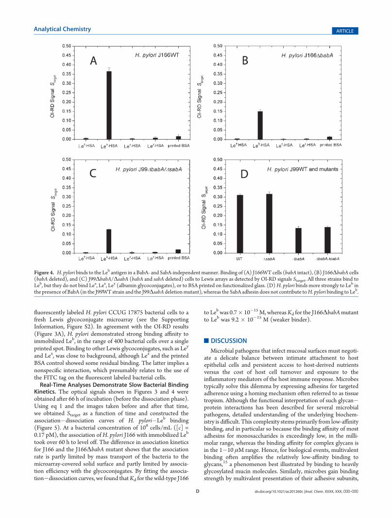
We determined Θtarget from the OI-RD microscopy imagesobtained before and after blocking with BSA (see the SupportingInformation, text S2). With the information on Θtarget andSblocking-BSA from the unprinted surface region, we calculatedStarget using eq 1 from the data shown in Figure 3. Parts A and B ofFigure 4 show, respectively, the results for H. pylori J166 (WT)and J166ΔbabA binding to the four Lewis glycoconjugates andprinted BSA control. Wild-type J166 binds to Leb with highstrength and specificity, whereas the J166ΔbabA mutant showslower but still specific affinity for Leb. The latter observationsupports the notion thatH. pylori J166 has both BabA-dependentand BabA-independent Leb-binding adhesin activity. Findingswere similar forH. pylori strain J99WT, which differs from J166 inthat it in addition to BabA also expresses the sialyl-Lex bindingprotein, SabA.14 Both J99WT and the J99ΔsabA mutant bind toLeb with high affinity and specificity, whereas the J99ΔbabAΔ-sabA and J99ΔbabA mutants bind to Leb with reduced but stillspecific affinity (Figure 4, parts C and D). These results suggestthat, like J166, the J99 strain also has additional Leb-bindingactivity due to one or more outer-membrane proteins. Thiscomplementary adhesin activity is specific for Leb (Figure 4C)and is not associated with the SabA adhesin, since the ΔsabAmutants exhibit intact Leb binding (Figure 4, parts C and D).The detection sensitivity of the OI-RD microscopy technique
was compared to that of conventional microscopy by applying
Figure 3. H. pylori binds to the Leb antigen in a BabA-independent manner. (A) The J166WT strain, (B) the J166ΔbabA deletion mutant strain, and(C) the J99ΔbabA deletion mutant strain (F) were incubated for 66 h at room temperature on an extended Lewis glycoconjugate microarray (Leb, Lea,Lex, Ley, each at four target concentrations). The OI-RD images show that both the WT and the babA deletion mutants bind specifically to the Leb
antigen, although the WT strain has higher binding strength. The results were obtained by subtracting the images taken before incubation from theimages taken after incubation.
D dx.doi.org/10.1021/ac201260c |Anal. Chem. XXXX, XXX, 000–000
Analytical Chemistry ARTICLE
fluorescently labeled H. pylori CCUG 17875 bacterial cells to afresh Lewis glycoconjugate microarray (see the SupportingInformation, Figure S2). In agreement with the OI-RD results(Figure 3A), H. pylori demonstrated strong binding affinity toimmobilized Leb, in the range of 400 bacterial cells over a singleprinted spot. Binding to other Lewis glycoconjugates, such as Ley
and Lex, was close to background, although Lea and the printedBSA control showed some residual binding. The latter implies anonspecific interaction, which presumably relates to the use ofthe FITC tag on the fluorescent labeled bacterial cells.Real-Time Analyses Demonstrate Slow Bacterial Binding
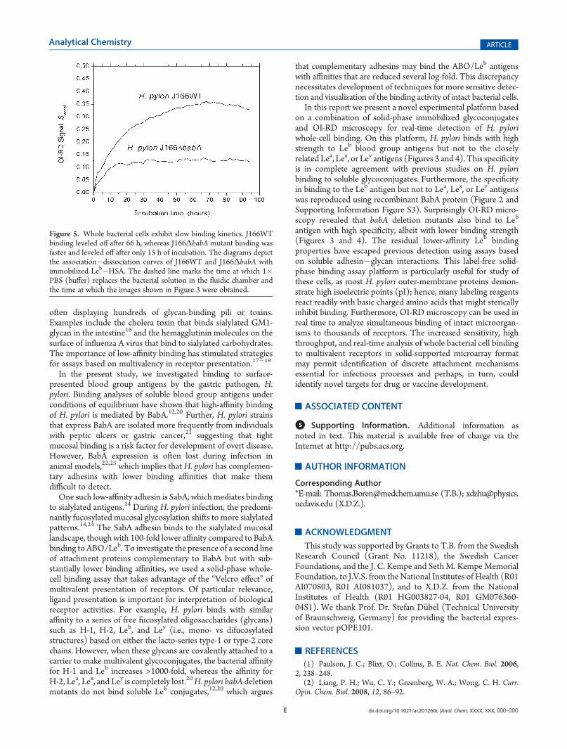
Kinetics. The optical signals shown in Figures 3 and 4 wereobtained after 66 h of incubation (before the dissociation phase).Using eq 1 and the images taken before and after that time,we obtained Starget as a function of time and constructed theassociation�dissociation curves of H. pylori�Leb binding(Figure 5). At a bacterial concentration of 108 cells/mL ([c] =0.17 pM), the association ofH. pylori J166 with immobilized Leb
took over 60 h to level off. The difference in association kineticsfor J166 and the J166ΔbabA mutant shows that the associationrate is partly limited by mass transport of the bacteria to themicroarray-covered solid surface and partly limited by associa-tion efficiency with the glycoconjugates. By fitting the associa-tion�dissociation curves, we found thatKd for the wild-type J166
to Leb was 0.7� 10�15M, whereasKd for the J166ΔbabAmutantto Leb was 9.2 � 10�15 M (weaker binder).
’DISCUSSION
Microbial pathogens that infect mucosal surfaces must negoti-ate a delicate balance between intimate attachment to hostepithelial cells and persistent access to host-derived nutrientsversus the cost of host cell turnover and exposure to theinflammatory mediators of the host immune response. Microbestypically solve this dilemma by expressing adhesins for targetedadherence using a homing mechanism often referred to as tissuetropism. Although the functional interpretation of such glycan�protein interactions has been described for several microbialpathogens, detailed understanding of the underlying biochem-istry is difficult. This complexity stems primarily from low-affinitybinding, and in particular so because the binding affinity of mostadhesins for monosaccharides is exceedingly low, in the milli-molar range, whereas the binding affinity for complex glycans isin the 1�10 μM range. Hence, for biological events, multivalentbinding often amplifies the relatively low-affinity binding toglycans,15 a phenomenon best illustrated by binding to heavilyglycosylated mucin molecules. Similarly, microbes gain bindingstrength by multivalent presentation of their adhesive subunits,
Figure 4. H. pylori binds to the Leb antigen in a BabA- and SabA-independent manner. Binding of (A) J166WT cells (babA intact), (B) J166ΔbabA cells(babA deleted), and (C) J99ΔbabA/ΔsabA (babA and sabA deleted) cells to Lewis arrays as detected by OI-RD signals Starget. All three strains bind toLeb, but they do not bind Lea, Lex, Ley (albumin glycoconjugates), or to BSA printed on functionalized glass. (D)H. pylori binds more strongly to Leb inthe presence of BabA (in the J99WT strain and the J99ΔsabA deletionmutant), whereas the SabA adhesin does not contribute toH. pylori binding to Leb.
E dx.doi.org/10.1021/ac201260c |Anal. Chem. XXXX, XXX, 000–000
Analytical Chemistry ARTICLE
often displaying hundreds of glycan-binding pili or toxins.Examples include the cholera toxin that binds sialylated GM1-glycan in the intestine16 and the hemagglutinin molecules on thesurface of influenza A virus that bind to sialylated carbohydrates.The importance of low-affinity binding has stimulated strategiesfor assays based on multivalency in receptor presentation.17�19
In the present study, we investigated binding to surface-presented blood group antigens by the gastric pathogen, H.pylori. Binding analyses of soluble blood group antigens underconditions of equilibrium have shown that high-affinity bindingof H. pylori is mediated by BabA.12,20 Further, H. pylori strainsthat express BabA are isolated more frequently from individualswith peptic ulcers or gastric cancer,21 suggesting that tightmucosal binding is a risk factor for development of overt disease.However, BabA expression is often lost during infection inanimal models,22,23 which implies that H. pylori has complemen-tary adhesins with lower binding affinities that make themdifficult to detect.
One such low-affinity adhesin is SabA, whichmediates bindingto sialylated antigens.14 During H. pylori infection, the predomi-nantly fucosylated mucosal glycosylation shifts to more sialylatedpatterns.14,24 The SabA adhesin binds to the sialylated mucosallandscape, though with 100-fold lower affinity compared to BabAbinding to ABO/Leb. To investigate the presence of a second lineof attachment proteins complementary to BabA but with sub-stantially lower binding affinities, we used a solid-phase whole-cell binding assay that takes advantage of the “Velcro effect” ofmultivalent presentation of receptors. Of particular relevance,ligand presentation is important for interpretation of biologicalreceptor activities. For example, H. pylori binds with similaraffinity to a series of free fucosylated oligosaccharides (glycans)such as H-1, H-2, Leb, and Ley (i.e., mono- vs difucosylatedstructures) based on either the lacto-series type-1 or type-2 corechains. However, when these glycans are covalently attached to acarrier to make multivalent glycoconjugates, the bacterial affinityfor H-1 and Leb increases >1000-fold, whereas the affinity forH-2, Lea, Lex, and Ley is completely lost.20H. pylori babA deletionmutants do not bind soluble Leb conjugates,12,20 which argues
that complementary adhesins may bind the ABO/Leb antigenswith affinities that are reduced several log-fold. This discrepancynecessitates development of techniques for more sensitive detec-tion and visualization of the binding activity of intact bacterial cells.
In this report we present a novel experimental platform basedon a combination of solid-phase immobilized glycoconjugatesand OI-RD microscopy for real-time detection of H. pyloriwhole-cell binding. On this platform, H. pylori binds with highstrength to Leb blood group antigens but not to the closelyrelated Lea, Lex, or Ley antigens (Figures 3 and 4). This specificityis in complete agreement with previous studies on H. pyloribinding to soluble glycoconjugates. Furthermore, the specificityin binding to the Leb antigen but not to Lea, Lex, or Ley antigenswas reproduced using recombinant BabA protein (Figure 2 andSupporting Information Figure S3). Surprisingly OI-RD micro-scopy revealed that babA deletion mutants also bind to Leb
antigen with high specificity, albeit with lower binding strength(Figures 3 and 4). The residual lower-affinity Leb bindingproperties have escaped previous detection using assays basedon soluble adhesin�glycan interactions. This label-free solid-phase binding assay platform is particularly useful for study ofthese cells, as most H. pylori outer-membrane proteins demon-strate high isoelectric points (pI); hence, many labeling reagentsreact readily with basic charged amino acids that might stericallyinhibit binding. Furthermore, OI-RD microscopy can be used inreal time to analyze simultaneous binding of intact microorgan-isms to thousands of receptors. The increased sensitivity, highthroughput, and real-time analysis of whole bacterial cell bindingto multivalent receptors in solid-supported microarray formatmay permit identification of discrete attachment mechanismsessential for infectious processes and perhaps, in turn, couldidentify novel targets for drug or vaccine development.
’ASSOCIATED CONTENT
bS Supporting Information. Additional information asnoted in text. This material is available free of charge via theInternet at http://pubs.acs.org.
’AUTHOR INFORMATION
Corresponding Author*E-mail: [email protected] (T.B.); [email protected] (X.D.Z.).
’ACKNOWLEDGMENT
This study was supported by Grants to T.B. from the SwedishResearch Council (Grant No. 11218), the Swedish CancerFoundations, and the J. C. Kempe and Seth M. KempeMemorialFoundation, to J.V.S. from the National Institutes of Health (R01AI070803, R01 AI081037), and to X.D.Z. from the NationalInstitutes of Health (R01 HG003827-04, R01 GM076360-04S1). We thank Prof. Dr. Stefan D€ubel (Technical Universityof Braunschweig, Germany) for providing the bacterial expres-sion vector pOPE101.
’REFERENCES
(1) Paulson, J. C.; Blixt, O.; Collins, B. E. Nat. Chem. Biol. 2006,2, 238–248.
(2) Liang, P. H.; Wu, C. Y.; Greenberg, W. A.; Wong, C. H. Curr.Opin. Chem. Biol. 2008, 12, 86–92.
Figure 5. Whole bacterial cells exhibit slow binding kinetics. J166WTbinding leveled off after 66 h, whereas J166ΔbabA mutant binding wasfaster and leveled off after only 15 h of incubation. The diagrams depictthe association�dissociation curves of J166WT and J166ΔbabA withimmobilized Leb�HSA. The dashed line marks the time at which 1�PBS (buffer) replaces the bacterial solution in the fluidic chamber andthe time at which the images shown in Figure 3 were obtained.
F dx.doi.org/10.1021/ac201260c |Anal. Chem. XXXX, XXX, 000–000
Analytical Chemistry ARTICLE
(3) Landry, J. P.; Zhu, X. D.; Gregg, J. Opt. Lett. 2004, 29, 581–583.(4) Zhu, X. D.; Landry, J. P.; Sun, Y. S.; Gregg, J. P.; Lam, K. S.; Guo,
X. W. Appl. Opt. 2007, 46, 1890–1895.(5) Landry, J. P.; Sun, Y. S.; Guo, X. W.; Zhu, X. D. Appl. Opt. 2008,
47, 3275–3288.(6) Fei, Y. Y.; Landry, J. P.; Sun, Y. S.; Zhu, X. D.; Luo, J. T.; Wang,
X. B.; Lam, K. S. Rev. Sci. Instrum. 2008, 79, 013708.(7) Sun, Y. S.; Landry, J. P.; Fei, Y. Y.; Zhu, X. D.; Luo, J. T.; Wang,
X. B.; Lam, K. S. Langmuir 2008, 24, 13399–13405.(8) Sun, Y. S.; Landry, J. P.; Fei, Y. Y.; Zhu, X. D.; Luo, J. T.; Wang,
X. B.; Lam, K. S. Anal. Chem. 2009, 81, 5373–5380.(9) Fei, Y. Y.; Landry, J. P.; Sun, Y. S.; Zhu, X. D.; Wang, X. B.; Wu,
C. Y.; Lam, K. S. J. Biomed. Opt. 2010, 15, 016018.(10) Kusters, J. G.; van Vliet, A. H.; Kuipers, E. J. Clin. Microbiol. Rev.
2006, 19, 449–490.(11) Bor�en, T.; Falk, P.; Roth, K. A.; Larson, G.; Normark, S. Science
1993, 262, 1892–1895.(12) Aspholm-Hurtig, M.; Dailide, G.; Lahmann, M.; Kalia, A.; Ilver,
D.; Ilver, D.; Roche, N.; Vikstr€om, S.; Sj€ostr€om, R.; Lind�en, S.;B€ackstr€om, A.; Lundberg, C.; Arnqvist, A.; Mahdavi, J.; Nilsson, U. J.;Velapatino, B.; Gilman, R. H.; Gerhard, M.; Alarcon, T.; Lopez-Brea, M.;Nakazawa, T.; Fox, J. G.; Correa, P.; Dominguez-Bello, M. G.; Perez-Perez, G. I.; Blaser, M. J.; Normark, S.; Carlstedt, I.; Oscarson, S.;Teneberg, S.; Berg, D. E.; Bor�en, T. Science 2004, 305, 519–522.(13) Alm, R. A.; Bina, J.; Andrews, B. M.; Doig, P.; Hancock, R. E.;
Trust, T. J. Infect. Immun. 2000, 68, 4155–4168.(14) Mahdavi, J.; Sond�en, B.; Hurtig, M.; Olfat, F. O.; Forsberg, L.;
Roche, N.; Ångstrom, J.; Larsson, T.; Teneberg, S.; Karlsson, K. A.;Altraja, S.; Wadstr€om, T.; Kersulyte, D.; Berg, D. E.; Dubois, A.;Petersson, C.; Magnusson, K. E.; Norberg, T.; Lindh, F.; Lundskog,B. B.; Arnqvist, A.; Hammarstr€om, L.; Bor�en, T. Science 2002,297, 573–578.(15) Adler, P.; Wood, S. J.; Lee, Y. C.; Lee, R. T.; Petri, W. A., Jr.;
Schnaar, R. L. J. Biol. Chem. 1995, 270, 5164–5171.(16) Holmgren, J.; L€onnroth, I.; M�ansson, J.; Svennerholm, L. Proc.
Natl. Acad. Sci. U.S.A. 1975, 72, 2520–2524.(17) Bor�en, T.; Normark, S.; Falk, P. Trends Microbiol. 1994,
2, 221–228.(18) Falk, P.; Bor�en, T.; Normark, S. Methods Enzymol. 1994,
236, 353–374.(19) Aspholm, M.; Kalia, A.; Ruhl, S.; Schedin, S.; Arnqvist, A.;
Lind�en, S.; Sj€ostr€om, R.; Gerhard, M.; Semino-Mora, C.; Dubois, A.;Unemo,M.; Danielsson, D.; Teneberg, S.; Lee,W. K.; Berg, D. E.; Bor�en,T. Methods Enzymol. 2006, 417, 293–339.(20) Ilver, D.; Arnqvist, A.; €Ogren, J.; Frick, I. M.; Kersulyte, D.; Rad,
R.; Incecik, E. T.; Berg, D. E.; Covacci, A.; Engstrand, L.; Bor�en, T.Science 1998, 279, 373–377.(21) Gerhard, M.; Lehn, N.; Neumayer, N.; Bor�en, T.; Rad, R.;
Schepp, W.; Miehlke, S.; Classen, M.; Prinz, C. Proc. Natl. Acad. Sci. U.S.A. 1999, 96, 12778–12783.(22) Solnick, J. V.; Hansen, L. M.; Salama, N. R.; Boonjakuakul, J. K.;
Syvanen, M. Proc. Natl. Acad. Sci. U.S.A. 2004, 101, 2106–2111.(23) Styer, C. M.; Hansen, L. M.; Cooke, C. L.; Gundersen, A. M.;
Choi, S. S.; Berg, D. E.; Benghezal, M.; Marshall, B. J.; Peek, R. M., Jr.;Bor�en, T.; Solnick, J. V. Infect. Immun. 2010, 78, 1593–1600.(24) Lind�en, S.; Mahdavi, J.; Semino-Mora, C.; Olsen, C.; Carlstedt,
I.; Bor�en, T.; Dubois, A. PLoS Pathog. 2008, 4, e2.(25) Solnick, J. V.; Hansen, L. M.; Canfield, D. R.; Parsonnet, J.
Infect. Immun. 2001, 69, 6887–6892.