Embed Size (px)
Citation preview
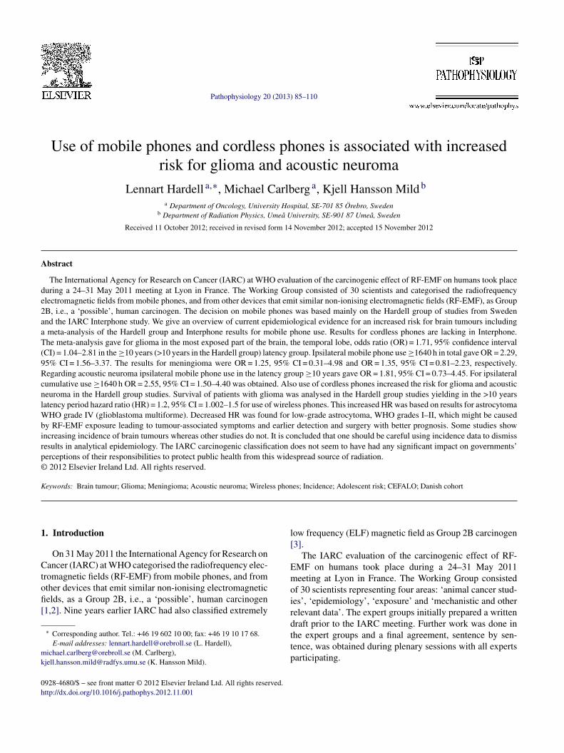
Pathophysiology 20 (2013) 85–110
Use of mobile phones and cordless phones is associated with increasedrisk for glioma and acoustic neuroma
Lennart Hardell a,!, Michael Carlberg a, Kjell Hansson Mild b
a Department of Oncology, University Hospital, SE-701 85 Örebro, Swedenb Department of Radiation Physics, Umeå University, SE-901 87 Umeå, Sweden
Received 11 October 2012; received in revised form 14 November 2012; accepted 15 November 2012
Abstract
The International Agency for Research on Cancer (IARC) at WHO evaluation of the carcinogenic effect of RF-EMF on humans took placeduring a 24–31 May 2011 meeting at Lyon in France. The Working Group consisted of 30 scientists and categorised the radiofrequencyelectromagnetic fields from mobile phones, and from other devices that emit similar non-ionising electromagnetic fields (RF-EMF), as Group2B, i.e., a ‘possible’, human carcinogen. The decision on mobile phones was based mainly on the Hardell group of studies from Swedenand the IARC Interphone study. We give an overview of current epidemiological evidence for an increased risk for brain tumours includinga meta-analysis of the Hardell group and Interphone results for mobile phone use. Results for cordless phones are lacking in Interphone.The meta-analysis gave for glioma in the most exposed part of the brain, the temporal lobe, odds ratio (OR) = 1.71, 95% confidence interval(CI) = 1.04–2.81 in the "10 years (>10 years in the Hardell group) latency group. Ipsilateral mobile phone use "1640 h in total gave OR = 2.29,95% CI = 1.56–3.37. The results for meningioma were OR = 1.25, 95% CI = 0.31–4.98 and OR = 1.35, 95% CI = 0.81–2.23, respectively.Regarding acoustic neuroma ipsilateral mobile phone use in the latency group "10 years gave OR = 1.81, 95% CI = 0.73–4.45. For ipsilateralcumulative use "1640 h OR = 2.55, 95% CI = 1.50–4.40 was obtained. Also use of cordless phones increased the risk for glioma and acousticneuroma in the Hardell group studies. Survival of patients with glioma was analysed in the Hardell group studies yielding in the >10 yearslatency period hazard ratio (HR) = 1.2, 95% CI = 1.002–1.5 for use of wireless phones. This increased HR was based on results for astrocytomaWHO grade IV (glioblastoma multiforme). Decreased HR was found for low-grade astrocytoma, WHO grades I–II, which might be causedby RF-EMF exposure leading to tumour-associated symptoms and earlier detection and surgery with better prognosis. Some studies showincreasing incidence of brain tumours whereas other studies do not. It is concluded that one should be careful using incidence data to dismissresults in analytical epidemiology. The IARC carcinogenic classification does not seem to have had any significant impact on governments’perceptions of their responsibilities to protect public health from this widespread source of radiation.© 2012 Elsevier Ireland Ltd. All rights reserved.
Keywords: Brain tumour; Glioma; Meningioma; Acoustic neuroma; Wireless phones; Incidence; Adolescent risk; CEFALO; Danish cohort
1. Introduction
On 31 May 2011 the International Agency for Research onCancer (IARC) at WHO categorised the radiofrequency elec-tromagnetic fields (RF-EMF) from mobile phones, and fromother devices that emit similar non-ionising electromagneticfields, as a Group 2B, i.e., a ‘possible’, human carcinogen[1,2]. Nine years earlier IARC had also classified extremely
! Corresponding author. Tel.: +46 19 602 10 00; fax: +46 19 10 17 68.E-mail addresses: [email protected] (L. Hardell),
[email protected] (M. Carlberg),[email protected] (K. Hansson Mild).
low frequency (ELF) magnetic field as Group 2B carcinogen[3].
The IARC evaluation of the carcinogenic effect of RF-EMF on humans took place during a 24–31 May 2011meeting at Lyon in France. The Working Group consistedof 30 scientists representing four areas: ‘animal cancer stud-ies’, ‘epidemiology’, ‘exposure’ and ‘mechanistic and otherrelevant data’. The expert groups initially prepared a writtendraft prior to the IARC meeting. Further work was done inthe expert groups and a final agreement, sentence by sen-tence, was obtained during plenary sessions with all expertsparticipating.
0928-4680/$ – see front matter © 2012 Elsevier Ireland Ltd. All rights reserved.http://dx.doi.org/10.1016/j.pathophys.2012.11.001
86 L. Hardell et al. / Pathophysiology 20 (2013) 85–110
The IARC decision on mobile phones was based mainlyon two sets of case-control human studies; the Hardell groupof studies from Sweden and the IARC Interphone study. Bothprovided complementary and supportive results on positiveassociations between two types of brain tumours; glioma andacoustic neuroma, and exposure to RF-EMF from wirelessphones.
The final IARC decision was confirmed by voting of 29scientists (one not present). A large majority of participantsvoted to classify RF-EMF radiation as ‘possibly carcino-genic’ to humans, Group 2B. The decision was also basedon occupational studies.
In this paper an up-to-date review of the evidence of anassociation between use of wireless phones and brain tumoursis presented. The Nordic countries were among the firstcountries in the world to widely adopt wireless telecommuni-cations technology. Analogue phones (NMT; Nordic MobileTelephone System) were introduced in the early 1980s usingboth 450 and 900 Megahertz (MHz) frequencies. NMT 450was used in Sweden from 1981 but closed down on 31December 2007, NMT 900 operated during 1986–2000.
The digital system (GSM; Global System for MobileCommunication) using dual band, 900 and 1800 MHz,started to operate in 1991 and dominates now the market.The third generation of mobile phones, 3G or UMTS(Universal Mobile Telecommunication System), using1900/2100 MHz RF fields has been introduced worldwide inrecent years, in Sweden in 2003. Currently the fourth gener-ation, 4G (Terrestrial 3G), operating at 800/2600 MHz andTrunked Radio Communication (TETRA 380–400 MHz)are being established in Sweden and elsewhere. Nowadaysmobile phones are used more than landline phones inSweden (http://www.pts.se/upload/Rapporter/Tele/2011/sv-telemarknad-halvar-2011-pts-er-2011-21.pdf). Worldwide,an estimate of 5.9 billion mobile phone subscriptions wasreported at the end of 2011 by the International Telecom-munication Union (ITU; http://www.itu.int/ITU-D/ict/facts/2011/material/ICTFactsFigures2011.pdf). Many usersare children and adolescents, which is of special concernregarding potential health effects.
Desktop cordless phones (DECT) have been used inSweden since 1988, first using analogue 800–900 MHz RFfields, but since early 1990s using a digital 1900 MHz system.The cordless phones are becoming more common than tradi-tional landlines. Also these phones emit RF-EMF radiationsimilar to that of mobile phones. Thus, it is also neces-sary to consider the usage of cordless phones along withmobile phones, when human health risks are evaluated. Itshould be noted that the usual cordless base stations emitRF-EMF continuously. They are often installed in officesclose to the person using a cordless phone handset or inhomes even in bedrooms next to the head of a sleeping per-son.
The real increase in use and exposure to electromagneticfields from wireless phones (mobile phones and cordlessphones) in most countries has occurred since the end of the
1990s. When used they emit RF-EMFs. The GSM phonesand to a lesser extent the cordless phones emit also ELF-EMF from the battery when used [4,5]. The brain is the maintarget organ during use of the handheld phone [6]. Thus, fearof an increased risk for brain tumours has dominated thedebate during the last one or two decades. While RF-EMFsdo not have sufficient energy to break chemical bonds likeionising radiation, at least not directly, they can neverthelesshave harmful effects on biological tissues. Plausible biologi-cal mechanisms for these effects include impairment of DNArepair mechanisms and epigenetic changes to DNA.
Primary brain tumours (central nervous system; CNS)constitute of a heterogeneous group of neoplasms dividedinto two major groups; malignant and benign. They are ofdifferent histological types depending on tissue of origin withdifferent growth patterns, molecular markers, anatomicallocalisations, and age and gender distributions. The clini-cal appearance, treatment and prognosis are quite differentdepending on tumour type.
Ionising radiation is an established risk factor for primarybrain tumours [7], but there are no well-established envi-ronmental causes. Higher socio-economic status tends to berelated to higher incidence and some rare inherited cancersyndromes account for a small fraction of tumours [7]. Famil-ial aggregation of glioma has been reported. In a large study77% more glioma cases than expected were reported amongfamily members [8].
The purpose of this article is to give a comprehensivereview of the association between use of mobile and cord-less phones and brain tumours, primarily based on the resultsof the major publications in this field. We include the Hardellgroup papers and the WHO Interphone study [9–11]. Alsosome additional analyses of the risk for brain tumours basedon these results are given. Some early studies not part of thesetwo major study groups are also included. More discussionof the results and responses, agreements and disagreementsof the findings for the Hardell group and Interphone stud-ies can be found elsewhere [12]. In addition, this reviewincludes studies published after the IARC evaluation in May2011.
2. Materials and methods
The PubMed database (www.ncbi.nlm.nih.gov) was usedfor an up-dated search of published studies in thisarea using mobile/cellular/cordless telephone and braintumour/neoplasm/acoustic neuroma/meningioma/glioma assearching terms. Personal knowledge of published studieswas also used in order to get as comprehensive a reviewas possible. All of the authors have long experience in thisresearch area and have published the pioneer studies indicat-ing an association between use of wireless phones and certaintypes of brain tumours. They represent different supportiveareas of competence such as oncology, cancer epidemiology,statistics and physics.
L. Hardell et al. / Pathophysiology 20 (2013) 85–110 87
Table 1Summary of studies on the use of mobile phones and brain tumour risk.
Study Years; study type Age Tumour type No. of exposed cases Odds ratio, 95%confidence interval
Comments
Hardell et al. [15,16]Sweden
1994–1996;Case-control
20–80 years Brain tumours(n = 209)
78 OR 0.98 (0.69–1.41) Analogue and digitalmobile phone use
34 OR 1.07 (0.64–1.80) Ipsilateral mobile phoneuse
16 OR 1.20 (0.56–2.59) >10 year latency,analogue mobile phoneuse
Muscat et al. [17]USA
1994–1998;Case-control
18–80 years Brain tumours(n = 469)
66 OR 0.8 (0.6–1.2) Mean duration of mobilephone use 2.8 years
Neuorepithelioma(n = 35)
14 OR 2.1 (0.9–4.7)
2.1. Statistical methods
All analyses in the Hardell group studies were done usingStataSE 10.1 (Stata/SE 10.1 for Windows; StataCorp., Col-lege Station TX). Odds ratios (OR) and 95% confidenceintervals (CI) were calculated using unconditional logisticregression analysis. Further details can be found in the pub-lications.
Meta-analyses were performed on use of mobile phonesin the Hardell group [13,14] and Interphone group [9,10]studies. No duplicate data from different articles publishedby the same group of authors were included. Model waschosen based on test for heterogeneity in the overall ("10years and "1640 h) groups. In the analysis of survival ofpatients with glioma, Cox proportional hazards model wasused to calculate hazard ratios (HR) and corresponding 95%confidence intervals. Follow-up time was counted from thedate of diagnosis to the date of death or until May 30, 2012for living cases.
3. Results
3.1. Brain tumours overall
The first study by Hardell et al. [15,16] included cases andcontrols during 1994–1996 in parts of Sweden and was thefirst published study on this issue. Only living cases diag-nosed during 1994–1996 were included. Two controls wereselected to each case from the Population Registry. In total209 (90%) of the cases and 425 (91%) of the controls thatmet the inclusion criteria answered the mailed questionnaire.Overall no association between mobile phone use and braintumours was found. A slightly increased, but not statisticallysignificant, risk was found for analogue phone (NMT) useand for a latency period greater than 10 years, OR = 1.20,95% CI = 0.56–2.59, Table 1.
Exposure to the radiation from the phones is generallyhigher in the temporal lobe, the part of the brain that is nearto the ear [6]. For tumours located in the temporal, occip-ital or temporoparietal lobe areas of the brain an increasedrisk was found for ipsilateral exposure, that is the telephone
was mostly used on the same side of the head as the tumourappeared, yielding OR = 2.42, 95% CI = 0.97–6.05 [16]. Thiswas the first study in the world that indicated an associa-tion between use of mobile phones and an increased riskfor brain tumours. However, all results were based on lownumbers of exposed subjects and different histopathologicaltypes of brain tumours so no firm conclusions could be drawn.Furthermore, this first study did not include use of cordlessphones.
Muscat et al. [17] studied patients with malignant braintumours from five different hospitals in USA, Table 1. Con-trols were hospital patients. Data from 469 (82%) cases and422 (90%) controls were available. Overall no associationwas found, OR for handheld cellular phones was 0.8, 95%CI = 0.6–1.2, but the mean duration of use was short, only 2.8years for cases and 2.7 years for controls. For neuroepithe-lioma OR = 2.1, 95% CI = 0.9–4.7, was reported. The studywas inconclusive since no data were available on long-termusers ("10 years latency period). Some support of an associa-tion was obtained since of 41 evaluable tumours, 26 occurredat the side of the head mostly used during calls and 15 on thecontralateral side.
3.2. Glioma
Glioma is the most common malignant brain tumour andrepresents about 60% of all central nervous system tumours.The most common glioma subtype is astrocytoma. Astrocytictumours are divided in two groups depending on the malig-nant potential; low-grade (WHO grades I–II) and high-grade(WHO grades III–IV). Low-grade astrocytoma has a rela-tively favourable prognosis, whereas survival is shorter forpatients with high-grade glioma. Glioblastoma multiforme(WHO grade IV) accounts for 60–75% of all astrocytoma.The peak incidence is between 45 and 75 years of age withmedian survival less than one year [18].
In the study by Hardell et al. [15] analysis of the caseswith astrocytoma produced OR = 1.09, 95% CI = 0.64–1.84(n = 36 cases), Table 2. OR increased further for ipsilat-eral exposure for right sided tumours, OR = 1.30, 95%CI = 0.54–3.13 (n = 13 cases), whereas no association was
88L.H
ardelletal./Pathophysiology20
(2013)85–110
Table 2Summary of studies on the use of wireless phones and glioma risk.
Study Years; study type Age Tumour type No. ofexposedcases
Odds ratio, 95%confidence interval
Comments
Hardell et al. [15] Sweden 1994–1996;Case-control
20–80 years Astrocytoma WHOgrade I–IV (n = 94)
36 OR 1.09 (0.64–1.84) Analogue and digital mobile phone use
13 OR 1.30 (0.54–3.13) Ipsilateral mobile phone use, right sided tumours3 OR 0.35 (0.07–1.81) Ipsilateral mobile phone use, left sided tumours
Inskip et al. [19] USA 1994–1998;Case-control
"18 years Glioma (n = 489) 11 OR 0.6 (0.3–1.4) "5 years of mobile phone use
Auvinen et al. [20] Finland 1996; Case-control,register based
20–69 years Glioma (n = 198) Not given OR 1.5 (1.0–2.4) Analogue and digital mobile phone “ever” use
25 OR 2.1 (1.3–3.4) Analogue mobile phone “ever” used11 OR 2.4 (1.2–5.1) Analogue mobile phone use, 1–2 years11 OR 2.0 (1.0–4.1) Analogue mobile phone use, >2 years
Hardell et al. [26–28] Carlberg,Hardell [29] Sweden
1997–2003;Case-control
20–80 years Glioma (n = 1148) 123 OR 2.5 (1.8–3.3) >10 year latency, mobile phone
57 OR 2.9 (1.8–4.7) >10 year latency, mobile phone, ipsilateral, only living50 OR 2.6 (1.7–4.1) >10 year latency, mobile phone only45 OR 1.7 (1.1–2.6) >10 year latency, cordless phone20 OR 3.8 (1.8–8.1) >10 year latency, cordless phone, ipsilateral, only living9 OR 1.2 (0.5–2.9) >10 year latency, cordless phone only; >5–10 year latency
OR 1.9 (1.3–2.9; n = 55)150 OR 2.1 (1.6–2.8) >10 year latency, wireless phone (mobile and cordless
phone)
Astrocytoma, highgrade (n = 820)
102 OR 3.0 (2.1–4.2) >10 year latency, mobile phone
47 OR 3.9 (2.3–6.6) >10 year latency, mobile phone, ipsilateral, only living37 OR 2.8 (1.7–4.6) >10 year latency, mobile phone only36 OR 2.0 (1.2–3.2) >10 year latency, cordless phone15 OR 5.5 (2.3–13) >10 year latency, cordless phone, ipsilateral, only living6 OR 0.9 (0.3–2.6) >10 year latency, cordless phone only; >5–10 year latency
OR 2.4 (1.6–3,7; n = 44)121 OR 2.5 (1.8–3.4) >10 year latency, wireless phone (mobile and cordless
phone)
Interphone Study Group [9] 13countries; Australia, Canada,Denmark, Finland, France,UK, Germany, Israel, Italy,Japan, New Zealand, Norway,Sweden
2000–2004, 2–4 yearsdepending on studyregion. Case-control
30–59 years Glioma (n = 2708) 1666 OR 0.81 (0.70–0.94) Regular use of mobile phone in the past "1 year
L. Hardell et al. / Pathophysiology 20 (2013) 85–110 89
210
OR
1.40
(1.0
3–1.
89)
Cum
ulat
ive
hour
sm
obile
phon
e"
1640
h78
OR
1.87
(1.0
9–3.
22)
Cum
ulat
ive
hour
sm
obile
phon
e"
1640
h,tu
mou
rsin
tem
pora
llob
e10
0O
R1.
96(1
.22–
3.16
)C
umul
ativ
eho
urs
mob
ileph
one
"16
40h,
ipsi
late
ral
mob
ileph
one
use
Inte
rpho
neSt
udy
Gro
up[9
]A
ppen
dix
220
00–2
004,
2–4
year
sde
pend
ing
onst
udy
regi
on.C
ase-
cont
rol
30–5
9ye
ars
Glio
ma
(n=
1211
)46
0O
R1.
68(1
.16–
2.41
)R
estr
icte
dto
ever
regu
lar
use
time
sinc
est
art2
–4ye
ars;
1–1.
9ye
ars
asre
fere
nce
entit
y
468
OR
1.54
(1.0
6–2.
22)
Res
tric
ted
toev
erre
gula
rus
etim
esi
nce
star
t5–9
year
s;1–
1.9
year
sas
refe
renc
een
tity
190
OR
2.18
(1.4
3–3.
31)
Res
tric
ted
toev
erre
gula
rus
etim
esi
nce
star
t10+
year
s;1–
1.9
year
sas
refe
renc
een
tity
160
OR
1.82
(1.1
5–2.
89)
Res
tric
ted
toev
erre
gula
rus
e"
1640
h,<5
has
refe
renc
een
tity
seen for astrocytoma in the left hemisphere and ipsilateralexposure, OR = 0.35, 95% CI = 0.07–1.81 (n = 3 cases).
The study by Inskip et al. [19] from USA had few long-term users of mobile phones. Only 11 cases with glioma, 6with meningioma and 5 with acoustic neuroma had "5 yearsregular use. No subject had "10 years use. Of the hospital-based cases 92% participated. The study comprised 489 caseswith glioma, 197 with meningioma and 96 with acousticneuroma, and 799 (86%) hospital-based controls. Proxy inter-views were necessary for 16% of the patients with glioma,8% of the patients with meningioma, 3% of the patients withacoustic neuroma, and 3% of the controls. Overall no statisti-cally significant associations were found, Table 2. Regardingdifferent types of glioma OR = 1.8, 95% CI = 0.7–5.1was found for anaplastic astrocytoma (WHO grade III).Regarding hospital-based interviews and use of proxy inter-views, see discussion below in relation to the Interphonestudy.
A register based case-control study on brain and salivarygland tumours was performed in Finland [20]. All casesaged 20–69 years diagnosed in 1996 were included; 398brain tumour cases and 34 salivary gland tumour cases. Theduration of mobile phone use was short, for analogue users2–3 years and for digital users less than one year. No asso-ciation was found for salivary gland tumours. For gliomaOR = 2.1, 95% CI = 1.3–3.4 was calculated for use of ana-logue phones, but no association was found for digital mobilephones, Table 2. When duration of use of analogue phoneswas used as a continuous variable an increased risk wasfound for glioma with OR = 1.2, 95% CI = 1.1–1.5 per yearof use.
The Hardell group in Sweden studied the associationbetween use of mobile and cordless phones and brain tumoursdiagnosed during 1997–2003. First, cases diagnosed during1 January 1997 to 30 June 2000 were included. These resultswere published separately [21,22]. This was followed by thenext study period, 1 July 2000 to 31 December 2003 [23,24].The methods were the same including the same inclusioncriteria and an identical questionnaire in both studies; see thepublications for further details.
Both men and women aged 20–80 years at the time ofdiagnosis were included and all were alive at the time ofinclusion in the study. They were reported from cancer reg-istries with a brain tumour verified by histopathology. TheSwedish Population Registry was used for identification ofmatched controls. The study included use of wireless phones(mobile and cordless phones), as well as asking questionson e.g., occupational exposures. Use of wireless phones wascarefully assessed by a self-administered questionnaire sup-plemented over the phone. The ear that had mostly been usedduring calls with mobile phone and/or cordless phone wasassessed by separate questions; >50% of the time for oneside, or equally for both sides. This information was checkedduring the supplementary phone calls and finally also bya separate letter with good agreement between these threemethods.
90 L. Hardell et al. / Pathophysiology 20 (2013) 85–110
Tumour localisation for the cases was defined by usingmedical records including computer tomography (CT) and/ormagnetic resonance imaging (MRI). The matched controlwas assigned the same side as the tumour of the respectivecase. Use of the wireless phone was defined as ipsilateral("50% of the time), or contralateral (<50% of the time) inrelation to tumour side. Further details can be found in thepublications.
In a review commissioned by the former Swedish Radia-tion Protection Agency (now called the Swedish RadiationSafety Authority) it was suggested that the exclusion ofdeceased cases was a source of bias in our studies [25].As a response to that critique we performed a study on thecases with a malignant brain tumour that had died beforeinclusion in the case-control studies 1997–2003. These casesrepresented patients with a poor prognosis, mostly with astro-cytoma WHO grade IV (glioblastoma multiforme). Controlswere selected from the Death Registry in Sweden.
The study encompassed 464 cases and 464 controls thathad died from a malignant disease and 463 controls with othercauses of death. Exposure was assessed by a questionnairesent to the next of kin to each deceased case and control. Thequestionnaire was similar as in previous studies.
This investigation confirmed the previous results of anassociation between mobile phones and malignant braintumours [26].
The Hardell group has previously published pooled anal-ysis of malignant brain tumours diagnosed during the period1997–2003 [27]. These results were updated including alsoresults for deceased cases with malignant brain tumours[28,29]. The results on use of wireless phones were basedon 1251 cases with malignant brain tumour (response rate85%) and 2438 controls (response rate 84%).
Most cases had glioma (n = 1148) so we present in the fol-lowing results for that type of tumour. Latency was dividedin three categories, >1–5 years, >5–10 years, and >10 yearsfrom first use of a wireless phone until diagnosis of glioma.Both use of mobile and cordless phone gave an increased riskoverall, highest in the latency group >10 years, increasing fur-ther for ipsilateral use yielding for mobile phone OR = 2.9,95% CI = 1.8–4.7 and for cordless phone OR = 3.8, 95%CI = 1.8–8.1, Table 2. Highest ORs were found in the >10 yearlatency group for total wireless phone use as well, OR = 2.1,95% CI = 1.6–2.8 or a doubling of glioma risk.
OR increased statistically significant for glioma for cumu-lative use of wireless phones per 100 h; OR = 1.014, 95%CI = 1.008–1.019, and per year of latency; OR = 1.056, 95%CI = 1.037–1.075 [29]. Separate calculations of mobile phoneand cordless phone use yielded similar results with statisti-cally significant increasing risks.
It is common for a person to use both a mobile and acordless phone. For only use of mobile phone OR increasedfor glioma with time since first use yielding for >10 yearslatency OR = 2.6, 95% CI = 1.7–4.1. For only cordless phoneuse highest risk was obtained in the >5–10 years latency time;OR = 1.9, 95% CI = 1.3–2.9. However, the calculations in the
longest latency period were based on few subjects regardingcordless phone.
In Table 2 results are presented for high-grade astrocy-toma (n = 820). The results are similar as for the whole gliomagroup. Low-grade glioma is less common and the results inthis study were based on 132 cases. Ipsilateral use of mobilephone yielded in total OR = 1.8, 95% CI = 1.02–3.1 (n = 39cases) and cordless phone OR = 1.7, 95% CI = 0.98–3.1(n = 34 cases, data not in Table). Further results and discus-sion may be found elsewhere [29].
The Interphone study was conducted at 16 research centresin 13 countries during varying time periods between 2000 and2004. It was an international collaboration on brain tumourrisk and mobile phone use conducted under the guidance ofIARC. The investigation was initiated by recommendationsfrom several expert groups including one of the authors, KjellHansson Mild as a member of the EU group, to study possiblehealth effects of exposure to RF-EMF [30,31]. It should benoted that there was no overlap of cases or controls betweenthe Hardell group studies and the Swedish part of Interphoneperformed by another research group.
Some of the separate country analyses of the Interphonestudy produced contradictory results, as we have discussedelsewhere [13,32]. An increased risk for brain tumour wasfound in some studies and decreased risk in other studies.After several years of delay the overall Interphone resultswere finally published in May 2010 [9].
The study included 4301 glioma cases and the results werebased on 2708 participating cases (response rate 64%, rangeby centre 36–92%). In total 14,354 potential controls wereidentified and interviews were completed with 7658 (53%,range 42–74%). The low participation rates in some centresmay have created selection bias, see Hardell et al. [32].
Regular use of mobile phone in the past "1 year gavefor glioma OR = 0.81, 95% CI = 0.70–0.94, Table 2. Sub-group analyses showed statistically significant increased riskin the highest exposure group, i.e., those with cumulativemobile phone use "1640 h, which corresponds to abouthalf an hour of use per day for ten years, OR = 1.40, 95%CI = 1.03–1.89. The risk increased further for glioma in thetemporal lobe yielding OR = 1.87, 95% CI = 1.09–3.22. In thesame exposure category, cumulative use "1640 h and ipsi-lateral exposure produced OR = 1.96, 95% CI = 1.22–3.16 intotal (no data given for temporal lobe).
In Appendix 2, available on the web [9] analysis wasrestricted to ever-regular users of mobile phones in the Inter-phone study. Cumulative call time "1640 h gave OR = 1.82,95% CI = 1.15–2.89 compared with use <5 h. Time since startof regular use (latency) "10 years produced OR = 2.18, 95%CI = 1.43–3.31; reference entity 1–1.9 years.
The Interphone study group concluded: “However, biasesand errors limit the strength of the conclusions we candraw from these analyses and prevent a causal interpreta-tion.” In an editorial accompanying the Interphone results themain conclusion of the Interphone results was described as“both elegant and oracular. . .(which) tolerates diametrically
L. Hardell et al. / Pathophysiology 20 (2013) 85–110 91
opposite readings” [33]. They also pointed out severalmethodological reasons why the Interphone results werelikely to have underestimated the risks, such as the shortlatency period since first exposures became widespread;less than 10% of the Interphone cases had more than 10years exposure. “None of the today’s established carcino-gens, including tobacco, could have been firmly identified asincreasing risk in the first 10 years or so since first expo-sure”.
As has pointed our elsewhere [32] there were differencesbetween the Hardell group studies and Interphone. Regardingage group the Hardell group studies included subjects aged20–80 years, versus 30–59 years in Interphone. Furthermoreuse of cordless phones was not properly assessed, analysedor reported in Interphone. These differences have been dis-cussed in detail by Hardell et al. [14]. Thus, it could be shownthat restricting the age group to 30–59 years and consider-ing subjects that used a cordless phone as unexposed in theHardell group studies reduced the OR and produced resultsquite similar to Interphone, Table 3; see also Table 11 asdiscussed below. Latency time >10 years for glioma in thetemporal lobe yielded OR = 1.40, 95% CI = 0.70–2.81 in theHardell group studies and OR = 1.36, 95% CI = 0.88–2.11in Interphone (latency "10 years). Unfortunately the Inter-phone study did not give results for glioma in the temporallobe in the analyses in Appendix 2. Thus, excluding exposureto RF-EMFs from cordless phones as in the Interphone study,as well as excluding the younger and older subjects biasedthe ORs towards unity, which likely dilutes the ability to seehealth risks.
Most mobile phone users have not been using one singletelephone. It is likely that they have changed their handsetseveral times if they have been using a mobile phone formore than a few years. Many users have also been usingdifferent phone systems, such as analogue and digital, andmany of them have also been using a cordless phone at homeor at work. It is not clear how to combine the use of differentphones with different power outputs, systems, frequenciesand anatomical specific absorption rate (SAR) distributionsinto one exposure and dose measure. The difficulties lie in thefact that there is no generally accepted mechanism(s) betweenthe electromagnetic fields emitted from the phone and thebiological organism. This includes a mechanism by whichRF-EMF exposure produces changes in DNA. The energylevel associated with exposure is too low to cause direct DNAstrand breaks and DNA cross links. However, DNA damagescan be caused by cellular biochemical activities such as freeradicals. Several studies indicate that RF-EMFs increase freeradical activity in cells, as reviewed by Phillips et al. [34].This process is probably mediated via the Fenton reaction. Itshould also be noted that possible biological effects might nothave linear dose–response as indicated in some studies [35]and that the effects are depending on the carrier frequencies[36].
The different types of phones have different outputpower. We applied different weighting factors according to
the mean output power of the phones using for analoguephones (NMT) = 1, GSM = 0.1 and cordless phones = 0.01.The cumulative time for use of the different phone types wasmultiplied with the respective weighting factor added intoone score. The median score among the controls was usedas the cut-off in the dose–response calculations. We appliedthis method for the study period 1 January 1997 to 30 June2000 [21,22]. Somewhat higher ORs were obtained using theweighting factor, especially with a >10-year latency period,compared with calculations based on cumulative use only,but overall the results were similar [37]. This was explainedby the fact that most subjects had used an analogue mobilephone with the weighting factor = 1, thus the weighting factorhad little impact on the results.
A further issue is that there is a difference in the out-put power level from mobile phones between urban andrural areas. This is caused by adaptive power control (APC)in the cellular telephone and is regulated by the distancebetween base stations. Thus, in areas with a long distancebetween base stations, usually rural areas, the output powerlevel is higher than in more densely populated areas; thatis, urban areas, with a shorter distance between base sta-tions. To further explore these circumstances we used theSwedish population register that contains information onpresent municipality for all residents. The municipalities areclassified by Statistics Sweden into so called homogeneityregions, six categories depending on the population den-sity, and the number of inhabitants in the nearest vicinityof the main city in that municipality. Thus, we used theseofficial statistics for grouping of the subjects in urban orrural areas for the study period 1 January 1997 to 30 June2000. For use of digital mobile phones (GSM) we founda clear effect of urban versus rural areas [38]. Living inrural areas yielded OR = 1.4, 95% CI = 0.98–2.0, increas-ing to 3.2, 95% CI = 1.2–8.4 with >5 year latency time fordigital phones. The corresponding ORs for living in urbanareas were 0.9, 95% CI = 0.8–1.2 and 0.9, 95% CI = 0.6–1.4,respectively. This effect was most obvious for malignant braintumours.
Estimated RF-EMF dose from mobile phone use in thetumour area was associated with an increased risk of glioma inparts of the Interphone study [11]. OR increased with increas-ing total cumulative dose of specific energy (J/kg) absorbed atthe estimated tumour centre for more than 7 years before diag-nosis giving OR = 1.91, 95% CI = 1.05–3.47 (p trend = 0.01)in the highest quintile of exposure. A similar study based onless sound methods was later published by another part of theInterphone study group [39]. The results seemed to contradictthe findings of Cardis et al. [11]. However, a different, lessclear method was used. Only 42 cases had used mobile phonefor more than 10 years and no analysis was made of the mostexposed group with longest duration of use. Thus, this studyis much less informative and less sophisticated than the oneby Cardis et al. [11]. It should have been of great value toapply the method by Cardis et al. for the whole Interphonestudy.
92L.H
ardelletal./Pathophysiology20
(2013)85–110
Table 3Comparison between Hardell group and Interphone using the same age group 30–59 years and excluding use of cordless phones.
Study Years; study type Age Tumour type No. ofexposed cases
Odds ratio, 95%confidence interval
Comments
Hardell et al. [14] 1997–2003;Case-control
30–59 years Glioma (n = 490) 56 OR 1.79 (1.19–2.70) >10 year latency, cordless phoneamong unexposed, age 30–59 years
29 OR 1.75 (1.02–3.00) Cumulative use "1640 h, cordlessphone among unexposed, age 30–59years
20 OR 2.18 (1.09–4.35) Cumulative use "1640 h, cordlessphone among unexposed, age 30–59years, ipsilateral
8 OR 1.48 (0.57–3.87) Cumulative use "1640 h, cordlessphone among unexposed, age 30–59years, contralateral
Interphone Study Group [9] 13countries; Australia, Canada,Denmark, Finland, France, UK,Germany, Israel, Italy, Japan, NewZealand, Norway, Sweden
2000–2004, 2–4 yearsdepending on studyregion. Case-control
30–59 years Glioma (n = 2708) 252 OR 0.98 (0.76–1.26) Regular use of mobile phone in thepast "1 year, latency "10 years
210 OR 1.40 (1.03–1.89) Cumulative hours mobile phone"1640 h
100 OR 1.96 (1.22–3.16) Cumulative hours mobile phone"1640 h, ipsilateral
39 OR 1.25 (0.64–2.42) Cumulative hours mobile phone"1640 h, contralateral
160 OR 1.82 (1.15–2.89) Restricted to ever regular use"1640 h, <5 h as reference entity,Appendix 2. Results for ipsilateraland contralateral use not reported.
L. Hardell et al. / Pathophysiology 20 (2013) 85–110 93
Table 4Use of mobile phones and glioma risk, meta-analysis of Hardell et al. [14] and Interphone [9]. Numbers of exposed cases (Ca) and controls (Co) are given.
Hardell et al. Interphone Meta-analysis
Ca/Co OR, CI Ca/Co OR, CI Ca/Co OR, CI
Latency "10 years-all 88/99 2.26 (1.60–3.19) 252/232 0.98 (0.76–1.26) 340/331 1.48 (0.65–3.35)-ipsilateral 57/45 2.84 (1.82–4.44) 108/82 1.21 (0.82–1.80) 165/127 1.84 (0.80–4.25)-contralateral 29/29 2.18 (1.24–3.85) 49/56 0.70 (0.42–1.15) 78/85 1.23 (0.40–3.73)-temporal lobe 28/99 2.26 (1.32–3.86) 94/69 1.36 (0.88–2.11) 122/168 1.71 (1.04–2.81)
Cumulative use "1640 h-all 42/43 2.31 (1.44–3.70) 210/154 1.40 (1.03–1.89) 252/197 1.74 (1.07–2.83)-ipsilateral 29/21 2.94 (1.60–5.41) 100/62 1.96 (1.22–3.16) 129/83 2.29 (1.56–3.37)-contralateral 12/12 2.10 (0.90–4.90) 39/31 1.25 (0.64–2.42) 51/43 1.52 (0.90–2.57)-temporal lobe 14/43 2.44 (1.21–4.95) 78/47 1.87 (1.09–3.22) 92/90 2.06 (1.34–3.17)
Random-effects model used for all meta-analyses, based on test for heterogeneity in the overall ("10 years and "1640 h) groups.
3.3. Meta-analysis glioma
We performed a meta-analysis of glioma on use of mobilephones based on Hardell et al. [14] and Interphone StudyGroup [9]. Random-effects model was used based on testfor heterogeneity in the overall ("10 years and "1640 h)groups. The analysis was based on published results in Inter-phone since we do not have access to their database. Ourresults were recalculated to these groups of exposure. Thus,results can be found in Table 4 for latency "10 years, (>10years in Hardell et al.), and cumulative use of mobile phone"1640 h. The meta-analysis yielded for mobile phone useOR = 1.71, 95% CI = 1.04–2.81 for glioma in the tempo-ral lobe in the "10 years latency group. Ipsilateral mobilephone use "1640 h in total gave the highest risk, OR = 2.29,95% CI = 1.56–3.37. Certainly the meta-analysis strength-ens a causal association between use of mobile phones andglioma.
3.4. Meningioma
Meningioma is the most common benign brain tumour. Itdevelops from the pia and arachnoid that covers the centralnervous system. Meningioma is an encapsulated and well-demarked tumour. It is rarely malignant. More women thanmen develop meningioma.
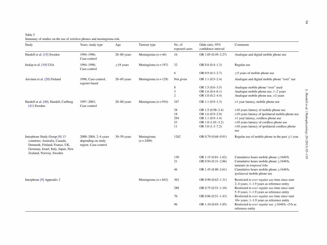
In the first study by Hardell et al. [15] only 46 cases hadmeningioma. No increased risk was found overall; OR = 1.05,95% CI = 0.49–2.27, Table 5. Only 16 cases had used a mobilephone. There was no pattern of increased risk for ipsilateraluse, although the results were based on low numbers.
The US study by Inskip et al. [19] included 197 cases withmeningioma. Regular mobile phone use produced OR = 0.8,95% CI = 0.4–1.3, Table 5. The risk did not increase withaverage daily use, cumulative use, or duration of regular use.However, results for duration of regular use "5 years wasbased on only 6 exposed cases.
The Finnish register based case-control study on braintumours by Auvinen et al. [20] included 129 cases with
meningioma. Ever use of mobile phone gave OR = 1.1,95% CI = 0.5–2.4, analogue phone use OR = 1.5, 95%CI = 0.6–3.5, Table 5. As discussed above the study waslimited by short latency and exposure based on subscriptioninformation.
The Hardell group made a pooled analysis of benignbrain tumours from the two case-control studies 1997–2003as discussed above [40,41]. Regarding meningioma use ofmobile phone gave OR = 1.1, 95% CI = 0.9–1.3, and cordlessphone OR = 1.1, 95% CI = 0.9–1.4, Table 5. Using >10 yearlatency period OR increased; for mobile phone to OR = 1.5,95% CI = 0.98–2.4, and for cordless phone to OR = 1.8,95% CI = 1.01–3.2. Ipsilateral mobile phone use in the >10years latency group yielded OR = 1.6, 95% CI = 0.9–2.9,and cordless phone OR = 3.0, 95% CI = 1.3–7.2. Theseresults were based on rather low numbers of exposed cases,however.
In the Interphone study [9] a statistically significantdecreased risk was found for meningioma for regularuse of mobile phone, OR = 0.79, 95% CI = 0.68–0.91,Table 5. The risk increased somewhat with cumulative use"1640 h and ipsilateral mobile phone use to OR = 1.45, 95%CI = 0.80–2.61. The overall pattern of no association did notchange if analysis was restricted to tumours in the temporallobe or only to the group of ever-regular use.
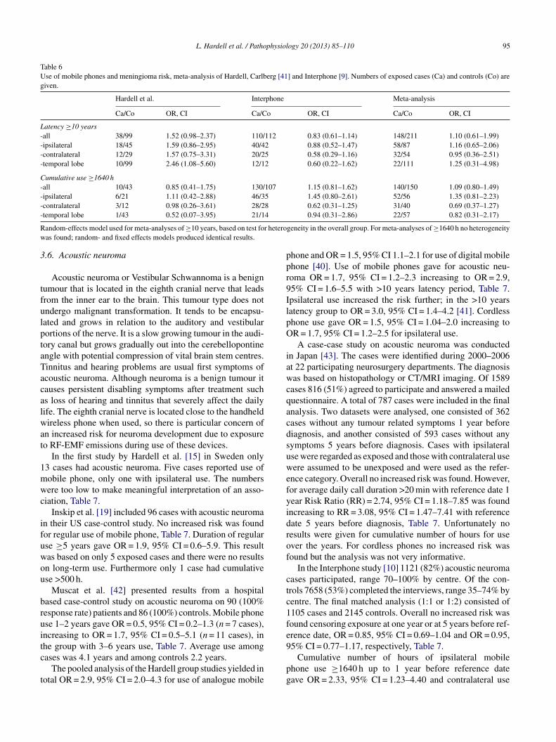
3.5. Meta-analysis meningioma
Similarly as for glioma we performed meta-analysis ofmeningioma for use of mobile phone on the Hardell groupand Interphone results, Table 6. Random-effects model wasused in the "10 years group based on test for heterogeneityin the overall group. For analyses of "1640 h no heterogene-ity was found in the heterogeneity test; random- and fixedeffects models produced identical results. In summary no sta-tistically significant decreased or increased risks were found.These results support the conclusion that up to latency "10years or cumulative use "1640 h there is not a consistentpattern of an association between use of mobile phones andmeningioma.
94L.H
ardelletal./Pathophysiology20
(2013)85–110
Table 5Summary of studies on the use of wireless phones and meningioma risk.
Study Years; study type Age Tumour type No. ofexposed cases
Odds ratio, 95%confidence interval
Comments
Hardell et al. [15] Sweden 1994–1996;Case-control
20–80 years Meningioma (n = 46) 16 OR 1.05 (0.49–2.27) Analogue and digital mobile phone use
Inskip et al. [19] USA 1994–1998;Case-control
"18 years Meningioma (n = 197) 32 OR 0.8 (0.4–1.3) Regular use
6 OR 0.9 (0.3–2.7) "5 years of mobile phone use
Auvinen et al. [20] Finland 1996; Case-control,register based
20–69 years Meningioma (n = 129) Not given OR 1.1 (0.5–2.4) Analogue and digital mobile phone “ever” use
8 OR 1.5 (0.6–3.5) Analogue mobile phone “ever” used3 OR 1.6 (0.4–6.1) Analogue mobile phone use, 1–2 years2 OR 1.0 (0.2–4.4) Analogue mobile phone use, >2 years
Hardell et al. [40], Hardell, Carlberg[41] Sweden
1997–2003;Case-control
20–80 years Meningioma (n = 916) 347 OR 1.1 (0.9–1.3) >1 year latency, mobile phone use
38 OR 1.5 (0.98–2.4) >10 years latency of mobile phone use18 OR 1.6 (0.9–2.9) >10 years latency of ipsilateral mobile phone use294 OR 1.1 (0.9–1.4) >1 year latency, cordless phone use23 OR 1.8 (1.01–3.2) >10 years latency of cordless phone use11 OR 3.0 (1.3–7.2) >10 years latency of ipsilateral cordless phone
use
Interphone Study Group [9] 13countries; Australia, Canada,Denmark, Finland, France, UK,Germany, Israel, Italy, Japan, NewZealand, Norway, Sweden
2000–2004, 2–4 yearsdepending on studyregion. Case-control
30–59 years Meningioma(n = 2409)
1262 OR 0.79 (0.68–0.91) Regular use of mobile phone in the past "1 year
130 OR 1.15 (0.81–1.62) Cumulative hours mobile phone "1640 h21 OR 0.94 (0.31–2.86) Cumulative hours mobile phone "1640 h,
tumours in temporal lobe46 OR 1.45 (0.80–2.61) Cumulative hours mobile phone "1640 h,
ipsilateral mobile phone use
Interphone [9] Appendix 2 Meningioma (n = 842) 362 OR 0.90 (0.62–1.31) Restricted to ever regular use time since start2–4 years; 1–1.9 years as reference entity
288 OR 0.75 (0.51–1.10) Restricted to ever regular use time since start5–9 years; 1–1.9 years as reference entity
76 OR 0.86 (0.51–1.43) Restricted to ever regular use time since start10+ years; 1–1.9 years as reference entity
96 OR 1.10 (0.65–1.85) Restricted to ever regular use "1640 h, <5 h asreference entity
L. Hardell et al. / Pathophysiology 20 (2013) 85–110 95
Table 6Use of mobile phones and meningioma risk, meta-analysis of Hardell, Carlberg [41] and Interphone [9]. Numbers of exposed cases (Ca) and controls (Co) aregiven.
Hardell et al. Interphone Meta-analysis
Ca/Co OR, CI Ca/Co OR, CI Ca/Co OR, CI
Latency "10 years-all 38/99 1.52 (0.98–2.37) 110/112 0.83 (0.61–1.14) 148/211 1.10 (0.61–1.99)-ipsilateral 18/45 1.59 (0.86–2.95) 40/42 0.88 (0.52–1.47) 58/87 1.16 (0.65–2.06)-contralateral 12/29 1.57 (0.75–3.31) 20/25 0.58 (0.29–1.16) 32/54 0.95 (0.36–2.51)-temporal lobe 10/99 2.46 (1.08–5.60) 12/12 0.60 (0.22–1.62) 22/111 1.25 (0.31–4.98)
Cumulative use "1640 h-all 10/43 0.85 (0.41–1.75) 130/107 1.15 (0.81–1.62) 140/150 1.09 (0.80–1.49)-ipsilateral 6/21 1.11 (0.42–2.88) 46/35 1.45 (0.80–2.61) 52/56 1.35 (0.81–2.23)-contralateral 3/12 0.98 (0.26–3.61) 28/28 0.62 (0.31–1.25) 31/40 0.69 (0.37–1.27)-temporal lobe 1/43 0.52 (0.07–3.95) 21/14 0.94 (0.31–2.86) 22/57 0.82 (0.31–2.17)
Random-effects model used for meta-analyses of "10 years, based on test for heterogeneity in the overall group. For meta-analyses of "1640 h no heterogeneitywas found; random- and fixed effects models produced identical results.
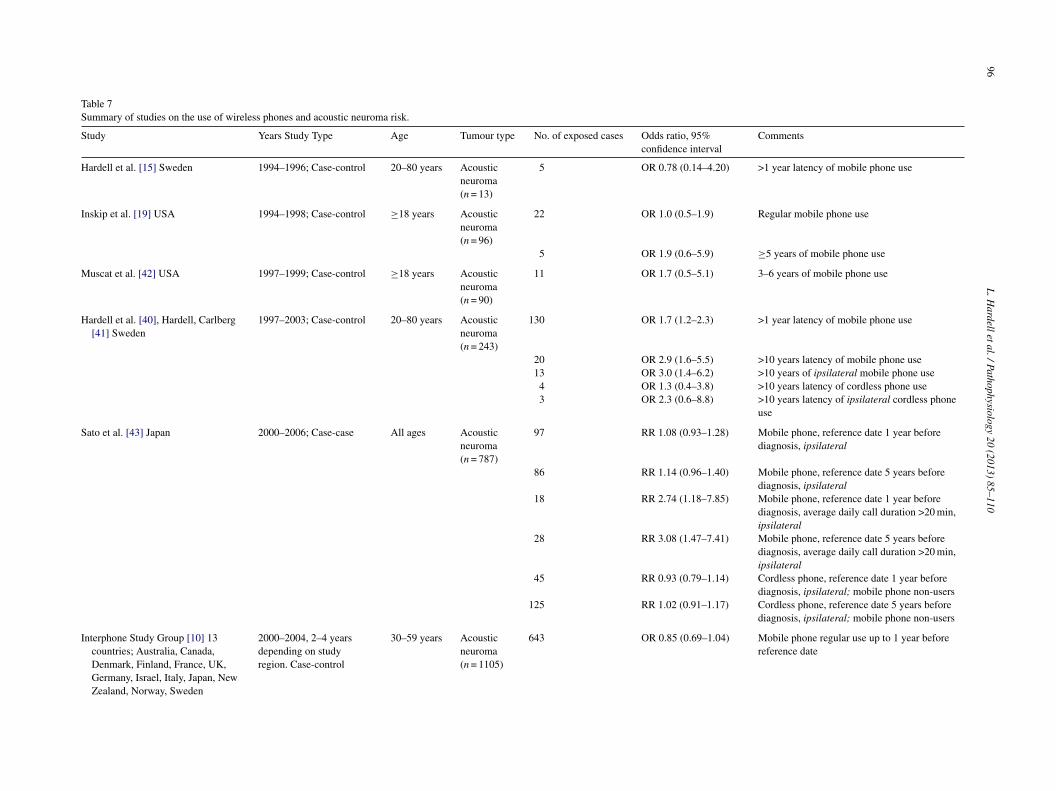
3.6. Acoustic neuroma
Acoustic neuroma or Vestibular Schwannoma is a benigntumour that is located in the eighth cranial nerve that leadsfrom the inner ear to the brain. This tumour type does notundergo malignant transformation. It tends to be encapsu-lated and grows in relation to the auditory and vestibularportions of the nerve. It is a slow growing tumour in the audi-tory canal but grows gradually out into the cerebellopontineangle with potential compression of vital brain stem centres.Tinnitus and hearing problems are usual first symptoms ofacoustic neuroma. Although neuroma is a benign tumour itcauses persistent disabling symptoms after treatment suchas loss of hearing and tinnitus that severely affect the dailylife. The eighth cranial nerve is located close to the handheldwireless phone when used, so there is particular concern ofan increased risk for neuroma development due to exposureto RF-EMF emissions during use of these devices.
In the first study by Hardell et al. [15] in Sweden only13 cases had acoustic neuroma. Five cases reported use ofmobile phone, only one with ipsilateral use. The numberswere too low to make meaningful interpretation of an asso-ciation, Table 7.
Inskip et al. [19] included 96 cases with acoustic neuromain their US case-control study. No increased risk was foundfor regular use of mobile phone, Table 7. Duration of regularuse "5 years gave OR = 1.9, 95% CI = 0.6–5.9. This resultwas based on only 5 exposed cases and there were no resultson long-term use. Furthermore only 1 case had cumulativeuse >500 h.
Muscat et al. [42] presented results from a hospitalbased case-control study on acoustic neuroma on 90 (100%response rate) patients and 86 (100%) controls. Mobile phoneuse 1–2 years gave OR = 0.5, 95% CI = 0.2–1.3 (n = 7 cases),increasing to OR = 1.7, 95% CI = 0.5–5.1 (n = 11 cases), inthe group with 3–6 years use, Table 7. Average use amongcases was 4.1 years and among controls 2.2 years.
The pooled analysis of the Hardell group studies yielded intotal OR = 2.9, 95% CI = 2.0–4.3 for use of analogue mobile
phone and OR = 1.5, 95% CI 1.1–2.1 for use of digital mobilephone [40]. Use of mobile phones gave for acoustic neu-roma OR = 1.7, 95% CI = 1.2–2.3 increasing to OR = 2.9,95% CI = 1.6–5.5 with >10 years latency period, Table 7.Ipsilateral use increased the risk further; in the >10 yearslatency group to OR = 3.0, 95% CI = 1.4–4.2 [41]. Cordlessphone use gave OR = 1.5, 95% CI = 1.04–2.0 increasing toOR = 1.7, 95% CI = 1.2–2.5 for ipsilateral use.
A case-case study on acoustic neuroma was conductedin Japan [43]. The cases were identified during 2000–2006at 22 participating neurosurgery departments. The diagnosiswas based on histopathology or CT/MRI imaging. Of 1589cases 816 (51%) agreed to participate and answered a mailedquestionnaire. A total of 787 cases were included in the finalanalysis. Two datasets were analysed, one consisted of 362cases without any tumour related symptoms 1 year beforediagnosis, and another consisted of 593 cases without anysymptoms 5 years before diagnosis. Cases with ipsilateraluse were regarded as exposed and those with contralateral usewere assumed to be unexposed and were used as the refer-ence category. Overall no increased risk was found. However,for average daily call duration >20 min with reference date 1year Risk Ratio (RR) = 2.74, 95% CI = 1.18–7.85 was foundincreasing to RR = 3.08, 95% CI = 1.47–7.41 with referencedate 5 years before diagnosis, Table 7. Unfortunately noresults were given for cumulative number of hours for useover the years. For cordless phones no increased risk wasfound but the analysis was not very informative.
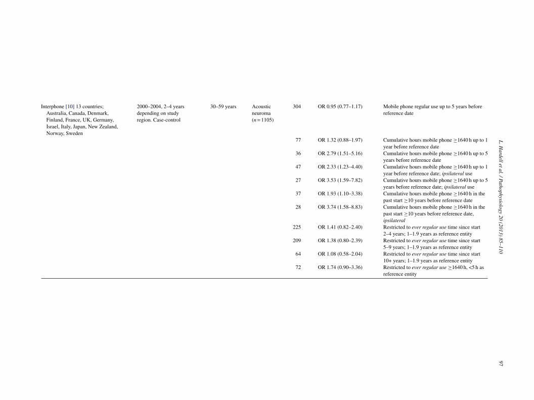
In the Interphone study [10] 1121 (82%) acoustic neuromacases participated, range 70–100% by centre. Of the con-trols 7658 (53%) completed the interviews, range 35–74% bycentre. The final matched analysis (1:1 or 1:2) consisted of1105 cases and 2145 controls. Overall no increased risk wasfound censoring exposure at one year or at 5 years before ref-erence date, OR = 0.85, 95% CI = 0.69–1.04 and OR = 0.95,95% CI = 0.77–1.17, respectively, Table 7.
Cumulative number of hours of ipsilateral mobilephone use "1640 h up to 1 year before reference dategave OR = 2.33, 95% CI = 1.23–4.40 and contralateral use
96L.H
ardelletal./Pathophysiology20
(2013)85–110
Table 7Summary of studies on the use of wireless phones and acoustic neuroma risk.
Study Years Study Type Age Tumour type No. of exposed cases Odds ratio, 95%confidence interval
Comments
Hardell et al. [15] Sweden 1994–1996; Case-control 20–80 years Acousticneuroma(n = 13)
5 OR 0.78 (0.14–4.20) >1 year latency of mobile phone use
Inskip et al. [19] USA 1994–1998; Case-control "18 years Acousticneuroma(n = 96)
22 OR 1.0 (0.5–1.9) Regular mobile phone use
5 OR 1.9 (0.6–5.9) "5 years of mobile phone use
Muscat et al. [42] USA 1997–1999; Case-control "18 years Acousticneuroma(n = 90)
11 OR 1.7 (0.5–5.1) 3–6 years of mobile phone use
Hardell et al. [40], Hardell, Carlberg[41] Sweden
1997–2003; Case-control 20–80 years Acousticneuroma(n = 243)
130 OR 1.7 (1.2–2.3) >1 year latency of mobile phone use
20 OR 2.9 (1.6–5.5) >10 years latency of mobile phone use13 OR 3.0 (1.4–6.2) >10 years of ipsilateral mobile phone use4 OR 1.3 (0.4–3.8) >10 years latency of cordless phone use3 OR 2.3 (0.6–8.8) >10 years latency of ipsilateral cordless phone
use
Sato et al. [43] Japan 2000–2006; Case-case All ages Acousticneuroma(n = 787)
97 RR 1.08 (0.93–1.28) Mobile phone, reference date 1 year beforediagnosis, ipsilateral
86 RR 1.14 (0.96–1.40) Mobile phone, reference date 5 years beforediagnosis, ipsilateral
18 RR 2.74 (1.18–7.85) Mobile phone, reference date 1 year beforediagnosis, average daily call duration >20 min,ipsilateral
28 RR 3.08 (1.47–7.41) Mobile phone, reference date 5 years beforediagnosis, average daily call duration >20 min,ipsilateral
45 RR 0.93 (0.79–1.14) Cordless phone, reference date 1 year beforediagnosis, ipsilateral; mobile phone non-users
125 RR 1.02 (0.91–1.17) Cordless phone, reference date 5 years beforediagnosis, ipsilateral; mobile phone non-users
Interphone Study Group [10] 13countries; Australia, Canada,Denmark, Finland, France, UK,Germany, Israel, Italy, Japan, NewZealand, Norway, Sweden
2000–2004, 2–4 yearsdepending on studyregion. Case-control
30–59 years Acousticneuroma(n = 1105)
643 OR 0.85 (0.69–1.04) Mobile phone regular use up to 1 year beforereference date
L.Hardelletal./Pathophysiology
20(2013)
85–11097
Interphone [10] 13 countries;Australia, Canada, Denmark,Finland, France, UK, Germany,Israel, Italy, Japan, New Zealand,Norway, Sweden
2000–2004, 2–4 yearsdepending on studyregion. Case-control
30–59 years Acousticneuroma(n = 1105)
304 OR 0.95 (0.77–1.17) Mobile phone regular use up to 5 years beforereference date
77 OR 1.32 (0.88–1.97) Cumulative hours mobile phone "1640 h up to 1year before reference date
36 OR 2.79 (1.51–5.16) Cumulative hours mobile phone "1640 h up to 5years before reference date
47 OR 2.33 (1.23–4.40) Cumulative hours mobile phone "1640 h up to 1year before reference date; ipsilateral use
27 OR 3.53 (1.59–7.82) Cumulative hours mobile phone "1640 h up to 5years before reference date; ipsilateral use
37 OR 1.93 (1.10–3.38) Cumulative hours mobile phone "1640 h in thepast start "10 years before reference date
28 OR 3.74 (1.58–8.83) Cumulative hours mobile phone "1640 h in thepast start "10 years before reference date,ipsilateral
225 OR 1.41 (0.82–2.40) Restricted to ever regular use time since start2–4 years; 1–1.9 years as reference entity
209 OR 1.38 (0.80–2.39) Restricted to ever regular use time since start5–9 years; 1–1.9 years as reference entity
64 OR 1.08 (0.58–2.04) Restricted to ever regular use time since start10+ years; 1–1.9 years as reference entity
72 OR 1.74 (0.90–3.36) Restricted to ever regular use "1640 h, <5 h asreference entity
98 L. Hardell et al. / Pathophysiology 20 (2013) 85–110
OR = 0.72, 95% CI = 0.34–1.53 for acoustic neuroma, Table 7[10]. For cumulative number of hours of ipsilateral mobilephone use "1640 h up to 5 years before reference dateOR = 3.53, 95% CI = 1.59–7.82, and for contralateral useOR = 1.69, 95% CI = 0.43–6.69 were obtained. The riskincreased further for cumulative ipsilateral use "1640 hwith start "10 years before reference date to OR = 3.74,95% CI = 1.58–8.83. Contralateral use in that group yieldedOR = 0.48, 95% CI = 0.12–1.94, however based on only 4exposed cases and 9 exposed controls. Overall OR = 1.93,95% CI = 1.10–3.38 was obtained for long-term use with start"10 years before reference date and cumulative call time"1640 h.
Similar analyses of the data as in Appendix 2 for glioma[9], yielded highest OR for acoustic neuroma in the shortestlatency group, 2–4 years before reference date, OR = 1.41,95% CI = 0.82–2.40 [10]. Lower OR was calculated in the"10 years group, OR = 1.08, 95% CI = 0.58–2.04. Somewhathigher risk than in total, OR = 1.32, 95% CI = 0.88–1.97, wasfound for cumulative mobile phone use "1640 h; OR = 1.74,95% CI = 0.90–3.36, in this analysis restricted to only regularusers. No results were given for ipsilateral use.
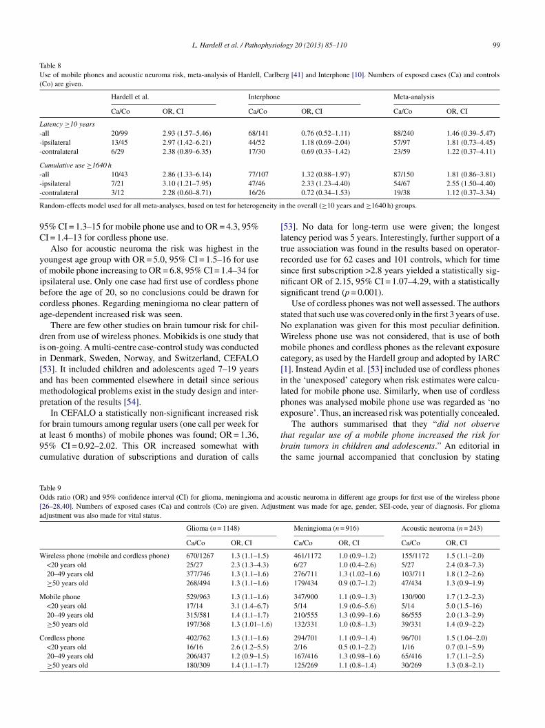
3.7. Meta-analysis acoustic neuroma
Table 8 shows results for use of mobile phone and theassociation with acoustic neuroma based on results by theHardell group and Interphone study. Random-effects modelwas used based on test for heterogeneity in the overall("10 years and "1640 h) groups. The same exposure groupsas in the meta-analyses of glioma and meningioma wereused. For the latency group "10 years highest risk wasobtained for ipsilateral use, OR = 1.81, 95% CI = 0.73–4.45.The risk increased further for cumulative use "1640 h yield-ing OR = 2.55, 95% CI = 1.50–4.40 for ipsilateral use. Themeta-analysis strengthens a causal association between useof mobile phones and acoustic neuroma.
3.8. Other types of brain tumours
Results for other types of brain tumours from the Hardellgroup diagnosed during 1997–2003 included medulloblas-toma (n = 6), ependymoma (n = 19) and other malignant types(n = 46). In total using >1 year latency time no statisticallysignificant increased risk was found for mobile phone use,OR = 1.2, 95% CI = 0.7–2.1 for these tumour types groupedtogether [41]. However, with >10 years latency the riskincreased to OR = 3.2, 95% CI = 1.2–8.8 in total; for ipsi-lateral use OR = 4.1, 95% CI = 1.03–16. For cordless phoneuse no statistically significant decreased or increased risk wasfound (data not in Table). For pituitary adenoma (n = 34) andother types of benign brain tumours (n = 62) no statisticallysignificant associations were found overall. In the >10 yearlatency group ipsilateral mobile phone use gave OR = 4.7,95% CI = 1.1–21 for benign tumours other than pituitary ade-noma (central location in the brain and not included in these
calculations) but based on only 4 exposed cases. Thus, severalof the calculations were based on low numbers.
Takebayashi et al. [44] included 102 cases with pituitaryadenoma in the Japanese part of Interphone from December2000 to November 2004. The response rate was 76%; 102out of 135 cases. Of the individually matched controls 208(49%) of 421 participated. In the statistical analysis 161 con-trols were used to 101 cases; one case was excluded since notdiagnosed within study period. Regular mobile phone useyielded OR = 0.90, 95% CI = 0.50–1.61. Cumulative lengthof use in years or cumulative call time in hours producedno pattern of an association and there was no statisticallysignificant trend. The cut off for highest quartile of cumula-tive use was 560 h producing OR = 1.33, 95% CI = 0.58–3.09(n = 21 cases, 27 controls exposed). Since pituitary adenomais a centrally located tumour in the pituitary gland in sellaturcica there was no laterality analysis.
In parallel with the Interphone study, pituitary tumourswere studied in Southeast England using the same protocol[45]. The inclusion period was from December 2000 untilFebruary 2005. In total 506 eligible cases were identified. Ofthem 317 (63%) were interviewed and 291 (58%) included inthe final analysis. Eligible controls from patient lists at gen-eral practitioners in the study region were 1464 subjects, and630 (43%) were interviewed. Regular use of mobile phonegave OR = 0.9, 95% CI = 0.7–1.3. No statistically significanttrend for the risk was found for lifetime use in years or cumu-lative use in hours. For "10 years since first use and "51 h ofcumulative use (median number in that category) OR = 1.6,95% CI = 0.8–3.6 (n = 16 cases, 23 controls exposed) wasfound.
3.9. Risks to children and adolescents
Children have smaller head and thinner skull bone thanadults. Their brain tissue has also higher conductivity andthese circumstances give higher absorption from RF-EMFthan for adults [6,46,47]. The developing brain is more sensi-tive to toxins [48] and it is still developing until about 20 yearsof age [49]. Use of wireless phones is widespread amongchildren and adolescents [50,51]. The greater absorption ofRF energy per unit of time, the greater sensitivity of theirbrains, and their longer lifetimes with the risk to develop abrain tumour leaves children at a higher risk than adults frommobile phone radiation.
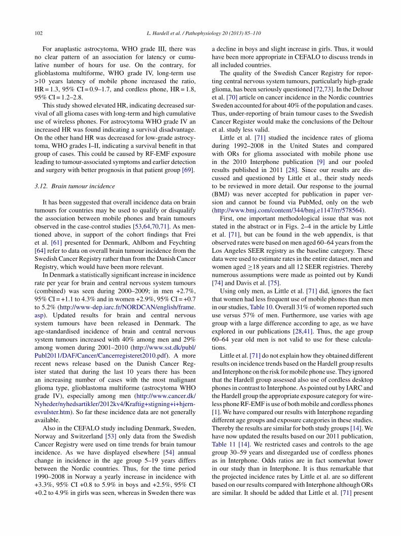
The Hardell group has published results for different agegroups at the time of diagnosis [52] or age at first use ofwireless phones [12,13,28]. Three age groups for first useof a wireless phone were used: <20 years, 20–49 years and50–80 years. Highest risk for glioma was found for firstuse of mobile phone or cordless phone before the age of20 years, Table 9. Thus, mobile phone yielded for gliomaOR = 3.1, 95% CI = 1.4–6.7 and cordless phone OR 2.6,95% CI = 1.2–5.5. The risk increased further for ipsilateralmobile phone use in the youngest age group to OR = 4.4,
L. Hardell et al. / Pathophysiology 20 (2013) 85–110 99
Table 8Use of mobile phones and acoustic neuroma risk, meta-analysis of Hardell, Carlberg [41] and Interphone [10]. Numbers of exposed cases (Ca) and controls(Co) are given.
Hardell et al. Interphone Meta-analysis
Ca/Co OR, CI Ca/Co OR, CI Ca/Co OR, CI
Latency "10 years-all 20/99 2.93 (1.57–5.46) 68/141 0.76 (0.52–1.11) 88/240 1.46 (0.39–5.47)-ipsilateral 13/45 2.97 (1.42–6.21) 44/52 1.18 (0.69–2.04) 57/97 1.81 (0.73–4.45)-contralateral 6/29 2.38 (0.89–6.35) 17/30 0.69 (0.33–1.42) 23/59 1.22 (0.37–4.11)
Cumulative use "1640 h-all 10/43 2.86 (1.33–6.14) 77/107 1.32 (0.88–1.97) 87/150 1.81 (0.86–3.81)-ipsilateral 7/21 3.10 (1.21–7.95) 47/46 2.33 (1.23–4.40) 54/67 2.55 (1.50–4.40)-contralateral 3/12 2.28 (0.60–8.71) 16/26 0.72 (0.34–1.53) 19/38 1.12 (0.37–3.34)
Random-effects model used for all meta-analyses, based on test for heterogeneity in the overall ("10 years and "1640 h) groups.
95% CI = 1.3–15 for mobile phone use and to OR = 4.3, 95%CI = 1.4–13 for cordless phone use.
Also for acoustic neuroma the risk was highest in theyoungest age group with OR = 5.0, 95% CI = 1.5–16 for useof mobile phone increasing to OR = 6.8, 95% CI = 1.4–34 foripsilateral use. Only one case had first use of cordless phonebefore the age of 20, so no conclusions could be drawn forcordless phones. Regarding meningioma no clear pattern ofage-dependent increased risk was seen.
There are few other studies on brain tumour risk for chil-dren from use of wireless phones. Mobikids is one study thatis on-going. A multi-centre case-control study was conductedin Denmark, Sweden, Norway, and Switzerland, CEFALO[53]. It included children and adolescents aged 7–19 yearsand has been commented elsewhere in detail since seriousmethodological problems exist in the study design and inter-pretation of the results [54].
In CEFALO a statistically non-significant increased riskfor brain tumours among regular users (one call per week forat least 6 months) of mobile phones was found; OR = 1.36,95% CI = 0.92–2.02. This OR increased somewhat withcumulative duration of subscriptions and duration of calls
[53]. No data for long-term use were given; the longestlatency period was 5 years. Interestingly, further support of atrue association was found in the results based on operator-recorded use for 62 cases and 101 controls, which for timesince first subscription >2.8 years yielded a statistically sig-nificant OR of 2.15, 95% CI = 1.07–4.29, with a statisticallysignificant trend (p = 0.001).
Use of cordless phones was not well assessed. The authorsstated that such use was covered only in the first 3 years of use.No explanation was given for this most peculiar definition.Wireless phone use was not considered, that is use of bothmobile phones and cordless phones as the relevant exposurecategory, as used by the Hardell group and adopted by IARC[1]. Instead Aydin et al. [53] included use of cordless phonesin the ‘unexposed’ category when risk estimates were calcu-lated for mobile phone use. Similarly, when use of cordlessphones was analysed mobile phone use was regarded as ‘noexposure’. Thus, an increased risk was potentially concealed.
The authors summarised that they “did not observethat regular use of a mobile phone increased the risk forbrain tumors in children and adolescents.” An editorial inthe same journal accompanied that conclusion by stating
Table 9Odds ratio (OR) and 95% confidence interval (CI) for glioma, meningioma and acoustic neuroma in different age groups for first use of the wireless phone[26–28,40]. Numbers of exposed cases (Ca) and controls (Co) are given. Adjustment was made for age, gender, SEI-code, year of diagnosis. For gliomaadjustment was also made for vital status.
Glioma (n = 1148) Meningioma (n = 916) Acoustic neuroma (n = 243)
Ca/Co OR, CI Ca/Co OR, CI Ca/Co OR, CI
Wireless phone (mobile and cordless phone) 670/1267 1.3 (1.1–1.5) 461/1172 1.0 (0.9–1.2) 155/1172 1.5 (1.1–2.0)<20 years old 25/27 2.3 (1.3–4.3) 6/27 1.0 (0.4–2.6) 5/27 2.4 (0.8–7.3)20–49 years old 377/746 1.3 (1.1–1.6) 276/711 1.3 (1.02–1.6) 103/711 1.8 (1.2–2.6)"50 years old 268/494 1.3 (1.1–1.6) 179/434 0.9 (0.7–1.2) 47/434 1.3 (0.9–1.9)
Mobile phone 529/963 1.3 (1.1–1.6) 347/900 1.1 (0.9–1.3) 130/900 1.7 (1.2–2.3)<20 years old 17/14 3.1 (1.4–6.7) 5/14 1.9 (0.6–5.6) 5/14 5.0 (1.5–16)20–49 years old 315/581 1.4 (1.1–1.7) 210/555 1.3 (0.99–1.6) 86/555 2.0 (1.3–2.9)"50 years old 197/368 1.3 (1.01–1.6) 132/331 1.0 (0.8–1.3) 39/331 1.4 (0.9–2.2)
Cordless phone 402/762 1.3 (1.1–1.6) 294/701 1.1 (0.9–1.4) 96/701 1.5 (1.04–2.0)<20 years old 16/16 2.6 (1.2–5.5) 2/16 0.5 (0.1–2.2) 1/16 0.7 (0.1–5.9)20–49 years old 206/437 1.2 (0.9–1.5) 167/416 1.3 (0.98–1.6) 65/416 1.7 (1.1–2.5)"50 years old 180/309 1.4 (1.1–1.7) 125/269 1.1 (0.8–1.4) 30/269 1.3 (0.8–2.1)
100 L. Hardell et al. / Pathophysiology 20 (2013) 85–110
that the study showed “no increased risk of brain tumorsin children and adolescents who are regular cell phoneusers” [55]. This was echoed by a news release fromthe Karolinska Institute in Stockholm claiming that theresults of no increased risk were ‘reassuring’ (http://ki.se/ki/jsp/polopoly.jsp?d=130&a=125250&l=en&newsdep=130).However, these statements go far beyond what the studyreally showed. In fact, the results indicate a moderatelyincreased risk, in spite of low exposure, short latency periodand limitations in study design and analyses. Aydin et al.discussed recall bias – that people tend to overestimate theirnumber of calls – and interestingly they showed that controlsoverestimated their number of calls more than cases [56]. Itwas concluded that it was unlikely that a false positive resultoccurred in CEFALO and that the OR was underestimatedfor heavy users. Certainly the results in the article [53]cannot be used as reassuring evidence against an association,as discussed in our commentary [54].
3.10. Danish cohort study on mobile phone users
Ideally a cohort study on wireless phone users wouldbe of substantial value. However, several problems exist toestablish a cohort with high quality assessed exposure. Forexample use of both mobile phones and cordless phones varyover time and exposure to RF-EMF emissions also dependson several physical characteristics for different phone types.An attempt to establish a cohort of mobile phone users wasmade in Denmark in co-operation between the Danish CancerSociety and the International Epidemiology Institute (IEI),Rockville, MD, USA. It was financed by grants from twoDanish telecom operation companies (TeleDenmark Mobiland Sonofon), IEI, and the Danish Cancer Society. The sourceof money for IEI has not been disclosed.
The first results from the Danish study on brain tumourrisk among mobile phone subscribers were published in2001 [57]. It included subjects from January 1, 1982 untilDecember 31, 1995 identified from the computerised filesof the two Danish operating companies, TeleDenmark Mobiland Sonofon. A total of 723,421 subscribers were initiallyidentified but the final cohort consisted of only 58% of thesesubjects. Due to lack of names of individual users 200,507corporate users were excluded. They were expected to be theheaviest users and such exclusion would underestimate anyrisk estimates. It should be noted that duration of subscriptionof a digital phone was at most "3 years (n = 9) and that twothirds of the subscriptions began in 1994 and 1995. In otherwords, the majority of the cohort members had two years orless of subscription time. This and other shortcomings in thiscohort study have been discussed elsewhere in detail [58].The Danish study was part of the IARC evaluation but itwas concluded that the methods used could have resulted inconsiderable misclassification in exposure assessment [1].
The first update of the Danish study gave follow-up datauntil 2002 [59]. The median time since first subscriptionwas this time 8.0 years. It was now stated that the cohort
members were excluded from the reference population,which seems not to have been the case in the first publication.The Standardised Incidence Ratio (SIR) for glioma was closeto unity, SIR = 1.01, 95% CI = 0.89–1.14. The highest SIRwas found for glioma in the temporal lobe where RF-EMFexposure from a mobile phone would be highest, SIR = 1.21,95% CI = 0.91–1.58 (n = 54 cases).
After the outcome of the IARC-evaluation was made pub-lic in June 2011 [1] two additional reports on the Danishcohort were soon published. Both were new up-dates ofmobile phone subscribers and included more information onrisk related to longer follow-up. One focused on acoustic neu-roma [60] while the other gave results both for all cancers andseparately for glioma and meningioma [61].
Approximately 2.9 million of the Danish population of 5.5million in total was included in the record linkage study onacoustic neuroma [60]. Of the 2.9 million subjects 420,095were mobile phone subscribers that started their subscription1987–1995 and in accordance with the aim of the study hadlasted for "11 years, i.e., 1998–2006 during which period thetumour cases were ascertained. No evidence of an increasedrisk was found for "11 years of subscription; adjusted Inci-dence Rate Ratio (IRR) was 0.87, 95% CI = 0.52–1.46.
The analysis of long-term exposure ("11 years) was basedon only 15 exposed cases with acoustic neuroma all of whichwere men. Analysis of tumour size was based on even fewercases; 8 had a subscription for "11 years. As for the riskrelated to laterality Schüz et al. [60] compared the location ofacoustic neuroma in long-term mobile phone subscribers withshorter use (<11 years) and non-subscribers to see if tumoursoccurred more frequently on the side which was assumed tobe the mostly exposed. This assumption was based on eco-logical data from the prospective study, COSMOS, as proxyfor laterality [62]. Due to these facts the argument of no lat-erality risk is not very impressive, especially when applied toonly 15 exposed cases.
The fourth report on the Danish mobile phone cohort ontumours of the central nervous system showed no overallincreased risk [61]. This was true also when restricted to theindividuals with the longest mobile phone use, "13 years ofassumed subscription.
This time the number of the cohort was reduced to 358,403(49.5%) of the initially identified subscribers (n = 723,421).This number was also used in the study on acoustic neu-roma [60]. The major additional exclusion (n = 54,350) wasdue to record linkage with the Danish so-called CANULIcohort on socioeconomic factors [63]. That register started1990 and included subjects from the age of 30. Subscriptionholders aged 18–29 years were excluded from the mobilephone cohort; this was also the case for the third publication(acoustic neuroma), see above. Follow-up of cancer startedat January 1, 1990, or at the age of 30 if occurred later, andended December 31, 2007.
The study period was 1990–2007 [61] but the cohort wasestablished during 1982–1995. Cancer cases before 1990were disregarded since the CANULI cohort started in 1990.
L. Hardell et al. / Pathophysiology 20 (2013) 85–110 101
The authors did not discuss the impact of the exclusion ofthese subscribers on the results. This exclusion would includethe early users of analogue phones, which seem to have hadhigher emissions of RF-EMF than the later digital system.The authors themselves also stated the following in theirdiscussion: “. . .we found indications that early subscriptionholders before 1995 were in fact heavier users (based on out-going calls) compared with all subscription holders in theyears 1996–2002.” Analysis of any early effect in the groupwho used phones with the highest emissions was most likelyhampered. Moreover, also the youngest users, aged 18–29years that had previously been included, were now excludedfrom the cohort. The fully adjusted model had no substan-tial effect on the risk estimates, so results adjusted for ageand calendar period should be possible also for the youngestusers. The exclusion of young subscribers could be of impor-tance since as discussed above studies have indicated highestrisk in subjects that started the use of a mobile or cordlessphone before the age of 20 [28,41].
Some of the many shortcomings of the Danish cohortstudy include: (a) no individual exposure data (e.g. on cumu-lative exposure, side of head mostly used, and use of cordlessphones); including users of cordless phones in the referencecategory; (b) no control for use of mobile phones in thepopulation after the establishment of the cohort; and (c) nooperator-verified data on years of subscription was available.These limitations are likely to have led to an underestimateof any risk in this study. One would expect considerable mis-classification of mobile phone use both among subscribersand the reference population since no new subscribers wereincluded in the exposed cohort after 1995.
The publication of the latest update of the Danish study[61] was accompanied by an editorial by Ahlbom and Fey-chting from the Karolinska Institute in Sweden [64]. It beganwith the statement: “Evidence is reassuring, but continuedmonitoring of health registers and prospective cohorts isstill warranted.” They pointed out methodological advan-tages, such as elimination of non-response and selectionbias, but did forget to mention that less than 50% of theinitial cohort remained for analysis. However, they weremore lenient on the methodological limitations that they hadpreviously pointed out as serious. In a letter to the Editorin 2007 on an earlier publication of the same cohort [59]they pointed out that several methodological shortcomingsundermined the authors’ conclusion that “any large asso-ciation of risk of cancer and cellular telephone use canbe excluded” [65]. Although more long-term data was nowavailable and adjustment for socioeconomic factors could bemade, the update by Frei et al. [61] suffers from basically thesame methodological limitations – mainly related to expo-sure assessment – as the first one did. Instead of addressingthe limitations of the Danish cohort study in full, Ahlbomand Feychting [64] used their space to selectively report onresults in the Hardell group studies choosing the time period2000–2003 [23,24] although the whole investigation periodwas 1997–2003 [27,40]. They discussed incidence data on
brain tumours in Sweden instead of Denmark, which wouldhave been more appropriate regarding a Danish cohort study.
The authors of the Danish study have themselves pointedout the main causes of such considerable exposure misclas-sifications [61]: mobile phone subscription holders not usingthe phone were classified as ‘exposed’, non-subscribers usingthe mobile phone were classified as ‘unexposed’; corporatesubscribers of mobile phones (200,507 people), which arelikely to have been heavy users, were classified as ‘unex-posed’; persons with a mobile phone subscription later than1995 were classified as ‘unexposed’ and users of cordlessphones not using a mobile phone were also classified as‘unexposed’.
Other limitations are the absence of analysis by laterality(the side of head where the phone is used in relation to the sideof the tumour) and the complete absence of actual exposuredata. These and other shortcomings in the cohort study havebeen discussed elsewhere in more detail [58,65].
It is clear from these limitations that the authors’ conclu-sion that: “In this update of a large nationwide cohort studyof mobile phone use, there were no increased risks of tumoursof the central nervous system, providing little evidence for acausal association” is not soundly based [61].
3.11. Hazard ratio (HR) for survival of patients withglioma
A poorer survival among children with acute lymphoblas-tic leukaemia exposed to ELF-EMF has been reported in twostudies [66,67]. These findings certainly strengthen a causalassociation between exposure to ELF-EMF and childhoodleukaemia. Thus, a carcinogenic effect of RF-EMF emis-sions would be strengthened if exposure might correlate withsurvival of glioma patients. To further elucidate that possi-bility we analysed survival of all cases with malignant braintumour (n = 1251) in our case-control studies [26–28]. Mostcases were diagnosed with glioma (n = 1132 in this study) soin the following results for glioma are presented in short, forfurther details see Hardell and Carlberg [68].
Hazard ratio (HR) for survival was close to unity forall glioma cases for use of wireless phones, HR = 1.1, 95%CI = 0.9–1.2. However, latency >10 years increased HR to1.2, 95% CI = 1.002–1.5. Increased ratio was found for bothmobile phone use, HR = 1.3, 95% CI = 1.0005–1.6, and cord-less phone use, HR = 1.3, 95% CI = 0.9–1.9. HR increasedalso with cumulative number of hours of use of mobile phoneand cordless phone with statistically significant trend for ter-tiles (p = 0.01) of use of both phone types.
Regarding different types of astrocytoma wireless phoneuse gave a decreased HR = 0.5, 95% CI = 0.3–0.9 for low-grade astrocytoma, WHO grades I–II. Similar results werefound for both mobile and cordless phones. Latency did notchange these results. Also cumulative numbers of hours foruse yielded decreased HR for both mobile and cordless phoneuse.
102 L. Hardell et al. / Pathophysiology 20 (2013) 85–110
For anaplastic astrocytoma, WHO grade III, there wasno clear pattern of an association for latency or cumu-lative number of hours for use. On the contrary, forglioblastoma multiforme, WHO grade IV, long-term use>10 years latency of mobile phone increased the ratio,HR = 1.3, 95% CI = 0.9–1.7, and cordless phone, HR = 1.8,95% CI = 1.2–2.8.
This study showed elevated HR, indicating decreased sur-vival of all glioma cases with long-term and high cumulativeuse of wireless phones. For astrocytoma WHO grade IV anincreased HR was found indicating a survival disadvantage.On the other hand HR was decreased for low-grade astrocy-toma, WHO grades I–II, indicating a survival benefit in thatgroup of cases. This could be caused by RF-EMF exposureleading to tumour-associated symptoms and earlier detectionand surgery with better prognosis in that patient group [69].
3.12. Brain tumour incidence
It has been suggested that overall incidence data on braintumours for countries may be used to qualify or disqualifythe association between mobile phones and brain tumoursobserved in the case-control studies [53,64,70,71]. As men-tioned above, in support of the cohort findings that Freiet al. [61] presented for Denmark, Ahlbom and Feychting[64] refer to data on overall brain tumour incidence from theSwedish Cancer Registry rather than from the Danish CancerRegistry, which would have been more relevant.
In Denmark a statistically significant increase in incidencerate per year for brain and central nervous system tumours(combined) was seen during 2000–2009; in men +2.7%,95% CI = +1.1 to 4.3% and in women +2.9%, 95% CI = +0.7to 5.2% (http://www-dep.iarc.fr/NORDCAN/english/frame.asp). Updated results for brain and central nervoussystem tumours have been released in Denmark. Theage-standardised incidence of brain and central nervoussystem tumours increased with 40% among men and 29%among women during 2001–2010 (http://www.sst.dk/publ/Publ2011/DAF/Cancer/Cancerregisteret2010.pdf). A morerecent news release based on the Danish Cancer Reg-ister stated that during the last 10 years there has beenan increasing number of cases with the most malignantglioma type, glioblastoma multiforme (astrocytoma WHOgrade IV), especially among men (http://www.cancer.dk/Nyheder/nyhedsartikler/2012kv4/Kraftig+stigning+i+hjern-esvulster.htm). So far these incidence data are not generallyavailable.
Also in the CEFALO study including Denmark, Sweden,Norway and Switzerland [53] only data from the SwedishCancer Registry were used on time trends for brain tumourincidence. As we have displayed elsewhere [54] annualchange in incidence in the age group 5–19 years differsbetween the Nordic countries. Thus, for the time period1990–2008 in Norway a yearly increase in incidence with+3.3%, 95% CI +0.8 to 5.9% in boys and +2.5%, 95% CI+0.2 to 4.9% in girls was seen, whereas in Sweden there was
a decline in boys and slight increase in girls. Thus, it wouldhave been more appropriate in CEFALO to discuss trends inall included countries.
The quality of the Swedish Cancer Registry for repor-ting central nervous system tumours, particularly high-gradeglioma, has been seriously questioned [72,73]. In the Deltouret al. [70] article on cancer incidence in the Nordic countriesSweden accounted for about 40% of the population and cases.Thus, under-reporting of brain tumour cases to the SwedishCancer Register would make the conclusions of the Deltouret al. study less valid.
Little et al. [71] studied the incidence rates of gliomaduring 1992–2008 in the United States and comparedwith ORs for glioma associated with mobile phone usein the 2010 Interphone publication [9] and our pooledresults published in 2011 [28]. Since our results are dis-cussed and questioned by Little et al., their study needsto be reviewed in more detail. Our response to the journal(BMJ) was never accepted for publication in paper ver-sion and cannot be found via PubMed, only on the web(http://www.bmj.com/content/344/bmj.e1147/rr/578564).
First, one important methodological issue that was notstated in the abstract or in Figs. 2–4 in the article by Littleet al. [71], but can be found in the web appendix, is thatobserved rates were based on men aged 60–64 years from theLos Angeles SEER registry as the baseline category. Thesedata were used to estimate rates in the entire dataset, men andwomen aged "18 years and all 12 SEER registries. Therebynumerous assumptions were made as pointed out by Kundi[74] and Davis et al. [75].
Using only men, as Little et al. [71] did, ignores the factthat women had less frequent use of mobile phones than menin our studies, Table 10. Overall 31% of women reported suchuse versus 57% of men. Furthermore, use varies with agegroup with a large difference according to age, as we haveexplored in our publications [28,41]. Thus, the age group60–64 year old men is not valid to use for these calcula-tions.
Little et al. [71] do not explain how they obtained differentresults on incidence trends based on the Hardell group resultsand Interphone on the risk for mobile phone use. They ignoredthat the Hardell group assessed also use of cordless desktopphones in contrast to Interphone. As pointed out by IARC andthe Hardell group the appropriate exposure category for wire-less phone RF-EMF is use of both mobile and cordless phones[1]. We have compared our results with Interphone regardingdifferent age groups and exposure categories in these studies.Thereby the results are similar for both study groups [14]. Wehave now updated the results based on our 2011 publication,Table 11 [14]. We restricted cases and controls to the agegroup 30–59 years and disregarded use of cordless phonesas in Interphone. Odds ratios are in fact somewhat lowerin our study than in Interphone. It is thus remarkable thatthe projected incidence rates by Little et al. are so differentbased on our results compared with Interphone although ORsare similar. It should be added that Little et al. [71] present
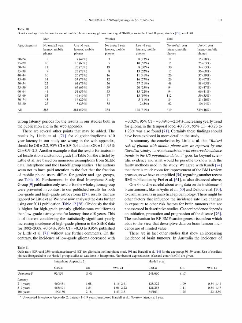
L. Hardell et al. / Pathophysiology 20 (2013) 85–110 103
Table 10Gender and age distribution for use of mobile phones among glioma cases aged 20–80 years in the Hardell group studies [28]; n = 1148.
Men Women Total
Age, diagnosis No use/#1 yearlatency, mobilephones
Use >1 yearlatency, mobilephones
No use/#1 yearlatency, mobilephones
Use >1 yearlatency, mobilephones
No use/#1 yearlatency, mobilephones
Use >1 yearlatency, mobilephones
20–24 8 7 (47%) 3 8 (73%) 11 15 (58%)25–29 10 15 (60%) 5 10 (67%) 15 25 (63%)30–34 11 26 (70%) 19 8 (30%) 30 34 (53%)35–39 9 23 (72%) 8 13 (62%) 17 36 (68%)40–44 10 26 (72%) 16 11 (41%) 26 37 (59%)45–49 14 37 (73%) 12 16 (57%) 26 53 (67%)50–54 22 61 (73%) 26 27 (51%) 48 88 (65%)55–59 35 65 (65%) 59 20 (25%) 94 85 (47%)60–64 41 51 (55%) 53 15 (22%) 94 66 (41%)65–69 55 46 (46%) 57 13 (19%) 112 59 (35%)70–74 43 16 (27%) 41 5 (11%) 84 21 (20%)75–80 27 8 (23%) 35 2 (5%) 62 10 (14%)
All 285 381 (57%) 334 148 (31%) 619 529 (46%)
wrong latency periods for the results in our studies both inthe publication and in the web appendix.
There are several other points that may be added. Theresults by Little et al. [71] for oligodendroglioma >10year latency in our study are wrong in the web appendix,should be OR = 2.2, 95% CI = 0.9–5.4 and not OR = 1.4, 95%CI = 0.9–2.3. Another example is that the results for anatomi-cal localisations and tumour grade [in Table 5 in the article] byLittle et al. are based on numerous assumptions from SEERdata, Interphone and the Hardell group studies. The authorsseem not to have paid attention to the fact that the fractionof mobile phone users differs for gender and age groups,see Table 10. Furthermore, in the final Interphone StudyGroup [9] publication only results for the whole glioma groupwere presented in contrast to our published results for bothlow-grade and high-grade astrocytoma [27], results that areignored by Little et al. We have now analysed the data furtherusing our 2011 publication, Table 12 [28]. Obviously the riskis higher for high-grade (mostly glioblastoma multiforme)than low-grade astrocytoma for latency time >10 years. Thisis of interest considering the statistically significant yearlyincreasing incidence of high-grade glioma in the SEER datafor 1992–2008, +0.64%, 95% CI = +0.33 to 0.95% publishedby Little et al. [71] without any further comments. On thecontrary, the incidence of low-grade glioma decreased with
$3.02%, 95% CI = $3.49 to$2.54%. Increasing yearly trendfor glioma in the temporal lobe, +0.73%, 95% CI = +0.23 to1.23% was also found [71]. Certainly these findings shouldhave been explored in more detail in the study.
In summary the conclusion by Little et al. that “Raisedrisk of glioma with mobile phone use, as reported by one(Swedish) study. . .are not consistent with observed incidencetrends in the US population data. . .” goes far beyond scien-tific evidence and what would be possible to show with thefaulty methods used in the study. We agree with Kundi [74]that there is much room for improvement of the BMJ reviewprocess, as we have exemplified [54] regarding another recentBMJ publication by Frei et al. [61], as also discussed above.
One should be careful about using data on the incidence ofbrain tumours, like in Aydin et al. [53] and Deltour et al. [70],to dismiss results in analytical epidemiology. There might beother factors that influence the incidence rate like changesin exposure to other risk factors for brain tumours that arenot assessed in descriptive studies. Cancer incidence dependson initiation, promotion and progression of the disease [76].The mechanism for RF-EMF carcinogenesis is unclear whichadds to the view that descriptive data on brain tumour inci-dence are of limited value.
There are in fact other studies that show an increasingincidence of brain tumours. In Australia the incidence of
Table 11Odds ratio (OR) and 95% confidence interval (CI) for glioma in the Interphone study [9] and Hardell et al. [14] for the age group 30–59 years. Use of cordlessphones disregarded in the Hardell group studies as was done in Interphone. Numbers of exposed cases (Ca) and controls (Co) are given.
Interphone Appendix 2 Hardell et al.
Ca/Co OR 95% CI Ca/Co OR 95% CI
Unexposeda 93/159 (1.0) – 241/660 (1.0) –
Latency2–4 years 460/451 1.68 1.16–2.41 128/322 1.09 0.84–1.415–9 years 468/491 1.54 1.06–2.22 121/258 1.11 0.84–1.4710+ years 190/150 2.18 1.43–3.31 84/103 1.75 1.23–2.50
a Unexposed Interphone Appendix 2: Latency 1–1.9 years; unexposed Hardell et al.: No use + latency # 1 year.
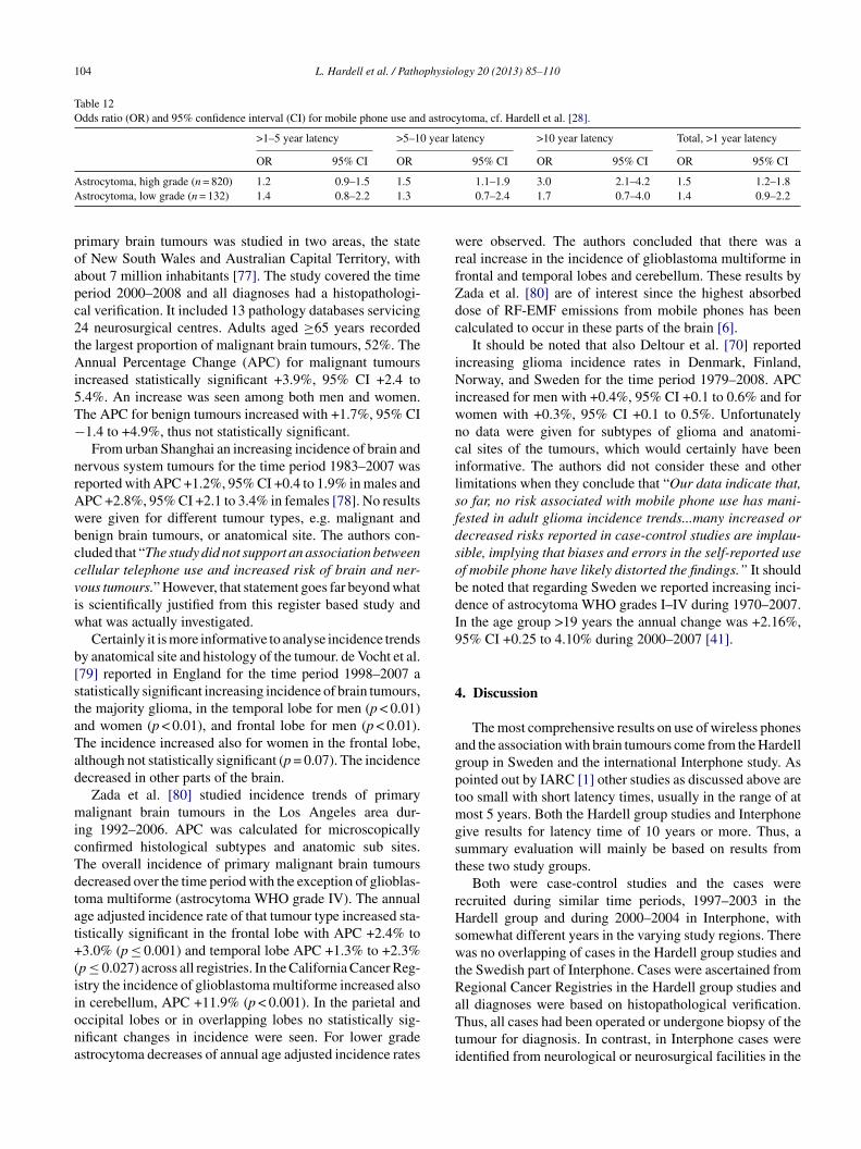
104 L. Hardell et al. / Pathophysiology 20 (2013) 85–110
Table 12Odds ratio (OR) and 95% confidence interval (CI) for mobile phone use and astrocytoma, cf. Hardell et al. [28].
>1–5 year latency >5–10 year latency >10 year latency Total, >1 year latency
OR 95% CI OR 95% CI OR 95% CI OR 95% CI
Astrocytoma, high grade (n = 820) 1.2 0.9–1.5 1.5 1.1–1.9 3.0 2.1–4.2 1.5 1.2–1.8Astrocytoma, low grade (n = 132) 1.4 0.8–2.2 1.3 0.7–2.4 1.7 0.7–4.0 1.4 0.9–2.2
primary brain tumours was studied in two areas, the stateof New South Wales and Australian Capital Territory, withabout 7 million inhabitants [77]. The study covered the timeperiod 2000–2008 and all diagnoses had a histopathologi-cal verification. It included 13 pathology databases servicing24 neurosurgical centres. Adults aged "65 years recordedthe largest proportion of malignant brain tumours, 52%. TheAnnual Percentage Change (APC) for malignant tumoursincreased statistically significant +3.9%, 95% CI +2.4 to5.4%. An increase was seen among both men and women.The APC for benign tumours increased with +1.7%, 95% CI$1.4 to +4.9%, thus not statistically significant.
From urban Shanghai an increasing incidence of brain andnervous system tumours for the time period 1983–2007 wasreported with APC +1.2%, 95% CI +0.4 to 1.9% in males andAPC +2.8%, 95% CI +2.1 to 3.4% in females [78]. No resultswere given for different tumour types, e.g. malignant andbenign brain tumours, or anatomical site. The authors con-cluded that “The study did not support an association betweencellular telephone use and increased risk of brain and ner-vous tumours.” However, that statement goes far beyond whatis scientifically justified from this register based study andwhat was actually investigated.
Certainly it is more informative to analyse incidence trendsby anatomical site and histology of the tumour. de Vocht et al.[79] reported in England for the time period 1998–2007 astatistically significant increasing incidence of brain tumours,the majority glioma, in the temporal lobe for men (p < 0.01)and women (p < 0.01), and frontal lobe for men (p < 0.01).The incidence increased also for women in the frontal lobe,although not statistically significant (p = 0.07). The incidencedecreased in other parts of the brain.
Zada et al. [80] studied incidence trends of primarymalignant brain tumours in the Los Angeles area dur-ing 1992–2006. APC was calculated for microscopicallyconfirmed histological subtypes and anatomic sub sites.The overall incidence of primary malignant brain tumoursdecreased over the time period with the exception of glioblas-toma multiforme (astrocytoma WHO grade IV). The annualage adjusted incidence rate of that tumour type increased sta-tistically significant in the frontal lobe with APC +2.4% to+3.0% (p # 0.001) and temporal lobe APC +1.3% to +2.3%(p # 0.027) across all registries. In the California Cancer Reg-istry the incidence of glioblastoma multiforme increased alsoin cerebellum, APC +11.9% (p < 0.001). In the parietal andoccipital lobes or in overlapping lobes no statistically sig-nificant changes in incidence were seen. For lower gradeastrocytoma decreases of annual age adjusted incidence rates
were observed. The authors concluded that there was areal increase in the incidence of glioblastoma multiforme infrontal and temporal lobes and cerebellum. These results byZada et al. [80] are of interest since the highest absorbeddose of RF-EMF emissions from mobile phones has beencalculated to occur in these parts of the brain [6].
It should be noted that also Deltour et al. [70] reportedincreasing glioma incidence rates in Denmark, Finland,Norway, and Sweden for the time period 1979–2008. APCincreased for men with +0.4%, 95% CI +0.1 to 0.6% and forwomen with +0.3%, 95% CI +0.1 to 0.5%. Unfortunatelyno data were given for subtypes of glioma and anatomi-cal sites of the tumours, which would certainly have beeninformative. The authors did not consider these and otherlimitations when they conclude that “Our data indicate that,so far, no risk associated with mobile phone use has mani-fested in adult glioma incidence trends...many increased ordecreased risks reported in case-control studies are implau-sible, implying that biases and errors in the self-reported useof mobile phone have likely distorted the findings.” It shouldbe noted that regarding Sweden we reported increasing inci-dence of astrocytoma WHO grades I–IV during 1970–2007.In the age group >19 years the annual change was +2.16%,95% CI +0.25 to 4.10% during 2000–2007 [41].
4. Discussion
The most comprehensive results on use of wireless phonesand the association with brain tumours come from the Hardellgroup in Sweden and the international Interphone study. Aspointed out by IARC [1] other studies as discussed above aretoo small with short latency times, usually in the range of atmost 5 years. Both the Hardell group studies and Interphonegive results for latency time of 10 years or more. Thus, asummary evaluation will mainly be based on results fromthese two study groups.
Both were case-control studies and the cases wererecruited during similar time periods, 1997–2003 in theHardell group and during 2000–2004 in Interphone, withsomewhat different years in the varying study regions. Therewas no overlapping of cases in the Hardell group studies andthe Swedish part of Interphone. Cases were ascertained fromRegional Cancer Registries in the Hardell group studies andall diagnoses were based on histopathological verification.Thus, all cases had been operated or undergone biopsy of thetumour for diagnosis. In contrast, in Interphone cases wereidentified from neurological or neurosurgical facilities in the
L. Hardell et al. / Pathophysiology 20 (2013) 85–110 105
study regions; in some centres also from cancer registries.The diagnoses of glioma, meningioma and acoustic neu-roma were based on histopathology or diagnostic imaging. Itshould be pointed out that the diagnosis of both meningiomaand acoustic neuroma has a rather high precision using CTand/or MRI. Regarding glioma it is certainly more difficult toestablish a valid diagnosis without histopathology, especiallywhen it comes to subgroups such as different grades of astro-cytoma (WHO grades I–IV). In the publication by Lahkolaet al. [81] most glioma diagnoses were based on histopatho-logy, whereas this has not been published for Interphone intotal. It is notable that Interphone [9] has not presented sepa-rate results for astrocytoma in total in contrast to the Hardellgroup. Especially results for high-grade glioma including themost common glioma type, glioblastoma multiforme (WHOgrade IV), would be of value since the highest risk was foundfor that subtype by Hardell et al., Table 12 [27,28]. It is also ofinterest that we found higher risk for use of mobile and cord-less phones for astrocytoma grades III–IV than for gradesI–II [82]. Some results were published for glioblastoma mul-tiforme from the 5 North European countries [81]. Certainlythe total result for glioma and >10 years since first ipsilateralmobile phone use with OR = 1.39, 95% CI = 1.01–1.92 (pfor trend 0.04) would have been of interest for glioblastomamultiforme separately in Lahkola et al. [81].
The Hardell group included cases aged 20–80 yearswhereas eligible cases in Interphone were aged 30–59 yearsat diagnosis. This difference is important since the highestincidence of astrocytoma WHO grade IV (glioblastoma mul-tiforme) is found in the age group 45–75 years with mean age61 years and 80% older than 50 years [83]. As can be seenin Table 10, the highest prevalence of use of mobile phonesin the Hardell group studies was up to the age of 54 years,so limiting the age to 59 years as in Interphone diminishesthe possibility to find an increased risk taking a reasonabletumour induction period. It seems as if the age distribution inInterphone was more decided by prevalence of mobile phoneuse in the population than age distribution for glioma cases.Excluding the age group 20–29 years, as in Interphone, makesalso an evaluation of young users more difficult, see Table 9.
Meningioma is a slow growing benign tumour with a peakincidence in the sixth and seventh decade of life with a 3:2–2:1female:male ratio [84]. As pointed out by Interphone [10]the incidence peak of acoustic neuroma is in the age group50–65 years. Thus, again limiting upper age to 59 years forcases in Interphone excluded a large proportion of cases withmeningioma or acoustic neuroma taking a reasonable latencyperiod.
One control subject matched on age, gender and geograph-ical area (region) to each case in the Hardell group studieswas drawn from the national population register. The registercovers the whole population and each person is assigned aunique id-number making it possible to trace current addressfor all inhabitants. In Interphone one control was selectedfor each case from a ‘locally appropriate population-basedsampling frame’. In Germany the centres used individual
matching or frequency matching. The matching variableswere age within 5 years, gender and region of residence; inIsrael also ethnic origin. When stratified matching was usedindividual matching was made afterwards from the wholecontrol sample with cases being assigned one control subject(two in Germany) interviewed as close as possible in timeto the case [9]. Regarding the Interphone study on acousticneuroma some centres sampled special controls to the cases,other draw controls from the pool of controls in the gliomaand meningioma studies, or used a mixture of both methods.
The Nordic countries have population registers that wereused in Denmark, Norway and Finland for recruitment ofcontrols in Interphone. Also Germany used a population reg-ister [85]. However, UK used general practioners’ lists [86]and in Japan random digit dialling was used [44,87]. Certainlythe methods used in Interphone may introduce selection bias.Patient lists are usually selective to use for drawing of controlsand do not represent the whole population which is the sourceof the cases. Also random digit dialling has the potential tointroduce selection bias since persons that are registered tosubscribe a phone are usually wealthier than non-subscribers.Furthermore, it seems not to be the most appropriate methodfor selection of controls in a study on mobile phone use, andcertainly not regarding cordless phones, since phone sub-scribers are selected as controls. Furthermore, later selectionof controls from a pool with individual matching may givethe possibility for selection bias if this is not done in a blindedmanner as to exposure status.
These methods contrast to the Hardell group where con-trols were drawn consequently to the cases and all controlsthat answered the questionnaire were included in the analy-ses. In Interphone proxy interviews were performed for 13%of glioma cases but only 1% of controls [9]. This is in contrastto the Hardell group study on deceased cases with malignantbrain tumours [26]. Deceased controls were drawn from theDeath Registry in Sweden. Relatives to both deceased casesand deceased controls were interviewed, thereby creating thesame condition for assessment of exposure among cases andcontrols. Although using proxy interviews for both cases andcontrols is the more appropriate method exclusion of proxyinterviews in Interphone had little impact on the overall resultin the sensitivity analysis.
Use of wireless phones was carefully assessed by a self-administered questionnaire in the Hardell et al. studies. Theinformation was supplemented over the phone by trainedinterviewers thereby using a structured protocol. This wasdone blinded as to case or control status. The ear that hadmostly been used during calls with mobile phone and/or cord-less phone was assessed by separate questions; >50% of thetime for one side, or equally for both sides. This informationwas checked during the supplementary phone calls. Moreoverevery person that had used a mobile phone received after thata letter asking them again to specify the ear that had been usedduring phone calls and to what extent that side of the headwas mostly used. There was a very good agreement of theresults using these three methods to assess these data. Also
106 L. Hardell et al. / Pathophysiology 20 (2013) 85–110
other exposures were assessed in the questionnaire. After theinterviews all personal data like names and addresses wereremoved from the questionnaires so that only an id-numberthat did not disclose if it was a case or a control was shown.Thus, coding of the data for statistical analysis was performedwithout personal data on the individual.
We investigated in more detail the possibility of recalland observational bias in our second case-control study [21].Reporting a previous cancer or if a relative helped to fill in thequestionnaire did not change the results, i.e., were no con-founding factors. Potential observational bias during phoneinterviews was analysed by comparing change of exposurein cases and controls after these interviews. No statisticallysignificant differences were found, showing that our resultscould not be explained by observational bias, for furtherdetails see discussion in that publication [21].
On the contrary information on past mobile phone usewas mostly collected during face-to-face interviews in Inter-phone obviously disclosing if it was a case or a control thatwas interviewed. These interviews were performed by a largenumber of interviewers at different participating centres. Inthe personal interviews a computer program that guided theinterview with questions read by the interviewer from a laptopcomputer screen was used. The interviews in the Swedish partlasted for about 45 min. The answers were entered directlyinto the computer by the interviewer. Cards were shown toif possible identify the model of the mobile phone [88]. Thepurpose of the study was thereby obviously disclosed to thecases and controls. This was in contrast to the Hardell groupmailed questionnaire that contained a large number of otherquestions without special attention to wireless phones.
We regard hospital based interviews of cases, as in theInterphone study, to be a major disadvantage and ethicallyquestionable. At that time the patient has not fully recoveredfrom e.g. surgery, may not have been fully informed about thediagnosis, treatment and prognosis and may even be undersedation by drugs. Using computer based face-to-face inter-views may also be a stressful situation for the patient. Infact patients scored significantly lower than controls due torecalling of words (aphasia), problems with writing and draw-ing due to paralysis in the Danish part of Interphone [89].Obviously observational bias could have been introduced bythe interview methods in Interphone. Only Finland used apaper version of the questionnaire, but Finland has never pub-lished country specific results on the different tumour types,which would certainly have been of interest. For unclear rea-sons the results on glioma were only included as part of theresults for the 5 North European countries [81] and as partof the whole Interphone study [9]. Furthermore, it has notbeen disclosed how the personal interviews were performedin sparsely populated areas, e.g. in the Northern Sweden. Didthe interviewers travel long distances for interviews of con-trols in rural areas or were all controls living in the largestcities thereby creating selection bias?
It should be noted that the number of participating casesand controls from each centre in Interphone was quite low. It
varied for glioma from 60 (Japan) to at most 421 (UK North),for meningioma from 52 (New Zealand) to 350 (Israel) andfor acoustic neuroma from 18 (New Zealand) to 152 (UKSouth). Similarly the number of controls varied according tocentre [9,10]. It is obvious that with so low number of inter-viewed subjects by many different interviewers the qualitymay have been hampered in Interphone by low training andexperience of certain interviewers. Experienced interviewerswere defined as those who conducted at least 20 interviews.In fact, in the sensitivity analysis the risk increased for gliomafor cumulative mobile phone use "1640 h from OR = 1.40,95% CI = 1.03–1.89 to OR = 1.50, 95% CI = 1.10–2.06 if‘experienced interviewers only’ were considered. In theHardell group studies few persons conducted all interviewsof the 1251 participating cases with malignant brain tumour,1254 cases with benign brain tumour, and 2438 controls (total4942; note one case had both a malignant and a benign braintumour). All interviewers were first educated; they used adefined protocol and gained considerable experience as inter-viewers. In fact, they were obliged to carry out the interviewsextensively to fulfil the quality in data assessment accordingto the structured protocol. It is obvious that the few inter-viewers in the Hardell group study must have been muchmore experienced than the diversity of interviewers in Inter-phone. The higher risk restricting analysis to ‘experiencedinterviewers’ in Interphone indicates observational bias dur-ing assessment of exposure decreasing the risk. Furthermore,20 interviews as the definition was in Interphone to be anexperienced interviewer, is after all a very low number.
Several other sensitivity analyses were performed in Inter-phone without any major impact on the results. It is discussedin the Interphone study [9] that the increased risk for gliomain the highest decile of cumulative exposure was caused by anumber of subjects reporting >5 h call time per day. This num-ber may be real in e.g. certain occupations using the phoneas a working tool. Furthermore, if call time was truncated to5 h per day no statistically significant difference of the riskwas found, OR = 1.38, 95% CI = 1.02–1.87 for glioma andOR = 3.03, 95% CI = 1.62–5.67 for acoustic neuroma (expo-sure up to 5 years before reference date). Certainly it is notjustified to exclude these subjects from the analysis as wasdone in some of the calculations in Interphone [9,10].
It is always essential to have a high response rate in case-control studies to get as valid results as possible. In the Hardellgroup studies the response rate was 85% (n = 1251) for caseswith malignant brain tumour, 88% (n = 1254) for cases withbenign brain tumour, and 84% (n = 2438) for controls [29,40].Lower response rates were obtained in the Interphone study,64%, range by centre 36–92%, (n = 2765) for glioma cases,78%, range 56–92%, (n = 2425) for meningioma cases, 82%,range 70–100% (n = 1121) for acoustic neuroma cases, and53%, range 42–74%, (n = 7658) for controls [9,10]. Certainlythese low response rates, less than half of the cases and con-trols in some centres, may have created the possibility ofconsiderable selection bias and are examples of the mul-tiple methodological problems in Interphone. As has been
L. Hardell et al. / Pathophysiology 20 (2013) 85–110 107
discussed elsewhere not responding controls in Interphonetended to be less frequent users of mobile phone than partic-ipating controls leading to underestimation of the risk [32].
There are other differences between the Hardell groupstudies and Interphone study such as restricting age to30–59 years in Interphone compared with 20–80 years in theHardell-group studies and considering use of cordless phonesas no exposure to RE-EMF in Interphone. Even if the preva-lence of mobile phone use is highest in the age group 30–59years, excluding older cases diminishes the possibility to findan increased risk, assuming a reasonable latency time. As dis-cussed above the peak incidence of most brain tumours is ata higher age. In a case series from Canada all brain tumoursshowed a bimodal age distribution with one peak in the 0–4age group and the other in the 60–69 age group [90]. Asshown elsewhere [14] step-wise exclusion of the age group20–29 years, 60–80 years and including cordless phone useamong unexposed reduced OR in the Hardell-group studiesto similar results as in Interphone [see Tables 1 and 2 in thepublication]. Thus, Interphone seems to have underestimatedthe risk also for these reasons.
Survival of patients with glioma has only been presentedby the Hardell group [68]. Decreased survival of glioma caseswith long-term and high cumulative use of wireless phoneswas found. We found a survival disadvantage for astrocy-toma WHO grade IV among cases using mobile phone orcordless phone indicating a worse prognosis in that patientgroup. On the contrary, a survival benefit for astrocytomaWHO grades I–II was observed. The fact that there was noclear trend with intensity or duration of wireless phone usefor low-grade astrocytoma does not speak in favour of aneffect of RF-EMF from such use. The exposure might, how-ever, produce awareness bias in these cases. RF-EMF maygive tumour promotion [91] inducing disease related person-ality disturbances and habit changes leading to earlier tumourdiagnosis than among unexposed patients. This would resultin earlier treatment with a better prognosis after surgery inthis patient group [69]. These findings indicate a complexbiological effect from RF-EMF exposure and strengthen acausal association between these tumour types, e.g. astrocy-toma WHO grade IV (glioblastoma multiforme), and use ofwireless phones.
By placing a strong emphasis on incidence data an asso-ciation between use of wireless phones and brain tumourshas been challenged [92]. The authors considered that, if theincreased risks seen in case-control studies reflect a causalrelationship, there would already be an increase in incidenceof brain and central nervous system tumours, for which thereseemed to be little evidence. This belief is unfounded fortwo reasons. The first relates to latent periods for gliomaand acoustic neuroma development, typically 10–40 years[93,94]. The results on long-term use of wireless phones arescanty and at most latency period of 10+ years have beenstudied. Furthermore, we know little about the earliest eventsin the genesis of glioma in humans for obvious reasons. How-ever, progression of glioma has been studied in large series of
tumours of different malignancy grades. Patients with low-grade glioma have been followed with later progression tohigh-grade glioma [95]. Thus, since the natural history ofmost glioma from earliest events to clinical manifestation isunknown, but most likely several decades, the exposure dura-tion in most studies is incompatible with a tumour initiatingeffect. An initiating effect is what would have the most directeffect on the incidence. The other reason concerns the possi-bility of an effect on tumour development (promotion) and itsconsequences on the increase in incidence that can possiblyoccur. If the exposure acts as a promoter, this would decreaselatency time for already existing tumours, giving a temporarybut not a continuous increase in incidence. In addition it hasto be pointed out that any such effect on tumour developmentis limited by the magnitude of the shift of the age-incidencefunction and its slope for the respective tumour type [91]. Itshould be noted that studies on tumour type and anatomicallocalisation indicate by now an effect from RF-EMF on theincidence of brain tumours [71,77,79,80].
5. Conclusions
There is a consistent pattern of increased risk of gliomaand acoustic neuroma associated with use of mobile phonesand cordless phones. The epidemiological evidence comesmainly from two study centres, the Hardell group and theInterphone study group. In the same studies by the Hardellgroup and Interphone study group no consistent pattern ofan increased risk was found for meningioma. These resultsstrengthen the other findings, i.e., increased risk for gliomaand acoustic neuroma, since a systematic bias in the studieswould also have been inherit for meningioma. Furthermore,a causal association between use of mobile phone and gliomaand acoustic neuroma comes from the meta-analyses as pre-sented in this publication and also reviewed elsewhere [96].Supportive evidence comes also from anatomical localisationof the tumour to the most exposed area of the brain, cumula-tive exposure and latency time that all add to the biologicalrelevance of an increased risk. In addition risk calculationbased on estimated absorbed dose gives strength to the find-ings as well as the impact on survival of glioma patientsrelating to their use of mobile and cordless phones.
Evidence is increasing that workers with heavy use ofwireless phones who develop glioma or acoustic neuromashould be compensated. In fact, the first case with such com-pensation has now been established in court. The ItalianSupreme Court affirmed a previous ruling that the InsuranceBody for Work (INAIL) must grant worker’s compensation toa businessman who had used wireless phones for 12 years anddeveloped a neuroma in the brain (www.applelettrosmog.it;www.microwavenews.com). He had used both mobile andcordless phones for five to six hours per day preferably onthe same side as the tumour developed. The neuroma waslocated in the trigeminal Gasser’s ganglion in the brain. Thisfifth cranial nerve controls facial sensations and muscles. It is
108 L. Hardell et al. / Pathophysiology 20 (2013) 85–110
the same type of tumour as the acoustic neuroma in the eighthcranial nerve located in the same area of the brain. The Ital-ian case fulfils the criteria for a causal association; more than10 years use of wireless phones, high cumulative exposureon the same side as the tumour appeared, and a tumour typethat would be predicted based on previous research on use ofwireless phones and brain tumour risk. No further appeal ofthe Supreme Court decision is possible.
In summary there is reasonable basis to conclude thatRF-EMFs are bioactive and have a potential to cause healthimpacts. There is a consistent pattern of increased risk forglioma and acoustic neuroma associated with use of wire-less phones (mobile phones and cordless phones) mainlybased on results from case-control studies from the Hardellgroup and Interphone Final Study results. Epidemiologicalevidence gives that RF-EMF should be classified as a humancarcinogen. The current safety limits and reference levelsare not adequate to protect public health. New public healthstandards and limits are needed.
Authors’ contributions
Lennart Hardell was responsible for drafting of themanuscript and Michael Carlberg made all statistical calcu-lations. Michael Carlberg and Kjell Hansson Mild read andgave valuable comments on the manuscript. All authors haveread and approved the final version. No conflicts of interestreported.
Acknowledgements
Supported by grants from Cancer-och Allergifonden,Cancerhjälpen, and Örebro University Hospital CancerFund.
References
[1] R. Baan, Y. Grosse, B. Lauby-Secretan, F. El Ghissassi, V.Bouvard, L. Benbrahim-Tallaa, et al., Carcinogenicity of radiofre-quency electromagnetic fields, Lancet Oncology 12 (2011)624–626.
[2] IARC Monographs on the Evaluation of Carcinogenic Risks toHumans, Volume 102, Non-Ionizing radiation, Part II: RadiofrequencyElectromagnetic Fields [includes mobile telephones], IARC Press2011, Lyon, France.
[3] IARC Monographs on the Evaluation of Carcinogenic Risks toHumans, Volume 80, Non-Ionizing Radiation, Part I: Static andExtremely Low-Frequency (ELF) Electric and Magnetic Fields, IARCPress 2002, Lyon, France.
[4] T. Linde, K. Hansson Mild, Measurement of low frequency magneticfields from digital cellular telephones, Bioelectromagnetics 18 (1997)184–186.
[5] K. Jokela, L. Puranen, A.P. Sihvonen, Assessment of the magnetic fieldexposure due to the battery current of digital mobile phones, HealthPhysics 86 (2004) 56–66.
[6] E. Cardis, I. Deltour, S. Mann, M. Moissonnier, M. Taki, N. Varsier,et al., Distribution of RF energy emitted by mobile phones in anatomicalstructures of the brain, Physics in Medicine and Biology 53 (2008)2771–2783.
[7] S. Preston-Martin, R. Munir, I. Chakrabarti, Nervous system, in: D.Schottenfeld, J.F. Fraumeni Jr. (Eds.), Cancer Epidemiology and Pre-vention, Oxford University Press, 2006, pp. 1173–1195.
[8] M.E. Scheurer, C.J. Etzel, M. Liu, J. Barnholtz-Sloan, F. Wik-lund, B. Tavelin, et al., Familial aggregation of glioma: apooled analysis, American Journal of Epidemiology 172 (2010)1099–1107.
[9] Interphone Study Group, Brain tumour risk in relation to mobiletelephone use: results of the INTERPHONE international case-control study, International Journal of Epidemiology 39 (2010)675–694.
[10] Interphone Study Group, Acoustic neuroma risk in relation to mobiletelephone use: results of the INTERPHONE international case-controlstudy, Cancer Epidemiology 35 (2011) 453–464.
[11] E. Cardis, B.K. Armstrong, J.D. Bowman, G.G. Giles, M. Hours, D.Krewski, et al., Risk of brain tumours in relation to estimated RF dosefrom mobile phones: results from five Interphone countries, Occupa-tional and Environmental Medicine 68 (2011) 631–640.
[12] L. Hardell, M. Carlberg, D. Gee, Mobile phone use and brain tumourrisk: early warnings, early actions? In: Late Lessons from Early War-nings, part 2, European Environment Agency, Copenhagen, Denmark2013, in press.
[13] L. Hardell, M. Carlberg, K. Hansson Mild, Epidemiological evidencefor an association between use of wireless phones and tumour diseases,Pathophysiology 16 (2009) 113–122.
[14] L. Hardell, M. Carlberg, K. Hansson Mild, Re-analysis of risk forglioma in relation to mobile telephone use: comparison with the resultsof the Interphone international case-control study, International Journalof Epidemiology 40 (2011) 1126–1128.
[15] L. Hardell, Å. Näsman, A. Påhlson, A. Hallquist, K. Hansson Mild,Use of cellular telephones and the risk for brain tumours: a case-controlstudy, International Journal of Oncology 15 (1999) 113–116.
[16] L. Hardell, K. Hansson Mild, A. Påhlson, A. Hallquist, Ionizing radi-ation, cellular telephones and the risk for brain tumours, EuropeanJournal of Cancer Prevention 10 (2001) 523–529.
[17] J.E. Muscat, M.G. Malkin, S. Thompson, R.E. Shore, S.D. Stell-man, D. McRee, et al., Handheld cellular telephone use and risk ofbrain cancer, Journal of the American medical Association 284 (2000)3001–3007.
[18] H. Ohgaki, P. Kleihues, Population-based studies on incidence, survivalrates, and genetic alterations in astocytic and oligodendroglial gliomas.Review, Journal of Neuropathology and Experimental Neurology 64(2005) 479–489.
[19] P.D. Inskip, R.E. Tarone, E.E. Hatch, T.C. Wilcosky, W.R. Shapiro, R.G.Selker, et al., Cellular-telephone use and brain tumours, New EnglandJournal of Medicine 344 (2001) 79–86.
[20] A. Auvinen, M. Hietanen, R. Luukkonen, R.S. Koskela, Brain tumoursand salivary gland cancers among cellular telephone users, Epidemiol-ogy 13 (2002) 356–359.
[21] L. Hardell, A. Hallquist, K. Hansson Mild, M. Carlberg, A. Påhlson, A.Lilja, Cellular and cordless telephones and the risk for brain tumours,European Journal of Cancer Prevention 11 (2002) 377–386.
[22] L. Hardell, K. Hansson Mild, M. Carlberg, Further aspects on cellularand cordless telephones and brain tumours, International Journal ofOncology 22 (2003) 399–407.
[23] L. Hardell, M. Carlberg, K. Hansson Mild, Case-control study of theassociation between the use of cellular and cordless telephones andmalignant brain tumours diagnosed during 2000-2003, EnvironmentalResearch 100 (2006) 232–241.
[24] L. Hardell, M. Carlberg, K. Hansson Mild, Case-control study on cel-lular and cordless telephones and the risk for acoustic neuroma ormeningioma in patients diagnosed 2000-2003, Neuroepidemiology 25(2005) 120–128.
L. Hardell et al. / Pathophysiology 20 (2013) 85–110 109
[25] J.D. Jr. Boice, J.K. McLaughlin, Epidemiologic Studies ofCellular Telephones and Cancer Risk—A Review. SSI Publica-tion 2002:16, accessed at http://www.stralsakerhetsmyndigheten.se/Publikationer/Rapport/Stralskydd/2002/200216/
[26] L. Hardell, M. Carlberg, K. Hansson Mild, Mobile phone use and therisk for malignant brain tumours: a case-control study on deceased casesand controls, Neuroepidemiology 35 (2010) 109–114.
[27] L. Hardell, M. Carlberg, K. Hansson Mild, Pooled analysis of twocase-control studies on use of cellular and cordless telephones and therisk for malignant brain tumours diagnosed in 1997-2003, InternationalArchives of Occupational Environmental Health 79 (2006) 630–639.
[28] L. Hardell, M. Carlberg, K. Hansson Mild, Pooled analysis of case-control studies on malignant brain tumours and the use of mobile andcordless phones including living and deceased subjects, InternationalJournal of Oncology 38 (2011) 1465–1474.
[29] M. Carlberg, L. Hardell, On the association between glioma, wirelessphones, heredity and ionising radiation, Pathophysiology 19 (2012)243–252.
[30] A. McKinlay, Possible health effects related to the use ofradiotelephones—recommendations of a European Commission ExpertGroup, Radiological Protection Bullentin 187 (1997) 9–16.
[31] E. Cardis, L. Richardson, I. Deltour, B. Armstrong, M. Feychting, C.Johansen, et al., The INTERPHONE study: design, epidemiologicalmethods, and description of the study population, European Journal ofEpidemiology 22 (2007) 647–664.
[32] L. Hardell, M. Carlberg, K. Hansson, Mild, methodological aspects ofepidemiological studies on the use of mobile phones and their asso-ciation with brain tumours, Open Environmental Sciences 2 (2008)54–61.
[33] R. Saracci, J. Samet, Commentary: call me on my mobile phone.orbetter not?—a look at the INTERPHONE study results, InternationalJournal of Epidemiology 39 (2010) 695–698.
[34] J.L. Phillips, N.P. Singh, H. Lai, Electromagnetic fields and DNA dam-age, Pathophysiology 16 (2009) 79–88.
[35] H. Nittby, A. Brun, J. Eberhardt, L. Malmgren, B.R. Persson, L.G.Salford, Increased blood-brain barrier permeability in mammalian brain7 days after exposure to the radiation from a GSM-900 mobile phone,Pathophysiology 16 (2009) 103–112.
[36] E. Markova, L.O.G. Malmgren, I.Y. Belyaev, Microwaves from mobilephones inhibit 53BP1 focus formation in human stem cells morestrongly than in differentiated cells: possible mechanistic link to cancer,Environmental Health Perspectives 118 (2010) 394–399.
[37] K. Hansson Mild, M. Carlberg, J. Wilén, L. Hardell, How to combinethe use of different mobile and cordless telephones in epidemiologicalstudies on brain tumours? European Journal of Cancer Prevention 14(2005) 285–288.
[38] L. Hardell, M. Carlberg, K. Hansson Mild, Use of cellular telephonesand brain tumour risk in urban and rural areas, Occupational and Envi-ronmental Medicine 62 (2005) 390–394.
[39] S. Larjavaara, J. Schüz, A. Swerdlow, M. Feychting, C. Johansen, S.Lagorio, et al., Location of gliomas in relation to mobile telephone use:a case-case and case-specular analysis, American Journal of Epidemi-ology 174 (2011) 2–11.
[40] L. Hardell, M. Carlberg, K. Hansson Mild, Pooled analysis of two case-control studies on the use of cellular and cordless telephones and therisk of benign brain tumours diagnosed during 1997-2003, InternationalJournal of Oncology 28 (2006) 509–518.
[41] L. Hardell, M. Carlberg, Mobile phones, cordless phones and the riskfor brain tumours, International Journal of Oncology 35 (2009) 5–17.
[42] J.E. Muscat, M.G. Malkin, R.E. Shore, S. Thompson, A.I. Neugut,S.D. Stellman, et al., Handheld cellular telephones and risk of acousticneuroma, Neurology 58 (2002) 1304–1306.
[43] Y. Sato, S. Akiba, O. Kubo, N. Yamaguchi, A case-case study of mobilephone use and acoustic neuroma risk in Japan, Bioelectromagnetics 32(2011) 85–93.
[44] T. Takebayashi, N. Varsier, Y. Kikuchi, K. Wake, M. Taki, S. Watanabe,et al., Mobile phone use, exposure to radiofrequency electromagnetic
field, and brain tumour: a case-control study, British Journal of Cancer98 (2008) 652–659.
[45] M.J. Schoemaker, A.J. Swerdlow, Risk of pituitary tumours in cellularphone users: a case-control study, Epidemiology 20 (2009) 348–354.
[46] A. Christ, M.C. Gosselin, M. Christopoulou, S. Kühn, N. Kuster, Age-dependent tissue-specific exposure of cell phone users, Physics inMedicine and Biology 55 (2010) 1767–1783.
[47] O.P. Gandhi, L.L. Morgan, A.A. de Salles, Y.Y. Han, R.B. Herber-man, D.L. Davis, Exposure limits: the underestimation of absorbedcell phone radiation, especially in children, Electromagnetic Biologyand Medicine 31 (2012) 34–51.
[48] L. Kheifets, M. Repacholi, R. Saunders, E. van Deventer, The sen-sitivity of children to electromagnetic fields, Pediatrics 116 (2005)303–313.
[49] N.U. Dosenbach, N. Nardos, A.L. Cohen, D.A. Fair, J.D. Power, J.A.Church, et al., Prediction of individual brain maturity using fMRI,Science 329 (2010) 1358–1361.
[50] F. Söderqvist, L. Hardell, M. Carlberg, K. Hansson Mild, Ownershipand use of wireless telephones: a population-based study of Swedishchildren aged 7-14 years, BMC Public Health 7 (2007) 105.
[51] F. Söderqvist, M. Carlberg, L. Hardell, Use of wireless telephonesand self-reported health symptoms: a population-based study amongSwedish adolescents aged 15–19 years, Environmental Health 7 (2008)18.
[52] L. Hardell, K. Hansson Mild, M. Carlberg, A. Hallquist, Cellular andcordless telephone use and the association with brain tumours in dif-ferent age groups, Archives of Occupational and Environmental Health59 (2004) 132–137.
[53] D. Aydin, M. Feychting, J. Schüz, T. Tynes, T.V. Andersen, L.S.Schmidt, et al., Mobile phone use and brain tumours in children andadolescents: a multicenter case-control study, Journal of the NationalCancer Institute 103 (2011) 1264–1276.
[54] F. Söderqvist, M. Carlberg, K. Hansson Mild, L. Hardell, Childhoodbrain tumour risk and its association with wireless phones: a commen-tary, Environmental Health 10 (2011) 106.
[55] J.D. Boice Jr., R.E. Tarone, Cell phones, cancer, and children, Journalof the National Cancer Institute 16 (2011) 1211–1213.
[56] D. Aydin, M. Feychting, J. Schüz, T.V. Andersen, A. Harbo Poulsen,M. Prochazka, et al., Impact of random and systematic recall errorsand selection bias in case-control studies on mobile phone use andbrain tumors in adolescents (CEFALO Study), Bioelectromagnetics 32(2011) 396–407.
[57] C. Johansen, J.D. Boice Jr., J. McLaughlin, J. Olsen, Cellular telephonesand cancer—a nationwide cohort study in Denmark, Journal of theNational Cancer Institute 93 (2001) 203–207.
[58] F. Söderqvist, M. Carlberg, L. Hardell, Review of four publica-tions on the Danish cohort study on mobile phone subscribers andrisk of brain tumours, Reviews Environmental Health 27 (2012)51–58.
[59] J. Schüz, R. Jacobsen, J.H. Olsen, J.D. Boice Jr., J.K. McLaughlin, C.Johansen, Cellular telephone use and cancer risk: update of a nation-wide Danish cohort, Journal of the National Cancer Institute 98 (2006)1707–1713.
[60] J. Schüz, M. Steding-Jessen, S. Hansen, S.E. Stangerup, P. Cayé-Thomasen, A.H. Poulsen, et al., Long-term mobile phone use andthe risk of vestibular schwannoma: a Danish nationwide cohort study,American Journal of Epidemiology 174 (2011) 416–422.
[61] P. Frei, A. Harbo Poulsen, C. Johansen, J.H. Olsen, M. Steding-Jessen, J. Schüz, Use of mobile phones and risk of brain tumours:update of Danish cohort study, British Medical Journal 343 (2011)d6387.
[62] J. Schüz, P. Elliott, A. Auvinen, H. Kromhout, A. Harbo Poulsen, C.Johansen, et al., An international prospective cohort study of mobilephone users and health (Cosmos): design considerations and enrolment,Cancer Epidemiology 35 (2011) 37–43.
[63] S.O. Dalton, M. Steding-Jessen, M. Gislum, K. Frederiksen, G.Engholm, J. Schüz, Social inequality and incidence of and survival
110 L. Hardell et al. / Pathophysiology 20 (2013) 85–110
from cancer in a population-based study in Denmark, 1994-2003: back-ground, aims, material and methods, European Journal of Cancer 44(2008) 1938–1949.
[64] A. Ahlbom, M. Feychting, Mobile telephones and brain tumours,British Medical Journal 343 (2011) d6605.
[65] A. Ahlbom, M. Feychting, E. Cardis, P. Elliott, Re: Cellular telephoneuse and cancer risk: update of a nationwide Danish cohort study, Journalof the National Cancer Institute 99 (2007) 655.
[66] D.E. Foliart, B.H. Pollock, G. Mezei, R. Iriye, J.M. Silva, K.L. Ebi,et al., Magnetic field exposure and long-term survival among childrenwith leukaemia, British Journal of Cancer 94 (2006) 161–164.
[67] A.-L. Svendsen, T. Weihkopf, P. Kaatsch, J. Schüz, Exposure to mag-netic fields and survival after diagnosis of childhood leukemia: aGerman cohort study, Cancer Epidemiology Biomarkers Prevention16 (2007) 1167–1171.
[68] L. Hardell, M. Carlberg, Use of mobile and cordless phones and survivalof patients with glioma, Neuroepidemiology 40 (2012) 101–108.
[69] F.T. Vertosick Jr., R.G. Selker, V.C. Arena, Survival of patients withwell-differentiated astrocytomas diagnosed in the era of computedtomography. Review, Neurosurgery 28 (1991) 496–501.
[70] I. Deltour, A. Auvinen, M. Feychting, C. Johansen, L. Klaeboe, R.Sankila, et al., Mobile phone use and incidence of glioma in theNordic countries 1997-2008: consistency check, Epidemiology 23(2012) 301–307.
[71] M.P. Little, P. Rajaraman, R.E. Curtis, S.S. Devesa, P. Inskip, D.P.Check, et al., Mobile phone use and glioma risk: comparison of epi-demiological study results with incidence trends in the United States,British Medical Journal 344 (2012) e1147.
[72] T. Bergenheim, A. Malmström, H. Bolander, A. Michanek, G.Stragliotto, L. Damber, et al., Registration on regional basis of patientswith primary brain tumours. Regional differences disclosed, Läkartid-ningen 104 (2007) 332–338, 340–341.
[73] L. Barlow, K. Westergren, L. Holmberg, M. Talbäck, The completenessof the Swedish Cancer Register: a sample survey for year 1998, ActaOncologica 48 (2009) 27–33.
[74] M. Kundi, Study of mobile phone use and glioma risk was fatallyflawed, British Medical Journal 344 (2012) e3078.
[75] D.L. Davis, A.B. Miller, A. Philips, Association of mobile phone usewith adult brain cancer remains plausible, British Medical Journal 344(2012) e3083.
[76] W.D. Hazelton, M.S. Clements, S.H. Moolgavkar, Multistage car-cinogenesis and lung cancer mortality in three cohorts, CancerEpidemiology Biomarkers and Prevention 14 (2005) 1171–1181.
[77] M. Dobes, B. Shadbolt, V.G. Khurana, S. Jain, S.F. Smith, R. Smee,et al., A multicenter study of primary brain tumour incidence inAustralia (2000-2008), Neuro Oncology 13 (2011) 783–790.
[78] L.-X. Ding, Y.-X. Wang, Increasing incidence of brain and nervoustumours in urban Shanghai, China, 1983-2007, Asian Pacific Journalof Cancer Prevention 12 (2011) 3319–3322.
[79] F. de Vocht, I. Burstyn, J.W. Cherrie, Time trends (1998-2007) in braincancer incidence rates in relation to mobile phone use in England,Bioelectromagnetics 32 (2011) 334–339.
[80] G. Zada, A.E. Bond, Y.-P. Wang, S.L. Giannotta, D. Deapan, Incidencetrends in the anatomic location of primary malignant brain tumours inthe United States: 1992-2006, World Neurosurgery 77 (2012) 518–524.
[81] A. Lahkola, A. Auvinen, J. Raitanen, M.J. Schoemaker, H.C. Chris-tensen, M. Feychting, et al., Mobile phone use and risk of glioma in 5North European countries, International Journal of Cancer 120 (2007)1769–1775.
[82] K. Hansson Mild, L. Hardell, M. Carlberg, Pooled analysis of twoSwedish case-control studies on the use of mobile and cordlesstelephones and the risk of brain tumours diagnosed 1997-2003, Inter-national Journal of Occupational Safety and Ergonomics 13 (2007)63–71.
[83] H. Ohgaki, P. Dessen, B. Jourde, S. Horstmann, T. Nishikawa, P.L.Di Patre, et al., Genetic pathways of glioblastoma: a population-basedstudy, Cancer Research 64 (2004) 6892–6899.
[84] C. Marosi, M. Hassle, K.R. Ssler, Guidelines to the treatment of menin-gioma, Forum 13 (2003) 76–89.
[85] B. Schlehofer, K. Schlaefer, M. Blettner, G. Berg, E. Böhler, I. Het-tinger, et al., Environmental risk factors for sporadic acoustic neuroma(Interphone Study Group, Germany), European Journal of Cancer 43(2007) 1741–1747.
[86] S.J. Hepworth, M.J. Schoemaker, K.R. Muir, A.J. Swerdlow, M.J. vanTongeren, P.A. McKinney, Mobile phone use and risk of glioma inadults: case-control study, British Medical Journal 332 (2006) 883–887.
[87] T. Takebayashi, S. Akiba, Y. Kikuchi, M. Taki, K. Wake, S. Watan-abe, et al., Mobile phone use and acoustic neuroma risk in Japan,Occupational Environmental Medicine 63 (2006) 802–807.
[88] S. Lönn, A. Ahlbom, P. Hall, M. Feychting, the Swedish InterphoneStudy Group, Long-term mobile phone use and brain tumour risk,American Journal of Epidemiology 161 (2005) 526–535.
[89] H.C. Christensen, J. Schüz, M. Kosteljanetz, H.S. Poulsen, J.D. BoiceJr., J.K. McLaughlin, et al., Cellular telephones and risk for braintumours: a population-based, incident case-control study, Neurology64 (2005) 1189–1195.
[90] G.R. Sutherland, R. Florell, D. Louw, N.W. Choi, A.A. Sima, Epi-demiology of primary intracranial neoplasms in Manitoba, Canada,Canadian Journal of Neurological Sciences 14 (1987) 586–592.
[91] M. Kundi, Essential problems in the interpretation of epidemiologic evi-dence for an association between mobile phone use and brain tumours,Comptes Rendus Physique 11 (2010) 556–563.
[92] A.J. Swerdlow, M. Feychting, A.C. Green, L. Kheifets, D.A. Savitz,International commission for non-ionizing radiation protection stand-ing committee on epidemiology. Mobile phones, brain tumours, andthe interphone study: where are we now? Environmental Health Per-spectives 119 (2011) 1534–1538.
[93] M. Kranzinger, N. Jones, O. Rittinger, P. Pilz, W.P. Piotrowski, M.Manzl, et al., Malignant glioma as a secondary malignant neoplasmafter radiation therapy for craniopharyngioma: report of a case andreview of reported cases, Onkologie 24 (2001) 66–72.
[94] A. Mohyuddin, E.A. Vokurka, D.G. Evans, R.T. Ramsden, A. Jack-son, Is clinical growth index a reliable predictor of tumour growth investibular schwannomas? Clinical Otolaryngology and Allied Sciences28 (2003) 85–90.
[95] V.P. Collins, Gliomas, Cancer Survival 32 (1998) 37–51.[96] A.G. Levis, N. Minicuci, P. Ricci, V. Gennaro, S. Garbisa, Mobile
phones and head tumours. The discrepancies in cause-effect rela-tionships in the epidemiological studies—how do they arise?Environmental Health 10 (2011) 59.