Embed Size (px)
Citation preview

Lin et al
A customized impression coping is often used in conjunction with conventional implant impression techniques to trans-fer a well-defined periimplant soft tissue profile resulting from an implant-supported interim restoration to the definitive cast with a removable gingival replica and achieve the desired esthetic outcome of the definitive restorations. However, a direct line of sight between the intraoral scanner and the periimplant soft tissue is needed during the data acquisition of the digital impression techniques, and it is not possible to use customized scannable impression copings to support periimplant soft tissue. This study describes a clinical technique with implant-supported interim restorations to transfer desired periimplant soft tissue profiles to the milled definitive polyurethane cast with a removable periimplant soft tissue replica to maximize the esthetic outcome of the definitive restorations. (J Prosthet Dent 2013;109:333-337)
Use of implant-supported interim restorations to transfer periimplant soft tissue profiles to a milled polyurethane definitive cast
Wei-Shao Lin, DDS,a Bryan T. Harris, DMD,b and Dean Morton, BDS, MSc
School of Dentistry, University of Louisville, Louisville, Ky
aAssistant Professor, Department of Oral Health and Rehabilitation.bAssistant Professor, Department of Oral Health and Rehabilitation.cDirector, Advanced Education in Prosthodontics; Professor, Chair, Department of Oral Health and Rehabilitation.
Digital impressions at the implant level became possible with the devel-opment of intraoral digital scanner systems in which data can be acquired by using a scannable impression cop-ing (Scan Body; Straumann AG, Ba-sel, Switzerland)1 with a compatible intraoral scanner (Cadent iTero; Ca-dent Inc, San Jose, Calif ).2 The scan-nable impression coping provides the 3-dimensional (3D) registration of the implant position, and the labora-tory-based computer-aided design/computer-aided manufacture (CAD/CAM) software (Straumann Cares Vi-sual 6.2; Straumann AG) can be used to place the virtual implant analog in the virtual definitive cast to design a customized anatomic abutment. Fur-thermore, the scanned data can be transmitted electronically to the mod-eling center (Cadent iTero; Cadent Inc), and an optional milled definitive polyurethane cast with a removable implant analog (Repositional Analog; Straumann AG) can be fabricated and returned to the dental laboratory for the fabrication of definitive restora-tions. Although a preliminary in vi-tro study3 demonstrated that using a
scannable impression coping to ob-tain a digital impression at the implant level is straightforward and effective, the authors believe its clinical applica-tion requires further validation.
Interim implant-supported res-torations are often used to establish the desired periimplant soft tissue contour and emergence profile, sta-ble and predictable overall esthetic outcome, and increased function.4,5
After the periimplant tissue has ma-tured, the customized impression coping can then be used to capture the emergence profile and transfer this information to the definitive cast with a removable periimplant soft tis-sue replica.6-8 Without the custom-ized impression coping, the soft tissue may collapse into the space above the implant immediately after the interim restoration is removed.7 A removable periimplant soft tissue replica with a well-defined gingival margin and the desired emergence profile can be used by the dental laboratory technician to maximize the esthetic outcome of the definitive restorations.9,10 A digital im-pression is limited by the line of sight during data acquisition, making the
use of customized scannable impres-sion copings to support periimplant soft tissue impossible. A direct line of sight between the intraoral scan-ner and the periimplant soft tissue is needed, and the use of any material to support the soft tissue during this process will result in an incomplete scan. A clinical technique with im-plant-supported interim restorations to transfer desired periimplant soft tissue profiles to the milled definitive polyurethane cast is described.
TECHNIQUE
First clinical appointment
1. Perform a thorough intraoral examination of the functions and es-thetics of existing implant-supported interim restorations and the periim-plant soft tissue profiles. Make clini-cal photographs to provide the dental laboratory technician with additional information for the fabrication of de-finitive restorations (Fig. 1).
2. Remove the implant-supported interim restorations (Fig. 2A) and se-cure scannable impression copings

334 Volume 109 Issue 5
The Journal of Prosthetic Dentistry Lin et al
(Scan Body; Straumann AG) to the implants with a torque control device (Torque control device and Ratchet; Straumann AG) to 15 Ncm (Fig. 2B).
3. Make the definitive impression with an intraoral digital scanner (Ca-dent iTero; Cadent Inc) and evaluate the obtained data to ensure a com-plete scan of the scannable impres-sion copings (Scan Body; Straumann AG) (Fig. 3). Approve the digital im-pression and forward the scan data to the manufacturer (Cadent iTero; Ca-dent Inc) and the selected dental lab-oratory for fabrication of the milled definitive polyurethane cast.
Laboratory procedure
1. Articulate the milled polyure-thane definitive cast and the oppos-
ing cast in a specifically designed hinge articulator (Itero Articulator; Cadent Inc). Select corresponding removable implant analogs (Repo-sition analog for Itero System, RC; Straumann AG) and insert the ana-logs completely into the milled poly-urethane definitive cast.
2. Adjust the periimplant areas on the definitive cast with a tung-sten carbide rotary cutting instru-ment (Coarse Staggered Toothing H79GE; Brasseler USA, Savannah, Ga) and scalpel (BD Bard-Parker; BD Medical, Franklin Lakes, NJ) to expose the top surfaces of the re-movable implant analogs (Reposi-tion analog for Itero system, RC; Straumann AG) and to create suf-ficient space for further customiza-tion with vinyl polysiloxane mate-
rial (Softissue Moulage; Kerr Dental Laboratory Products, Orange, Ca-lif ) (Fig. 4). Remove the analogs during the adjustment if necessary to avoid damage from the rotary cutting instrument.
Second clinical appointment
1. Make a diagnostic impression (with the implant-supported interim restorations in situ) with irreversible hydrocolloid impression material (Jeltrate Alginate; Dentsply Caulk, Milford, Del) (Fig. 5A). Adjust the diagnostic impression with a scalpel (BD Bard-Parker; BD Medical) to re-move the impression material extend-ing beyond 6 to 7 mm from the soft tissue margins. Reposition the defini-tive cast into the adjusted diagnostic
1 Existing implant-supported interim restorations in maxillary canine and premolar areas with satisfactory esthetics, function, and periimplant soft tissue profile.
2 A, Implant-supported interim restorations are removed and periimplant emergence profiles created by interim restorations can be observed. B, Scannable impression copings are secured to dental implants. Periimplant soft tis-sues collapse immediately after interim restorations are removed.
A B

335May 2013
Lin et al
impression and remove any excessive impression material that may inter-fere with the complete seating of the definitive cast (Fig. 5B).
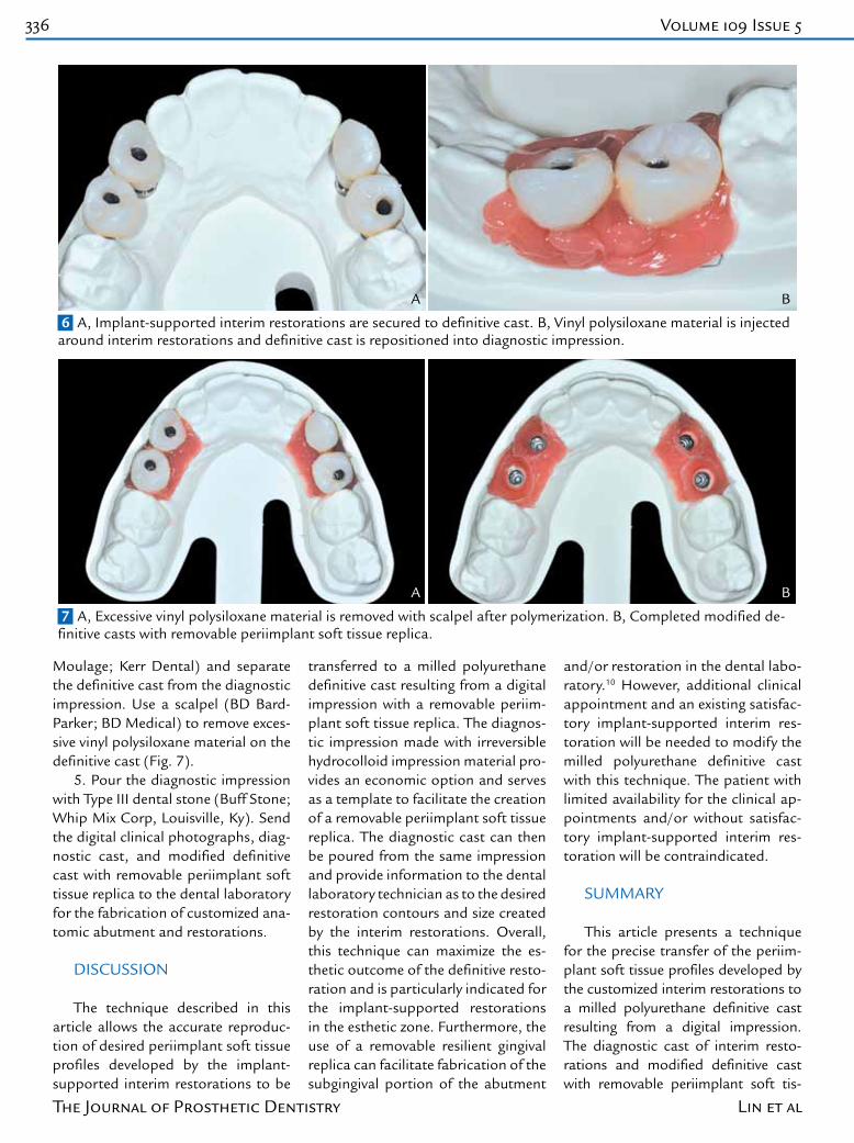
2. Remove the implant-supported interim restorations and secure them to the definitive cast with a torque
control device (Torque control device and Ratchet; Straumann AG) to 15 Ncm torque (Fig. 6A).
3. Inject vinyl polysiloxane mate-rial (Softissue Moulage; Kerr Dental) with a disposable curved utility syringe (Henry Schein, Melville, NY) around
the interim restorations on the defini-tive cast (Fig. 6B) and reposition the definitive cast into the adjusted diag-nostic impression. Ensure the com-plete seating of the definitive cast.
4. Allow polymerization of the vi-nyl polysiloxane material (Softissue
3 Digital impression acquired with intraoral scanner.
5 A, Diagnostic impression is made with implant-supported interim restorations in situ and adjustments are made to accommodate definitive cast. B, Definitive cast and diagnostic impression assembly.
4 A, Milled polyurethane definitive cast received from manufacturer’s facility. Periimplant soft tissue profiles are not evident. B, Periimplant areas of cast adjusted for subsequent soft tissue replica fabrication. Areas above removable implant analogs should be completely removed.
A
A
B
B
336 Volume 109 Issue 5
The Journal of Prosthetic Dentistry Lin et al
Moulage; Kerr Dental) and separate the definitive cast from the diagnostic impression. Use a scalpel (BD Bard-Parker; BD Medical) to remove exces-sive vinyl polysiloxane material on the definitive cast (Fig. 7).
5. Pour the diagnostic impression with Type III dental stone (Buff Stone; Whip Mix Corp, Louisville, Ky). Send the digital clinical photographs, diag-nostic cast, and modified definitive cast with removable periimplant soft tissue replica to the dental laboratory for the fabrication of customized ana-tomic abutment and restorations.
DISCUSSION
The technique described in this article allows the accurate reproduc-tion of desired periimplant soft tissue profiles developed by the implant-supported interim restorations to be
transferred to a milled polyurethane definitive cast resulting from a digital impression with a removable periim-plant soft tissue replica. The diagnos-tic impression made with irreversible hydrocolloid impression material pro-vides an economic option and serves as a template to facilitate the creation of a removable periimplant soft tissue replica. The diagnostic cast can then be poured from the same impression and provide information to the dental laboratory technician as to the desired restoration contours and size created by the interim restorations. Overall, this technique can maximize the es-thetic outcome of the definitive resto-ration and is particularly indicated for the implant-supported restorations in the esthetic zone. Furthermore, the use of a removable resilient gingival replica can facilitate fabrication of the subgingival portion of the abutment
and/or restoration in the dental labo-ratory.10 However, additional clinical appointment and an existing satisfac-tory implant-supported interim res-toration will be needed to modify the milled polyurethane definitive cast with this technique. The patient with limited availability for the clinical ap-pointments and/or without satisfac-tory implant-supported interim res-toration will be contraindicated.
SUMMARY
This article presents a technique for the precise transfer of the periim-plant soft tissue profiles developed by the customized interim restorations to a milled polyurethane definitive cast resulting from a digital impression. The diagnostic cast of interim resto-rations and modified definitive cast with removable periimplant soft tis-
6 A, Implant-supported interim restorations are secured to definitive cast. B, Vinyl polysiloxane material is injected around interim restorations and definitive cast is repositioned into diagnostic impression.
7 A, Excessive vinyl polysiloxane material is removed with scalpel after polymerization. B, Completed modified de-finitive casts with removable periimplant soft tissue replica.
A
A
B
B

337May 2013
sue replica can be used by the dental laboratory technician to achieve a pre-dictable esthetic outcome for implant-supported definitive restorations.
REFERENCES
1. Patel N. Integrating three-dimensional digital technologies for comprehensive implant den-tistry. J Am Dent Assoc 2010;141:20S-4S.
2. Henkel GL. A comparison of fixed prosthe-ses generated from conventional vs digitally scanned dental impressions. Compend Contin Educ Dent 2007;28:422-31.
3. Lee SJ, Gallucci GO. Digital vs. conventional implant impressions: efficiency outcomes. Clin Oral Implants Res 2013;24:111-5.
4. Chee WW, Donovan T. Use of provisional restorations to enhance soft-tissue con-tours for implant restorations. Compend Contin Educ Dent 1998;19:481-9.
5. Rodriguez AM, Rosenstiel SF. Esthetic considerations related to bone and soft tis-sue maintenance and development around dental implants: report of the Committee on Research in Fixed Prosthodontics of the American Academy of Fixed Prosthodon-tics. J Prosthet Dent 2012;108:259-67.
6. Santosa RE. Provisional restoration op-tions in implant dentistry. Aust Dent J 2007;52:234-42.
7. Polack MA. Simple method of fabricat-ing an impression coping to reproduce peri-implant gingiva on the master cast. J Prosthet Dent 2002;88:221-3.
8. Schoenbaum TR, Han TJ. Direct custom implant impression copings for the preser-vation of the pontic receptor site architec-ture. J Prosthet Dent 2012;107:203-6.
9. Azer SS. A simplified technique for creating a customized gingival emergence profile for implant-supported crowns. J Prosthodont 2010;19:497-501.
10.Orenstein IH, Petrazzuolo V, Gorczyca P, Chun JH. Use of transparent polyvinylsilox-ane to replicate gingival peri-implant soft tissue. J Prosthet Dent 2003;90:410-2.
Corresponding author:Dr Wei-Shao LinDepartment of Oral Health and RehabilitationUniversity of Louisville, School of Dentistry501 S. Preston Street, Louisville, KY 40202Fax: 502-852-1317E-mail: [email protected]
AcknowledgementsThe authors thank Stephanie Tinsley, CDT, and Roy Dental Laboratory, New Albany, IN for assistance in this study.
Copyright © 2013 by the Editorial Council for The Journal of Prosthetic Dentistry.
Access to The Journal of Prosthetic Dentistry Online is reserved for print subscribers!
Full-text access to The Journal of Prosthetic Dentistry Online is available for all print subscribers. To activate your individu-al online subscription, please visit The Journal of Prosthetic Dentistry Online. Point your browser to http://www.journals.elsevierhealth.com/periodicals/ympr/home, follow the prompts to activate online access here, and follow the instruc-tions. To activate your account, you will need your subscriber account number, which you can find on your mailing label (note: the number of digits in your subscriber account number varies from 6 to 10). See the example below in which the subscriber account number has been circled.
Personal subscriptions to The Journal of Prosthetic Dentistry Online are for individual use only and may not be trans-ferred. Use of The Journal of Prosthetic Dentistry Online is subject to agreement to the terms and conditions as indicated online.
This is your subscription account number
Sample mailing label
1 V97-3 J010 12345678-9J. H. DOE531 MAIN STCENTER CITY, NY 10001-001
*********AUTO**SCH 3-DIGIT 001
Lin et al


![A Technique to Improve an Ailing Interim Implant-Supported ... · assess proposed occlusal schemes before definitive restoration is . placed [9]. Provisional restorations designs](https://img.pdfslide.us/doc/110x75/5f85d815ef129373f346690c/a-technique-to-improve-an-ailing-interim-implant-supported-assess-proposed-occlusal.jpg)


![Newsletter 10 October 2019 - Dental Implant CE & Training ......Oct 08, 2019 · 1] Tarica DY, Survey of United States dental schools on cementation protocols for implant crown restorations](https://img.pdfslide.us/doc/110x75/6003d9df670b8e5e18015533/newsletter-10-october-2019-dental-implant-ce-training-oct-08-2019.jpg)


![Metal-free implant-supported restorations in the ... · screw-retained implant-supported res-torations has become very popular in recent years [1, 10]. Implantological concepts such](https://img.pdfslide.us/doc/110x75/5f8954cfeb23b875ef263f7d/metal-free-implant-supported-restorations-in-the-screw-retained-implant-supported.jpg)