Embed Size (px)
Citation preview

Governors State UniversityOPUS Open Portal to University Scholarship
All Capstone Projects Student Capstone Projects
Spring 2015
Use Of Functional Manual Therapy and aStrengthening Program in the Treatment of a41-Year-Old Female with Low Back and SciaticaPain: A Case ReportJustin JonesGovernors State University
Follow this and additional works at: http://opus.govst.edu/capstones
Part of the Analytical, Diagnostic and Therapeutic Techniques and Equipment Commons, andthe Physical Therapy Commons
For more information about the academic degree, extended learning, and certificate programs of Governors State University, go tohttp://www.govst.edu/Academics/Degree_Programs_and_Certifications/
Visit the Governors State Physical Therapy DepartmentThis Project Summary is brought to you for free and open access by the Student Capstone Projects at OPUS Open Portal to University Scholarship. Ithas been accepted for inclusion in All Capstone Projects by an authorized administrator of OPUS Open Portal to University Scholarship. For moreinformation, please contact [email protected].
Recommended CitationJones, Justin, "Use Of Functional Manual Therapy and a Strengthening Program in the Treatment of a 41-Year-Old Female with LowBack and Sciatica Pain: A Case Report" (2015). All Capstone Projects. 103.http://opus.govst.edu/capstones/103
USE OF FUNCTIONAL MANUAL THERAPY AND A STRENGTHENING
PROGRAM IN THE TREATMENT OF A 41-YEAR-OLD FEMALE WITH
LOW BACK AND SCIATICA PAIN: A CASE REPORT
By
Justin Jones
B.S., Olivet Nazarene University, 2012
CAPSTONE PROJECT
Submitted in partial fulfillment of the requirements
For the Degree of Doctor of Physical Therapy
Governors State University
University Park, IL 60484
2015

i
ABSTRACT
Background and Purpose: Although low back pain is a common diagnosis
treated in physical therapy clinics, there is disagreement in the literature as
to the preferred interventions for this patient population. The purpose of this
case report is to describe the outcome of a patient with acute onset of low
back and sciatica pain with a treatment directed towards Functional Manual
Therapy and a strengthening program based on initial examination findings.
Case Description: The patient was a 41 year-old female secretary with four
day history of low back and sciatica pain that initiated after straining during
a bowel movement. The client presented with pain, decreased ROM,
decreased strength, and functional disability. Intervention was directed by
initial examination findings and consisted of components including Functional
Manual Therapy and a strengthening program.
Outcomes: All of the patient’s impairments improved and she was able to
return to work at the beginning of the final week of treatment without any
functional difficulties.
Discussion: Use of Functional Manual Therapy and a strengthening program
may result in positive outcomes related to pain and functional disability in
patients with acute onset of low back and sciatica pain.

Jones 1
BACKGROUND AND PURPOSE
Previous research demonstrates that approximately 60-80% of the
Western world’s population will experience low back pain (LBP) at some
point in their lives.1 In addition, nearly 50% of all patients presenting to
outpatient physical therapy clinics present with LBP of some kind.2 Despite
the great number of LBP cases treated by physical therapists every year,
there is still controversy as to which treatments are most effective for this
patient population.1
This controversy is compounded by the lack of consensus found in the
literature. In a randomized, controlled trial comparing the effectiveness of
manual therapy to exercise therapy in patients with chronic LBP, Aure et al
notes that although a number of conservative treatment methods for LBP
have been studied, wide disagreement still remains as to the preferred
treatment.1 Cherkin et al also reports that although there are many non-
surgical treatments for LBP, there is little evidence that any are effective.3 In
addition, Fritz et al has concluded that although a variety of interventions
are accepted as standard care for patients with LBP, there is a lack of high-
quality evidence from randomized clinical trials that offers conclusive support
for most interventions.2
The debate as to which interventions are most successful in LBP
patients is made even more unclear by studies that have found success with

Jones 2
treatments in this patient population. Petersen et al concluded that the
McKenzie Method and intensive dynamic strengthening training seem to be
equally effective in the treatment of patients with LBP, while Unlu et al found
traction, ultrasound, and low-power laser therapies to all be effective in a
group of patients with acute lumbar disc herniation.4, 5
Despite the abundance of research regarding conservative
management of LBP, the evidence remains inconsistent and inconclusive.6 A
possible explanation for the insufficient evidence for commonly accepted
interventions involves study designs with broad inclusion criteria, resulting in
diverse samples.7 It is also possible that research attempting to identify the
best interventions for patients with LBP does not take into account a
common belief amongst clinicians: that it is unreasonable to expect all
patients with LBP to respond to any single treatment approach.8 This school
of thought has prompted researchers to investigate methods to place
patients into groups to be matched to interventions that will produce positive
outcomes.9
In an attempt to rectify the discrepancy noted in the research, Delitto
et al presented a treatment-based classification approach to the
conservative management of LBP.10 This study was one of the first to give
physical therapists a working framework to classify patients with LBP into
different categories based on evaluation findings in order to direct
treatment. Three categories were proposed: (1) patients in the acute phase

Jones 3
with the goal of symptom relief, (2) patients in the subacute phase where
symptom relief and a quick return to function are the focus, and (3) patients
who must return to participation in activities that are highly physically
demanding. Furthermore, once the phase of a patient’s condition was
determined, patients were then placed in treatment categories based on
evaluative data. These treatment categories included manipulation,
stabilization, specific exercise, and traction. Delitto et al concludes that the
classification of patients into different categories and matching treatments to
those patients that fall into a certain treatment category will result in faster,
more efficient, and more cost-effective care.10
A common impairment that often accompanies LBP is that of
lumbosacral radicular syndrome, also called sciatica.11 Characteristics of
sciatica include radiating pain in the lower extremities with related
disabilities.12 Sciatica can often be accompanied by nerve root tension or
neurological deficits. Sciatica is frequently caused by spinal stenosis, tumors,
and/or radiculitis, but caused by herniated disc with nerve root compression
in 90% of cases.11, 12
A consensus between research and clinical practice determined the
management of sciatica should be conservative in the first 6-8 weeks of
onset.12 In fact, most cases of acute sciatica have displayed a favorable
prognosis with resolution of symptoms in two weeks. However, up to 20-
30% of patients with sciatica have been shown to have pain for one year or

Jones 4
longer. Despite a generally favorable prognosis with this condition, it is still
unclear in the literature as to what conservative management of sciatica
should consist of.12 In a systematic review of conservative treatments of
sciatica, Luijsterburg et al evaluated injections, traction, physical therapy,
bed rest, manipulation, medication, and acupuncture as treatments for
sciatica.13 After examining the evidence, the researchers concluded there
was no conclusive data indicating one type of conservative treatment was
superior to the others.13
Given the disparity found in the literature there is a need for continued
research investigating preferred treatments for patients presenting to
physical therapy with LBP and also those presenting with symptoms of
sciatica. Continued research to contribute to the literature on categorizing
LBP patients in order to direct treatment will only lead to more positive
outcomes. Thus, the purpose of this case report is to describe the outcome
of a patient referred to physical therapy with acute onset of low back and
sciatica pain with a treatment directed towards functional manual therapy
and a strengthening program based on initial examination findings.

Jones 5
CASE DESCRIPTION
Subject
LD was a 41 year-old Caucasian female employed as a secretary with
a four day history of severe LBP with painful symptoms radiating down her
left buttock and posterior thigh to just above the knee. LD was referred to
physical therapy with a medical diagnosis of lumbar radiculopathy. The
patient reported she was using the restroom and “pushed too hard” during a
bowel movement. This resulted in immediate sharp pain in her lower back,
left buttock, and posterior thigh region. LD reported experiencing almost
constant pain that was interfering with her ability to perform daily tasks such
as sitting, bending forward, lifting, standing, walking, going to the bathroom,
and sleeping. The client was able to get some relief from lying in supine and
applying ice to her low back region. LD reported her pain was intensified
during sitting and forward bending.
Relevant medical history included a diagnoses of herniated discs at L3-
L4 discovered after LD received an MRI after experiencing mild LBP over one
year prior to the current episode. The symptoms from the previous episode
resolved without requiring the patient to seek any physical therapy
treatment. Ibuprofen was the only medication the client was taking during
the current onset of LBP. LD reported she hoped physical therapy would help
her to be able to return to living without constantly being in pain. The client

Jones 6
had taken the previous three days off of work due to inability to sit for long
periods of time and hoped to return as soon as possible.
Systems Review
Through a combination of the subjective examination and the primary
physician’s report, the client’s integumentary and cardiovascular systems
were found to be unimpaired.
Impairments were noted in LD’s musculoskeletal system including
elevated left shoulder, rounded shoulder posture, anterior pelvic shear, a
shift of the upper thorax to the right, and a leg length discrepancy noted in
supine with left leg found to be shorter than right leg. Tenderness was
detected in the patient’s bilateral lumbar paraspinal muscles, left posterior
superior iliac spine (PSIS), bilateral sacroiliac (SI) joints, coccyx, and
bilateral ischial tuberosities. Hypomobility was noted in the client’s lumbar
spine, SI joints, and sacrococcygeal symphysis through manual spring
testing. LD ambulated with an antalgic gait displaying decreased weight-
bearing of the left lower extremity, decreased bilateral lower extremity
push-off, and decreased trunk reciprocation. LD also presented with
decreased range of motion of lumbar spine and left hip with decreased
strength noted of core and bilateral hip musculature.

Jones 7
Neuromuscular impairments included radicular symptoms into the
patient’s left buttock and posterior thigh intensified with lumbar flexion,
lumbar extension, sitting, lifting, walking, and standing.
Clinical Impression #1
Based upon data from the subjective examination and systems review,
relevant tests and measures were selected to attain a more complete
understanding of LD’s clinical picture. Due to the client’s report of intense
pain, the Numerical Pain Rating Scale (NPRS) was selected to obtain a
baseline pain level.
Decreased active range of motion (AROM) was assessed through
standard goniometric measurement to assess baseline lumbar spine and
bilateral hip AROM. Passive range of motion (PROM) was not taken in this
case based on the treating clinician’s clinical judgment that AROM
measurements would be a better measure of function than PROM
measurements.
Decreased muscle strength was assessed through manual muscle
testing to find baseline data on the strength of LD’s core and hip
musculature.
Special tests were selected to objectify ROM limitations and identify
the source of radicular symptoms. The Straight Leg Raising Test and Thomas
Test were used to ascertain measurements of hamstring flexibility and hip

Jones 8
flexor flexibility, respectively. The Extension-Rotation Test was used to
identify zygapophyseal joint pain, and the Slump Test was used to assist in
the identification of lower extremity radicular symptom patterns.
LD was also given the Oswestry Disability Index (ODI) to fill out in
order to attain an objective measure of the degree of disability her LBP was
causing at initial examination.
Tests and Measures
Numerical Pain Rating Scale
The first test and measure used in this case was the Numerical Pain
Rating Scale (NPRS) in order to give an objective measure of LD’s pain. The
client was asked to rate her current pain level, best pain level, and worst
pain level since the onset of the episode on a 0-10 scale. A score of 0
indicates the subject was in no pain and a score of 10 indicates the subject
is in need of emergency medical attention. Williamson and Hoggart report
the NPRS is valid, reliable, and appropriate for use in clinical practice.14 For
general purposes, the NPRS has good sensitivity and generates data that can
be statistically analyzed for audit purposes.14 LD’s NPRS ratings at initial
evaluation can be found in Table 3 below.
Range of Motion
The positions used for measuring active range of motion (AROM) in
this case are as described in Reese and Bandy.15 The AROM of LD’s lumbar

Jones 9
spine and bilateral hips were measured in this case with decreased ROM
noted in lumbar spine and left hip ROM. Nussbaumer et al described
goniometric measurement of hip ROM to have good concurrent validity for
hip abduction and internal rotation with intraclass correlation coefficients
(ICC) of 0.94 and 0.88, respectively.16 Test-retest reliability was found to be
good with ICCs above 0.90 in all planes, except for hip adduction (0.82-
0.84). Fitzgerald et al found standard goniometry of thoracolumbar
extension and lateral flexion to be reliable.17 Specific goniometric
measurements taken at initial evaluation can be found in Table 3 below.
Muscle Strength
Muscle strength was measured through manual muscle testing (MMT)
with techniques as described in Hislop and Montgomery.18 MMT was
performed on LD’s core and hip musculature with strength deficits noted in
core and bilateral hip muscles. Fan et al found MMT to have excellent inter-
rater reliability in trained examiners and to be a reliable method of
comprehensively assessing muscle strength.19 Results from MMT at initial
examination can be found in Table 3 below.
Special Tests
In order to attain a more complete clinical picture, special orthopedic
tests were used to identify impairments in muscle length and get a better
idea of the nature of the subject’s symptoms. Special test positions and

Jones 10
procedures are as described in Cook and Hegedus.20 The Thomas Test was
used as a test of muscle length in this case, while the Slump Test and
Extension-Rotation Test were used to identify the nature of the LD’s lumbar
radiculopathy. The Straight Leg Raising (SLR) Test was used as both a
muscle length test and a test for lumbar radiculopathy. Results from special
testing can be found in Table 3 below. Psychometric data for special tests
used in this case can be found in Table 1 below:
Table 1: Special Test Psychometric Data
(NR=Not reported; NA= Not applicable)
Test Reliability Sensitivity Specificity LR+ LR-
Thomas Test20 NR NR NR NA NA
SLR Test21 NR .52 .89 4.72 0.53
Slump Test21 NR .84 .83 4.94 0.19
Ext.-Rot. Test20 NR 1.00 .22 1.28 0.00
Oswestry Disability Index
The Oswestry Disability Index (ODI) was used to quantify LD’s LBP and
how her pain was restricting her function. The ODI is one of the most
commonly recommended condition specific outcome measures for spinal
disorders used to track patient progress.22 This questionnaire asks the
patient to rate his/her disability on 10 function-related topics on a 0-5 scale
for each topic. A score is determined as a percentage with 0% meaning no

Jones 11
disability and 100% indicating maximal disability.22 LD’s ODI score at initial
examination can be seen in Table 3 below. Psychometric data on the ODI
can be found in Table 2 below:
Table 2: ODI Psychometric Data
Minimally Clinical Important
Difference23
Minimal Detectable Change23
Test-Retest Reliability24
Criterion Validity23
Construct Validity24
12.8
11.67
Excellent (ICC=0.97; 95% CI)
r=0.11 rho=0.35 rho=0.46
(r = 0.607, p < 0.001); (r = 0.56, p < 0.001)
Table 3: Initial Examination Tests and Measures
Pain (NPRS)
(0-10)
AROM
(degrees)
MMT
(0-5)
Special Tests ODI
Pain at Initial
Exam: 8/10
Worst Pain
Since Onset:
8/10
Best Pain Since
Onset: 5/10
Location: Low
back, (L)
buttock, and (L)
posterior thigh
Lumbar-
Flexion: 70
Extension: 10
Side Bending: 20
Hips-
(L) Hip-
Flexion: 100
External Rotation: 45
Internal Rotation: 5
Extension: 0
(R) Hip-
Flexion: 120
Extension: 10
Internal rotation: 25
All Other Planes:
WNL
Upper Abs: 3+/5
Low Abs: 3-/5
Back Ext: 3+/5
Bilateral Hips-
Flexion: 3+/5
Abduction: 4/5
Adduction: 4/5
External Rotation: 4/5
Internal Rotation: 3+/5
Extension: 3+/5
SLR:
(L) 45 degrees
(R) 80 degrees
(+) (L) for lumbar
radiculopathy
Thomas Test:
(+) bilaterally
Extension-
Rotation Test:
(+) (L)
Slump Test:
(+) for lumbar
radiculopathy
68%
(Crippling
Back Pain)

Jones 12
Clinical Impression #2
The initial evaluation showed the patient to be in severe pain, with
limited ROM in the lumbar spine and bilateral hips.14, 15 LD was also found to
have weakness of core and bilateral hip musculature.18 Positive findings
were found for the Thomas Test, SLR Test, Extension-Rotation Test, and
Slump Test indicating decreased muscle length and signs of lumbar
radiculopathy.20 The client’s ODI score indicated she was experiencing back
pain that could be considered crippling in severity.22
Due to objective findings from the initial examination, LD’s plan of care
included a variety of interventions designed to reduce pain, increase ROM of
the lumbar spine and bilateral hips, strengthen core and hip musculature,
decrease lower extremity radicular symptoms, and improve the patient’s
ability to perform daily tasks as measured by patient report and the ODI.
LD’s symptoms were found to be intensified with lumbar flexion and
lumbar extension movements. A directional preference is defined as a
situation in which movement in one direction improves pain and limitation of
ROM, and movement in the opposite direction causes signs and symptoms to
worsen.2 Since there was no particular movement that brought on an
improvement in pain and ROM, a directional preference could not be
identified in this case.

Jones 13
Client outcomes were determined through a physical therapy re-
evaluation that was taken three weeks after the initial evaluation. All tests
and measures performed at the initial evaluation were tested again at the
re-evaluation with the most important measures being those related to pain
and patient ability to perform daily tasks.
PT Diagnosis
After the initial evaluation, LD’s condition was found to be best
categorized into Preferred Physical Therapist Practice Pattern 4F: Impaired
Joint Mobility, Motor Function, Muscle Performance, Range of Motion, and
Reflex Integrity Associated with Spinal Disorders.25
Prognosis
Past research has found the prognosis for patients with acute LBP to
be generally good. Aure et al concluded that clients with LBP who seek
treatment in the acute stages enjoy a favorable prognosis with 80%-90% of
patients improving considerably within six to eight weeks.1 In a systematic
review, Pengel et al found most patients with acute LBP to have rapid
improvements in pain and disability within one month with a return to work
within that same one month period.26 However, it is not uncommon for low
levels of persisting pain and disability to persist from three to at least 12
months.26
Jones 14
In this case, LD was determined to have a good prognosis based on
the literature and past clinical experience of the treating clinician. LD was
expected to display decreased symptoms and improved function within four
to eight weeks.
Plan of Care
LD’s plan of care was designed to include evidence-based interventions
to improve deficits noted in the initial evaluation and improve functional
limitations. The treating clinicians planned to employ a variety of
interventions and use patient response to guide treatment. For example, if
the client reported a certain manual technique provided pain relief, the
treating clinician would make this intervention a regular part of the patient’s
plan of care.
Based on objective findings, past clinical experience, and support in
the literature, a variety of interventions were included in the plan for this
case including: AROM, strengthening, stretching, stabilization activities,
patient education, joint mobilization, therapeutic exercise, functional
activities, manual therapy, neuromuscular re-education, gait training,
cardiovascular exercise, modalities, and a home exercise program (HEP).
The patient planned to attend physical therapy treatment sessions of one to
two hours duration three times per week for at least three weeks.

Jones 15
Goals
Physical therapy goals for this case were as follows:
Short-Term (2 weeks):
1. Client will learn HEP and perform HEP independently.
2. Client will restore functional ROM and mobility in lumbar, sacrum, and
coccyx area.
3. Client will restore functional sitting postural control with no symptoms.
Long-Term (4 weeks):
1. Client will restore core and leg muscle strength to at least 4+/5.
2. Client will restore functional standing postural control.
3. Client will restore functional gait pattern.
4. Client will be able to perform all daily activities including: transferring,
sitting, standing, lifting, and sleeping at night with no symptoms.
Interventions
In accordance with normal protocol at the outpatient clinic at which LD
received her physical therapy treatment, the patient received treatment
from one physical therapist, one physical therapist assistant, and one
student physical therapist over the course of her eight PT visits. The physical
therapist involved in this case was extensively trained in Functional Manual
Therapy (FMT) techniques.27 FMT is described as an integrated treatment

Jones 16
system which couples mechanical treatment of the joints, soft tissues,
visceral, and neurovascular systems with manual neuromuscular facilitation
to enhance optimum motor control and human function. The Institute of
Physical Art offers a variety of continuing education courses, certifications,
and fellowship programs for clinicians to gain competency in FMT.27
In this case, FMT techniques were applied to LD’s lumbar spine, sacral,
and coccygeal region in order to decrease pain noted upon palpation,
improve joint hypomobility detected with spring testing, and lead to an
improvement of poor movement patterns found upon observation of the
patient. FMT was typically used near the beginning of a treatment session to
decrease pain and allow the subject to perform more functional interventions
to the best of her ability.
An example of an FMT technique utilized in this case includes a hold-
relax proprioceptive neuromuscular facilitation technique designed to
increase mobility of the bilateral sacroiliac joints and decrease pain in the
region. The subject was placed in prone while the therapist used one hand to
apply pressure to block the mobile sacroiliac joint segment. The therapist’s
other hand is used to provide resistance to the patient’s ankle joint with the
patient’s knee bent to 90 degrees to employ the hold-relax portion of the
technique. The patient’s lower extremity is moved through different hip
internal/external ranges of motion as the client is instructed to resist the
therapist’s manual force at different points. By using manual force to block

Jones 17
the mobile SI joint, the PT hoped to improve joint mobility of the SI joint
lacking mobility and reduce the patient’s pain.
Other manual techniques utilized during the client’s plan of care
included soft tissue mobilization (STM) in order to relieve symptoms through
the breaking up of soft tissue restrictions and improve movement patterns.
STM was performed to this subject’s lumbar paraspinal and gluteal
musculature to improve range of motion, relieve symptoms, and break up
any soft tissue restrictions to facilitate full participation in activities.
An STM technique utilized in this case involved application of STM to
the sciatic nerve as it passes through the gluteal region. With the patient in
side-lying, STM was applied to the sciatic nerve as the patient performed an
active-assisted straight leg raise. The patient was instructed to repeatedly
raise and lower the leg as the therapist provided STM to the sciatic nerve in
a longitudinal manner. The performance of this technique is as described in
Cleland et al.28 The other STM techniques utilized in this case that were
applied to the lumbar and gluteal regions are as described in Kisner and
Colby.29
Many of LD’s treatment sessions began with a 10 minute warm-up
period on the NuStep T4 recumbent cross trainer in order to increase blood
flow to lower extremity musculature and incorporate cardiovascular exercise
into the patient’s program. NuStep cross trainers are manufactured by the

Jones 18
NuStep Corporation out of Ann Arbor, Michigan. LD used the Nu-Step for the
first time during her third visit and reported discomfort after two minutes,
thus the intervention was discontinued at that session. LD was able to
complete the full 10 minutes on the Nu-Step during treatments 4-8.
Stretching of the patient’s bilateral hip flexors, hamstrings, quadriceps,
hip internal/external rotators, and low back musculature was included using
manual, passive, active, and active-assisted methods. An example of a
stretching exercise utilized in this case is that of prone press-ups in order to
improve lumbar extension ROM. Prone press-ups were only utilized after
lumbar extension was found to not provoke painful symptoms. Stretching to
increase range of motion was included in every treatment session and
included in the client’s home exercise program using methods as described
in Kisner and Colby.29 Stretching activities were typically utilized after the
client completed a warm-up session on the Nu-Step machine and typically 5
repetitions of 15-20 seconds were completed for each stretch.
Strengthening of core stabilizers, low back musculature, and hip
musculature was included in every treatment session and included in the
client’s home exercise program. Strengthening exercises varied and began
with basic table exercises near the beginning of treatment that progressed to
more functional activities in standing as the patient progressed. For
example, early in treatment, the client would perform the side-lying
clamshell exercise with an exercise band around her knees to provide
Jones 19
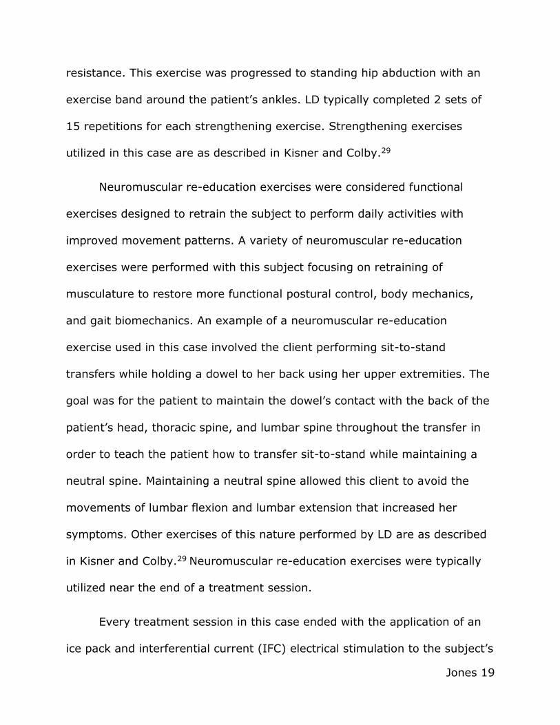
resistance. This exercise was progressed to standing hip abduction with an
exercise band around the patient’s ankles. LD typically completed 2 sets of
15 repetitions for each strengthening exercise. Strengthening exercises
utilized in this case are as described in Kisner and Colby.29
Neuromuscular re-education exercises were considered functional
exercises designed to retrain the subject to perform daily activities with
improved movement patterns. A variety of neuromuscular re-education
exercises were performed with this subject focusing on retraining of
musculature to restore more functional postural control, body mechanics,
and gait biomechanics. An example of a neuromuscular re-education
exercise used in this case involved the client performing sit-to-stand
transfers while holding a dowel to her back using her upper extremities. The
goal was for the patient to maintain the dowel’s contact with the back of the
patient’s head, thoracic spine, and lumbar spine throughout the transfer in
order to teach the patient how to transfer sit-to-stand while maintaining a
neutral spine. Maintaining a neutral spine allowed this client to avoid the
movements of lumbar flexion and lumbar extension that increased her
symptoms. Other exercises of this nature performed by LD are as described
in Kisner and Colby.29 Neuromuscular re-education exercises were typically
utilized near the end of a treatment session.
Every treatment session in this case ended with the application of an
ice pack and interferential current (IFC) electrical stimulation to the subject’s

Jones 20
lower back region for 15 minutes in order to provide LD with further pain
relief.30
Patient education was provided throughout each physical therapy
session. Education topics included disc herniation pathology, postural
education, proper body mechanics training, gait training, and HEP
instruction. The client’s HEP was added to as LD progressed. Appropriate
exercises performed during therapy sessions were often added to the
patient’s HEP throughout the course of treatment. Exercises included in LD’s
HEP included stretching exercises of the lower back/hips, core/hip/lower
back strengthening exercises, and neuromuscular re-education exercises.
Any questions the patient had were answered in full to provide the best
comprehensive care possible. Types of interventions employed in a particular
session can be seen in Table 4 below:

Jones 21
Table 4: Interventions- (Recorded per session)
1=patient education 5=STM 2=stretching 6=neuromuscular re-education 3=strengthening 7=Ice/Electrical stimulation (IFC) 4=FMT
OUTCOMES
After attending eight total physical therapy sessions over three weeks,
LD was re-evaluated with all the tests and measures used at the initial
evaluation. Observation found the patient to have improved sitting and
standing postural control, no leg length discrepancy in supine, and no
tenderness noted in the client’s lumbar and pelvic regions. Improved
mobility of LD’s lumbar spine, SI joints, and sacrococcygeal symphysis was
found upon spring testing. The patient’s gait pattern was found to be
Session
#
Intervention
1
Intervention
2
Intervention
3
Intervention
4
Intervention
5
Intervention
6
Intervention
7
1 1 2 3 4 7
2 1 2 3 4 5 6 7
3 1 2 3 4 6 6 7
4 1 2 3 4 6 7
5 1 2 3 5 6 7
6 1 2 3 4 5 6 7
7 1 2 3 5 6 7
8 1 2 3 6 7

Jones 22
improved with equal weight distribution, improved bilateral lower extremity
push-off, and improved trunk reciprocation.
Subjectively, LD reported no LBP, no radicular symptoms, and no
problems with daily tasks. LD reported she was able to return to work at the
beginning of the third week of treatment without any difficulty. In addition,
improved range of motion of the lumbar spine and the patient’s left hip was
noted with improvements in strength of core and hip musculature. All special
tests were found to be negative and the client showed improvement on her
ODI outcome measure.
The client was discharged from physical therapy treatment after re-
evaluation due to the completion of all functional therapy goals,
improvement of Oswestry Disability Index score, no reported difficulty with
any daily activities, and reported relief of all symptoms. The client was given
a home exercise program to continue to follow upon discharge. Specific
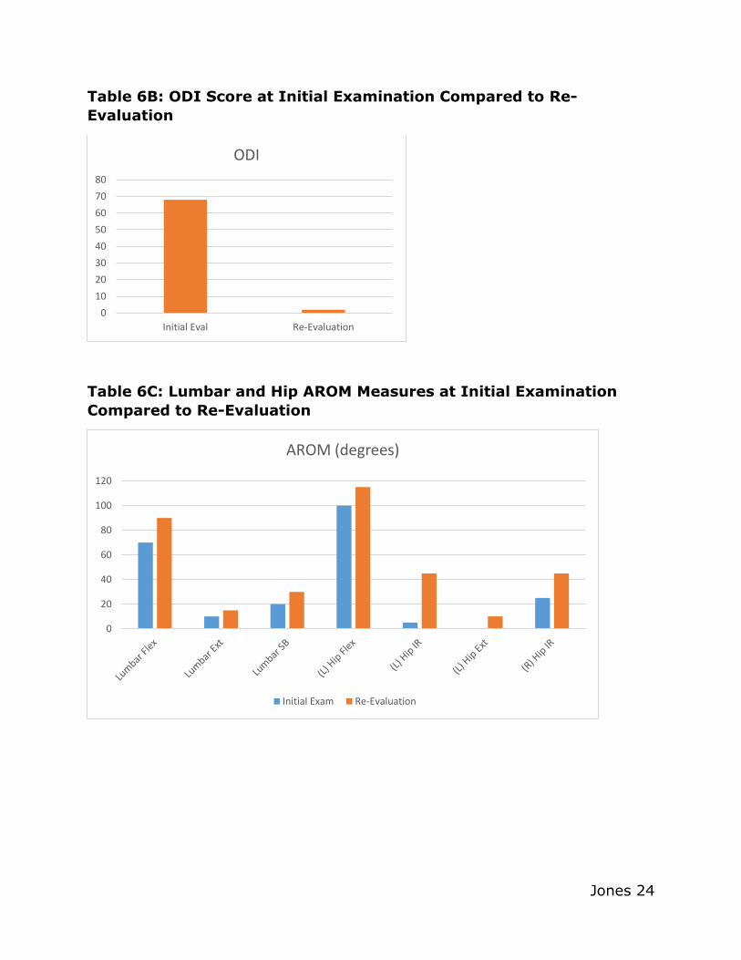
measurements taken at re-evaluation can be found in Table 5 below. A
comparison of measures taken at the initial examination and re-evaluation
can be found in Tables 6A-6D.

Jones 23
Table 5: Re-Evaluation Tests and Measures
Pain (NPRS)
(0-10)
AROM
(degrees)
MMT
(0-5)
Special Tests ODI
Pain at Re-
evaluation: 0/10
Worst Pain in
Previous Week:
0/10
Best Pain in
Previous Week:
0/10
Lumbar-
Flexion: 90
Extension: 15
Side Bending: 30
Hips-
(L) Hip-
Flexion: 115
External Rotation: 45
Internal Rotation: 45
Extension: 10
(R) Hip-
Flexion: 120
Extension: 10
Internal rotation: 45
All Other Planes:
WNL
Upper Abs: 4/5
Low Abs: 4-/5
Back Ext: 4/5
Bilateral Hips-
Flexion: 4+/5
Abduction: 4+/5
Adduction: 5/5
External Rotation: 4+/5
Internal Rotation: 4+/5
Extension: 4+/5
SLR:
(L) 73 degrees
(R) 80 degrees
Thomas Test:
(-) bilaterally
Extension-
Rotation Test: (-)
Slump Test: (-)
2%
(Minimal
Disability)
Table 6A: Pain Level at Initial Examination Compared to Re-
Evaluation as Measured by NPRS
0
1
2
3
4
5
6
7
8
9
Initial Exam Re-Evaluation
Pain (0-10)
Jones 24
Table 6B: ODI Score at Initial Examination Compared to Re-
Evaluation
Table 6C: Lumbar and Hip AROM Measures at Initial Examination
Compared to Re-Evaluation
0
10
20
30
40
50
60
70
80
Initial Eval Re-Evaluation
ODI
0
20
40
60
80
100
120
AROM (degrees)
Initial Exam Re-Evaluation

Jones 25
Table 6D: MMT at Initial Examination Compared to Re-Evaluation
Area Tested Initial Examination Re-Evaluation
Upper Abs 3+/5 4/5
Lower Abs 3-/5 4-/5
Back Ext 3+/5 4/5
Hip Flexion 3+/5 4+/5
Hip Extension 4/5 4+/5
Hip Abduction 4/5 5/5
Hip Adduction 4/5 4+/5
Hip ER 3+/5 4+/5
Hip IR 3+/5 4+/5
DISCUSSION AND CONCLUSION
This case report demonstrated how Functional Manual Therapy and a
strengthening program can be utilized to treat a 41 year-old female with a
four day history of acute low back and sciatica pain. Although previous
studies found a variety of treatments to be effective in the treatment of
patients with acute LBP and sciatica symptoms, there is still a disparity in
the research as to which treatments are the most effective.1, 6 Due to this
disparity, researchers have found that the classification of patients into
treatment categories based on examination findings may help to direct
treatment and lead to more positive outcomes.2, 10 The positive outcomes
found in this case report could be helpful in adding to the current literature
on effective interventions for patients with similar diagnoses. In addition,

Jones 26
this case report may prove beneficial in the treatment of LBP clients based
on categorization of symptoms.
The categorization of patients to direct LBP treatment was first
described by Delitto et al and expanded upon by Fritz et al.2, 10 Based on the
work of Fritz et al, the symptoms displayed by LD would lead to this client
falling into the manipulation category of treatment.2 In the creation of LD’s
plan of care, the treating clinicians decided to incorporate interventions from
this category and assess patient response to these interventions.
FMT to the client’s lumbar and pelvic regions was chosen as the
intervention to address the manipulation categorization of LD’s treatment
with the purpose of decreasing pain, restoring proper joint mobility, and
improving movement patterns.27 Although the degree of effectiveness of
FMT on the outcomes of this case is unknown, LD repeatedly reported
decreased pain after the application of FMT. Utilization of these pain-
reducing manual techniques near the beginning of a treatment session may
have allowed LD to more fully participate in strengthening and
neuromuscular re-education interventions typically performed later in a
treatment session.
According to the Fear-Avoidance Model of Pain, a patient’s
interpretation of their acute pain may lead to avoidance behaviors that may,
in turn, lead to greater disability.31 Reduction of a client’s pain early in
Jones 27
treatment is essential in the facilitation of functional movement patterns
leading to more positive outcomes.31 The utilization of FMT in LD’s plan of
care proved effective in the reduction of the patient’s pain and may have led
to improved movement patterns leading to improvement after three weeks
of treatment.
In order to directly address the symptoms of sciatica displayed in this
case, a technique that involved STM to the sciatic nerve with a straight leg
raising component was utilized, as described previously. Past research
suggests that improving the range of SLR has a beneficial effect in restoring
normal movement and reducing the degree of impairment due to low back
dysfunction.32 On several occasions, LD reported decreased radicular
symptoms after the performance of this manual technique and was found to
have improved SLR range of motion of the left lower extremity at re-
evaluation compared to initial examination. Thus, this manual technique may
have been an important component in the relief of LD’s radicular symptoms.
Strengthening exercises were another large component of LD’s plan of
care. These exercises included various core stabilization and hip
strengthening exercises designed to address muscular weaknesses found in
the patient’s core and hip muscles. Although strength training has shown to
be no more effective than other interventions in the treatment of LBP,
strengthening exercises were made a priority in this case to improve
significant muscular weaknesses in LD’s hips leading to non-functional gait

Jones 28
biomechanics.4, 29 Previous studies have shown core and hip musculature
weaknesses to be a contributor to gait deformities and a cause of LBP.29, 33
Thus, strengthening of the hip and core musculature may have been an
important factor in LD’s rehabilitation.
Although a positive outcome was seen in LD’s case with the utilization
of FMT and a strengthening program, other factors and limitations in this
study may have contributed to the patient’s outcome. LD reported a
previous episode of acute LBP that had resolved without treatment. It is
unknown whether the current episode of LBP would have healed without
physical therapy intervention. In addition, this patient responded well to the
interventions selected in this case report, however, the client may have
responded better to another set of interventions. Finally, with the use of
several interventions in this case, it is uncertain which interventions may
have actually been effective and which interventions were ineffective.
This case report also identified several topics for future studies.
Although different aspects of the techniques of FMT are supported in the
literature, there have been no studies that have looked specifically at the
effects of FMT as a physical therapy intervention. Also, the use of FMT and a
strengthening program in this case led to a positive outcome, but future
studies are obviously needed to assess these interventions in larger
samples. Follow-ups at six and 12 months after discharge should also be
included to assess the long-term effectiveness of these interventions.

Jones 29
In conclusion, this case report described the successful treatment of a
41 year-old female with a four day history of acute low back and sciatica
pain utilizing FMT and a strengthening program to lead to a resolution of
symptoms in three weeks. The use of FMT and a strengthening program may
result in positive outcomes related to pain and functional disability in this
patient population.

Jones 30
REFERENCES
1. Aure OF, Nilsen JH, Vaseljen O. Manual therapy and exercise therapy
in patients with chronic low back pain: A randomized, controlled trial
with 1-year follow-up. Spine. 2003;28(6):525-532.
2. Fritz JM, Cleland JA, Childs JD. Subgrouping patients with low back
pain: Evolution of a classification approach to physical therapy. J
Orthop Sports Phys Ther. 2007;37(6):290-302.
3. Cherkin DC, Deyo RA, Battie M, Street J, Barlow W. A comparison of
physical therapy, chiropractic manipulation, and provision of an
educational booklet for the treatment of patients with low back pain.
The New England Journal of Medicine. 1998;339:1021-1029.
4. Petersen T, Kryger P, Ekdahl C, Olsen S, Jacobsen S. The effect of
McKenzie therapy as compared with that of intensive strengthening
training for the treatment of patients with subacute or chronic low
back pain: A randomized controlled trial. Spine. 2002;27(16):1702-
1708.
5. Unlu Z, Tasci S, Tarhan S, Pabuscu Y, Islak S. Comparison of 3
physical therapy modalities for acute pain in lumbar disc herniation
measured by clinical evaluation and magnetic resonance imaging.
Journal of Manipulative and Physiological Therapeutics.
2008;31(3):191-198.

Jones 31
6. Hayden JA, van Tulder MW, Tomlinson G. Systematic review:
strategies for using exercise therapy to improve outcomes in chronic
low back pain. Ann Intern Med. 2005;142:776-785.
7. Delitto A. Research in low back pain: time to stop seeking the elusive
“magic bullet”. Phys Ther. 2005;85:206-208.
8. Kent P, Keating J. Do primary-care clinicians think that nonspecific low
back pain is one condition? Spine. 2004;29:1022-1031.
9. Koes BW, van Tulder MW, Thomas S. Diagnosis and treatment of low
back pain. BMJ. 2006;332:1430-1434.
10. Delitto A, Erhard RE, Bowling RW. A treatment-based
classification approach to low back syndrome: Identifying and staging
patients for conservative treatment. Phys Ther. 1995;75:470-485.
11. Luijsterburg PA, Verhagen AP, Ostelo RW, et al. Physical therapy
plus general practitioners’ care versus general practitioners’ care alone
for sciatica: A randomised clinical trial with a 12-month follow-up.
European Spine Journal. 2008;17(4):509-517.
12. Koes BW, van Tulder MW, Peul WC. Diagnosis and treatment of
sciatica. BMJ. 2007;334:1313–1317.
13. Luijsterburg PAJ, Verhagen AP, Ostelo RWJG, van Os TA, Peul
WC, Koes BW. Effectiveness of conservative treatments for the
lumbosacral radicular syndrome: a systematic review. Eur Spine J.
2007;16:881–899.

Jones 32
14. Williamson A, Hoggart B. Pain: a review of three commonly used
pain rating scales. Journal of Clinical Nursing. 2005;14(7): 798–804.
15. Reese NR, Bandy WD. Joint Range of Motion and Muscle Length
Testing. 2nd ed. St.Louis: Saunders; 2012.
16. Nussbaumer S, Leunig M, Glatthorn JF, Stauffacher S, Gerber H,
Maffiuletti NA. Validity and test-retest reliability of manual goniometers
for measuring passive hip range of motion in femoroacetabular
impingement patients. BMC Musculoskeletal Disorders. 2010;11:194.
17. Fitzgerald GK, Wynveen KJ, Rheault W, et al. Objective
assessment with establishment of normal values for lumbar spinal
range of motion. Phys Ther. 1983;63:1776-1781.
18. Hislop HJ, Montgomery J. Daniels and Worthingham’s Muscle
Testing: Techniques of Manual Examination. 8th ed. St. Louis:
Saunders; 2007.
19. Fan E, Ciesla ND, Truong AD, Bhoopathi V, Zeger SL, Needham
DM. Inter-rater reliability of manual muscle strength testing in ICU
survivors and simulated patients. Intensive Care Medicine.
2010;36(6):1038-1043.
20. Cook CE, Hegedus EJ. Orthopedic Physical Examination Tests: An
Evidence-Based Approach. 2nd ed. New Jersey: Pearson; 2013.

Jones 33
21. Majlesi J, Togay H, Unalan H, Toprak S. The sensitivity and
specificity of the Slump and the Straight Leg Raising tests in patients
with lumbar disc herniation. J Clin Rheumatol. 2008;14:87-91.
22. Fairbank JC, Pynsent PB. The oswestry disability index. Spine.
2000;25(22):2940-2953.
23. Copay AG, Glassman SD, et al. Minimum clinically important
difference in lumbar spine surgery patients: a choice of methods using
the Oswestry Disability Index, Medical Outcomes Study questionnaire
Short Form 36, and pain scales. Spine. 2008;8(6):968-974.
24. Miekisiak G, Kollataj M, et al. Validation and cross-cultural
adaptation of the Polish version of the Oswestry Disability Index.
Spine. 2013;38(4):237-243.
25. Guide to Physical Therapist Practice. 2nd ed. Phys Ther.
2001;81:9-744.
26. Pengel LH, Herbert RD, Maher CG, Refshauge KM. Acute low
back pain: systematic review of its prognosis. BMJ. 2003;327:323.
27. Institute of Physical Art. Welcome to the Institute of Physical Art.
2011. Available at: http://www.instituteofphysicalart.com/. Accessed
November 11, 2014.
28. Cleland J, Hunt GC, Palmer J. Effectiveness of neural mobilization
in the treatment of a patient with lower extremity neurogenic pain: A

Jones 34
single-case design. The Journal of Manual & Manipulative Therapy.
2004;12(3):143-152.
29. Kisner C, Colby LA. Therapeutic Exercise: Foundations and
Techniques. 6th ed. Philadelphia: F.A. Davis Company; 2012.
30. Prentice WE. Therapeutic Modalities in Rehabilitation. 3rd ed. New
York: McGraw-Hill; 2005.
31. Leeuw M, Goossens ME, Linton SJ, Crombez G, Boersma K,
Vlaeyen JW. The fear-avoidance model of musculoskeletal pain:
Current state of scientific evidence. Journal of Behavioral Medicine.
2007;30(1):77-94.
32. Adel SM. Efficacy of neural mobilization in treatment of low back
dysfunctions. Journal of American Science. 2011;7(4):566-573.
33. Leinonen V, Kankaanpää M, Airaksinen O, Hänninen O. Back and
hip extensor activities during trunk flexion/extension: Effects of low
back pain and rehabilitation. Archives of Physical Medicine and
Rehabilitation. 2000;81(1):32-37.


![Light therapy for managing cognitive, sleep, functional ... · [Intervention Review] Light therapy for managing cognitive, sleep, functional, behavioural, or psychiatric disturbances](https://img.pdfslide.us/doc/110x75/5f16ae12df4c3207b02ccd6b/light-therapy-for-managing-cognitive-sleep-functional-intervention-review.jpg)