Embed Size (px)
Citation preview

10.1128/MCB.21.20.6820-6832.2001.
2001, 21(20):6820. DOI:Mol. Cell. Biol. Michael Q. Zhang and Peggy J. FarnhamAmy S. Weinmann, Stephanie M. Bartley, Theresa Zhang, Clone Novel E2F Target PromotersUse of Chromatin Immunoprecipitation To
http://mcb.asm.org/content/21/20/6820Updated information and services can be found at:
These include:
REFERENCEShttp://mcb.asm.org/content/21/20/6820#ref-list-1at:
This article cites 49 articles, 29 of which can be accessed free
CONTENT ALERTS more»articles cite this article),
Receive: RSS Feeds, eTOCs, free email alerts (when new
http://journals.asm.org/site/misc/reprints.xhtmlInformation about commercial reprint orders: http://journals.asm.org/site/subscriptions/To subscribe to to another ASM Journal go to:
on July 16, 2014 by St B
artholomew
's & the R
oyalhttp://m
cb.asm.org/
Dow
nloaded from
on July 16, 2014 by St B
artholomew
's & the R
oyalhttp://m
cb.asm.org/
Dow
nloaded from
MOLECULAR AND CELLULAR BIOLOGY,0270-7306/01/$04.00�0 DOI: 10.1128/MCB.21.20.6820–6832.2001
Oct. 2001, p. 6820–6832 Vol. 21, No. 20
Copyright © 2001, American Society for Microbiology. All Rights Reserved.
Use of Chromatin Immunoprecipitation To Clone Novel E2FTarget Promoters
AMY S. WEINMANN,1 STEPHANIE M. BARTLEY,1 THERESA ZHANG,2 MICHAEL Q. ZHANG,2 AND
PEGGY J. FARNHAM1*
McArdle Laboratory for Cancer Research, University of Wisconsin Medical School, Madison, Wisconsin,1 andCold Spring Harbor Laboratory, Cold Spring Harbor, New York2
Received 9 May 2001/Returned for modification 25 June 2001/Accepted 5 July 2001
We have taken a new approach to the identification of E2F-regulated promoters. After modification of achromatin immunoprecipitation assay, we cloned nine chromatin fragments which represent both strong andweak in vivo E2F binding sites. Further characterization of three of the cloned fragments revealed that they arebound in vivo not only by E2Fs but also by members of the retinoblastoma tumor suppressor protein familyand by RNA polymerase II, suggesting that these fragments represent promoters regulated by E2F transcrip-tion complexes. In fact, database analysis indicates that all three fragments correspond to genomic DNAlocated just upstream of start sites for previously identified mRNAs. One clone, ChET 4, corresponds to thepromoter region for beclin 1, a candidate tumor suppressor protein. We demonstrate that another of the clones,ChET 8, is strongly bound by E2F family members in vivo but does not contain a consensus E2F binding site.However, this fragment functions as a promoter whose activity can be repressed by E2F1. Finally, we demon-strate that the ChET 9 promoter contains a consensus E2F binding site, can be activated by E2F1, and drivesexpression of an mRNA that is upregulated in colon and liver tumors. Interestingly, the characterized ChETpromoters do not display regulation patterns typical of known E2F target genes in a U937 cell differentiationsystem. In summary, we have provided evidence that chromatin immunoprecipitation can be used to identifyE2F-regulated promoters which contain both consensus and nonconsensus binding sites and have shown thatnot all E2F-regulated promoters show identical expression profiles.
The E2F family consists of six E2Fs which heterodimerizewith one of two different DP proteins to create 12 differentDNA binding transcriptional regulators (7, 9). The E2F factorscan be divided into three subgroups: (i) E2F1, E2F2, andE2F3, which are highly related and display maximal expressionin late G1 to early S phases; (ii) E2F4 and E2F5, which are lessresponsive to changes in proliferation and lack an N-terminaldomain contained within E2Fs 1 to 3; and (iii) E2F6, a recentlycloned E2F family member that lacks both the N-terminalregion common to E2Fs 1 to 3 and the C-terminal transacti-vation domain common to E2Fs 1 to 5. Known E2F targetgenes include those for critical cell cycle regulators (e.g., cy-clins, Cdks, and Cdk inhibitors), as well as important mediatorsof DNA synthesis (e.g., DNA polymerase alpha, DHFR, andthymidine kinase). Genes controlled by E2F show low pro-moter activity in quiescent and early G1 phase cells and highpromoter activity in late G1 and S phase cells. Many studieshave shown that the E2Fs can also bind to the pocket proteinsretinoblastoma protein (Rb), p107, and p130, and it is believedthat the interactions between the pocket proteins and the E2Fsare critical in E2F-mediated cell cycle regulation of transcrip-tion (7).
Several lines of evidence suggest that proper regulation ofE2F target genes is critical to maintain normal cell prolifera-tion. For example, many human tumors have suffered muta-tions in the regulators of E2F activity, suggesting that loss of
E2F target gene regulation contributes to neoplastic transfor-mation. Also, we and others have shown that overexpression ofE2Fs has severe consequences in both normal and neoplasti-cally transformed cells (19, 23, 24, 37, 40, 51). Studies such asthese suggest that deregulation of certain E2F target genes isdetrimental to proper cell growth control. E2F family membershave been reported to bind to and regulate approximately 30different target genes. However, the fact that E2F overexpres-sion can have severe biological consequences without largechanges in expression of these known target genes (reference24 and unpublished data) suggests that E2F factors may reg-ulate a set of target genes that have not been previously iden-tified by the candidate gene approach. In fact, a recent mi-croarray study suggested that hundreds of genes are affected bythe overexpression of E2Fs (31), but the exact role E2F playsin the regulation of these genes needs to be examined in moredetail.
Recently, a computer analysis of promoter databases wasused to search for new E2F-regulated promoters (21). Thisstudy suggested that approximately 7% of mammalian promot-ers may be regulated by E2F factors. However, our previousapproach had two main limitations. First, only previously char-acterized promoters are present in the current databases;therefore, promoters for as-yet-uncharacterized genes cannotbe analyzed. Second, the computer-assisted approach wasbased on screening for sequences having high homology to theE2F consensus site. This consensus site was developed using asmall subset of E2F binding sites identified in cell cycle-regu-lated promoters. If E2Fs bind to additional sites, either aloneor in cooperation with other DNA binding proteins, these siteswould be overlooked in the database search. Therefore, it
* Corresponding author. Mailing address: The McArdle Laboratoryfor Cancer Research, The University of Wisconsin Medical School,1400 University Ave., Madison, WI 53706. Phone: (608) 262-2071. Fax:(608) 262-2824. E-mail: [email protected].
6820
on July 16, 2014 by St B
artholomew
's & the R
oyalhttp://m
cb.asm.org/
Dow
nloaded from
seems clear that it is necessary to take an unbiased approach toidentify additional E2F target genes. Accordingly, we haveused a modification of a chromatin immunoprecipitation assayto clone novel E2F binding sites, most of which do not havestrong homology to the E2F consensus site developed usingcell cycle-regulated promoters. Interestingly, characterizationof several of these novel E2F binding promoters revealedunique gene expression profiles.
MATERIALS AND METHODS
Cell culture and promoter and mRNA analyses. HeLa cells were grown in 50%alpha minimal essential medium and 50% Joklik’s medium with 5% supplemen-tal calf serum (HyClone) and 1% penicillin-streptomycin (Gibco). The cells weregrown to a density of 2 � 105 to 5 � 105 per ml before being harvested forcross-linking experiments. For all chromatin immunoprecipitation experimentspresented in this study, asynchronously growing HeLa cells were used. Foranalysis of transcriptional properties of the ChET (for chromatin-precipitatedE2F target) 8 promoter, a fragment was obtained by PCR using primers havingsequences complementary to the �477 to � 94 region of the ChET 8 promoterplus restriction sites. The ChET 9 promoter studies were performed with a293-bp segment of the ChET 9 clone. The PCR fragment was digested andinserted in either orientation into the HindIII site in pGL2 basic (Promega). Foranalysis of promoter activity and responsiveness to E2F1, NIH 3T3 cells (Amer-ican Type Culture Collection) were maintained and transfected as describedpreviously (14). pCMVE2F1 and the control vector pcDNA3 were describedpreviously (25). U937 cells were maintained and RNA was prepared as describedpreviously (8). Human tissue was procured at the University of Wisconsin Sur-gical Pathology Department; as required by our institutional review board pro-tocol, the identities of the patients were unknown. The excess tissue was frozenafter surgery, stored at �70°C, and prepared as described previously (11). Re-verse transcription (RT)-PCR analyses were performed as described previously(11). Details of the primers used and the required hybridization temperaturescan be found on our website at http://mcardle.oncology.wisc.edu/farnham. Allprimers were synthesized at the University of Wisconsin Biotechnology Center.
Chromatin immunoprecipitation. Formaldehyde (Fisher Scientific) was addedat a final concentration of 1% directly to cell culture media of nonadherentlog-phase HeLa cells. Fixation proceeded at 22°C for 10 min and was stopped bythe addition of glycine to a final concentration of 0.125 M. The HeLa cells werecollected by centrifugation and rinsed in cold phosphate-buffered saline. The cellpellets were resuspended in swelling buffer (10 mM potassium acetate, 15 mMmagnesium acetate, 0.1 M Tris [pH 7.6], 0.5 mM phenylmethylsulfonyl fluoride,and 100 ng of leupeptin and aprotinin/ml), incubated on ice for 20 min, and thenDounce homogenized. The nuclei were collected by microcentrifugation andthen resuspended in sonication buffer (1% sodium dodecyl sulfate, 10 mMEDTA, 50 mM Tris-HCl [pH 8.1], 0.5 mM phenylmethylsulfonyl fluoride, and100 ng of leupeptin and aprotinin/ml) and incubated on ice for 10 min. Prior tosonication, 0.1 g of glass beads (212- to 300-�m diameter; Sigma) was added toeach sample. The samples were sonicated on ice with an Ultrasonics sonicator atsetting 10 for six 20-s pulses to an average length of approximately 1,000 bp andthen microcentrifuged. The chromatin solution was precleared with the additionof Staphylococcus aureus protein A-positive cells for 15 min at 4°C. Prior to use,the Staph A cells were blocked with 1 �g of sheared herring sperm DNA/�l and1 �g of bovine serum albumin/�l for at least 4 h at 4°C. Precleared chromatinfrom 107 cells was incubated with 1 �g of affinity-purified rabbit polyclonalantibody or no antibody and rotated at 4°C for approximately 12 to 16 h.Antibodies used included E2F1 no. 05-379 (UBI), E2F2 C-20 no. SC-633X(Santa Cruz), E2F3 C-18 no. SC-878X (Santa Cruz), E2F4 C-20 no. SC-866X(Santa Cruz), E2F5 E-19 no. SC999X (Santa Cruz), E2F6 E-20 no. SC-8366(Santa Cruz), RNA polymerase II (a gift from David Bentley), p107 C-18 no.SC318X, p130 C-20 no. SC 317X, and Rb C-15 no. SC-50X. Immunoprecipita-tion, washing, and elution of immune complexes was carried out as previouslydescribed (3). Prior to the first wash, 20% of the supernatant from the reactionwith no primary antibody for each time point was saved as total input chromatinand was processed with the eluted immunoprecipitates beginning at the cross-link reversal step. Cross-links were reversed by the addition of NaCl to a finalconcentration of 200 mM, and RNA was removed by the addition of 10 �g ofRNase A per sample followed by incubation at 65°C for 4 to 5 h. The sampleswere then precipitated at �20°C overnight by the addition of 2.5 volumes ofethanol and then pelleted by microcentrifugation. The samples were resus-pended in 100 �l of Tris-EDTA (pH 7.5), 25 �l of 5� proteinase K buffer (1.25%
sodium dodecyl sulfate, 50 mM Tris [pH 7.5], and 25 mM EDTA), and 1.5 �l ofproteinase K (Boehringer Mannheim) and incubated at 45°C for 2 h. Sampleswere extracted with phenol-chloroform-isoamyl alcohol (25:24:1) followed byextraction with chloroform-isoamyl alcohol and then precipitated with 1/10 vol-ume of 3 M NaOAc (pH 5.3), 5 �g of glycogen, and 2.5 volumes of ethanol. Thepellets were collected by microcentrifugation, resuspended in 30 �l of H2O, andanalyzed by PCR. A detailed protocol can be found at http://mcardle.oncology.wisc.edu/farnham.
PCR mixtures contained 2 �l of immunoprecipitate or 2 �l of a 1:100 dilutionof the total sample; 50 ng of each primer; 0.88 mM MgCl2; 0.2 mM (each) dATP,dCTP, dGTP, and dTTP; 1� thermophilic buffer (Promega); and 1.25 U of TaqDNA polymerase (Promega) in a total volume of 20 �l. Following 32 to 35 cyclesof amplification, the PCR products were run on a 1.0% agarose gel and analyzedby ethidium bromide staining. The PCR primers used to analyze target genes canbe found on our web site (http://mcardle.oncology.wisc.edu/farnham).
Cloning novel E2F targets. Several modifications of the chromatin immuno-precipitation protocol were required for the cloning of novel target genes. Thefirst modification occurred at the elution step. Instead of eluting twice using 150�l of elution buffer each time, the immunoprecipitated chromatin was elutedfrom the Staph A cells once, using 30 �l of elution buffer. The eluate was thendiluted with 270 �l of immunoprecipitation dilution buffer to a total volume of300 �l, and a new aliquot of the same antibody as that used in the first immu-noprecipitation was added for an overnight incubation at 4°C. The next morning,the samples were processed in the standard manner (i.e., they were washed andeluted, cross-links were reversed, and the samples were proteinase K digested,followed by phenol extraction and ethanol precipitation). At this point, an ali-quot of the immunoprecipitated samples was used in a reaction with b-myb ordhfr primers to demonstrate that E2F targets had been selected. The remainingimmunoprecipitated DNA was then treated with T4 DNA polymerase andcloned into either the zero-blunt vector (Invitrogen) or HincII-digested puc 19.Colonies having inserts were identified by restriction enzyme digestion usingenzymes in the polylinker. Plasmids having inserts greater than 500 bp werechosen for further analysis. The sequence of each of the cloned fragments, detailsconcerning the sequences of the primers used to analyze each clone, the requiredhybridization temperatures, and the product sizes can be found on our web site(http://mcardle.oncology.wisc.edu/farnham). All primers were obtained at theUniversity of Wisconsin Biotechnology Center.
Electromobility shift assays. In vitro E2F DNA binding activity was assayed byincubating about 6 �g of HeLa nuclear extract with 2.5 �g of sonicated salmonsperm DNA and 2 �l of 5X-500 buffer (100 mM HEPES [pH 7.4], 500 mM KCl,5 mM MgCl2, 0.5 mM EDTA, 35% glycerol, and 5 mM NaF) in a total volumeof 18 �l for 10 min at room temperature. Either a 34-bp double-strandedoligonucleotide containing the E2F site from the b-myb promoter or a 22-bpdouble-stranded oligonucleotide containing the E2F site from ChET 9 (both ofwhich were end labeled using T4 DNA kinase and [�-32P]ATP as describedpreviously [28]) was then added in 2 �l of water, and the incubation continuedfor 20 min. The reactions were electrophoresed for 2 h on a 4% polyacrylamidegel that had been preelectrophoresed for 30 min. When competition assays wereperformed, the competitor DNA was included in the first incubation at a 50-foldmolar excess to the labeled probe.
RESULTS
In vivo isolation of E2F binding sites. Conventional targetgene identification methods, such as microarray analysis orsubtractive hybridization, examine changes in gene expressionprofiles. Although valuable information can be obtained fromsuch studies, the disadvantage of using these techniques toclone target genes of transcription factors is that the observedchanges may in fact be the result of indirect pathways influ-encing gene expression patterns. For example, approximately7% of the mRNAs on a human cDNA microarray respondedto overexpression of an E2F (31). However, promoter analyseswere not performed on these genes. Therefore it is not knownif the genes in that study are regulated by promoters thatcontain consensus E2F binding sites or if the promoters aredirectly bound by E2F. In contrast, one major advantage ofutilizing the chromatin immunoprecipitation method to iden-tify novel target genes is that it selects for sites bound by the
VOL. 21, 2001 NOVEL E2F TARGETS IDENTIFIED VIA CLONING 6821
on July 16, 2014 by St B
artholomew
's & the R
oyalhttp://m
cb.asm.org/
Dow
nloaded from
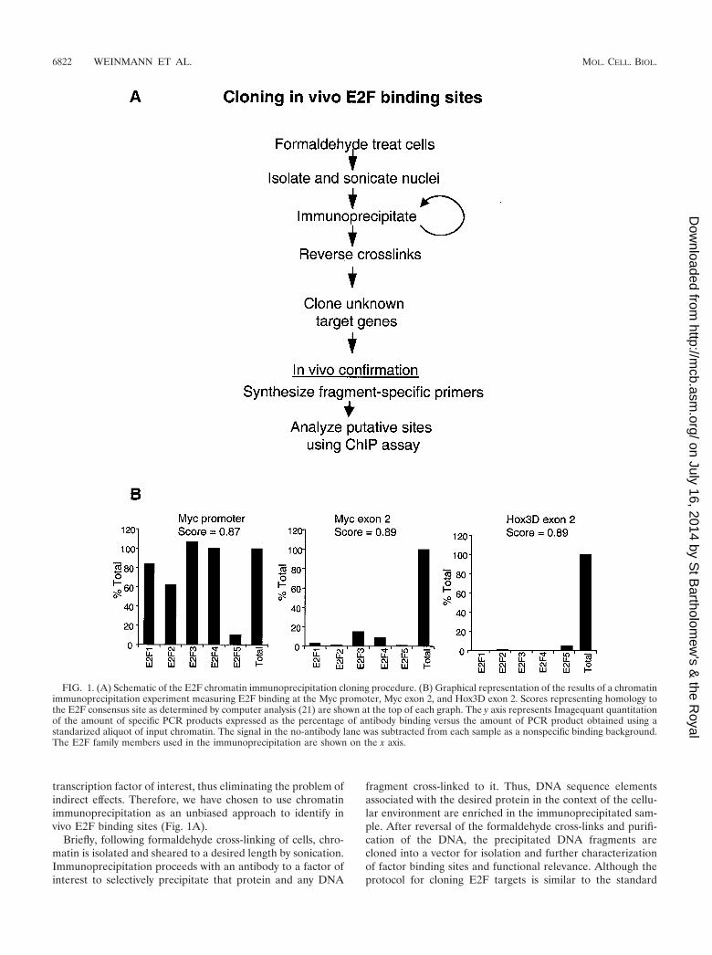
transcription factor of interest, thus eliminating the problem ofindirect effects. Therefore, we have chosen to use chromatinimmunoprecipitation as an unbiased approach to identify invivo E2F binding sites (Fig. 1A).
Briefly, following formaldehyde cross-linking of cells, chro-matin is isolated and sheared to a desired length by sonication.Immunoprecipitation proceeds with an antibody to a factor ofinterest to selectively precipitate that protein and any DNA
fragment cross-linked to it. Thus, DNA sequence elementsassociated with the desired protein in the context of the cellu-lar environment are enriched in the immunoprecipitated sam-ple. After reversal of the formaldehyde cross-links and purifi-cation of the DNA, the precipitated DNA fragments arecloned into a vector for isolation and further characterizationof factor binding sites and functional relevance. Although theprotocol for cloning E2F targets is similar to the standard
FIG. 1. (A) Schematic of the E2F chromatin immunoprecipitation cloning procedure. (B) Graphical representation of the results of a chromatinimmunoprecipitation experiment measuring E2F binding at the Myc promoter, Myc exon 2, and Hox3D exon 2. Scores representing homology tothe E2F consensus site as determined by computer analysis (21) are shown at the top of each graph. The y axis represents Imagequant quantitationof the amount of specific PCR products expressed as the percentage of antibody binding versus the amount of PCR product obtained using astandarized aliquot of input chromatin. The signal in the no-antibody lane was subtracted from each sample as a nonspecific binding background.The E2F family members used in the immunoprecipitation are shown on the x axis.
6822 WEINMANN ET AL. MOL. CELL. BIOL.
on July 16, 2014 by St B
artholomew
's & the R
oyalhttp://m
cb.asm.org/
Dow
nloaded from
chromatin immunoprecipitation assay that has been previouslydescribed (3, 33, 49), several modifications were made to theassay. Importantly, we performed two sequential chromatinimmunoprecipitations using the same antibody for both thefirst and second steps. In preliminary studies, we found that alarge number of nonspecific fragments were cloned if only oneimmunoprecipitation was performed. Therefore, the secondimmunoprecipitation was used to decrease the amount of non-specific DNA present to enable more efficient cloning of thespecific fragments. Also, a portion of the immunoprecipitatedsamples was retained prior to cloning to analyze known targetgenes as a positive control for the immunoprecipitation. Othermodifications important for cloning the immunoprecipitatedproducts can be found on our web site (http://mcardle.oncology.wisc.edu/farnham).
Previous studies have demonstrated the validity of using thechromatin immunoprecipitation protocol to identify site-spe-cific interactions of transcription factors and promoter DNA inthe context of a living cell. Importantly, we have shown thatbinding of E2F to target promoters is site specific. For exam-ple, the chromatin immunoprecipitation assay has been used todemonstrate that E2F4 binds with high affinity to the dhfrpromoter (which is known to be regulated by E2F) but doesnot bind to the cad promoter (which is a Myc target gene) (3).Also, in a previous report (49) we showed that a site-specificmutation in the dhfr promoter eliminates binding of E2F4, asmonitored by the chromatin immunoprecipitation assay. Inaddition, high-affinity binding of E2F family members to themyc promoter is abolished when a point mutation is introducedinto the E2F site of that promoter (2). Finally, we have alsoshown that E2Fs do not bind to promoters which are regulatedby liver-specific transcription factors (C. R. Graveel and P. J.Farnham, unpublished data). Clearly, the chromatin immuno-precipitation assay has been useful in demonstrating that notall cellular promoters bind E2Fs and that those that do requirea specific site on the DNA for high-affinity in vivo binding.However, we felt that additional controls were needed prior tousing this assay to clone novel targets. Namely, we wished toclone regulatory E2F binding sites and not clone random,nonfunctional E2F binding sites present in nonpromoter re-gions of the genome. Previous analysis of promoter and exon 2databases suggested that E2F sites are found at a much higherfrequency in promoters (21). However, sequences having high-score matches to the E2F consensus site (scores of 0.86 orbetter in the computer analysis) can be found in nonpromoterregions as well. To explore whether E2F is bound to these sitesin living cells, we identified two such consensus sites located inthe second exon of the myc and the hox3D genes. Because themyc promoter has previously been shown to be an E2F target(30, 34, 50), we examined binding of E2F family members tothe exon sites in comparison to binding at the E2F site in theMyc promoter. As shown in Fig. 1B, E2F binding to the mycgene promoter is at much higher levels than binding to the mycand hox3D exon 2 regions in the same chromatin sample, eventhough the score match to the consensus E2F site is very highin the exon 2 regions.
Having assured ourselves that the chromatin immunopre-cipitation assay can be used to detect promoter-specific andsite-specific binding of E2F, we performed immunoprecipita-tions from HeLa cell chromatin using antibodies against either
E2F1 or E2F4 and proceeded with the cloning procedure. Wechose clones having inserts of 500 bp or greater and examined11 clones obtained by immunoprecipitation with the E2F1antibody and 7 clones obtained by immunoprecipitation withthe E2F4 antibody for further analysis. The first step in thecharacterization of the ChET clones was to determine whichones contained bona fide E2F binding sites and which werefalse positives.
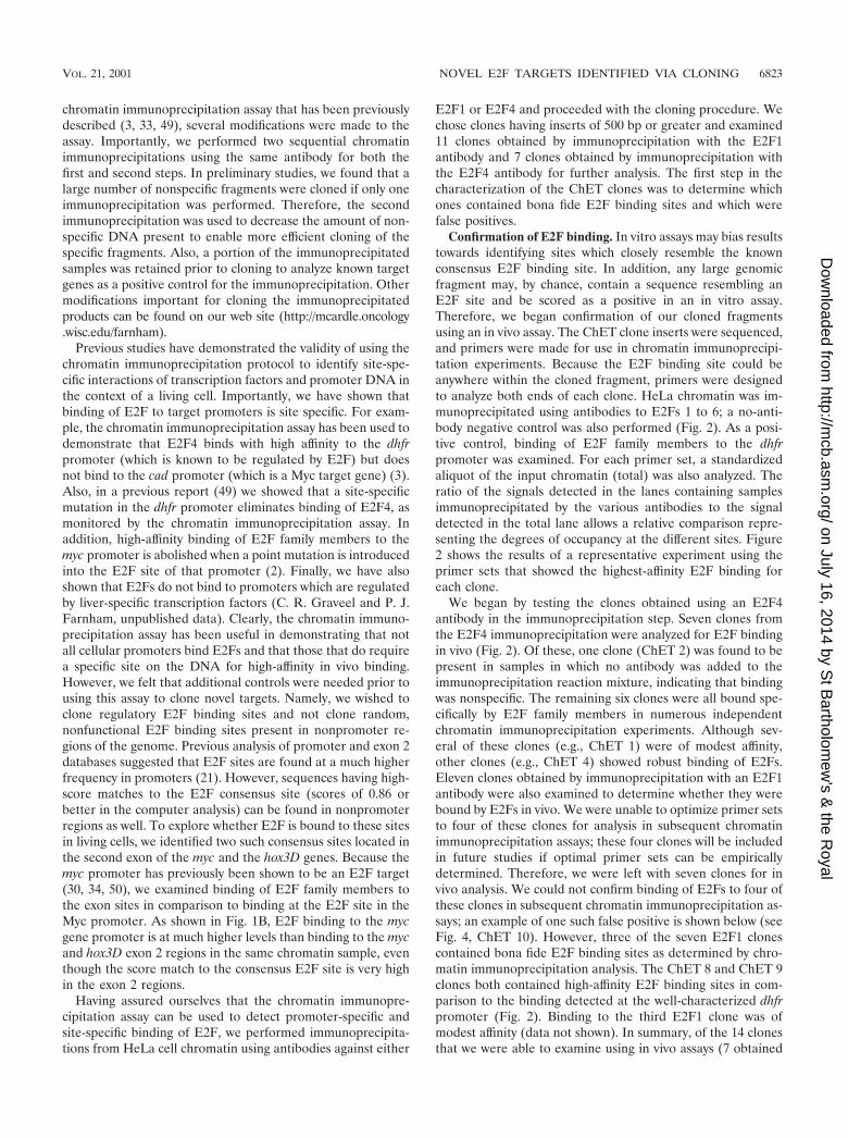
Confirmation of E2F binding. In vitro assays may bias resultstowards identifying sites which closely resemble the knownconsensus E2F binding site. In addition, any large genomicfragment may, by chance, contain a sequence resembling anE2F site and be scored as a positive in an in vitro assay.Therefore, we began confirmation of our cloned fragmentsusing an in vivo assay. The ChET clone inserts were sequenced,and primers were made for use in chromatin immunoprecipi-tation experiments. Because the E2F binding site could beanywhere within the cloned fragment, primers were designedto analyze both ends of each clone. HeLa chromatin was im-munoprecipitated using antibodies to E2Fs 1 to 6; a no-anti-body negative control was also performed (Fig. 2). As a posi-tive control, binding of E2F family members to the dhfrpromoter was examined. For each primer set, a standardizedaliquot of the input chromatin (total) was also analyzed. Theratio of the signals detected in the lanes containing samplesimmunoprecipitated by the various antibodies to the signaldetected in the total lane allows a relative comparison repre-senting the degrees of occupancy at the different sites. Figure2 shows the results of a representative experiment using theprimer sets that showed the highest-affinity E2F binding foreach clone.
We began by testing the clones obtained using an E2F4antibody in the immunoprecipitation step. Seven clones fromthe E2F4 immunoprecipitation were analyzed for E2F bindingin vivo (Fig. 2). Of these, one clone (ChET 2) was found to bepresent in samples in which no antibody was added to theimmunoprecipitation reaction mixture, indicating that bindingwas nonspecific. The remaining six clones were all bound spe-cifically by E2F family members in numerous independentchromatin immunoprecipitation experiments. Although sev-eral of these clones (e.g., ChET 1) were of modest affinity,other clones (e.g., ChET 4) showed robust binding of E2Fs.Eleven clones obtained by immunoprecipitation with an E2F1antibody were also examined to determine whether they werebound by E2Fs in vivo. We were unable to optimize primer setsto four of these clones for analysis in subsequent chromatinimmunoprecipitation assays; these four clones will be includedin future studies if optimal primer sets can be empiricallydetermined. Therefore, we were left with seven clones for invivo analysis. We could not confirm binding of E2Fs to four ofthese clones in subsequent chromatin immunoprecipitation as-says; an example of one such false positive is shown below (seeFig. 4, ChET 10). However, three of the seven E2F1 clonescontained bona fide E2F binding sites as determined by chro-matin immunoprecipitation analysis. The ChET 8 and ChET 9clones both contained high-affinity E2F binding sites in com-parison to the binding detected at the well-characterized dhfrpromoter (Fig. 2). Binding to the third E2F1 clone was ofmodest affinity (data not shown). In summary, of the 14 clonesthat we were able to examine using in vivo assays (7 obtained
VOL. 21, 2001 NOVEL E2F TARGETS IDENTIFIED VIA CLONING 6823
on July 16, 2014 by St B
artholomew
's & the R
oyalhttp://m
cb.asm.org/
Dow
nloaded from
using an E2F1 antibody and 7 obtained using an E2F4 anti-body), 9 were confirmed to contain bona fide in vivo E2Fbinding sites (Table 1). Although false positives are unavoid-able, we believe that the two sequential immunoprecipitationreactions greatly enriched for clones containing bona fide E2Fbinding sites. It is important to note that the in vivo binding ofE2Fs to the ChET clones has been confirmed in numerousexperiments. For example, the experiments shown in Fig. 2 andbelow (see Fig. 4) are completely independent from each otherand from the chromatin immunoprecipitation experiment usedto clone the fragments. We have also confirmed binding of
E2Fs to ChET 4, ChET 8, and ChET 9 in other human celltypes (data not shown).
Either an E2F1 or an E2F4 antibody was initially used forthe cloning procedure; however, we found that the cloned sitesdid not reveal an E2F binding pattern to suggest family mem-ber binding specificity. Rather, binding of multiple E2F familymembers to each clone was detected in vivo (Fig. 2). Theseresults, suggesting a lack of DNA binding specificity among thedifferent E2Fs, are similar to those of our previous studies ofknown E2F target genes (49). E2F family members contain ahighly conserved DNA binding domain; therefore, it is notsurprising that these family members have the ability to bind tothe same sequence in vivo. It is unlikely that our results indi-cate that multiple E2F family members are simultaneouslybinding to the same site, but the more likely explanation is thata dynamic exchange occurs at a given site and various E2Fs aretrapped in different cells during the cross-linking procedure.There are precedents for this hypothesis, as dynamic exchangeof the glucocorticoid receptor has been demonstrated in livingcells (29). With this in mind, it is worth noting that the clonedsites can be separated into three basic categories. The first type(e.g., ChET 9) showed an in vivo binding pattern very similar
FIG. 2. Confirmation of ChET clones by examining E2F binding in vivo. A representative chromatin immunoprecipitation experiment withHeLa cells is shown. Immunoprecipitation proceeded utilizing antibodies (Ab) against E2F1 (lane 1), E2F2 (lane 2), E2F3 (lane 3), E2F4 (lane4), E2F5 (lane 5), and E2F6 (lane 6) or no antibody (lane 8). Following DNA purification, samples were subjected to PCR with primers designedfor the individual E2F clones or the dhfr promoter as a control (labeled on the right). A portion of the total input was also examined by PCR (lane7).
TABLE 1. Summary of ChET cloning
ParameterValue
E2F4a E2F1a Total (%)
No. of fragments analyzed 7 7 14No. of false positives 1 4 5 (36)No. of clones specifically
bound by E2Fs6 3 9 (64)
a Antibody to the indicated E2F was used to clone the fragments.
6824 WEINMANN ET AL. MOL. CELL. BIOL.
on July 16, 2014 by St B
artholomew
's & the R
oyalhttp://m
cb.asm.org/
Dow
nloaded from
to that of the dhfr promoter, i.e., strong binding of E2F1 toE2F4 and very little binding of E2F5 or E2F6. The second type(e.g., ChET 4 and ChET 8) showed strong binding of E2F1 toE2F4 and little binding of E2F5 but detectable binding ofE2F6. Finally, the third type of site (e.g., ChET 1) showedequal low-affinity binding of E2F1 to E2F6. The significance ofthese distinct binding patterns is unknown, and further analysiswill be required to elucidate the functional consequences, ifany.
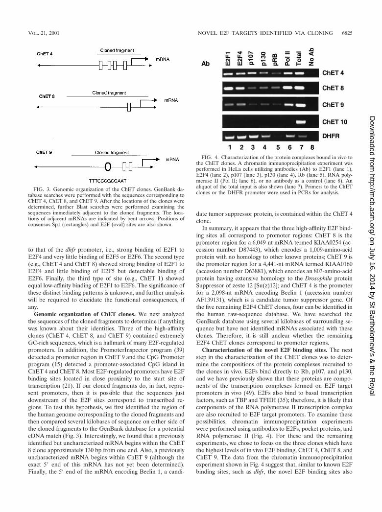
Genomic organization of ChET clones. We next analyzedthe sequences of the cloned fragments to determine if anythingwas known about their identities. Three of the high-affinityclones (ChET 4, ChET 8, and ChET 9) contained extremelyGC-rich sequences, which is a hallmark of many E2F-regulatedpromoters. In addition, the PromoterInspector program (39)detected a promoter region in ChET 9 and the CpG Promoterprogram (15) detected a promoter-associated CpG island inChET 4 and ChET 8. Most E2F-regulated promoters have E2Fbinding sites located in close proximity to the start site oftranscription (21). If our cloned fragments do, in fact, repre-sent promoters, then it is possible that the sequences justdownstream of the E2F sites correspond to transcribed re-gions. To test this hypothesis, we first identified the region ofthe human genome corresponding to the cloned fragments andthen compared several kilobases of sequence on either side ofthe cloned fragments to the GenBank database for a potentialcDNA match (Fig. 3). Interestingly, we found that a previouslyidentified but uncharacterized mRNA begins within the ChET8 clone approximately 130 bp from one end. Also, a previouslyuncharacterized mRNA begins within ChET 9 (although theexact 5� end of this mRNA has not yet been determined).Finally, the 5� end of the mRNA encoding Beclin 1, a candi-
date tumor suppressor protein, is contained within the ChET 4clone.
In summary, it appears that the three high-affinity E2F bind-ing sites all correspond to promoter regions: ChET 8 is thepromoter region for a 6,049-nt mRNA termed KIAA0254 (ac-cession number D87443), which encodes a 1,009-amino-acidprotein with no homology to other known proteins; ChET 9 isthe promoter region for a 4,441-nt mRNA termed KIAA0160(accession number D63881), which encodes an 803-amino-acidprotein having extensive homology to the Drosophila proteinSuppressor of zeste 12 [Su(z)12]; and ChET 4 is the promoterfor a 2,098-nt mRNA encoding Beclin 1 (accession numberAF139131), which is a candidate tumor suppressor gene. Ofthe five remaining E2F4 ChET clones, four can be identified inthe human raw-sequence database. We have searched theGenBank database using several kilobases of surrounding se-quence but have not identified mRNAs associated with theseclones. Therefore, it is still unclear whether the remainingE2F4 ChET clones correspond to promoter regions.
Characterization of the novel E2F binding sites. The nextstep in the characterization of the ChET clones was to deter-mine the compositions of the protein complexes recruited tothe clones in vivo. E2Fs bind directly to Rb, p107, and p130,and we have previously shown that these proteins are compo-nents of the transcription complexes formed on E2F targetpromoters in vivo (49). E2Fs also bind to basal transcriptionfactors, such as TBP and TFIIH (35); therefore, it is likely thatcomponents of the RNA polymerase II transcription complexare also recruited to E2F target promoters. To examine thesepossibilities, chromatin immunoprecipitation experimentswere performed using antibodies to E2Fs, pocket proteins, andRNA polymerase II (Fig. 4). For these and the remainingexperiments, we chose to focus on the three clones which havethe highest levels of in vivo E2F binding, ChET 4, ChET 8, andChET 9. The data from the chromatin immunoprecipitationexperiment shown in Fig. 4 suggest that, similar to known E2Fbinding sites, such as dhfr, the novel E2F binding sites also
FIG. 3. Genomic organization of the ChET clones. GenBank da-tabase searches were performed with the sequences corresponding toChET 4, ChET 8, and ChET 9. After the locations of the clones weredetermined, further Blast searches were performed examining thesequences immediately adjacent to the cloned fragments. The loca-tions of adjacent mRNAs are indicated by bent arrows. Positions ofconsensus Sp1 (rectangles) and E2F (oval) sites are also shown.
FIG. 4. Characterization of the protein complexes bound in vivo tothe ChET clones. A chromatin immunoprecipitation experiment wasperformed in HeLa cells utilizing antibodies (Ab) to E2F1 (lane 1),E2F4 (lane 2), p107 (lane 3), p130 (lane 4), Rb (lane 5), RNA poly-merase II (Pol II; lane 6), or no antibody as a control (lane 8). Analiquot of the total input is also shown (lane 7). Primers to the ChETclones or the DHFR promoter were used in PCRs for analysis.
VOL. 21, 2001 NOVEL E2F TARGETS IDENTIFIED VIA CLONING 6825
on July 16, 2014 by St B
artholomew
's & the R
oyalhttp://m
cb.asm.org/
Dow
nloaded from
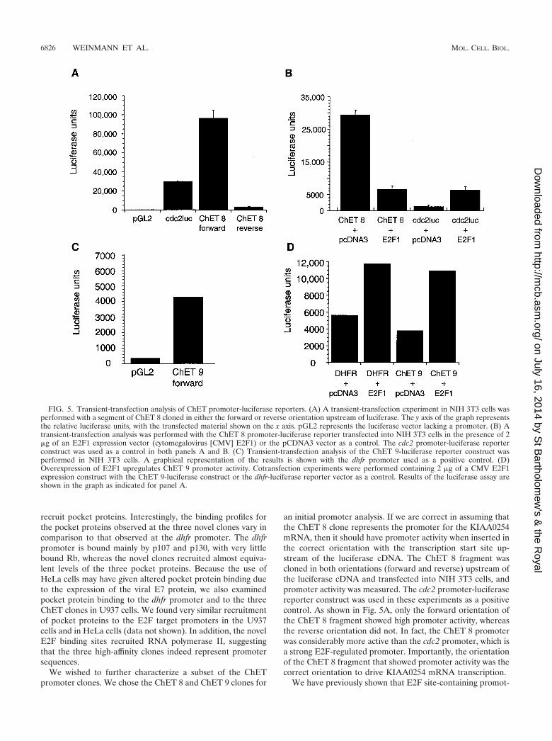
recruit pocket proteins. Interestingly, the binding profiles forthe pocket proteins observed at the three novel clones vary incomparison to that observed at the dhfr promoter. The dhfrpromoter is bound mainly by p107 and p130, with very littlebound Rb, whereas the novel clones recruited almost equiva-lent levels of the three pocket proteins. Because the use ofHeLa cells may have given altered pocket protein binding dueto the expression of the viral E7 protein, we also examinedpocket protein binding to the dhfr promoter and to the threeChET clones in U937 cells. We found very similar recruitmentof pocket proteins to the E2F target promoters in the U937cells and in HeLa cells (data not shown). In addition, the novelE2F binding sites recruited RNA polymerase II, suggestingthat the three high-affinity clones indeed represent promotersequences.
We wished to further characterize a subset of the ChETpromoter clones. We chose the ChET 8 and ChET 9 clones for
an initial promoter analysis. If we are correct in assuming thatthe ChET 8 clone represents the promoter for the KIAA0254mRNA, then it should have promoter activity when inserted inthe correct orientation with the transcription start site up-stream of the luciferase cDNA. The ChET 8 fragment wascloned in both orientations (forward and reverse) upstream ofthe luciferase cDNA and transfected into NIH 3T3 cells, andpromoter activity was measured. The cdc2 promoter-luciferasereporter construct was used in these experiments as a positivecontrol. As shown in Fig. 5A, only the forward orientation ofthe ChET 8 fragment showed high promoter activity, whereasthe reverse orientation did not. In fact, the ChET 8 promoterwas considerably more active than the cdc2 promoter, which isa strong E2F-regulated promoter. Importantly, the orientationof the ChET 8 fragment that showed promoter activity was thecorrect orientation to drive KIAA0254 mRNA transcription.
We have previously shown that E2F site-containing promot-
FIG. 5. Transient-transfection analysis of ChET promoter-luciferase reporters. (A) A transient-transfection experiment in NIH 3T3 cells wasperformed with a segment of ChET 8 cloned in either the forward or reverse orientation upstream of luciferase. The y axis of the graph representsthe relative luciferase units, with the transfected material shown on the x axis. pGL2 represents the luciferase vector lacking a promoter. (B) Atransient-transfection analysis was performed with the ChET 8 promoter-luciferase reporter transfected into NIH 3T3 cells in the presence of 2�g of an E2F1 expression vector (cytomegalovirus [CMV] E2F1) or the pCDNA3 vector as a control. The cdc2 promoter-luciferase reporterconstruct was used as a control in both panels A and B. (C) Transient-transfection analysis of the ChET 9-luciferase reporter construct wasperformed in NIH 3T3 cells. A graphical representation of the results is shown with the dhfr promoter used as a positive control. (D)Overexpression of E2F1 upregulates ChET 9 promoter activity. Cotransfection experiments were performed containing 2 �g of a CMV E2F1expression construct with the ChET 9-luciferase construct or the dhfr-luciferase reporter vector as a control. Results of the luciferase assay areshown in the graph as indicated for panel A.
6826 WEINMANN ET AL. MOL. CELL. BIOL.
on July 16, 2014 by St B
artholomew
's & the R
oyalhttp://m
cb.asm.org/
Dow
nloaded from
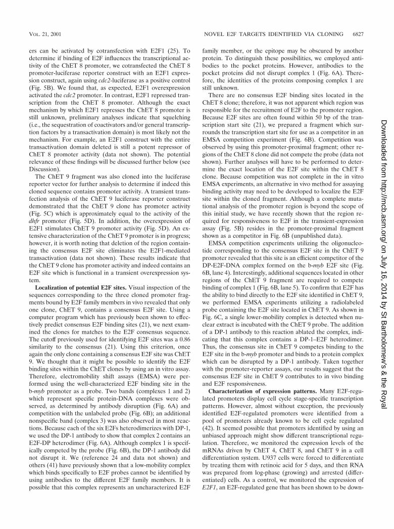
ers can be activated by cotransfection with E2F1 (25). Todetermine if binding of E2F influences the transcriptional ac-tivity of the ChET 8 promoter, we cotransfected the ChET 8promoter-luciferase reporter construct with an E2F1 expres-sion construct, again using cdc2-luciferase as a positive control(Fig. 5B). We found that, as expected, E2F1 overexpressionactivated the cdc2 promoter. In contrast, E2F1 repressed tran-scription from the ChET 8 promoter. Although the exactmechanism by which E2F1 represses the ChET 8 promoter isstill unknown, preliminary analyses indicate that squelching(i.e., the sequestration of coactivators and/or general transcrip-tion factors by a transactivation domain) is most likely not themechanism. For example, an E2F1 construct with the entiretransactivation domain deleted is still a potent repressor ofChET 8 promoter activity (data not shown). The potentialrelevance of these findings will be discussed further below (seeDiscussion).
The ChET 9 fragment was also cloned into the luciferasereporter vector for further analysis to determine if indeed thiscloned sequence contains promoter activity. A transient trans-fection analysis of the ChET 9 luciferase reporter constructdemonstrated that the ChET 9 clone has promoter activity(Fig. 5C) which is approximately equal to the activity of thedhfr promoter (Fig. 5D). In addition, the overexpression ofE2F1 stimulates ChET 9 promoter activity (Fig. 5D). An ex-tensive characterization of the ChET 9 promoter is in progress;however, it is worth noting that deletion of the region contain-ing the consensus E2F site eliminates the E2F1-mediatedtransactivation (data not shown). These results indicate thatthe ChET 9 clone has promoter activity and indeed contains anE2F site which is functional in a transient overexpression sys-tem.
Localization of potential E2F sites. Visual inspection of thesequences corresponding to the three cloned promoter frag-ments bound by E2F family members in vivo revealed that onlyone clone, ChET 9, contains a consensus E2F site. Using acomputer program which has previously been shown to effec-tively predict consensus E2F binding sites (21), we next exam-ined the clones for matches to the E2F consensus sequence.The cutoff previously used for identifying E2F sites was a 0.86similarity to the consensus (21). Using this criterion, onceagain the only clone containing a consensus E2F site was ChET9. We thought that it might be possible to identify the E2Fbinding sites within the ChET clones by using an in vitro assay.Therefore, electromobility shift assays (EMSA) were per-formed using the well-characterized E2F binding site in theb-myb promoter as a probe. Two bands (complexes 1 and 2)which represent specific protein-DNA complexes were ob-served, as determined by antibody disruption (Fig. 6A) andcompetition with the unlabeled probe (Fig. 6B); an additionalnonspecific band (complex 3) was also observed in most reac-tions. Because each of the six E2Fs heterodimerizes with DP-1,we used the DP-1 antibody to show that complex 2 contains anE2F-DP heterodimer (Fig. 6A). Although complex 1 is specif-ically competed by the probe (Fig. 6B), the DP-1 antibody didnot disrupt it. We (reference 24 and data not shown) andothers (41) have previously shown that a low-mobility complexwhich binds specifically to E2F probes cannot be identified byusing antibodies to the different E2F family members. It ispossible that this complex represents an uncharacterized E2F
family member, or the epitope may be obscured by anotherprotein. To distinguish these possibilities, we employed anti-bodies to the pocket proteins. However, antibodies to thepocket proteins did not disrupt complex 1 (Fig. 6A). There-fore, the identities of the proteins composing complex 1 arestill unknown.
There are no consensus E2F binding sites located in theChET 8 clone; therefore, it was not apparent which region wasresponsible for the recruitment of E2F to the promoter region.Because E2F sites are often found within 50 bp of the tran-scription start site (21), we prepared a fragment which sur-rounds the transcription start site for use as a competitor in anEMSA competition experiment (Fig. 6B). Competition wasobserved by using this promoter-proximal fragment; other re-gions of the ChET 8 clone did not compete the probe (data notshown). Further analyses will have to be performed to deter-mine the exact location of the E2F site within the ChET 8clone. Because competition was not complete in the in vitroEMSA experiments, an alternative in vivo method for assayingbinding activity may need to be developed to localize the E2Fsite within the cloned fragment. Although a complete muta-tional analysis of the promoter region is beyond the scope ofthis initial study, we have recently shown that the region re-quired for responsiveness to E2F in the transient-expressionassay (Fig. 5B) resides in the promoter-proximal fragmentshown as a competitor in Fig. 6B (unpublished data).
EMSA competition experiments utilizing the oligonucleo-tide corresponding to the consensus E2F site in the ChET 9promoter revealed that this site is an efficient competitor of theDP-E2F-DNA complex formed on the b-myb E2F site (Fig.6B, lane 4). Interestingly, additional sequences located in otherregions of the ChET 9 fragment are required to competebinding of complex 1 (Fig. 6B, lane 5). To confirm that E2F hasthe ability to bind directly to the E2F site identified in ChET 9,we performed EMSA experiments utilizing a radiolabeledprobe containing the E2F site located in ChET 9. As shown inFig. 6C, a single lower-mobility complex is detected when nu-clear extract is incubated with the ChET 9 probe. The additionof a DP-1 antibody to this reaction ablated the complex, indi-cating that this complex contains a DP-1–E2F heterodimer.Thus, the consensus site in ChET 9 competes binding to theE2F site in the b-myb promoter and binds to a protein complexwhich can be disrupted by a DP-1 antibody. Taken togetherwith the promoter-reporter assays, our results suggest that theconsensus E2F site in ChET 9 contributes to in vivo bindingand E2F responsiveness.
Characterization of expression patterns. Many E2F-regu-lated promoters display cell cycle stage-specific transcriptionpatterns. However, almost without exception, the previouslyidentified E2F-regulated promoters were identified from apool of promoters already known to be cell cycle regulated(42). It seemed possible that promoters identified by using anunbiased approach might show different transcriptional regu-lation. Therefore, we monitored the expression levels of themRNAs driven by ChET 4, ChET 8, and ChET 9 in a celldifferentiation system. U937 cells were forced to differentiateby treating them with retinoic acid for 5 days, and then RNAwas prepared from log-phase (growing) and arrested (differ-entiated) cells. As a control, we monitored the expression ofE2F1, an E2F-regulated gene that has been shown to be down-
VOL. 21, 2001 NOVEL E2F TARGETS IDENTIFIED VIA CLONING 6827
on July 16, 2014 by St B
artholomew
's & the R
oyalhttp://m
cb.asm.org/
Dow
nloaded from
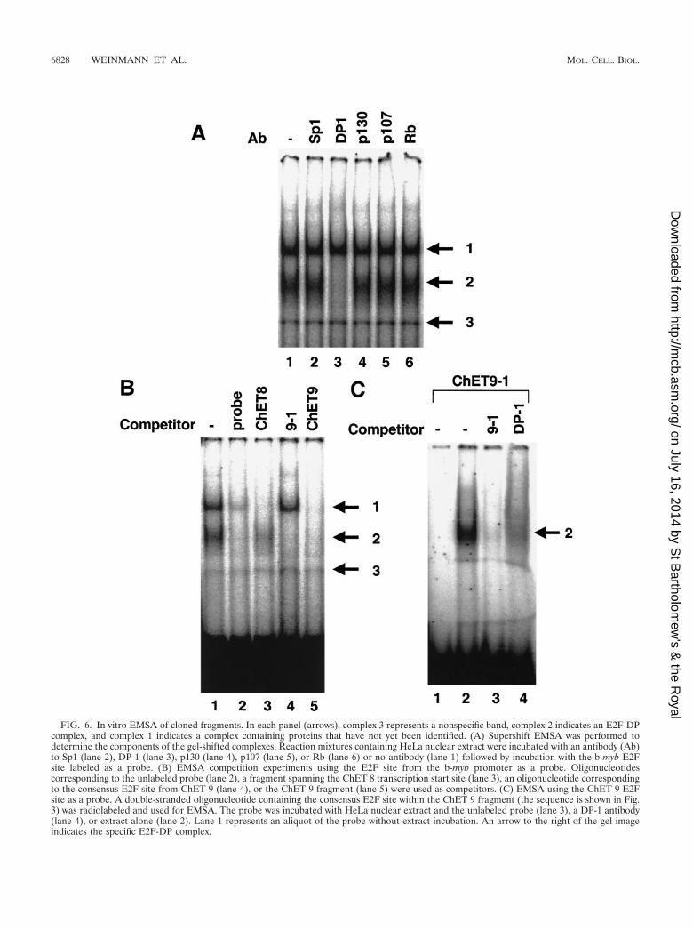
FIG. 6. In vitro EMSA of cloned fragments. In each panel (arrows), complex 3 represents a nonspecific band, complex 2 indicates an E2F-DPcomplex, and complex 1 indicates a complex containing proteins that have not yet been identified. (A) Supershift EMSA was performed todetermine the components of the gel-shifted complexes. Reaction mixtures containing HeLa nuclear extract were incubated with an antibody (Ab)to Sp1 (lane 2), DP-1 (lane 3), p130 (lane 4), p107 (lane 5), or Rb (lane 6) or no antibody (lane 1) followed by incubation with the b-myb E2Fsite labeled as a probe. (B) EMSA competition experiments using the E2F site from the b-myb promoter as a probe. Oligonucleotidescorresponding to the unlabeled probe (lane 2), a fragment spanning the ChET 8 transcription start site (lane 3), an oligonucleotide correspondingto the consensus E2F site from ChET 9 (lane 4), or the ChET 9 fragment (lane 5) were used as competitors. (C) EMSA using the ChET 9 E2Fsite as a probe. A double-stranded oligonucleotide containing the consensus E2F site within the ChET 9 fragment (the sequence is shown in Fig.3) was radiolabeled and used for EMSA. The probe was incubated with HeLa nuclear extract and the unlabeled probe (lane 3), a DP-1 antibody(lane 4), or extract alone (lane 2). Lane 1 represents an aliquot of the probe without extract incubation. An arrow to the right of the gel imageindicates the specific E2F-DP complex.
6828 WEINMANN ET AL. MOL. CELL. BIOL.
on July 16, 2014 by St B
artholomew
's & the R
oyalhttp://m
cb.asm.org/
Dow
nloaded from
regulated when growing cells exit the cell cycle (14, 18, 32, 43).As expected, levels of E2F1 mRNA decreased upon differen-tiation and cell cycle arrest (Fig. 7A). In contrast, the levels ofmRNA for the three novel clones did not decrease significantlyin the U937 cell population upon differentiation, which sug-gests that these promoters are not cell cycle regulated. Thesedata indicate that E2F family members may regulate both cellcycle- and non cell cycle-responsive promoters. To determinewhether another unbiased approach would also yield con-stitutively active E2F-regulated promoters, we examined themRNAs for two promoters that were predicted to be regulatedby E2F from a computer-assisted identification of consensusE2F binding sites in the promoter regions. The large subunit ofRNAPII and XRCC2 were both identified in a previous studywhich scanned the eukaryotic promoter databases for E2Ftarget promoters (21). Chromatin immunoprecipitation analy-sis indicated that indeed the promoters for RNAPII largesubunit and XRCC2 are bound by E2F (data not shown).Analysis of mRNA levels in the U937 cells indicated that oneof the two computer-identified genes was constitutively ex-pressed whereas the other was downregulated upon differen-
tiation. Therefore, four of the five E2F-bound promoters iden-tified by using unbiased approaches are not regulated upondifferentiation of U937 cells. It is also important to note thatalthough the amount of E2F1 declines during differentiation ofU937 cells, the overall amount of E2F activity remains highdue to the constitutive expression of E2F4. In fact, we haveshown that the amount of E2F4 bound to the ChET promotersis unchanged after differentiation of U937 cells (data notshown). Perhaps only those E2F target genes which areuniquely responsive to E2F1 versus E2F4 will show a decline inactivity upon differentiation.
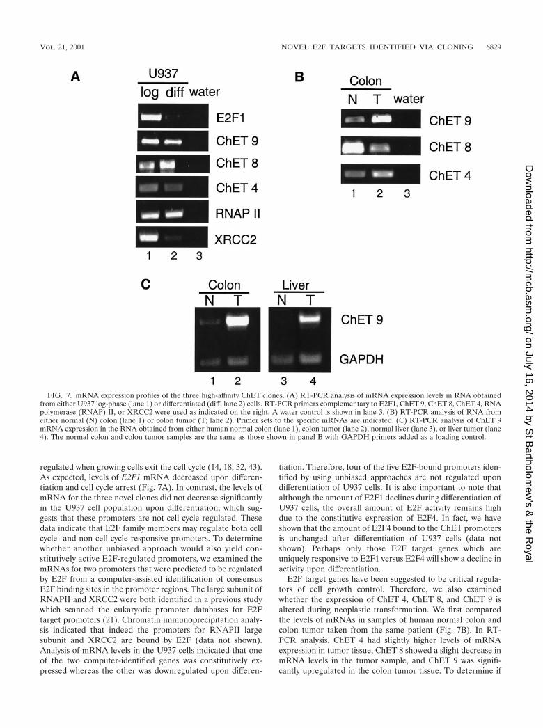
E2F target genes have been suggested to be critical regula-tors of cell growth control. Therefore, we also examinedwhether the expression of ChET 4, ChET 8, and ChET 9 isaltered during neoplastic transformation. We first comparedthe levels of mRNAs in samples of human normal colon andcolon tumor taken from the same patient (Fig. 7B). In RT-PCR analysis, ChET 4 had slightly higher levels of mRNAexpression in tumor tissue, ChET 8 showed a slight decrease inmRNA levels in the tumor sample, and ChET 9 was signifi-cantly upregulated in the colon tumor tissue. To determine if
FIG. 7. mRNA expression profiles of the three high-affinity ChET clones. (A) RT-PCR analysis of mRNA expression levels in RNA obtainedfrom either U937 log-phase (lane 1) or differentiated (diff; lane 2) cells. RT-PCR primers complementary to E2F1, ChET 9, ChET 8, ChET 4, RNApolymerase (RNAP) II, or XRCC2 were used as indicated on the right. A water control is shown in lane 3. (B) RT-PCR analysis of RNA fromeither normal (N) colon (lane 1) or colon tumor (T; lane 2). Primer sets to the specific mRNAs are indicated. (C) RT-PCR analysis of ChET 9mRNA expression in the RNA obtained from either human normal colon (lane 1), colon tumor (lane 2), normal liver (lane 3), or liver tumor (lane4). The normal colon and colon tumor samples are the same as those shown in panel B with GAPDH primers added as a loading control.
VOL. 21, 2001 NOVEL E2F TARGETS IDENTIFIED VIA CLONING 6829
on July 16, 2014 by St B
artholomew
's & the R
oyalhttp://m
cb.asm.org/
Dow
nloaded from
upregulation of ChET 9 mRNA was specific to colon tumors,we also compared normal liver and liver tumor mRNA levels.To control for equivalent RNA sample concentrations, primersfor GAPDH (glyceraldehyde-3-phosphate dehydrogenase)were included in the reaction mixtures. The results show thatChET 9 mRNA was upregulated in both tumor types (Fig. 7C),which suggests that ChET 9 expression may be generally de-regulated in human tumors. We have also confirmed thatChET 9 mRNA is upregulated in eight of nine additionalhuman colon tumor samples (unpublished data).
DISCUSSION
To our knowledge, this is the first demonstration that thechromatin immunoprecipitation assay can be used to clonepromoters which are direct in vivo targets of a mammaliansite-specific DNA binding protein. This provides a powerfulnew approach to examine direct transcription factor targets inan unbiased manner which does not rely on previous charac-terization of a consensus sequence or a prior knowledge ofgene expression patterns. Although others have used similarapproaches to isolate genomic fragments (6, 36, 38), thosestudies did not use subsequent experiments to confirm in vivobinding of the factor of interest to the isolated DNA. Due tothis lack of in vivo confirmation, it is difficult to assess thevalidity of the previous protocols. Also, sequence analysis ofthe clones isolated in the previous studies indicated that thecloned fragments corresponded to nonpromoter regions, suchas introns (5, 10, 22, 45, 46). One study did find that 3 out of 43clones isolated after in vitro incubation of genomic DNA withpurified Ets-1 protein were promoters; however, the authorsdid not confirm in vivo binding of ETS1 to these 3 clones or tothe other 40 isolated clones. Therefore, it is difficult to be sureif any of the clones in that study were bona fide in vivo targetsof ETS1.
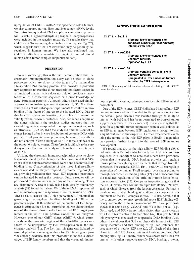
Utilizing the chromatin immunoprecipitation assay to clonefragments bound by E2F family members, we found that 64%(9 of 14) of the clones characterized were bona fide in vivo E2Fbinding sites. Characterization of the three highest-affinityclones revealed that they correspond to promoter regions (Fig.8), providing validation that novel E2F-regulated promoterscan be isolated by using this protocol. Future studies will beperformed to determine whether any of the remaining clonesare promoters. A recent study using high-density microarrayanalysis (31) found that about 7% of the mRNAs representedon the microarray were responsive to overexpression of E2Fs.These results suggest that a high percentage of mammaliangenes might be regulated by direct binding of E2F to thepromoter region. If this estimate of the number of E2F targetgenes is correct, then it is not surprising that we did not isolateone of the several dozen well-characterized E2F target pro-moters in the set of nine positive clones that we analyzed.However, one of our ChET clones (ChET 9, which corre-sponds to the promoter region of the KIAA0160 gene) wasshown to be upregulated by E2F overexpression in the mi-croarray analysis (31). The fact that this gene was isolated bytwo independent screening methods for E2F target genes pro-vides strong evidence that this promoter is indeed a directtarget of E2F family members and that the chromatin immu-
noprecipitation cloning technique can identify E2F-regulatedpromoters.
One of the E2F4 clones, ChET 4, displayed high-affinity E2Fbinding in vivo and corresponded to the promoter region forthe beclin 1 gene. Beclin 1 was isolated through its ability tointeract with bcl-2 and has been postulated to possess tumorsuppressor activity in breast cancer (1). It is interesting that thegene for a potential tumor suppressor protein was isolated asan E2F target gene because E2F regulation is thought to playa significant role in tumorigenisis. Further experiments exam-ining the nature of the role E2F plays in Beclin 1 regulationmay provide further insight into the role of E2F in tumordevelopment.
We found that two of the high-affinity E2F binding clonesdid not contain E2F sites which closely matched the consensussequence. It is important to note that others have previouslyshown that site-specific DNA binding proteins can regulatetranscription through sequence elements that diverge from theconsensus. For example, CREB, Ets-1, and AML1 can regulateexpression of the human T-cell receptor beta chain promoterthrough nonconsensus binding sites (12) and a nonconsensussite mediates regulation of the atrial natriuretic factor by se-rum response factor (13). Computer inspection suggests thatthe ChET clones may contain multiple low-affinity E2F sites,each of which diverges from the known consensus. Perhaps acombination of weak binding sites allows for cooperative re-cruitment of the E2F complex in vivo. It is also possible thatthe promoter context may greatly influence E2F binding effi-ciency within the cellular environment. We have previouslyshown that some (e.g., CCAAT and YY1) but not all (e.g.,Oct1, Ap2, and NF1) transcription factor sites can synergizewith E2F sites to activate transcription (47). It is possible thatthis synergy was mediated by cooperative DNA binding. Also,others have shown that Sp1 can physically interact with E2Ffamily members and that binding of Sp1 can influence theoccupancy of a nearby E2F site (20, 27). Each of the threecharacterized ChET clones contains at least one consensus Sp1binding site (Fig. 3). Finally, others have shown that E2Fs caninteract with other sequence-specific DNA binding proteins,
FIG. 8. Summary of information obtained relating to the ChETpromoter clones.
6830 WEINMANN ET AL. MOL. CELL. BIOL.
on July 16, 2014 by St B
artholomew
's & the R
oyalhttp://m
cb.asm.org/
Dow
nloaded from
such as C/EBP� (17, 44). Interestingly, we have recently shownthat E2F1 can be recruited to promoters which containC/EBP� binding sites but lack E2F consensus sites (Graveeland Farnham, unpublished). It remains to be determined ifC/EBP� and/or other protein-protein interactions are mediat-ing the recruitment of E2F to the promoters we have cloned.However, recruitment of E2F through the recognition se-quence of another DNA binding protein could explain whysome of the cloned fragments failed to show robust competi-tion of a consensus E2F site in vitro.
To date, the majority of well-characterized E2F target pro-moters have been shown to be cell cycle regulated and acti-vated by E2F overexpression. In contrast, our three novel E2Ftarget promoters are constitutively expressed in growing versusdifferentiated U937 cells. It is perhaps not surprising that E2Ftarget promoters isolated using an unbiased approach showexpression profiles different from those of the well-character-ized E2F target promoters. According to microarray analyses,hundreds of genes are regulated by E2F family members (16,31). It is highly unlikely that this large number of mRNAs,which encode proteins having highly diverse biological func-tions, will all show exactly the same expression pattern in allcell types.
It is interesting that the mRNA produced by each of thethree novel promoters displayed unique expression profileswhen normal versus tumor human primary samples were ex-amined; one mRNA was constitutively expressed, one mRNAwas downregulated in the tumor sample, and one mRNA washighly upregulated in tumor RNA. Interestingly, one of thepromoters that we cloned which displayed high-affinity bindingin vivo was shown to be repressed, not activated, by E2F1.Although most E2F target genes studied to date are activatedin response to overexpression of E2F1, it has been shown thatthe cyclin D1 promoter is also repressed by E2F1 (48). Inaddition, the recent microarray analysis by Muller et al. pro-vided evidence that E2Fs can both activate and repress cellulargenes, although their data did suggest that most E2F-mediatedrepression was indirect (31). Additional evidence supportingE2F-mediated repression of the ChET 8 promoter can beextrapolated from a recent study examining the cell cycle fluc-tuations of thousands of human mRNAs (4). We have ex-tracted the expression profiles of E2F1 and KIAA0254, themRNA driven by the ChET 8 promoter, from the publishedmicroarray data. Interestingly, ChET 8 mRNA levels are in-versely related to E2F1 mRNA levels (data not shown). Col-lectively, these findings support a role for E2F1 in repression ofthe ChET 8 promoter. Further experiments need to be per-formed to characterize similarities and differences between thepromoters which are directly activated and those which aredirectly repressed upon overexpression of E2F1. However,these observations suggest that the nature and context of theE2F binding site may influence the role that E2F plays inregulation of a promoter.
In summary, the data presented in this paper establish thebasis for cloning novel promoters regulated by specific tran-scription factors through chromatin immunoprecipitation tech-niques. Our initial data suggest that the E2F consensus bindingsequence may not account for all potential in vivo E2F targets,possibly due to the roles of interacting proteins within thecellular environment. Importantly, the possibility that E2F
family members can regulate promoters that lack consensusbinding sites may aid in the understanding of microarray stud-ies which show that hundreds of mRNAs can respond to over-expression of E2Fs (16, 31). Also, we find it most interestingthat the expression profiles of the genes identified by using thisunbiased approach are quite different from the expression pro-files of the previously characterized E2F target genes. Finally,of particular interest are ChET 9 and ChET 4. ChET 9 con-tains a consensus E2F binding site and shows high-affinitybinding in vivo and in vitro. Interestingly, the KIAA0160mRNA which is transcribed by ChET 9 is upregulated in twodifferent tumor types. The protein encoded by the KIAA0160mRNA has high homology to a Drosophila protein calledSu(z)12. This protein was isolated as a suppressor of a muta-tion of the gene for zeste, a site-specific DNA binding tran-scription factor. Although no characterizations of Su(z)12 havebeen performed; another suppressor of zeste, Su(z)2, is knownto be a locus-specific chromosome binding protein. Therefore,it is possible that KIAA0160 will be involved in transcriptionalregulation. ChET 4, which shows high-affinity E2F in vivobinding but does not contain a consensus E2F site, is thepromoter region for the beclin 1 gene, a putative tumor sup-pressor gene. The Beclin 1 protein is thought to effect thedegradation of cellular proteins and has been shown to besignificantly downregulated in human breast carcinomas (26).Our future studies will be focused on understanding the role ofBeclin 1 and KIAA1060 in neoplastic transformation.
ACKNOWLEDGMENTS
This work was supported in part by Public Health Service grantCA45250 (to P.J.F.), CA07175 (an NCI Cancer Center Core grant),HG01696 and GM61503 (to M.Q.Z.), and training grant CA09681(A.S.W.) from the National Institutes of Health.
We thank David Bentley for the RNA polymerase II antibody,Alexander Kel for exon 2 computer sequence analysis, Scott Eber-hardy, Carrie Graveel, and Tadge Kanjo for RNA samples, Julie Wellsfor technical assistance, and members of the Farnham laboratory forhelpful discussions.
REFERENCES
1. Aita, V. M., X. H. Liang, V. V. Murty, D. L. Pincus, W. Yu, E. Cayanis, S.Kalachikov, T. C. Gilliam, and B. Levine. 1999. Cloning and genomic orga-nization of beclin 1, a candidate tumor suppressor gene on chromosome17q21. Genomics 59:59–65.
2. Albert, T., J. Wells, J.-O. Funk, A. Pullner, E.-E. Raschke, G. Stelzer, M.Meisterernst, P. J. Farnham, and D. Eick. 2001. The chromatin structure ofthe dual c-Myc promoter P1/P2 is regulated by separate elements. J. Biol.Chem. 276:20482–20490.
3. Boyd, K. E., J. Wells, J. Gutman, S. M. Bartley, and P. J. Farnham. 1998.c-Myc target gene specificity is determined by a post-DNA-binding mecha-nism. Proc. Natl. Acad. Sci. USA 95:13887–13892.
4. Bussemaker, H. J., H. Li, and E. D. Siggia. 2001. Regulatory element de-tection using correlation with expression. Nat. Genet. 27:167–174.
5. Cohen-Kaminsky, S., L. Maouche-Chretien, L. Vitelli, M.-A. Vinit, I. Blan-chard, M. Yamamoto, C. Peschle, and P.-H. Romeo. 1998. Chromatin im-munoselection defines a TAL-1 target. EMBO J. 17:5151–5160.
6. de Belle, I., D. Mercola, and E. D. Adamson. 2000. Method for cloning invivo targets of the Egr-1 transcription factor. BioTechniques 29:162–169.
7. Dyson, N. 1998. The regulation of E2F by pRB-family proteins. Genes Dev.12:2245–2262.
8. Eberhardy, S. E., C. A. D’Cunha, and P. J. Farnham. 2000. Direct exami-nation of histone acetylation on Myc target genes using chromatin immuno-precipitation. J. Biol. Chem. 275:33798–33805.
9. Farnham, P. J., J. E. Slansky, and R. Kollmar. 1993. The role of E2F in themammalian cell cycle. Biochim. Biophys. Acta 1155:125–131.
10. Grandori, C., J. Mac, F. Siebelt, D. E. Ayer, and R. N. Eisenman. 1996.Myc-Max heterodimers activate a DEAD box gene and interact with multi-ple E box-related sites in vivo. EMBO J. 15:4344–4357.
11. Graveel, C. R., T. Jatkoe, S. J. Madore, A. L. Holt, and P. J. Farnham. 2001.
VOL. 21, 2001 NOVEL E2F TARGETS IDENTIFIED VIA CLONING 6831
on July 16, 2014 by St B
artholomew
's & the R
oyalhttp://m
cb.asm.org/
Dow
nloaded from
Expression profiling and identification of novel genes in hapatocellular car-cinomas. Oncogene 20:2704–2712.
12. Halle, J. P., P. Haus-Seuffert, C. Woltering, G. Stelzer, and M. Meisterernst.1997. A conserved tissue-specific structure at a human T-cell receptor beta-chain core promoter. Mol. Cell. Biol. 17:4220–4229.
13. Hines, W. A., J. Thorburn, and A. Thorburn. 1999. A low-affinity serumresponse element allows other transcription factors to activate induciblegene expression in cardiac myocytes. Mol. Cell. Biol. 19:1841–1852.
14. Hsiao, K.-M., S. L. McMahon, and P. J. Farnham. 1994. Multiple DNAelements are required for the growth regulation of the mouse E2F1 pro-moter. Genes Dev. 8:1526–1537.
15. Ioshikhes, I. P., and M. Q. Zhang. 2000. Large-scale human promotermapping using CpG islands. Nat. Genet. 26:61–63.
16. Ishida, S., E. Huang, H. Zuzan, R. Spang, G. Leone, M. West, and J. R.Nevins. 2001. Role for E2F in control of both DNA replication and mitoticfunctions as revealed from DNA microarray analysis. Mol. Cell. Biol. 21:4684–4699.
17. Johansen, L. M., A. Iwama, T. A. Lodie, K. Sasaki, D. W. Felsher, T. R.Golub, and D. G. Tenen. 2001. c-Myc is a critical target for C/EBP� ingranulopoiesis. Mol. Cell. Biol. 21:3789–3806.
18. Johnson, D. G., K. Ohtani, and J. R. Nevins. 1994. Autoregulatory control ofE2F1 expression in response to positive and negative regulators of cell cycleprogression. Genes Dev. 8:1514–1525.
19. Johnson, D. G., J. K. Schwarz, W. D. Cress, and J. R. Nevins. 1993. Expres-sion of transcription factor E2F1 induces quiescent cells to enter S phase.Nature 365:349–352.
20. Karlseder, J., H. Rotheneder, and E. Wintersberger. 1996. Interaction of Sp1with the growth- and cell cycle-regulated transcription factor E2F. Mol. Cell.Biol. 16:1659–1667.
21. Kel, A. E., O. V. Kel-Margoulis, P. J. Farnham, S. M. Bartley, E. Wingender,and M. Q. Zhang. 2001. Computer-assisted identification of cell cycle-relatedgenes—new targets for E2F transcription factors. J. Mol. Biol. 309:99–120.
22. Kim, J. H., P. Hui, D. Yue, J. Aycock, C. Leclerc, A. R. Bjoring, and A. S.Perkins. 1998. Identification of candidate target genes for EVI-1, a zincfinger oncoprotein, using a novel selection strategy. Ocogene 17:1527–1538.
23. Kowalik, T. F., J. DeGregori, J. K. Schwarz, and J. R. Nevins. 1995. E2F1overexpression in quiescent fibroblasts leads to induction of cellular DNAsynthesis and apoptosis. J. Virol. 69:2491–2500.
24. Lee, T. A., and P. J. Farnham. 2000. Exogenous E2F1 is growth inhibitorybefore, during, and after neoplastic transformation. Oncogene 19:2257–2268.
25. Li, Y., J. E. Slansky, D. J. Myers, N. R. Drinkwater, W. G. Kaelin, and P. J.Farnham. 1994. Cloning, chromosomal location, and characterization ofmouse E2F1. Mol. Cell. Biol. 14:1861–1869.
26. Liang, X. H., S. Jackson, M. Seaman, K. Brown, B. Kempkes, H. Hibshoosh,and B. Levine. 1999. Induction of autophagy and inhibition of tumorigenesisby beclin 1. Nature 402:672–676.
27. Lin, S. Y., A. R. Black, D. Kostic, S. Pajovic, C. N. Hoover, and J. C.Azizkhan. 1996. Cell cycle-regulated association of E2F1 and Sp1 is relatedto their functional interaction. Mol. Cell. Biol. 16:1668–1675.
28. Maniatis, T., E. F. Fritsch, and J. Sambrook. 1982. Molecular cloning: alaboratory manual. Cold Spring Harbor Laboratory, Cold Spring Harbor,New York.
29. McNally, J. G., W. G. Muller, D. Walker, R. Wolford, and G. L. Hager. 2000.The glucocorticoid receptor: rapid exchange with regulatory sites in livingcells. Science 287:1262–1265.
30. Moberg, K. H., T. J. Logan, W. A. Tyndall, and D. J. Hall. 1992. Threedistinct elements within the murine c-myc promoter are required for tran-scription. Oncogene 7:411–421.
31. Muller, H., A. P. Bracken, R. Vernell, M. C. Moroni, F. Christians, E.Grassilli, E. Prosperini, E. Vigo, J. D. Oliner, and K. Helin. 2001. E2Fsregulate the expression of genes involved in differentiation, development,
proliferation, and apoptosis. Genes Dev. 15:267–285.32. Neuman, E., E. K. Flemington, W. R. Sellers, and W. G. Kaelin, Jr. 1994.
Transcription of the E2F-1 gene is rendered cell cycle dependent by E2FDNA-binding sites within its promoter. Mol. Cell. Biol. 14:6607–6615. (Er-ratum, 15:4660, 1995.)
33. Orlando, V., H. Strutt, and R. Paro. 1997. Analysis of chromatin structure byin vivo formaldehyde cross-linking. Methods 11:205–214.
34. Oswald, F., H. Lovec, T. Moroy, and M. Lipp. 1994. E2F-dependent regu-lation of human MYC: trans-activation by cyclins D1 and A overrides tumoursuppressor protein functions. Oncogene 9:2029–2036.
35. Pearson, A., and J. Greenblatt. 1997. Modular organization of the E2F1activation domain and its interaction with general transcription factors TBPand TFIIH. Oncogene 15:2643–2658.
36. Phelps, D. E., and G. R. Dressler. 1996. Identification of novel Pax-2 bindingsites by chromatin precipitation. J. Biol. Chem. 271:7978–7985.
37. Qin, X.-Q., D. M. Livingston, W. G. Kaelin, and P. D. Adams. 1994. Dereg-ulated transcription factor E2F-1 expression leads to S-phase entry andp53-mediated apoptosis. Proc. Natl. Acad. Sci. USA 91:10918–10922.
38. Robinson, L., A. Panayiotakis, T. S. Papas, I. Kola, and A. Seth. 1997. ETStarget genes: identification of egr1 as a target by RNA differential displayand whole genome PCR techniques. Proc. Natl. Acad. Sci. USA 94:7170–7175.
39. Scherf, M., A. Klingenhoff, and T. Werner. 2000. Highly specific localizationof promoter regions in large genomic sequences by PromoteInspector: anovel context analysis approach. J. Mol. Biol. 297:599–606.
40. Shan, B., and W.-H. Lee. 1994. Deregulated expression of E2F1 inducesS-phase entry and leads to apoptosis. Mol. Cell. Biol. 14:8166–8173.
41. Singh, P., S. H. Wong, and W. Hong. 1994. Overexpression of E2F-1 in ratembryo fibroblasts leads to neoplastic transformation. EMBO J. 13:3329–3338.
42. Slansky, J. E., and P. J. Farnham. 1993. The role of the transcription factorE2F in the growth regulation of DHFR. Plenum Press, New York, N.Y.
43. Slansky, J. E., Y. Li, W. G. Kaelin, and P. J. Farnham. 1993. A proteinsynthesis-dependent increase in E2F1 mRNA correlates with growth regu-lation of the dihydrofolate reductase promoter. Mol. Cell. Biol. 13:1610–1618. (Erratum, 13:7201.)
44. Slomiany, B. A., K. L. D’Arigo, M. M. Kelly, and D. T. Kurtz. 2000. C/EBP�inhibits cell growth via direct repression of E2F-DP-mediated transcription.Mol. Cell. Biol. 20:5986–5987.
45. Strutt, D. I., and R. A. H. White. 1994. Characterisation of T48: a target ofhomeotic gene regulation in Drosophila embryogenesis. Mech. Dev. 46:27–39.
46. Tomotsune, D., H. Shoji, Y. Wakamatsu, H. Kondoh, and N. Takahashi.1993. A mouse homologue of the Drosophila tumor suppressor gene 1(2)glcontrolled by HoxC8 in vivo. Nature 365:69–72.
47. van Ginkel, P. R., K.-M. Hsiao, H. Schjerven, and P. J. Farnham. 1997.E2F-mediated growth regulation requires transcription factor cooperation.J. Biol. Chem. 272:18367–18374.
48. Watanabe, G., C. Albanese, R. J. Lee, A. Reutens, G. Vairo, B. Henglein, andR. G. Pestell. 1998. Inhibition of cyclin D1 kinase activity is associated withE2F-mediated inhibition of cyclin D1 promoter activity through E2F andSp1. Mol. Cell. Biol. 18:3212–3222.
49. Wells, J., K. E. Boyd, S. M. Bartley, and P. J. Farnham. 2000. Bindingspecificity of E2F and pocket protein family members in living cells. Mol.Cell. Biol. 20:5797–5807.
50. Wong, K. K., X. Zou, K. T. Merrell, A. J. Patel, K. B. Marcu, S. Chellappan,and K. Calame. 1995. v-Abl activates c-myc transcription through the E2Fsite. Mol. Cell. Biol. 15:6535–6544.
51. Wu, X., and A. J. Levine. 1994. p53 and E2F-1 cooperate to mediate apo-ptosis. Proc. Natl. Acad. Sci. USA 91:1–5.
6832 WEINMANN ET AL. MOL. CELL. BIOL.
on July 16, 2014 by St B
artholomew
's & the R
oyalhttp://m
cb.asm.org/
Dow
nloaded from


![[ICH E2F] [MODEL DSUR – Non-Commercial Sponsor ... · PDF fileFICTIONAL DOCUMENT, FOR DISCUSSION PURPOSES ONLY Document Date: 5 October 2010 [ICH E2F] [MODEL DSUR – Non-Commercial](https://img.pdfslide.us/doc/110x75/5a7875f27f8b9a1f128b509d/ich-e2f-model-dsur-non-commercial-sponsor-fictional-document-for.jpg)