Embed Size (px)
Citation preview
Acta Cirúrgica Brasileira - Vol. 26 (1) 2011 - 7
3 – ORIGINAL ARTICLEMaterials Testing
Use of calcium phosphate cement scaffolds for bone tissue engineering: in vitro study1
A utilização do cimento de fosfato de cálcio como arcabouço para engenharia de tecido ósseo:estudo in vitro
Taís Somacal Novaes SilvaI, Bruno Tochetto PrimoII, Aurelício Novaes Silva JúniorIII, Denise Cantarelli MachadoIV, ChristianViezzerV, Luis Alberto SantosVI
1 Research performed at the Institute of Biomedical Research, Sao Lucas Hospital, Pontifical Catholic University of Rio Grande do Sul (PUCRS),Brazil. Part of a thesis: Tutor, Denise Cantarelli Machado.
I Fellow PhD degree, Department of Oral and Maxillofacial Surgery (OMFS), School of Dentistry, PUCRS, Porto Alegre-RS, Brazil. Main author.Conception, design, intellectual and scientific content of the study.II Fellow Master degree, OMFS, School of Dentistry, Lutheran University of Brazil (ULBRA), Canoas-RS, Brazil. Acquisition and interpretationof data.III PhD, Full Professor, OMFS, School of Dentistry, ULBRA, Canoas-RS, Brazil. Responsible for conception of the study and critical revision.IV PhD, Full Professor, Department of Cellular Biology and Respiratory Diseases, School of Dentistry, PUCRS, Porto Alegre-RS, Brazil. Tutor.Supervised all phases of the study and manuscript writing.V Fellow Master degree, Department of Cellular Biology and Respiratory Diseases, School of Dentistry, PUCRS, Porto Alegre-RS, Brazil. Acquisitionand interpretation of data.VI PhD, Full Professor, Department of Materials, School of Mechanical Engineering, UFRGS, Porto Alegre-RS, Brazil. Critical revision.
ABSTRACTPurpose: To evaluate the ability of macroporous tricalcium phosphate cement (CPC) scaffolds to enable the adhesion, proliferation,and differentiation of mesenchymal stem cells derived from human bone marrow. Methods: Cells from the iliac crest of an adulthuman donor were processed and cultured on macroporous CPC discs. Paraffin spheres sized between 100 and 250µm were used asporogens. Cells were cultured for 5, 10, and 15 days. Next, we assessed cells’ behavior and morphology on the biomaterial byscanning electron microscopy. The expression levels of the BGLA and SSP1 genes and the alkaline phosphatase (ALP) activity werequantified by the quantitative real-time polymerase chain reaction technique (QT-PCR) using the fluorophore SYBR GREEN®.Results: QT-PCR detected the expression of the BGLA and SSP1 genes and the ALP activity in the periods of 10 and 15 days ofculture. Thus, we found out that there was cell proliferation and differentiation in osteogenic cells. Conclusion: Macroporous CPC,with pore sized between 100 and 250µm and developed using paraffin spheres, enables adhesion, proliferation, and differentiation ofmesenchymal stem cells in osteogenic cells and can be used as a scaffold for bone tissue engineering.Key words: Biocompatible Materials. Stem Cells. Bioengineering. Bone Cements.
RESUMOObjetivo: Avaliar a capacidade de suportes tridimensionais macroporosos de cimento de fosfato de cálcio (CFC), de permitir a adesão,proliferação e diferenciação de células-tronco mesenquimais derivadas da medula óssea humana. Métodos: células obtidas da cristailíaca de um doador humano adulto foram processadas e cultivadas sobre suportes de CFC, macroporosos, que tiveram como corpogerador de poros, microesferas de parafina, com tamanho entre 100 e 250µm. Os períodos de cultura estabelecidos foram de cinco, 10e 15 dias. Após estes períodos, o comportamento e a morfologia das células junto ao biomaterial foram avaliados por meio de MicroscopiaEletrônica de Varredura. Os níveis de expressão dos genes BGLA e SSP1 bem como a atividade da Fosfatase Alcalina (ALP) foramquantificados pela técnica de PCR em Tempo Real (QT-PCR) utilizando o fluoróforo SYBR Green®. Resultados: O QT-PCR detectoua expressão dos genes BGLA e SSP1 e a atividade da fosfatase alcalina nos períodos de 10 e 15 dias de cultura. No período de cincodias, não foi observada a expressão de nenhum dos genes investigados. Conclusão: O CFC, macroporoso, com tamanho de porosentre 100 e 250µm, criados por meio da utilização de microesferas de parafina, permite a adesão, proliferação e diferenciação decélulas-tronco mesenquimais em células osteogênicas, podendo ser utilizado como arcabouço para engenharia de tecido ósseo.Descritores: Materiais Biocompatíveis. Células-Tronco. Bioengenharia. Cimentos para ossos.
Silva TSN et al
8 - Acta Cirúrgica Brasileira - Vol. 26 (1) 2011
Introduction
The strategies commonly used to treat bone defectsin the maxillofacial region include autogenous bone grafts,allografts, metals and ceramics. All these alternatives havespecific limitations such as restricted amount of tissue obtainedwith autogenous grafts, difficulty of preparing grafts with thedesired shape, and risk of transferring pathogens when usingsynthetic implants and allograft1.
The regenerative medicine that makes use of stem cellsis a fast developing field in the treatment of bone defects2. Itsbasic principle involves the use of an appropriate type of cell anda biocompatible and bioabsorbable scaffold (matrix, carrier) toproduce a biosystem that mimics the function and structure of aspecific type of tissue3.
Recently, much attention has been paid to the use ofcalcium phosphate cement (CPC) as a bone substitute. Suchbiomaterial has a composition and structure very similar tothe mineral portion of the bone tissue and has been consideredappropriate to develop scaffolds for bone tissue engineering4. Inaddition to these characteristics, CPCs are also easily handledand molded.
The main objective of this study was to assess theviability of using a CPC made of Brazilian raw material, withporosity created by paraffin spheres, as a scaffold for bonetissue engineering by analyzing the behavior of mesenchymal
stem cells cultivated in osteogenic induction medium on thisbiomaterial.
Methods
CPC scaffolds
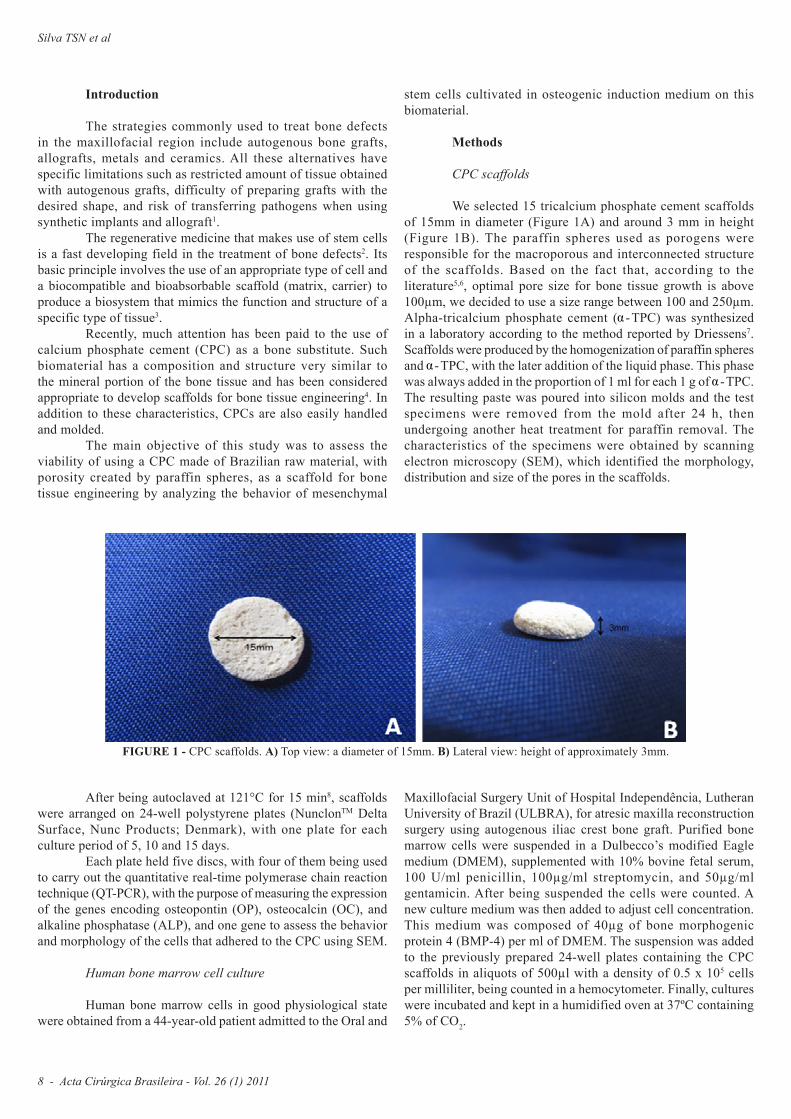
We selected 15 tricalcium phosphate cement scaffoldsof 15mm in diameter (Figure 1A) and around 3 mm in height(Figure 1B). The paraffin spheres used as porogens wereresponsible for the macroporous and interconnected structureof the scaffolds. Based on the fact that, according to theliterature5,6, optimal pore size for bone tissue growth is above100µm, we decided to use a size range between 100 and 250µm.Alpha-tricalcium phosphate cement ( -TPC) was synthesizedin a laboratory according to the method reported by Driessens7.Scaffolds were produced by the homogenization of paraffin spheresand -TPC, with the later addition of the liquid phase. This phasewas always added in the proportion of 1 ml for each 1 g of -TPC.The resulting paste was poured into silicon molds and the testspecimens were removed from the mold after 24 h, thenundergoing another heat treatment for paraffin removal. Thecharacteristics of the specimens were obtained by scanningelectron microscopy (SEM), which identified the morphology,distribution and size of the pores in the scaffolds.
FIGURE 1 - CPC scaffolds. A) Top view: a diameter of 15mm. B) Lateral view: height of approximately 3mm.
After being autoclaved at 121°C for 15 min8, scaffoldswere arranged on 24-well polystyrene plates (NunclonTM DeltaSurface, Nunc Products; Denmark), with one plate for eachculture period of 5, 10 and 15 days.
Each plate held five discs, with four of them being usedto carry out the quantitative real-time polymerase chain reactiontechnique (QT-PCR), with the purpose of measuring the expressionof the genes encoding osteopontin (OP), osteocalcin (OC), andalkaline phosphatase (ALP), and one gene to assess the behaviorand morphology of the cells that adhered to the CPC using SEM.
Human bone marrow cell culture
Human bone marrow cells in good physiological statewere obtained from a 44-year-old patient admitted to the Oral and
Maxillofacial Surgery Unit of Hospital Independência, LutheranUniversity of Brazil (ULBRA), for atresic maxilla reconstructionsurgery using autogenous iliac crest bone graft. Purified bonemarrow cells were suspended in a Dulbecco’s modified Eaglemedium (DMEM), supplemented with 10% bovine fetal serum,100 U/ml penicillin, 100µg/ml streptomycin, and 50µg/mlgentamicin. After being suspended the cells were counted. Anew culture medium was then added to adjust cell concentration.This medium was composed of 40µg of bone morphogenicprotein 4 (BMP-4) per ml of DMEM. The suspension was addedto the previously prepared 24-well plates containing the CPCscaffolds in aliquots of 500µl with a density of 0.5 x 10⁵ cellsper milliliter, being counted in a hemocytometer. Finally, cultureswere incubated and kept in a humidified oven at 37ºC containing5% of CO2.
Acta Cirúrgica Brasileira - Vol. 26 (1) 2011 - 9
Use of calcium phosphate cement scaffolds for bone tissue engineering: in vitro study
Analysis of the expression of the bgla, ssp1 and alkalinephosphatase genes
The expression levels of the BGLA and SSP1 genes,which encode OC and OP, respectively, and the ALP activity werequantified by the QT-PCR technique using fluorophore SYBRGREEN®. The QT-PCR technique used in the present studywas based on the amplification of the cDNA with gene-specificprimer oligonucleotides and detection using a laser beam ableto capture the fluorescence emitted by the fluorophore SYBRGreen® at each synthesis cycle. This process made it possibleto quantify the levels of the sequences complementary to mRNAfor those genes present in the specimens analyzed.
Scanning electron microscopy
After each culture period, the discs were assessed bySEM to analyze the behavior of the cells in contact with thebiomaterial surface. The scanning electron microscope used inour study was a PHILIPS® device, model XL30. A thin layer ofgold was sputtered on the specimens analyzed.
This research project was approved by the Scientificand Ethics Committee of the School of Dentistry of PUCRSand by the Research Ethics Committee of PUCRS. This studyis in accordance with items III.3.i and III.3.t of the BrazilianGuidelines and Norms for Research involving Humans.
Results
Characterization of the specimens
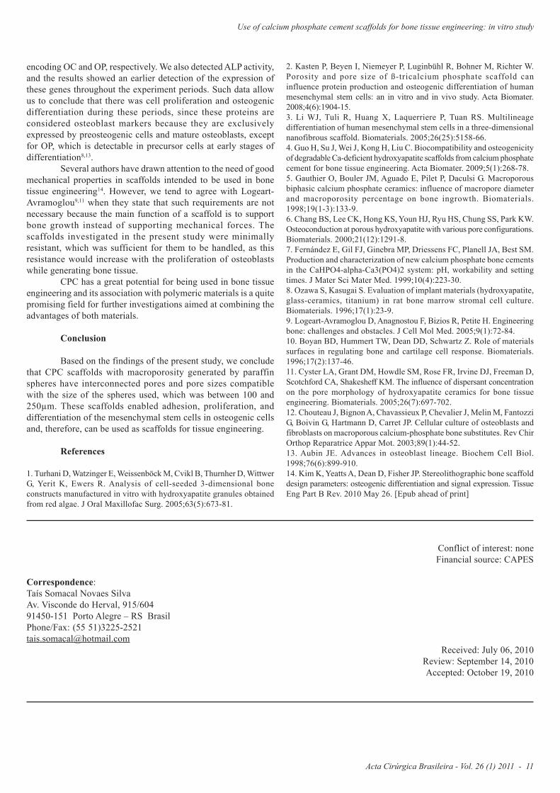
The SEM images showed the spherical shape of the poresand their interconnectivity (Figure 2). The amount of spheres addedis linked to the formation of a thin layer of cement between thespheres, enabling them to have contact with one another and,thus, after paraffin removal, promoting the establishment ofinterconnections between the pores generated by the paraffinspheres. The size of the pores we found is the same as the size ofthe paraffin spheres used, ranging from 100 and 250 µm.
FIGURE 2 - Scanning electron micrography of the macroporous CPCsurface. Magnification of 200x.
Scanning electron microscopy
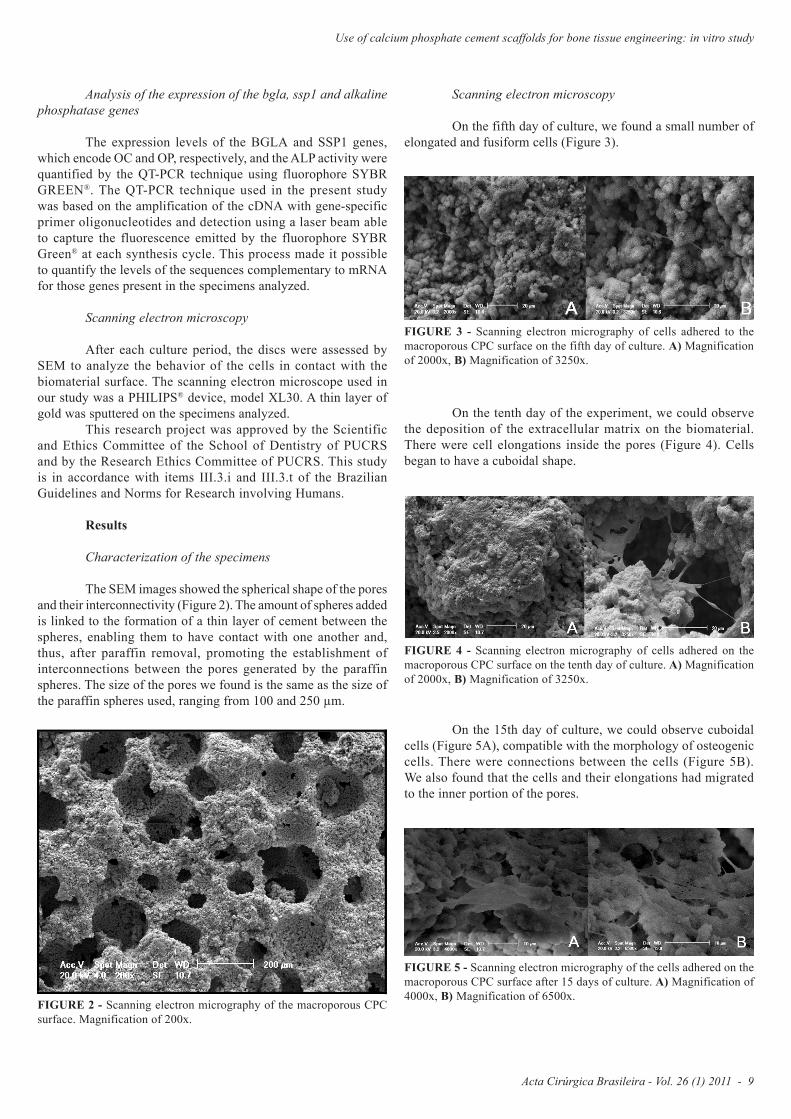
On the fifth day of culture, we found a small number ofelongated and fusiform cells (Figure 3).
FIGURE 3 - Scanning electron micrography of cells adhered to themacroporous CPC surface on the fifth day of culture. A) Magnificationof 2000x, B) Magnification of 3250x.
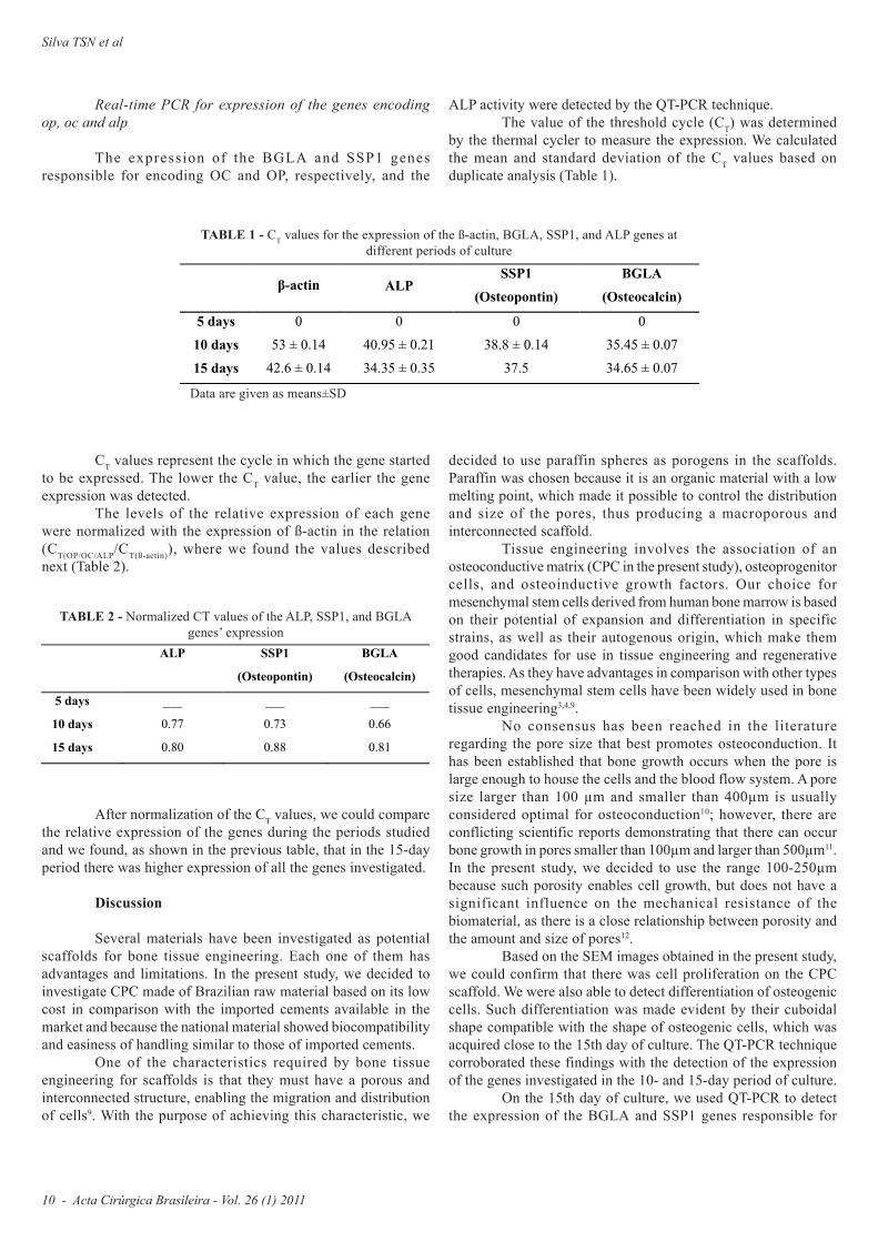
On the tenth day of the experiment, we could observethe deposition of the extracellular matrix on the biomaterial.There were cell elongations inside the pores (Figure 4). Cellsbegan to have a cuboidal shape.
FIGURE 4 - Scanning electron micrography of cells adhered on themacroporous CPC surface on the tenth day of culture. A) Magnificationof 2000x, B) Magnification of 3250x.
On the 15th day of culture, we could observe cuboidalcells (Figure 5A), compatible with the morphology of osteogeniccells. There were connections between the cells (Figure 5B).We also found that the cells and their elongations had migratedto the inner portion of the pores.
FIGURE 5 - Scanning electron micrography of the cells adhered on themacroporous CPC surface after 15 days of culture. A) Magnification of4000x, B) Magnification of 6500x.
Silva TSN et al
10 - Acta Cirúrgica Brasileira - Vol. 26 (1) 2011
CT values represent the cycle in which the gene startedto be expressed. The lower the CT value, the earlier the geneexpression was detected.
The levels of the relative expression of each genewere normalized with the expression of ß-actin in the relation(CT(OP/OC/ALP/CT(ß-actin)), where we found the values describednext (Table 2).
Real-time PCR for expression of the genes encodingop, oc and alp
The expression of the BGLA and SSP1 genesresponsible for encoding OC and OP, respectively, and the
-actin ALP SSP1
(Osteopontin)
BGLA
(Osteocalcin)
5 days 0 0 0 0
10 days 53 ± 0.14 40.95 ± 0.21 38.8 ± 0.14 35.45 ± 0.07
15 days 42.6 ± 0.14 34.35 ± 0.35 37.5 34.65 ± 0.07
TABLE 1 - CT values for the expression of the ß-actin, BGLA, SSP1, and ALP genes atdifferent periods of culture
ALP SSP1
(Osteopontin)
BGLA
(Osteocalcin)
5 days ___ ___ ___
10 days 0.77 0.73 0.66
15 days 0.80 0.88 0.81
TABLE 2 - Normalized CT values of the ALP, SSP1, and BGLAgenes’ expression
Data are given as means±SD
ALP activity were detected by the QT-PCR technique.The value of the threshold cycle (CT) was determined
by the thermal cycler to measure the expression. We calculatedthe mean and standard deviation of the CT values based onduplicate analysis (Table 1).
After normalization of the CT values, we could comparethe relative expression of the genes during the periods studiedand we found, as shown in the previous table, that in the 15-dayperiod there was higher expression of all the genes investigated.
Discussion
Several materials have been investigated as potentialscaffolds for bone tissue engineering. Each one of them hasadvantages and limitations. In the present study, we decided toinvestigate CPC made of Brazilian raw material based on its lowcost in comparison with the imported cements available in themarket and because the national material showed biocompatibilityand easiness of handling similar to those of imported cements.
One of the characteristics required by bone tissueengineering for scaffolds is that they must have a porous andinterconnected structure, enabling the migration and distributionof cells9. With the purpose of achieving this characteristic, we
decided to use paraffin spheres as porogens in the scaffolds.Paraffin was chosen because it is an organic material with a lowmelting point, which made it possible to control the distributionand size of the pores, thus producing a macroporous andinterconnected scaffold.
Tissue engineering involves the association of anosteoconductive matrix (CPC in the present study), osteoprogenitorcells, and osteoinductive growth factors. Our choice formesenchymal stem cells derived from human bone marrow is basedon their potential of expansion and differentiation in specificstrains, as well as their autogenous origin, which make themgood candidates for use in tissue engineering and regenerativetherapies. As they have advantages in comparison with other typesof cells, mesenchymal stem cells have been widely used in bonetissue engineering3,4,9.
No consensus has been reached in the literatureregarding the pore size that best promotes osteoconduction. Ithas been established that bone growth occurs when the pore islarge enough to house the cells and the blood flow system. A poresize larger than 100 µm and smaller than 400µm is usuallyconsidered optimal for osteoconduction10; however, there areconflicting scientific reports demonstrating that there can occurbone growth in pores smaller than 100µm and larger than 500µm11.In the present study, we decided to use the range 100-250µmbecause such porosity enables cell growth, but does not have asignificant influence on the mechanical resistance of thebiomaterial, as there is a close relationship between porosity andthe amount and size of pores12.
Based on the SEM images obtained in the present study,we could confirm that there was cell proliferation on the CPCscaffold. We were also able to detect differentiation of osteogeniccells. Such differentiation was made evident by their cuboidalshape compatible with the shape of osteogenic cells, which wasacquired close to the 15th day of culture. The QT-PCR techniquecorroborated these findings with the detection of the expressionof the genes investigated in the 10- and 15-day period of culture.
On the 15th day of culture, we used QT-PCR to detectthe expression of the BGLA and SSP1 genes responsible for
Acta Cirúrgica Brasileira - Vol. 26 (1) 2011 - 11
Use of calcium phosphate cement scaffolds for bone tissue engineering: in vitro study
2. Kasten P, Beyen I, Niemeyer P, Luginbühl R, Bohner M, Richter W.Porosity and pore size of ß-tricalcium phosphate scaffold caninfluence protein production and osteogenic differentiation of humanmesenchymal stem cells: an in vitro and in vivo study. Acta Biomater.2008;4(6):1904-15.3. Li WJ, Tuli R, Huang X, Laquerriere P, Tuan RS. Multilineagedifferentiation of human mesenchymal stem cells in a three-dimensionalnanofibrous scaffold. Biomaterials. 2005;26(25):5158-66.4. Guo H, Su J, Wei J, Kong H, Liu C. Biocompatibility and osteogenicityof degradable Ca-deficient hydroxyapatite scaffolds from calcium phosphatecement for bone tissue engineering. Acta Biomater. 2009;5(1):268-78.5. Gauthier O, Bouler JM, Aguado E, Pilet P, Daculsi G. Macroporousbiphasic calcium phosphate ceramics: influence of macropore diameterand macroporosity percentage on bone ingrowth. Biomaterials.1998;19(1-3):133-9.6. Chang BS, Lee CK, Hong KS, Youn HJ, Ryu HS, Chung SS, Park KW.Osteoconduction at porous hydroxyapatite with various pore configurations.Biomaterials. 2000;21(12):1291-8.7. Fernández E, Gil FJ, Ginebra MP, Driessens FC, Planell JA, Best SM.Production and characterization of new calcium phosphate bone cementsin the CaHPO4-alpha-Ca3(PO4)2 system: pH, workability and settingtimes. J Mater Sci Mater Med. 1999;10(4):223-30.8. Ozawa S, Kasugai S. Evaluation of implant materials (hydroxyapatite,glass-ceramics, titanium) in rat bone marrow stromal cell culture.Biomaterials. 1996;17(1):23-9.9. Logeart-Avramoglou D, Anagnostou F, Bizios R, Petite H. Engineeringbone: challenges and obstacles. J Cell Mol Med. 2005;9(1):72-84.10. Boyan BD, Hummert TW, Dean DD, Schwartz Z. Role of materialssurfaces in regulating bone and cartilage cell response. Biomaterials.1996;17(2):137-46.11. Cyster LA, Grant DM, Howdle SM, Rose FR, Irvine DJ, Freeman D,Scotchford CA, Shakesheff KM. The influence of dispersant concentrationon the pore morphology of hydroxyapatite ceramics for bone tissueengineering. Biomaterials. 2005;26(7):697-702.12. Chouteau J, Bignon A, Chavassieux P, Chevalier J, Melin M, FantozziG, Boivin G, Hartmann D, Carret JP. Cellular culture of osteoblasts andfibroblasts on macroporous calcium-phosphate bone substitutes. Rev ChirOrthop Reparatrice Appar Mot. 2003;89(1):44-52.13. Aubin JE. Advances in osteoblast lineage. Biochem Cell Biol.1998;76(6):899-910.14. Kim K, Yeatts A, Dean D, Fisher JP. Stereolithographic bone scaffolddesign parameters: osteogenic differentiation and signal expression. TissueEng Part B Rev. 2010 May 26. [Epub ahead of print]
encoding OC and OP, respectively. We also detected ALP activity,and the results showed an earlier detection of the expression ofthese genes throughout the experiment periods. Such data allowus to conclude that there was cell proliferation and osteogenicdifferentiation during these periods, since these proteins areconsidered osteoblast markers because they are exclusivelyexpressed by preosteogenic cells and mature osteoblasts, exceptfor OP, which is detectable in precursor cells at early stages ofdifferentiation8,13.
Several authors have drawn attention to the need of goodmechanical properties in scaffolds intended to be used in bonetissue engineering14. However, we tend to agree with Logeart-Avramoglou9,11 when they state that such requirements are notnecessary because the main function of a scaffold is to supportbone growth instead of supporting mechanical forces. Thescaffolds investigated in the present study were minimallyresistant, which was sufficient for them to be handled, as thisresistance would increase with the proliferation of osteoblastswhile generating bone tissue.
CPC has a great potential for being used in bone tissueengineering and its association with polymeric materials is a quitepromising field for further investigations aimed at combining theadvantages of both materials.
Conclusion
Based on the findings of the present study, we concludethat CPC scaffolds with macroporosity generated by paraffinspheres have interconnected pores and pore sizes compatiblewith the size of the spheres used, which was between 100 and250µm. These scaffolds enabled adhesion, proliferation, anddifferentiation of the mesenchymal stem cells in osteogenic cellsand, therefore, can be used as scaffolds for tissue engineering.
References
1. Turhani D, Watzinger E, Weissenböck M, Cvikl B, Thurnher D, WittwerG, Yerit K, Ewers R. Analysis of cell-seeded 3-dimensional boneconstructs manufactured in vitro with hydroxyapatite granules obtainedfrom red algae. J Oral Maxillofac Surg. 2005;63(5):673-81.
Conflict of interest: noneFinancial source: CAPES
Correspondence:Taís Somacal Novaes SilvaAv. Visconde do Herval, 915/60491450-151 Porto Alegre – RS BrasilPhone/Fax: (55 51)[email protected]
Received: July 06, 2010Review: September 14, 2010Accepted: October 19, 2010