Embed Size (px)
Citation preview
1
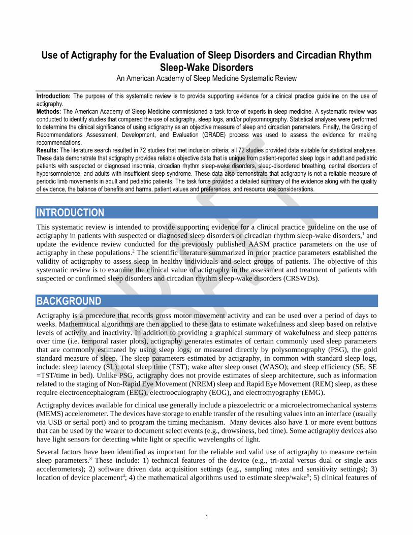
Use of Actigraphy for the Evaluation of Sleep Disorders and Circadian Rhythm Sleep-Wake Disorders
An American Academy of Sleep Medicine Systematic Review
Introduction: The purpose of this systematic review is to provide supporting evidence for a clinical practice guideline on the use of actigraphy. Methods: The American Academy of Sleep Medicine commissioned a task force of experts in sleep medicine. A systematic review was conducted to identify studies that compared the use of actigraphy, sleep logs, and/or polysomnography. Statistical analyses were performed to determine the clinical significance of using actigraphy as an objective measure of sleep and circadian parameters. Finally, the Grading of Recommendations Assessment, Development, and Evaluation (GRADE) process was used to assess the evidence for making recommendations. Results: The literature search resulted in 72 studies that met inclusion criteria; all 72 studies provided data suitable for statistical analyses. These data demonstrate that actigraphy provides reliable objective data that is unique from patient-reported sleep logs in adult and pediatric patients with suspected or diagnosed insomnia, circadian rhythm sleep-wake disorders, sleep-disordered breathing, central disorders of hypersomnolence, and adults with insufficient sleep syndrome. These data also demonstrate that actigraphy is not a reliable measure of periodic limb movements in adult and pediatric patients. The task force provided a detailed summary of the evidence along with the quality of evidence, the balance of benefits and harms, patient values and preferences, and resource use considerations.
INTRODUCTION
This systematic review is intended to provide supporting evidence for a clinical practice guideline on the use of
actigraphy in patients with suspected or diagnosed sleep disorders or circadian rhythm sleep-wake disorders,1 and
update the evidence review conducted for the previously published AASM practice parameters on the use of
actigraphy in these populations.2 The scientific literature summarized in prior practice parameters established the
validity of actigraphy to assess sleep in healthy individuals and select groups of patients. The objective of this
systematic review is to examine the clinical value of actigraphy in the assessment and treatment of patients with
suspected or confirmed sleep disorders and circadian rhythm sleep-wake disorders (CRSWDs).
BACKGROUND
Actigraphy is a procedure that records gross motor movement activity and can be used over a period of days to
weeks. Mathematical algorithms are then applied to these data to estimate wakefulness and sleep based on relative
levels of activity and inactivity. In addition to providing a graphical summary of wakefulness and sleep patterns
over time (i.e. temporal raster plots), actigraphy generates estimates of certain commonly used sleep parameters
that are commonly estimated by using sleep logs, or measured directly by polysomnography (PSG), the gold
standard measure of sleep. The sleep parameters estimated by actigraphy, in common with standard sleep logs,
include: sleep latency (SL); total sleep time (TST); wake after sleep onset (WASO); and sleep efficiency (SE; SE
=TST/time in bed). Unlike PSG, actigraphy does not provide estimates of sleep architecture, such as information
related to the staging of Non-Rapid Eye Movement (NREM) sleep and Rapid Eye Movement (REM) sleep, as these
require electroencephalogram (EEG), electrooculography (EOG), and electromyography (EMG).
Actigraphy devices available for clinical use generally include a piezoelectric or a microelectromechanical systems
(MEMS) accelerometer. The devices have storage to enable transfer of the resulting values into an interface (usually
via USB or serial port) and to program the timing mechanism. Many devices also have 1 or more event buttons
that can be used by the wearer to document select events (e.g., drowsiness, bed time). Some actigraphy devices also
have light sensors for detecting white light or specific wavelengths of light.
Several factors have been identified as important for the reliable and valid use of actigraphy to measure certain
sleep parameters.3 These include: 1) technical features of the device (e.g., tri-axial versus dual or single axis
accelerometers); 2) software driven data acquisition settings (e.g., sampling rates and sensitivity settings); 3)
location of device placement4; 4) the mathematical algorithms used to estimate sleep/wake5; 5) clinical features of
2
the population being studied, 6) utilization of a standardized scoring approach to setting rest activity intervals; and
7) training of patients in data collection procedures.6 Standardized information on the technical aspects of actigraphy
and analysis and interpretation procedures for clinical and research use have recently been published.7 It is important
to note that the basic technology in products sold “direct to consumers” may differ significantly from what is
available for clinical application. It is important to note that the basic technology in products sold “direct to
consumers” may differ significantly from what is available for clinical application. The basic technology in products
sold “direct to consumers” may differ significantly from what is available for clinical application. At the present
time, data are not adequate to suggest that consumer products can be used as a replacement for clinical devices
using validated sleep scoring algorithms, technologies, and procedures.
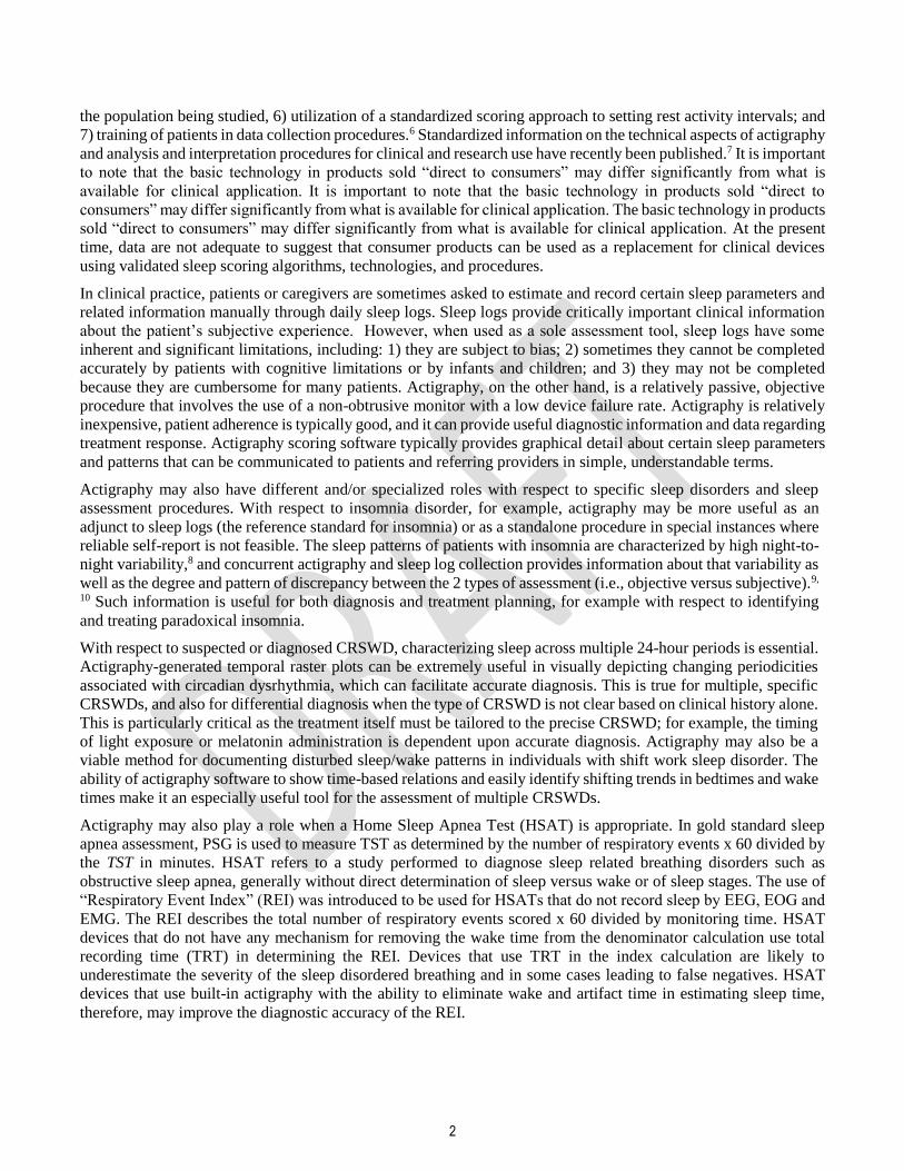
In clinical practice, patients or caregivers are sometimes asked to estimate and record certain sleep parameters and
related information manually through daily sleep logs. Sleep logs provide critically important clinical information
about the patient’s subjective experience. However, when used as a sole assessment tool, sleep logs have some
inherent and significant limitations, including: 1) they are subject to bias; 2) sometimes they cannot be completed
accurately by patients with cognitive limitations or by infants and children; and 3) they may not be completed
because they are cumbersome for many patients. Actigraphy, on the other hand, is a relatively passive, objective
procedure that involves the use of a non-obtrusive monitor with a low device failure rate. Actigraphy is relatively
inexpensive, patient adherence is typically good, and it can provide useful diagnostic information and data regarding
treatment response. Actigraphy scoring software typically provides graphical detail about certain sleep parameters
and patterns that can be communicated to patients and referring providers in simple, understandable terms.
Actigraphy may also have different and/or specialized roles with respect to specific sleep disorders and sleep
assessment procedures. With respect to insomnia disorder, for example, actigraphy may be more useful as an
adjunct to sleep logs (the reference standard for insomnia) or as a standalone procedure in special instances where
reliable self-report is not feasible. The sleep patterns of patients with insomnia are characterized by high night-to-
night variability,8 and concurrent actigraphy and sleep log collection provides information about that variability as
well as the degree and pattern of discrepancy between the 2 types of assessment (i.e., objective versus subjective).9,
10 Such information is useful for both diagnosis and treatment planning, for example with respect to identifying
and treating paradoxical insomnia.
With respect to suspected or diagnosed CRSWD, characterizing sleep across multiple 24-hour periods is essential.
Actigraphy-generated temporal raster plots can be extremely useful in visually depicting changing periodicities
associated with circadian dysrhythmia, which can facilitate accurate diagnosis. This is true for multiple, specific
CRSWDs, and also for differential diagnosis when the type of CRSWD is not clear based on clinical history alone.
This is particularly critical as the treatment itself must be tailored to the precise CRSWD; for example, the timing
of light exposure or melatonin administration is dependent upon accurate diagnosis. Actigraphy may also be a
viable method for documenting disturbed sleep/wake patterns in individuals with shift work sleep disorder. The
ability of actigraphy software to show time-based relations and easily identify shifting trends in bedtimes and wake
times make it an especially useful tool for the assessment of multiple CRSWDs.
Actigraphy may also play a role when a Home Sleep Apnea Test (HSAT) is appropriate. In gold standard sleep
apnea assessment, PSG is used to measure TST as determined by the number of respiratory events x 60 divided by
the TST in minutes. HSAT refers to a study performed to diagnose sleep related breathing disorders such as
obstructive sleep apnea, generally without direct determination of sleep versus wake or of sleep stages. The use of
“Respiratory Event Index” (REI) was introduced to be used for HSATs that do not record sleep by EEG, EOG and
EMG. The REI describes the total number of respiratory events scored x 60 divided by monitoring time. HSAT
devices that do not have any mechanism for removing the wake time from the denominator calculation use total
recording time (TRT) in determining the REI. Devices that use TRT in the index calculation are likely to
underestimate the severity of the sleep disordered breathing and in some cases leading to false negatives. HSAT
devices that use built-in actigraphy with the ability to eliminate wake and artifact time in estimating sleep time,
therefore, may improve the diagnostic accuracy of the REI.
3
Actigraphy may be especially useful in documenting insufficient sleep both for the purpose of improving the
interpretation of Multiple Sleep Latency Testing (MSLT) in patients with suspected disorders of hypersomnolence
and for assessing insufficient sleep syndrome (ISS), especially in high risk occupations such as medicine,
transportation and the military. Objective measurement may be especially important in facilitating the sometime
complex medical and occupational risks associated with ISS. Some studies have sought to evaluate whether
actigraphy worn on the ankles might provide a reasonable estimate of periodic limb movements, although it is
increasingly clear that additional measures of arousal may be important in evaluating the clinical significance of
PLMS. Finally, actigraphy has been utilized in several studies of infants and children ranging in ages between
several months to 18 years old and to identify sleep disruption in psychiatric, neurodevelopmental, medical, and
sleep disorders including: insomnia, CRSWDs, and SDB.11-21 In each of these cases, standard sleep logs and
laboratory PSG both have limitations, and actigraphy might significantly add to the clinical evaluation of a patient.
METHODOLOGY
Expert Task Force
The AASM commissioned a task force (TF) of sleep medicine clinicians with expertise in the use of actigraphy in
patients with suspected sleep disorders to develop this systematic review. These content experts were required to
disclose all potential conflicts of interest (COI) according to the AASM’s COI policy prior to being appointed to
the TF, and throughout the development of this document. In accordance with the AASM’s conflicts of interest
policy, TF members with a Level 1 conflict were not allowed to participate. TF members with a Level 2 conflict
were required to recuse themselves from any related discussion or writing responsibilities. All relevant conflicts of
interest are listed in the Disclosures section.
PICO Questions
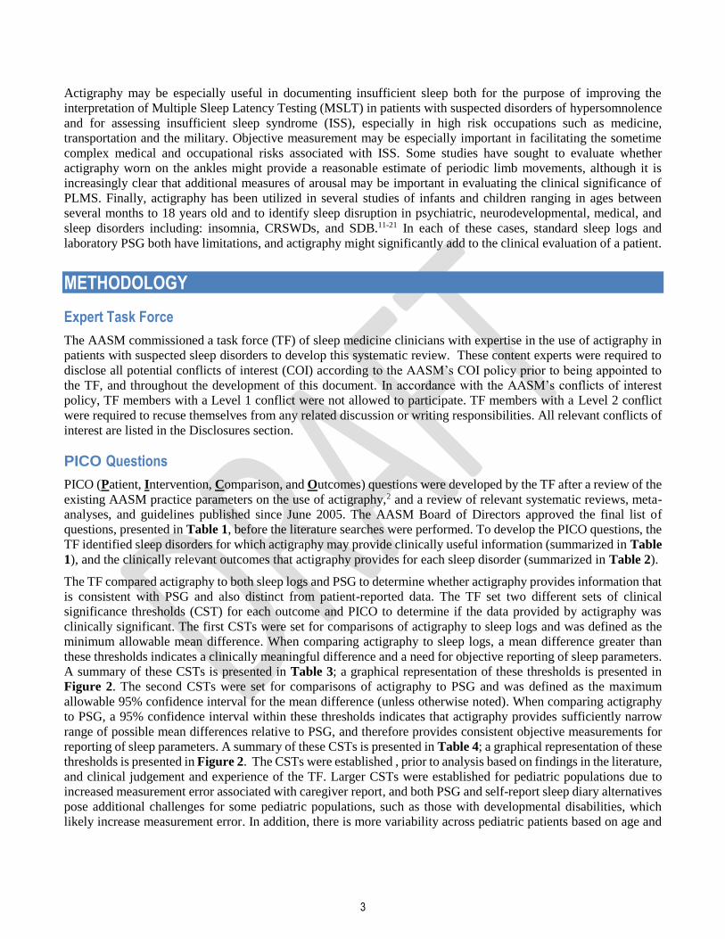
PICO (Patient, Intervention, Comparison, and Outcomes) questions were developed by the TF after a review of the
existing AASM practice parameters on the use of actigraphy,2 and a review of relevant systematic reviews, meta-
analyses, and guidelines published since June 2005. The AASM Board of Directors approved the final list of
questions, presented in Table 1, before the literature searches were performed. To develop the PICO questions, the
TF identified sleep disorders for which actigraphy may provide clinically useful information (summarized in Table
1), and the clinically relevant outcomes that actigraphy provides for each sleep disorder (summarized in Table 2).
The TF compared actigraphy to both sleep logs and PSG to determine whether actigraphy provides information that
is consistent with PSG and also distinct from patient-reported data. The TF set two different sets of clinical
significance thresholds (CST) for each outcome and PICO to determine if the data provided by actigraphy was
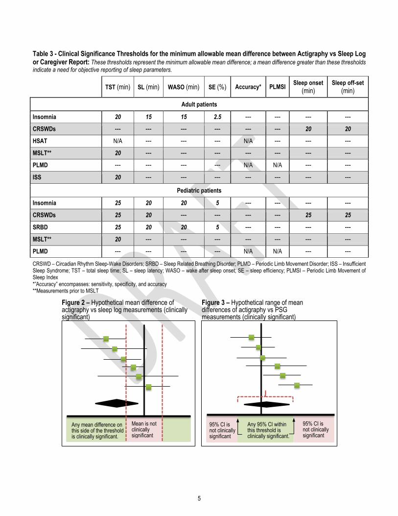
clinically significant. The first CSTs were set for comparisons of actigraphy to sleep logs and was defined as the
minimum allowable mean difference. When comparing actigraphy to sleep logs, a mean difference greater than
these thresholds indicates a clinically meaningful difference and a need for objective reporting of sleep parameters.
A summary of these CSTs is presented in Table 3; a graphical representation of these thresholds is presented in
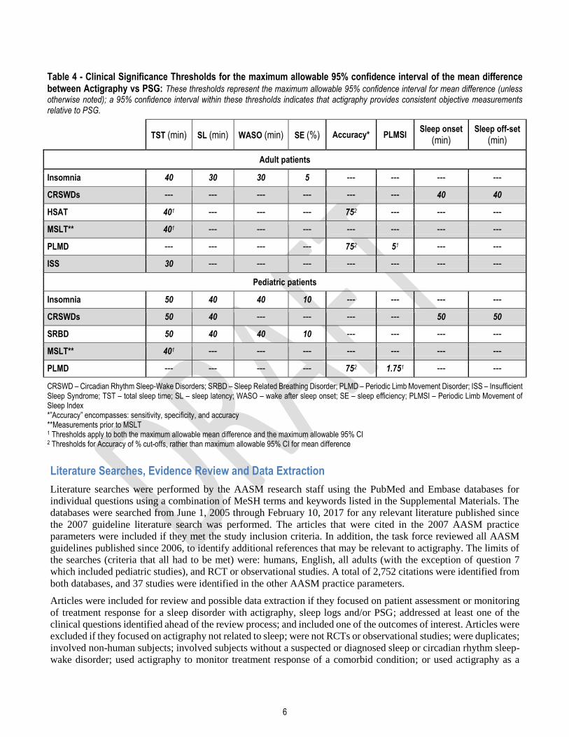
Figure 2. The second CSTs were set for comparisons of actigraphy to PSG and was defined as the maximum
allowable 95% confidence interval for the mean difference (unless otherwise noted). When comparing actigraphy
to PSG, a 95% confidence interval within these thresholds indicates that actigraphy provides sufficiently narrow
range of possible mean differences relative to PSG, and therefore provides consistent objective measurements for
reporting of sleep parameters. A summary of these CSTs is presented in Table 4; a graphical representation of these
thresholds is presented in Figure 2. The CSTs were established , prior to analysis based on findings in the literature,
and clinical judgement and experience of the TF. Larger CSTs were established for pediatric populations due to
increased measurement error associated with caregiver report, and both PSG and self-report sleep diary alternatives
pose additional challenges for some pediatric populations, such as those with developmental disabilities, which
likely increase measurement error. In addition, there is more variability across pediatric patients based on age and
4
other factors. The TF endeavored to balance the need for accuracy, care giver burden, and the differential sleep
needs of pediatric groups relative to adults.
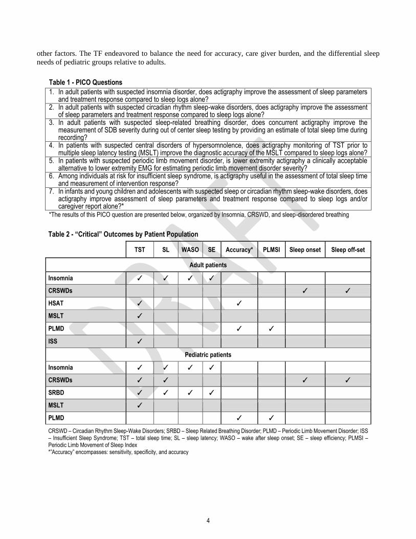
Table 1 - PICO Questions
1. In adult patients with suspected insomnia disorder, does actigraphy improve the assessment of sleep parameters and treatment response compared to sleep logs alone?
2. In adult patients with suspected circadian rhythm sleep-wake disorders, does actigraphy improve the assessment of sleep parameters and treatment response compared to sleep logs alone?
3. In adult patients with suspected sleep-related breathing disorder, does concurrent actigraphy improve the measurement of SDB severity during out of center sleep testing by providing an estimate of total sleep time during recording?
4. In patients with suspected central disorders of hypersomnolence, does actigraphy monitoring of TST prior to multiple sleep latency testing (MSLT) improve the diagnostic accuracy of the MSLT compared to sleep logs alone?
5. In patients with suspected periodic limb movement disorder, is lower extremity actigraphy a clinically acceptable alternative to lower extremity EMG for estimating periodic limb movement disorder severity?
6. Among individuals at risk for insufficient sleep syndrome, is actigraphy useful in the assessment of total sleep time and measurement of intervention response?
7. In infants and young children and adolescents with suspected sleep or circadian rhythm sleep-wake disorders, does actigraphy improve assessment of sleep parameters and treatment response compared to sleep logs and/or caregiver report alone?*
*The results of this PICO question are presented below, organized by Insomnia, CRSWD, and sleep-disordered breathing
Table 2 - “Critical” Outcomes by Patient Population
TST SL WASO SE Accuracy* PLMSI Sleep onset Sleep off-set
Adult patients
Insomnia ✓ ✓ ✓ ✓
CRSWDs ✓ ✓
HSAT ✓ ✓
MSLT ✓
PLMD ✓ ✓
ISS ✓
Pediatric patients
Insomnia ✓ ✓ ✓ ✓
CRSWDs ✓ ✓ ✓ ✓
SRBD ✓ ✓ ✓ ✓
MSLT ✓
PLMD ✓ ✓
CRSWD – Circadian Rhythm Sleep-Wake Disorders; SRBD – Sleep Related Breathing Disorder; PLMD – Periodic Limb Movement Disorder; ISS – Insufficient Sleep Syndrome; TST – total sleep time; SL – sleep latency; WASO – wake after sleep onset; SE – sleep efficiency; PLMSI – Periodic Limb Movement of Sleep Index *”Accuracy” encompasses: sensitivity, specificity, and accuracy
5
Table 3 - Clinical Significance Thresholds for the minimum allowable mean difference between Actigraphy vs Sleep Log or Caregiver Report: These thresholds represent the minimum allowable mean difference; a mean difference greater than these thresholds indicate a need for objective reporting of sleep parameters.
TST (min) SL (min) WASO (min) SE (%) Accuracy* PLMSI
Sleep onset
(min) Sleep off-set
(min)
Adult patients
Insomnia 20 15 15 2.5 --- --- --- ---
CRSWDs --- --- --- --- --- --- 20 20
HSAT N/A --- --- --- N/A --- --- ---
MSLT** 20 --- --- --- --- --- --- ---
PLMD --- --- --- --- N/A N/A --- ---
ISS 20 --- --- --- --- --- --- ---
Pediatric patients
Insomnia 25 20 20 5 --- --- --- ---
CRSWDs 25 20 --- --- --- --- 25 25
SRBD 25 20 20 5 --- --- --- ---
MSLT** 20 --- --- --- --- --- --- ---
PLMD --- --- --- --- N/A N/A --- ---
CRSWD – Circadian Rhythm Sleep-Wake Disorders; SRBD – Sleep Related Breathing Disorder; PLMD – Periodic Limb Movement Disorder; ISS – Insufficient Sleep Syndrome; TST – total sleep time; SL – sleep latency; WASO – wake after sleep onset; SE – sleep efficiency; PLMSI – Periodic Limb Movement of Sleep Index *”Accuracy” encompasses: sensitivity, specificity, and accuracy **Measurements prior to MSLT
Mean is not clinically significant
Figure 2 – Hypothetical mean difference of actigraphy vs sleep log measurements (clinically significant)
Any mean difference on this side of the threshold is clinically significant.
Figure 3 – Hypothetical range of mean differences of actigraphy vs PSG measurements (clinically significant)
Any 95% CI within this threshold is clinically significant.
95% CI is not clinically significant
95% CI is not clinically significant
6
Table 4 - Clinical Significance Thresholds for the maximum allowable 95% confidence interval of the mean difference between Actigraphy vs PSG: These thresholds represent the maximum allowable 95% confidence interval for mean difference (unless otherwise noted); a 95% confidence interval within these thresholds indicates that actigraphy provides consistent objective measurements relative to PSG.
TST (min) SL (min) WASO (min) SE (%) Accuracy* PLMSI
Sleep onset
(min) Sleep off-set
(min)
Adult patients
Insomnia 40 30 30 5 --- --- --- ---
CRSWDs --- --- --- --- --- --- 40 40
HSAT 401 --- --- --- 752 --- --- ---
MSLT** 401 --- --- --- --- --- --- ---
PLMD --- --- --- --- 752 51 --- ---
ISS 30 --- --- --- --- --- --- ---
Pediatric patients
Insomnia 50 40 40 10 --- --- --- ---
CRSWDs 50 40 --- --- --- --- 50 50
SRBD 50 40 40 10 --- --- --- ---
MSLT** 401 --- --- --- --- --- --- ---
PLMD --- --- --- --- 752 1.751 --- ---
CRSWD – Circadian Rhythm Sleep-Wake Disorders; SRBD – Sleep Related Breathing Disorder; PLMD – Periodic Limb Movement Disorder; ISS – Insufficient Sleep Syndrome; TST – total sleep time; SL – sleep latency; WASO – wake after sleep onset; SE – sleep efficiency; PLMSI – Periodic Limb Movement of Sleep Index *”Accuracy” encompasses: sensitivity, specificity, and accuracy **Measurements prior to MSLT 1 Thresholds apply to both the maximum allowable mean difference and the maximum allowable 95% CI 2 Thresholds for Accuracy of % cut-offs, rather than maximum allowable 95% CI for mean difference
Literature Searches, Evidence Review and Data Extraction
Literature searches were performed by the AASM research staff using the PubMed and Embase databases for
individual questions using a combination of MeSH terms and keywords listed in the Supplemental Materials. The
databases were searched from June 1, 2005 through February 10, 2017 for any relevant literature published since
the 2007 guideline literature search was performed. The articles that were cited in the 2007 AASM practice
parameters were included if they met the study inclusion criteria. In addition, the task force reviewed all AASM
guidelines published since 2006, to identify additional references that may be relevant to actigraphy. The limits of
the searches (criteria that all had to be met) were: humans, English, all adults (with the exception of question 7
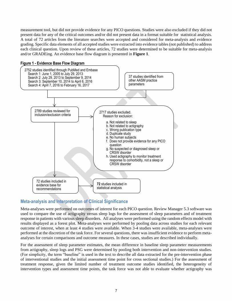
which included pediatric studies), and RCT or observational studies. A total of 2,752 citations were identified from
both databases, and 37 studies were identified in the other AASM practice parameters.
Articles were included for review and possible data extraction if they focused on patient assessment or monitoring
of treatment response for a sleep disorder with actigraphy, sleep logs and/or PSG; addressed at least one of the
clinical questions identified ahead of the review process; and included one of the outcomes of interest. Articles were
excluded if they focused on actigraphy not related to sleep; were not RCTs or observational studies; were duplicates;
involved non-human subjects; involved subjects without a suspected or diagnosed sleep or circadian rhythm sleep-
wake disorder; used actigraphy to monitor treatment response of a comorbid condition; or used actigraphy as a
7
measurement tool, but did not provide evidence for any PICO questions. Studies were also excluded if they did not
present data for any of the critical outcomes and/or did not present data in a format suitable for statistical analysis.
A total of 72 articles from the literature searches were accepted and considered for meta-analysis and evidence
grading. Specific data elements of all accepted studies were extracted into evidence tables (not published) to address
each clinical question. Upon review of these articles, 72 studies were determined to be suitable for meta-analysis
and/or GRADEing. An evidence base flow diagram is presented in Figure 1.
Figure 1 - Evidence Base Flow Diagram
Meta-analysis and Interpretation of Clinical Significance
Meta-analyses were performed on outcomes of interest for each PICO question. Review Manager 5.3 software was
used to compare the use of actigraphy versus sleep logs for the assessment of sleep parameters and of treatment
response in patients with various sleep disorders. All analyses were performed using the random effects model with
results displayed as a forest plot. Meta-analyses were performed by pooling data across studies for each relevant
outcome of interest, when at least 4 studies were available. When 3-4 studies were available, meta-analyses were
performed at the discretion of the task force. For several questions, there was insufficient evidence to perform meta-
analyses for certain comparisons and outcome measures. In these cases, studies are described individually.
For the assessment of sleep parameter estimates, the mean difference in baseline sleep parameter measurements
from actigraphy, sleep logs and PSG were determined by pooling both intervention and non-intervention studies.
(For simplicity, the term “baseline” is used in the text to describe all data extracted for the pre-intervention phase
of interventional studies and the initial assessment time point for cross sectional studies.) For the assessment of
treatment response, given the limited number of treatment outcome studies identified, the heterogeneity of
intervention types and assessment time points, the task force was not able to evaluate whether actigraphy was
72 studies included in evidence base for recommendations
72 studies included in statistical analysis
2789 studies reviewed for inclusion/exclusion criteria
2717 studies excluded. Reason for exclusion:
a. Not related to sleep b. Not related to actigraphy c. Wrong publication type d. Duplicate study e. No human subjects f. Does not provide evidence for any PICO
question g. No suspected or diagnosed sleep or
CRSW disorder h. Used actigraphy to monitor treatment
response to comorbidity, not a sleep or CRSW disorder
2752 studies identified through PubMed and Embase Search 1: June 1, 2005 to July 29, 2013 Search 2: July 29, 2013 to September 9, 2014 Search 3: September 10, 2014 to April 6, 2016 Search 4: April 7, 2016 to February 16, 2017
37 studies identified from other AASM practice parameters
8
sensitive to change relative to sleep logs or PSG. Instead, the TF analyzed the mean difference of post-treatment
measurements from actigraphy, sleep logs and PSG. The pooled results for each continuous outcome measure are
expressed as the mean difference between the intervention and comparator. The results of the meta-analyses are
presented in the Supplemental Materials.
Interpretation of clinical significance for the outcomes of interest was conducted in two different ways. First, by
comparing the mean difference in measurements of actigraphy and sleep logs against their CSTs (see Table 3).
Next, by comparing the 95% confidence interval of the mean difference of actigraphy vs PSG measurements to
their CSTs (see Table 4). For comparisons of actigraphy to sleep logs, the CST was defined as the minimum
allowable mean difference; a mean difference greater than the threshold demonstrates that actigraphy provides
unique information from sleep logs, and objective measurements are warranted (see Figure 2, which shows an
example of a clinically significant mean difference). For comparisons of actigraphy to PSG, the CST was defined
as the maximum allowable 95% confidence interval for the mean difference between actigraphy and PSG (unless
otherwise noted); a 95% confidence interval within the threshold demonstrates that actigraphy provides sufficiently
narrow range of possible mean differences relative to PSG (regardless of the mean difference, unless otherwise
noted). A sufficiently narrow range of mean differences indicates that actigraphy provides consistent objective
measurements and may be useful as an objective measurement of sleep parameters (see Figure 3, which shows an
example of a sufficiently narrow range of mean differences).
A detailed review of the evidence and clinical significance of the findings for all critical outcome are provided for
each PICO question.
GRADE Assessment for Developing Recommendations
The evidence was assessed according to the Grading of Recommendations Assessment, Development and
Evaluation (GRADE) process.22, 23 The TF considered the following four GRADE domains: quality of evidence,
balance of beneficial and harmful effects, patient values and preferences and resource use, as described below.
1. Quality of evidence – based on an assessment of the overall risk of bias (randomization, blinding, allocation
concealment, selective reporting), imprecision (95% confidence interval relative to the CST, sample size <
200), inconsistency and indirectness (study population), and risk of publication bias (funding sources), the
TF determined their overall confidence that the estimated differences in measurements found in the body
of evidence were representative of the true differences in measurements that patients would experience.
The overall quality of the evidence was based on all outcomes that the TF deemed critical for decision
making.
2. Benefits vs. Harms – based on any harms/side effects reported within the accepted literature, and the clinical
expertise of the TF, the TF determined if the beneficial outcomes of using actigraphy outweighed any
harmful side effects.
3. Patient values and preferences – based on the clinical expertise of the TF members and any data published
on the topic relevant to patient preferences, the TF determined if patient values and preferences would be
consistent across the majority of patients, and if patients would use actigraphy based on the body of
evidence.
4. Resource use – based on the clinical expertise of the TF members, the TF determined if accessibility and
costs associated with actigraphy compared favorably to alternative measurement tools. Information on both
costs to patients and to the healthcare system were considered.
A summary of each GRADE domain is provided after the detailed evidence review for each PICO.
Public Comment and Final Approval
A draft of the systematic review and accompanying guideline were made available for public comment for a two-
week period on the AASM website. The TF took into consideration all the comments received and made decisions
about whether to revise the draft based on the comments. The public comments and revised documents were
submitted to the AASM BOD who subsequently approved them for publication.
9
The AASM expects this systematic review to have an impact on professional behavior, patient outcomes, and,
possibly, health care costs. This review reflects the state of knowledge at the time of publication and will be
reviewed and updated as new information becomes available.
THE USE OF ACTIGRAPHY
The aims of the current systematic reviews and data analyses were to address 7 PICO questions pertaining to the
use of actigraphy relative to sleep logs across a wide range of clinical populations, and in conjunction with HSAT
and MSLT. While sufficient data were available for meta-analyses for most PICO questions, there are caveats that
should be considered with respect to interpreting the results. With regard to sleep parameters, the TF noted
variability across studies with respect to definitions and technical details such as algorithms and sensitivity threshold
settings used or reported. As is common practice, many studies utilized information noted by the patient in a “sleep
log” for the analysis and interpretation of actigraphy-estimated sleep parameters. This is important particularly with
respect to determining bedtime (“lights off”) to calculate SL. Other studies relied completely on actigraphy
algorithms to estimate SL, while some studies failed to report these details. The TF decided not to analyze the
number of nightly awakenings as a sleep parameter of interest, since actigraphy definitions of what constitutes an
awakening are variable and comparison across studies would not be possible. The TF also cautions that
generalizability of some of the meta-analytic findings may be limited due to a small number of studies meeting the
inclusion/exclusion criteria and/or a small number of patients across studies. Generalizability to the broad spectrum
of sleep disorder patients seen in clinical settings may also be limited by heterogeneity across sleep disorder severity
and subpopulations with clinical comorbidities, both of which may influence validity.
Below are detailed summaries of the evidence identified in the literature searches and the statistical analyses
performed by the task force. Each evidence summary is accompanied by a discussion of the quality of evidence,
balance of benefits and harms, patient values and preferences, and resource use considerations that contributed to
the development of the recommendations. The recommendations are provided in the accompanying clinical practice
guideline.1
Use of Actigraphy in the Evaluation of Insomnia in Adults
Our review of the literature identified 42 studies10, 24-64 that used actigraphy concurrent with sleep logs and/or PSG
in adults with suspected or diagnosed insomnia. Both non-intervention and intervention studies met the eligibility
criteria and were included. The number of studies included in the analyses varied by sleep parameter and whether
the comparison was to sleep logs or PSG. Overall, more studies were identified that provided comparisons to sleep
logs than to PSG.
The data for examining the use of actigraphy for assessment were either based on a single night or drawn from the
baseline periods of intervention trials with insomnia and represent sleep parameter values averaged over 1 to 2
weeks. Similarly, data for analyses examining the use of actigraphy to assess treatment response were either based
on a single night or were drawn from sleep parameter values averaged over 1 to 2 weeks following treatment. The
vast majority of the intervention studies reviewed involved 1 or more components of cognitive-behavioral treatment
for insomnia.
The meta-analyses and figures are provided in the Supplemental Materials, Figure S1a through Figure S8b.
Summary of Findings tables are provided in the Supplemental Materials, Table S1a through S2b. A summary of
the evidence for each outcome is provided below.
TOTAL SLEEP TIME: A meta-analysis of 37 studies10, 24-59 compared actigraphy to sleep logs for the assessment of TST
in patients with suspected or diagnosed insomnia (see Supplemental Materials, Figure S1a). The meta-analysis
showed a clinically significant mean difference of 27.7 minutes higher (95% CI: 11.6 to 43.8 minutes higher) TST
as assessed by actigraphy compared to sleep logs. This difference indicates actigraphy and sleep logs provide
10
distinct information when assessing TST. The quality of evidence was moderate due to imprecision.
A meta-analysis of 14 studies24, 25, 27, 29-31, 33, 36, 42, 48-50, 52, 55 compared actigraphy to PSG for the assessment of TST
in patients with suspected or diagnosed insomnia (see Supplemental Materials, Figure S1b). The meta-analysis
showed a clinically significant range of possible mean differences of 36.7 minutes (95% CI: -9.7 minutes lower to
27.0 minutes higher) with an overall mean difference of 8.6 minutes. This range is narrow enough that actigraphy
can be reliably used to provide an objective assessment of TST for the purpose of making clinical care decisions.
The quality of evidence was high.
A meta-analysis of 2710, 34-59 studies compared actigraphy to sleep logs for the assessment of treatment response in
TST in patients with suspected or diagnosed insomnia (see Supplemental Materials, Figure S5a). The meta-analysis
demonstrated a clinically insignificant mean difference in TST measured by actigraphy of 8.1 minutes higher (95%
CI: 10.3 minutes lower to 26.5 minutes higher) as compared to logs. This small difference indicates actigraphy and
sleep logs provide similar measurements of treatment-related changes in TST. The quality of evidence was moderate
due to imprecision.
A meta-analysis of 7 studies36, 42, 48-50, 52, 55 compared actigraphy to PSG for the assessment of treatment response in
TST in patients with suspected or diagnosed insomnia (see Supplemental Materials, Figure S5b). The meta-analysis
demonstrated a clinically insignificant range of possible mean differences of 83.3 minutes (95% CI: 37.0 minutes
lower to 46.2 minutes higher) with an overall mean difference of 4.6 minutes. This large range indicates actigraphy
and PSG provide distinct information and should not be used interchangeably for the assessment of treatment-
related changes in TST. The quality of evidence was moderate due to imprecision.
SLEEP LATENCY: A meta-analysis of 34 studies10, 24-31, 33, 35, 37-41, 43-51, 53-59, 62, 64 compared actigraphy to sleep logs for
the assessment of SL in patients with suspected or diagnosed insomnia (see Supplemental Materials, Figure S2a).
The meta-analysis showed a clinically significant mean difference in SL measured by actigraphy of 23.6 minutes
lower (95% CI: 20.3 to 26.9 minutes lower) as compared to sleep logs. This difference indicates actigraphy and
sleep logs provide distinct information when assessing SL. The quality of evidence was high.
A meta-analysis of 11 studies24, 25, 27, 29-31, 33, 48-50, 55 compared actigraphy to PSG for the assessment of SL in patients
with suspected or diagnosed insomnia (see Supplemental Materials, Figure S2b). The meta-analysis showed a
clinically significant range of possible mean differences of 6.4 minutes (95% CI: 2.2 to 8.6 minutes lower) with a
mean difference of 5.4 minutes. This range is narrow enough that actigraphy can be reliably used to provide an
objective assessment of SL for the purpose of making clinical care decisions. The quality of evidence was high.
A meta-analysis of 25 studies10, 35, 37-41, 43-51, 53-59, 62, 64 compared actigraphy to sleep logs for the assessment of
treatment response in SL in patients with suspected or diagnosed insomnia (see Supplemental Materials, Figure
S6a). The meta-analysis demonstrated a clinically insignificant mean difference in SL measured by actigraphy of
10.5 minutes lower (95% CI: 8.1 to 13.0 minutes lower) as compared to sleep logs. This small difference indicates
actigraphy and sleep logs provide similar measurements of treatment-related changes in SL. The quality of evidence
was high.
Four studies48-50, 55 compared actigraphy to PSG for the assessment of treatment response in SL in patients with
suspected or diagnosed insomnia (see Supplemental Materials, Figure S6b). All studies reported a clinically
significant range of possible mean differences, with the largest range of differences being 29.8 minutes (95% CI:
12.1 minutes lower to 17.7 minutes higher). This small range indicates actigraphy and PSG provide similar
information for the assessment of treatment-related changes in SL. The quality of evidence was moderate due to
imprecision due to small sample size.
WAKE AFTER SLEEP ONSET: A meta-analysis of 33 studies10, 24-32, 34, 36, 37, 39-41, 43-52, 55-59, 62, 64 compared actigraphy to
sleep logs for the assessment of WASO in patients with suspected or diagnosed insomnia (see Supplemental
Materials, Figure S3a). The meta-analysis showed a clinically insignificant mean difference in WASO measured
11
by actigraphy of 5.2 minutes lower (95% CI: 17.3 minutes lower to 6.9 minutes higher) as compared to sleep logs.
This difference indicates actigraphy and sleep logs do not provide distinct information when assessing WASO. The
quality of evidence was high.
A meta-analysis of 12 studies24, 25, 27, 29-31, 36, 48-50, 52, 55 compared actigraphy to PSG for the assessment of WASO in
patients with suspected or diagnosed insomnia (see Supplemental Materials, Figure S3b). The meta-analysis
showed a clinically insignificant range of possible mean differences of 34.7 minutes (95% CI: 15.8 minutes lower
to 18.9 minutes higher), with a mean difference of 1.5 minutes. This large range indicates actigraphy cannot be
reliably used to provide an objective assessment of WASO that is comparable with PSG. The quality of evidence
was downgraded to moderate due to imprecision.
A meta-analysis of 24 studies10, 34, 36, 37, 39-41, 43-52, 55-59, 62, 64 compared actigraphy to sleep logs for the assessment of
treatment response in WASO in patients with suspected or diagnosed insomnia (see Supplemental Materials, Figure
S7a). The meta-analysis demonstrated a clinically insignificant mean difference in WASO measured by actigraphy
of 9.6 minutes higher (95% CI: 2.1 minutes lower to 21.2 minutes higher) as compared to sleep logs. This small
difference indicates actigraphy and sleep logs provide similar measurements of treatment-related changes in
WASO. The quality of evidence was moderate due to imprecision.
A meta-analysis of 6 studies36, 48-50, 52, 55 compared actigraphy to PSG for the assessment of treatment response in
WASO in patients with suspected or diagnosed insomnia (see Supplemental Materials, Figure S7b). The meta-
analysis demonstrated a clinically insignificant range of possible mean difference in WASO measured by actigraphy
as compared to PSG of 86.0 minutes (95% CI: 53.2 minutes lower to 32.8 minutes higher) with a mean difference
of 10.2 minutes. This large range indicates actigraphy and PSG provide distinct information and cannot be used
interchangeably for the assessment of treatment-related changes in WASO. The quality of evidence was moderate
due to imprecision.
SLEEP EFFICIENCY: A meta-analysis of 32 studies10, 24, 25, 27-29, 32, 34, 37-52, 55-60, 62, 64 compared actigraphy to sleep logs for
the assessment of SE in patients with suspected or diagnosed insomnia (see Supplemental Materials, Figure S4a).
The meta-analysis showed a clinically significant mean difference in SE measured by actigraphy of 7.6% higher
(95% CI: 5.3% to 10.0% higher) as compared to sleep logs. This difference indicates actigraphy and sleep logs
provide distinct information when assessing SE. The quality of evidence was high.
A meta-analysis of 9 studies24, 25, 27, 29, 42, 48-50, 55 compared actigraphy to PSG for the assessment of SE in patients
with suspected or diagnosed insomnia (see Supplemental Materials, Figure S4b). The meta-analysis showed a
clinically insignificant range of possible mean differences of 7.8% (95% CI: 4.9% lower to 3.0% higher), with a
mean difference of 1%. This large range indicates actigraphy cannot be reliably used to provide an objective
assessment of SE that is comparable with PSG. The quality of evidence was moderate due to imprecision.
A meta-analysis of 28 studies10, 34, 37-52, 55-60, 62-64 compared actigraphy to sleep logs for the assessment of treatment
response in SE in patients with suspected or diagnosed insomnia (see Supplemental Materials, Figure S8a). The
meta-analysis demonstrated a clinically insignificant mean difference in SE measured by actigraphy of 1.9% higher
(95% CI: .8% lower to 4.5% higher) as compared to sleep logs. This small difference indicates actigraphy and sleep
logs provide similar measurements of treatment-related changes in SE. The quality of evidence was moderate due
to imprecision.
A meta-analysis of 5 studies42, 48-50, 55 compared actigraphy to PSG for the assessment of treatment response in SE
in patients with suspected or diagnosed insomnia (see Supplemental Materials, Figure S8b). The meta-analysis
demonstrated a clinically insignificant range of possible mean difference in SE measured by actigraphy as compared
to PSG of 7.9% (95% CI: .2% to 8.1%), with a mean difference of 4.2%. This large range indicates actigraphy and
PSG provide distinct information and cannot be used interchangeably for the assessment of treatment-related
12
changes in SE. The quality of evidence was moderate due to imprecision.
OVERALL QUALITY OF EVIDENCE: The quality of evidence for actigraphy for both assessment and the evaluation of
treatment response for critical clinical outcomes for insomnia was moderate to high depending on the outcome. The
reason for downgrading the quality of evidence for some comparisons or outcomes was imprecision. Thus, the
overall quality of evidence is moderate.
BENEFITS VS HARMS: Actigraphy may be useful to assess TST and SL in patients with suspected and diagnosed
insomnia disorder. Such benefits could include convenience, and relatively low patient burden. Another
convenience relative to PSG is that actigraphy requires considerably less time to prepare the patient and the patient
can remove the actigraphy device as easily as taking off a watch. Actigraphy’s ability to provide relatively low
burden, and high convenience longitudinal assessment of sleep patterns and response to treatment is another benefit.
Actigraphy-derived short sleep in patients with insomnia is associated with negative health outcomes (e.g.,
cardiometabolic risk, hypertension, depression).65-69 Thus, actigraphy may provide additional benefits for certain
patient subgroups, including those with suspected paradoxical insomnia or at risk for cardiometabolic and other
medical and psychiatric comorbidities impacted by short sleep duration. These benefits must be weighed against
the potential for harm. The TF determined that there were no clinically significant and undesirable outcomes
associated with actigraphy. Therefore, the TF determined that if actigraphy is used in the context described in the
recommendation and remarks, the risk of harm is minimized and the probability of clinical benefits increased.
PATIENTS’ VALUES AND PREFERENCES: Complaints of not getting enough sleep and difficulties falling and/or staying
asleep are all primary reasons prompting treatment-seeking. Although SL, WASO, and SE are often the targets of
treatment, TST is also a relevant outcome for some patients. One study70 showed a different treatment response
based on objective TST in patients with insomnia. Thus, TST, SL, WASO, and SE are all sleep parameters that
patients value. Patients may prefer actigraphy to completing daily sleep logs and/or undergoing overnight PSG,
because it is less burdensome. Sleep logs require daily completion over multiple days. In contrast, PSG requires
either an overnight stay in the sleep lab or a home-based study. Although individuals with insomnia often sleep
better away from their home environment where conditioning often reinforces and perpetuates their insomnia,
patients nonetheless can express concern and anxiety regarding their ability to sleep in the lab. For both lab and
home based studies, patients can experience burden and anxiety related to both the process of being prepared for
the study and their ability to sleep while wearing the leads and other PSG-related equipment. PSG is not
recommended for the routine assessment of insomnia, but is an added diagnostic procedure (which increases
burden) when other sleep disorders are suspected. The TF noted that the use of actigraphy (as reported in the studies
evaluated) did not completely eliminate the need for patients to provide some daily self-report data as reported in
and out of bed times were frequently used to set the sleep window used to score the actigraphic data. Some patients
may prefer the combined approach of completing sleep logs and actigraphy. Some patients may object to
actigraphy, because the wrist band can aggravate sensitive skin. Addressing the skin irritation (different band, lining
the band) may address this objection for some patients. The TF determined that actigraphy provides outcomes that
patients value with minimal undesired effects.
RESOURCE USE: Actigraphy is more costly than sleep logs in terms of equipment, scoring software, and personnel.
However, those costs are relatively low and compare favorably to the device, scoring, and personnel costs associated
with PSG. Economic analyses comparing the cost-effectiveness of these devices for the assessment of insomnia or
the evaluation of treatment response have not been conducted. The TF concluded actigraphy may be more cost
effective if an objective measurement of sleep is needed.
Use of Actigraphy in the Evaluation of Insomnia in Pediatric Populations
Our review of the literature identified a total of 4 studies meeting inclusion criteria. Three studies71-73 reported
mean differences between actigraphy and sleep logs for TST (2 studies),71, 72 SL (2 studies),71, 72 and WASO (2
studies)72, 73. Data also included the review of one study15 of non-specific sleep disorders (included participants
with insomnia) in children with autism. We also identified 3 intervention studies72-74 for meta-analysis that reported
13
posttreatment actigraphy and sleep log estimates of TST (3 studies),72-74 SL (2 studies)72, 74 and WASO (3 studies)72-
74. We also reviewed post intervention data from the study of non-specific sleep disorders (included participants
with insomnia) in children with autism. This study reported posttreatment data on TST and SL.15
Regarding studies reporting baseline data on TST and SL, one was a case-control study comparing young children
(mean age=6.6±1.1 years) with insomnia to healthy controls, and healthy snorers.71 The other study reported data
on TST, SL and WASO and was a randomized control clinical trial (RCT) of group cognitive behavior therapy for
insomnia in adolescents.72 A single arm pilot study of CBT-I in adolescents also reported baseline data for WASO
only (ages 11-18).73 The study of non-specific sleep disorders provided baseline data on TST and SL and was a
randomized clinical trial testing the effects of a weighted blanket in children with autism whose parents reported
sleep problems (mean age = 9, range 5-16 years).15 With the exception of this study, which was comprised of 81%
males, the other studies were comprised primarily of female participants (>75%).
The studies reporting post treatment data included an RCT of CBT-I with behavioral treatment for anxiety in
children (mean age =9.3±1.9; 43% female)74 and two studies72, 73 of CBT-I in adolescents (75% female).
Posttreatment data was also reviewed for the RCT testing the effects of a weighted blanket in children with autism
(mean age = 9, range 5-16 years).15 The meta-analyses and figures are provided in the Supplemental Material,
Figure S9 through Figure S14. Summary of Findings tables are provided in the Supplemental Material, Table S3
and Table S4. A summary of the evidence for each outcome is provided below.
TOTAL SLEEP TIME: For baseline TST, both studies15, 71 met our clinical significance threshold of 25 minutes,
indicating that actigraphy and sleep logs provide distinct information when assessing TST. Actigraphy estimated
lower TST compared to sleep logs by a large mean difference of 119.8 minutes (95% CI: 114.4 to 25.2 minutes
lower) in one study71 (N=327) and 27.0 minutes (95% CI: 4.1 to 49.9 minutes lower) in the other72 (N=116). One
additional study of children with autism15 (N=59) also met clinical threshold, demonstrating that actigraphy
estimated lower TST compared to sleep logs by a large mean difference of 79.0 minutes (95% CI: 49.2 to108.9
minutes lower). (See Supplemental Materials, Figure S9 and S28) The quality of evidence was low due to the small
sample size and imprecision.
With respect to treatment response, meta-analysis of three studies72-74 (N=138) demonstrated that actigraphy TST
met the clinical significance threshold of 25 minutes, indicating that actigraphy and sleep logs provide distinct
information when assessing posttreatment TST. Actigraphy estimated lower TST compared to sleep logs by a large
mean difference of 26.5 minutes (95% CI: 0.3 to 52.65 minutes lower). One additional study of children with
autism15 (N=59) also met clinical threshold, finding that actigraphy estimated lower TST compared to sleep logs by
a large mean difference of 74.5 minutes (95% CI: 40.5 to 108.50 minutes lower). (See Supplemental Materials,
Figure S12 and S30) The quality of evidence was low due the small sample size and imprecision.
SLEEP LATENCY: For baseline SL, neither of the two insomnia studies71, 72 demonstrated that actigraphy estimates of
SL met the clinical significance threshold of 20 minutes, suggesting they provide similar estimates. One study72
(N=116) demonstrated a mean difference in SL of 10.0 minutes lower (95% CI: 0.04 to 20.0 minutes lower)
compared to sleep logs, and the other71 (N=327) demonstrated a mean difference in SL of 2.9 minutes higher (95%
CI: 1.4 to 4.4 minutes higher) compared to sleep logs. Additionally, the study of children with autism15 (N=60) also
failed to reach clinical significance, demonstrating a small mean difference of +6.60 minutes higher (95%CI: -9.7
minutes lower to 22.9 minutes higher) compared to sleep logs. (See Supplemental Materials, Figures S10 and S29)
The quality of evidence was low due to the small sample size and imprecision.
With respect to treatment response, meta-analysis of 2 studies72, 74 (overall N= 98) demonstrated that actigraphy and
sleep logs yielded similar estimates of posttreatment SL with a small mean difference in SL of 6.2 minutes higher
(95% CI: 4.7 minutes lower to 17.0 minutes higher) compared to sleep logs. Additionally, the study of children with
autism15 also failed to meet clinical significance with a small posttreatment mean difference of 18.70 minutes higher
(95%CI: 3.3 to 34.1 minutes higher). (See Supplemental Materials, Figure S13 and S31) The quality of evidence
14
was low due to the small sample size and imprecision.
WAKE AFTER SLEEP ONSET: The baseline studies assessing WASO,72, 73 demonstrated that both met the clinical
significance threshold of 20 minutes, suggesting that actigraphy and sleep logs provide distinct information when
assessing WASO. One study72 (N=116) demonstrated that actigraphy estimated a large mean difference in WASO
of 23.0 minutes higher (95% CI: 12.8 to 33.2 minutes higher) compared to sleep logs, and the other73 (N= 40)
demonstrated that actigraphy estimated a mean difference in WASO of 46.0 minutes higher (95%CI: 35.7 to 56.3
higher) compared to sleep logs. (See Supplemental Materials, Figure S11). The quality of evidence was low due to
the small sample size and imprecision.
With respect to treatment response, meta-analysis of 3 studies (N=138),72-74 demonstrated a clinically significant
mean difference in WASO of 46.4 minutes higher (95% CI: 14.8 to 77.9 minutes higher) with actigraphy compared
to sleep logs, suggesting that actigraphy and sleep logs provide distinct information when assessing posttreatment
WASO. (See Supplemental Materials, Figure S14) The quality of evidence was moderate due to imprecision.
SLEEP EFFICIENCY: None of the accepted studies provided data on SE.
OVERALL QUALITY OF EVIDENCE: The overall quality of evidence was low due to the small sample sizes and imprecision.
Given the heterogeneous nature of pediatric populations, which ranged in age from 3 to19, a span involving
changing sleep needs and insomnia symptom presentations and potential distinct insomnia causes, the small number
of studies meeting eligibility criteria significantly limit generalizability of the findings.
BENEFITS VS HARMS: Potential benefits of actigraphy include: 1) increased sensitivity over sleep logs in identifying
short sleep and increased WASO; 2) the ability to obtain reliable sleep parameter estimates when many pediatric
patients may be unable to reliably report sleep parameters or when caregiver burden and accuracy is an issue.
Potential harms of actigraphy are mild and include skin irritation. When evaluating potential benefits versus harm,
the task force considered the vulnerability of this population, the relatively high prevalence of insomnia in
pediatric populations75-77 and findings that sleep disturbance can impact growth and development, psychological
and cognitive functions and may be an indicator of medical and psychiatric disorder.75, 78-82 Although studies with
PSG data were not identified meeting our eligibility criteria, PSG validation studies have, however, demonstrated
acceptable validity of actigraphy in infants and children, particularly in healthy normal subjects.83-86 Based on
their clinical expertise, the task force determined that the potential benefits of actigraphy outweighed potential
harms.
PATIENTS’ VALUES AND PREFERENCES: Although minimal data exists related to patient values and preferences on the
use of actigraphy versus sleep logs for assessing insomnia in pediatric populations, the task force’s experience and
opinion is that the use of actigraphy is favored by the majority of patients and caregivers with no important
uncertainty or variability due to: 1) the relatively unobtrusive nature and minor burden of this relatively passive
monitoring procedure; 2) the fact that monitoring sleep patterns over multiple days as is required to assess insomnia,
imposes a major burden to caregivers of young children unable to accurately report sleep parameters; 3) the utility
of objective data monitoring to compliment patient self-report and 4) the increased accuracy that actigraphy data
provides to inform clinical diagnosis, decision making, and monitoring treatment response. Patients and caregivers
sometimes express concern about out of pocket expenses related to inconsistent third-party reimbursements.
RESOURCE USE: The cost of actigraphy is higher than paper sleep log monitoring, but much less expensive than PSG
and other home sleep testing devices with multiple sensor technologies. Moreover, these devices are not well
tolerated over multiple consecutive monitoring periods. Minimal data exist evaluating the cost benefit, but potential
savings to medical healthcare systems and third-party payers and employers is potentially high. Actigraphy has the
potential to improve the accurate detection of insomnia and treatment and policy interventions related to these data
could reduce downstream healthcare expenses. At the present time, cost benefits of the use of actigraphy to assess
15
pediatric insomnia and treatment response require systematic study.
Use of Actigraphy in the Evaluation of Circadian Rhythm Sleep-Wake Disorders in Adults
Our review of the literature identified 2 studies87, 88 meeting inclusion criteria. A cross-sectional study87 compared
craniopharyngioma patients, who are at risk for damage to the sleep-wake and circadian rhythm systems, to matched
healthy controls. The study included actigraphy and sleep log assessment of sleep onset and sleep offset, as well as
melatonin secretion.87 Another study88 assessed sleep and circadian rhythms in hospitalized patients with
decompensated cirrhosis. This patient population often exhibits poor sleep/wake, which may be linked to altered
circadian rhythms. The figures are provided in the Supplemental Materials, Figure S15 through Figure S18.
Summary of Findings tables are provided in the Supplemental Materials, Table S5 and Table S6. A summary of
the evidence for each outcome is provided below.
SLEEP ONSET: One study87 measured sleep onset time in patients with suspected CRSWD due to craniopharyngioma
or consequent surgery. In this study,87 the mean difference in sleep onset time was 0.3 hours later (95% CI 0.8 hours
earlier to 1.4 hours later) with sleep logs compared to actigraphy. (See Supplemental Materials, Figure S15) A
second study88 evaluated the effects of a circadian rhythm intervention (light therapy) on hospitalized patients with
liver cirrhosis and found that the difference in measurement of a treatment effect for actigraphy compared to sleep
logs was 0.60 hours later (95% CI = 0.1 to 1.1 hours later). These differences crossed the clinical significance
thresholds established by the TF. (See Supplemental Materials, Figure S17) The quality of evidence for sleep onset
was very low due to small sample size and imprecision.
SLEEP OFFSET: The two studies described above87, 88 also assessed sleep offset time. One study87 reported a mean
difference between actigraphy and sleep logs of 0.2 hours later (95% CI 1.0 hours earlier to 0.6 hours later) for
sleep offset time (see Supplemental Materials, Figure S16), and the other study88 found a mean difference in the
measured treatment effect between actigraphy and sleep logs of 0.4 hours earlier (95% CI: 0.9 hours earlier to 0.1
hours later) with actigraphy compared to sleep logs (see Supplemental Materials, Figure S18). These differences
crossed the clinical significance thresholds established by the TF. The quality of evidence for sleep onset was very
low due to small sample size and imprecision.
OVERALL QUALITY OF EVIDENCE: The overall quality of evidence was very low due to small sample sizes and
imprecision. The two available studies used concurrent measurement; however, the sample sizes in these studies
were small. In addition, there was imprecision, with the 95% CI crossing the clinical significance threshold for
assessment of treatment response as determined by the TF.
BENEFITS VS HARMS: The main benefit of actigraphy is that it can be worn outside of the sleep laboratory and requires
only minimal effort in tracking sleep onset and sleep offset times by patients. There are minimal harms associated
with the use of actigraphy. In some patient populations (e.g., frail older adults in long-term care) where skin health
is an issue, the risk of irritation under the device may be higher; however, this risk appears very low (<1%) in studies
recording actigraphy for up to 1 week. Based on their clinical expertise, the task force determined that the benefit
of accurate assessment with minimal burden outweigh the potential harms associated with actigraphy devices.
PATIENTS’ VALUES AND PREFERENCES: Indirect evidence suggests actigraphy is acceptable to patients with CRSWDs
as shown by high patient acceptance of actigraphy in reviewed studies. Patients with CRSWDs may find it difficult
to complete sleep logs for extended periods of time, and actigraphy may be a less cumbersome alternative. Also,
given the useful information on sleep parameters that can be obtained with actigraphy, most patients are likely use
actigraphy in place of sleep logs alone. Laboratory PSG may also prevent assessment of “natural” sleep onset or
sleep offset times in patients with very late or very sleep onset or sleep offset times. As a result, actigraphy is likely
to provide more useful information to clinicians about sleep onset and sleep offset, and is likely to be more
acceptable to patients than in-laboratory assessment of these parameters with PSG.
RESOURCE USE: Actigraphy is more expensive than sleep logs, and therefore may be more resource intensive. In the
absence of a widely available objective method for assessment of circadian rhythms in the home environment,
16
however, actigraphy is currently the most widely available tool for this purpose. Actigraphy is not routinely paid
for by insurers for evaluation of sleep patterns in patients with suspected CRSWDs, and as a result, the cost to
patients may be higher. The cost to the healthcare system may also be higher than sleep logs alone; however, some
of the higher costs of diagnosis may be offset by reduced costs associated with delay in identifying appropriate
interventions (e.g., light therapy) and avoiding inappropriate ones (e.g., hypnotic medications) for patients with
CRSWDs.
MELATONIN LEVELS AND PROFILES: In addition to the above outcomes, the use of actigraphy is supported by multiple
studies conducted to evaluate actigraphy-based estimates of sleep that included biological markers of circadian
phase such as dim light melatonin onset (DLMO) and melatonin secretion profiles in patients with suspected or
confirmed CRSWDs. Studies with actigraphy and melatonin assessments included patients with Advanced Sleep-
Wake Phase Disorder (ASWPD), Delayed Sleep-Wake Phase Disorder (DSWPD), Non-24-Hour Sleep-Wake
Rhythm Disorder (N24SWD), and Irregular Sleep-Wake Rhythm Disorder (ISWRD), and the results of these
studies informed the recent AASM clinical practice guidelines for the treatment of CRSWDs.89 Studies show that
actigraphy can reflect changes in endogenous melatonin in patients with DSWPD,90-92 and after circadian
interventions for patients with DSWPD, ASWPD and shift work sleep/wake phase disorder.90, 93-96
Use of Actigraphy in the Evaluation of Circadian Rhythm Sleep-Wake Disorders in Pediatric Populations
Our literature review identified 3 studies12, 97, 98 meeting eligibility criteria for pediatric populations with CRSWD.
TST actigraphy and sleep log data were available from baseline and posttreatment assessments and are included in
the meta-analyses. We also reviewed TST data from a heterogenous study that included participants with suspected
CRSWD, phase delay and/or insomnia.11 Regarding SL, data were available from three studies12, 97, 98 for baseline
and posttreatment assessment. Only 1 study98 reported baseline and posttreatment data on sleep onset and sleep
offset. All of the studies were randomized controlled clinical trials testing melatonin and / or light therapy for
delayed sleep phase syndrome in children with a wide age range (2-21 years old). Most of the studies included both
male and female participants who were largely school age children; one study97 included children primarily in their
late adolescents/early adulthood. Two studies11, 12 involved children with neurodevelopmental disorders. No studies
meeting our inclusion criteria included PSG assessments. PSG validation studies84-86 have however, demonstrated
acceptable validity of actigraphy in infants and children, particularly in healthy normal subjects. Overall, because
infants and children are often unable to accurately or reliably keep sleep logs, repeated PSG are not always feasible,
sole reliance on caregiver data yields variable quality, and the meta-analytic evidence indicates that actigraphy may
be more sensitive than self-report in detecting reduced sleep in children with CRSWD, the task force recommended
actigraphy to be used in the assessment and treatment of pediatric patients with CRSWD. The meta-analyses and
figures are provided in the Supplemental Materials, Figure S19 through Figure S26. Summary of Findings tables
are provided in the Supplemental Materials, Table S7 and Table S8. A summary of the evidence for each outcome
is provided below.
TOTAL SLEEP TIME: For baseline sleep parameters, meta-analysis of 3 studies12, 97, 98 (N=328) demonstrated that the
clinical significance criteria of 25 minutes was met, indicating that actigraphy and sleep logs provide distinct
information when assessing TST. Meta-analysis demonstrated a large mean difference in TST of 47.4 minutes lower
(95% CI: 4.5 minutes higher to 99.4 minutes lower) for actigraphy compared to sleep logs. This was not statistically
significant, however (P=.07). One additional study11 of non-specific sleep disorders in children with developmental
disorders (N=81), also met the clinical significance threshold for TST. This study demonstrated a large mean
difference in TST of 96.6 minutes lower (95% CI: 65.2 to 128.0 minutes lower) for actigraphy compared to sleep
logs.11 (See Supplemental Materials, Figure S19 and S28) The quality of evidence was low due to imprecision and
the small sample size.
With respect to treatment response, meta-analysis of three studies12, 97, 98 (N=136) demonstrated that actigraphy TST
met the clinical significance threshold of 25 minutes, indicating that actigraphy and sleep logs provide distinct
information when assessing posttreatment TST. Meta-analysis demonstrated a large mean difference of 52.7
17
minutes lower TST (95% CI: 20.8 minutes lower to 84.6 minutes lower) for actigraphy estimates compared to sleep
logs. The study of non-specific sleep disorders in children with developmental disorders,11 which was not included
in the meta-analysis (N=81), also met the clinical significance threshold for TST. This study demonstrated a large
mean difference in posttreatment TST of 121.4 minutes lower (95% CI: 88.4 minutes lower to 154.4 minutes lower)
for actigraphy estimates compared to sleep logs. (See Supplemental Materials, Figure S23 and S30) Interventions
included melatonin supplementation and / or bright light therapy. Taken together, these data indicate that actigraphy
measures of TST yield lower estimates compared to sleep logs at baseline and posttreatment, suggesting that
actigraphy may be more sensitive at detecting sleep loss in pediatric populations with CRSWD. The quality of
evidence was low due to imprecision and small sample size
SLEEP LATENCY: Three studies12, 97, 98 reported baseline and posttreatment SL estimates. Meta-analyses for both
baseline and posttreatment estimates of SL demonstrated that the small mean differences did not meet the clinical
significance threshold of 20 minutes, indicating that actigraphy and sleep logs provide similar estimates. The mean
difference for baseline SL was 3.0 minutes lower (95% CI: 14.9 minutes higher to 20.9 minutes lower) for
actigraphy compared to sleep logs. Only one baseline study97 met the clinical significance criteria, demonstrating a
mean difference in SL of 20 minutes lower (95%CI: 6.8 to 33.12 minutes higher) for actigraphy estimates compared
to sleep logs. The other two studies12, 98 suggested actigraphy estimated slightly longer SL relative to sleep logs.
One additional study of non-specific sleep disorders in children with developmental disorders11 (N=78) met the
clinical threshold reporting a large mean difference in SL of 24.8 minutes higher (95% CI: 9.71 minutes lower to
59.3 minutes higher) for actigraphy estimates compared to sleep logs. (See Supplemental Materials, Figure S20
and S29) The quality of evidence was low due to imprecision and small sample sizes.
With respect to treatment response, the small mean difference for posttreatment SL of 1.1 minutes lower (95%CI:
11.1 minutes lower to 9.0 minute higher) for actigraphy compared to sleep logs, was not clinically significant,
suggesting that actigraphy and sleep logs provide similar estimates. Only one arm (N=10) testing light therapy of
one study97 met the clinical significance threshold, reporting a mean difference in posttreatment SL of 24.0 minutes
lower (95%CI: 37.9 minutes lower to 10.1 to higher) for actigraphy estimates compared to sleep logs. (See
Supplemental Materials, Figure S24) The quality of evidence was low due to imprecision and small sample size.
SLEEP ONSET: Only one study98 (N=84) reported baseline sleep onset and the small mean difference between
actigraphy and sleep logs estimates did not meet the clinical significance threshold of 25 mins, suggesting that
actigraphy and sleep logs provide similar estimates. This study98 found a mean difference in sleep onset of 0 minutes
(95% CI: 0.24 minutes lower to 0.24 minutes higher) between actigraphy and sleep logs. This study98 also reported
a mean difference in post-treatment sleep onset of 0 minutes (95% CI: 0.20 minutes lower to 0.20 minutes higher)
between actigraphy and sleep logs. (See Supplemental Materials, Figures S21 and S25 respectively) The quality of
evidence was very low due to imprecision and very small sample size.
SLEEP OFFSET: Only one study98 (N=84) was identified that reported baseline sleep offset. The mean difference
between actigraphy and sleep log estimates met the clinical significance threshold of 25 minutes, suggesting
actigraphy and sleep provide distinct estimates. This clinical trial of melatonin and light therapy in school aged
children with likely delayed sleep phase syndrome demonstrated a large mean difference in baseline sleep offset of
1.4 hours lower (95% CI: 1.2 hours lower to 1.6 hours lower) for actigraphy estimates compared to sleep logs.98
With respect to treatment response, a large mean difference of 1.7 hours lower (95% CI: 1.5 to 1.9 hours lower) for
actigraphy estimates compared to sleep logs was found. (See Supplemental Materials, Figures S22 and S26) The
quality of evidence was very low due to imprecision and very small sample size.
OVERALL QUALITY OF EVIDENCE: The overall quality of evidence was low due to the small sample sizes, and imprecision.
Given the heterogenous nature of pediatric populations, which ranged in age from 2 to 21 years, a developmental
span involving changing sleep and circadian rhythm patterns, the small number of studies meeting eligibility criteria
significantly limit generalizability of the findings.
BENEFITS VS HARMS: Given that many pediatric patients are unable to accurately monitor and record their sleep and
18
caregiver sleep logs are burdensome for caregivers and prone to error, actigraphy may be the only feasible means
to assess certain sleep parameters over multiple nights. Based on their clinical expertise and the above reviewed
data, the task force determined that the benefits that actigraphy provides outweigh potential minor harms. Benefits
of actigraphy include a relatively unobtrusive, passive, and objective measure of sleep in pediatric populations.
Alternative, more intensive home sleep testing devices, which also provide objective sleep parameter estimates,
using multiple and more obtrusive sensor technologies may not be as well tolerated over multiple consecutive
monitoring periods. The evidence base reviewed above suggests that actigraphy, compared to sleep logs, provides
distinct estimates for some key sleep parameters, notably TST. The finding that actigraphy may be more sensitive
than sleep logs in detecting reduced sleep time in pediatric populations is an important potential benefit. Minimal
adverse effects associated with actigraphy monitoring are that the device may cause contact, dermatitis, which is
typically mild. When evaluating the benefit versus harm ratio, the task force considered the vulnerability of this
population and the relatively high prevalence of CRSWD in pediatric populations. 76, 78-81
PATIENTS’ VALUES AND PREFERENCES: Although minimal data exists related to patient values and preferences on the
use of actigraphy versus sleep logs for assessing CRSWD in pediatric populations, the task force’s experience and
opinion is that the use of actigraphy is favored by the majority of patients and caregivers. This is due to: 1) the
relatively unobtrusive nature and minor burden of the monitoring procedure; 2) the fact that monitoring sleep
patterns over multiple days as is required to assess CRSWD, which imposes a major burden on caregivers of young
children who may be unable to accurately report sleep parameters; 3) the utility of objective data monitoring to
compliment patient self-report and 4) the increased accuracy that actigraphy data provides to inform clinical
diagnosis, decision making, and monitoring treatment response. Patients and caregivers sometimes express concern
about out of pocket expenses related to inconsistent third-party reimbursements.
RESOURCE USE: The cost of actigraphy is higher than paper sleep log monitoring, but much less expensive than PSG
and other home sleep testing devices with multiple sensor technologies. Minimal data exist evaluating the cost
benefit, but potential savings to medical healthcare systems and third-party payers and employers is potentially
high. Actigraphy has the potential improve the accurate detection of CRSWD and treatment and policy interventions
related to these data could reduce downstream healthcare expenses. At the present time, however, cost benefits of
the use of actigraphy to assess pediatric CRSWD and treatment response is unclear and require systematic study.
Use of Actigraphy in the Evaluation of Sleep-Disordered Breathing with Home Sleep Apnea Tests in Adults
Our review of the literature identified 5 studies99-103 which examined the concomitant use of actigraphy with HSAT
in the evaluation of SDB. It is important to note that the TF was unable to identify a single study which directly
addresses the clinical question, which ideally should include data on comparing the accuracies of REI determination
with and without actigraphy in HSAT use, and simultaneously compared that to AHI determined by PSG as gold
standard. Four of the studies contained data on comparing estimated TST by actigraphy against measured TST by
PSG in patient population with SDB. Only one study used a HSAT device with built-in actigraphy. 102
The meta-analyses are provided in the Supplemental Materials, Figure S27. Summary of Findings tables are
provided in the Supplemental Materials, Table S9. A summary of the evidence for each outcome is provided
below.
TOTAL SLEEP TIME: In order to determine the utility of adding actigraphy to HSAT, the first critical outcome examined
the accuracy of TST estimation by actigraphy compared to PSG in patients with suspected or diagnosed SDB. Four
studies99-101, 103 were captured and included in the meta-analysis. Of note, two of the studies99, 103 did not study the
use of HSAT; they contained data on the comparison of TST between actigraphy and PSG in the setting of OSA
and were therefore included in the meta-analysis. Actigraphy appeared to be less accurate in estimating TST as
PSG-determined AHI increases, likely due to movements related to severe and frequent apneas. The overall results
showed a mean difference in TST measured by actigraphy as compared to PSG of 10.6 minutes lower (95% CI:
55.4 minutes lower to 34.2 minutes higher) which indicated a sufficiently small mean difference, however, a large
19
variability was present. These results are consistent with other studies104-106 which have demonstrated the validity
of actigraphy in estimating TST in the setting of SDB. (See Supplemental Materials, Figure S27) The quality of
evidence was moderate due to imprecision.
ACCURACY: One study100 compared AHI values obtained by PSG versus AHI values calculated by simplified
polygraphy (akin to a HSAT setup) with or without actigraphy-estimated TST in 20 subjects with SDB. Using
actigraphy-estimated TST to calculate AHI improved both sensitivity (88% AHI-act versus 50% AHI-tib) and
negative predictive value (92.5% AHI-act versus 75% AHI-tib) in the subset of patients with severe OSA (AHI >30
per hour). However, for the diagnosis of moderate OSA (defined as AHI >10 to 29 in this study) by simplified
polygraphy, sensitivity and specificity were the same (at 100%) with or without actigraphy-estimated TST data.
Another study101 compared a biomotion sensor and actigraphy-estimated TST with standard PSG. In a post hoc
analysis, the use of actigraphy-estimated TST resulted in a reduced number of misclassifications of SDB severity
categorizations compared to using total recording time (TRT) (7 misclassifications with actigraphy versus 10
misclassifications using TRT).
In one other study102, AHI/RDI thresholds of 10, 15, and 30 events/hr were used to compare the accuracy of PSG
vs an HSAT device with built-in actigraphy. Based on the manual analysis of two “observers”, the sensitivity ranged
between 94.6% and 100%, and the specificity between 88% and 96.7% for the different cutoff points of the apnea-
hypopnea indexes studied. This study showed increased sensitivity with the addition of actigraphy TST, compared
to using recording time in HSAT, with the increased sensitivity was primarily observed in patients with severe OSA
(RDI >30 per hour).
Another study107 that met inclusion criteria but was not included in our meta-analysis, Taiwanese bus drivers were
studied for SDB. They used AHI thresholds of 5 and 15 events/hr and showed an increase in AHI when measured
with actigraphy-estimated TST as compared with recording time, but this was not statistically significant.107 The
quality of evidence was low due to imprecision and indirectness.
OVERALL QUALITY OF EVIDENCE: The overall quality of evidence on the use of actigraphy with HSAT to estimate TST
(monitoring time) during recording, in the absence of alternative objective measurements of TST, in adult patients
suspected of SDB was low due to imprecision and indirectness of additional evidence from other sleep disorders
supporting that TST estimated by actigraphy is reliably accurate when compared to PSG. The quality of evidence
in assessing the accuracy of REI by the addition of actigraphy with HSAT was also downgraded due to small sample
size.
BENEFITS VS HARMS: By providing an improved TST estimation (monitoring time) over total time in bed or total
recording time, actigraphy may improve the diagnostic accuracy of HSAT in calculating respiratory event indices
and thus the diagnostic accuracy of HSAT in detecting SDB in the evaluation of patients suspected or diagnosed
with SDB. In addition, the TF considered the empirical concern that in patients with short sleep duration or chronic
insomnia (TST < 6 hours), simply using total time in bed or total recording time may increase the denominator in
calculating the AHI, thereby underestimating the severity of OSA or missing the diagnosis completely. Hence, the
TF suggests that actigraphy maybe particularly useful in such patients with short sleep duration or chronic insomnia
to help improve the diagnostic accuracy of HSAT. The TF determined that only actigraphy integrated within HSAT
devices should be used in the clinical settings as adding actigraphy separately to a HSAT study will be impractical
to do so. The TF cautions the limitation in actigraphy use in cases with limited upper extremity mobility (e.g., stroke
patients). Based on their clinical expertise and the above reviewed data, the TF determined that there were no
clinically significant and undesirable outcomes associated with actigraphy (integrated within HSAT devices).
PATIENTS’ VALUES AND PREFERENCES: In adult patients with suspected sleep-related breathing disorder, currently no
available studies to draw from in assessing patients’ values and preferences on actigraphy use in conjunction with
HSAT. However, patients will likely value the potentially more accurate assessment of SDB severity that could be
obtained with the addition of actigraphy (integrated within HSAT devices) which can impact access to treatment
20
(e.g., based on cut-off requirements of third-party payers, job requirements, disability benefits, etc.). Actigraphy
should carry minimal burden (if at all) onto the patients.
RESOURCE USE: From a resource use perspective, it would be most appropriate to compare the use of actigraphy
integrated within HSAT device versus HSAT without actigraphy. It is neither feasible (for billing reason) to obtain
actigraphy testing separately during a HSAT study, nor practical to separately collect actigraphy data and
synchronize with the HSAT recording. Several HSAT systems already have integrated actigraphy. However,
economic analyses comparing the cost-effectiveness on HSAT with integrated actigraphy for the assessment of
sleep-disordered breathing have not been conducted. The TF concluded using HSAT with integrated actigraphy
function may be more cost effective by potentially improving the diagnostic accuracy of OSA by HSAT when
compared with only using total time in bed or total recording time.
Use of Actigraphy in the Evaluation of Central Disorders of Hypersomnolence with the Multiple Sleep Latency Test
The MSLT measures the physiologic sleep tendency of an individual,83 and is recommended in the diagnostic
evaluation of narcolepsy and other central nervous system disorders of hypersomnolence.108 The MSLT can be
influenced by a number of factors, including sleep duration prior to testing. 109 A sleep study with EEG, EMG and
EOG recording is recommended as standard procedure for the night prior to the MSLT to identify any underlying
clinical conditions that could result in sleep fragmentation and to document that the patient had a sufficient amount
of sleep the night prior to the study. Although the overnight PSG will rule out acute insufficient sleep that might
influence interpretation of the findings for diagnosing disorders of hypersomnolence, chronic insufficient sleep time
may also negatively influence the MSLT study, and should be ruled out prior to the MSLT as well. Sleep-wake
patterns over a period of time are most commonly assessed using sleep logs rather than actigraphy. Sleep logs,
however may be subject to bias (e.g. motivational factors) resulting in patients overestimating, or in some cases,
underestimating their TST. Objective data documenting historical sleep time prior to MSLT may be especially
critical when MSLTs are utilized for occupational health purposes where a lack of daytime vigilance poses serious
safety concerns.
The figures are provided in the Supplemental Materials, Figure S32a and S32b. Summary of Findings tables are
provided in the Supplemental Materials, Table S10a and S10b. A summary of the evidence for each outcome is
provided below.
TOTAL SLEEP TIME: In this review, we identified only one study109 that examined the nightly sleep duration by both
actigraphy as well as sleep logs in the 2-week period prior to a MSLT in patients with excessive daytime sleepiness.
It found that most patients (50/54) recorded longer sleep duration by an average of 1.43 ± 1.31 hours per night on
sleep logs than was measured by actigraphy (see Supplemental Materials, Figure S32a), a large mean difference
that was clinically significant.109 Hence, the task force determined that the use of actigraphy to ascertain nightly
sleep duration prior to MSLT in addition to sleep logs would be helpful. When comparing the TST recorded by
actigraphy and PSG on the night before the MSLT, the study reported a mean difference of 15.60 mins, which is
not clinically significant, however the 95% confidence interval of 49.40 min (-40.30, 9.10) exceeded the clinical
significance threshold (see Supplemental Materials, Figure S32b).109 In addition, in their subgroup analysis,
patients who had a mean sleep latency (MSL) of less than 8 mins in the MSLT were found to have a mean nightly
sleep duration of only 4.53 ± 1.37 hours by actigraphy, while patients who had a MSL of more than or equal to 8
mins were found to have a mean nightly sleep duration of 6.10 ±1.37 hours by actigraphy.109 This difference in
mean nightly sleep duration between the two groups of patients was reported to be statistically significant. 109
However, in terms of sleep logs-recorded mean nightly sleep duration, no significant difference was found between
these two groups of patients (7.08 ± 0.70 hours for patients with MSL < 8 mins vs 6.94 ± 0.93 hours for patients
with MSL ≥ 8 mins).109 Results from this study suggests that patients with a MSL < 8 mins on the MSLT were
more likely to overestimate their nightly sleep duration on sleep logs compared to actigraphy, suggesting that sleep
logs may be unreliable in patients with a reduced SL on the MSLT.109 It is likely that some patients who were
referred for an MSLT in the evaluation for hypersomnia disorders had unrecognized insufficient sleep syndrome.
21
The task force noted that this study was limited by a military sleep center setting, a relatively small sample size,
and patients consisted of mostly men (87%). The quality of evidence was moderate due to imprecision and small
sample size.
OVERALL QUALITY OF EVIDENCE: The overall quality of evidence on the use of actigraphy to monitor TST prior to MSLT
testing in adult and pediatric patients with suspected hypersomnia was moderate. The quality of evidence was
downgraded due to imprecision because of small sample size from one single study. The quality of evidence was
also downgraded due to indirectness of evidence; that is, some evidence supporting this recommendation was based
on studies evaluating the accuracy of TST versus sleep logs in patients with a variety of sleep disorders or
complaints. Despite looking broadly at available literature, no pediatric data were currently available. However, the
TF determined that the findings and recommendation reported here could be extended to the pediatric population,
particularly in adolescents, where differentiating CRSWDs from hypersomnia conditions can be clinically
challenging.
BENEFITS VS HARMS: Actigraphy’s ability to provide longitudinal assessment of TST and sleep patterns in patients
with suspected hypersomnia disorder may improve the diagnostic accuracy of MSLT and potentially reveal other
sleep disorders or circadian rhythm sleep-wake disorders. Actigraphy is a non-invasive test that can be conducted
over multiple nights, which is not feasible with PSG. The TF determined that there were no clinically significant
and undesirable outcomes associated with actigraphy. In light of the ICSD-3 diagnostic recommendations on
hypersomnia disorders in which insufficient sleep should be ruled out, taken together, the task force determined
that there is evidence to suggest that actigraphy be used in combination with sleep logs prior to MSLT in adults
suspected of central disorders of hypersomnolence to improve the diagnostic accuracy of the MSLT.
PATIENTS’ VALUES AND PREFERENCES: Actigraphy is able to provide objective sleep duration data prior to MSLT which
could improve the diagnostic accuracy of the MSLT compared to sleep logs alone. Patients with suspected
hypersomnia condition will benefit from a more accurate diagnosis by use of actigraphy prior to MSLT. The TF
determined that the vast majority of patients would want to receive a correct clinical diagnosis in the evaluation for
hypersomnia disorders. However, little evidence exists to indicate how much patients value the main outcome. The
use of actigraphy under consideration here requires patients to wear a wrist watch device continuously for up to two
weeks prior to MSLT.
RESOURCE USE: Actigraphy device is reusable and data can be collected from patient over a period of time prior to
MSLT. In practical terms, actigraphy studies can be obtained over a period of 7-14 days, though currently there is
no available data to help us determine the optimal length of study prior to MSLT. It is a relatively low cost medical
diagnostic test.
Use of Actigraphy in the Evaluation of Insufficient Sleep Syndrome in Adults
Our review of the literature identified 11 studies110-120 permitting the comparison of actigraphy and sleep log
estimates of TST for routine assessment in participants at risk for insufficient sleep syndrome. These studies
included data from male and female participants, ranging in age between 18-57.9 years. The majority of the studies
were within-subject, case control or quasi experimental designs evaluating workers with occupations involving
extended shifts / duty hour schedules curtailing sleep opportunity relative to off duty hours. Occupations included
pilots/astronauts (4 studies),110, 114, 117, 118 medical interns/residents (3 studies),112, 115, 119 oil rig workers (1 study),113
tunnel workers (1 study),116 and ballet dancers (1 study) 111. Three intervention studies114, 117, 120 meeting eligibility
criteria were identified, which assessed post intervention TST. Due to the small number of intervention studies and
heterogeneity in the sample characteristics, interventions deployed, we did not conduct meta-analyses evaluating
treatment response for intervention studies. Overall, The TF found actigraphy estimates of
TST were significantly lower compared to sleep log estimates for individuals at risk for insufficient sleep syndrome
(ISS). The meta-analyses and figures are provided in the Supplemental Materials, Figure S33 and S34. Summary
of Findings tables are provided in the Supplemental Materials, Table S11 and S12. A summary of the evidence for
22
each outcome is provided below.
TOTAL SLEEP TIME: Overall, meta-analysis of the 10 baseline assessment studies110-119 demonstrated that sleep logs
estimated greater baseline TST relative to actigraphy by a large mean difference of 38.5 minutes (95% CI: 27.0 to
49.2 minutes higher), which exceeded the clinical significance threshold of 20 minutes. This finding suggests that
actigraphy may be more sensitive than sleep logs in detecting short sleep in individuals at risk for ISS. (See
Supplemental Materials, Figure S33) The quality of evidence was high.
With respect to treatment response, only three studies114, 117, 120 were identified. Similar to the baseline assessment
studies, two studies, one in pilots114 and the other astronauts,117 demonstrated that sleep logs estimated greater post
treatment TST compared to actigraphy by large mean differences of 57.00 minutes (95% CI: 87.44 minutes higher
to 26.56 minutes higher) and 26 minutes (95% CI: 39.96 minutes to 12.04 minutes higher), respectively. The mean
differences in both studies were clinically significant. Interventions included a behavioral counter fatigue
intervention in airline pilots114 conducted in within subjects experimental study and sedative medications for on
duty astronauts117 in an observational study. A study113 of offshore oil platform workers with difficulty adjusting to
shiftwork, however, found that sleep logs tended to yield lower estimates of TST relative to actigraphy in this
randomized cross-over experiment comparing light therapy and melatonin against placebo. The light therapy
intervention arm (N=15) demonstrated that post treatment sleep logs estimated lower TST compared to actigraphy
by a large mean difference of 38 minutes (95% CI: 76.7 minutes lower to .70 minutes high), which was clinically
significant.113 This mean difference, however, was not statistically significant. The melatonin intervention arm sleep
log estimated lower TST compared to actigraphy by a small mean difference of 5.5 minutes (95% CI: 37.11 minutes
lower to 26.11 minutes higher), which was neither clinically nor statistically significant.113 (See Supplemental
Materials, Figure S34) These post treatment data suggest that actigraphy estimates of post treatment TST generally
yield lower estimates of TST compared to sleep logs, though the direction of the differences may not be uniformly
consistent and may be specific to a particular intervention types or subpopulations. The quality of posttreatment
evidence was low due small sample size, imprecision, and heterogeneity of the studies.
OVERALL QUALITY OF EVIDENCE: The overall quality of the evidence was judged to be, moderate with downgrading due
primarily to the three treatment response studies. Treatment response studies were downgraded because of
heterogeneity, and imprecision, i.e., one study had 95% C.I. crossing the clinically significance threshold. The
evidence pertaining the 10 assessment studies of baseline data was judged to be of high quality.
BENEFITS VS HARMS: The potential benefits of actigraphy assessment of TST in patients at risk for ISS are strong
relative to the minor undesirable effects, which include as small risk of skin irritation. The majority of the studies
demonstrate that actigraphy estimates of TST yield evidence of greater sleep loss compared to sleep log estimates.
This indicates that Actigraphy may be more sensitive in detecting insufficient sleep disorders compared to sleep
logs. This is important because insufficient sleep is highly prevalent,121-124 associated with motor vehicle accidents,
diminished work-related productivity and medical and psychiatric morbidity.65-69, 123-128 The discrepancy of -36
minutes between actigraphy and sleep logs is clinically significant in that this differential degree of chronic sleep
loss would be expected to impact sleep debt and be expected to be more robustly associated with physiologic and
neurobehavioral risk factors of medical and psychiatric morbidity. Based on their clinical expertise, and the meta-
analyses, the task force determined that the potential benefits of actigraphy outweighed its potential harms. PATIENTS’ VALUES AND PREFERENCES: Although minimal data exists related to patient values and preferences on the
use of actigraphy versus sleep logs for assessing insufficient sleep, the task force’s experience and opinion is that
the use of actigraphy is favored by the majority of patients with no important uncertainty or variability due to: 1)
the relatively unobtrusive nature and minor burden of this relatively passive monitoring procedure; 2) the utility of
objective data monitoring to compliment patient self-report; and 3) the increased accuracy that actigraphy data
provides to inform clinical diagnosis, decision making, and monitoring treatment response. Patients sometimes
express concern about out of pocket expenses related to inconsistent third-party reimbursements and variable co-
pays.
RESOURCE USE: The cost of actigraphy is higher than paper sleep log monitoring, but much less expensive than PSG
23
and other home sleep testing devices with multiple sensor technologies. Minimal data exist evaluating the cost
benefit, but potential savings to medical healthcare systems and third-party payers and employers is potentially
high. Actigraphy is expected to improve the accurate detection of insufficient sleep and treatment and policy
interventions related to these data could reduce downstream healthcare expenses, lost productivity, diminished
accidents and other deleterious effects of insufficient sleep. At the present time cost benefits of the use of actigraphy
to assess treatment response are less certain due to limitations in the small number of well-designed outcome studies
and mixed findings related clinical significance.
Use of Actigraphy in the Evaluation of Periodic Limb Movement Disorder
A review of the literature to identify studies including both EMG and EEG during in-center PSG and actigraphy to
estimate periodic limb movement frequency yielded 3 studies129-131 meeting our inclusion/exclusion criteria. One
study129 compared two different actigraphy devices to EMG. The small number of studies precluded meta-analysis;
however, summary information for each study are shown in the Supplemental Materials, Figure S35. Summary of
Findings tables are provided in the Supplemental Materials, Table S13. A summary of the evidence for each
outcome is provided below.
ACCURACY: None of the included studies provided information on the accuracy of PLMD diagnosis using current
diagnostic criteria. One study131 of adults provided sensitivity/specificity using a PLMSI cutoff of 15 on PSG and a
PLMSI cut-off of 16 on the actigraphy device, and reported sensitivity of 82.4% and specificity of 70.8%, a false-
positive rate of 31.8 and a false-negative rate of 26.3. The PLMSI threshold of 16 events per hour is not routinely
used for diagnostic purposes in clinical practice. The quality of evidence was moderate due to small sample size.
PERIODIC LIMB MOVEMENT INDEX: The correspondence between the PLMSI derived from actigraphy varied widely. In
one study129 compared EMG to two different actigraphy devices to EMG in patients with PLMSI>5 at baseline.
They found that the average PLMSI using one device was 34.4 (SD=30.7) measured on both legs with one device,
and 63.6 (SD=39.3) measured on both legs with the second device, while the PLMSI based on EMG during
laboratory PSG was 37.0 (SD=30.7). In a second study131 patients with suspected PLMD were studied, and EMG
derived PLMSI was compared to one actigraphy device worn for 5 consecutive nights (4 nights at home). The mean
PLMSI was 30.4 (SD=34.3) on actigraphy, compared to 21.0 (SD=28.9) as measured by EMG during laboratory
PSG. In a study of pediatric patients,130 the mean PLMSI based on EMG was 4.0 (SD=1.3) for left leg and 4.0
(SD=1.5) for right leg, while the PLMSI based on actigraphy was 6.4 (SD=4.1) on the left leg and 7.9 (SD=3.9) on
the right leg. (See Supplemental Materials, Figure S35). The quality of evidence was moderate due to imprecision.
OVERALL QUALITY OF EVIDENCE: The overall quality of evidence was moderate. The three available studies used
concurrent measurement; however, the evidence was drawn from only three studies with small sample sizes, and
only two devices were studied. In addition, there was imprecision, with the 95% CI crossing the clinical significance
threshold as determined by the TF for both adult and pediatric studies.
BENEFITS VS HARMS: The main benefit of actigraphy is that it can potentially be worn outside of the sleep laboratory,
and may provide a simpler alternative for patients; however, the potential harms of misclassification of patients
with and without PLMD outweighs the benefit of increased convenience. Given that actigraphy both over and under-
estimated PLMSI compared to EMG during PSG, it cannot be viewed as a substitute for EMG during laboratory
PSG in the diagnosis of PLMD.
PATIENTS’ VALUES AND PREFERENCES: While patients may prefer a simpler diagnostic tool, diagnostic accuracy is also
important to patients. The TF concluded that most patients would prefer EMG during PSG over actigraphy.
RESOURCE USE: Actigraphy may be less expensive than laboratory PSG; however, actigraphy is not routinely covered
by insurers for diagnosis of PLMD. As a result, the cost to patients may be higher for actigraphy compared to in-
laboratory PSG with EMG. Although data are limited, given the low diagnostic utility, there could also be added
cost to the healthcare system from repeat diagnostic testing or use of inappropriate treatments, even if the cost was
24
covered by insurers.
DISCUSSION & FUTURE DIRECTIONS
Our review and analyses support the utility of actigraphy as a relatively low cost, objective measure of sleep patterns
and certain estimated sleep parameters in both children and adults, across a wide range of sleep disorders, when
conducted using validated algorithms with attention to sensitivity settings and standardized scoring procedures.
Overall, our meta-analyses indicated that actigraphy yields significantly distinct estimates of sleep patterns when
compared to sleep logs, suggesting that, although the 2 measures are often correlated, they provide unique
information contributing to clinical understanding of patients with sleep disorders. With respect to specific sleep
and CRSWDs, the utility of actigraphy in objective estimation of sleep and wake parameters across multiple
consecutive 24-hour periods renders it a very useful tool for assessing circadian dysrhythmia. With respect to
insomnia disorder, there is ample evidence of its validity and utility in assessing sleep continuity in conjunction
with sleep logs both in terms of general diagnostic assessment as well as post-treatment assessment. Actigraphy is
also especially useful to assess sleep continuity in patients who are typically unable to complete sleep logs reliably,
including children and individuals with cognitive impairment. Finally, actigraphy may be especially useful in
assessing TST in individual at risk for ISS. The data in populations at risk for insufficient sleep, suggest that
actigraphy estimated shorter sleep duration compared to sleep log estimate and therefore may be especially useful
in identifying short sleep, which contributes to increased medical and psychiatric morbidity, injuries and workplace
accidents.65-69, 123-128
Future scientific reports using actigraphy should uniformly publish detailed technical and scoring procedures
including sensitivity settings, scoring algorithms, and scoring procedures so that future research can more fully
establish validity, particularly in special patient populations. A major finding across disorders is that actigraphy
generally yields distinct information from sleep log estimates, and in some cases, actigraphy estimates in
comparison to those from sleep logs correspond more closely with PSG measures. More research that compares all
3 approaches across patients with different types of sleep disorders is warranted. Given that actigraphy and sleep
logs often generate distinct parameter estimates for the same variables, there is an important research imperative to
establish normative data that account for demographic and developmental factors such as age, sex, ethnicity, as well
as disease type (e.g., sleep disorders, healthy individuals, medical and psychiatric disorders).
A key strength of actigraphy is that it provides relatively unobtrusive monitoring of sleep patterns over long periods
of time. In addition, the use of devices to measure sleep behavior is becoming broadly acceptable, and the experience
of the task force is that actigraphy is largely acceptable to patients with sleep disorders; however, data are needed
to understand patient preferences based on sleep disorder, age, and other factors. Future research should also explore
statistical models that capitalize on these micro-longitudinal data, evaluating day-to-day variation in sleep
parameters and trajectories over time rather than relying exclusively on aggregated, mean level data. Sleep disorders
such as chronic insomnia disorder and CRSWDs often involve considerable variability in symptoms and sleep
parameters, which may be readily captured via actigraphy and analyzed using time series data analytic approaches.
In addition, this information can be displayed graphically to patients, enabling them to understand diagnostic
decisions and evaluate their own response to treatment. The review and meta-analyses that the TF performed
highlighted some important gaps that would benefit from future investigation. In particular, the TF identified very
few studies that have evaluated the relative benefit of actigraphy-based TST estimates used in conjunction with
HSAT devices that do not determine actual sleep time by EEG, EOG and EMG. Similarly, more studies are needed
to evaluate the use of actigraphy prior to MSLT in assessment for narcolepsy and other central disorders of
hypersomnolence. In pediatric patients, more research is needed to establish whether actigraphy can reliably detect
response to well-established treatments. A similar need exists to determine the sensitivity of actigraphy to
behavioral interventions that target extension of habitual sleep duration and quality in individuals with ISS.

25
References
1. Medicine AAoS. Clinical Practice Guideline on the Use of Actigraphy for the Evaluation of Sleep Disorders and Circadian Rhythm Sleep-Wake Disorders. JCSM. 2018.
2. Morgenthaler T, Alessi C, Friedman L et al. Practice parameters for the use of actigraphy in the assessment of sleep and sleep disorders: an update for 2007. Sleep. 2007;30(4):519-529.
3. Blackwell T, Ancoli-Israel S, Redline S, Stone KL, Osteoporotic Fractures in Men Study G. Factors that may influence the classification of sleep-wake by wrist actigraphy: the MrOS Sleep Study. J Clin Sleep Med. 2011;7(4):357-367.
4. Zinkhan M, Berger K, Hense S et al. Agreement of different methods for assessing sleep characteristics: a comparison of two actigraphs, wrist and hip placement, and self-report with polysomnography. Sleep Med. 2014;15(9):1107-1114.
5. Sadeh A. Iii. Sleep assessment methods. Monogr Soc Res Child Dev. 2015;80(1):33-48. 6. Patel SR, Weng J, Rueschman M et al. Reproducibility of a Standardized Actigraphy Scoring Algorithm for Sleep in a US
Hispanic/Latino Population. Sleep. 2015;38(9):1497-1503. 7. Ancoli-Israel S, Martin JL, Blackwell T et al. The SBSM Guide to Actigraphy Monitoring: Clinical and Research Applications. Behav
Sleep Med. 2015;13 Suppl 1(S4-S38. 8. Buysse DJ, Cheng Y, Germain A et al. Night-to-night sleep variability in older adults with and without chronic insomnia. Sleep Med.
2010;11(1):56-64. 9. Williams JM, Kay DB, Rowe M, McCrae CS. Sleep discrepancy, sleep complaint, and poor sleep among older adults. J Gerontol B
Psychol Sci Soc Sci. 2013;68(5):712-720. 10. Kay DB, Buysse DJ, Germain A, Hall M, Monk TH. Subjective-objective sleep discrepancy among older adults: associations with
insomnia diagnosis and insomnia treatment. J Sleep Res. 2015;24(1):32-39. 11. Appleton RE, Jones AP, Gamble C et al. The use of MElatonin in children with neurodevelopmental disorders and impaired sleep: a
randomised, double-blind, placebo-controlled, parallel study (MENDS). Health Technol Assess. 2012;16(40):i-239. 12. Wasdell MB, Jan JE, Bomben MM et al. A randomized, placebo-controlled trial of controlled release melatonin treatment of delayed
sleep phase syndrome and impaired sleep maintenance in children with neurodevelopmental disabilities. J Pineal Res. 2008;44(1):57-64.
13. Hvolby A, Bilenberg N. Use of Ball Blanket in attention-deficit/hyperactivity disorder sleeping problems. Nord J Psychiatry. 2011;65(2):89-94.
14. Axelson DA, Bertocci MA, Lewin DS et al. Measuring mood and complex behavior in natural environments: use of ecological momentary assessment in pediatric affective disorders. J Child Adolesc Psychopharmacol. 2003;13(3):253-266.
15. Gringras P, Green D, Wright B et al. Weighted blankets and sleep in autistic children--a randomized controlled trial. Pediatrics. 2014;134(2):298-306.
16. Faedda GL, Ohashi K, Hernandez M et al. Actigraph measures discriminate pediatric bipolar disorder from attention-deficit/hyperactivity disorder and typically developing controls. J Child Psychol Psychiatry. 2016.
17. Richdale AL, Baker E, Short M, Gradisar M. The role of insomnia, pre-sleep arousal and psychopathology symptoms in daytime impairment in adolescents with high-functioning autism spectrum disorder. Sleep Med. 2014;15(9):1082-1088.
18. McArthur AJ, Budden SS. Sleep dysfunction in Rett syndrome: a trial of exogenous melatonin treatment. Dev Med Child Neurol. 1998;40(3):186-192.
19. Wee R, Van Gelder RN. Sleep disturbances in young subjects with visual dysfunction. Ophthalmology. 2004;111(2):297-302; discussion 302-293.
20. Vallance K, Yang J, Li J, Crabtree VM, Hinds PS, Mandrell BN. Disturbed sleep in pediatric patients with leukemia: the potential role of interleukin-6 (-174GC) and tumor necrosis factor (-308GA) polymorphism. Oncol Nurs Forum. 2011;38(5):E365-372.
21. Kushnir J, Cohen-Zrubavel V, Kushnir B. Night diapers use and sleep in children with enuresis. Sleep Med. 2013;14(10):1013-1016. 22. Guyatt GH, Oxman AD, Vist GE et al. GRADE: an emerging consensus on rating quality of evidence and strength of
recommendations. BMJ. 2008;336(7650):924-926. 23. Morgenthaler TI, Lee-Chiong T, Alessi C et al. Practice parameters for the clinical evaluation and treatment of circadian rhythm sleep
disorders. An American Academy of Sleep Medicine report. Sleep. 2007;30(11):1445-1459. 24. Creti L, Libman E, Baltzan M, Rizzo D, Bailes S, Fichten CS. Impaired sleep in chronic fatigue syndrome: how is it best measured? J
Health Psychol. 2010;15(4):596-607. 25. Campbell CM, Buenaver LF, Finan P et al. Sleep, Pain Catastrophizing, and Central Sensitization in Knee Osteoarthritis Patients
With and Without Insomnia. Arthritis Care Res (Hoboken). 2015;67(10):1387-1396. 26. Floam S, Simpson N, Nemeth E, Scott-Sutherland J, Gautam S, Haack M. Sleep characteristics as predictor variables of stress
systems markers in insomnia disorder. J Sleep Res. 2015;24(3):296-304. 27. Lichstein KL, Stone KC, Donaldson J et al. Actigraphy validation with insomnia. Sleep. 2006;29(2):232-239. 28. Levenson JC, Troxel WM, Begley A et al. A quantitative approach to distinguishing older adults with insomnia from good sleeper
controls. J Clin Sleep Med. 2013;9(2):125-131.
26
29. Vallieres A, Morin CM. Actigraphy in the assessment of insomnia. Sleep. 2003;26(7):902-906. 30. McCall C, McCall WV. Objective vs. subjective measurements of sleep in depressed insomniacs: first night effect or reverse first
night effect? J Clin Sleep Med. 2012;8(1):59-65. 31. McCall C, McCall WV. Comparison of actigraphy with polysomnography and sleep logs in depressed insomniacs. J Sleep Res.
2012;21(1):122-127. 32. Straus LD, Drummond SP, Nappi CM, Jenkins MM, Norman SB. Sleep variability in military-related PTSD: a comparison to primary
insomnia and healthy controls. J Trauma Stress. 2015;28(1):8-16. 33. Winkelman JW, Benson KL, Buxton OM et al. Lack of hippocampal volume differences in primary insomnia and good sleeper
controls: an MRI volumetric study at 3 Tesla. Sleep Med. 2010;11(6):576-582. 34. Lovato N, Lack L, Wright H, Kennaway DJ. Evaluation of a brief treatment program of cognitive behavior therapy for insomnia in
older adults. Sleep. 2014;37(1):117-126. 35. Lengacher CA, Reich RR, Paterson CL et al. The effects of mindfulness-based stress reduction on objective and subjective sleep
parameters in women with breast cancer: a randomized controlled trial. Psychooncology. 2015;24(4):424-432. 36. Talbot LS, Maguen S, Metzler TJ et al. Cognitive behavioral therapy for insomnia in posttraumatic stress disorder: a randomized
controlled trial. Sleep. 2014;37(2):327-341. 37. Jarnefelt H, Lagerstedt R, Kajaste S, Sallinen M, Savolainen A, Hublin C. Cognitive behavioral therapy for shift workers with chronic
insomnia. Sleep Med. 2012;13(10):1238-1246. 38. Edinger JD, Wohlgemuth WK, Radtke RA, Coffman CJ, Carney CE. Dose-response effects of cognitive-behavioral insomnia therapy:
a randomized clinical trial. Sleep. 2007;30(2):203-212. 39. Taylor DJ, Zimmerman MR, Gardner CE et al. A pilot randomized controlled trial of the effects of cognitive-behavioral therapy for
insomnia on sleep and daytime functioning in college students. Behav Ther. 2014;45(3):376-389. 40. Gross CR, Kreitzer MJ, Reilly-Spong M et al. Mindfulness-based stress reduction versus pharmacotherapy for chronic primary
insomnia: a randomized controlled clinical trial. Explore (NY). 2011;7(2):76-87. 41. Edinger JD, Olsen MK, Stechuchak KM et al. Cognitive behavioral therapy for patients with primary insomnia or insomnia associated
predominantly with mixed psychiatric disorders: a randomized clinical trial. Sleep. 2009;32(4):499-510. 42. Ong JC, Manber R, Segal Z, Xia Y, Shapiro S, Wyatt JK. A randomized controlled trial of mindfulness meditation for chronic
insomnia. Sleep. 2014;37(9):1553-1563. 43. Edinger JD, Wohlgemuth WK, Krystal AD, Rice JR. Behavioral insomnia therapy for fibromyalgia patients: a randomized clinical trial.
Arch Intern Med. 2005;165(21):2527-2535. 44. Taibi DM, Vitiello MV. A pilot study of gentle yoga for sleep disturbance in women with osteoarthritis. Sleep Med. 2011;12(5):512-
517. 45. Garland SN, Carlson LE, Stephens AJ, Antle MC, Samuels C, Campbell TS. Mindfulness-based stress reduction compared with
cognitive behavioral therapy for the treatment of insomnia comorbid with cancer: a randomized, partially blinded, noninferiority trial. J Clin Oncol. 2014;32(5):449-457.
46. Espie CA, Fleming L, Cassidy J et al. Randomized controlled clinical effectiveness trial of cognitive behavior therapy compared with treatment as usual for persistent insomnia in patients with cancer. J Clin Oncol. 2008;26(28):4651-4658.
47. Harris J, Lack L, Kemp K, Wright H, Bootzin R. A randomized controlled trial of intensive sleep retraining (ISR): a brief conditioning treatment for chronic insomnia. Sleep. 2012;35(1):49-60.
48. Tyagi S, Resnick NM, Perera S, Monk TH, Hall MH, Buysse DJ. Behavioral treatment of chronic insomnia in older adults: does nocturia matter? Sleep. 2014;37(4):681-687.
49. Buysse DJ, Germain A, Moul DE et al. Efficacy of brief behavioral treatment for chronic insomnia in older adults. Arch Intern Med. 2011;171(10):887-895.
50. Mundt JM, Crew EC, Krietsch K et al. Measuring Treatment Outcomes in Comorbid Insomnia and Fibromyalgia: Concordance of Subjective and Objective Assessments. J Clin Sleep Med. 2016;12(2):215-223.
51. Falloon K, Elley CR, Fernando A, 3rd, Lee AC, Arroll B. Simplified sleep restriction for insomnia in general practice: a randomised controlled trial. Br J Gen Pract. 2015;65(637):e508-515.
52. Friedman L, Zeitzer JM, Kushida C et al. Scheduled bright light for treatment of insomnia in older adults. J Am Geriatr Soc. 2009;57(3):441-452.
53. Yamadera W, Sato M, Harada D et al. Comparisons of short-term efficacy between individual and group cognitive behavioral therapy for primary insomnia. Sleep Biol Rhythms. 2013;11(3):176-184.
54. Sato M, Yamadera W, Matsushima M, Itoh H, Nakayama K. Clinical efficacy of individual cognitive behavior therapy for psychophysiological insomnia in 20 outpatients. Psychiatry Clin Neurosci. 2010;64(2):187-195.
55. Friedman L, Benson K, Noda A et al. An actigraphic comparison of sleep restriction and sleep hygiene treatments for insomnia in older adults. J Geriatr Psychiatry Neurol. 2000;13(1):17-27.
56. Yeung WF, Chung KF, Tso KC, Zhang SP, Zhang ZJ, Ho LM. Electroacupuncture for residual insomnia associated with major depressive disorder: a randomized controlled trial. Sleep. 2011;34(6):807-815.
27
57. Lovato N, Lack L, Kennaway DJ. Comparing and contrasting therapeutic effects of cognitive-behavior therapy for older adults suffering from insomnia with short and long objective sleep duration. Sleep Med. 2016;22(4-12.
58. Tang NK, Goodchild CE, Salkovskis PM. Hybrid cognitive-behaviour therapy for individuals with insomnia and chronic pain: a pilot randomised controlled trial. Behav Res Ther. 2012;50(12):814-821.
59. Yeung WF, Chung KF, Zhang SP, Yap TG, Law AC. Electroacupuncture for primary insomnia: a randomized controlled trial. Sleep. 2009;32(8):1039-1047.
60. Alessi C, Martin JL, Fiorentino L et al. Cognitive Behavioral Therapy for Insomnia in Older Veterans Using Nonclinician Sleep Coaches: Randomized Controlled Trial. J Am Geriatr Soc. 2016;64(9):1830-1838.
61. Epstein DR, Sidani S, Bootzin RR, Belyea MJ. Dismantling multicomponent behavioral treatment for insomnia in older adults: a randomized controlled trial. Sleep. 2012;35(6):797-805.
62. Espie CA, MacMahon KM, Kelly HL et al. Randomized clinical effectiveness trial of nurse-administered small-group cognitive behavior therapy for persistent insomnia in general practice. Sleep. 2007;30(5):574-584.
63. Kang SG, Kang JM, Cho SJ et al. Cognitive Behavioral Therapy Using a Mobile Application Synchronizable With Wearable Devices for Insomnia Treatment: A Pilot Study. J Clin Sleep Med. 2017;13(4):633-640.
64. Chung KF, Yeung WF, Yu YM et al. Acupuncture for residual insomnia associated with major depressive disorder: a placebo- and sham-controlled, subject- and assessor-blind, randomized trial. J Clin Psychiatry. 2015;76(6):e752-760.
65. Covassin N, Singh P. Sleep Duration and Cardiovascular Disease Risk: Epidemiologic and Experimental Evidence. Sleep Med Clin. 2016;11(1):81-89.
66. Diem SJ, Blackwell TL, Stone KL et al. Measures of Sleep-Wake Patterns and Risk of Mild Cognitive Impairment or Dementia in Older Women. Am J Geriatr Psychiatry. 2016;24(3):248-258.
67. Gangwisch JE. A review of evidence for the link between sleep duration and hypertension. Am J Hypertens. 2014;27(10):1235-1242. 68. Song Y, Dzierzewski JM, Fung CH et al. Association Between Sleep and Physical Function in Older Veterans in an Adult Day
Healthcare Program. J Am Geriatr Soc. 2015;63(8):1622-1627. 69. Xi B, He D, Zhang M, Xue J, Zhou D. Short sleep duration predicts risk of metabolic syndrome: a systematic review and meta-
analysis. Sleep Med Rev. 2014;18(4):293-297. 70. Bathgate CJ, Edinger JD, Krystal AD. Insomnia Patients With Objective Short Sleep Duration Have a Blunted Response to Cognitive
Behavioral Therapy for Insomnia. Sleep. 2017;40(1). 71. Dayyat EA, Spruyt K, Molfese DL, Gozal D. Sleep estimates in children: parental versus actigraphic assessments. Nat Sci Sleep.
2011;3(115-123. 72. de Bruin EJ, Bogels SM, Oort FJ, Meijer AM. Efficacy of Cognitive Behavioral Therapy for Insomnia in Adolescents: A Randomized
Controlled Trial with Internet Therapy, Group Therapy and A Waiting List Condition. Sleep. 2015;38(12):1913-1926. 73. Palermo TM, Beals-Erickson S, Bromberg M, Law E, Chen M. A Single Arm Pilot Trial of Brief Cognitive Behavioral Therapy for
Insomnia in Adolescents with Physical and Psychiatric Comorbidities. J Clin Sleep Med. 2017;13(3):401-410. 74. Paine S, Gradisar M. A randomised controlled trial of cognitive-behaviour therapy for behavioural insomnia of childhood in school-
aged children. Behav Res Ther. 2011;49(6-7):379-388. 75. Badin E, Haddad C, Shatkin JP. Insomnia: the Sleeping Giant of Pediatric Public Health. Curr Psychiatry Rep. 2016;18(5):47. 76. Owens J. Classification and epidemiology of childhood sleep disorders. Prim Care. 2008;35(3):533-546, vii. 77. Owens JA, Mindell JA. Pediatric insomnia. Pediatr Clin North Am. 2011;58(3):555-569. 78. Tham EK, Schneider N, Broekman BF. Infant sleep and its relation with cognition and growth: a narrative review. Nat Sci Sleep.
2017;9(135-149. 79. Beebe DW. Cognitive, behavioral, and functional consequences of inadequate sleep in children and adolescents. Pediatr Clin North
Am. 2011;58(3):649-665. 80. Coogan AN, McGowan NM. A systematic review of circadian function, chronotype and chronotherapy in attention deficit hyperactivity
disorder. Atten Defic Hyperact Disord. 2017;9(3):129-147. 81. Lunsford-Avery JR, Krystal AD, Kollins SH. Sleep disturbances in adolescents with ADHD: A systematic review and framework for
future research. Clin Psychol Rev. 2016;50(159-174. 82. Meltzer LJ, Mindell JA. Systematic review and meta-analysis of behavioral interventions for pediatric insomnia. J Pediatr Psychol.
2014;39(8):932-948. 83. Littner MR, Kushida C, Wise M et al. Practice parameters for clinical use of the multiple sleep latency test and the maintenance of
wakefulness test. Sleep. 2005;28(1):113-121. 84. Sadeh A, Lavie P, Scher A, Tirosh E, Epstein R. Actigraphic home-monitoring sleep-disturbed and control infants and young
children: a new method for pediatric assessment of sleep-wake patterns. Pediatrics. 1991;87(4):494-499. 85. Meltzer LJ, Montgomery-Downs HE, Insana SP, Walsh CM. Use of actigraphy for assessment in pediatric sleep research. Sleep
Med Rev. 2012;16(5):463-475. 86. Hyde M, O'Driscoll DM, Binette S et al. Validation of actigraphy for determining sleep and wake in children with sleep disordered
breathing. J Sleep Res. 2007;16(2):213-216.
28
87. Pickering L, Jennum P, Gammeltoft S, Poulsgaard L, Feldt-Rasmussen U, Klose M. Sleep-wake and melatonin pattern in craniopharyngioma patients. Eur J Endocrinol. 2014;170(6):873-884.
88. De Rui M, Middleton B, Sticca A et al. Sleep and circadian rhythms in hospitalized patients with decompensated cirrhosis: effect of light therapy. Neurochem Res. 2015;40(2):284-292.
89. Auger RR, Burgess HJ, Emens JS, Deriy LV, Thomas SM, Sharkey KM. Clinical Practice Guideline for the Treatment of Intrinsic Circadian Rhythm Sleep-Wake Disorders: Advanced Sleep-Wake Phase Disorder (ASWPD), Delayed Sleep-Wake Phase Disorder (DSWPD), Non-24-Hour Sleep-Wake Rhythm Disorder (N24SWD), and Irregular Sleep-Wake Rhythm Disorder (ISWRD). An Update for 2015: An American Academy of Sleep Medicine Clinical Practice Guideline. J Clin Sleep Med. 2015;11(10):1199-1236.
90. Cole RJ, Smith JS, Alcala YC, Elliott JA, Kripke DF. Bright-light mask treatment of delayed sleep phase syndrome. J Biol Rhythms. 2002;17(1):89-101.
91. Mundey K, Benloucif S, Harsanyi K, Dubocovich ML, Zee PC. Phase-dependent treatment of delayed sleep phase syndrome with melatonin. Sleep. 2005;28(10):1271-1278.
92. Serfaty M, Kennell-Webb S, Warner J, Blizard R, Raven P. Double blind randomised placebo controlled trial of low dose melatonin for sleep disorders in dementia. Int J Geriatr Psychiatry. 2002;17(12):1120-1127.
93. Palmer CR, Kripke DF, Savage HC, Jr., Cindrich LA, Loving RT, Elliott JA. Efficacy of enhanced evening light for advanced sleep phase syndrome. Behav Sleep Med. 2003;1(4):213-226.
94. Mishima K, Okawa M, Hishikawa Y, Hozumi S, Hori H, Takahashi K. Morning bright light therapy for sleep and behavior disorders in elderly patients with dementia. Acta Psychiatr Scand. 1994;89(1):1-7.
95. Dorrian J, Darwent D, Dawson D, Roach GD. Predicting pilot's sleep during layovers using their own behaviour or data from colleagues: implications for biomathematical models. Accid Anal Prev. 2012;45 Suppl(17-21.
96. Arendt J, Middleton B, Williams P, Francis G, Luke C. Sleep and circadian phase in a ship's crew. J Biol Rhythms. 2006;21(3):214-221.
97. Saxvig IW, Wilhelmsen-Langeland A, Pallesen S, Vedaa O, Nordhus IH, Bjorvatn B. A randomized controlled trial with bright light and melatonin for delayed sleep phase disorder: effects on subjective and objective sleep. Chronobiol Int. 2014;31(1):72-86.
98. van Maanen A, Meijer AM, van der Heijden KB, Oort FJ. The effects of light therapy on sleep problems: A systematic review and meta-analysis. Sleep Med Rev. 2016;29(52-62.
99. Kim MJ, Lee GH, Kim CS et al. Comparison of three actigraphic algorithms used to evaluate sleep in patients with obstructive sleep apnea. Sleep Breath. 2013;17(1):297-304.
100. Elbaz M, Roue GM, Lofaso F, Quera Salva MA. Utility of actigraphy in the diagnosis of obstructive sleep apnea. Sleep. 2002;25(5):527-531.
101. Pallin M, O'Hare E, Zaffaroni A et al. Comparison of a novel non-contact biomotion sensor with wrist actigraphy in estimating sleep quality in patients with obstructive sleep apnoea. J Sleep Res. 2014;23(4):475-484.
102. Garcia-Diaz E, Quintana-Gallego E, Ruiz A et al. Respiratory polygraphy with actigraphy in the diagnosis of sleep apnea-hypopnea syndrome. Chest. 2007;131(3):725-732.
103. Sharif MM, Bahammam AS. Sleep estimation using BodyMedia's SenseWear armband in patients with obstructive sleep apnea. Ann Thorac Med. 2013;8(1):53-57.
104. Kushida CA, Chang A, Gadkary C, Guilleminault C, Carrillo O, Dement WC. Comparison of actigraphic, polysomnographic, and subjective assessment of sleep parameters in sleep-disordered patients. Sleep Med. 2001;2(5):389-396.
105. Gagnadoux F, Nguyen XL, Rakotonanahary D, Vidal S, Fleury B. Wrist-actigraphic estimation of sleep time under nCPAP treatment in sleep apnoea patients. Eur Respir J. 2004;23(6):891-895.
106. Hedner J, Pillar G, Pittman SD, Zou D, Grote L, White DP. A novel adaptive wrist actigraphy algorithm for sleep-wake assessment in sleep apnea patients. Sleep. 2004;27(8):1560-1566.
107. Ting H, Huang RJ, Lai CH et al. Evaluation of candidate measures for home-based screening of sleep disordered breathing in Taiwanese bus drivers. Sensors (Basel). 2014;14(5):8126-8149.
108. Medicine AAoS. International classification of sleep disorders, 3rd edition: Diagnostic and coding manual. Darien, Illinois: American Academy of Sleep Medicine, 2014.
109. Bradshaw DA, Yanagi MA, Pak ES, Peery TS, Ruff GA. Nightly sleep duration in the 2-week period preceding multiple sleep latency testing. J Clin Sleep Med. 2007;3(6):613-619.
110. Simons R, Wilschut ES, Valk PJ. Sleep and alertness in North Sea helicopter operations. Aviat Space Environ Med. 2011;82(7):704-710.
111. Fietze I, Strauch J, Holzhausen M et al. Sleep quality in professional ballet dancers. Chronobiol Int. 2009;26(6):1249-1262. 112. Kim HJ, Kim JH, Park KD, Choi KG, Lee HW. A survey of sleep deprivation patterns and their effects on cognitive functions of
residents and interns in Korea. Sleep Med. 2011;12(4):390-396. 113. Thorne H, Hampton S, Morgan L, Skene DJ, Arendt J. Differences in sleep, light, and circadian phase in offshore 18:00-06:00 h and
19:00-07:00 h shift workers. Chronobiol Int. 2008;25(2):225-235.
29
114. Rosekind MR, Gregory KB, Mallis MM. Alertness management in aviation operations: enhancing performance and sleep. Aviat Space Environ Med. 2006;77(12):1256-1265.
115. Gohar A, Adams A, Gertner E et al. Working memory capacity is decreased in sleep-deprived internal medicine residents. J Clin Sleep Med. 2009;5(3):191-197.
116. Forberg K, Waage S, Moen B, Bjorvatn B. Subjective and objective sleep and sleepiness among tunnel workers in an extreme and isolated environment: 10-h shifts, 21-day working period, at 78 degrees north. Sleep Med. 2010;11(2):185-190.
117. Barger LK, Flynn-Evans EE, Kubey A et al. Prevalence of sleep deficiency and use of hypnotic drugs in astronauts before, during, and after spaceflight: an observational study. Lancet Neurol. 2014;13(9):904-912.
118. Signal TL, Gale J, Gander PH. Sleep measurement in flight crew: comparing actigraphic and subjective estimates to polysomnography. Aviat Space Environ Med. 2005;76(11):1058-1063.
119. Rose M, Manser T, Ware JC. Effects of call on sleep and mood in internal medicine residents. Behav Sleep Med. 2008;6(2):75-88. 120. Bjorvatn B, Stangenes K, Oyane N et al. Randomized placebo-controlled field study of the effects of bright light and melatonin in
adaptation to night work. Scand J Work Environ Health. 2007;33(3):204-214. 121. Ohayon MM. Epidemiology of insomnia: what we know and what we still need to learn. Sleep Med Rev. 2002;6(2):97-111. 122. Centers for Disease C, Prevention. Short sleep duration among workers--United States, 2010. MMWR Morb Mortal Wkly Rep.
2012;61(16):281-285. 123. Morita Y, Sasai-Sakuma T, Asaoka S, Inoue Y. Prevalence and Correlates of Insufficient Sleep Syndrome in Japanese Young
Adults: A Web-Based Cross-Sectional Study. J Clin Sleep Med. 2015;11(10):1163-1169. 124. Liu Y, Wheaton AG, Chapman DP, Cunningham TJ, Lu H, Croft JB. Prevalence of Healthy Sleep Duration among Adults--United
States, 2014. MMWR Morb Mortal Wkly Rep. 2016;65(6):137-141. 125. Centers for Disease C, Prevention. Drowsy driving - 19 states and the District of Columbia, 2009-2010. MMWR Morb Mortal Wkly
Rep. 2013;61(51-52):1033-1037. 126. Geiger-Brown J, Rogers VE, Trinkoff AM, Kane RL, Bausell RB, Scharf SM. Sleep, sleepiness, fatigue, and performance of 12-hour-
shift nurses. Chronobiol Int. 2012;29(2):211-219. 127. Hartzler BM. Fatigue on the flight deck: the consequences of sleep loss and the benefits of napping. Accid Anal Prev. 2014;62(309-
318. 128. Dashti HS, Scheer FA, Jacques PF, Lamon-Fava S, Ordovas JM. Short sleep duration and dietary intake: epidemiologic evidence,
mechanisms, and health implications. Adv Nutr. 2015;6(6):648-659. 129. Gschliesser V, Frauscher B, Brandauer E et al. PLM detection by actigraphy compared to polysomnography: a validation and
comparison of two actigraphs. Sleep Med. 2009;10(3):306-311. 130. Montgomery-Downs HE, Crabtree VM, Gozal D. Actigraphic recordings in quantification of periodic leg movements during sleep in
children. Sleep Med. 2005;6(4):325-332. 131. Kobayashi M, Namba K, Ito E et al. The validity of the PAM-RL device for evaluating periodic limb movements in sleep and an
investigation on night-to-night variability of periodic limb movements during sleep in patients with restless legs syndrome or periodic limb movement disorder using this system. Sleep Med. 2014;15(1):138-143.