Embed Size (px)
Citation preview
ORIGINAL ARTICLE
Use of a continual sweep motion to compare air polishing devices,powders and exposure time on unexposed root cementum
Mandy L. Herr1 • Ralph DeLong2 • Yuping Li3 • Scott A. Lunos4 • Jill L. Stoltenberg1
Received: 19 March 2016 / Accepted: 1 October 2016 / Published online: 9 January 2017
� The Society of The Nippon Dental University 2017
Abstract Low abrasive air polishing powders are a viable
method for subgingival biofilm removal. This in vitro study
evaluated the effects of air polishing using a standard tip on
cementum following clinically recommended protocols.
Forty-eight teeth were randomly divided into eight groups
with six teeth per group. Teeth were treated using either a
Hu-Friedy EMS or DENTSPLY Cavitron� air polishing
device. One of three glycine powders (Air-flow 25 lm,
Clinpro 45 lm, Clinpro?TCP 45 lm) or a sodium bicar-
bonate powder (NaHCO3 85 lm) was sprayed on
cementum using a clinically relevant sweeping motion.
Volume and depth of cementum removed after 5 and 90 s
exposures were calculated. Surface texture was evaluated
using SEMs taken following the last exposure. After 5 s
exposures, neither unit nor powder had a substantial effect
on volume loss or defect depth. After 90 s exposures,
differences between powders existed only for the
DENTSPLY unit (p\ 0.0001). Pairwise comparisons for
this unit revealed mean volume loss and maximum defect
depth were greater for NaHCO3 85 lm than the glycine
powders (p\ 0.0001). The 90 s exposure produced greater
mean volume loss and defect depth for all powders
(p\ 0.0001). SEM images revealed dentinal tubule expo-
sure with all powders; however, exposed tubules were
larger and more prevalent for NaHCO3 85 lm. Root sur-
face loss was similar for glycine powders evaluated in this
study. Differences in powder performance between units
may be related to tip apertures and spray patterns. Addi-
tional research is needed to determine if cementum loss is
greater than what occurs with conventional biofilm
removal methods, such as curets and ultrasonic scalers.
Keywords Air polishing � Standard tip � Glycine powders �Sodium bicarbonate powder � Dental cementum
Introduction
Air polishing has been available for the removal of
supragingival plaque and stain since the late 1970s. Using a
combination of pressurized air, water and abrasive powder,
such as sodium bicarbonate, scientific evidence has docu-
mented time-saving advantages as well as many disad-
vantages, including damage to the gingiva and dentin
[1–7]. These studies overwhelmingly concluded air pol-
ishing of exposed root surfaces with conventional sodium
bicarbonate powder was contraindicated.
With the development of low abrasive powders, studies
focusing on the safety and efficacy of air polishing for
subgingival biofilm removal have been conducted [8–11].
The main objective of such therapy is improvement and
maintenance of periodontal health with no or minimal
damage to the surrounding structures (gingiva and
cementum). Using a standard tip directed at the gingival
margin, Petersilka et al. found air polishing with a low
abrasive glycine powder superior to curets for removal of
& Jill L. Stoltenberg
1 Department of Primary Dental Care, School of Dentistry,
University of Minnesota, 9-372 Moos HST, 515 Delaware
Street S.E., Minneapolis, USA
2 Department of Restorative Sciences, School of Dentistry,
University of Minnesota, Minneapolis, USA
3 Department of Restorative Sciences, Minnesota Dental
Research Center for Biomaterials and Biomechanics, School
of Dentistry, University of Minnesota, Minneapolis, USA
4 Biostatistical Design and Analysis Center, Clinical and
Translational Sciences Institute, University of Minnesota,
Minneapolis, USA
123
Odontology (2017) 105:311–319
DOI 10.1007/s10266-016-0282-1
subgingival plaque at interdental sites with up to 5 mm
probing depths [9] and at buccal and lingual sites with
pocket depths of 3–5 mm without major trauma to the
gingival tissue [10]. Glycine is an amino acid with a par-
ticle size ranging from 25 to 65 lm. This powder has also
been found to be minimally abrasive on root dentin [12]
and cementum [13]. Use of air polishing in this manner has
the potential to be less damaging and more effective and
efficient than conventional hand instruments (curets) and
ultrasonic scalers [14–19].
Few studies have examined the effect of air polishing
with glycine powders on root surfaces with intact cemen-
tum such as those found in the gingival sulcus. In addition,
most studies evaluating the effects of air polishing have
used a fixed point exposure versus a continual sweep
motion traditionally used in clinical practice. Air polishing
restricted to one specific area may over estimate root sur-
face damage. The purpose of this study was to evaluate the
in vitro influence of two air polishing devices and four
abrasive powders on previously unexposed root surfaces
using standardized air polishing protocols. The null
hypotheses stated that (a) there were no significant root
surface volume loss or defects following exposure to air
polishing; and that there were no significant differences
between (b) the air polishing units used, (c) the abrasive
powders or (d) the treatment times on root surface volume
loss or defects.
Methods and materials
Collection and processing of teeth
Following Institutional Review Board and Minnesota
Dental Research Center for Biomaterials and Biome-
chanics approvals, one hundred extracted human teeth
(premolars and molars), with root surfaces not previously
exposed, were collected and stored in sterilized glass jars
containing a neutral buffered 10% formalin solution. The
collected teeth were free of root surface caries, root
defects, calculus and restorations extending onto the
cementum. From this sample, forty-eight teeth were ran-
domly selected and divided into eight treatment groups of
six teeth each. Use of each unit was alternated and the
sequence of powder use was randomly determined. Root
cementum was examined using the Olympus DP71
MVX10 Macroview Microscope for soft tissue remnants
and other debris that were gently removed with a non-
serrated gingival cord packer. All selected teeth were
lightly polished with a dry rotary brush and mounted in
acrylic resin allowing for exposure of a single dimension
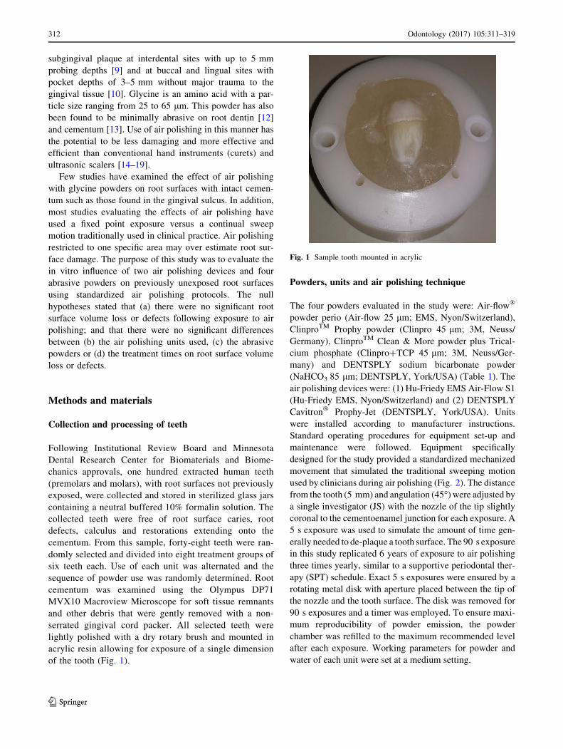
of the tooth (Fig. 1).
Powders, units and air polishing technique
The four powders evaluated in the study were: Air-flow�
powder perio (Air-flow 25 lm; EMS, Nyon/Switzerland),
ClinproTM Prophy powder (Clinpro 45 lm; 3M, Neuss/
Germany), ClinproTM Clean & More powder plus Trical-
cium phosphate (Clinpro?TCP 45 lm; 3M, Neuss/Ger-
many) and DENTSPLY sodium bicarbonate powder
(NaHCO3 85 lm; DENTSPLY, York/USA) (Table 1). The
air polishing devices were: (1) Hu-Friedy EMS Air-Flow S1
(Hu-Friedy EMS, Nyon/Switzerland) and (2) DENTSPLY
Cavitron� Prophy-Jet (DENTSPLY, York/USA). Units
were installed according to manufacturer instructions.
Standard operating procedures for equipment set-up and
maintenance were followed. Equipment specifically
designed for the study provided a standardized mechanized
movement that simulated the traditional sweeping motion
used by clinicians during air polishing (Fig. 2). The distance
from the tooth (5 mm) and angulation (45�) were adjusted bya single investigator (JS) with the nozzle of the tip slightly
coronal to the cementoenamel junction for each exposure. A
5 s exposure was used to simulate the amount of time gen-
erally needed to de-plaque a tooth surface. The 90 s exposure
in this study replicated 6 years of exposure to air polishing
three times yearly, similar to a supportive periodontal ther-
apy (SPT) schedule. Exact 5 s exposures were ensured by a
rotating metal disk with aperture placed between the tip of
the nozzle and the tooth surface. The disk was removed for
90 s exposures and a timer was employed. To ensure maxi-
mum reproducibility of powder emission, the powder
chamber was refilled to the maximum recommended level
after each exposure. Working parameters for powder and
water of each unit were set at a medium setting.
Fig. 1 Sample tooth mounted in acrylic
312 Odontology (2017) 105:311–319
123

Table
1Airpolishingpowdersusedin
thisstudy
Brandnam
eCode
Manufacturer
Type
Particlegrain
size
(d50)(lm)a
SEM
b
Air-Flow�Perio
Air-flow
25lm
Hu-Friedy
Glycine
25
Clinpro
TMProphy
Clinpro
45lm
3M
ESPE
Glycine
45
Clinpro
TMClean
&More
plusTCP
Clinpro
?TCP45lm
3M
ESPE
Glycineplustricalcium
phosphate(TCP)
45
DENTSPLY
Prophy
NaH
CO385lm
DENTSPLY
Sodium
bicarbonate
85
aManufacturerproduct
inform
ation;d50=
medianparticlegrain
size
bScanningelectronmicroscopeim
ages
3009
forDENTSPLY
powder;allother
powders8009
Odontology (2017) 105:311–319 313
123

Quantification of measurement error and root
substance loss
Root surface volume loss and defect depth resulting from
exposure to air polishing were quantified using an optical
three-dimensional laser scanner (3M ESPE LavaTM Scan-
ner ST). Briefly, each randomly selected tooth in the study
was scanned by a white light beam and the lateral dis-
placement of the beam’s reflection was detected by a
charge-coupled device (CCD) chip at baseline, 5 and 90 s
exposure times. Scanned images of the root surfaces were
superimposed and subtracted using CUMULUS 64.0 soft-
ware (� Regents, University of Minnesota, Minneapolis,
MN, USA) allowing measurements within accuracy of
*2 lm. This measurement error was quantified prior to
the conduct of the study by scanning 18 teeth twice. (Data
on file) For each tooth, two data points for maximum defect
depth were reported from the scanner and the average of
those values was used in the analysis. In addition, two teeth
from each treatment group were randomly selected for
SEM analyses following the last exposure.
Scanning electron microscopy (SEM)
Surfacemorphology of the root cementumafter treatmentwas
examined by a semi-environmental tabletop SEM (TM-3000,
Hitachi, Japan) operated at an accelerating voltage of 15 kV.
After 30 min of air drying, specimens were directly mounted
on aluminum stubs with double sided carbon tapes. No con-
ductive coatingwas applied.A charge-up reductionmodewas
selected for image acquisition to counteract the charging
effect. A working distance of*8 mm was used.
Statistical analyses
Statistical analysis was done using SAS V9.3 (SAS Institute,
Inc., Cary, NC,USA) software. Two-way analysis of variance
(ANOVA) models were used to determine differences in
mean volume loss and maximum mean depth between units
and powders. If the interaction was not significant, the main
effects model was run. If there was a significant interaction,
powders were compared within each unit. Pairwise compar-
isons were adjusted using the Tukey’s method. A paired t test
was used to compare the two exposure times. The sample size
of 48 (six per group) had a[85% power to detect main effect
sizes of 1.5 using a two-way ANOVA. P values less than 0.05
were considered statistically significant.
Results
Length of exposure and volume loss of cementum
A 5 s exposure to air polishing had little effect on the mean
volume loss of the cemental surface. At this exposure
Fig. 2 Equipment used for standardizing protocols, including sweep-
ing motion, angulation and nozzle distance from the tooth
Table 2 Mean maximum
defect depth (lm) and mean
volume loss (mm3) after 5 and
90 s of air polishing using
different units and powders
(N = 48)
Mean (SD) maximum defect deptha (lm) Mean (SD) volume loss (mm3)
Hu-Friedy EMS unit 5 sb 90 sb 5 sb 90 sb
Air-flow 25 lm 14 (4) 70 (15) 0.016 (0.008) 0.230 (0.085)
Clinpro 45 lm 31 (28) 69 (31) 0.011 (0.005) 0.174 (0.066)
Clinpro?TCP 45 lm 14 (3) 113 (32) 0.019 (0.014) 0.401 (0.122)
NaHCO3 85 lm 25 (15) 73 (44) 0.016 (0.008) 0.248 (0.195)
DENTSPLY unit
Air-flow 25 lm 14 (8) 39 (30) 0.019 (0.020) 0.100 (0.076)
Clinpro 45 lm 11 (4) 34 (18) 0.002 (0.003) 0.137 (0.103)
Clinpro?TCP 45 lm 13 (10) 43 (34) 0.005 (0.004) 0.150 (0.230)
NaHCO3 85 lm 21 (27) 154 (35)c 0.001 (0.002) 0.653 (0.159)c
a Determined by the average of two reported values per toothb n = 6 for all groupsc Mean maximum defect depth and mean volume loss were greater for NaHCO3 85 lm when compared to
Air-flow 25 lm, Clinpro 45 lm and Clinpro?TCP 45 lm (p\ 0.0001)
314 Odontology (2017) 105:311–319
123

interval, the interaction between units and powders was not
statistically significant (p = 0.1287). A 90 s exposure
resulted in greater mean volume loss of root cementum
than the 5 s exposure (0.251 ± 0.217; p\ 0.0001). In
addition, the interaction between unit and powder was
statistically significant (p\ 0.0001). While there were no
significant differences in mean volume loss between
powders for the Hu-Friedy EMS unit (p = 0.0501), dif-
ferences were identified between powders for the
DENTSPLY unit (p\ 0.0001). The mean volume loss for
NaHCO3 85 lm was greater than the other powders
(p\ 0.0001) (Table 2; Fig. 3).
Length of exposure and mean defect depth
The results for 5 s of exposure for mean maximum depth
between units (p = 0.1914) and powders (p = 0.3427)
were not statistically significant. The interaction was also
not statistically significant (p = 0.3879). At 90 s of expo-
sure there were no significant differences between powders
for Hu-Friedy EMS unit (p = 0.0538), however, differ-
ences in powders were observed with the DENTSPLY unit.
For this unit, the mean maximum defect depth for NaHCO3
85 lm was greater than the other powders (p\ 0.0001)
(Table 2; Fig. 4).
Correlation of volume loss and defect depth
The measures for volume loss and maximum defect depth
appeared to be positively correlated, slightly at 5 s and
strongly at 90 s. Pearson’s correlation coefficient was
(r = 0.172) and (r = 0.896), respectively.
Unit comparisons
Comparisons between units for each powder revealed no
significant differences in volume loss for Air-flow 25 lm(p = 0.1200) or Clinpro 45 lm (p = 0.6519); however,
differences were identified for Clinpro?TCP 45 lm(p = 0.0038) and NaHCO3 85 lm (p\ 0.0001). Similarly,
no significant differences in defect depth were identified
for Air-flow 25 lm (p = 0.0856) or Clinpro 45 lm(p = 0.0579); however, differences were identified for
Clinpro?TCP 45 lm (p = 0.0004) and NaHCO3 85 lm(p\ 0.0001).
SEM analyses
SEM images following the 90 s exposure to air polishing
revealed visible dentinal tubules on all selected test teeth
regardless of unit or powder used (Fig. 5). Comparisons of
images for each unit revealed more cementum loss and
exposure of dentinal tubules with NaHCO3 85 lm when
used in the DENTSPLY unit. Overall, dentinal tubule
exposure and root surface irregularities and defects were
more noticeable with NaHCO3 85 lm than with the glycine
powders. SEM images revealed that specimens exposed to
glycine powders showed less damage to the cemental layer
and fewer exposed dentinal tubules than specimens
exposed to NaHCO3 85 lm.
Fig. 3 A comparison of mean
volume loss (mm3) by powder
and unit at 5 and 90 s exposure
times to air polishing with
various powders (* p\ 0.05)
Odontology (2017) 105:311–319 315
123

SEM images also revealed differences between glycine
powders. Specimens exposed to Clinpro 45 lm had more
exposed dentinal tubules than Air-flow 25 lm. Exposure to
Air-flow 25 lm resulted in specimens with a smooth
homogenous surface and minimal dental tubule exposure.
Specimens exposed to Clinpro?TCP 45 lm appeared to
have fewer dentinal tubules exposed than those exposed to
Clinpro 45 lm. SEM imagery also showed that Clin-
pro?TCP 45 lm, regardless of unit, appeared to have the
fewest number of exposed dentinal tubules. Overall, no
cratering was observed in any of the SEM images (Fig. 5).
Discussion
A primary goal of preventive dental care is to achieve and
maintain a healthy periodontium. Decreasing inflammation
caused by the host response to biofilm along the gingival
margin and subgingival is essential to achieve this goal.
For patients with 3–5 mm pocket depths, conventional
methods include daily oral hygiene by the patient and
professional care including the use of hand and ultrasonic
instruments. Both hand and ultrasonic instrumentation
skills require years of practice, are technically demanding
and time consuming. Major limitations include the
incomplete removal of biofilm [20, 21] and damage to the
root structure [15, 22–25]. Studies have reported loss of
root structure by hand [15, 22] and ultrasonic instrumen-
tation [23]. Alterations of root surface anatomy (the cre-
ation of grooves) with hand instruments [24] and stippling
or wash-board effects on the root surface following ultra-
sonic use have also been reported [23, 25]. Therefore,
improved methods for biofilm removal along the gingival
margin and subgingivally would be beneficial for clinicians
and patients.
This in vitro study was designed to evaluate and quan-
tify the effect of two air polishing units, four powders, and
two exposure times on root surfaces with intact cementum
using standard air polishing tips. To properly evaluate these
outcomes, steps were taken to standardize treatment
parameters, such as tip angulation, distance of the tip from
the tooth surface tip motion and unit settings. The powder
level in the chamber was monitored and maintained at the
manufacturer’s recommended level. Exposure time of 5 s
simulated a single appointment, whereas the 90 s exposure
simulated multiple (repeated) exposures over time (the
equivalent of three times yearly for 6 years). Measurement
error was also determined to identify significant differences
as accurately as possible. Therefore, the outcomes of this
study reflect the influence of the device, powders, and
exposure times on the root cementum.
The results revealed that at a single 5 s exposure, no
significant differences existed between powders. Both
volume loss and defect depth were minimal. Therefore, the
study hypothesis was supported for a single, short exposure
to air polishing with either NaHCO3 85 lm or any of the
glycine powders. Previous research on unexposed root
surfaces has reported root irregularities ranging from 109.6
to 592 ± 153 lm (mean = 323 lm) following 5 s expo-
sure to air polishing with sodium bicarbonate powder [3]
Fig. 4 A comparison of mean
maximum defect depth (lm) by
powder and unit at 5 and 90 s
exposure times to air polishing
with various powders
(* p\ 0.05)
316 Odontology (2017) 105:311–319
123
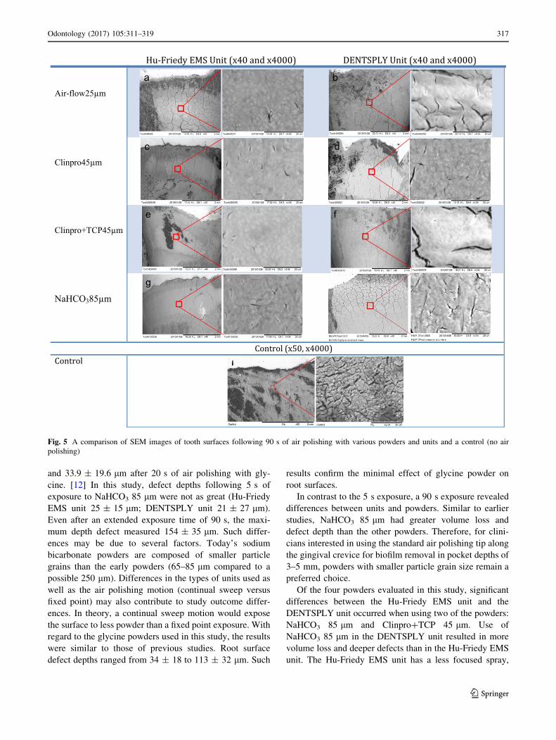
and 33.9 ± 19.6 lm after 20 s of air polishing with gly-
cine. [12] In this study, defect depths following 5 s of
exposure to NaHCO3 85 lm were not as great (Hu-Friedy
EMS unit 25 ± 15 lm; DENTSPLY unit 21 ± 27 lm).
Even after an extended exposure time of 90 s, the maxi-
mum depth defect measured 154 ± 35 lm. Such differ-
ences may be due to several factors. Today’s sodium
bicarbonate powders are composed of smaller particle
grains than the early powders (65–85 lm compared to a
possible 250 lm). Differences in the types of units used as
well as the air polishing motion (continual sweep versus
fixed point) may also contribute to study outcome differ-
ences. In theory, a continual sweep motion would expose
the surface to less powder than a fixed point exposure. With
regard to the glycine powders used in this study, the results
were similar to those of previous studies. Root surface
defect depths ranged from 34 ± 18 to 113 ± 32 lm. Such
results confirm the minimal effect of glycine powder on
root surfaces.
In contrast to the 5 s exposure, a 90 s exposure revealed
differences between units and powders. Similar to earlier
studies, NaHCO3 85 lm had greater volume loss and
defect depth than the other powders. Therefore, for clini-
cians interested in using the standard air polishing tip along
the gingival crevice for biofilm removal in pocket depths of
3–5 mm, powders with smaller particle grain size remain a
preferred choice.
Of the four powders evaluated in this study, significant
differences between the Hu-Friedy EMS unit and the
DENTSPLY unit occurred when using two of the powders:
NaHCO3 85 lm and Clinpro?TCP 45 lm. Use of
NaHCO3 85 lm in the DENTSPLY unit resulted in more
volume loss and deeper defects than in the Hu-Friedy EMS
unit. The Hu-Friedy EMS unit has a less focused spray,
Hu-Friedy EMS Unit (x40 and x4000) DENTSPLY Unit (x40 and x4000)
Air-flow25µm
Clinpro45µm
Clinpro+TCP45µm
NaHCO385µm
Control (x50, x4000)
Control
Fig. 5 A comparison of SEM images of tooth surfaces following 90 s of air polishing with various powders and units and a control (no air
polishing)
Odontology (2017) 105:311–319 317
123

which may have resulted in less powder reaching the tooth
surface at the desired 45� angle (Fig. 6). The standard tip ofthe Hu-Friedy EMS unit is also smaller in diameter
(0.65 mm) than the tip of the DENTSPLY unit (0.75 mm),
potentially limiting larger particle grains such as those in
the sodium bicarbonate powder from exiting the tip of the
nozzle. This confirms the results of previous studies that
found results vary depending on the unit used [11]. In
contrast, the Hu-Friedy EMS unit caused more volume loss
and deeper defects than the DENTSPLY unit for Clin-
pro?TCP 45 lm. Estimation of powder emissions of each
unit prior to the conduct of the study may have been
beneficial, however, technical differences of the units
appeared to have minimal or no effect with the other gly-
cine powders.
SEM images of specimens exposed to NaHCO3 85 lmconfirmed the abrasive nature of this powder on the
cementum. While SEM images also revealed differences in
the appearance of cementum exposed to glycine powders,
the clinical importance of such differences is unknown. If
no statistically significant differences in the amount of
cementum removed from the surface exist, the quantity of
exposed tubules may not be clinically relevant. Clin-
pro?TCP 45 lm had fewer observable tubules; therefore,
it may have an improved ability to block or plug tubules
once exposed. Theories on root hypersensitivity involve the
exposure of the surface to stimuli in the oral environment
including hot and cold liquids [26]. Dentinal tubules in the
gingival sulcus area experience limited stimuli, and there-
fore hypersensitivity may not occur even when exposed
tubules exist. While irregularities were observed on some
SEM specimens, none of the specimens had cratering.
Cratering has been frequently observed in studies using a
fixed point exposure [1, 3–5, 11, 12, 27, 28]. Lack of
cratering in this study indicates the importance of clini-
cians’ use of the recommended continual sweep motion to
avoid such damage. Use of such a motion may also mini-
mize the effect of particle grain diameter differences
observed in previous studies on root defects between
similar type powders [27, 28].
In conclusion, root surface volume loss and defect depth
caused by air polishing using a standard tip differed
depending on the type of powder, unit used, and exposure
time. Sodium bicarbonate powder produced greater volume
loss and defect depth of root surface cementum when used in
the DENTSPLY unit. Differences in powder performance
between units may be related to tip aperture and spray pat-
terns. While more research is needed, the results demon-
strated that the glycine powders used in this study (1) had
minimal effect on root surfaces and (2) performed similarly
regardless of differences in particle grain size. Therefore,
glycine powders may be a viable treatment option for use on
unexposed sulcular root surfaces at regular dental visits.
Acknowledgements This research was supported by NIH Grant
#UL1TR000114 of the National Center for Advancing Translational
Sciences (NCATS). 3M ESPE provided the DENTSPLY unit and air
polishing powders used in this study. The authors acknowledge the
Minnesota Dental Research Center for Biomaterials and Biome-
chanics (MDRCBB) for the technical support provided during the
conduct of this study.
Compliance with ethical standards
Conflict of interest The authors declare they have no conflicts of
interest.
References
1. Atkinson DR, Cobb CM, Killoy WJ. The effect of an air powder
abrasive system on in vitro root surfaces. J Periodontol.
1984;55:13–8.
2. Boyde A. Air polishing effects on enamel, dentine, cement and
bone. Br Dent J. 1984;156(8):287–91.
Fig. 6 A comparison of tip design and powder flow for each unit: Hu-Friedy EMS unit (left) and DENTSPLY unit (right)
318 Odontology (2017) 105:311–319
123

3. Agger M, Horsted-Bindslev P, Hovgaard O. Abrasiveness of an
air-powder polishing system on root surfaces in vitro. Quintes-
sence lnt. 2001;32:407–11.
4. Galloway SE, Pashley DH. Rate of removal of tooth structure by
the use of the Prophy-Jet device. J Periodontol. 1987;58:464–9.
5. Petersilka GJ, Bell M, Mehl A, Hickel R, Flemmig TF. Root
defects following air polishing. J Clin Periodontol.
2003;30:165–70.
6. Graumann SJ, Sensat ML, Stoltenberg JL. Air polishing: a review
of the current literature. J Dent Hyg. 2013;87(4):173–80.
7. Kozlovsky A, Artzi Z, Nemcovsky CE, Hirshberg A. Effect of
air-polishing devices on the gingiva: histologic study in the
canine. J Clin Periodontol. 2005;32(4):329–34.
8. Daubert DM. Subgingival air polishing. Dimens Dent Hyg.
2013;12:1–6.
9. Petersilka GJ, Tunkel J, Barakos K, Heinecke A, Haberlein I,
Flemmig TF. Subgingival plaque removal at interdental sites
using a low-abrasive air polishing powder. J Periodontol.
2003;74(3):307–11.
10. Petersilka GJ, Steinmann D, Haberlein I, Heinecke A, Flemmig
TF. Subgingival plaque removal in buccal and lingual sites using
a novel low abrasive air-polishing powder. J Clin Periodontol.
2003;30(4):328–33.
11. Pelka M, Trautmann S, Petschelt A, Lohbauer U. Influence of air-
polishing devices and abrasives on root dentin—an in vitro
confocal laser scanning microscope study. Quintessence Int.
2010;41(7):141–8.
12. Petersilka GJ, Bell M, Haberlein I, Mehl A, Hickel R, Flemmig
TF. In vitro evaluation of novel low abrasive air polishing
powders. J Clin Periodontol. 2003;30(1):9–13.
13. Sahrmann P, Ronay V, Schmidlin PR, Attin T, Paque F. 3-D
defect evaluation of air polishing on extracted human roots.
J Periodontol. 2014;85(8):1107–14.
14. Berkstein S, Reiff RL, McKinney JF. Supragingival root surface
removal during maintenance procedures utilizing an air-powder
abrasive system or hand scaling. J Periodontol.
1987;58(5):327–30.
15. Coldiron NB, Yukna RA, Weir J, Caudill RF. A quantitative
study of cementum removal with hand curettes. J Periodontol.
1990;61(5):293–9.
16. Gantes BG, Nilveus R. The effects of different hygiene instru-
ments on titanium surfaces: SEM observations. Int J Periodontics
Restor Dent. 1991;11:225–39.
17. Kocher T, Konig J, Hansen P, Ruhling A. Subgingival polishing
compared to scaling with steel curettes: a clinical pilot study.
J Clin Periodontol. 2001;28(2):194–9.
18. Pameijer CH, Stallard RE, Hiep N. Surface characteristics of
teeth following periodontal instrumentation: a scanning electron
microscope study. J Periodontol. 1972;43(10):628–33.
19. Woodruff HC, Levin MP, Brady JM. The effects of two ultra-
sonic instruments on root surfaces. J Periodontol.
1975;46(2):119–26.
20. Waerhaug J. Healing of the dento-epithelial junction following
subgingival plaque control. II: as observed on extracted teeth.
J Periodontol. 1978;49(30):119–1934.
21. Jones S, Lozdan J, Boyde. Tooth surfaces treated in situ with
periodontal instruments: scanning electron microscopic studies.
Br Dent J. 1972;132(2):57–64.
22. Zappa U, Smith B, Simona C, Graf H, Case D, Kim W. Root
substance removal by scaling and root planing. J Periodontol.
1991;62(12):750–4.
23. Flemmig TF, Petersilka GJ, Mehl A, Hickel R, Klaiber B.
Working parameters of a magnetostrictive ultrasonic scaler
influencing root substance removal in vitro. J Periodontol.
1998;69(5):547–53.
24. Waerhaug J. Pathogenesis of periodontal diseases. Br Dent J.
1970;129(4):181–2.
25. Belting CM, Spjut PJ. Effects of high-speed periodontal instru-
ments on the root surface during subgingival calculus removal.
J Am Dent Assoc. 1964;69(11):578–84.
26. Kim JJ. Dentinal hypersensitivity management. In: Darby ML,
Walsh MM, editors. Dental hygiene theory and practice. 4th ed.
St. Louis: Elsevier; 2015. p. 696–706.
27. Tada K, Kiyoshi K, Ogura H, Sato S. Effect of particle diameter
on air polishing of dentin surfaces. Odontology. 2010;98:31–6.
28. Tada K, Wiroj S, Inatomi M, Sato S. The characterization of
dentin defects produced by air polishing. Odontology.
2012;100:41–6.
Odontology (2017) 105:311–319 319
123

![Microanalysis of Root Cementum in Patients with Rapidly ......at the exposed cementum [4]. Chemical analysis of the exposed cementum has shown an increase in calcium, magnesium, and](https://img.pdfslide.us/doc/110x75/5f237b2b5d795a336e24c740/microanalysis-of-root-cementum-in-patients-with-rapidly-at-the-exposed-cementum.jpg)


![Adv in Cementum Devt[1]](https://img.pdfslide.us/doc/110x75/55cf99ce550346d0339f453c/adv-in-cementum-devt1.jpg)