Embed Size (px)
Citation preview

US Pattern Recognition:
abdominal wall
This presentation covers the anatomy of the abdominal wall, and the ultrasound
appearances as relevant to the rectus sheath, transversus abdominal plane and
ilioinguinal blocks.ilioinguinal blocks.

Anatomy
T10
Caudad
Cephalad
Rectus muscle
Linea alba
Semilunaris
Caudad
External oblique
Internal oblique
Transverse abdominis

AnatomyIf the LA is injected between the rectus
muscle and its’ posterior fascia cephalad
and caudad spread occurs. The
intertendinous intersections that form the
“six pack do NOT attach to the posterior
fascia.
skin
Posterior
sheath
Anterior
sheath
muscle
fat
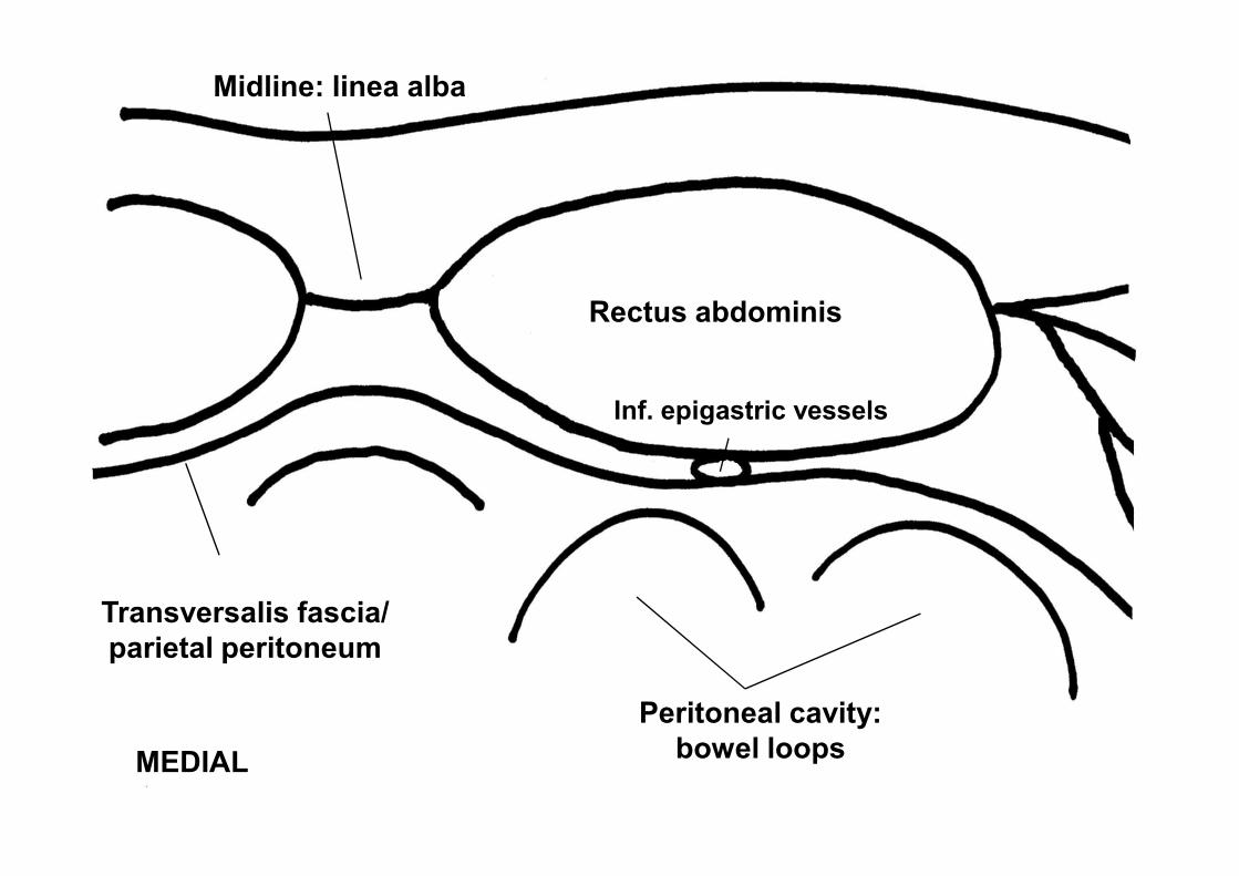
Midline: linea alba
Rectus abdominis
Inf. epigastric vessels
MEDIAL
Peritoneal cavity:
bowel loops
Transversalis fascia/
parietal peritoneum


Sonoanatomy
Right Rectus sheath

Left rectus IP technique, needle
and LA

Ext. oblique
Int. oblique
LATERALTAP
Rectus Trans. abdominis


TAP image, probe over midaxillary line
between costal margin and iliac crest.

TAP IP needle + LA

TAP IP needle + LA

Tips:
• Scan from midline laterally
• Identify known structures
– muscles
– peritoneum– peritoneum
– linea semilunaris
• Determine individual fascial planes
• Nerves lie between internal oblique and
transversus abdominis
• In-plane vs. out-of-plane approaches

EO
IO
Iliohypogastric & ilioinguinal
nervesIlioinguinal
Rectus TA
IliacusFemoral nerve,
not always visible
A
S
I
S

Sonoanatomy Ilioinguinal

M. Weintraud, P. Marhofer, A. Bosenberg, S. Kapral, H. Willschke, M.
Ilioinguinal/Iliohypogastric Blocks in Children: Where Do We Administer the Local Anesthetic Without
Direct Visualization?
Anesth. Analg., January 1, 2008; 106(1): 89 - 93.