Embed Size (px)
Citation preview
Us ing T andem Mas s S pec trometry to R es olve and Inves tigate Interferenc es in G as C hromatography S ingleQuadrupole Mas s S pec trometry (G C /MS )
Us ing T andem Mas s S pec trometry to R es olve and Inves tigate Interferenc es in G as C hromatography S ingleQuadrupole Mas s S pec trometry (G C /MS )
J ason C .C . S awyer*1, G ordon J . Nelson1, P eter Hancock2, Alan L. R ockwood1, B . Derric Maxfield1, S teve G riffin2 1AR UP Institute for C linical and E xperimental P athology, S alt Lake C ity, Utah; 2Waters C orporation, Manchester, United K ingdom
MethodsOverviewPurpose
Evaluate the interferences observed in metanephrine using the single quadrupole GC/MS, by running the same samples on the tandem quadrupole GC-MS/MS
Methods
Urine samples were extracted using a solid-phase extraction to isolate metanephrine.
Extracted samples were injected into an Agilent 6890/5973 GC/MS and then injected into a Waters Micromass Quattro Micro GC-MS/MS
GC/MS quantified by single ion monitoring (SIM).
GC-MS/MS quantified by multiple reaction monitoring (MRM).
Results
GC/MS results for metanephrine display a frequent interference at m/z 523.
Ion ratios (qualitative/quantitative) for biological samples on the GC/MS ran high by 31% when compared to pure standards, consistent with the existence of interference. For GC-MS/MS this improved to 9.7%.
The coefficient of variation for the ion ratios on the GC/MS was 10.8%, compared to 5.6% on the GC-MS/MS.
The interference reduction from the GC/MS to the GC-MS/MS allowed for an increase in precision when the allowable range of the standard ion ratio range is lowered from +/-50% to +/-10%.
Biological samples containing metanephrine are monitored for the diagnosis and follow-up of patients with pheochromocytoma and related neurogenic tumors.
To prevent potential interference from causing incorrect compound identification, more than a single ion is usually monitored in selective ion mode (SIM) of single quadrupole GC-MS analysis. Interference is further evaluated by comparing the ratio of the abundances of the monitored ions of a compound to the same ratio produced by a known standard. Measuring metanephrine levels in urine by a single quadrupole GC-MS method can be challenging due to the size of the molecule, and the number of other analytes present in the sample with similar size and structure.
The purpose of this study was to compare results from biological samples displaying interference in GC/single quadrupole analysis to results for the same biological samples analyzed by GC/tandem quadrupole analysis.
Introduction
Apparatus
Single Quadrupole MSGC/MS: Agilent 6890/5973Column: DB-5MS 15m x 25mm with an internal diameter of 0.25µm, J&W Scientific. Injection volume: 0.5µL.Carrier gas: Helium Initial Oven Temp: 100°CMaximum Oven Temperature: 325°CInlet Mode: Pulsed SplitlessAcquisition Mode: SIM (single ion monitoring)
Tandem Quadrupole MSGC-MS/MS: Agilent 6890 GC, CTC CombiPal autosampler system, and Water's Quattro Micro GCSource: Electron Impact (EI)Column: DB-5MS 15m x 25mm with an internal diameter of 0.25µm, J&W Scientific. Injection volume: 1.0µLCarrier gas: HeliumCollision gas: ArgonInitial Oven Temperature: 100°CMaximum Oven Temperature: 320°CInlet Mode: SplitlessAcquisition mode: MRM (multiple reaction monitoring)
Sample Preparation
Urine samples, calibrator, and controls were mixed by inversion 10 times prior to aliquoting 500 µL into 16x100 mm glass test tubes.
Added 50µL of 6M HCL, Incubated 30 min, centrifuged 5 min @ 2500 rpm, added 2.0mL 0.2M phosphate buffer, and added 50µL 6M NaOH.
Added 100µL working internal standard, 1µg/µL Metanephrine-d3
SPE (solid phase extraction).
SPE eluate evaporated @ 37°C for approx. 15 min.
Derivitized with 50µL MSTFA, 25µL MBHFBA, vortexed for 5 sec and transferred to glass autosampler vials.
Ion Ratio Interference Study
39 unknown specimens analyzed.
One batch was run on each of the instruments (GC-MS and GC-MS/MS)
Concentrations from approximately 20-2800µg/L for metanephrine.
Ion ratio = (qualitative ion peak area)/(quantitative ion peak area).
Ion ratio investigation: ± 10-50% of calibrator average.
ConclusionsMRM ion monitoring on the GC-MS/MS eliminates the observed interference of the GC/MS in SIM.
Ions ratios (qualitative/quantitative) in GC-MS/MS were more reproducible than in GC-MS.
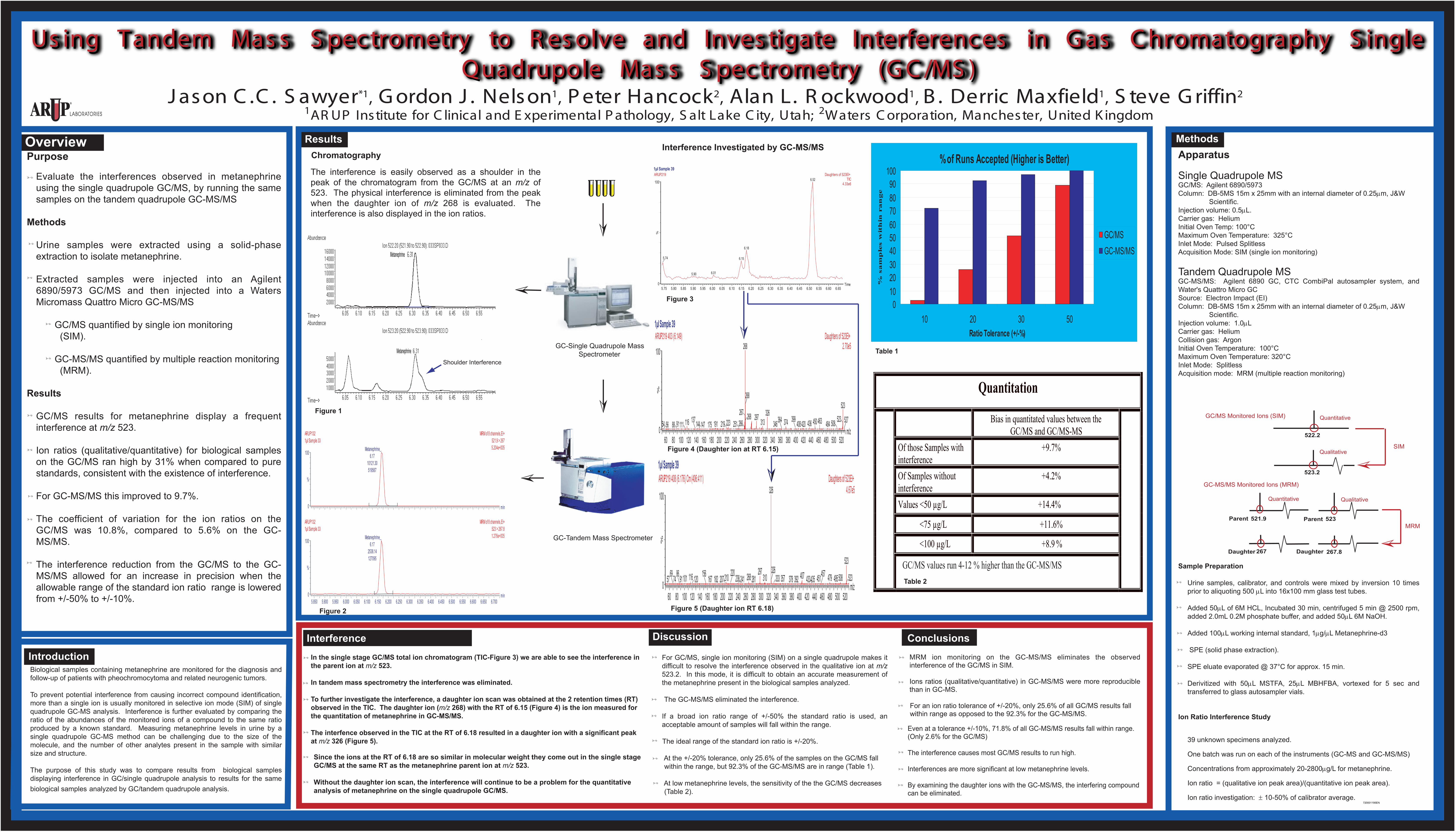
For an ion ratio tolerance of +/-20%, only 25.6% of all GC/MS results fall within range as opposed to the 92.3% for the GC-MS/MS.
For GC/MS, single ion monitoring (SIM) on a single quadrupole makes it difficult to resolve the interference observed in the qualitative ion at m/z 523.2. In this mode, it is difficult to obtain an accurate measurement of the metanephrine present in the biological samples analyzed.
The GC-MS/MS eliminated the interference.
If a broad ion ratio range of +/-50% the standard ratio is used, an acceptable amount of samples will fall within the range.
The ideal range of the standard ion ratio is +/-20%. At the +/-20% tolerance, only 25.6% of the samples on the GC/MS fall within the range, but 92.3% of the GC-MS/MS are in range (Table 1).
At low metanephrine levels, the sensitivity of the the GC/MS decreases (Table 2).
522.2
523.2
Discussion
The interference is easily observed as a shoulder in the peak of the chromatogram from the GC/MS at an m/z of 523. The physical interference is eliminated from the peak when the daughter ion of m/z 268 is evaluated. The interference is also displayed in the ion ratios.
ResultsChromatography
Interference Investigated by GC-MS/MS
GC/MS Monitored Ions (SIM)
GC-MS/MS Monitored Ions (MRM)
521.9 523
267 267.8
QualitativeQuantitative
Parent Parent
Daughter Daughter
SIM
MRM
min5.850 5.900 5.950 6.000 6.050 6.100 6.150 6.200 6.250 6.300 6.350 6.400 6.450 6.500 6.550 6.600 6.650 6.700
%
0
100
MRM of 8 channels,EI+523 > 267.81.276e+005
ARUP132 1µl Sample 33
Metanephrine6.17
2538.14127095
min
%
0
100
MRM of 8 channels,EI+521.9 > 2675.204e+005
ARUP132 1µl Sample 33
Metanephrine6.17
10121.30519567
Quantitation
Bias in quantitated values between the GC/MS and GC/MS-MS
Of those Samples with interference
+9.7%
Of Samples without interference
+4.2%
Values <50 µg/L +14.4%
<75 µg/L +11.6%
<100 µg/L +8.9 %
Even at a tolerance +/-10%, 71.8% of all GC-MS/MS results fall within range. (Only 2.6% for the GC/MS)
The interference causes most GC/MS results to run high.
Interferences are more significant at low metanephrine levels.
By examining the daughter ions with the GC-MS/MS, the interfering compound can be eliminated.
1µl Sample 39
Time5.75 5.80 5.85 5.90 5.95 6.00 6.05 6.10 6.15 6.20 6.25 6.30 6.35 6.40 6.45 6.50 6.55 6.60 6.65
%
0
100
ARUP219 Daughters of 523EI+ TIC
4.33e66.52
6.18
6.155.74
6.015.90
1µl Sample 39
m/z60 80 100 120 140 160 180 200 220 240 260 280 300 320 340 360 380 400 420 440 460 480 500 520
%
0
100
ARUP219 403 (6.149) Daughters of 523EI+ 2.70e5268
2671169154 56 88 111 223219191144137 147 175 266251
268523
326268 297 313 523463450374346364 436420408399 508484 527
1µl Sample 39
m/z60 80 100 120 140 160 180 200 220 240 260 280 300 320 340 360 380 400 420 440 460 480 500 520
%
0
100
ARUP219 408 (6.176) Cm (406:411) Daughters of 523EI+ 4.67e5326
29726811610191745769 86 210167156120 182 207220 238 241 281 310
523407326
357333 378 392 479451435433 457 508495 523
InterferenceIn the single stage GC/MS total ion chromatogram (TIC-Figure 3) we are able to see the interference inthe parent ion at m/z 523. In tandem mass spectrometry the interference was eliminated.
To further investigate the interference, a daughter ion scan was obtained at the 2 retention times (RT) observed in the TIC. The daughter ion (m/z 268) with the RT of 6.15 (Figure 4) is the ion measured for the quantitation of metanephrine in GC-MS/MS.
The interfence observed in the TIC at the RT of 6.18 resulted in a daughter ion with a significant peak at m/z 326 (Figure 5).
Figure 3
Figure 4 (Daughter ion at RT 6.15)
Figure 5 (Daughter ion RT 6.18)
Figure 1
Figure 2
Since the ions at the RT of 6.18 are so similar in molecular weight they come out in the single stage GC/MS at the same RT as the metanephrine parent ion at m/z 523.
Without the daughter ion scan, the interference will continue to be a problem for the quantitative analysis of metanephrine on the single quadrupole GC/MS.
Table 1
Table 2
GC-Single Quadrupole Mass Spectrometer
GC-Tandem Mass Spectrometer
Shoulder Interference
%of Runs Accepted (Higher is Better)
0102030405060708090
100
10 20 30 50Ratio Tolerance (+/-%)
% s
am
ple
s w
ith
in r
an
ge
GC/MSGC-MS/MS
GC/MS values run 4-12 % higher than the GC-MS/MS
Metanephrine
Metanephrine
Qualitative
Quantitative