Embed Size (px)
Citation preview
CASE REPORT Open Access
Urticaria as initial finding of a patient withcarcinoid tumorIvan Cherrez Ojeda1,2*, Juan Carlos Calderon1,2, Karin Plaza1, Emanuel Vanegas1, Annia Cherrez2,3 and José Cano1
Abstract
Background: Typical carcinoid syndrome is characterized by flushing, abdominal pain and diarrhea and occurs in<10 % of carcinoid tumor patients. Very rarely, initial signs include skin manifestations. Our purpose is to highlightcutaneous manifestations in the diagnosis and assessment of a patient with atypical manifestation of type I gastriccarcinoid tumor.
Case presentation: A 50-year-old woman presented with anemia, chronic urticaria and angioedema. Urticaria wastriggered principally by seafood and appeared in the first hour after. Urticaria Activity Score 7 was 24, and qualityof life (CU-Q2oL) was 3.61. P. Laboratory findings showed anemia, diminished iron, ferritin, and vitamin B12, withincreased gastrin and anti-parietal cell antibody levels. 15 gastric carcinoids 5 mm in diameter were observed in thegreater curvature of the stomach during gastric endoscopy and confirmed by biopsy, suggesting that this patienthad type I gastric carcinoids. Four additional tumors were found in the small intestine upon examination via videocapsule. Endoscopic argon plasma therapy was performed. The patient experienced definitive improvement inquality of life and urticaria activity score.
Conclusion: This patient, whose principal symptoms were anemia, urticaria and angioedema, was found to haveatypical carcinoid syndrome, with tumors located in the stomach. Allergists, immunologists, internists and primarycare physicians should consider the possibility of neuroendocrine malignancies, specifically type I carcinoid tumors,when evaluating patients with urticaria, and consider screening patients with chronic urticaria for elevated anti-parietalcell antibody levels.
Keywords: Urticaria, Quality of life, Carcinoid tumors, Kinin, Serotonin, Enterochromaffin cells, Antibodies
BackgroundCarcinoid tumors are rare, slow-growing neuroendocrinetumors arising from the enterochromaffin cells. They aremost commonly found in the gastrointestinal tract (64 %)[1], although they can involve any organ. These tumorscan secrete biogenic amines and peptides, such as ACTH,5-HTP and serotonin [2]. Classical carcinoid syndromeoccurs in <10 % of patients with carcinoid tumors. Itsmost typical clinical manifestations include cutaneousflushing, most often on the face, neck, and upper chest,diarrhea and abdominal pain [3]. Other manifestationsmay occasionally be encountered, including bronchialasthma, wheezing, or very rarely, skin manifestations [4].
Gastric carcinoid tumors are classified as those associ-ated with chronic gastritis type A (CAG-A, or type I),those that concur with the Zollinger-Ellison syndrome(type II), and sporadic gastric tumors (type III). Type Igastric carcinoids account for 70 to 80 % of neuroendocrinegastric malignancies. They are associated with autoimmuneatrophic gastritis, hypergastrinemia, and pernicious anemia[5]. Type I gastric carcinoids often measure less than 1 cmin diameter and are typically located in the body or fundusof the stomach [6, 7]. They are multicentric, polypoid,small, limited to the mucosa or submucosa, without angio-invasion, well-differentiated, and tend to display a benignbehavior. They primarily affect women between the ages of50 and 70 years and have a good prognosis, with a low rateof metastasis ranging between 2 and 5 % [8].More than half of patients with CAG-A–associated car-
cinoids also have pernicious anemia. Pernicious anemia isassociated with increased the risk of gastric carcinoid
* Correspondence: [email protected] de Especialidades Espíritu Santo, School of Medicine,Samborondón, Ecuador2Respiralab Research Group, Clínica Kennedy, Guayaquil, EcuadorFull list of author information is available at the end of the article
journal
© 2015 Cherrez Ojeda et al. Open Access This article is distributed under the terms of the Creative Commons Attribution 4.0International License (http://creativecommons.org/licenses/by/4.0/), which permits unrestricted use, distribution, andreproduction in any medium, provided you give appropriate credit to the original author(s) and the source, provide a link tothe Creative Commons license, and indicate if changes were made. The Creative Commons Public Domain Dedication waiver(http://creativecommons.org/publicdomain/zero/1.0/) applies to the data made available in this article, unless otherwise stated.
Cherrez Ojeda et al. World Allergy Organization Journal (2015) 8:34 DOI 10.1186/s40413-015-0083-y
tumors, presumably due to prolonged achlorhydria. It re-sults in parietal cell loss, compensatory hypergastrinemia,and argyrophilic cell hyperplasia [9].Persistent achlorhydria produces G cell hyperplasia
and more gastrin secretion, leading to hypergastrinemia[10]. Gastrin stimulates CCK-B/gastrin receptor (CCK2R)which further activates genes that encode HDC (histidinedecarboxylase), VMAT2 (vesicular monoamine transportermolecule 2) and CgA (Chromogranin-A), but the signalingpathway leading to enterochromaffin cells hyperplasia re-mains unknown [11]. The widespread use of proton pumpinhibitors can also induce gastric achlorhydria, thus con-tributing to hypergastrinemia.Type II carcinoids present with hypergastrinemia and
are associated with Zollinger-Ellison syndrome. In con-trast to type I gastric carcinoids, the hypergastrinemiaassociated with type II gastric carcinoids is secondary togastrin secreting G cell neoplasia rather than parietal cellloss. Type II gastric carcinoids are very uncommon; themajority have good prognosis. The last type of gastric car-cinoid, type III, may be associated with atypical carcinoidsyndrome [12, 13]. Atypical carcinoid syndrome is mani-fested by flushing, and tumors tend to be metastatic attheir presentation, and thus have the worst prognosis [14].Radical gastrectomy is the most common treatment fortype III gastric carcinoids [15].Here we report urticaria, angioedema and anemia as
symptoms in an atypical presentation of type I gastriccarcinoid. Our purpose is to highlight the relevance ofcutaneous manifestations in the diagnosis and assessmentof a patient with an underlying neuroendocrine malig-nancy. We also remark on the effectiveness of endoscopicablation. And we encourage all medical practitioners to beaware that skin changes may provide an early clue for clin-ical management of gastric carcinoids.
Case presentationWe report a case of a 50-year-old woman with atypicaltype I gastric carcinoid tumor. The patient presented withanemia, chronic urticaria and angioedema. The urticariabegan one year prior to her evaluation by us, and was trig-gered by seafood. Other triggers included vitamin B12,desserts, and maize, as well as friction from shoes whilerunning, and doing handicrafts. Lesions usually began inthe first hour after the stimuli. Three months prior to de-veloping urticaria, the patient was diagnosed with anemia.In the year since her urticaria began, the patient had lost20 pounds without effort. She reported that her symptomsimproved after taking the proton pump inhibitor (PPI)pantoprazole, but she only used the PPI sporadically. Thereason for this response to PPI is not clear.Initial symptoms of the urticaria were nausea ac-
companied by erythema, edema, pruritus and wheals.Wheals were larger than 5 mm, and were accompanied by
erythema. They were associated with pain, burning andmorning-time livedo reticularis. Wheals lasted more than12 h, and were localized in palm, soles, armpits, feet,hands, buttocks, shoulders, trunk, legs and back. The pa-tient also experienced generalized angioedema, which wastriggered by seafood, corn, some desserts and ibuprofen,as well as blurred vision and sore throat. Urticaria ActivityScore 7 (UAS 7) was 24, and the mean score on the CU-Q2oL quality of life measure was 3.61. Fecal exam,immunological tests (complement levels and immuno-globulins) and skin prick allergy tests for shrimp, crab,dust mite, and dog hair were all normal. There were nosigns of vasculitis in the skin biopsy.Laboratory tests showed diminished iron, ferritin, and
vitamin B12 (patient had stopped taking B12, as it trig-gered the hives). The gastrin level was 286 pg/ml (refer-ence value <100 pg/ml) and parietal cell antibody was106.53 U/ml (reference value Negative: < or =20.0 U,Equivocal: 20.1-24.9 U, Positive: > or =25.0 U) [16]. RDW(Red Cell Distribution Width) was high, and anisocytosiswas found. Microcytosis was also observed, perhaps dueto iron deficiency, as well as megaloblasts.Gastric endoscopy was performed and 15 sessile polyps
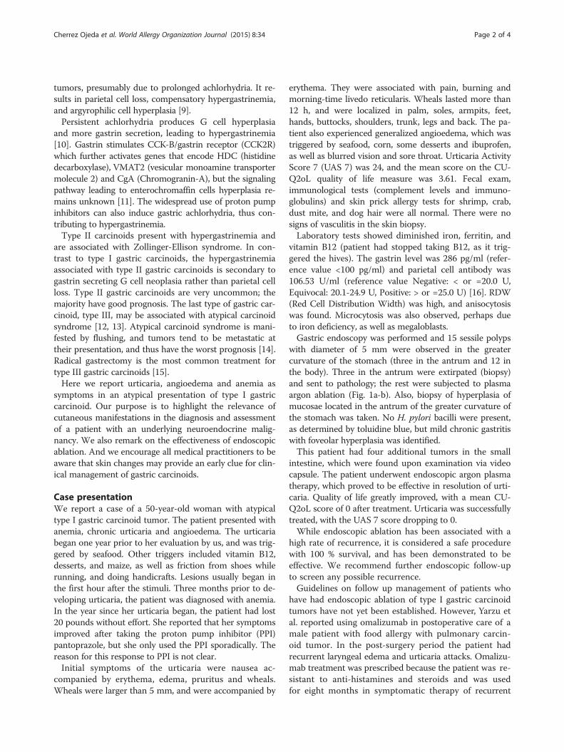
with diameter of 5 mm were observed in the greatercurvature of the stomach (three in the antrum and 12 inthe body). Three in the antrum were extirpated (biopsy)and sent to pathology; the rest were subjected to plasmaargon ablation (Fig. 1a-b). Also, biopsy of hyperplasia ofmucosae located in the antrum of the greater curvature ofthe stomach was taken. No H. pylori bacilli were present,as determined by toluidine blue, but mild chronic gastritiswith foveolar hyperplasia was identified.This patient had four additional tumors in the small
intestine, which were found upon examination via videocapsule. The patient underwent endoscopic argon plasmatherapy, which proved to be effective in resolution of urti-caria. Quality of life greatly improved, with a mean CU-Q2oL score of 0 after treatment. Urticaria was successfullytreated, with the UAS 7 score dropping to 0.While endoscopic ablation has been associated with a
high rate of recurrence, it is considered a safe procedurewith 100 % survival, and has been demonstrated to beeffective. We recommend further endoscopic follow-upto screen any possible recurrence.Guidelines on follow up management of patients who
have had endoscopic ablation of type I gastric carcinoidtumors have not yet been established. However, Yarzu etal. reported using omalizumab in postoperative care of amale patient with food allergy with pulmonary carcin-oid tumor. In the post-surgery period the patient hadrecurrent laryngeal edema and urticaria attacks. Omalizu-mab treatment was prescribed because the patient was re-sistant to anti-histamines and steroids and was usedfor eight months in symptomatic therapy of recurrent
Cherrez Ojeda et al. World Allergy Organization Journal (2015) 8:34 Page 2 of 4
laryngeal edema and urticaria attacks During the fouryears of follow up, no carcinoid tumor recurrence wasnoted [17].
ConclusionsThere are only a few reported cases of urticaria or angio-edema associated with carcinoid tumors [4, 18]. The casereported here is particularly interesting because the tu-mors were located in the foregut. Foregut tumors gener-ally do not secrete as much of the urticaria mediator kininas do midgut tumors, and only secrete a small amount ofthe possible mediator serotonin [19]. However, foreguttumors may secrete 5-hydroxytryptophan (5-HTP), hista-mine or adrenocorticotropic hormone [2, 20]. Histaminerelease could lead to urticaria, which should respond toH1-antagonists [20]. Our patient was treated with H1antagonists without response.This patient presented with urticaria and angioedema
as symptoms. Remarkably, urticaria was the reason formedical consultation. The patient had no diarrhea, and nosigns or symptoms of bronchoconstriction or heart failure.This, then, would not fit the definition of a classical car-cinoid syndrome [21]. The patient also presented withhypergastrinemia and anemia. This anemia can be attrib-uted to several etiologies, since RDW was high, and aniso-cytosis, microcytosis and megaloblasts were observed. Thepatient presented chronic autoimmune gastritis, with hightiters of anti-parietal cell antibodies, suggesting parietalcell loss as the etiology of G cell hyperplasia.We strongly encourage all physicians who are looking
for the etiology of any chronic urticaria, be they allergists,dermatologists, internists or primary care physicians, toconsider the possibility of neuroendocrine malignanciesand to screen these patients for elevated anti-parietal cellantibodies.
ConsentWritten informed consent was obtained from the patientfor publication of this Case Report and any accompanyingimages. A copy of the written consent is available forreview by the Editor-in-Chief of this journal.
Abbreviations5-HTP: 5-hydroxytryptophan; ACTH: adrenocorticotropic hormone;CAG-A: chronic gastritis type A; CU: chronic urticaria; CU-Q2oL: ChronicUrticaria Quality of Life Questionnaire; UAS 7: Urticaria Activity Score - 7.
Competing interestsThe authors declare that they have no competing interests.
Authors’ contributionsPK, VE, CHA and CJ collaborated in literature searching, interviewing andmonitoring the patient. CHOI and CJC prepared and wrote the draft ofmanuscript. CHA and VE collaborated in correcting the manuscript. Allauthors read and approved the final manuscript.
AcknowledgementsWe acknowledge to MECOR Program, specially to Sonia Buist and AnaMenezes for motivation in researching activities. Furthermore, we expressour appreciation to Mary Ckaiken for her editorial support in English as weprepared the manuscript.
Author details1Universidad de Especialidades Espíritu Santo, School of Medicine,Samborondón, Ecuador. 2Respiralab Research Group, Clínica Kennedy,Guayaquil, Ecuador. 3University of Heidelberg, School of Medicine,Heidelberg, Germany.
Received: 21 August 2015 Accepted: 11 November 2015
References1. Tun NT, Oza R. Atypical presentation of carcinoid tumor with unresolved
right shoulder pain: a case report. J Med Case Rep. 2014;8(1):142.2. Ha J, Tan W. Gastrointestinal carcinoid tumours: a review. J Gastrointest Dig
Syst. 2012;02(02):1–7.3. Oates JA. The carcinoid syndrome. NEJM. 1986;315:702–4.4. Bożek A, Rachowska R, Krajewska J, Paliczka-Cieślik E, Filipowska B, Jarzab J.
Carcinoid syndrome with angioedema and urticaria. JAMA Dermatology.2008;144(5):691–2.
5. Stilo F. Gastric carcinoid tumors. J Surg Oncol. 2006;93(5):347–9.
Fig. 1 Examples of observed polypoids. a Sessile polypoid located in the greater curvature (magnification of b), 5 mm in diameter, depressed inits center and umbilicated, and alteration of glandular pattern (red arrow in b). b Black arrows indicate many sessile polypoids, with characteristicsas described in a, located in the greater curvature in the body of stomach
Cherrez Ojeda et al. World Allergy Organization Journal (2015) 8:34 Page 3 of 4
6. Basuroy R, Srirajaskanthan R, Prachalias A, Quaglia A, Ramage J. Reviewarticle: the investigation and management of gastric neuroendocrinetumours. Aliment Pharmacol Ther. 2014;39(10):1071–84.
7. Rindi G, Bordi C, Rappel S, La Rosa S, Stolte M, Solcia E. Gastric carcinoidsand neuroendocrine carcinomas: pathogenesis, pathology, and behavior.World J Surg[Internet]. 2015 Jan [cited 2015 Sep 19]; 1996;20(2):168–72.Available from: http://www.ncbi.nlm.nih.gov/pubmed/8661813.
8. Nikou GC, Angelopoulos TP. Current concepts on gastric carcinoid tumors.Gastroenterol Res Pract. 2012;2012:287825.
9. Harvey RF, Davidson CM, Bradshaw MJ, Wilkinson SP, Davies PS. Multifocalgastric carcinoid tumours, achlorhydria, and hypergastrinaemia.Lancet[Internet]. 2015 Jan [cited 2015 Sep 20]; 1985;325(8435):951–4.Available from: http://www.sciencedirect.com/science/article/pii/S0140673685917271.
10. Binstock AJ, Johnson CD, Stephens DH, Lloyd RV, Fletcher JG. Carcinoidtumors of the stomach: a clinical and radiographic study. AJR Am JRoentgenol[Internet]. 2015 Jan [cited 2015 Oct 20]; 2001;176(4):947–51.Available from: http://www.ajronline.org/doi/abs/10.2214/ajr.176.4.1760947.
11. Hocker M. Molecular mechanisms of gastrin-dependent gene regulation.Ann N Y Acad Sci[Internet]. 2015 Jan [cited 2015 Sep 20]; 2004;1014:97–109.Available from: http://www.ncbi.nlm.nih.gov/pubmed/15153424.
12. Kulke M, Mayer R. Carcinoid tumors. NEJM. 1999;340(11):858–68.13. Bordi C, D’Adda T, Azzoni C, Ferraro G. Pathogenesis of ECL cell tumors in
humans. Yale J Biol Med. 1999;71(3–4):273–84.14. Modlin IM, Lye KD, Kidd M. Carcinoid tumors of the stomach. Surg Oncol.
2003;12(2):153–72.15. Kwon YH, Jeon SW, Kim GH, Kim JI, Chung IK, Jee SR, et al. Long-term
follow up of endoscopic resection for type 3 gastric NET. World JGastroenterol. 2013;19(46):8703–8.
16. Test Catalog - Mayo Medical Laboratories [Internet]. [cited 2015 Sep 20].Available from: http://www.mayomedicallaboratories.com/test-catalog/index.html.
17. Yalcin AD. Advances in anti-IgE therapy. Biomed Res Int [Internet]. 2015 Jan[cited 2015 Sep 9];2015:317465. Available from: http://www.hindawi.com/journals/bmri/2015/317465/.
18. Wymenga AN, de Monchy JG, de Vries EG. The carcinoid syndrome andangioedema. Ann Intern Med[Internet]. 2015 Jan [cited 2015 Sep 20]; 1995;123(8):636. Available from: http://annals.org/article.aspx?articleid=709146.
19. Woodside KJ, Townsend CM, Evers BM. Current management ofgastrointestinal carcinoid tumors. Soc Surg Aliment Tract. 2004;8(6):742–56.
20. Kölby L, Wängberg B, Ahlman H, Jansson S, Forssell-Aronsson E, Erickson JD,et al. Gastric carcinoid with histamine production, histamine transporter andexpression of somatostatin receptors. Digestion[Internet]. 2015 Jan [cited2015 Sep 19]; 1998;59(2):160–6. Available from: http://www.ncbi.nlm.nih.gov/pubmed/9586830.
21. Soga J. Early-stage carcinoids of the gastrointestinal tract: an analysis of1914 reported cases. Cancer. 2005;103(8):1587–95.
Submit your next manuscript to BioMed Centraland take full advantage of:
• Convenient online submission
• Thorough peer review
• No space constraints or color figure charges
• Immediate publication on acceptance
• Inclusion in PubMed, CAS, Scopus and Google Scholar
• Research which is freely available for redistribution
Submit your manuscript at www.biomedcentral.com/submit
Cherrez Ojeda et al. World Allergy Organization Journal (2015) 8:34 Page 4 of 4