Embed Size (px)
Citation preview
UROLITHIASISUROLITHIASIS
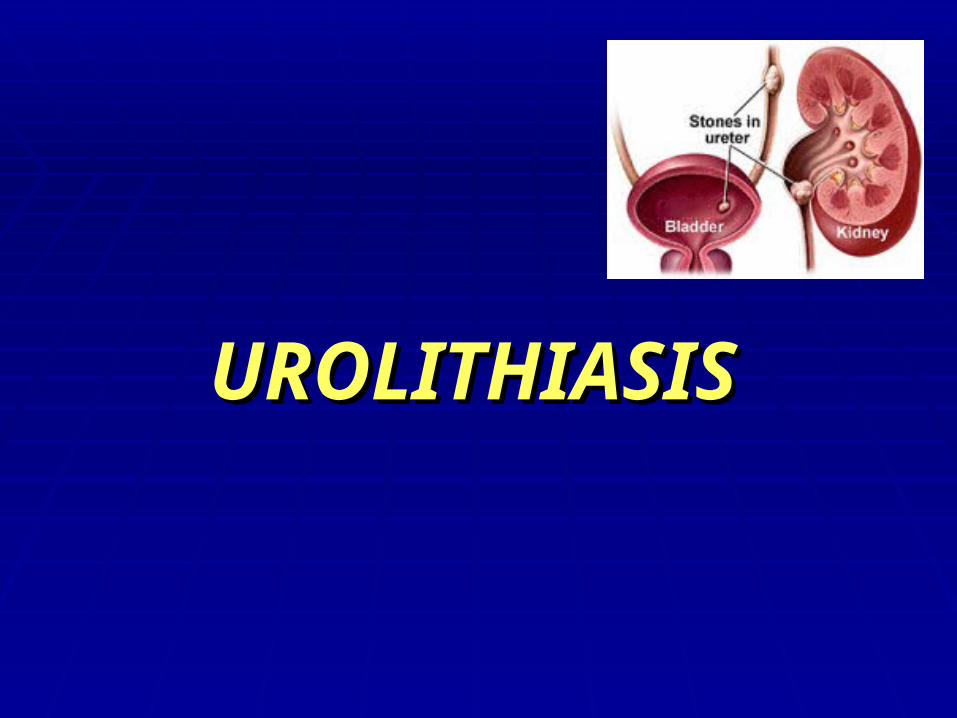
Theories of Stone FormationTheories of Stone Formation
A. Nucleation Theory B. Stone Matrix Theory C. Inhibitor of Crystallization Theory
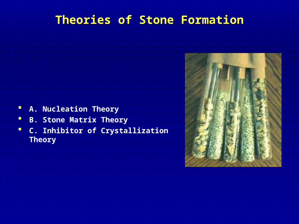
EpidemiologyEpidemiology
Men > WomenMen > Women
4 % of population (Germany)4 % of population (Germany)
Maximum rate at 30 – 50 y.o. Maximum rate at 30 – 50 y.o.
In children - rareIn children - rare
GeographyGeography
EthnologyEthnology
RISK FACTORSRISK FACTORS
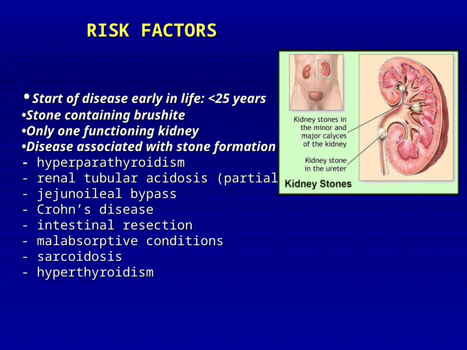
••Start of disease early in life: <25 yearsStart of disease early in life: <25 years••Stone containing brushiteStone containing brushite••Only one functioning kidneyOnly one functioning kidney••Disease associated with stone formationDisease associated with stone formation: : - - hyperparathyroidism hyperparathyroidism - renal tubular acidosis (partial/complete) - renal tubular acidosis (partial/complete) - jejunoileal bypass - jejunoileal bypass - Crohn’s disease - Crohn’s disease - intestinal resection - intestinal resection - malabsorptive conditions - malabsorptive conditions - sarcoidosis - sarcoidosis - hyperthyroidism- hyperthyroidism
Renal CalculiRenal Calculi
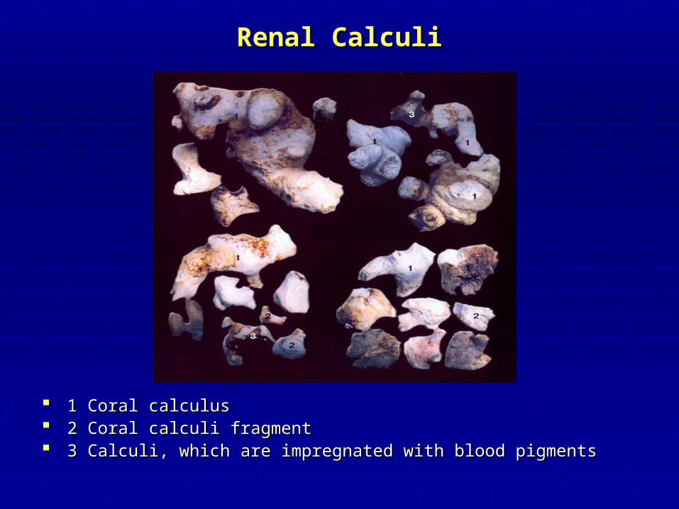
1 Coral calculus1 Coral calculus 2 Coral calculi fragment2 Coral calculi fragment 3 Calculi, which are impregnated with blood pigments3 Calculi, which are impregnated with blood pigments
TYPES OF KIDNEY STONESTYPES OF KIDNEY STONES
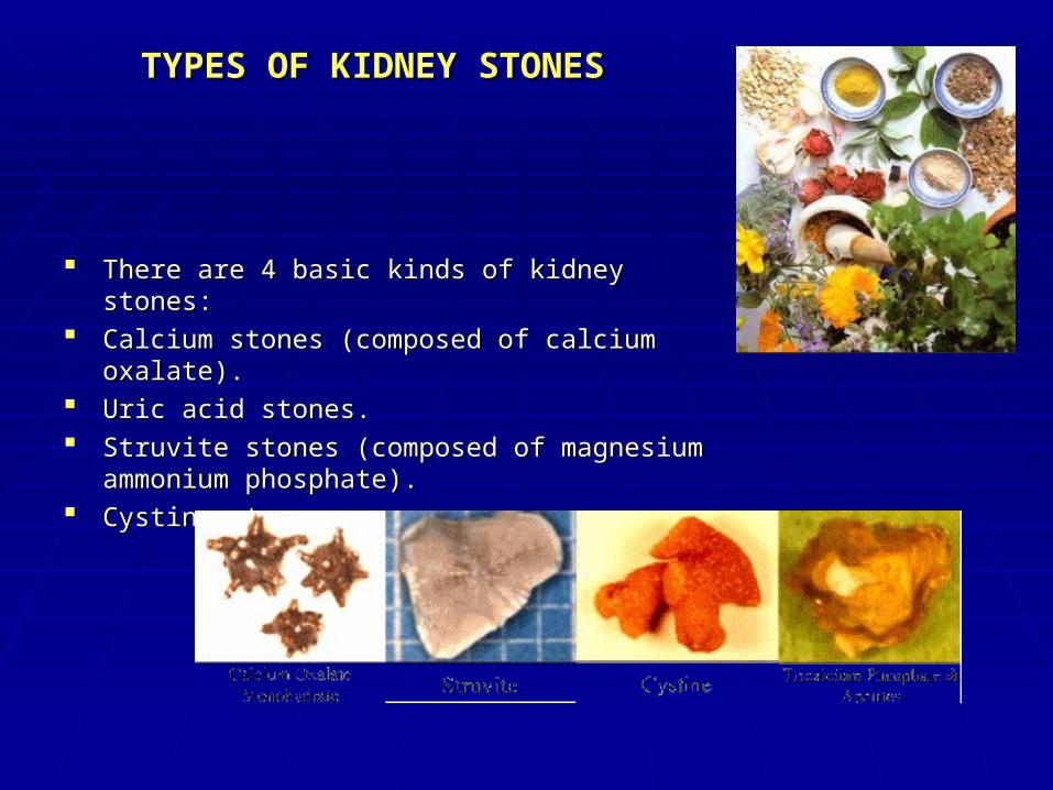
There are 4 basic kinds of kidney stones: There are 4 basic kinds of kidney stones: Calcium stones (composed of calcium oxalate).Calcium stones (composed of calcium oxalate). Uric acid stones.Uric acid stones. Struvite stones (composed of magnesium Struvite stones (composed of magnesium
ammonium phosphate).ammonium phosphate). Cystine stone.Cystine stone.
Clinical ManifestationsClinical Manifestations
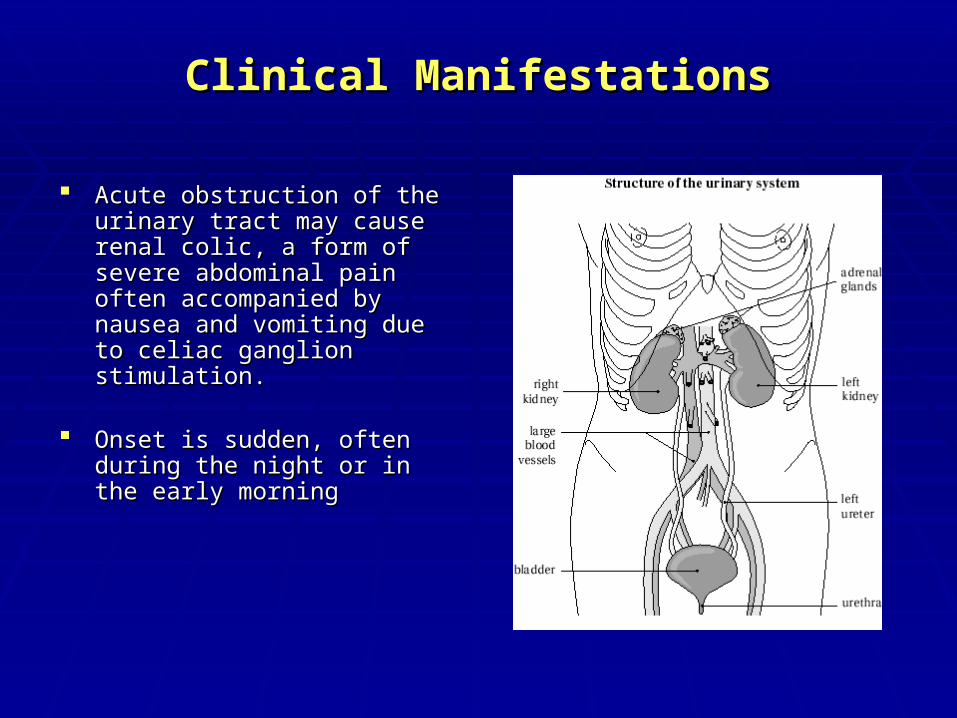
Acute obstruction of the urinary Acute obstruction of the urinary tract may cause renal colic, a form tract may cause renal colic, a form of severe abdominal pain often of severe abdominal pain often accompanied by nausea and accompanied by nausea and vomiting due to celiac ganglion vomiting due to celiac ganglion stimulation. stimulation.
Onset is sudden, often during the Onset is sudden, often during the night or in the early morningnight or in the early morning
Clinical ManifestationsClinical Manifestations
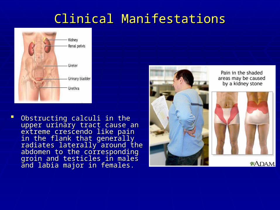
Obstructing calculi in the upper urinary Obstructing calculi in the upper urinary tract cause an extreme crescendo like tract cause an extreme crescendo like pain in the flank that generally radiates pain in the flank that generally radiates laterally around the abdomen to the laterally around the abdomen to the corresponding groin and testicles in corresponding groin and testicles in males and labia major in females.males and labia major in females.

Laboratry InvestigationsLaboratry Investigations
Stone analysisStone analysis: In every patient one stone should: In every patient one stone should
be analysed.be analysed.
Blood analysisBlood analysis: Calcium Albumin Creatinine Urate: Calcium Albumin Creatinine Urate
Urinalysis: Urinalysis: Fasting morning spot urine sampleFasting morning spot urine sample
Dip-stick test: pH, Leucocytes/BacteriaDip-stick test: pH, Leucocytes/Bacteria
Cystine test, Ca, P, citrate, urateCystine test, Ca, P, citrate, urate
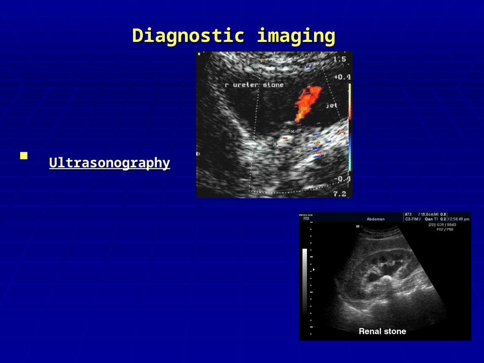
Diagnostic imagingDiagnostic imaging
UltrasonographyUltrasonography
Diagnostic imagingDiagnostic imaging
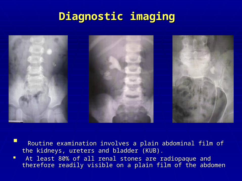
Routine examination involves a plain abdominal film of the kidneys, ureters Routine examination involves a plain abdominal film of the kidneys, ureters and bladder (KUB)and bladder (KUB)..
At least 80% of all renal stones are radiopaque and therefore readily visible on At least 80% of all renal stones are radiopaque and therefore readily visible on a plain film of the abdomena plain film of the abdomen
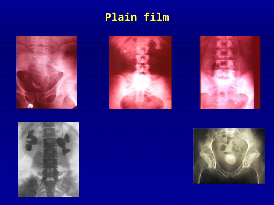
Plain filmPlain film
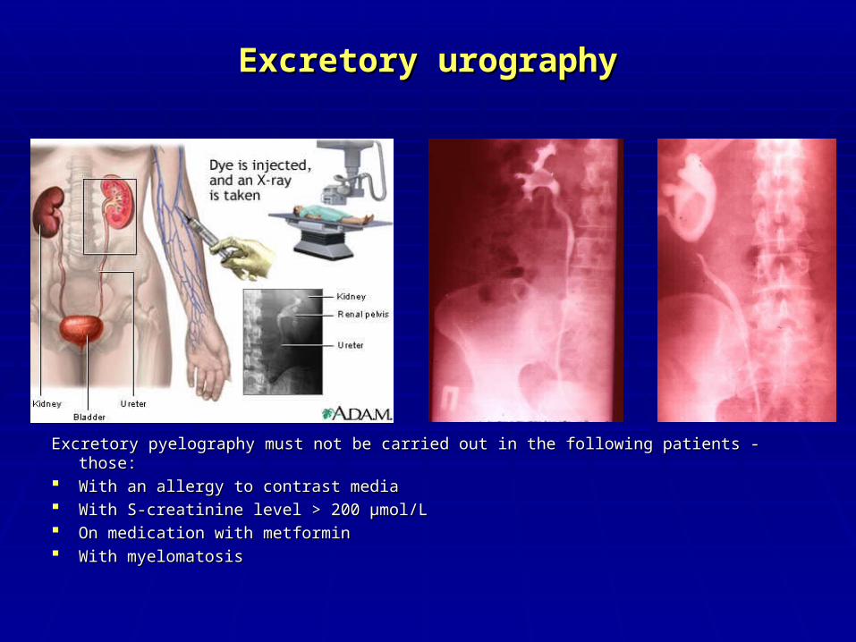
Excretory Excretory urourographygraphy
Excretory pyelography must not be carried out in the following patients - those: Excretory pyelography must not be carried out in the following patients - those: With an allergy to contrast media With an allergy to contrast media With S-creatinine level > 200 µmol/L With S-creatinine level > 200 µmol/L On medication with metformin On medication with metformin With myelomatosisWith myelomatosis
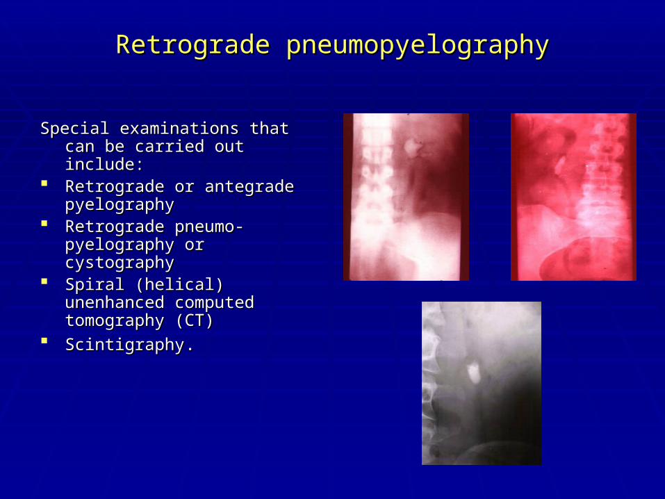
Retrograde pneumopyelographyRetrograde pneumopyelography
Special examinations that can be Special examinations that can be carried out include: carried out include:
Retrograde or antegrade Retrograde or antegrade pyelography pyelography
Retrograde pneumo-pyelography Retrograde pneumo-pyelography or cystographyor cystography
Spiral (helical) unenhanced Spiral (helical) unenhanced computed tomography (CT) computed tomography (CT)
ScintigraphyScintigraphy. .
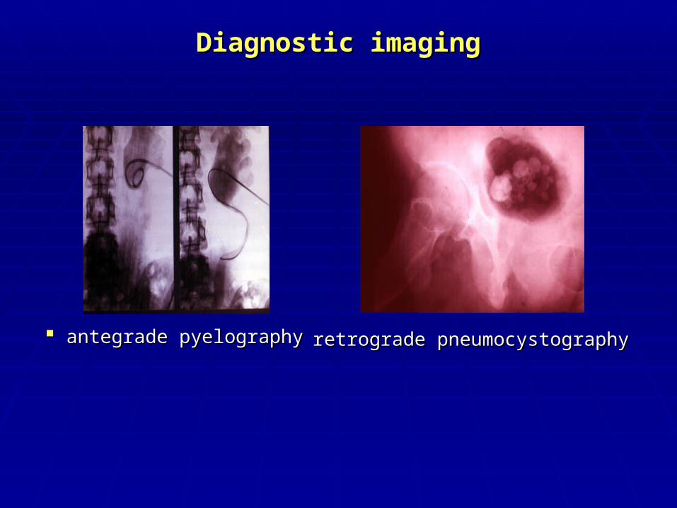
Diagnostic imagingDiagnostic imaging
antegrade pyelographyantegrade pyelography retrograde pneumocystographyretrograde pneumocystography
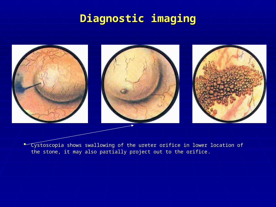
Diagnostic imagingDiagnostic imaging
CCystoscopia shows swallowing of the ureter orifice in lower location of ystoscopia shows swallowing of the ureter orifice in lower location of the stone, it may also partially project out to the orificethe stone, it may also partially project out to the orifice . .
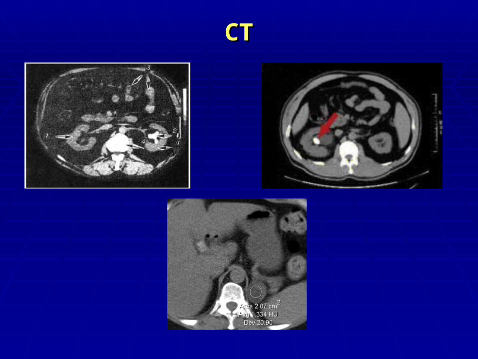
СТСТ
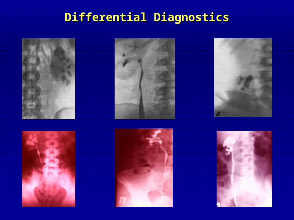
Differential DiagnosticsDifferential Diagnostics
TREATMENTTREATMENT
ConservativeConservative InstrumentalInstrumental SurgicalSurgical
Pain reliefPain relief
Pain relief involves the administration by various routes of Pain relief involves the administration by various routes of the following agents:the following agents:
Diclofenac sodium Diclofenac sodium Indomethacin Indomethacin Hydromorphone hydrochloride + atropine sulphate Hydromorphone hydrochloride + atropine sulphate BaralginBaralgin No-spae + AnalgineNo-spae + Analgine Tramadol Tramadol
Pain reliefPain relief
Warm bath Warm bath Spasmolytic “cocktails” (with papaverine, Spasmolytic “cocktails” (with papaverine,
spasmalgone, no-spanum, promedole) should be spasmalgone, no-spanum, promedole) should be taken. taken.
A high dosage of the cystenal or urolesan (20 drops A high dosage of the cystenal or urolesan (20 drops on the piece of sugar) is rather effective at the start on the piece of sugar) is rather effective at the start of the renal colic.of the renal colic.
Physical method.Physical method.
Pain reliefPain relief
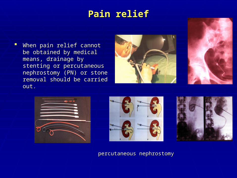
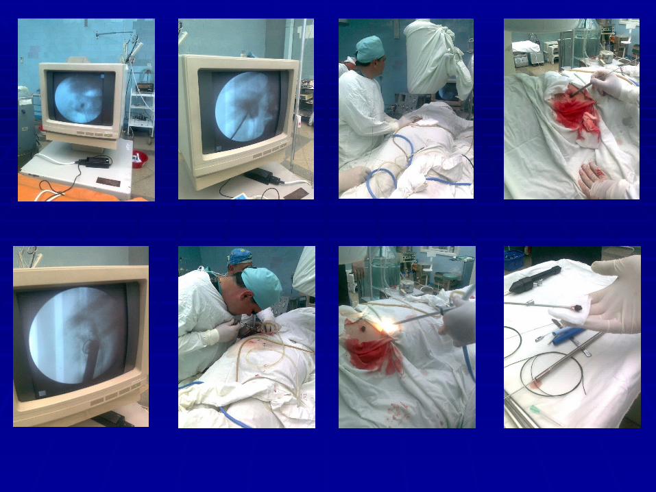
When pain relief cannot be When pain relief cannot be obtained by medical means, obtained by medical means, drainage by stenting or drainage by stenting or percutaneous nephrostomy (PN) percutaneous nephrostomy (PN) or stone removal should be or stone removal should be carried out.carried out.
percutaneous nephrostomypercutaneous nephrostomy
Stone removalStone removal
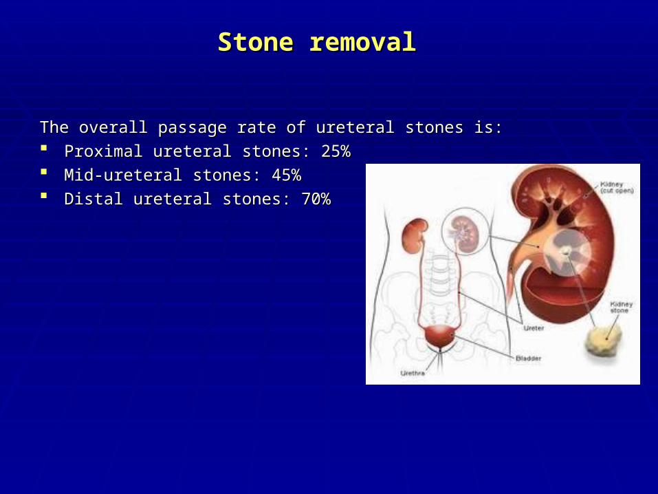
The overall passage rate of ureteral stones is: The overall passage rate of ureteral stones is: Proximal ureteral stones: 25% Proximal ureteral stones: 25% Mid-ureteral stones: 45% Mid-ureteral stones: 45% Distal ureteral stones: 70% Distal ureteral stones: 70%
Indications for Active Indications for Active Stone removalStone removal
Active stone removal is strongly Active stone removal is strongly recommended in patients fulfilling the recommended in patients fulfilling the following criteria: following criteria:
- p- persistent pain despite adequate ersistent pain despite adequate medicationmedication;;
- p- persistent obstruction with risk of ersistent obstruction with risk of impaired renal functionimpaired renal function;;
- - stone with urinary tract infectionstone with urinary tract infection;; - r- risk of pyonephrosis or urosepsisisk of pyonephrosis or urosepsis;; - b- bilateral obstructionilateral obstruction;; - obstructing calculus in a solitary - obstructing calculus in a solitary
functioning kidney.functioning kidney.
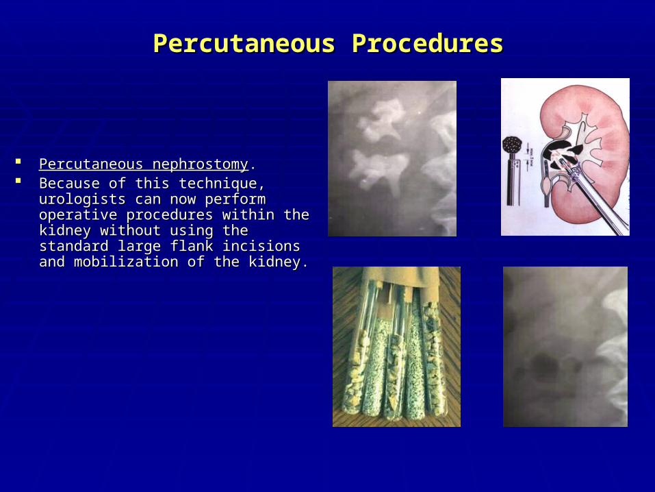
Percutaneous ProceduresPercutaneous Procedures
Percutaneous nephrostomyPercutaneous nephrostomy. . Because of this technique, urologists can Because of this technique, urologists can
now perform operative procedures within now perform operative procedures within the kidney without using the standard large the kidney without using the standard large flank incisions and mobilization of the flank incisions and mobilization of the kidney. kidney.
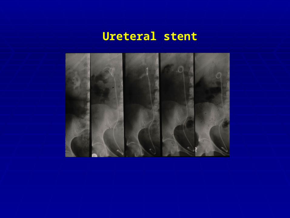
Ureteral stent
ContraindicationsContraindications
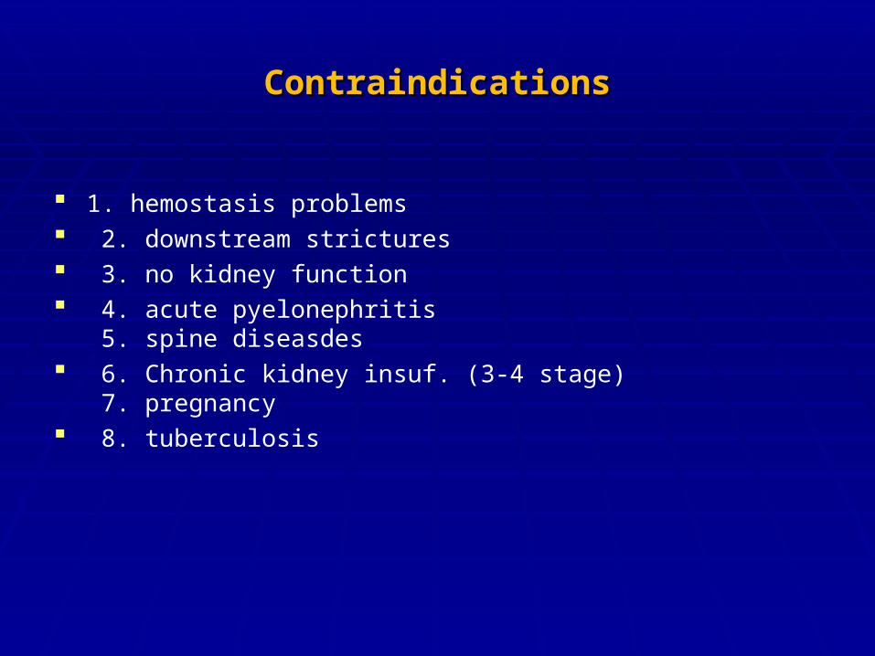
1. hemostasis problems 2. downstream strictures 3. no kidney function 4. acute pyelonephritis
5. spine diseasdes 6. Chronic kidney insuf. (3-4 stage)
7. pregnancy 8. tuberculosis
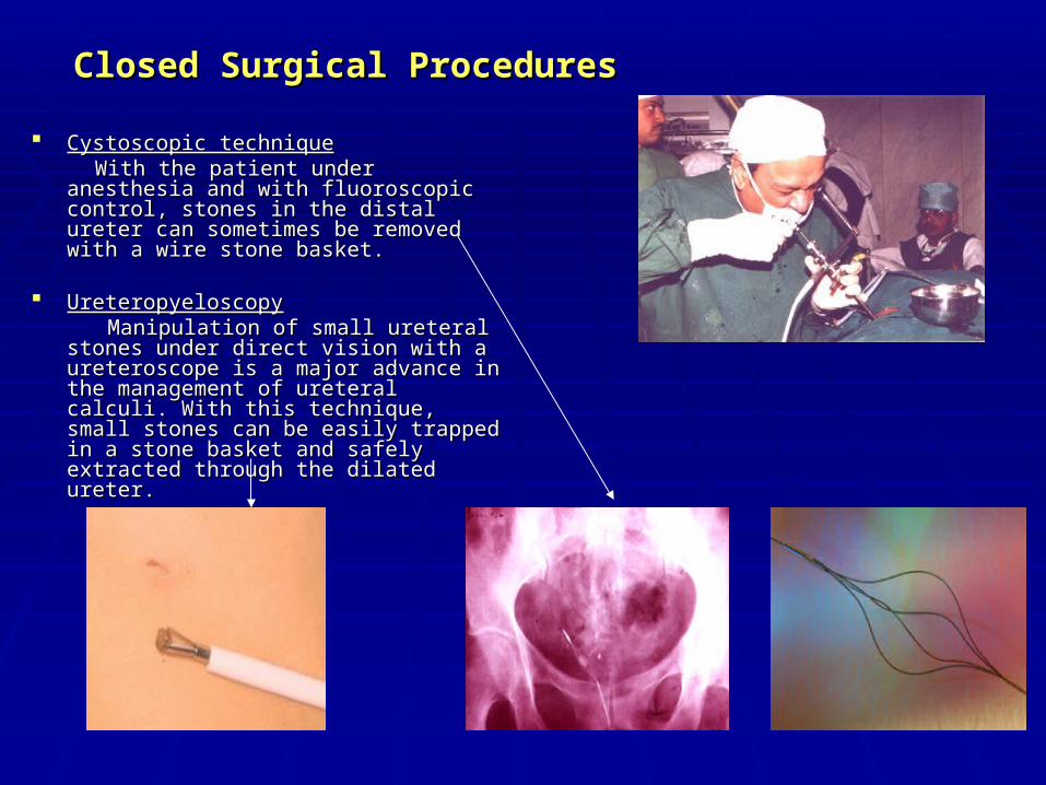
Closed Surgical ProceduresClosed Surgical Procedures
Cystoscopic techniqueCystoscopic technique With the patient under anesthesia and with With the patient under anesthesia and with
fluoroscopic control, stones in the distal fluoroscopic control, stones in the distal ureter can sometimes be removed with a ureter can sometimes be removed with a wire stone basket.wire stone basket.
UreteropyeloscopyUreteropyeloscopy Manipulation of small ureteral stones Manipulation of small ureteral stones
under direct vision with a ureteroscope is under direct vision with a ureteroscope is a major advance in the management of a major advance in the management of ureteral calculi. With this technique, small ureteral calculi. With this technique, small stones can be easily trapped in a stone stones can be easily trapped in a stone basket and safely extracted through the basket and safely extracted through the dilated ureter. dilated ureter.
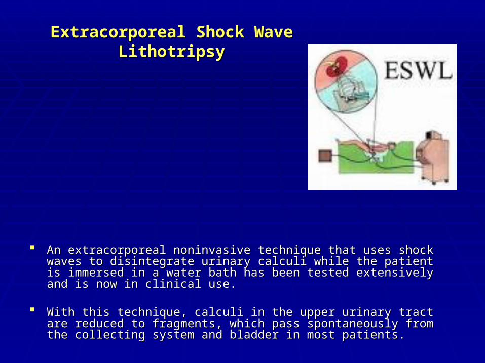
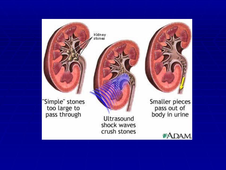
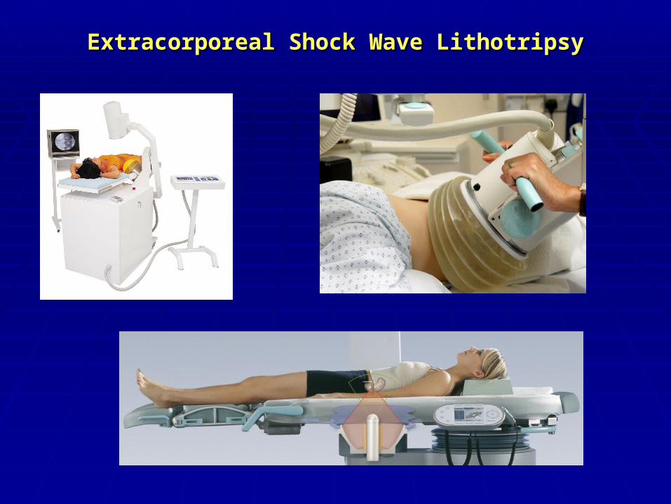
Extracorporeal Shock Wave Extracorporeal Shock Wave LithotripsyLithotripsy
An extracorporeal noninvasive technique that uses shock waves to An extracorporeal noninvasive technique that uses shock waves to disintegrate urinary calculi while the patient is immersed in a water bath disintegrate urinary calculi while the patient is immersed in a water bath has been tested extensively and is now in clinical use. has been tested extensively and is now in clinical use.
With this technique, calculi in the upper urinary tract are reduced to With this technique, calculi in the upper urinary tract are reduced to fragments, which pass spontaneously from the collecting system and fragments, which pass spontaneously from the collecting system and bladder in most patients.bladder in most patients.
Extracorporeal Shock Wave LithotripsyExtracorporeal Shock Wave Lithotripsy
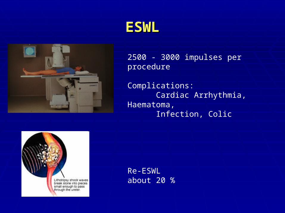
ESWLESWL
2500 - 3000 impulses per procedure
Complications:Cardiac Arrhythmia, Haematoma, Infection, Colic
Re-ESWLabout 20 %
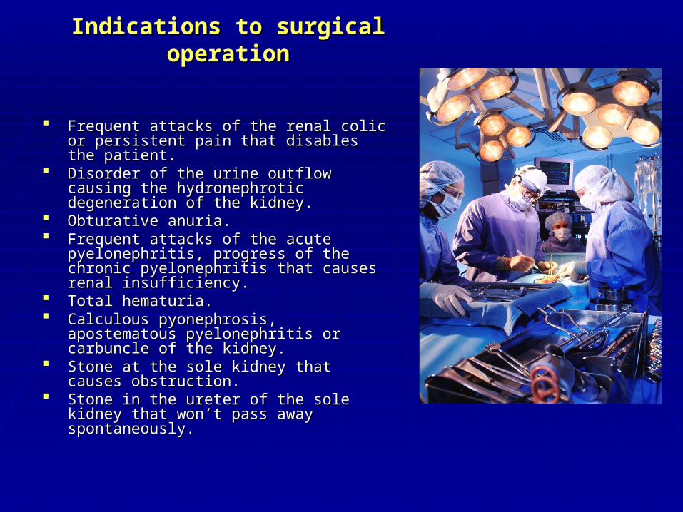
Indications to surgical operationIndications to surgical operation
Frequent attacks of the renal colic or persistent Frequent attacks of the renal colic or persistent pain that disables the patient.pain that disables the patient.
Disorder of the urine outflow causing the Disorder of the urine outflow causing the hydronephrotic degeneration of the kidney.hydronephrotic degeneration of the kidney.
Obturative anuria.Obturative anuria. Frequent attacks of the acute pyelonephritis, Frequent attacks of the acute pyelonephritis,
progress of the chronic pyelonephritis that progress of the chronic pyelonephritis that causes renal insufficiency.causes renal insufficiency.
Total hematuria.Total hematuria. Calculous pyonephrosis, apostematous Calculous pyonephrosis, apostematous
pyelonephritis or carbuncle of the kidney.pyelonephritis or carbuncle of the kidney. Stone at the sole kidney that causes obstruction.Stone at the sole kidney that causes obstruction. Stone in the ureter of the sole kidney that won’t Stone in the ureter of the sole kidney that won’t
pass away spontaneously.pass away spontaneously.
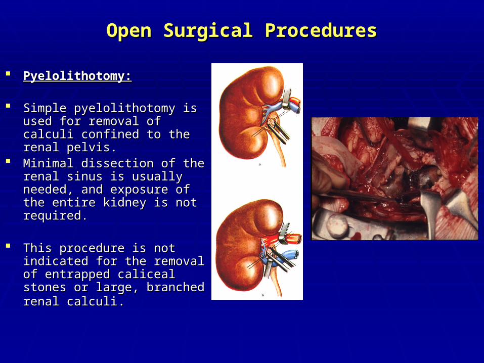
Open Surgical ProceduresOpen Surgical Procedures
Pyelolithotomy:Pyelolithotomy: Simple pyelolithotomy is used for Simple pyelolithotomy is used for
removal of calculi confined to the removal of calculi confined to the renal pelvis. renal pelvis.
Minimal dissection of the renal Minimal dissection of the renal sinus is usually needed, and sinus is usually needed, and exposure of the entire kidney is exposure of the entire kidney is not required. not required.
This procedure is not indicated for This procedure is not indicated for the removal of entrapped caliceal the removal of entrapped caliceal stones or large, branched renal stones or large, branched renal calculicalculi..
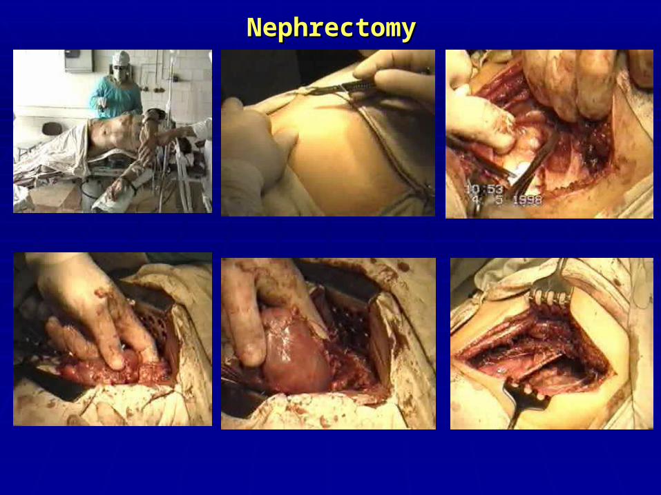
NephrectomyNephrectomy
Open Surgical ProceduresOpen Surgical Procedures
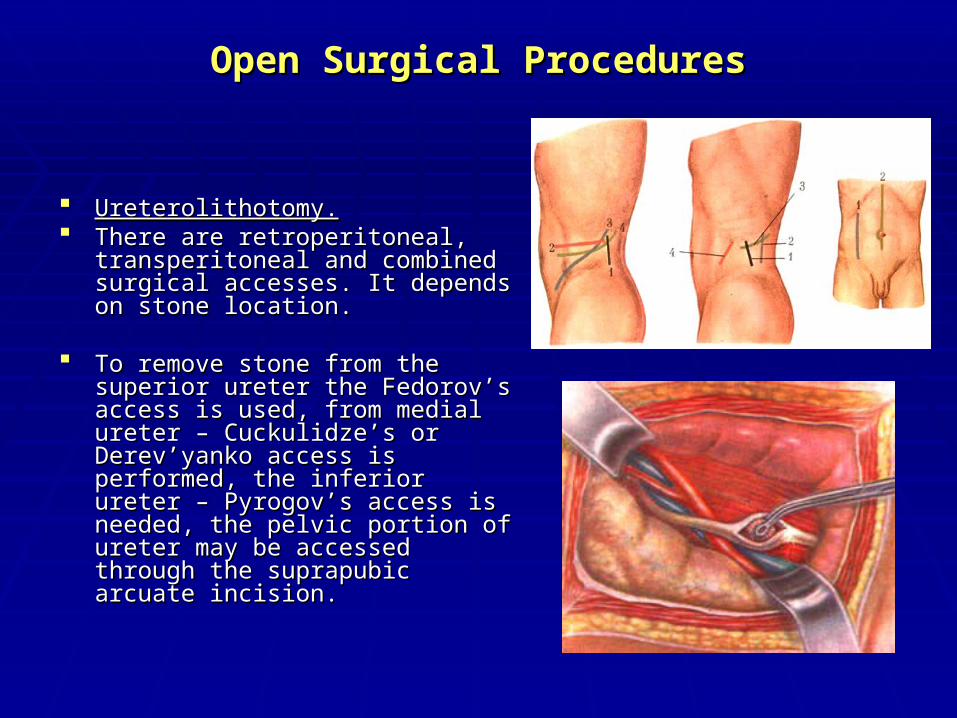
Ureterolithotomy.Ureterolithotomy. There are retroperitoneal, There are retroperitoneal,
transperitoneal and combined surgical transperitoneal and combined surgical accesses. It depends on stone location.accesses. It depends on stone location.
To remove stone from the superior To remove stone from the superior
ureter the Fedorov’s access is used, ureter the Fedorov’s access is used, from medial ureter – Cuckulidze’s or from medial ureter – Cuckulidze’s or Derev’yanko access is performed, the Derev’yanko access is performed, the inferior ureter – Pyrogov’s access is inferior ureter – Pyrogov’s access is needed, the pelvic portion of ureter needed, the pelvic portion of ureter may be accessed through the may be accessed through the suprapubic arcuate incision.suprapubic arcuate incision.
Open Surgical ProceduresOpen Surgical Procedures
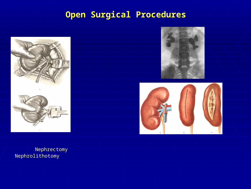
Nephrectomy NephrolithotomyNephrectomy Nephrolithotomy
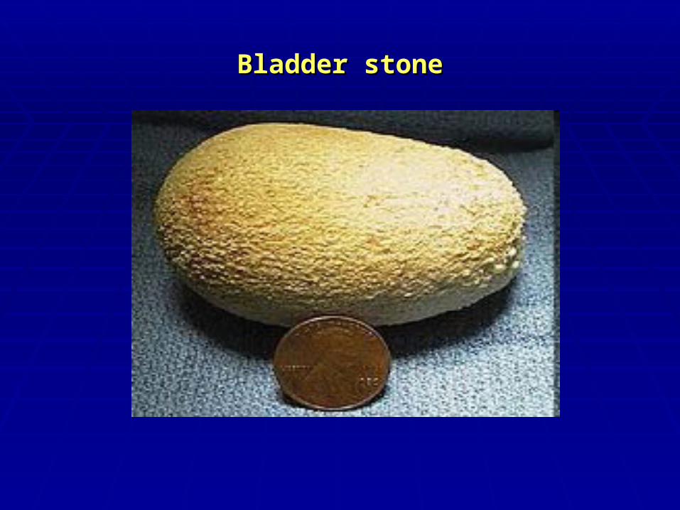
Bladder stoneBladder stone
BLADDER STONESBLADDER STONES
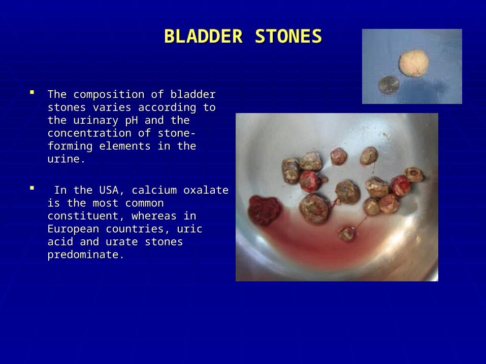
The composition of bladder stones The composition of bladder stones varies according to the urinary pH and varies according to the urinary pH and the concentration of stone-forming the concentration of stone-forming elements in the urine.elements in the urine.
In the USA, calcium oxalate is the In the USA, calcium oxalate is the most common constituent, whereas in most common constituent, whereas in European countries, uric acid and European countries, uric acid and urate stones predominate.urate stones predominate.
Diagnostic EvaluationDiagnostic Evaluation
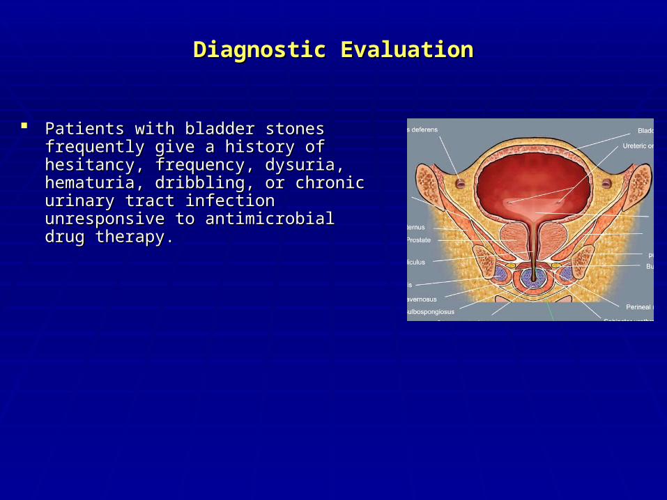
Patients with bladder stones frequently give Patients with bladder stones frequently give a history of hesitancy, frequency, dysuria, a history of hesitancy, frequency, dysuria, hematuria, dribbling, or chronic urinary tract hematuria, dribbling, or chronic urinary tract infection unresponsive to antimicrobial drug infection unresponsive to antimicrobial drug therapy.therapy.
Diagnostic EvaluationDiagnostic Evaluation
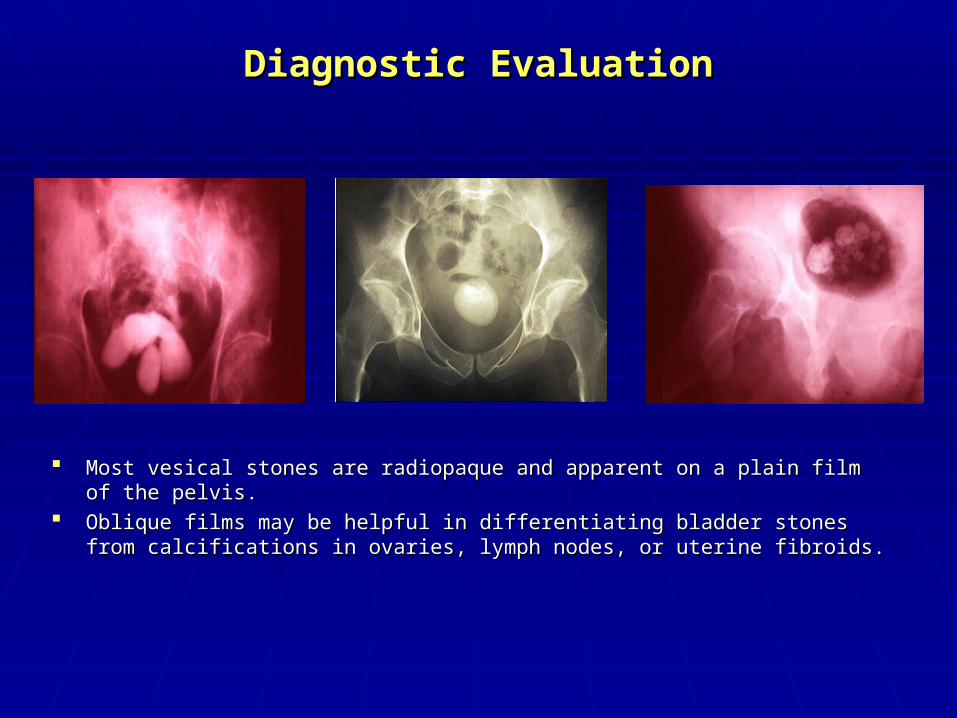
Most vesical stones are radiopaque and apparent on a plain film of the pelvis. Most vesical stones are radiopaque and apparent on a plain film of the pelvis. Oblique films may be helpful in differentiating bladder stones from calcifications in Oblique films may be helpful in differentiating bladder stones from calcifications in
ovaries, lymph nodes, or uterine fibroids.ovaries, lymph nodes, or uterine fibroids.
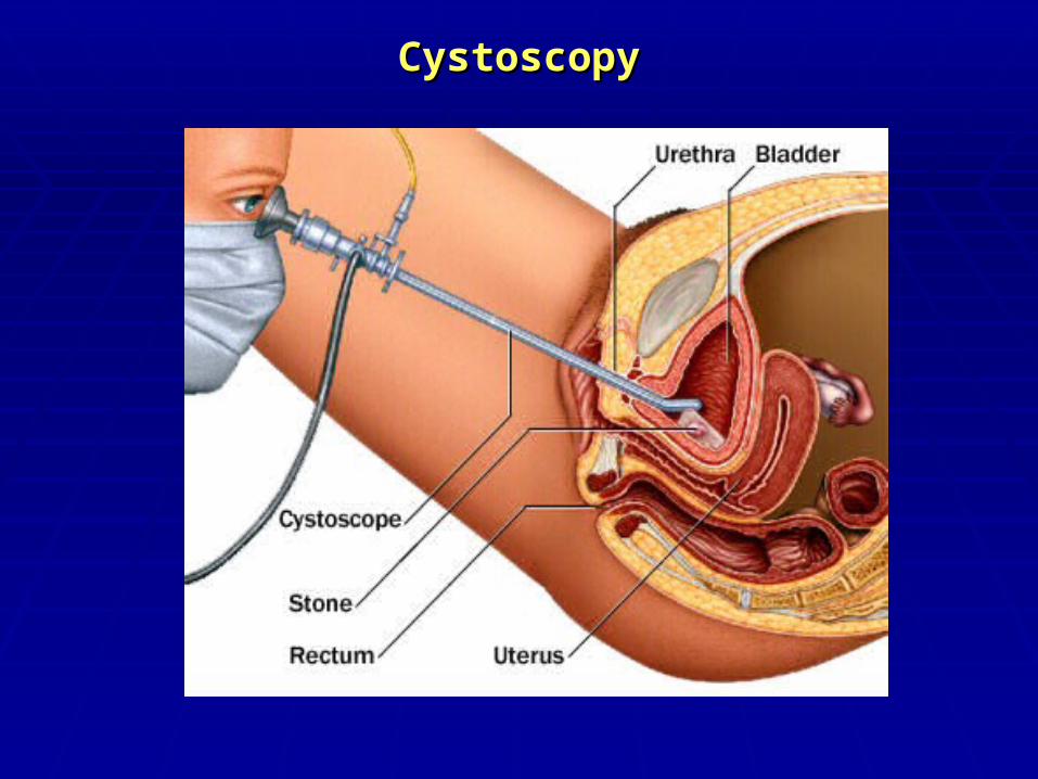
CystoscopyCystoscopy
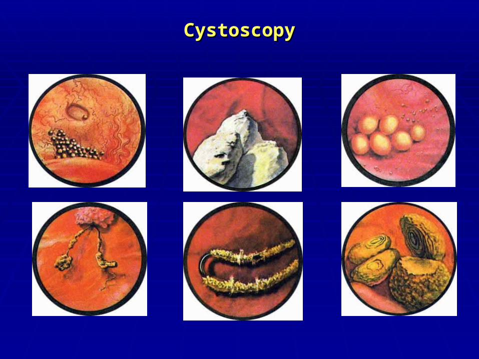
CystoscopyCystoscopy
TreatmentTreatment
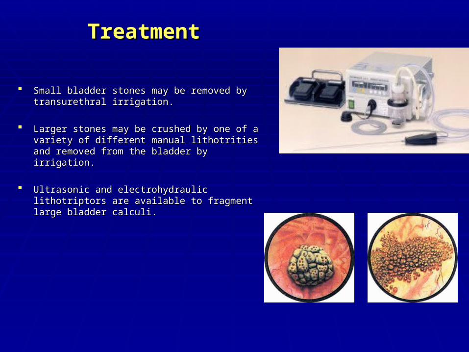
Small bladder stones may be removed by Small bladder stones may be removed by transurethral irrigation.transurethral irrigation.
Larger stones may be crushed by one of a variety Larger stones may be crushed by one of a variety of different manual lithotrities and removed from of different manual lithotrities and removed from the bladder by irrigation. the bladder by irrigation.
Ultrasonic and electrohydraulic lithotriptors are Ultrasonic and electrohydraulic lithotriptors are available to fragment large bladder calculi.available to fragment large bladder calculi.
TreatmentTreatment
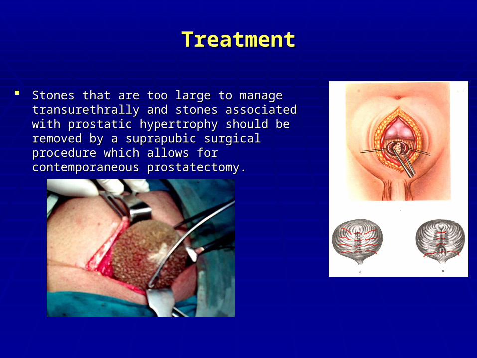
Stones that are too large to manage transurethrally Stones that are too large to manage transurethrally and stones associated with prostatic hypertrophy and stones associated with prostatic hypertrophy should be removed by a suprapubic surgical should be removed by a suprapubic surgical procedure which allows for contemporaneous procedure which allows for contemporaneous prostatectomy. prostatectomy.
Preventive treatment in calcium stone diseasePreventive treatment in calcium stone disease
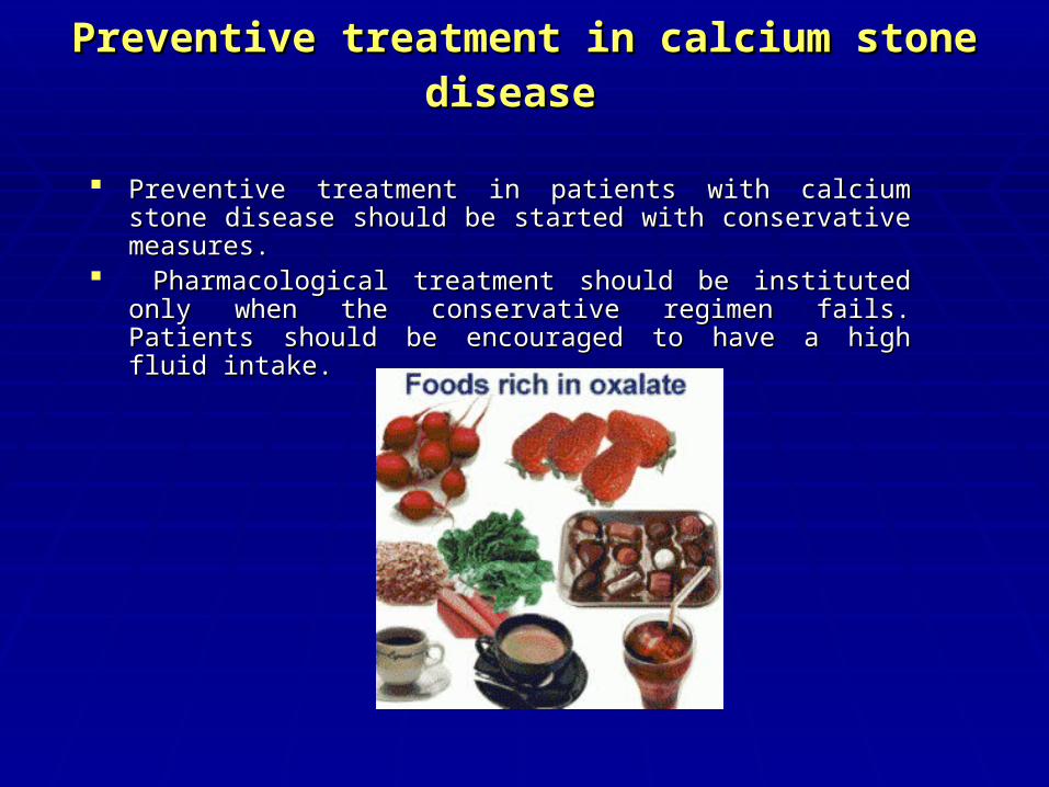
Preventive treatment in patients with calcium stone disease should Preventive treatment in patients with calcium stone disease should be started with conservative measures.be started with conservative measures.
Pharmacological treatment should be instituted only when the Pharmacological treatment should be instituted only when the conservative regimen fails. Patients should be encouraged to have conservative regimen fails. Patients should be encouraged to have a high fluid intake. a high fluid intake.
Preventive treatment in calcium stone diseasePreventive treatment in calcium stone disease
Diet should be of a 'common sense' type - a mixed balanced diet with Diet should be of a 'common sense' type - a mixed balanced diet with contributions from all food groups but without excesses of any kind.contributions from all food groups but without excesses of any kind.
The intake of fruits and vegetables should be encouraged because of the The intake of fruits and vegetables should be encouraged because of the beneficial effects of fibre. Care must be taken, however, to avoid fruits and beneficial effects of fibre. Care must be taken, however, to avoid fruits and vegetables that are rich in oxalate. Wheat bran is rich in oxalate and should be vegetables that are rich in oxalate. Wheat bran is rich in oxalate and should be avoided. In order to avoid an oxalate load, the excessive intake of products avoided. In order to avoid an oxalate load, the excessive intake of products rich in oxalate should be limited or avoided. This is of particular importance in rich in oxalate should be limited or avoided. This is of particular importance in patients in whom high excretion of oxalate has been demonstrated. patients in whom high excretion of oxalate has been demonstrated.
The following products have a high content of oxalateThe following products have a high content of oxalate : : Rhubarb 530 mg oxalate/100 g Rhubarb 530 mg oxalate/100 g Spinach 570 mg oxalate/100 g Spinach 570 mg oxalate/100 g Cocoa 625 mg oxalate/100 g Cocoa 625 mg oxalate/100 g Tea leaves 375-1450 mg oxalate/100 g Tea leaves 375-1450 mg oxalate/100 g Nuts 200-600 mg oxalate/100 g. Nuts 200-600 mg oxalate/100 g.
Preventive treatment in calcium stone diseasePreventive treatment in calcium stone disease
Vitamin C in doses up to 4 g/day can be taken without increasing the risk of Vitamin C in doses up to 4 g/day can be taken without increasing the risk of stone formation. stone formation.
Animal protein should not be ingested in excessive amounts. It is Animal protein should not be ingested in excessive amounts. It is recommended that the animal protein intake is limited to approximately 150 recommended that the animal protein intake is limited to approximately 150 g/day.g/day.
Calcium intake should not be restricted unless there are very strong reasons Calcium intake should not be restricted unless there are very strong reasons for such advice. The minimum daily requirement for calcium is 800 mg and the for such advice. The minimum daily requirement for calcium is 800 mg and the general recommendation is 1000 mg/day. Supplements of calcium are not general recommendation is 1000 mg/day. Supplements of calcium are not recommended except in cases of enteric hyperoxaluria, in which additional recommended except in cases of enteric hyperoxaluria, in which additional calcium should be ingested with meals. calcium should be ingested with meals.
Preventive treatment in uric acid stone stone diseasePreventive treatment in uric acid stone stone disease
The intake of foodstuffs particularly rich in urate should The intake of foodstuffs particularly rich in urate should be restricted in patients with hyperuricosuric calcium be restricted in patients with hyperuricosuric calcium oxalate stone disease , as well as in patients with uric oxalate stone disease , as well as in patients with uric acid stone disease. The intake of urate should not be acid stone disease. The intake of urate should not be more than 500 mg/day. more than 500 mg/day.
Below are examples of food rich in urate : Below are examples of food rich in urate : Calf thymus 900 mg urate/100 g Calf thymus 900 mg urate/100 g Liver 260-360 mg urate/100 g Liver 260-360 mg urate/100 g Kidneys 210-255 mg urate/100 g Kidneys 210-255 mg urate/100 g Poultry skin 300 mg urate/100 g Poultry skin 300 mg urate/100 g Herring with skin, sardines, anchovies, sprats 260-500 Herring with skin, sardines, anchovies, sprats 260-500
mg urate/100 g. mg urate/100 g.