Embed Size (px)
Citation preview
Urinary System
Dr. Michael P. Gillespie
Major Components
2 kidneys. 2 ureters. 1 urinary bladder. 1 urethra.
Functions Of Kidneys
Excrete wastes in urine. Regulate blood volume. Regulate blood composition. Regulate blood pressure. Synthesize glucose. Release erythropoietin. Participate in vitamin D synthesis.

Functions Of Urinary System Continued
Ureters transport urine from the kidneys to the urinary bladder.
Urinary bladder stores urine. Urethra discharges urine from the body.
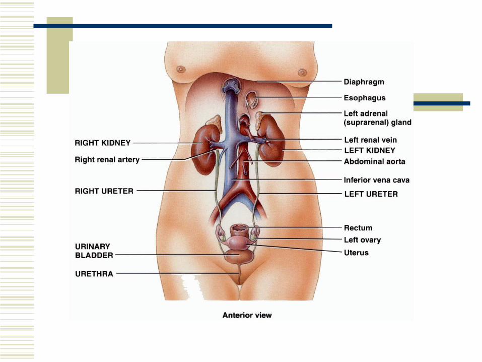
Kidneys
The paired kidneys are reddish, kidney-bean-shaped organs.
They are located above the waist between the peritoneum and the posterior wall of the abdomen.
They are retroperitoneal. The right kidney is slightly lower than the left due
to the presence of the liver.

External Anatomy Of The Kidneys
A typical kidney is 10-12 cm long, 5-7 cm wide and 3 cm thick.
The concave medial border of the kidneys faces the vertebral column.
The renal hilus is a deep vertical fissure through which the ureter, blood vessels, lymphatic vessels, and nerves pass.
External Anatomy Of The Kidneys
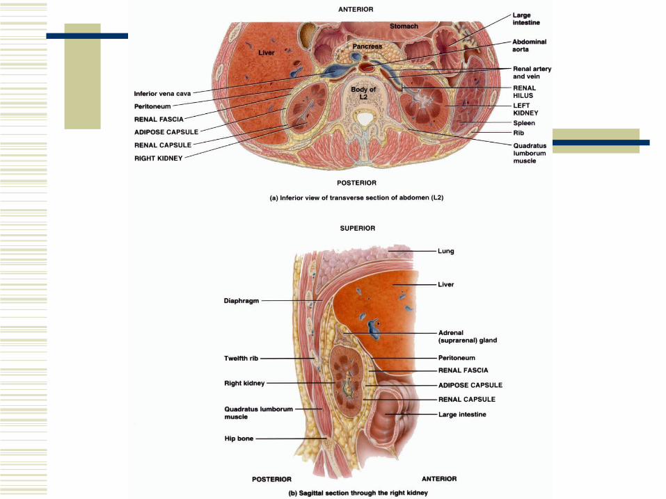
Layers of tissue around the kidneys. Renal capsule – deep layer. Smooth transparent
sheet of dense irregular connective tissue. Maintains the shape of the kidney.
Adipose capsule – mass of fatty tissue. Protects the kidney from trauma and holds it in place.
Renal fascia – superficial layer. Anchors the kidney to the surrounding structures and to the abdominal wall.

Nephroptosis
Nephroptosis or floating kidney is an inferior displacement or dropping of the kidney.
It occurs when the kidney slips from its normal position.
Nephroptosis
This happens when it is not securely held in place by adjacent organs or its covering of fat.
It occurs most often in very thin people who have a deficient adipose capsule or renal fascia.
The ureter may kink and block urine flow.
Internal Anatomy Of The Kidneys
Renal cortex – superficial layer. Renal medulla – inner region.
8-18 cone-shaped renal pyramids which taper to a renal papilla.
The renal columns are portions of the cortex that extend between the pyramids.
A renal lobe consists of a renal pyramid, overlying renal cortex, and one-half of each adjacent renal column.
Internal Anatomy Of The Kidneys
Together, the renal cortex and renal pyramids constitute the parenchyma (functional portion) of the kidney.
The nephrons (functional units) of the kidney are within the parenchyma.
Urine formed by the nephrons drains into the papillary ducts, which drain into the minor and eventually major calyces.
Internal Anatomy Of The Kidneys
Each kidney has 8 to 18 minor calyces and 2 to 3 major calyces.
The major calyces drain urine into a large cavity called the renal pelvis.
The hilus expands into the renal sinus (a cavity within the kidney).

Blood & Nerve Supply Of The Kidneys
The kidneys have abundant blood vessels. The kidneys remove wastes from the blood and
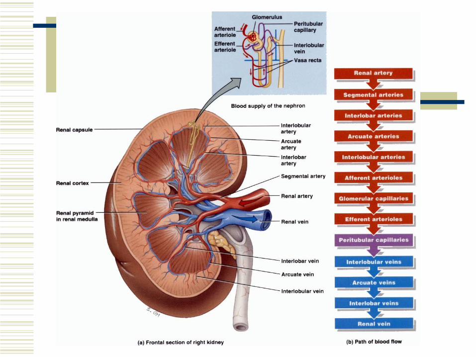
regulate its volume and ionic composition. Right and left renal arteries supple the kidneys. They branch into segmental arteries, which branch
into interlobar arteries.
Blood & Nerve Supply Of The Kidneys
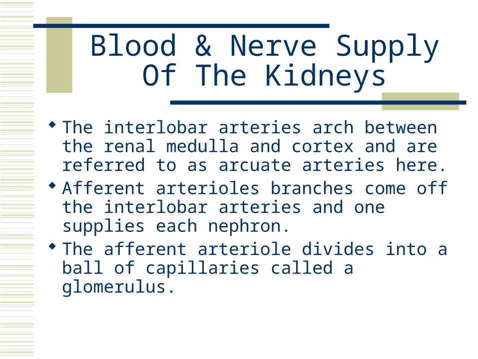
The interlobar arteries arch between the renal medulla and cortex and are referred to as arcuate arteries here.
Afferent arterioles branches come off the interlobar arteries and one supplies each nephron.
The afferent arteriole divides into a ball of capillaries called a glomerulus.
Blood & Nerve Supply Of The Kidneys
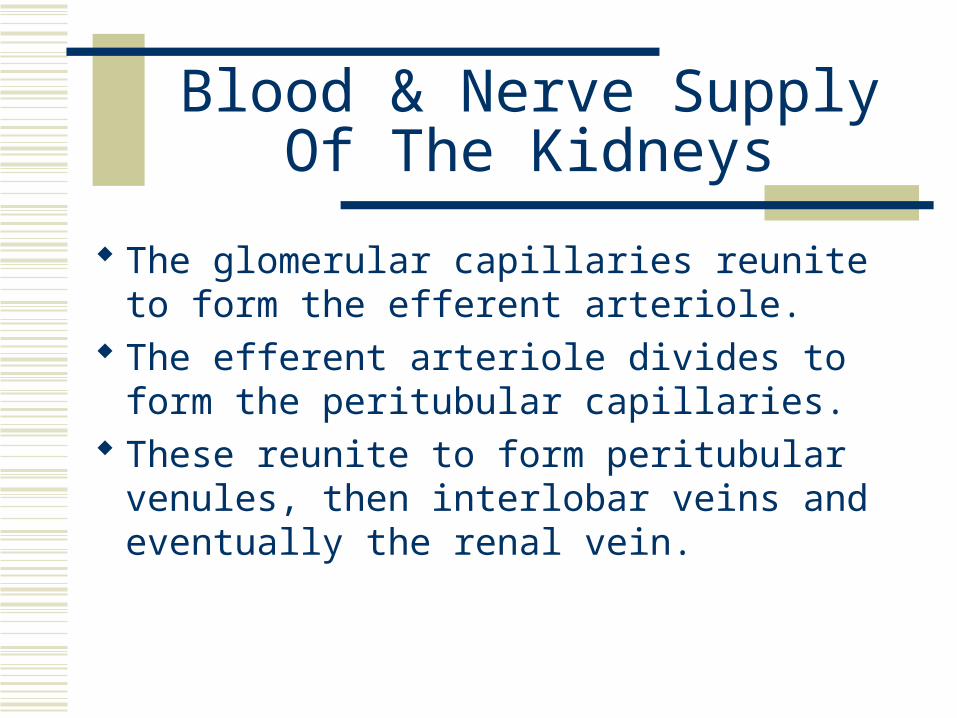
The glomerular capillaries reunite to form the efferent arteriole.
The efferent arteriole divides to form the peritubular capillaries.
These reunite to form peritubular venules, then interlobar veins and eventually the renal vein.
Blood & Nerve Supply Of The Kidneys
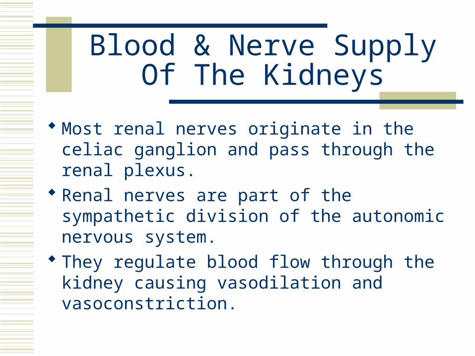
Most renal nerves originate in the celiac ganglion and pass through the renal plexus.
Renal nerves are part of the sympathetic division of the autonomic nervous system.
They regulate blood flow through the kidney causing vasodilation and vasoconstriction.

Nephron
Nephrons are the functional units of the kidneys.
Two main parts: Renal corpuscle – where blood plasma is filtered. Renal tubule – into which the filtered fluid
passes.
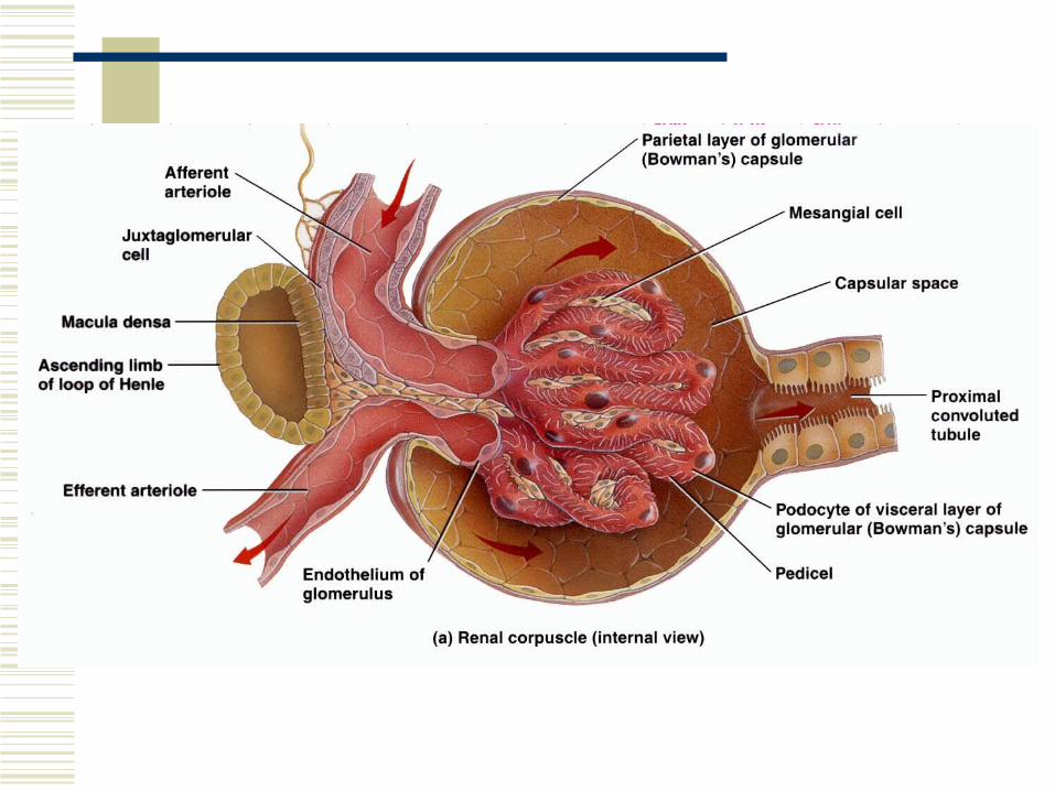
Renal Corpuscle
Two components: Glomerulus – a capillary network. Glomerular (bowman’s) capsule – a double
walled epithelial cup that surrounds the glomerular capillaries.
Renal Tubule
3 main sections: Proximal convoluted tubule. Loop of Henle (nephron loop). Distal convoluted tubule.
Nephron Continued
The distal convoluted tubules of several nephrons empty into a single collecting duct.
Collecting ducts then unite and converge into papillary ducts, which drain into minor calyces.
1 kidney has approximately 1 million nephrons.
Loop Of Henle
The loop of Henle connects the proximal and distal convoluted tubules.
It consists of a descending limb and an ascending limb.
Types Of Nephrons
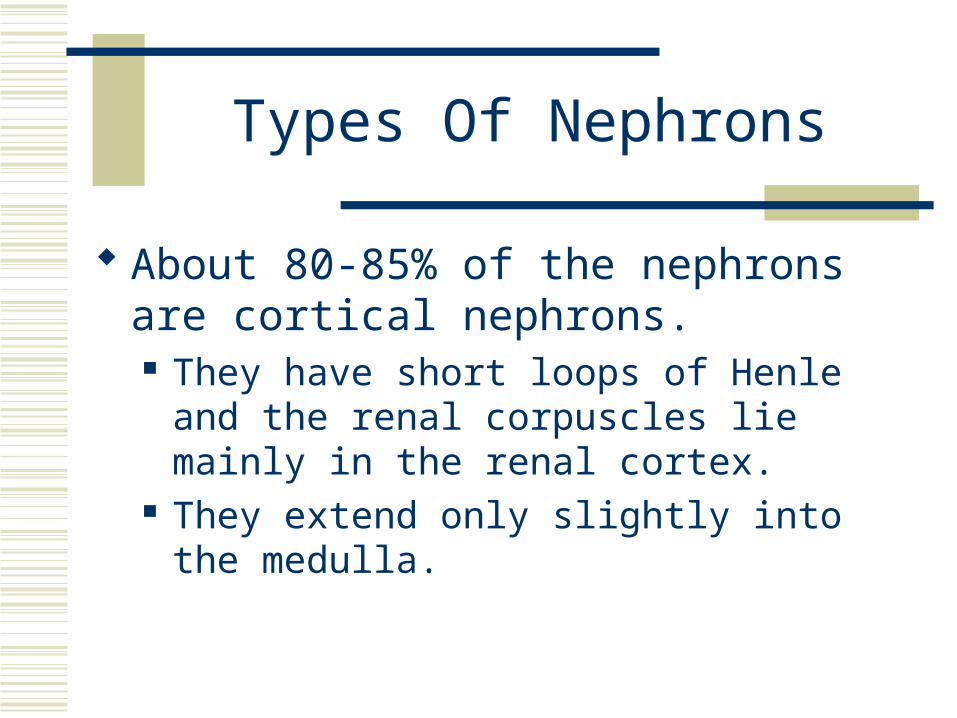
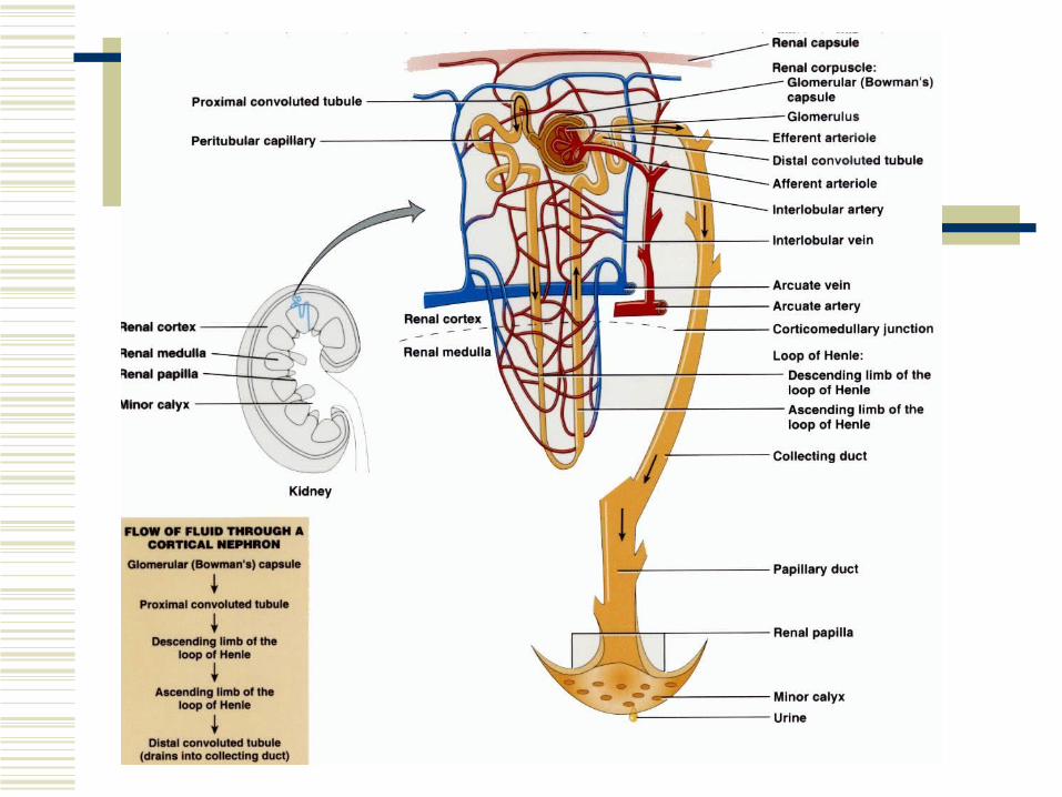
About 80-85% of the nephrons are cortical nephrons. They have short loops of Henle and the renal
corpuscles lie mainly in the renal cortex. They extend only slightly into the medulla.
Types Of Nephrons
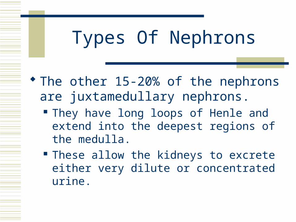
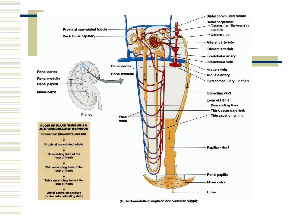
The other 15-20% of the nephrons are juxtamedullary nephrons. They have long loops of Henle and extend into
the deepest regions of the medulla. These allow the kidneys to excrete either very
dilute or concentrated urine.
Histology Of The Nephron & Collecting Duct
A single layer of epithelial cells forms the entire wall of the glomerular capsule, renal tubule, and ducts.
Glomerular Capsule
The glomerular (bowman’s) capsule consists of visceral and parietal layers with a capsular (bowman’s) space in between. Visceral layer – modified simple squamous
epithelial cells called podocytes. Parietal layer – simple squamous epithelium.
Fluid filtered from the glomerular capillaries enters the capsular space.
Renal Tubule & Collecting Duct
Proximal convoluted tubule cells are simple cuboidal epithelial cells with a brush border of microvilli.
The descending limb and the first part of the ascending limb are composed of simple squamous epithelium.
The thick ascending limb is composed of simple cuboidal epithelium to low columnar epithelium.

Number Of Nephrons
The number of nephrons is constant from birth.
Growth in kidney size is due to growth in size of the nephrons, not increase in number.
Signs of kidney dysfunction do not usually become apparent until function declines to less than 25% of normal.
Number Of Nephrons
The remaining functional nephrons adapt to form a larger than normal load.
Surgical removal of one kidney stimulates hypertrophy of the other kidney.
One kidney can eventually filter blood at a rate of 80% of two normal kidneys.
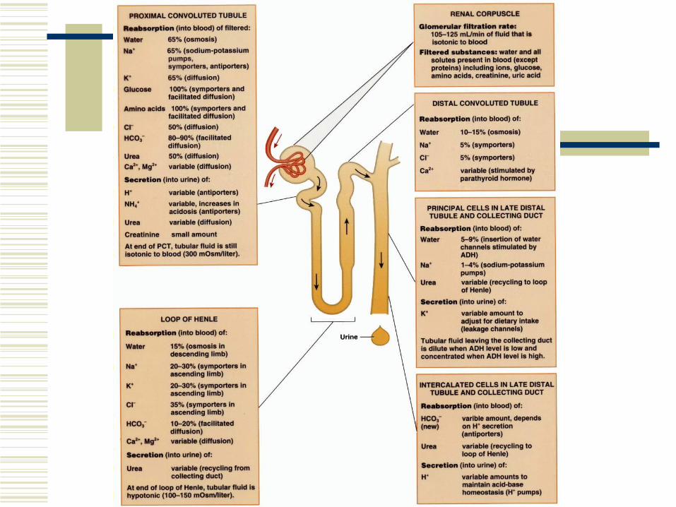
Functions Of Nephrons & Collecting Ducts
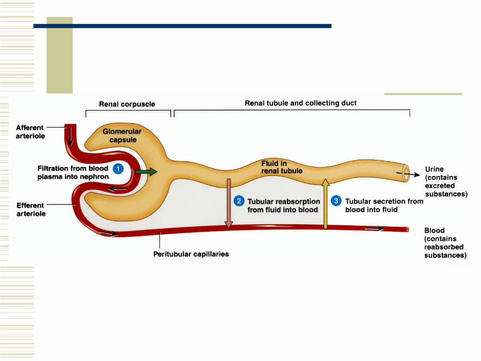
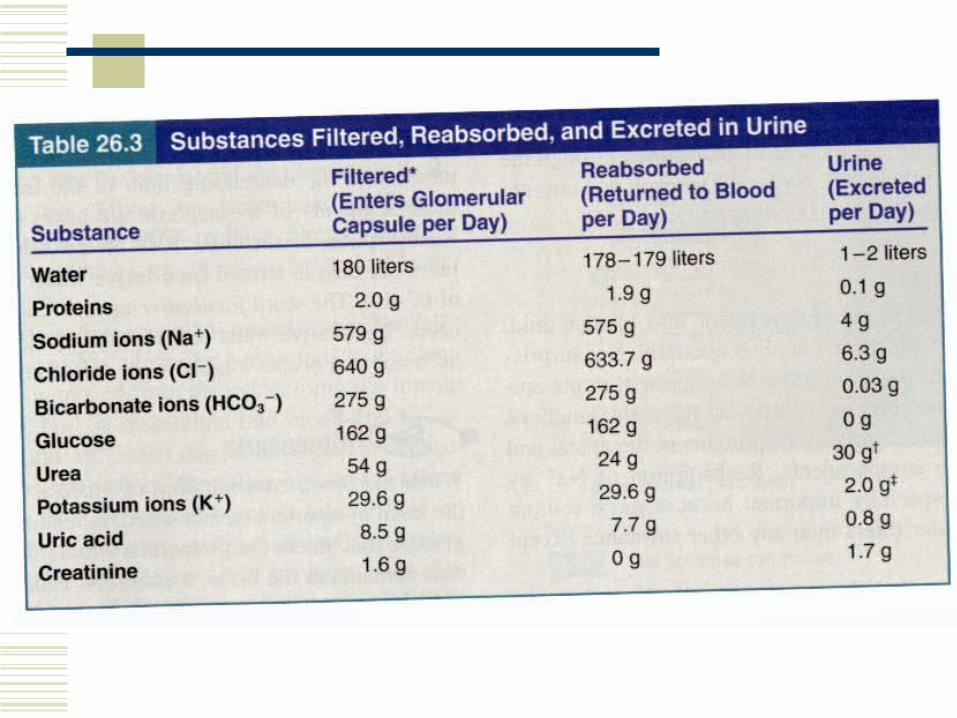
Glomerular filtration – water and most solutes move across the wall of glomerular capillaries into the glomerular capsule and into the renal tubule.
Tubular reabsorption – tubule cells reabsorb about 99% of the water and many useful solutes into the peritubular capillaries.
Tubular secretion – the tubule cells secrete wastes, drugs, and excess ions into the fluid as it moves through the tubule and collecting duct.
Glomerular Filtration
Glomerular filtrate – the fluid that enters the capsular space.
Filtration fraction – the fraction of blood plasma in the afferent arterioles of the kidneys that becomes filtrate (typically 16-20%).
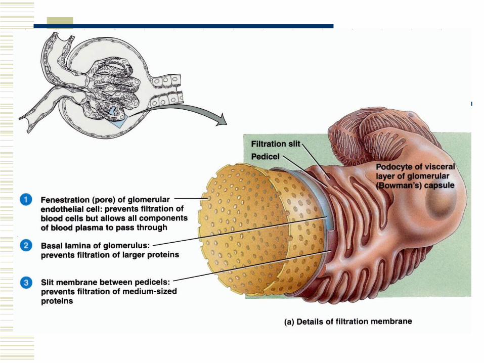
Filtration Membrane
The endothelial cells of the glomerular capillaries and the podocytes, which encircle the capillaries, form a leaky barrier known as the filtration membrane.
Filtration Membrane
Fenestrations (pores) in the glomerular epithelial cells cause them to be quite leaky.
Filtration Membrane
Filtration slits are spaces between the pedicels (footlike processes from the podocytes), which allow passage of molecules smaller than 6-7 nm. Water, glucose, vitamins, amino acids, very small
plasma proteins, ammonia, urea, and ions can pass through.
Albumin is too large to easily pass through the slits.
Filtration utilizes pressure to drive fluids and solutes through a membrane.
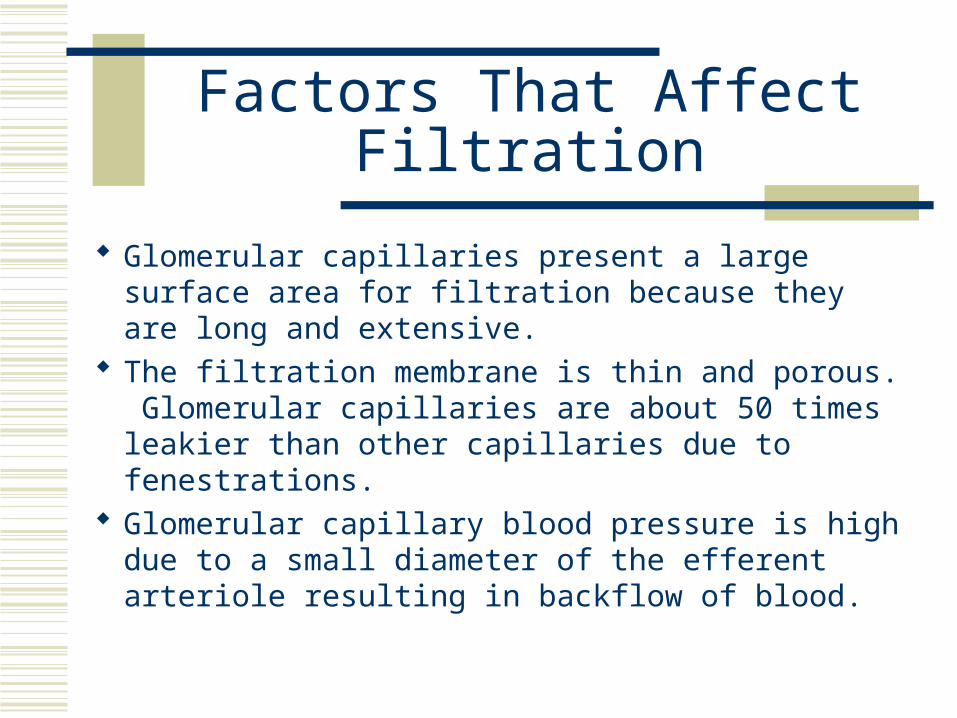
Factors That Affect Filtration
Glomerular capillaries present a large surface area for filtration because they are long and extensive.
The filtration membrane is thin and porous. Glomerular capillaries are about 50 times leakier than other capillaries due to fenestrations.
Glomerular capillary blood pressure is high due to a small diameter of the efferent arteriole resulting in backflow of blood.
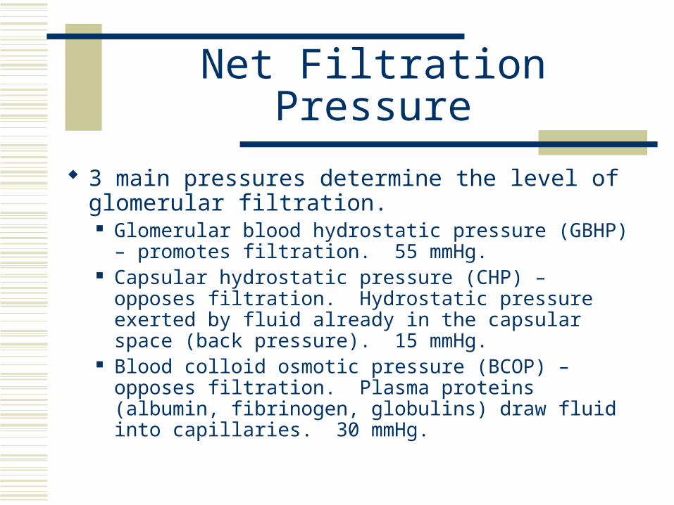
Net Filtration Pressure
3 main pressures determine the level of glomerular filtration. Glomerular blood hydrostatic pressure (GBHP) –
promotes filtration. 55 mmHg. Capsular hydrostatic pressure (CHP) – opposes filtration.
Hydrostatic pressure exerted by fluid already in the capsular space (back pressure). 15 mmHg.
Blood colloid osmotic pressure (BCOP) – opposes filtration. Plasma proteins (albumin, fibrinogen, globulins) draw fluid into capillaries. 30 mmHg.

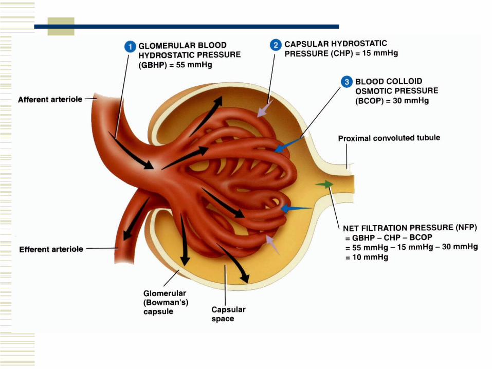
Net Filtration Pressure
Net filtration pressure (NFP) = GBHP – CHP – BCOP.
NFP = 55mmhg – 15mmhg – 30mmhg = 10mmhg.
Loss Of Plasma Proteins
Kidney disease can cause damage to glomerular capillaries allowing them to be permeable to plasma proteins.
As plasma proteins filter out, the osmotic pressure of the blood decreases, allowing water to be drawn from the blood plasma into interstitial tissues. This results in edema.
Glomerular Filtration Rate
The amount of filtrate formed in all the renal corpuscles of both kidneys each minute is the glomerular filtration rate (GFR).
If the GFR is too high, substances may pass too quickly through the tubules that they are not reabsorbed.
If the GFR is too low, nearly all the filtrate may be reabsorbed resulting in inadequate excretion.
Reabsorption
Primary and secondary active transport mechanisms are utilized to “pump” a substance across a membrane.
Obligatory water reabsorption occurs due to the solute reabsorption and corresponding osmotic pressure created. Water follows the solutes.
Glucosuria
When the blood concentration of glucose is above 200 mg/ml, the renal symporters cannot work fast enough to reabsorb all of the glucose that enters the glomerular filtrate.
Some of the glucose remains in the urine (glucosuria).
Diabetes mellitus is the most common cause of glucosuria.

Hormonal Regulation Of Tubular Reabsorption &
Secretion Angiotensin II – increases reabsorption of
Na+, other solutes, and water, which increases blood volume.
Aldosterone – increases secretion of K+ and reabsorption of Na+, Cl-. This increases reabsorption of water and increases blood volume.
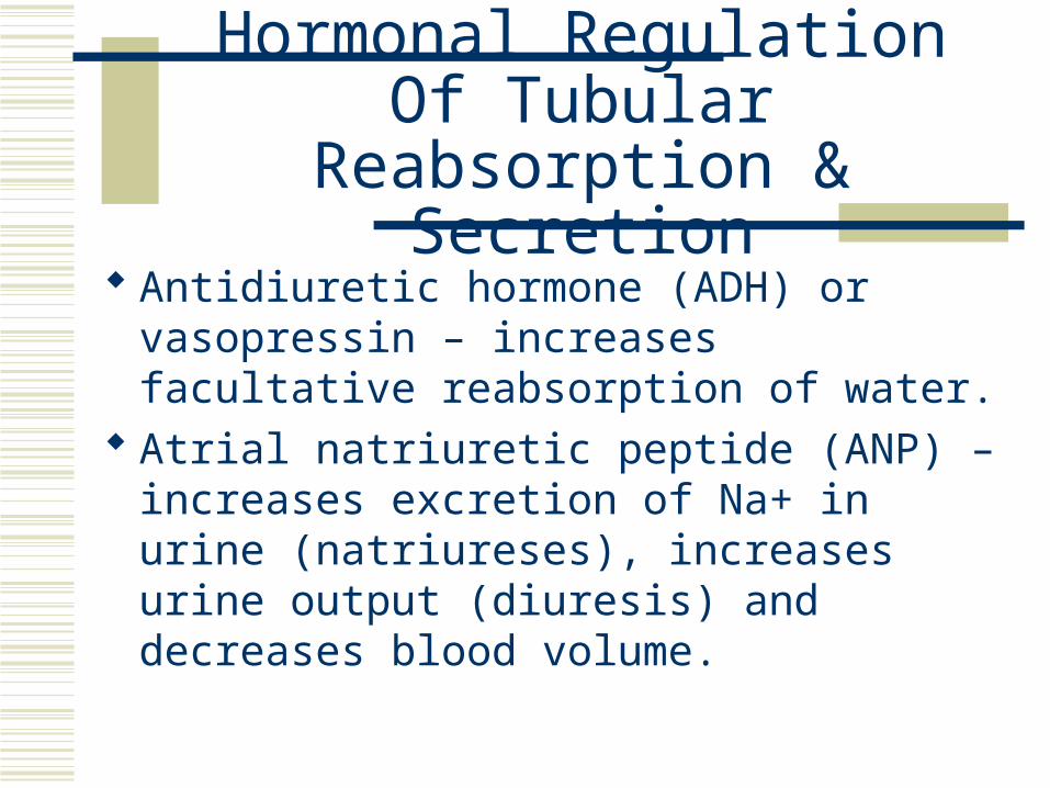
Hormonal Regulation Of Tubular Reabsorption &
Secretion Antidiuretic hormone (ADH) or vasopressin
– increases facultative reabsorption of water. Atrial natriuretic peptide (ANP) – increases
excretion of Na+ in urine (natriureses), increases urine output (diuresis) and decreases blood volume.
Diuretics
Diuretics are substances that slow renal reabsorption of water and thereby causes diuresis, an elevated urine flow rate, which in turn reduces blood volume.
Diuretics are prescribed to treat hypertension. Naturally occuring diuretics include caffeine
which inhibits Na+ reabsorption, and alcohol which inhibits secretion of ADH.
Evaluation Of Kidney Function
The kidneys are evaluated by assessing the quantity of urine, the quality of urine, and the level of wastes in blood.
Urinalysis, blood urea nitrogen (BUN) test, plasma creatinine, and renal plasma clearance tests are utilized to assess kidney functioning.
Characteristics Of Normal Urine
Volume – 1 to 2 liters / 24 hours (varies). Color – yellow or amber, but varies with
concentration and diet. Concentrated urine is darker. Diet (reddish color from beets), medications, and diseases may affect color. Kidney stones can produce blood in urine.
Turbidity – transparent when freshly voided, but becomes turbid (cloudy) upon standing.
Characteristics Of Normal Urine
Odor – mildly aromatic but becomes ammonia-like upon standing. Urine of diabetics has a fruity odor due to ketone bodies.
pH – ranges between 4.6 and 8.0 (average 6.0). High protein diets increases acidity, vegetarian diets increase alkalinity.
Specific gravity (density) – ranges from 1.001 to 1.035. Greater concentration of solutes yields greater specific gravity.
Blood Urea Nitrogen (BUN)
This test measures the blood nitrogen that is part of the urea resulting from catabolism and deamination of amino acids.
BUN rises as the glomerular filtration rate decreases due to renal disease or obstruction of the urinary tract.
Decreasing protein intake decreases urea production.
Plasma Creatinine
Plasma creatinine results from the catabolism of creatinine phosphate from skeletal muscle.
Creatinine levels above 1.5 mg/dL indicate poor renal function. Decreased levels indicated decreased muscle mass (I.e. muscular dystrophy).
Renal Plasma Clearance
Renal plasma clearance is the volume of blood that is “cleaned” or cleared of a substance per unit of time.
High renal plasma clearance indicates efficient excretion of a substance in the urine; low clearance indicates inefficient clearance.

Dialysis
If a person’s kidneys are so impaired by disease or injury that they are uable to function, the blood must be cleansed artificially by dialysis.
Dialysis is the separation of large solutes from smaller ones through the use of a selectively permeable membrane.

Dialysis
An artificial kidney machine performs hemodialysis. It directly filters a patient’s blood.
After passing though the dialysis tubing, the cleansed blood flows back into the patient’s body.
Urine Transportation, Storage, & Elimination
The urine drains from collecting ducts through papillary ducts into the minor calyces, which join the major calyces, that unite to form the renal pelvis.
From the renal pelvis, the urine drains into the ureters and then into the urinary bladder.
Urine is discharged from the body through a single urethra.
Ureters
Each of the two ureters transport urine from the renal pelvis of one kidney to the urinary bladder.
Peristaltic contractions of the muscular walls of the ureters push the urine towards the bladder.
No anatomical valve exists between the ureters and bladder; however, a physiological one exists. Pressure from the filling bladder compresses the openings of the ureters preventing backflow of urine and microbes.
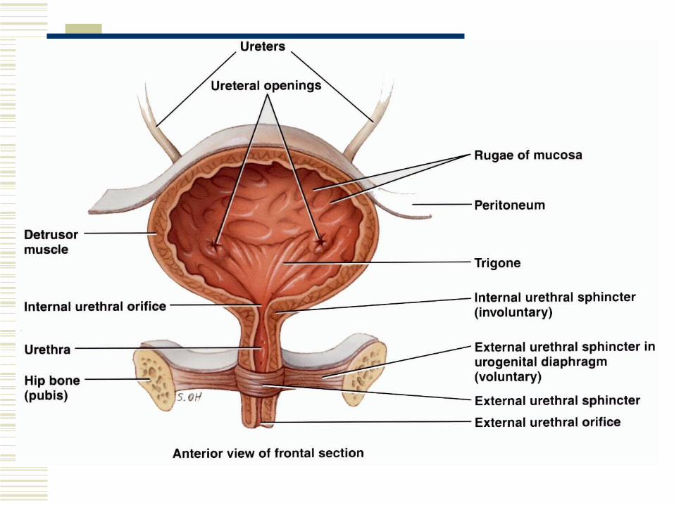
Urinary Bladder
The urinary bladder is a hollow, distensible muscular organ.
It resides in the pelvic cavity posterior to the pubic symphysis.
Urinary Bladder
When the bladder is empty, it is collapsed. When it is full, it becomes spherical in shape.
The muscularis, also called the detrusor muscle, consists of smooth muscle.
An internal urethral sphincter of smooth muscle and an external urethral sphincter of skeletal muscle exist.
Micturition
Micturition is discharge of urine from the urinary bladder. It is also known as urination or voiding.
The micturition reflex occurs when volume within the bladder exceeds 200 – 400 mL and causes stretch of the bladder wall.
Urethra
The urethra is a small tube leading from the internal urethral orifice in the floor of the urinary bladder to the exterior of the body.
It is the terminal portion of the urinary system.
In males, it discharges semen from the body as well as urine.
Urinary Incontinence
A lack of voluntary control over micturition is called urinary incontinence.
Stress incontinence – physical stresses that increase abdominal pressure such as coughing, sneezing, laughing, exercising, pregnancy, or walking can cause leakage of urine from the bladder.
Those who smoke have twice the risk or developing urinary incontinence.